cartilage and bone
TRANSCRIPT

Cartilage and Bone
VIBS 443/602

OBJECTIVES
GENERAL ORGANIZATION
MECHANISM OF GROWTH
CHARACTERISTICS OF CELLS

CELLS OF CT
FIBROBLASTS
MESENCHYMAL
CELLS and RBC
ADIPOSE CELLS
MACROPHAGE
PLASMA CELLS
MAST CELLS and WBC
CHONDROBLASTS
CHONDROCYTES
OSTEOBLASTS
OSTEOCYTES
OSTEOCLASTS
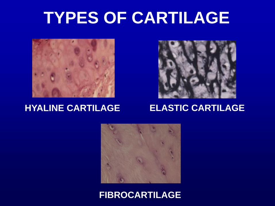
TYPES OF CARTILAGE
FIBROCARTILAGE
HYALINE CARTILAGE ELASTIC CARTILAGE
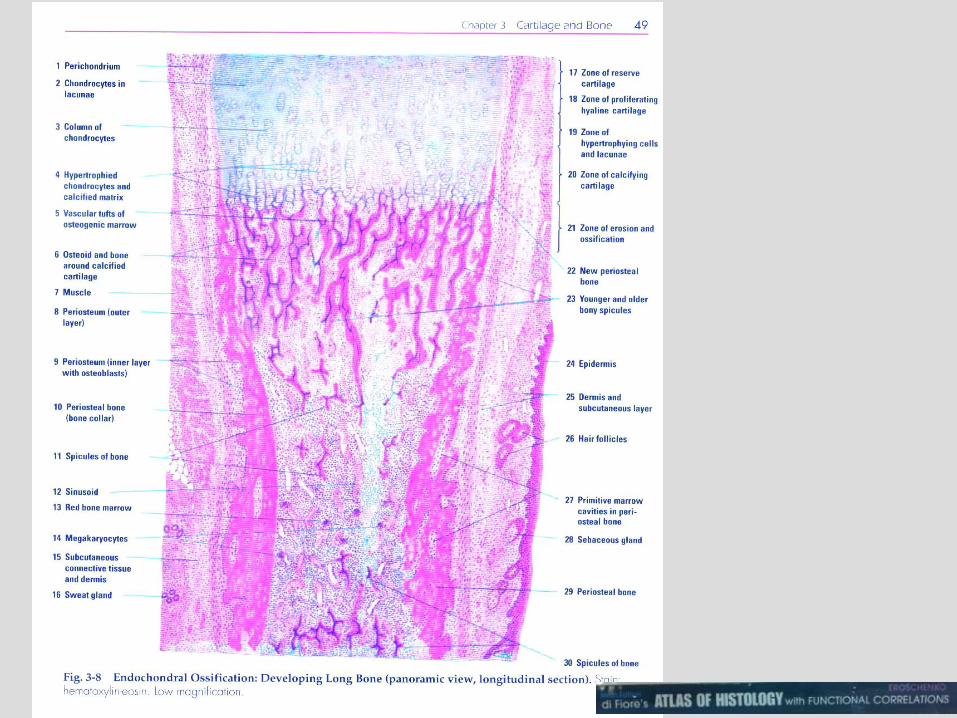


primary spongiosa of the Fetal finger
220

Fetal elbow 194

Fetal elbow 194

Tibia, fetal 421

Trachea, monkey 133

EM 11: cartilage
1. Chondrocyte
2. Fibroblast
3. Appositional
growth region
4. Type I collagen
5. Type II collagen
Appositional growth of
cartilage
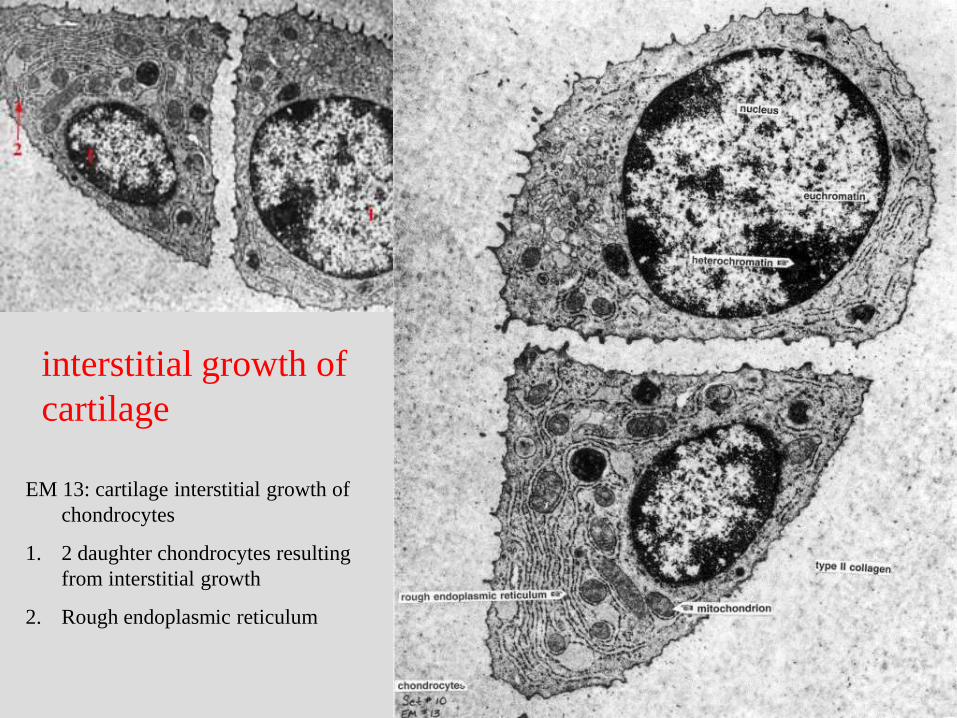
EM 13: cartilage interstitial growth of
chondrocytes
1. 2 daughter chondrocytes resulting
from interstitial growth
2. Rough endoplasmic reticulum
interstitial growth of
cartilage

HYALINE
CARTILAGE
EPIPHYSEAL PLATE,
RESPONSIBLE FOR
LONGITUDINAL GROWTH
OF LONG BONES

cartilage growth in endochondral bone
formation of the Fetal finger
220

OSTEOCLAST
OSTEOBLAST

OSTEOCLAST
OSTEOBLAST

Elastic cartilage in pina of ear
#19762
Elastic cartilage
Connective
tissue
epithelium Connective
tissue
epithelium
Elastic cartilage

FIBROCARTILAGE
INTERMEDIATE BETWEEN
DENSE REGULAR
CONNECTIVE TISSUE
AND HYALINE
CARTILAGE
NO PERICHONDRIUM

Fibrocartilage of Fetal elbow
194
Fibrocartilage
Fibrocartilage

Fibrocartilage of Fetal elbow
194
Fibrocartilage

Fibrocartilage of Fetal elbow 194
Fibrocartilage

Fetal finger – fibroblasts in tendon
220
Fibrocartilage Tendon

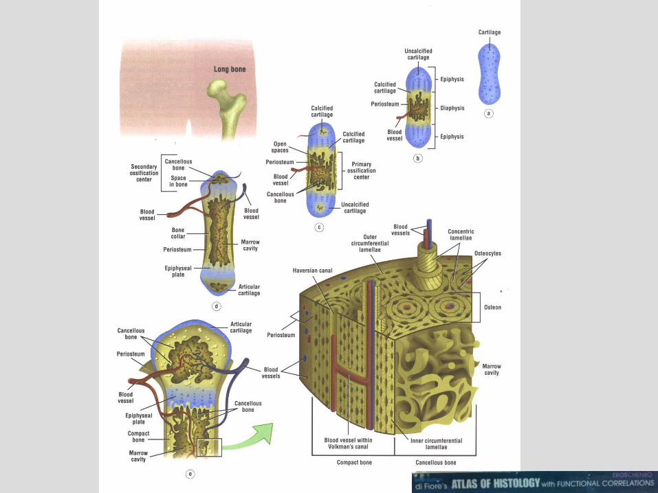
endochondral
bone formation

CELLS OF BONE
OSTEOBLASTS - SECRETE OSTEOID - BONE
– EXPAND BONE BY APPOSITIONAL GROWTH
OSTEOCYTE =
OSTEOBLAST
TRAPPED IN
MATRIX OF
BONE

EM 12: osteoblast adjacent to
osteoid; bone; RER and Golgi
1. Rough endoplasmic reticulum
2. Golgi
3. Osteoid
4. Nucleus of osteoblast
APPOSITIONAL GROWTH

CELLS OF BONE

FUNCTIONS OF BONE
CALCIUM REGULATION Parathroid hormone (BONE
RESORPTION)
Calcitonin (PREVENTS RESORPTION)
These HORMONES are INVOLVED IN TIGHT REGULATION as
1/4 OF FREE CA++ IN BLOOD IS EXCHANGED EACH MINUTE.
HEMOPOIESIS

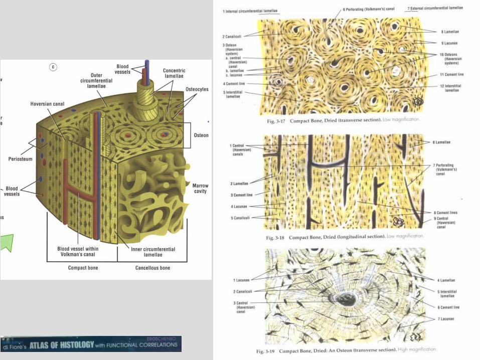
COMPACT BONE
HAVERSIAN
SYSTEMS - LAMELLAE OF BONE
AROUND HAVERSIAN
CANAL LINKED BY
VOLKMANN’S CANAL


COMPACT BONE REMODELING
Over time

COMPACT BONE REMODELING
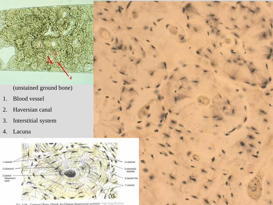
Slide 121: compact bone
(unstained ground bone)
1. Blood vessel
2. Haversian canal
3. Interstitial system
4. Lacuna
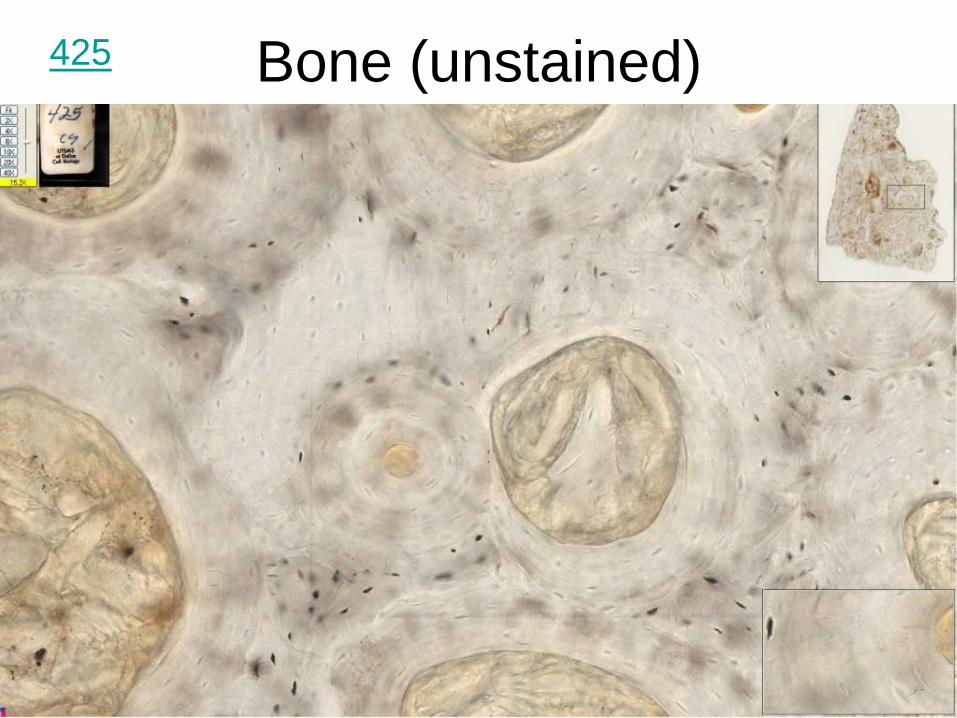
Bone (unstained) 425

Bone (unstained) 425
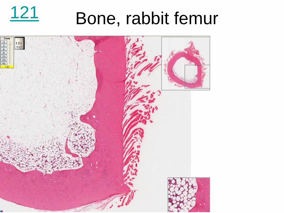
Bone, rabbit femur 121

Bone, rabbit femur 121

Bone, rabbit femur 121

Tibia, fetal 421

Osteoblasts in the Fetal jaw 195

Osteoclasts, osteoblasts, and
osteocytes in Fetal jaw 195
osteocytes
osteoclasts
osteoblasts


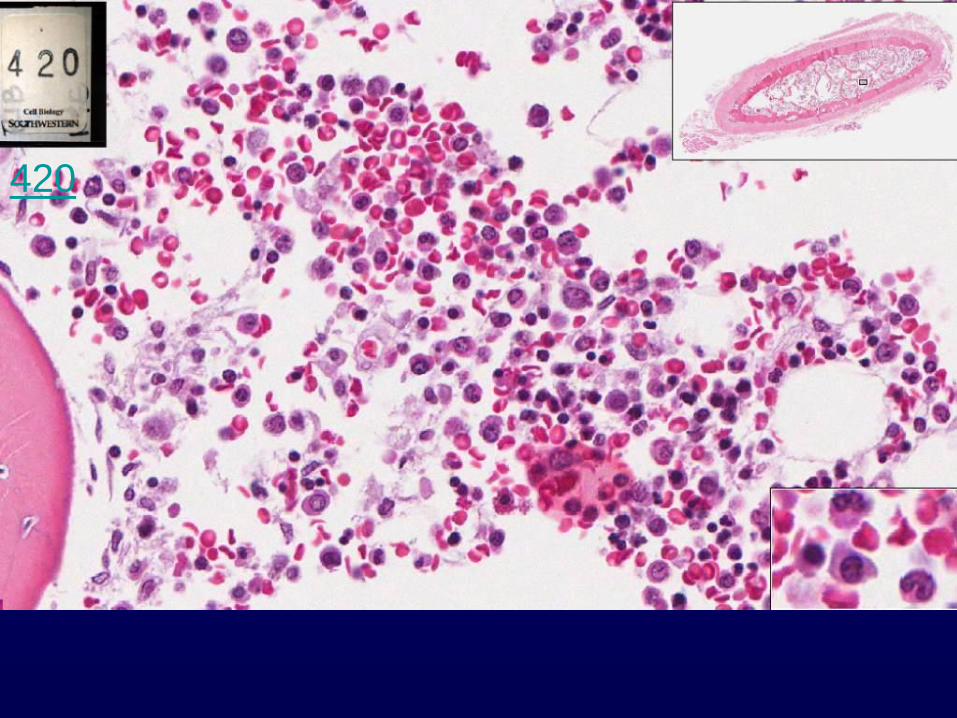
How many Osteoclasts of Fetal
finger can you spot?
Osteoclasts
220

ENDOCHONDRAL (SPONGY/
CANCELLOUS) BONE FORMATION
CARTILAGE MODEL
CENTERS OF OSSIFICATION
PRIMARY CENTER OF
OSSIFICATION
• DIAPHYSIS
SECOND CENTER OF
OSSIFICATION
• CENTER OF EACH EPIPHYSIS

cartilage growth in endochondral bone formation of the Fetal finger
220

Articular surfaces of hyaline cartilage of the
Fetal elbow 194

HISTOGENESIS OF BONE INTRAMEMBRANOUS
OSSIFICATION
FLAT BONES
OF SKULL

SKELETAL CONNECTIVE
TISSUE


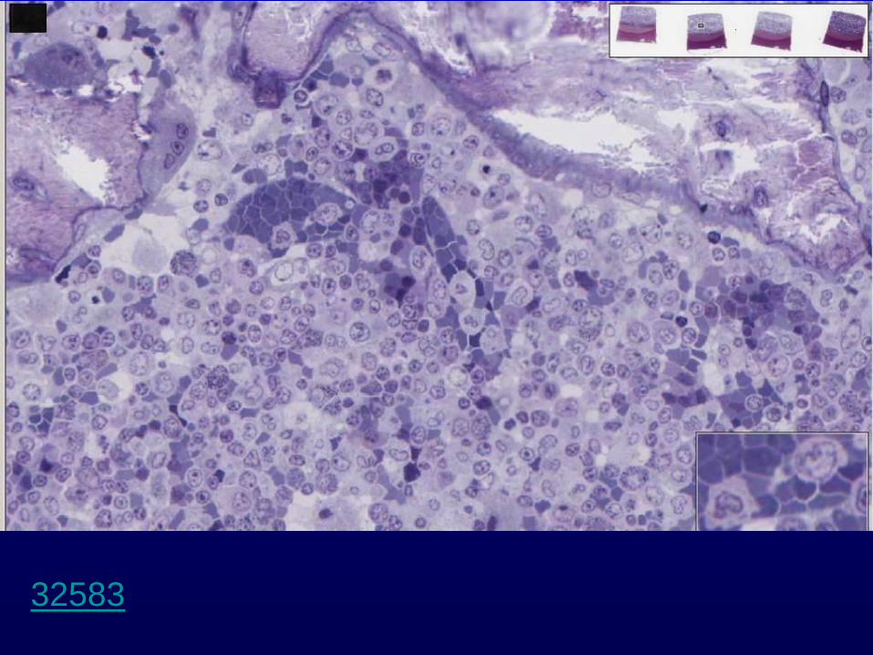

Polychromatic
normoblast
32583

