bone and cartilage regeneration news 01/2011chondrogide/chondro_gide/b… · 2 2 geistlich surgery...
TRANSCRIPT
Contents
GEISTLICH SURGERY03 Introduction04 new Distribution Partners BONE AND CARTILAGE REGENERATION05 Bone Regeneration Concept with orthoss®
10 Case Report: Aneurysmal Bone Cyst13 Posters and Abstracts23 Review 9th ICRs World Congress26 Congress Preview 2011 DISTRIBUTION
Bone & Cartilage RegenerationNews 01 11

2 Geistlich Surgery News 01-20112 Geistlich Surgery News 01-2011
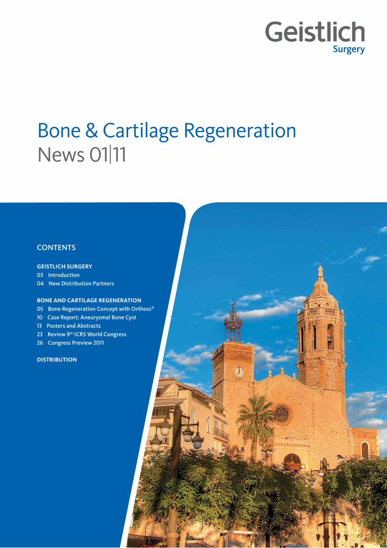
Title Image: Church of San Bartolomé y Santa Tecla (17th century) in Sitges, Spain
Orthoss®, the natural choice in bone regeneration. The excellent biofunctionality makes Orthoss® the ideal bone graft substitute. Bone regeneration ma-terials from Geistlich have been used successfully in more than three million patients.
Chondro-Gide®, the leading natural collagen matrix in cartilage regeneration. This standardised, easy to handle matrix can be used to treat cartilage defects using both AMIC® and ACI. The product includes a sterile Aluminium Template, ideal for creating an accurate impression of the defect.
3Geistlich Surgery News 01-2011
Dear Readers,
the International Cartilage Repair Society (ICRS) Congress in Sitges in September was definitely a major highlight for us in 2010. We successfully launched AMIC® as a treat-ment option of talar cartilage lesions!
During the last years we established AMIC®, which was developed more than 7 years ago together with leading cartilage specialists, as an effective and cost efficient treatment for cartilage defects in the knee. The time had come to transfer and adapt our expertise in knee car-tilage regeneration to the ankle.
Talar cartilage, which is the cartilage covering the ankle, is different from knee cartilage due to the specific ana-tomical requirements of this joint. An adaptation of the AMIC® technique, now available as surgical DVD, paved the way for the launch of AMIC® talus at the ICRS Congress.
As a highlight in Sitges, Geistlich Surgery was able to unite leading cartilage experts in a very well attended lunch symposium discussing the question: Is it worth repairing talar cartilage? The symposium was chaired by Prof. Dr. Valderrabano, from the University Hospital in Basel, who presented cartilage repair strategies in the ankle and the first clinical data on AMIC® talus. This was complemented by Prof. Richter from Heidelberg who gave an outstanding overview about the biological basics of in-situ cartilage repair followed by an extensive litera-ture summary from Dr. Becher, Hannover. And guess what the answer to the question is! You will find a full report on the 9th ICRS on pages 23 to 25.
Based on our 26 years of experience in bone regeneration we have developed a bone regeneration concept based on Orthoss® which we would like to share with you today on pages 5 to 9. It not only gives an overview on bone regeneration but also seeks to provide guidance to sur-geons in selecting suitable treatment options when treating bony defects.
Our success is based on our scientific approach, substanti-ated with sound clinical data. Accordingly, on pages 10 to 22 of this edition you will find an interesting contribution from Prof. Dr. Jäger to “The management of bone healing in an osteoclastic environment” as well as current abstracts on relevant publications in cartilage regeneration.
We are pleased to inform you that in 2011 we have new distribution partners in Austria and Finland in order to offer better support to our local customers. Please find more information on our distribution partners on page 4.
I wish you enjoyable reading and informative insights into bone and cartilage regeneration with the current Bone and Cartilage Regeneration News.
Kind regards,
Paul NoteCEO Geistlich Pharma AG
3Geistlich Surgery News 01-2011
4 Geistlich Surgery News 01-20114 Geistlich Surgery News 01-2011
New Geistlich Surgery Distribution Partners
Geistlich Surgery is pleased to announce, that it will be distributing its products in Finland through Articular Ab, a family owned com-pany. The company is specialised in orthopaedics and traumatology and is focused on selling high quality, innovative products. Articular Ab has imported and marketed finest quality healthcare products and equipment for medical profes-sionals in Finland since 1985. The company is located in Helsinki and has sales representatives working with their customers allover Finland.
Articular Ab places great value on customer service and professional education and is able to provide a competitive advantage in providing the latest innovations to surgeons in a rapidly growing and developing medical market.
Geistlich Surgery is pleased to have Articular Ab as a strong distribution partner in Finland.
Articular Ab, Vattuniemenranta 2, 2. krs, FI-00210 HelsinkiPhone +358 (0)9 4153 5555, Fax +358 (0)9 4153 [email protected]
For many years, Intraplant GmbH has been one of the leading companies in Austria in the field of reconstruction including export activities. Intraplant is ISO 9001 and 13485 certified and an accredited tissue bank under Art. 22 of the (Austrian) Tissue Safety Act.
As distributor for numerous partners, the company offers a complete, high-quality product portfolio in the areas of hip, knee and shoulder endoprosthetics, traumatology, foot surgery and orthobiologics. As well as distribution, Intraplant is also the manufacturer of the ANA.NOVA Hip Prosthesis System.
Continuous research, development and the highest standards are at the fore at Intraplant, and, for our customers and patients, we strive to fulfil the promise of our motto, “We bring movement back into your life”!
Intraplant GmbH, Grenzgasse 38 a, A-2340 Mödling Phone +43 (0)2236-865 232, Fax +43 (0)2236-411 [email protected], www.intraplant.at
Finland – Articular AB
Austria – Intraplant GmbH
5Geistlich Surgery News 01-2011 5Geistlich Surgery News 01-2011
Restoring skeletal integrity in the presence of osseous defects remains a significant challenge. The decision as to which treatment option will be the most successful is influenced by the etiology and pathogenesis of the osseous defect, as well as by the location of the defect, vitality of the surrounding bone and previous, possibly unsuccessful, surgeries.
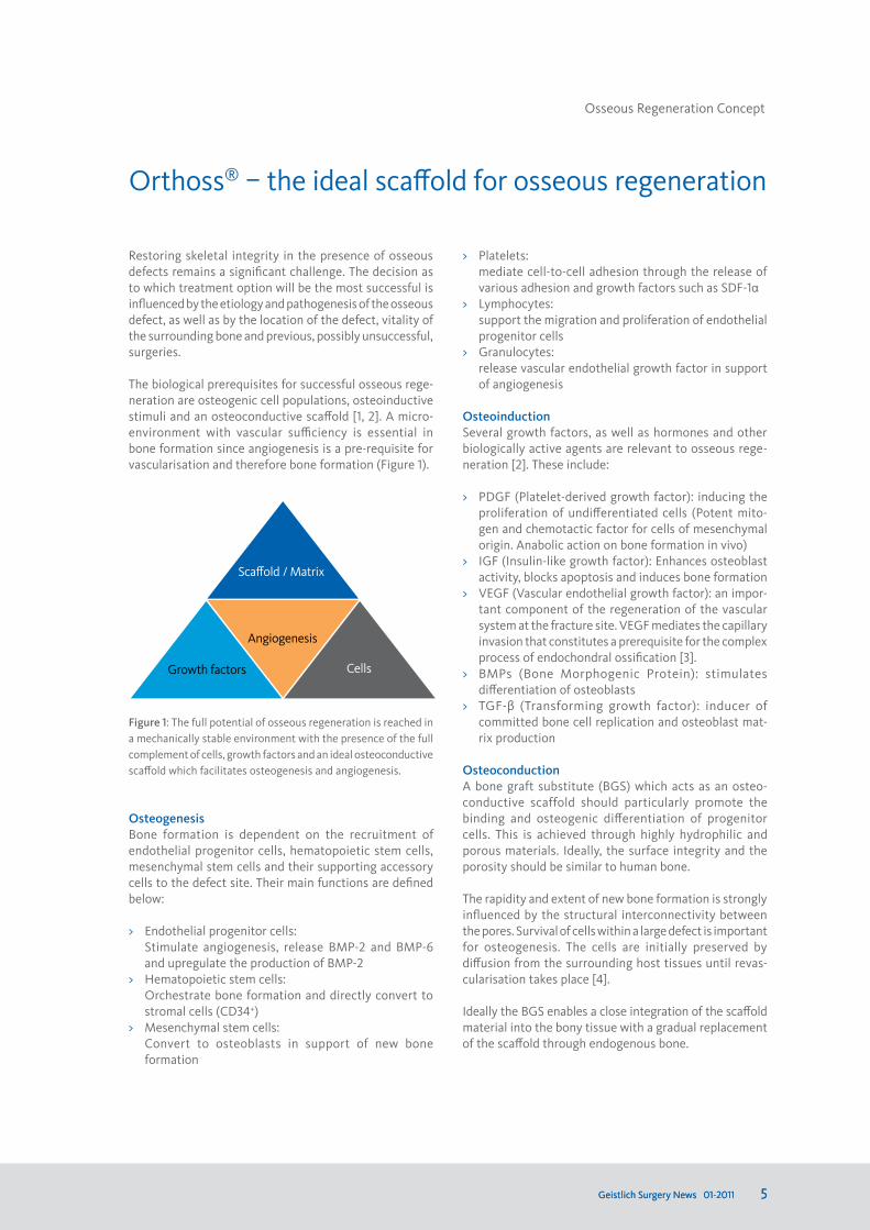
The biological prerequisites for successful osseous rege-neration are osteogenic cell populations, osteoinductive stimuli and an osteoconductive scaffold [1, 2]. A micro- environment with vascular sufficiency is essential in bone formation since angiogenesis is a pre-requisite for vascularisation and therefore bone formation (Figure 1).
Figure 1: The full potential of osseous regeneration is reached in a mechanically stable environment with the presence of the full complement of cells, growth factors and an ideal osteoconductive scaffold which facilitates osteogenesis and angiogenesis.
osteogenesisBone formation is dependent on the recruitment of endothelial progenitor cells, hematopoietic stem cells, mesenchymal stem cells and their supporting accessory cells to the defect site. Their main functions are defined below:
> Endothelial progenitor cells: Stimulate angiogenesis, release BMP-2 and BMP-6
and upregulate the production of BMP-2> Hematopoietic stem cells: Orchestrate bone formation and directly convert to
stromal cells (CD34+)> Mesenchymal stem cells: Convert to osteoblasts in support of new bone
formation
Orthoss® – the ideal scaffold for osseous regeneration
> Platelets: mediate cell-to-cell adhesion through the release of
various adhesion and growth factors such as SDF-1α> Lymphocytes: support the migration and proliferation of endothelial
progenitor cells> Granulocytes: release vascular endothelial growth factor in support
of angiogenesis
osteoinductionSeveral growth factors, as well as hormones and other biologically active agents are relevant to osseous rege-neration [2]. These include:
> PDGF (Platelet-derived growth factor): inducing the proliferation of undifferentiated cells (Potent mito-gen and chemotactic factor for cells of mesenchymal origin. Anabolic action on bone formation in vivo)
> IGF (Insulin-like growth factor): Enhances osteoblast activity, blocks apoptosis and induces bone formation
> VEGF (Vascular endothelial growth factor): an impor-tant component of the regeneration of the vascular system at the fracture site. VEGF mediates the capillary invasion that constitutes a prerequisite for the complex process of endochondral ossification [3].
> BMPs (Bone Morphogenic Protein): stimulates differentiation of osteoblasts
> TGF-β (Transforming growth factor): inducer of committed bone cell replication and osteoblast mat-rix production
osteoconductionA bone graft substitute (BGS) which acts as an osteo-conductive scaffold should particularly promote the binding and osteogenic differentiation of progenitor cells. This is achieved through highly hydrophilic and porous materials. Ideally, the surface integrity and the porosity should be similar to human bone.
The rapidity and extent of new bone formation is strongly influenced by the structural interconnectivity between the pores. Survival of cells within a large defect is important for osteogenesis. The cells are initially preserved by diffusion from the surrounding host tissues until revas-cularisation takes place [4].
Ideally the BGS enables a close integration of the scaffold material into the bony tissue with a gradual replacement of the scaffold through endogenous bone.
Scaffold / Matrix
Angiogenesis
Growth factors Cells
Osseous Regeneration Concept
6 Geistlich Surgery News 01-2011
Osseous Regeneration Concept
Simple
High
Complex
Low
Defect type
Healing Potential
sufficientVascularity
Orthoss® Orthoss®
Autologous BoneOrthoss®
BMA(Autologous Bone)
Orthoss®
BMAC(Autologous Bone)
Orthoss®
BMAC & BMP(Autologous Bone)
PoorVascularity
Contributing Comorbidities and Etiology:> Diabetes> Alcohol & drug abuse> Smoking> Sickle cell disease> Corticosteroid use
CompromisedVascularity
Treatment concept
Composite grafting
osseous regeneration options with orthoss®
Autologous cancellous bone graft is an effective grafting material because it provides all elements required to induce osteogenic regeneration. It is however associated with the limitations of donor site morbidity (including bleeding, infection, and chronic pain at the donor site) and insufficient amount or quality of available autologous material.
BGS represent a valid alternative to supplement or even replace autologous bone and are available from natural or synthetic origin. Natural materials are manufactured from human, animal or coralline sources whereas synthetic bone grafting materials are nonhuman and artificially produced. Examples include Demineralised Bone Matrix (DBM), hydroxyapatite (HA), calcium sulphate, calcium phosphate, beta-tricalcium phosphate (ß-TCP), collagen composites and bioactive glass (bioglass). The large variety of substitute materials available have varying compositions and geometry as well as diverse biological properties.
In contrast to synthetic materials, which are often very compact with limited interconnecting pore system, Orthoss® as a natural hydroxyapatite offers the advantage
of being very similar to human bone with regard to pore morphology, porosity and crystalline structure.
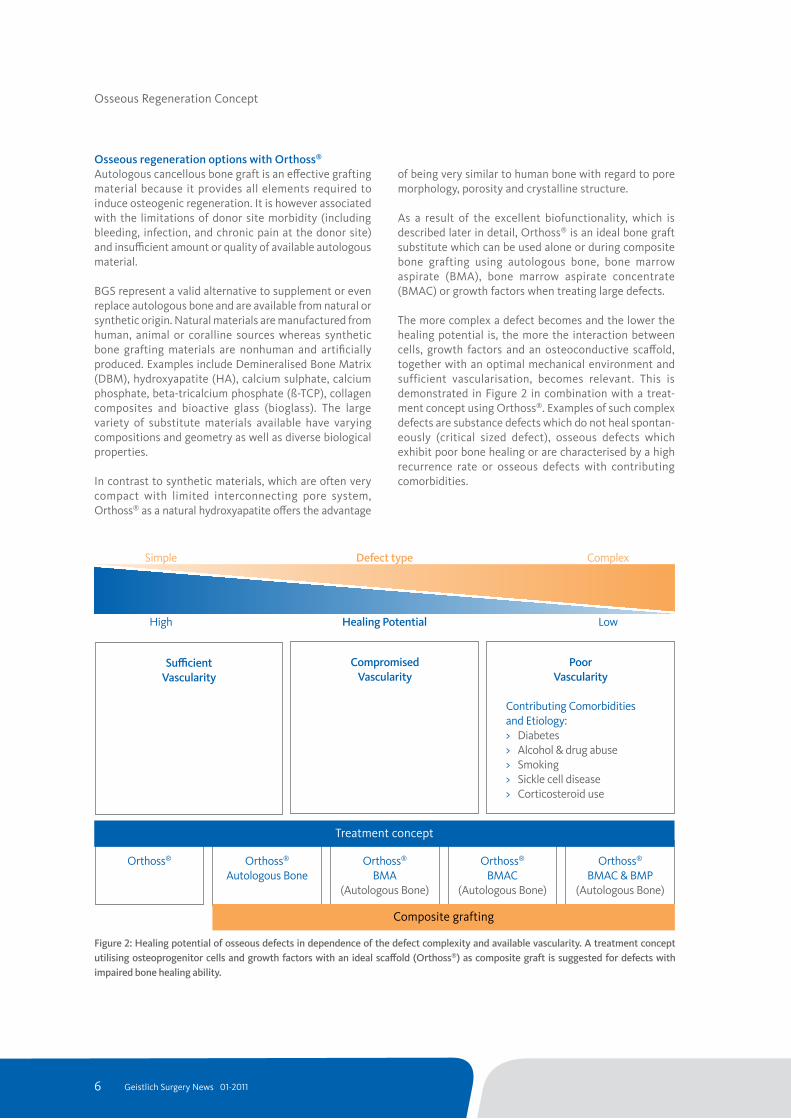
As a result of the excellent biofunctionality, which is described later in detail, Orthoss® is an ideal bone graft substitute which can be used alone or during composite bone grafting using autologous bone, bone marrow aspirate (BMA), bone marrow aspirate concentrate (BMAC) or growth factors when treating large defects. The more complex a defect becomes and the lower the healing potential is, the more the interaction between cells, growth factors and an osteoconductive scaffold, together with an optimal mechanical environment and sufficient vascularisation, becomes relevant. This is demonstrated in Figure 2 in combination with a treat-ment concept using Orthoss®. Examples of such complex defects are substance defects which do not heal spontan- eously (critical sized defect), osseous defects which exhibit poor bone healing or are characterised by a high recurrence rate or osseous defects with contributing comorbidities.
Figure 2: Healing potential of osseous defects in dependence of the defect complexity and available vascularity. A treatment concept utilising osteoprogenitor cells and growth factors with an ideal scaffold (orthoss®) as composite graft is suggested for defects with impaired bone healing ability.

7Geistlich Surgery News 01-2011
orthoss® and orthoss® with autologous boneThe biofunctionality of Orthoss® comprises several properties which make it a superior osteoconductive scaffold and carrier matrix when compared to other bone graft substitutes. These are:
> a morphology similar to that of human bone> an interconnecting pore system> a distinct high porosity and large inner surface area
comparable to human bone> a unique bimodal pore structure> exceptional osteoconductivity and osseointegration
The trabecular structure, porosity and cristallinity of Orthoss® are comparable to human bone. Due to the interconnecting pore system, a large surface area of 80.3 ± 1.2 m2/g with a porosity 77 ± 2 Vol.-% is created. The capillary effect of the nanopores (10-20nm) facilitates complete and spontaneous wetting whilst the macro- porous surface (100-200μm) promotes osteoblast passage and adherence.
These characteristics allow for rapid absorption of proteins and enable adequate conditions throughout the scaffold whilst facilitating the migration and distribution of cells involved in bone formation and vascularisation throughout the matrix.
Therefore, Orthoss® is ideal as BGS in filling smaller bone voids with sufficient vascularity and is well suited as a volume extender for composite bone grafting using autologous bone for larger defects. Orthoss® combined with 25% autologous bone has been shown to be suffi-cient to accelerate new bone formation in the treatment of critical sized defects, thereby limiting the amount of harvested bone and reducing potential complications [5].
orthoss® and BMAFor osseous regeneration in larger defects with compro-mised vascularity the addition of osteoprogenitor cells and growth factors is recommended [1, 2, 6, 7]. Orthoss®, together with bone marrow aspirate (BMA) from the iliac crest offers a suitable, cost efficient source for achieving this goal whilst decreasing the limitations of autologous bone harvesting.
orthoss® and BMACCritical sized defects or defects with an impaired bone healing ability as a result of poor vascularity or contri-buting comorbidities, require a complex strategy to encourage osseointegration. Utilisation of a high concen- tration of biological factors (growth factors and cells) in combination with a suitable carrier matrix with osteoconductive properties has been shown to encourage osseous regeneration [8].
The iliac crest contains bone marrow which is a rich source of the regenerative cells needed for angiogenesis, optimal bone formation and healing. Concentrated nucleated cells from marrow aspirate (BMAC) offer an alternative to iliac crest autograft.
Systems available on the market for the concentration of bone marrow aspirate (Harvest BMAC system, Harvest Technologies GmbH, Germany) offer a safe and easy to use, autologous system that can rapidly produce a con-centration of mononuclear cells and growth factors to help optimise the conditions for healing. Jäger et al. [7] have reported an average concentration of BMAC compared to the initial BMA of 5.2±1.3 (n=17) using this system.
The macroporosity of Orthoss® allows bony ingrowth and a solid integration within the transplantation site. It is incorporated into the physiological remodelling process and therefore has a volume maintaining effect which, in comparison to rapidly resorbing synthetic BGS, reduces the risk of persisting local bone defects or fractures occurring.
Using Orthoss® in combination with BMAC system offers an osseous regeneration solution with a highly osteo-conductive carrier matrix in combination with con-centrated regenerative cells and growth factors. This combination has shown excellent results in bone regen-eration with an accelerated healing in comparison to other scaffolds [8]. The use of Orthoss® and BMAC could result in a reduction or substitution of autologous bone transplants.
orthoss® with BMAC and BMPTransforming growth factor beta one (TGF-ß1) is known to affect osteogenesis and chondrogenesis by stimulating mesenchymal cells [9, 10].
The bone morphogenetic proteins (BMP-2 and BMP-7) represent members of the TGF- ß superfamily which stimulate the formation of new bone and offer an established method in clinical pratice for biological healing enhancement in areas of delayed fracture healing or nonunions which are complicated by local environment adverse circumstances [1, 11,12].
An in vitro study from Gille et al. [13] showed that Orthoss® functions as a carrier for growth hormones and continuously release TGF-ß1 during the investigated time period of 28 days. This time frame matches the early window between 2 and 6 weeks in which TGF-ß1 stimu-lates bone ingrowth according to Goodman et al. [14].

8 Geistlich Surgery News 01-2011
Osseous Regeneration Concept
Osteoconduction:Scaffold
Osteoinduction: Cytokines & growth factors
Osteogenesis:Regenerative cells
Angiogenesis / Vascularity
Most comprehensive solution
DBM
Allograft or β-TCP
Orthoss
Autograft
BMAC
Orthoss® with BMAC
Absent Present in low concentration Present in high concentration
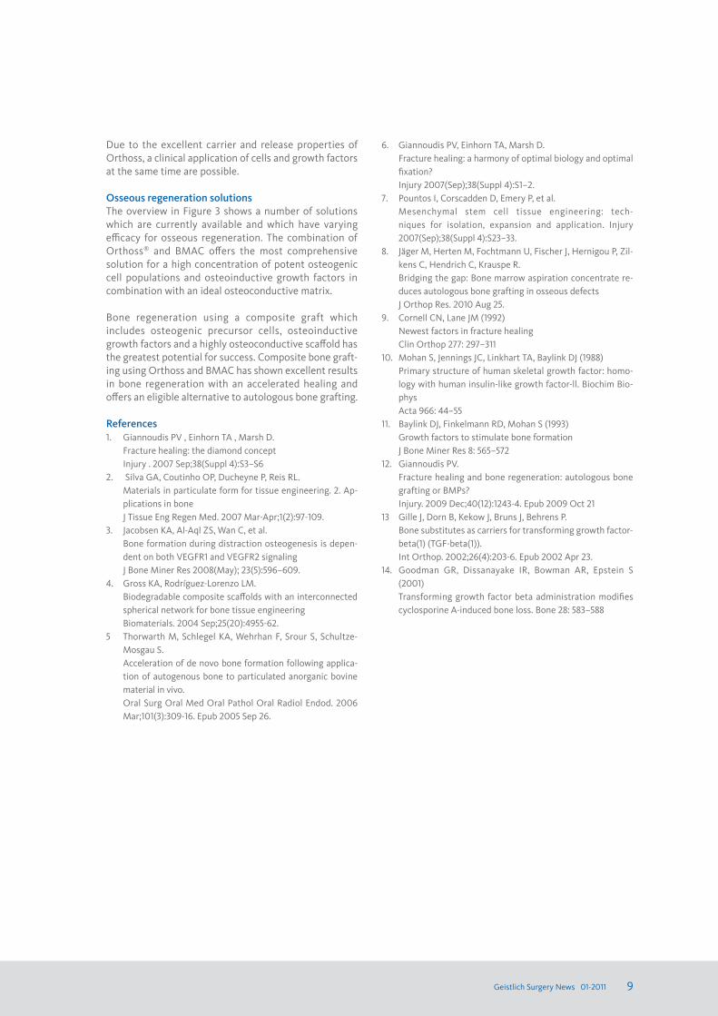
Figure 3: Suitability of available options for osseous regeneration. The inverted triangle symbolises the „V“ for vascularity.
9Geistlich Surgery News 01-2011
6. Giannoudis PV, Einhorn TA, Marsh D. Fracture healing: a harmony of optimal biology and optimal
fixation? Injury 2007(Sep);38(Suppl 4):S1–2.7. Pountos I, Corscadden D, Emery P, et al. Mesenchymal stem cell tissue engineering: tech-
niques for isolation, expansion and application. Injury 2007(Sep);38(Suppl 4):S23–33.
8. Jäger M, Herten M, Fochtmann U, Fischer J, Hernigou P, Zil-kens C, Hendrich C, Krauspe R.
Bridging the gap: Bone marrow aspiration concentrate re-duces autologous bone grafting in osseous defects
J Orthop Res. 2010 Aug 25. 9. Cornell CN, Lane JM (1992) Newest factors in fracture healing Clin Orthop 277: 297–31110. Mohan S, Jennings JC, Linkhart TA, Baylink DJ (1988) Primary structure of human skeletal growth factor: homo-
logy with human insulin-like growth factor-ll. Biochim Bio-phys
Acta 966: 44–5511. Baylink DJ, Finkelmann RD, Mohan S (1993) Growth factors to stimulate bone formation J Bone Miner Res 8: 565–57212. Giannoudis PV. Fracture healing and bone regeneration: autologous bone
grafting or BMPs? Injury. 2009 Dec;40(12):1243-4. Epub 2009 Oct 2113 Gille J, Dorn B, Kekow J, Bruns J, Behrens P. Bone substitutes as carriers for transforming growth factor-
beta(1) (TGF-beta(1)). Int Orthop. 2002;26(4):203-6. Epub 2002 Apr 23.14. Goodman GR, Dissanayake IR, Bowman AR, Epstein S
(2001) Transforming growth factor beta administration modifies
cyclosporine A-induced bone loss. Bone 28: 583–588
Due to the excellent carrier and release properties of Orthoss, a clinical application of cells and growth factors at the same time are possible.
osseous regeneration solutionsThe overview in Figure 3 shows a number of solutions which are currently available and which have varying efficacy for osseous regeneration. The combination of Orthoss® and BMAC offers the most comprehensive solution for a high concentration of potent osteogenic cell populations and osteoinductive growth factors in combination with an ideal osteoconductive matrix.
Bone regeneration using a composite graft which includes osteogenic precursor cells, osteoinductive growth factors and a highly osteoconductive scaffold has the greatest potential for success. Composite bone graft-ing using Orthoss and BMAC has shown excellent results in bone regeneration with an accelerated healing and offers an eligible alternative to autologous bone grafting.
References1. Giannoudis PV , Einhorn TA , Marsh D. Fracture healing: the diamond concept Injury . 2007 Sep;38(Suppl 4):S3–S62. Silva GA, Coutinho OP, Ducheyne P, Reis RL. Materials in particulate form for tissue engineering. 2. Ap-
plications in bone J Tissue Eng Regen Med. 2007 Mar-Apr;1(2):97-109.3. Jacobsen KA, Al-Aql ZS, Wan C, et al. Bone formation during distraction osteogenesis is depen-
dent on both VEGFR1 and VEGFR2 signaling J Bone Miner Res 2008(May); 23(5):596–609.4. Gross KA, Rodríguez-Lorenzo LM. Biodegradable composite scaffolds with an interconnected
spherical network for bone tissue engineering Biomaterials. 2004 Sep;25(20):4955-62.5 Thorwarth M, Schlegel KA, Wehrhan F, Srour S, Schultze-
Mosgau S. Acceleration of de novo bone formation following applica-
tion of autogenous bone to particulated anorganic bovine material in vivo.
Oral Surg Oral Med Oral Pathol Oral Radiol Endod. 2006 Mar;101(3):309-16. Epub 2005 Sep 26.
10 Geistlich Surgery News 01-2011
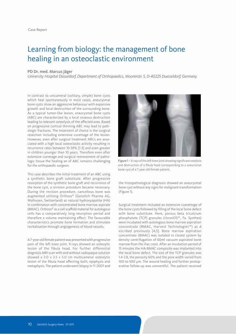
the histopathological diagnosis showed an aneurysmal bone cyst without any signs for malignant transformation (Figure 1).
Surgical treatment included an extensive cuerettage of the bone cysts followed by filling of the local bone defect with bone substitute. Here, porous beta tricalcium phosphatate (TCP) granules (chronOS™, Fa. Synthes) were incubated with autologous bone marrow aspiration concentrate (BMAC, Harvest Technologies™) as d escribed previously [4,5]. Bone marrow aspiration concentrate (BMAC) was isolated in closed system by density centrifugation of 60ml vacuum aspirated bone marrow from the iliac crest. After an incubation period of 15 minutes the HA-BMAC composite was implanted into the local bone defect. The size of the TCP granules was 1.4-2.8, the porosity 60% and the pore width varied from 100 to 500 µm. The wound healing and further postop-erative follow-up was uneventful. The patient received
Case Report
In contrast to unicameral (solitary, simple) bone cysts which heal spontaneously in most cases, aneurysmal bone cysts show an aggressive behaviour with expansive growth and local destruction of the surrounding bone. As a typical tumor-like lesion, aneurysmal bone cysts (ABC) are characterized by a local osseous destruction leading to relevant osteolysis of the affected area. Based on progressive cortical thinning ABC may lead to path-ologic fractures. The treatment of choice is the surgical resection including extensive curettage of the lesion. However, even after surgical treatment ABCs are asso-ciated with a high local osteoclastic activity resulting in recurrence rates between 10-59% [1-3] and even greater in children younger than 10 years. Therefore even after extensive curretage and surgical removement of patho-logic tissue the healing an of ABC remains challenging for the orthopaedic surgeon.
This case describes the initial treatment of an ABC using a synthetic bone graft substitute. After progressive resorption of the synthetic bone graft and recurrence of the bone cyst, a revision procedure became necessary. During the revision procedure, cancellous bone was augmented utilising Orthoss® (Geistlich Pharma AG, Wolhusen, Switzerland) as natural hydroxyapatite (HA) in combination with concentrated bone marrow aspirate (BMAC). Orthoss® as a cell scaffold material for autologous cells has a comparatively long resorption period and therefore a volume maintaining effect. The favourable characteristics promote bone formation and stimulate revitalisation through angiogenesis of blood vessels.
A 7-year old female patient was presented with progressive pain of the left knee joint. X-rays showed an osteoytic lesion of the fibula head. For further differential diagnosis MRI scan with and without radiopaque solution showed a 2.0 x 2.5 x 5.0 cm multicameral osteolytic lesion of the fibula head affecting both, epiphysis and metaphysis. The patient underwent biopsy in 11-2007 and
Learning from biology: the management of bone healing in an osteoclastic environmentPD Dr. med. Marcus JägerUniversity Hospital Düsseldorf, Department of Orthopaedics, Moorenstr. 5, D-40225 Duesseldorf, Germany
Figure 1 – X-rays of the left knee joint showing significant ostelysis and destruction of a fibula head corresponding to a aneurismal bone cyst of a 7-year old female patient.
11Geistlich Surgery News 01-2011
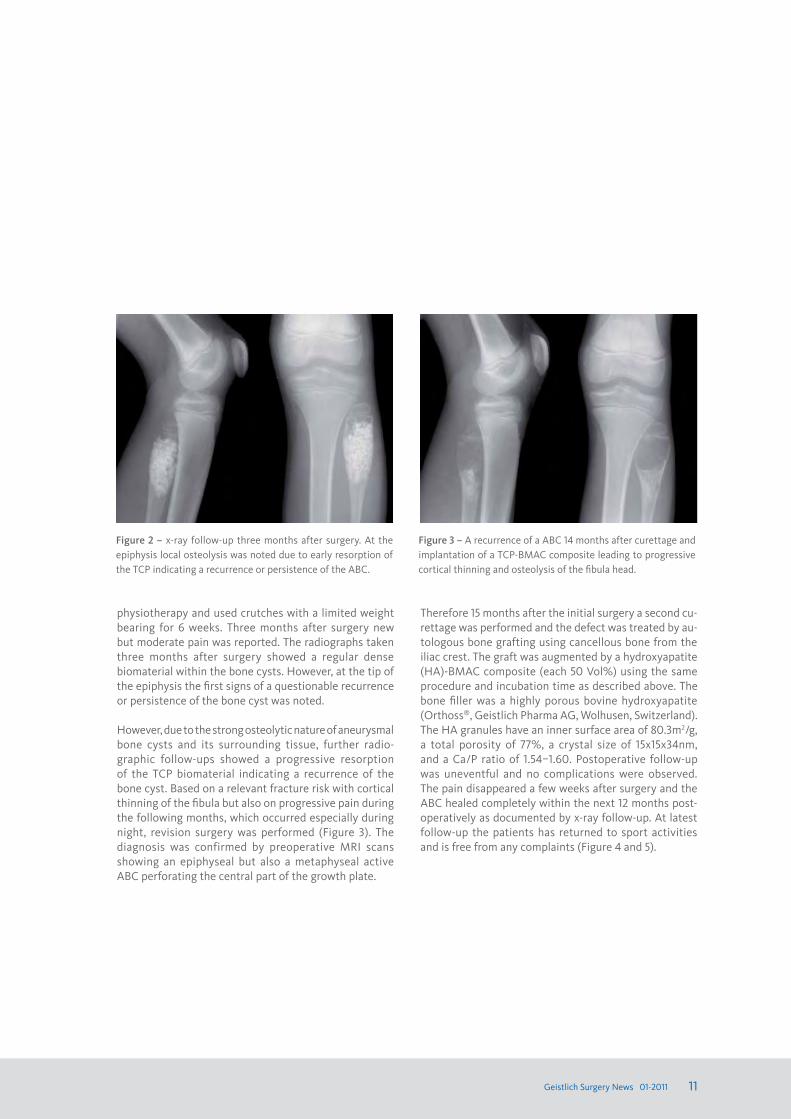
physiotherapy and used crutches with a limited weight bearing for 6 weeks. Three months after surgery new but moderate pain was reported. The radiographs taken three months after surgery showed a regular dense biomaterial within the bone cysts. However, at the tip of the epiphysis the first signs of a questionable recurrence or persistence of the bone cyst was noted.
However, due to the strong osteolytic nature of aneurysmal bone cysts and its surrounding tissue, further radio-graphic follow-ups showed a progressive resorption of the TCP biomaterial indicating a recurrence of the bone cyst. Based on a relevant fracture risk with cortical thinning of the fibula but also on progressive pain during the following months, which occurred especially during night, revision surgery was performed (Figure 3). The diagnosis was confirmed by preoperative MRI scans showing an epiphyseal but also a metaphyseal active ABC perforating the central part of the growth plate.
Figure 3 – A recurrence of a ABC 14 months after curettage and implantation of a TCP-BMAC composite leading to progressive cortical thinning and osteolysis of the fibula head.
Figure 2 – x-ray follow-up three months after surgery. At the epiphysis local osteolysis was noted due to early resorption of the TCP indicating a recurrence or persistence of the ABC.
Therefore 15 months after the initial surgery a second cu-rettage was performed and the defect was treated by au-tologous bone grafting using cancellous bone from the iliac crest. The graft was augmented by a hydroxyapatite (HA)-BMAC composite (each 50 Vol%) using the same procedure and incubation time as described above. The bone filler was a highly porous bovine hydroxyapatite (Orthoss®, Geistlich Pharma AG, Wolhusen, Switzerland). The HA granules have an inner surface area of 80.3m2/g, a total porosity of 77%, a crystal size of 15x15x34nm, and a Ca/P ratio of 1.54–1.60. Postoperative follow-up was uneventful and no complications were observed. The pain disappeared a few weeks after surgery and the ABC healed completely within the next 12 months post- operatively as documented by x-ray follow-up. At latest follow-up the patients has returned to sport activities and is free from any complaints (Figure 4 and 5).

12 Geistlich Surgery News 01-2011
Figure 4 – Two moths after revision surgery the osteolytic de-fect is completely covered with autologous bone combined with a HA-BMAC composite.
Figure 5 – Complete healing of the ABC 12 weeks after revision surgery using a HA bone substitute combined with autologous cell therapy (BMAC). Cortical bone remodeling has lead to relevant thickening.
This case report indicates that bone substitutes with a high in vivo resorption or degradation rate may have disadvantages in an aggressive and osteoclastic environ-ment such in ABC, giant cell tumor or enchdondroma. In addition, local bone defects in biomechanical relevant re-gions of the skeleton have to compensate relevant forces, e.g. during weight bearing. To our opinion affected patients with one of the mentioned lesion may benefit from bone fillers which show a slow resorption rate and a high biomechanical stability towards pressure and torque forces. Here HA, with a large surface area pro-
Literature 1. Basarir K, Piskin A, Guclu B, Yildiz Y, Saglik Y. Aneurysmal bone cyst recurrence in children: a review of 56 patients. J Pediatr
Orthop 2007;27(8):938-43.2. Dormans JP, Hanna BG, Johnston DR, Khurana JS. Surgical treatment and recurrence rate of aneurysmal bone cysts in children. Clin
Orthop Relat Res 2004(421):205-11.3. Mankin HJ, Hornicek FJ, Ortiz-Cruz E, Villafuerte J, Gebhardt MC. Aneurysmal bone cyst: a review of 150 patients. J Clin Oncol
2005;23(27):6756-62.4. Jäger M, Hernigou P, Zilkens C, Herten M, Fischer J, Krauspe R. Cell therapy in bone-healing disorders. Orthopäde;39(4):449-62.5. Jäger M, Herten M, Fochtmann U, Fischer J, Hernigou P, Zilkens C, Hendrich C, Krauspe R. Bridging the gap: Bone marrow aspira-
tion concentrate reduces autologous bone grafting in osseous defects. J Orthop Res. 2011;29(2):173-80.6. Hernigou P, Beaujean F. Treatment of osteonecrosis with autologous bone marrow grafting. Clin Orthop Relat Res 2002(405):14-23.7. Hernigou P, Daltro G, Filippini P, Mukasa MM, Manicom O. Percutaneous implantation of autologous bone marrow osteoprogeni-
tor cells as treatment of bone avascular necrosis related to sickle cell disease. Open Orthop J 2008;2:62-5.8. Hernigou P, Poignard A, Manicom O, Mathieu G, Rouard H. The use of percutaneous autologous bone marrow transplantation in
nonunion and avascular necrosis of bone. J Bone Joint Surg Br 2005;87(7):896-902.
Case Report
moting binding of extracellular matrix proteins but also the attachment and adherence of local cells, is a useful option compared to other biomaterials. Although 50 Vol% of the local bone defect was treated by autologous bone grafting, a combination with the HA carrier described above and a BMAC, has lead to complete healing of the ABC. It is evident that bone marrow cells have been applied successfully since many years in other bone healing disturbances such as osteonecrosis or pseudar-throsis [6-8]. Further clinical studies with statistical relevant cohorts have to show the efficiency of cell therapy in bone regeneration.

Posters and Abstracts
Mid-term results of Autologous Matrix Induced Chondrogenesis (AMIC) for treatment of focal cartilage defects in the knee
Gille J., Wimmer J., schuseil e., Gellissen J., Wallstabe s., Behrens P.Knee Surg Sports Traumatol Arthrosc. 2010 Nov;18(11):1456-64. Epub 2010 Feb 2.
AbstractArticular cartilage defects heal poorly. Autologous Matrix-Induced Chondrogenesis (AMIC) is an innovative treatment for localized full-thickness cartilage defects combining the well-known microfracturing with collagen scaffold and fibrin glue. The purpose of this prospective study was to evaluate the medium-term results of this enhanced microfracture technique for the treatment of chondral lesions of the knee. Thirty-two chondral lesions in 27 patients were treated with AMIC. Within the context of clinical follow-up, these patients were evaluated for up to 5 years after the intervention.
Five different scores (Meyer score, Tegner score, Lysholm score, ICRS score, Cincinatti score) as well as radiographs were used for outcome analysis. Articular resurfacing was assessed by magnetic resonance imaging (MRI). The average age of patients (11 females, 16 males; mean body mass index 26, range 20–32) was 37 years (range 16–50 years). The mean defect size of the chondral lesions was 4.2 cm2 (range 1.3–8.8 cm2). All defects were classified as grade IV according to the Outerbridge classification.
The follow-up period was between 24 and 62 months with a mean of 37 months. Twenty out of 23 individuals (87%) questioned were subjectively highly satisfied with the results after surgery. Significant improvement (P\0.05) of all scores was observed as early as 12 months after AMIC, and further increased values were notable up to 24 months post-operatively. MRI analysis showed moderate to complete filling with a normal to incidentally hyperintense signal in most cases. Results did not show a clinical impact of patient’s age at the time of operation, body mass index and number of previous operations (n.s.). In contrast, males showed significant higher values in the ICRS score compared to their female counterparts.
AMIC is an effective and safe method of treating symptomatic full-thickness chondral defects of the knee in appropriately selected cases. However, further studies with long-term follow-up are needed to determine whether the grafted area will maintain structural and functional integrity over time.
Posters and Abstracts
Cell-Laden and Cell-Free Matrix-Induced Chondrogenesis versus Microfracture for the Treatment of Articular Cartilage Defects: A Histological and Biomechanical Study in Sheep
Gille J., Kunow J., Boisch L., Behrens P., Bos I., Hoffmann C., Köller W., Russlies M., Kurz B.Cartilage, Jan 2010; vol. 1: pp. 29 - 42.
AbstractObjective: The aim of this study was to evaluate the regenerative potential of cell-laden and cell-free collagen matrices in comparison to microfracture treatment applied to full-thickness chondral defects in an ovine model.
Methods: Animals (n = 30) were randomized into 5 treatment groups, and 7-mm full-cartilage-thickness defects were set at the trochlea and medial condyle of both knee joints and treated as follows: 2 scaffolds in comparison (collagen I/III, Chondro-Gide®; collagen II, Chondrocell®) for covering microfractured defects (autologous matrix-induced chondro- genesis), both scaffolds colonized in vitro with autologous chondrocytes (matrix-associated chondrocyte transplantation), or scaffold-free microfracture technique. One year after surgery, cartilage lesions were biomechanically (indentation test), histologically (O’Driscoll score), and immunohistochemically (collagen type I and II staining) evaluated.
Results: All treatment groups of the animal model induced more repair tissue and showed better histological scores and biomechanical properties compared to controls. The average thickness of the repair tissue was significantly greater when a scaffold was used, especially the collagen I/III membrane. However, none of the index procedures surpassed the others from a biomechanical point of view or based on the histological scoring. Collagen type II expression was better in condylar defects compared to the trochlea, especially in those treated with collagen I/III membranes.
Conclusion: Covering of defects with suitable matrices promotes repair tissue formation and is suggested to be a promising treatment option for cartilage defects. However, it failed to improve the biomechanical and histological properties of regenerated articular cartilage compared to microfracture alone in an ovine model under the given circumstances.
Treatment Selection in Articular Cartilage Lesions of the Knee
The subchondral bone in articular cartilage repair: current problems in the surgical management
Bekkers J.e., Inklaar M., saris D.B.Am J Sports Med. 2009 Nov;37 Suppl 1:148S-55S
Gomoll A.H., Madry H., Knutsen G., Van Dijk n., seil R., Brittberg M., Kon e. Am J Sports Med. 2009 Knee Surg Sports Traumatol Arthrosc. 2010 Apr;18(4):434-47. Epub 2010 Feb 4
AbstractBackground: Several treatment options are available to repair articular cartilage lesions of the knee; however, evidence-based parameters for treatment selection are lacking.
Purpose: To identify parameters for valid treatment selection in the repair of articular cartilage lesions of the knee.
Study Design: Systematic review.
Methods: A systematic search was conducted in the databases EMBASE, MEDLINE, and the Cochrane collaboration. The retrieved articles were screened for relevance on title and abstract followed by a full-text study quality appraisal of the remaining articles. Eventually, a total of 4 randomized controlled trials were included.
Results: Lesion size, activity level, and age were the influencing parameters for the outcome of articular cartilage repair surgery. Lesions greater than 2.5 cm2 should be treated with sophisticated techniques, such as autologous chondrocyte implantation or osteochondral autologous transplantation, while microfracture is a good first-line treatment option for smaller (<2.5 cm2) lesions. Patients who are active show better results after autologous chondrocyte implantation or osteochondral autologous transplantation when compared with microfracture. Younger patients (<30 years) seem to benefit more from any form of cartilage repair surgery compared with those over 30 years of age.
Conclusion: Lesion size, activity level, and patient age are factors that should be considered in selecting treatment of articular cartilage lesions of the knee. In addition, these factors are a step toward evidence-based, instead of surgeon-preferred, treatment of articular cartilage lesions of the knee.
AbstractAs the understanding of interactions between articular cartilage and subchondral bone continues to evolve, increased attention is being directed at treatment options for the entire osteochondral unit, rather than focusing on the articular surface only. It is becoming apparent that without support from an intact subchondral bed, any treatment of the surface chondral lesion is likely to fail. This article reviews issues affecting the entire osteochondral unit, such as subchondral changes after marrow-stimulation techniques and meniscectomy or large osteochondral defects created by prosthetic resurfacing techniques. Also discussed are surgical techniques designed to address these issues, including the use of osteochondral allografts, autologous bone grafting, next generation cell-based implants, as well as strategies after failed subchondral repair and problems specific to the ankle joint. Lastly, since this area remains in constant evolution, the requirements for prospective studies needed to evaluate these emerging technologies will be reviewed.
15Geistlich Surgery News 01-2011
16 Geistlich Surgery News 01-2011
Posters and Abstracts
Effectiveness of Composite Bone Graft Substitute Plugs in the Treatment of Chondral and Osteochondral Lesions of the Talus
Lin J.s., Andersen L.B., Juliano P.J.J Foot Ankle Surg. 2010 May-Jun;49(3):224-31. Epub 2010 Mar 31
AbstractA review of outcomes in 13 patients with talar dome osteochondral or chondral lesions treated with a bone graft substitute plug was undertaken in an effort to evaluate its effectiveness in comparison with other reported surgical techniques.
Mean patient age was 36.4 (range 16 to 57) years. Mean follow-up was 30.1 (range 7 to 43) months. Medial malleolar osteotomy was performed in 9 (69.23%) cases. Average defect diameter was 9.8 (range 5 to 20) mm. Pain decreased significantly from 6.2 (range 3 to 9) to 4.0 (range 0 to 9) (P = .009). Postoperative American Orthopaedic Foot and Ankle Society Ankle-Hindfoot scores averaged 67.3 (range 26 to 100). Younger age, smaller defect size, and avoidance of medial malleolar osteotomy resulted in better outcomes. Mean Short Form-36 scores for the study group fell below US norms in all categories, and 12 (92.31%) ankles demonstrated persistent lesions radiographically. Postoperative magnetic resonance imaging in 2 (15.39%) patients demonstrated enlarged lesions, and 4 (30.77%) patients underwent revision surgery that revealed abnormal cartilage around the implant site. Complications included 1 (7.69%) deep venous throm-bosis, 1 (7.69%) arthrofibrosis, and 1 (7.69%) superficial neuritis.
Despite some improvement in pain, comparison of functional outcome showed bone graft substitute plug implantation to be less effective overall than other operative interventions. Future investigations with more specific selection criteria are warranted to gain further insight into the efficacy of these bone graft substitute plugs.
Augmentation Strategies following the Microfracture Technique for Repair of Focal Chondral Defects
strauss e.J., Barker J.U., Kercher J.s., Cole B., Mithoefer K.Cartilage, April 2010; vol. 1, 2: pp. 145-152
AbstractThe operative management of focal chondral lesions continues to be problematic for the treating orthopedic surgeon secondary to the limited regenerative capacity of articular cartilage. Although many treatment options are currently available, none fulfills the criteria for an ideal repair solution, including a hyaline repair tissue that completely fills the defect and integrates well with the surrounding normal cartilage.
The microfracture technique is an often-utilized, first-line treatment modality for chondral lesions within the knee, resulting in the formation of a fibrocartilaginous repair tissue with inferior biochemical and biomechanical properties compared to normal hyaline cartilage. Although symptomatic improvement has been shown in the short term, concerns about the durability and longevity of the fibrocartilaginous repair have been raised.
In response, a number of strategies and techniques for augmentation of the first-generation microfracture procedure have been introduced in an effort to improve repair tissue characteristics and reduce long-term deterioration. Recent experimental approaches utilize modern tissue-engineering technologies including local supplementation of chondrogenic growth factors, hyaluronic acid, or cytokine modulation. Other second-generation microfracture-based techniques use different types of scaffold-guided in situ chondroinduction. The current article presents a comprehensive overview of both the experimental and early clinical results of these developing microfracture augmentation techniques
AMIC technique enhanced by the use of concentrated bone marrow for the treatment of chondral lesion
AbstractPurpose: AMIC® (autologous matrix induced chondrogenesis) combines microfracturing with the application of Chondro-Gide®, a porcine collagen type I/III bilayer matrix. However, all marrow stimulation techniques collect only a poor number of mesenchymal stem cells and this number is variable among patients. For this reason it could be useful to enhance these techniques with progenitor cells harvested from iliac crest bone marrow, in order to provide a better and faster defect resurfarcing. Here we present the clinical outcome of 30 patients treated either with the standard AMIC® technique or combined with the use of concentrated bone marrow (cBM) from a minimum follow up of 6 months up to 24 months.
Methods and Materials: 30 patients (range 18-50 y/o) presenting focal cartilage lesions of the knee were treated either with the standard (n=11) or the modified AMIC® technique (n=19). Clinical evaluation was based on Lysholm Knee Score, IKDC and VAS pain scale. For each patient MRI was performed preoperatively and 6, 12 month and 24 months post- operatively.
Results: Significant differences between pre- and post-operative score values have been observed for all patients start-ing from 6 months. After 12 months significant best results were found in patients treated with the modified AMIC®
technique (n=11) respect to those treated with standard AMIC® (n=8), in particular in term of pain reduction (2.5±0.7 and 0.8±0.4, respectively, p<.05) and Lysholm score (84±1.4 and 95.4±4.4, p<.05). MRI showed good healing processes of the cartilage defects already after 6 months in both groups of patients.
Conclusions: Enhancing the AMIC® technique with concentrated bone marrow seems to be an effective and safe method. However, longer follow-up times are necessary to evaluate the long-term regeneration success.
de Girolamo L., Adravanti P., schoenhuber H., Volpi P.P337, Poster, 9th ICRS Congress, Sitges, Spain

Posters and Abstracts
Autologous Matrix Induced Chondrogenesis (AMIC® plus) for the treatment of patellar cartilage defects in the knee
Dhollander A.A.M., De neve F., Almqvist K.F., Verdonk R., Verbruggen G., Verdonk P.C.P274, Poster, 9th ICRS Congress, Sitges, Spain
AbstractPurpose: The present study was designed to evaluate the AMIC plus technique for the treatment of symptomatic patellar cartilage defects in the knee. MRI was used for the morphological analysis of cartilage repair.
Methods and materials: The AMIC plus technique (combination of the original AMIC technique and the use of platelet rich plasma) was used for the treatment of symptomatic chondral and osteochondral patellar lesions in the knee. Five patients were clinically prospectively evaluated with use of the Knee Injury and Osteoarthritis Outcome Score (KOOS), a Visual Analogue Scale (VAS) for pain, the Tegner activity level scale and Kujala scale preoperatively and at 12 and 24 months of follow-up. All 5 patients had consented to follow the postoperative MRI evaluation protocol. MRI data were analyzed based on the original MOCART (Magnetic Resonance Observation of Cartilage Repair Tissue) scoring system.
Results: A statistically significant clinical improvement became apparent after 24 months of follow-up. The MOCART scoring system revealed no significant deterioration or improvement of the repair tissue between one and two years of follow-up. Twenty-four months after the operation hypertrophy was found in 40%. Subchondral bone changes and intralesional osteophytes were seen in all cases (100%). Synovitis and adhesions were not observed in the study patients at that time of follow-up.
Conclusions: AMIC plus resulted in clinically significant improvement in all patients. The favourable clinical outcome of the AMIC plus technique was not confirmed by the MRI findings as determined by the MOCART score. More specifically, all cases showed subchondral lamina and bone changes, including intralesional osteophytes.
18 Geistlich Surgery News 01-2011
Reduced chondrocyte viability is associated with the use of marker pen ink
Getgood A., Mcnamara I., Kili s., Bhullar t., Henson F.M.P65, Poster, 9th ICRS Congress, Sitges, Spain
AbstractPurpose: The aim of this study was to investigate whether the addition of two different marker pen inks to a collagen membrane would affect chondrocyte viability, and therefore be potentially detrimental to articular cartilage repair in autologous chondrocyte transplantation.
Methods and Materials: Human chondrocytes were applied to Chondro-Gide® collagen membranes at a volume of 12 million cells/ml. Two sterile marker pens were used to mark the membranes; one contained methylene blue, the other contained crystal violet inks. Three groups of membrane were tested in duplicate for each pen. Group A consisted of no ink mark, group B had only the uppermost ‘smooth’ layer marked, and group C had the lower ‘porous’ layer marked. All membranes were then cultured in standard growth media for 24 hours. Cell viability was assessed at 24 hours on all membranes using a live/dead cell viability assay. Cell viability was quantified with florescent microscopy with mean number of cells compared to control membranes using the students t test (p<0.05).
Results: Table 1 shows the mean number of viable cells in each group. Control membranes (group A) with no ink showed excellent cell viability. A statistically significant reduction in cell viability with both methylene blue (p<0.0001) and crystal violet (p<0.0001) was found adjacent to the ink mark on the smooth side (group B) and on the porous side remote from the ink (group C). Marked cell death was seen with both dyes (p<0.0001) adjacent to the ink on the porous side. A small number of cells cultured with crystal violet were live – no live cells were detected in the presence of methylene blue.
Conclusions: Chondrocyte viability is significantly reduced when cells are cultured in-vitro on collagen membranes marked with methylene blue and crystal violet pen ink. Surgeons should be aware of the potential negative impact of marker pens in cell based therapies.

Posters and Abstracts
Autologous Matrix-Induced Chondrogenesis (AMIC) on the patella plusperiosteal coverage on the trochlea combined with mechanical realignment– A New Treatment Option in Symptomatic Isolated Femoropatellar Osteoarthritis due to Subluxation of the Patella
Kusano t., Marti C., Jacobi M., Jakob R.17.4.5, Free Paper, 9th ICRS Congress, Sitges, Spain
AbstractPurpose: Symptomatic isolated femoropatellar osteoarthritis is reported for 8% of women and 2% of men over the age of 55 years. Surgical options include osteotomy of the tibial tubercle with patellar debridement and realignment (1), patellar hemiarthroplasty and femoropatellar arthroplasty and patellectomy. We present our results of a new biological treatment option, in which (1) is combined with the AMIC procedure (autologous matrix-induced chondrogenesis) and periosteal resurfacing of the lateral trochlea femoris.
Methods and Materials: Only symptomatic isolated lateral femoropatellar osteoarthritis with an unsuccessful conservative treatment was included in the study. The surgery consisted of an AMIC procedure described by Behrens on the retropa-tellar cartilage defect and a periosteal coverage of the trochlear cartilage defect, both naked surfaces prepared by 1, 1 mm drillings combined with resection of the lateral pole of the patella with lateral release, medial reefing and a tibial tubercle medialisation and advancement. Lack of improvementand need for reoperation were defined as endpoints.
Results: From 2003 to 2009 a total number of 21 patients underwent this described new procedure of which two were operated bilaterally. 1/4 of patients had a lack of improvement, thus 3/4 were satisfied. One patient who underwent bilateral surgery was satisfied at the last follow-up but died later of an unrelated cause. The other patient with bilateral surgery had persistent anterior knee pain on the one knee and a good result on the other. One patient had a reoperation with the AMIC procedure and is now satisfied. Another patient underwent partial meniscectomy. His arthroscopy showed fibrous cartilage coverage. One patient had total knee replacement. The oldest patient in this series showed radiographic signs of patellar osteolysis. Patients over the age of 75 years all were failures.
Conclusions: The proposed combination of mechanical and biologic treatment modalities has proven to be a valid alternative to debridement and alignment alone or to arthroplasty in patients under the age of 75 years. It shows that there is a chondroplastic potential in osteoarthritis and advanced age.
Posters and Abstracts
The effect of cellular passage, quantity and in vitro 3-D matrix culturing time on gene expression profiles before and after matrix-assisted chondrocyte transplantation (MACT) in a leporine animal model
salzmann G.M., sauerschnig M., Berninger M., sütfels t., schönfelder M., schöttle P.B., Imhoff A.B.12.1.5, Free Paper, 9th ICRS Congress, Sitges, Spain
AbstractPurpose: Matrix-assisted chondrocyte transplantation (MACT) still lacks any standardization in its execution.
Methods and Materials: Post puberty NZW-rabbit knee articular chondrocytes were seeded within 3-D matrices (Chondro-Gide, Geistlich, Switzerland) at different passages (P 1, 3, 5); cellular yields (C: 2x10⁵/matrix=C1, 1x10⁶/matrix=C2, 3x10⁶/matrix=C3) and in vitro membrane-holding times (T: 24 hours=T1, 2 weeks=T2) to define 18 different groups. Each time, two cell-matrix-constructs (CMC, n=6/group) were in identical duplicate whereof one was for in vitro (CMCi) analysis directly prior to re-implantation of the other duplicate which was press-fit implanted (autologous) (CMCe) into trochlear full-thickness chondral defects (n=2/animal) of the biopsy-contralateral knee, reproducing a MACT situation. 12 weeks postimplantation the regenerates were analysed for Collagen-1,-2,-10, COMP, Aggrecan, Sox9 mRNA expression. Data were statistically compared using a mixed linear model, significance was at P<0.05 for all tests.
Results: Generally, CMCi values were higher than CMCe values for all differentiation targets, while the opposite was true for dedifferentiation targets. There appeared a general linearity between CMCi and CMCe values to potentially predict the CMCe outcome based on CMCi quality. Typically, animals improved, target-specific, a disadvantageous CMCi profile into an advantegeous CMCe expression, while generally advantageous CMCi values persisted within CMCes. CMCi values were significantly different between groups for all targets analysed, while the difference was not significant for Collagen-1,-10, Aggrecan among CMCes. Usually, interacting P and T resulted in significant different results, while this was true with much lesser frequency for C. A combination of low P, of medium C and short T appeared to result in an optimal CMCi and CMCe outcome, while generally P3, more pronounced P5 and foremost T2 (as well their combination) strongly impaired the outcome, with much lesser impact of C.
Conclusions: The results demonstrate that both in vitro and in vivo performance are strongly affected by cellular passage, density, membrane-holding time.
21Geistlich Surgery News 01-2011
22 Geistlich Surgery News 01-201122 Geistlich Surgery News 01-2011
Characterized Chondrocyte Implantation in the Patellofemoral Joint: a 1 to 3 Year Follow-Up
Vanlauwe J., Claes t.P240, Poster, 9th ICRS Congress, Sitges, Spain
AbstractPurpose: To assess clinical outcome in patients treated with Characterized Chondrocyte Implantation (CCI) for full thickness lesions in the patellofemoral joint up to 36 months post surgery.
Methods and Materials: Patients with symptomatic patellofemoral full thickness cartilage lesions were treated with ACI using characterized chondrocytes (ChondroCelect®) covered with a Chondro-Gide® membrane. Clinical outcome was assessed using the Knee Injury and Osteoarthritis Outcome score (KOOS) and a Visual Analogue Scale (VAS) for pain. Responders were defined using 5 categories (>10 points and >20, 30, 50, 70%) based on KOOS and VAS.
Results: Thirty-six patients, with a mean defect size of 4.89 cm² (1.5 to 11 cm²), were treated in the patella (n=25), trochlea (n=8) or kissing lesion (trochlea and patella; n=3). The mean follow-up period was 24.67 months . Treated patients showed statistically significant improvements in KOOS (at 6, 12, 18, 24 and 36 months) and VAS (at 12 and 24 months). Responder analysis identified approximately 75% of patients with a clinically relevant improvement greater than 10 points. Treatment failure was observed in 4 patients. The most commonly reported adverse events were joint crepitation (n=17) and arthrofibrosis (n=7).
Conclusions: CCI resulted in statistically significant and clinically relevant improvement over time. These results add to the evidence demonstrating that ACI is a useful technique for the repair of patellofemoral cartilage lesions and illustrate the potential of CCI in the context.
ACI or AMIC for osteochondritis dissecans of the knee? A 3-year follow-up study
Widuchowski W., Łukasik P., Bursig H., Wawrzynek W., szczesniak M., Widuchowski J.P258, Poster, 9th ICRS Congress, Sitges, Spain
AbstractPurpose: The aim of the study was to compare the results of Autologous Chondrocyte Implantation (ACI) and Autologous Matrix-Induced Chondrogenesis for osteochondritis dissecans treatment of the knee joint.
Methods and Materials: Clinical evaluation of the results are based on established scores (Lysholm, Tegner, ICRS). MRI was performed for each patient; preoperatively and 6, 12, 24 and 36 months postoperatively.The study comprised of 31 patients (21 male, 10 female, average age at surgery 28; range 15-32) affected by single grade III and IV isolated focal lesion (3-8 cm2; average 4,1 cm2). 17 patients were treated with ACI and 14 with AMIC. Both groups were evaluated prospectively.
Results: Significant (p<0.05) differences between pre- and postoperative values have been observed for all patients within both groups, but without distinction between groups. MRI evaluation revealed better defect filling in ACI patients, but difference was not statistically significant. Osseous or cartilagenous hypertrophy was observed in 30% (ACI) of cases in ACI group and in 40% of cases (AMIC).
Conclusions: Clinical and MRI scores demonstrate that both ACI and AMIC techniques allow to obtain good short-term results in the treatment of OD of the knee.
Posters and Abstracts
23Geistlich Surgery News 01-2011
In september 2010 the International Cartilage Repair society (ICRs) held its 9th World Congress in sitges near Barcelona, spain. the goal of this years meeting was to broaden the scope of presenta-tions provided and to promote interdisciplinary education and understanding between the clinical and preclinical scientists. over 1100 people attended the ICRs congress which was distinguished by diverse, well attended sessions and excellent symposia.
Geistlich Surgery was present as gold sponsor with the spotlight on a new treatment option for cartilage lesions in the ankle – Autologous Matrix Induced Chondrogenesis (AMIC®) with Chondro-Gide, the leading matrix in carti-lage regeneration.
Acute ankle sprains are one of the most common injuries of athletes and other young, active adults. According to Brooks et al. (1981) approximately 1 per 10,000 people per day are affected. This results in an incidence of 37 per 1000 persons per annum. The total number of acute ankle sprains in Germany can therefore be estimated to be 3 million per annum. According to Bruns et al. (1998), in 6.5% of acute ankle sprains an osteochondral lesion (OCL) can be diagnosed in the talus. This results in roughly 200’000 traumatic OCL of the talus per annum in Germany alone and extrapolated an estimated 1.5 million OCL per annum in Europe.
Already a couple of years ago first clinical investigations into the efficacy of treating talar OCL with AMIC® started. Meanwhile, the promising results led to the promotion of AMIC® for the treatment of cartilage damage in the ankle and the official launch at this years ICRS congress. Apart from the new surgical technique DVD published during the congress, scientific and clinical data was presented at the Geistlich symposium supporting this treatment option.
Is it worth repairing cartilage in the talus?The Geistlich Surgery satellite symposium held on Monday, September 27th 2010, focussed on AMIC® as alternative treatment option for osteochondral talar lesions. It was one of the highlights of the industry-organised symposia with Prof. Dr. med. Dr. phil. V. Valderrabano as chairman.
Prof. Dr. Wiltrud Richter gave an overview on mesen-chymal stem cells and in situ cartilage regeneration, presenting essential biological basics on cartilage re-generation based on mesenchymal stem cells and microfracturing. She stated that marrow stimulating techniques have evolved, being first reported as Pridie Drilling (Pridie, 1959), then developed into the abrasion
technologies (Johnson et. al., 1970s) and microfracturing introduced by Steadman (Steadman et. al. 1999) with a shift to AMIC®, microfracturing in conjunction with a collagen matrix (Behrens, 2005).
In her talk, Prof. Richter summarised the initial underlying steps and requirements for successful cartilage repair using marrow stimulating techniques which are outlined below:
1. Stem cells first have to be recruited into the defect.2. The cells need to adhere and remain in the defect,
which is enhanced through the use of a suitable matrix. This is then followed by activation and extensive proliferation of these cells to provide the necessary high numbers of chondroprogenitor cells required for the production of new cartilage tissue.
3. The expanded progenitor cells then have to undergo chondrogenic differentiation in order to deposit the extracellular matrix and to build up the shock ab-sorbance and gliding characteristics for proper tissue function.
4. Integration of the repair tissue with the adjacent cartilage and the subchondral bone.
5. For durable cartilage repair, the tissue eventually needs to regenerate a tidemark, adapt to biomechanical loading and build up a balanced tissue homeostasis.
9th ICRS Congress, Sitges ES
Review ICRS World Congress
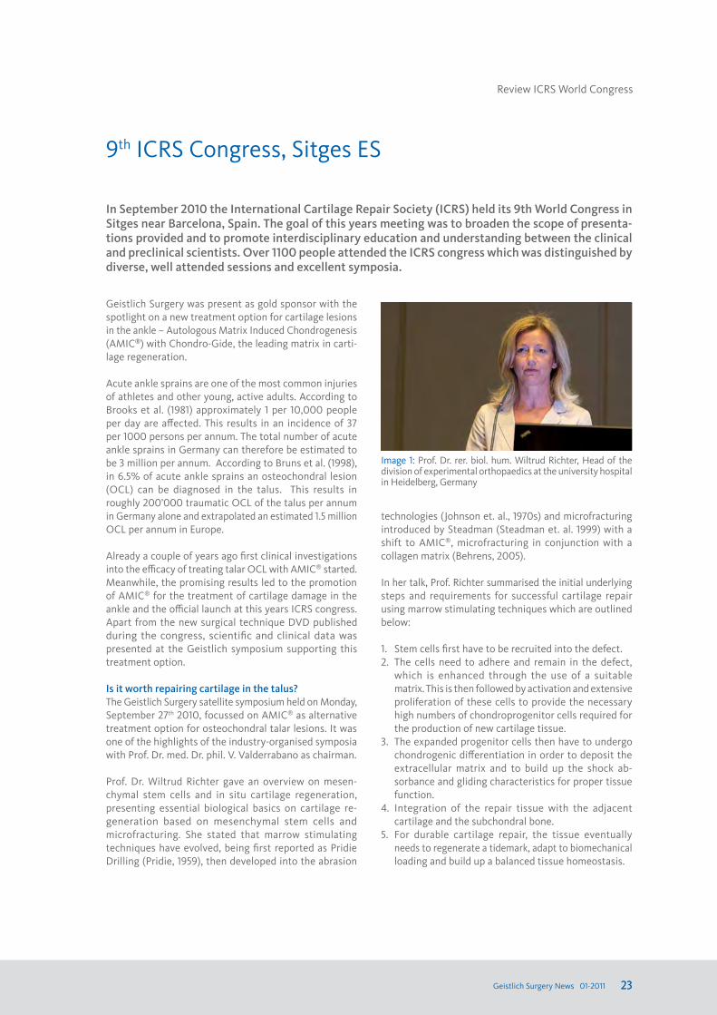
Image 1: Prof. Dr. rer. biol. hum. Wiltrud Richter, Head of the division of experimental orthopaedics at the university hospital in Heidelberg, Germany
23

Review ICRS World Congress
Referencing a recent paper from Jones et. al. 2010, Prof. Richter suggested that the recruitment of mesenchymal stem cells via microfracturing is not rate-limiting. The paper showed that recruiting of mesenchymal stem cells from the subchondral bone during microfracture shows higher number of mesenchymal stem cells (15-fold more MSC colonies and 65-fold CD271*+ cells) locally than compared to bone marrow aspirate from the illiac crest.
According to Prof. Richter, the super-clot which is formed during microfracturing alone, which is a relatively soft structure, undergoes a considerable amount of shrinkage during maturation, facilitated by non-adhesive charac-teristic of the cartilage surface. She stated that this may lead to only partial defect filling and facilitate an early loss of repair tissue from the cartilage lesion and argued that using a matrix which stabilises the super-clot, results in less shrinkage and therefore allows treatment of larger sized defects.
The results from a publication (Dickhut et. al., 2009) showed that the use of the Chondro-Gide® improves chondrogenesis of mesenchymal stem cells and proteo-glycan deposition in vitro. A significantly higher proteo-glycan content was observed in a fibrin-glue construct (8.5-fold, p=0.043) and in a fibrin-glue with a collagen-carrier construct (15.2-fold, p=0.043) when compared with a biomaterial-free group. The use of Chondro-Gide® with fibrin glue is supportive for an enhanced deposition of a proteoglycan-rich extracellular matrix.
Prof. Richter proposed that the Chondro-Gide® matrix may also have a protective function against inhibitory influences during cartilage repair which arise from the contralateral cartilage surface or from the synovial fluid.
An example for such an inhibitory factor which she mentioned is the Lubricin molecule which has been shown to inhibit the proliferation and maturation and to reduce the integrative cartilage repair capacity.
She underlined that the clinical goal of cartilage repair surgery is to treat the patient’s pain symptoms, to main-tain function and to prolong the time point for arthro-plasty for as long as possible. Prof. Richter concluded that marrow stimulating techniques such as Microfracture and AMIC® are very attractive for treating cartilage de-fects and for reaching this clinical goal. She stated that the information on mesenchymal stem cell biology can now be used to further dissect which of the individual steps are rate-limiting for the final outcome. Prof. Richter predicted that future improvements of these techniques could include a controlled factor release from the biomaterial to enhance the microenvironment, stimulate the cells, shield inhibitors, inhibit catabolism and enhance anabolism to achieve an improved and durable repair.
Dr. Christoph Becher from Hannover, Germany, presented an extensive overview of cartilage repair strategies in the ankle. He concluded that the knowledge of the knee cannot be transferred to the talus. In contrast to the knee, the ankle joint has a high congruency. Kuettner et. al. (2005) showed that talar cartilage has a higher content of proteoglycans and water and an increased rate of proteoglycan turnover and synthesis resulting in an increased stiffness and reduced permeability. He further showed that cartilage in the talus has a greater capacity for repair as talar chondrocytes synthesize proteoglycans at a higher rate in response to damage and have a decreased response to catabolic factors such as interleukin-1 and fibronectin fragments.
Image 2: Sitges, Spain as a conference venue for the 2010 ICRS World Congress

25Geistlich Surgery News 01-2011
The majority of the defects in the talus are osteochondral lesions caused by trauma or repeated microtrauma. According to van Dijk et. al. (2010), compression of the cartilage forces water into the microfractured subchondral bone during loading. The increased flow and pressure of fluid in the subchondral bone can cause osteolysis and the development of a subchondral cyst. Treatment strat-egies should therefore address the subchondral bone and include comorbidities such as ankle alignment or instability to prevent of further degeneration. Surgical treatment should however be restricted to symptomatic lesions where conservative treatment has failed.
The detailed literature summary presented by Dr. Becher is available for viewing on the ICRS educational services (http://icrs.onsite.tv/2010).
Image 3: Dr. Christoph Becher, orthopaedic hospital of the Hannover Medical School in Hannover, Germany
Image 4: Prof. Dr. med. Dr. phil. Victor Valderrabano, Head of orthopaedics at the university hospital in Basel, Switzerland
After treating osteochondral defects with a variety of techniques, Prof. Dr. med. Dr. phil. Victor Valderrabano reported that he is now reaching a satisfactory level of results using the AMIC® surgical technique. In his pres-entation, Prof. Valderrabano elaborated on the etiology, pathology, diagnostic methods and surgical technique for the treatment of osteochondral lesions in the talus.
For the diagnostic imaging investigation Prof. Valderrabano recommends the use of radiographic imaging and magnetic resonance imaging (MRT) as essential and SPECT/CT as optional techniques. In his experience, SPECT/CT shows the size of the osseous lesion with more precision than MRI and allows the surgeon to ascertain the extent of the scintigraphic activity along with the anatomically correct position.
Prof. Valderrabano indicated the importance to correct attendant pathologies, such as instability of the joint or an osseous malalignment, as well as the osseous and chondral reconstruction using AMIC®.
The presented 2-year data of 30 patients (m=15; f=15) with a mean age of 36 years (range 17-55 years) showed an etiology of mainly chronic instability caused by trauma as well as calcanear malalignment. The mainly medial defects were located centrally. The AOFAS score improved to an average of 83 points (p<0.001) after two years.
Prof. Valderrabano concluded that the AMIC® technique offers promising results with an excellent cost/benefit ratio. The principles and advantages of the AMIC® technique as described by Prof. Valderrabano are available as a surgical technique brochure and DVD on www.geistlich-pharma.com.
The satellite symposium was recorded by the ICRS and is available on the ICRS educational services (http://icrs.onsite.tv/2010) or as a complimentary copy from Geistlich Surgery. Please contact us via [email protected] for more information.
Autologous Matrix Induced Chondrogenesis, AMIC®, is an innovative biological surgical procedure developed by Geistlich Surgery for the treatment of traumatic chondral and osteochondral lesions. This unique single-step procedure combines the microfracturing method, which is an established first-line treatment, with the application of Chondro-Gide®. Clinical results are set-ting the AMIC® surgical technique as a new standard in the treatment of osteochondral defects of the talus. Both at the knee and at the talus, the AMIC® method shows clinically comparable results to autologous chondrocyte implantation and supports the body in the formation of functional cartilaginous repair tissues.
Geistlich Surgery is looking forward to the next international ICRS congress in Montreal, Canada in May 2012.

Congress Preview 2011The planning for 2011 has already begun. To assist in your planning, we have included a selection of congresses and events in 2011. Up-to-date information of the congresses at which we will be present is available on our internet site. We look forward to seeing you during the course of 2011 at our stand or symposia.
31. March – 03. April 2011Going, Austria – 9th International Meeting of the Austrian Foot Society
05. – 07. May 2011Vicenza, Italy – XXXIV Congresso Società Italiana Chirurgia Vertebrale - GIS
01. – 04. June 2011Copenhagen, Denmark – 12th EFORT Congress
13. – 14. June 2011Warwick, UK – Sports Foot & Ankle 2011
22. – 24. June 2011Lausanne, Switzerland – 71. Jahrestagung SGOT-SSOT
22. – 24. september 2011Regensburg, Germany – 28th AGA Kongress
01. – 05. october 2011Linz, Austria – 31. Jahrestagung ÖGO
01. – 05. october 2011Rimini, Italy – 16° SIOT (Congresso Nazionale della Società Italiana di Orthopedia e Traumatologia)
19. – 21. october 2011Milan, Italy – Eurospine
25. – 28. october 2011Berlin, Germany – DKOU (Deutscher Kongress für Orthopädie und Unfallchirurgie)
05. – 10. november 2011Paris, France – 86. SOFCOT (Société Française de Chirurgie Orthopédique et Traumatologique)
Further InformationPlease consult our web page for further information and invitations to the lunch symposia.
www.geistlich-pharma.com
Congresses and Events
26 Geistlich Surgery News 01-2011

27Geistlich Surgery News 01-2011

FranceGeistlich Pharma France SAParc des Nations – Paris Nord II385 rue de la Belle EtoileBP 43073 Roissy en FranceFR-95913 Roissy CDG CedexPhone +33 1 48 63 90 26Fax +33 1 48 63 90 [email protected]
GermanyGeistlich BiomaterialsVertriebsgesellschaft GmbHSchneidweg 5D-76534 Baden-BadenPhone +49 7223 96 24 0Fax +49 7223 96 24 [email protected]
ItalyGeistlich Biomaterials Italia S.r.lVia Castelletto, 28I-36016 Thiene VIPhone +39 0445 370 890Fax +39 0445 370 [email protected]
United KingdomGeistlich Sons Ltd.Long LaneGB-Chester CH2 2PFPhone +44 1244 347 534Fax +44 1244 319 [email protected]
Headquarters switzerlandGeistlich Pharma AGBusiness Unit SurgeryBahnhofstrasse 40CH-6110 WolhusenPhone +41 41 492 55 55Fax +41 41 492 56 [email protected]
3145
2.1/
110
2/en
©
20
11 G
eist
lich
Phar
ma
AG
– S
ubje
ct to
mod
ifica
tion
s