bone densitometry for technologists - home - springer978-1-4614-3625-6/1.pdf · bone densitometry...
TRANSCRIPT

Bone Densitometry for Technologists

Bone Densitometryfor TechnologistsThird Edition
Sydney Lou Bonnick, md, facp, ccdLori Ann Lewis, mrt, cdt
Clinical Research Center of North TexasDenton, TX, USA
ISBN 978-1-4614-3624-9 ISBN 978-1-4614-3625-6 (eBook)DOI 10.1007/978-1-4614-3625-6Springer New York Heidelberg Dordrecht London
Library of Congress Control Number: 2012947987
© Springer Science+Business Media New York 2013This work is subject to copyright. All rights are reserved by the Publisher, whether the whole or part of the material is concerned, speci fi cally the rights of translation, reprinting, reuse of illustrations, recitation, broadcasting, reproduction on micro fi lms or in any other physical way, and transmission or information storage and retrieval, electronic adaptation, computer software, or by similar or dissimilar methodology now known or hereafter developed. Exempted from this legal reservation are brief excerpts in connection with reviews or scholarly analysis or material supplied speci fi cally for the purpose of being entered and executed on a computer system, for exclusive use by the purchaser of the work. Duplication of this publication or parts thereof is permitted only under the provisions of the Copyright Law of the Publisher’s location, in its current version, and permission for use must always be obtained from Springer. Permissions for use may be obtained through RightsLink at the Copyright Clearance Center. Violations are liable to prosecution under the respective Copyright Law.The use of general descriptive names, registered names, trademarks, service marks, etc. in this publication does not imply, even in the absence of a speci fi c statement, that such names are exempt from the relevant protective laws and regulations and therefore free for general use.While the advice and information in this book are believed to be true and accurate at the date of publication, neither the authors nor the editors nor the publisher can accept any legal responsibility for any errors or omissions that may be made. The publisher makes no warranty, express or implied,with respect to the material contained herein.
Printed on acid-free paper
Springer is part of Springer Science+Business Media (www.springer.com)
Sydney Lou BonnickClinical Research Center of North Texas Denton, TX, USA
Lori Ann LewisClinical Research Center of North Texas Denton, TX, USA

Dedication
For Aubrey and Miller. Have a great life! (S.L.B)
For Colton and Kent. I love you. (L.A.L)


vii
Preface
Bone densitometry is an extraordinary clinical and research tool. Most of us think of densitometry as a relatively recent technological development, but in fact, its history began over 100 years ago. In the fi eld of dentistry, crude devices by today’s standards were developed in the late nineteenth century to evaluate the density of the bone in the mandible. The advances in technology continued, albeit slowly for the fi rst half of the twentieth century, gaining some speed in the 1960s and 1970s. The introduction of dual-energy X-ray absorptiometry in the late 1980s truly opened the door to clinician’s of fi ces for bone densitometry. In the last 20 years, the advances in technology and the introduction of new machines of various types have occurred with almost blinding speed compared to the pace of development during most of the twentieth century.
As densitometry has matured as a fi eld, the number of disease states in which bone density is known to be affected has increased. With this knowl-edge, physicians in many different fi elds of medicine now recognize the need to measure the bone density as part of the management of their patient. More studies are being requested now than ever before. This demand for densitometry has also led to an increased need for quali fi ed technologists to operate these machines.
Densitometry is one of the many quantitative techniques in use in clinical medicine today. That is, the technology is used to measure a quantity, the bone density, just as the measurement of blood pressure or cholesterol is also a quantitative measurement technique. But of all the quantitative tech-niques in use in clinical medicine today, there is none that has the potential to be more accurate or precise than bone densitometry. The technology is highly sophisticated. All of the devices in use today employ computer tech-nology. Even with all this mechanical sophistication, however, the technol-ogy will only be as good as the technologist.
The densitometry technologist must have knowledge of skeletal anatomy, densitometry techniques, radiation safety, basic statistics, quality control procedures, and various disease processes like osteoporosis. The technologist must often make decisions about the conduct of testing without immediate input from the physician. The circumstances in which densitometry is usu-ally performed create the opportunity for extended technologist–patient interaction and discussion. For technologists accustomed to performing
Prefaceviii
radiological procedures, this degree of interaction is unprecedented. Today’s densitometry technologist must be prepared for these encounters.
There is no substitute for the thoughtful training provided by the manu-facturers of the various types of densitometry equipment when the devices are installed. There is also no substitute for careful study of the operator’s manuals that are supplied with these machines. The exact operation of each machine is different. Ultimately, to be pro fi cient on any densitometry device, the technologist must be trained on that speci fi c device. There is a broad knowledge base however that all technologists should possess. This text is intended to help provide that base.
This book is overwhelmingly focused on the bone density technology known as dual-energy X-ray absorptiometry or DXA. While other technolo-gies are discussed, clinical practice guidelines from national and interna-tional organizations that have evolved in recent years call for the use of DXA studies of the spine and proximal femur in the diagnosis and manage-ment of osteoporosis. Consequently, other technologies, although not with-out merit, currently have lesser roles in clinical practice.
It is always dif fi cult to know where to begin. Like so many other fi elds of medicine, densitometry has its own language and conventions that must be explained so that in-depth discussions of densitometry are understood. Chapter 1 is an introduction to the terminology and conventions used in bone densitometry. In Chap. 2 , a review of the various techniques and tech-nologies used in quantifying the bone mass is presented. This review pro-vides some of the historical development of the fi eld as well as discussing the attributes of the various technologies and the differences between them. In Chap. 3 , the skeletal anatomy of commonly measured densitometry sites is discussed with an emphasis on those attributes of anatomy that are either unique to densitometry or which would have an effect on the measurement of bone density at that site. This is logically followed by Chap. 4 , in which the performance of DXA lumbar spine, proximal femur, and forearm studies is discussed. This chapter is new to the third edition of this book. While each manufacturer will provide speci fi c instruction on positioning and analysis of studies performed on their machines, there are aspects of scan performance that are common to all. Chapter 5 is a discussion of radiation safety in general and in bone densitometry speci fi cally. Most, but not all, densitometers are X-ray devices. Radiation safety then must be a concern. Fortunately, both patient and technologist exposures from X-ray densitom-etry are incredibly small. Nevertheless, the concept of ALARA (as low as reasonably achievable) demands that the patient, the public, and the tech-nologist be protected from unnecessary exposure to ionizing radiation. In Chap. 5 , radiation safety concepts are discussed with recommendations made for radiation safety procedures at densitometry facilities.
Preface ix
All densitometers, as sophisticated as they are, are ultimately mechanical devices. Things can and do go wrong. It is imperative that machine mal-functions be recognized as soon as possible. Otherwise, the data from the machine that are provided by the technologist to the physician will be fl awed. This means that a good quality control program must be in place. This is normally the responsibility of the technologist, not only to create but also to monitor. Therefore, an understanding of quality control procedures, quality control phantoms, and the development of a quality control program is absolutely necessary, although it is recognized that these are not particu-larly popular subjects. Quality control issues are discussed in Chap. 6 .
It is equally important that the technologist understand the concept of precision and how to measure and apply it. This is presented in Chap. 7 . Without careful attention to precision, those factors that affect it and know-ing how to calculate it, the physician to whom the results are given will not be able to interpret follow-up bone density studies to determine if the bone density has changed.
Also new to this edition is Chap. 8 . One of the major applications of bone densitometry data is the prediction of fracture risk. The manner in which this determination is made has changed in only the last few years. Older expressions of risk such as relative risk are no longer considered appropri-ate. Instead, there are several absolute fracture risk prediction tools that are achieving widespread use. Because some of these are being incorporated into densitometry software and because the technologist may be asked to access and employ others to calculate fracture risk, a discussion of these new fracture risk prediction tools is included in this edition.
Two chapters of this book may seem unusual in a book for technologists. Chapter 9 is a review of the disease for which densitometry is most com-monly used, osteoporosis. Chapter 10 is a review of how the data that comes from these machines is actually interpreted to diagnose osteoporosis and predict fracture risk. These chapters might at fi rst seem more appropriate in a book written for physicians. The densitometry technologist, however, normally spends a signi fi cant amount of time with the patient. There is ample opportunity for the patient to ask questions of the technologist about osteoporosis and about the test that he or she is about to undergo. The knowledgeable technologist can be a vital link in the education of the patient. He or she can allay unnecessary fears and encourage appropriate medical follow-up. The technologist is not usurping the role of the physi-cian by doing so if the technologist understands the issues involved. Indeed, the complete medical care of the patient must involve a partnership between the technologist and the physician. The fi nal diagnosis and treatment recom-mendations for any patient must be left to the physician, but within those bounds, there is much the technologist can do that will actually strengthen

Prefacex
the patient’s trust in the quality of their care and improve compliance with the medical recommendations. The technologist who understands as much as possible about what the physician will consider as he or she looks at the densitometry report will only be better able to aid that physician in the per-formance of their profession. Since the publication of the last edition of Bone Densitometry for Technologists, new drugs have been approved for the prevention and/or treatment of osteoporosis. This information has been added to Chap. 9 in this edition.
Chapter 11 is also new to this edition. In this chapter, a series of DXA images in which artifacts or structural changes can be seen are reproduced. Often in densitometry in illustrating the utility of the technology, scan images of perfect spines, free of artifacts, are shown. In clinical practice, the spines of our patients are often not as perfect. While some of these less than perfect scan images are self-explanatory, others are not. Realizing that there is no substitute for having seen it before, these images are provided in the hope of aiding the technologist in recognizing these artifacts and changes in their patients. It is imperative that the technologist do so, because many of these artifacts and structural changes will affect the measured BMD. Their recognition is critical to the appropriate interpretation of the study.
In the last few years, densitometry has found increasing applications in pediatrics. The technical considerations are different from adult densitom-etry, and the interpretation of densitometry data is even more complex than its adult counterpart. The 2002, 2004, and 2007 ISCD guidelines that apply to pediatric densitometry are reviewed here. This is an area that is expected to grow however, and so many of the confounding issues in pediatric den-sitometry are addressed in Chap. 12 .
Chapters 13 and 14 deal with some of the newer applications for DXA. In Chap. 13 , there is a review of vertebral fracture assessment (VFA) imaging. VFA imaging with fan-array DXA devices is now being utilized to perform vertebral fracture diagnosis and aortic calci fi cation assessment. Proximal femur morphometry and hip structural analysis can be performed using proximal femor studies. Body composition analysis with DXA, which is discussed in Chap. 14 , is probably the least utilized application of DXA, but its advantages become obvious when compared with other body composition methods. Its potential utility, particularly in the context of growing concerns about the metabolic syndrome, is enormous.
The 12 appendices have been updated wherever necessary to re fl ect the most current information available. Contact information for organizations of interest can be found in Appendix A. Every attempt was made to verify the accuracy of this information at the time this book went to press. The World Health Organization criteria for the diagnosis of osteoporosis are summarized in Appendix B, and the conversion of the criteria to T-scores is
Preface xi
illustrated. Guidelines for bone density testing from ISCD and other organi-zations are found in Appendices C and E, and the new ISCD-IOF position on the use of FRAX ® is found in Appendix D. Appendices F–K summarize the Medicare Bone Mass Measurement Act of 1997, DXA PA lumbar spine labeling guidelines, frequently used conversion formulas, short-term preci-sion study procedures, and the calculation of the LSC and quality control Shewhart rules.
Finally, in Appendix L, the contents of the accompanying CD-ROM are reviewed. On this CD, you will fi nd the Precision Calculator Companion that was fi rst included with the second edition of Bone Densitometry in Clinical Practice. With this calculator, you will be able to calculate the short-term precision and least signi fi cant change values for your facility as well as the statistical con fi dence level for any measured change in BMD. These concepts are discussed thoroughly in Chap. 7 . There is also a densit-ometry patient questionnaire that may be customized for your facility. This questionnaire was designed to ensure the capture of necessary responses in a form suitable for use with FRAX ® . A continuing education review is also found on the CD, which, if successfully completed, will result in the award-ing of 16 hours of Category A credit acceptable to the American Society of Radiologic Technologists. ( Those readers who do not have access to the CD can download the material at http://mixmastermedia.com/BDT3 .)
As a technology, bone densitometry is really quite extraordinary. The ability to quantify the density of the bones at a variety of skeletal sites has truly revolutionized the approach to a number of diseases, the most impor-tant of which is osteoporosis. Using the information from the machines, physicians can recommend and prescribe interventions that will slow bone loss and reduce the risk of disabling fractures. The remarkable advances in skeletal imaging with densitometry devices have made possible quantitative and diagnostic assessments of skeletal structure. But it is in fact the skill and concern of the technologist that enable all of this to happen. It is our hope that this book assists you in your pursuit of excellence in your profession.
Sydney Lou Bonnick, MD, FACP, CCD
Lori Ann Lewis, MRT, CDT

xiii
About the Authors
Sydney Lou Bonnick, M.D., F.A.C.P., C.C.D. Dr. Bonnick is a native of Dallas, Texas, and a graduate of Southern
Methodist University and the University of Texas Southwestern Medical School. She is the medical director of the Clinical Research Center of North Texas and an adjunct professor at the University of North Texas in Denton, Texas.
Dr. Bonnick is one of the founders and past Secretary of the International Society for Clinical Densitometry. Dr. Bonnick helped to create and teach the original physician and technologist bone densitometry certi fi cation courses for ISCD. She is the 1999 winner of the International Society for Clinical Densitometry President’s Award and a past recipient of the American Medical Women’s Association President’s Recognition Award and the AMWA’s Calcium Education Nutrition Award. She is a fellow in the American College of Physicians and a member of the American Society for Bone and Mineral Research, National Osteoporosis Foundation, International Society for Clinical Densitometry, and the North American Menopause Society. She is a former member of the osteoporosis advisory committee for the Texas Department of Health.
Dr. Bonnick has served as a primary investigator in numerous research trials in the fi eld of the prevention and treatment of osteoporosis and has published extensively. In addition to being the coauthor of this book, she is also the author of Bone Densitometry in Clinical Practice from Humana Press. She lives in Denton, Texas.
Lori Ann Lewis, M.R.T., C.D.T. Ms. Lewis is a medical radiologic technologist and recognized by the
International Society for Clinical Densitometry as a certi fi ed densitometry technologist. She is one of the original members of the technologist teaching faculty for ISCD and the 1997 winner of the ISCD technologist of the year award. She is the clinical research coordinator for Dr. Sydney Bonnick and bone density technologist at the Clinical Research Center of North Texas in Denton, Texas. She has extensive experience in bone densitometry on all types of equipment over the last 25 years. She lives in Denton, Texas.

xv
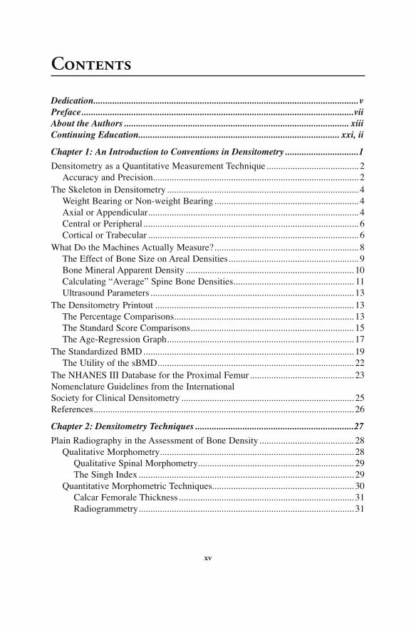
Contents
Dedication ................................................................................................................vPreface ...................................................................................................................viiAbout the Authors ............................................................................................... xiiiContinuing Education..................................................................................... xxi, ii
Chapter 1: An Introduction to Conventions in Densitometry ...............................1
Densitometry as a Quantitative Measurement Technique .......................................2Accuracy and Precision .......................................................................................2
The Skeleton in Densitometry .................................................................................4Weight Bearing or Non-weight Bearing .............................................................4Axial or Appendicular .........................................................................................4Central or Peripheral ...........................................................................................6Cortical or Trabecular .........................................................................................6
What Do the Machines Actually Measure? .............................................................8The Effect of Bone Size on Areal Densities .......................................................9Bone Mineral Apparent Density .......................................................................10Calculating “Average” Spine Bone Densities ................................................... 11Ultrasound Parameters ......................................................................................13
The Densitometry Printout ....................................................................................13The Percentage Comparisons ............................................................................13The Standard Score Comparisons .....................................................................15The Age-Regression Graph ...............................................................................17
The Standardized BMD .........................................................................................19The Utility of the sBMD ...................................................................................22
The NHANES III Database for the Proximal Femur ............................................23Nomenclature Guidelines from the International Society for Clinical Densitometry .........................................................................25References ..............................................................................................................26
Chapter 2: Densitometry Techniques ...................................................................27
Plain Radiography in the Assessment of Bone Density ........................................28Qualitative Morphometry ..................................................................................28
Qualitative Spinal Morphometry..................................................................29The Singh Index ...........................................................................................29
Quantitative Morphometric Techniques ............................................................30Calcar Femorale Thickness ..........................................................................31Radiogrammetry ...........................................................................................31

Contentsxvi
The Radiologic Osteoporosis Score ..................................................................33Radiographic Photodensitometry ...........................................................................33Radiographic Absorptiometry ................................................................................35Photon Absorptiometry Techniques .......................................................................37
Single-Photon Absorptiometry ..........................................................................38Dual-Photon Absorptiometry ............................................................................40Dual-Energy X-Ray Absorptiometry ................................................................44Peripheral DXA .................................................................................................49Single-Energy X-Ray Absorptiometry ..............................................................49Quantitative Computed Tomography ................................................................50Peripheral QCT .................................................................................................56
Quantitative Ultrasound Bone Densitometry ........................................................56References ..............................................................................................................59
Chapter 3: Skeletal Anatomy in Densitometry .....................................................63
The Spine in Densitometry ....................................................................................64Vertebral Anatomy ............................................................................................64Artifacts in PA Spine Densitometry ..................................................................69
Vertebral Fractures .......................................................................................70Degenerative Changes and Dystrophic Calcification ..................................71
The Effect of Vertebral Rotation on PA Lumbar Spine Bone Density .............83The Spine in the Lateral Projection ..................................................................84
The Proximal Femur in Densitometry ...................................................................85Proximal Femur Anatomy .................................................................................85The Effect of Scoliosis, Osteoarthritis, Osteophytes, Surgery, and Fracture on BMD in the Proximal Femur ..................................................86
The Forearm in Densitometry ................................................................................89Nomenclature ....................................................................................................89
The Effect of Artifacts on BMD in the Forearm ...................................................90The Metacarpals, Phalanges, and Calcaneus .........................................................92References ..............................................................................................................95
Chapter 4: Performing a DXA PA Lumbar Spine, Proximal Femur, or Forearm DXA Study ............................................................97
The PA Lumbar Spine Study .................................................................................98The Effect of Vertebral Rotation .......................................................................99
The Proximal Femur Study ..................................................................................101The Effect of Proximal Femoral Rotation ......................................................106The Effect of Leg Dominance ........................................................................107The Effect of Lumbar Scoliosis ......................................................................108The Effect of Femoral Shaft Adduction and Abduction .................................109
Unilateral Versus Bilateral Proximal Femur Bone Density Measurements ...............................................................................109
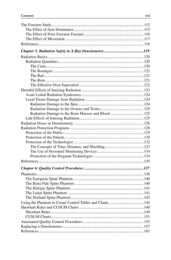
Contents xvii
The Forearm Study .............................................................................................. 112The Effect of Arm Dominance........................................................................ 115The Effect of Prior Forearm Fracture ............................................................. 116The Effect of Movement ................................................................................. 117
References ............................................................................................................ 118
Chapter 5: Radiation Safety in X-Ray Densitometry .........................................119
Radiation Basics ..................................................................................................120Radiation Quantities ........................................................................................120
The Curie ....................................................................................................120The Roentgen .............................................................................................121The Rad ......................................................................................................121The Rem .....................................................................................................121The Effective Dose Equivalent ..................................................................122
Harmful Effects of Ionizing Radiation ................................................................123Acute Lethal Radiation Syndromes ................................................................124Local Tissue Damage from Radiation ............................................................124
Radiation Damage to the Skin ...................................................................124Radiation Damage to the Ovaries and Testes ............................................125Radiation Damage to the Bone Marrow and Blood ..................................125
Late Effects of Ionizing Radiation ..................................................................125Radiation Doses in Densitometry ........................................................................126Radiation Protection Programs ............................................................................128
Protection of the Public ...................................................................................129Protection of the Patient ..................................................................................130Protection of the Technologist ........................................................................132
The Concepts of Time, Distance, and Shielding ........................................133The Use of Personnel Monitoring Devices ................................................134Protection of the Pregnant Technologist ....................................................134
References ............................................................................................................135
Chapter 6: Quality Control Procedures .............................................................137
Phantoms ..............................................................................................................138The European Spine Phantom .........................................................................140The Bona Fide Spine Phantom .......................................................................140The Hologic Spine Phantom ...........................................................................141The Lunar Spine Phantom ..............................................................................141The Norland Spine Phantom ...........................................................................143
Using the Phantom to Create Control Tables and Charts ....................................144Shewhart Rules and CUSUM Charts ..................................................................148
Shewhart Rules ...............................................................................................149CUSUM Charts ...............................................................................................151
Automated Quality Control Procedures ..............................................................153Replacing a Densitometer ....................................................................................157References ............................................................................................................161
Contentsxviii
Chapter 7: The Importance of Precision ...........................................................163
The Concept of Precision ....................................................................................163Performing a Precision Study ..............................................................................164
Short-Term Precision Studies .........................................................................166Mathematical Procedures Used to Calculate Precision ..................................168Long-Term Precision Studies ..........................................................................171
Applying the Precision Value to Serial Measurements .......................................171The Determination of Least Significant Change ............................................172A Case in Point ...............................................................................................174
Determining the Level of Confidence for Any Change and Precision ................176The Importance of Precision ................................................................................176Which Skeletal Sites Should Be Used for Monitoring? ......................................177How Frequently Should Measurements Be Repeated? .......................................180A Final Consideration ..........................................................................................181References ............................................................................................................182
Chapter 8: Using Absolute Risk to Predict Fracture Risk in Clinical Practice .....................................................................................183
Relative Fracture Risk in Clinical Trials .............................................................184Absolute Fracture Risk in Clinical Practice ........................................................185Absolute Fracture Risk Prediction Tools .............................................................186
FRAX® ............................................................................................................186Using the T-Score with FRAX® .................................................................192FRAX® Filters ............................................................................................193
FORE FRC ......................................................................................................194Canadian Association of Radiologists and Osteoporosis Canada (CAROC) Fracture Risk Assessment System ....................................196Garvan Fracture Risk Calculator ....................................................................200
References ............................................................................................................204
Chapter 9: An Overview of Osteoporosis ...........................................................207
The Definition of Osteoporosis ............................................................................208The 1991 and 1993 Consensus Development Conferences ...........................208The 1994 World Health Organization Criteria for Diagnosis of Osteoporosis .........................................................................209The 2000 National Institutes of Health Consensus Conference Definition of Osteoporosis ........................................................... 211
The Prevalence of Osteoporosis .......................................................................... 211Consequences of Osteoporosis ............................................................................213Risk Factors for Osteoporosis ..............................................................................213
The Attainment of Peak Bone Density ...........................................................213The Maintenance of Bone Density .................................................................214
Guidelines for Bone Mass Measurements ...........................................................215The 1997 Bone Mass Measurement Act ..............................................................215
Contents xix
Treatment Guidelines for Osteoporosis ...............................................................217The NOF Guidelines .......................................................................................217Treatment Guidelines from AACE and NAMS ..............................................217Treatment Guidelines from Osteoporosis Canada and NOGG.......................217
Interventions in Osteoporosis ..............................................................................218Nonprescription Interventions.........................................................................218
Lifestyle Modifications ..............................................................................218Calcium, Vitamin D, and Exercise .............................................................219
Prescription Interventions ...............................................................................221Estrogen Therapy .......................................................................................222The Selective Estrogen Receptor Modulator Raloxifene ..........................222Synthetic Salmon Calcitonin ......................................................................222The Bisphosphonates..................................................................................223Denosumab .................................................................................................232Parathyroid Hormone .................................................................................233
References ............................................................................................................236
Chapter 10: Interpretation of Bone Densitometry Data ....................................241
The Results ..........................................................................................................242The Skeletal Image .........................................................................................242The Measured and Calculated Bone Density Parameters ...............................245Comparisons to the Reference Database ........................................................249The sBMD .......................................................................................................250The Age-Regression Graph .............................................................................251Assignment of Diagnostic Category Based on the WHO Criteria .................254
Conflicting Diagnoses from the Measurement of Multiple Sites ........................254Report Review .....................................................................................................259References ............................................................................................................262
Chapter 11: Less Than Perfect Scan Images .....................................................265
Chapter 12: Pediatric Densitometry ...................................................................295
Pediatric Scan Acquisition and Analysis .............................................................296Radiation Safety Issues in Pediatric Densitometry .............................................297Bone Age ..............................................................................................................298Sexual Maturation Stage ......................................................................................301Considerations of Bone Size and Shape ..............................................................301Skeletal Development and the Use of Standard Scores in Pediatric Densitometry ....................................................................................304
Skeletal Development .....................................................................................304The Use of Standard Scores in Pediatric Densitometry .................................306
Pediatric Reference Databases .............................................................................3082003, 2004, and 2007 International Society for Clinical Densitometry Guidelines for Children .............................................309The Specialty of Pediatric Densitometry .............................................................310References ............................................................................................................ 311
Contentsxx
Chapter 13: VFA Imaging, Femoral Morphometry, and Hip Structural Analysis ...............................................................................313
VFA Imaging ........................................................................................................314The Relationship Between Prevalent Spine Fracture and Future Fracture Risk .................................................................................314Diagnosing Vertebral Fractures .......................................................................316
Vertebral Fracture Assessment with Genant Semiquantitative Technique .......................................................................316Vertebral Fracture Assessment with Quantitative Techniques ...................317Performance Comparisons of Semiquantitative and Quantitative Techniques ......................................................................319
Spine Imaging with DXA for Diagnosis of Vertebral Fracture ......................319VFA Patient Selection and Reporting .............................................................323Aortic Calcification Assessment .....................................................................323
Aortic Calcification Scoring Systems ........................................................325Reporting Aortic Calcification on VFA Studies .........................................328
Proximal Femur Morphometry ............................................................................328Hip Axis Length ..............................................................................................329The Femoral Neck–Shaft Angle .....................................................................330Femoral Neck Width .......................................................................................332The Upper Femoral Neck ...............................................................................332
Hip Structural Analysis ........................................................................................333HSA with QCT ................................................................................................335
References ............................................................................................................337
Chapter 14: Body Composition Analysis............................................................341
The Body Mass Index ..........................................................................................342Body Composition Methods ................................................................................344
2-Compartment Body Composition Measurement Techniques ......................344Underwater Weighing .................................................................................344Skinfold Measurements ..............................................................................345Bioelectrical Impedance Analysis ..............................................................347Air Displacement Plethysmography ..........................................................347
3-Compartment Body Composition Measurement Techniques ......................348Near-Infrared Interactance .........................................................................348Dual-Energy X-Ray Absorptiometry .........................................................348
The Metabolic Syndrome ....................................................................................355References ............................................................................................................359
Appendix A: Contact Information for Organizations of Interest ..................363
Appendix B: World Health Organization Criteria for the Diagnosis of Osteoporosis Based on the Measurement of Bone Density ......365
Appendix C: 2007 ISCD Official Positions .....................................................367
Appendix D: 2010 ISCD-IOF Official Positions on FRAX® ..........................383

Contents xxi
Appendix E: Guidelines for Bone Density Testing from Major Organizations Other Than ISCD .............................387
Appendix F: Bone Mass Measurement Act of 1997 .......................................391
Appendix G: DXA PA Lumbar Spine Labeling Guidelines ...........................393
Appendix H: Conversion Formulas .................................................................395
Appendix I: Recommended Procedures for Short-Term Precision Studies .....................................................399
Appendix J: Calculation of Least Significant Change (LSC) ........................403
Appendix K: Quality Control Shewhart Rules ................................................405
Appendix L: The CD-ROM Companion .........................................................407
Index ....................................................................................................................409


xxiii
Continuing Education
This companion CD for this book contains a continuing education test good for 16 hours of Category A credit from The American Society of Radiologic Technologists (ASRT). Instructions for the test are contained within the program. The CD also includes links to patient questionnaires for men and women in Word format and a precision calculator for bone densi-tometry technologists in Excel format.
For those readers viewing the material in a library setting where the disk is not available, the material outlined above can also be downloaded from the following website: http://mixmastermedia.com/BDT3.