botulism in alaskadhss.alaska.gov/dph/epi/id/siteassets/pages/botulism/monograph.pdf · 3. the...
TRANSCRIPT
Botulism in AlaskaNovember 2017
Department of Health and Social ServicesDivision of Public Health, Section of EpidemiologySTATE OF ALASKA

Botulism in Alaska – November 2017: http://dhss.alaska.gov/dph/Epi/id/SiteAssets/Pages/botulism/monograph.pdf 1
BOTULISM IN ALASKA NOVEMBER 2017
TABLE OF CONTENTS
INTRODUCTION 2
REPORTING AND OUTBREAK RESPONSE 3
GENERAL REVIEW 4
SURVEILLANCE DATA 7
CLINICAL CONSIDERATIONS 17
LABORATORY EVALUATION 23
PREVENTION 25
INFANT BOTULISM 27
WOUND BOTULISM 29
BIOTERRORISM CONSIDERATIONS 29
REFERENCES 30
ADDITIONAL READING 32
ACKNOWLEDGEMENTS 34

Botulism in Alaska – November 2017: http://dhss.alaska.gov/dph/Epi/id/SiteAssets/Pages/botulism/monograph.pdf 2
INTRODUCTION
The Alaska Division of Public Health, the Arctic Investigations Program of the U.S. Centers for Disease Control and Prevention (CDC), and the Alaska Area Native Health Service of the U.S. Indian Health Service first produced a botulism monograph in 1993 to give Alaska health care providers a comprehensive overview of botulism in Alaska. Alaska Division of Public Health staff updated the original hard-copy monograph in 1998, 2005, and 2011. In 2017, the monograph is being produced primarily in an electronic format.
Botulism can result from several different circumstances, the most common of which in Alaska is from consumption of preformed toxin in food. Cases of foodborne botulism in the United States have been associated with consumption of home-canned products, rarely with consumption of commercially available products, and in Alaska, all cases have occurred among Alaska Natives who had a history of consuming Alaska Native traditional foods.
This current 2017 monograph includes the following changes:
Epidemiologic data for confirmed foodborne botulism cases in Alaska from 1950 through 2016, as well as inclusion of all “probable” cases.
Addition of a case of infant botulism.
Changes to the protocol for administration of botulism antitoxin.
Changes to the algorithms for testing of lab specimens.
Botulism is relatively uncommon and health care providers unfamiliar with its epidemiology and presentation in Alaska may not think to include botulism in their differential diagnosis when examining patients with mild symptoms. It is critical that health care providers in Alaska are able to accurately diagnose botulism for several important reasons:
Botulism is a life-threatening disease.
Botulism is a public health emergency. The occurrence of a single case implies that other persons may also be at risk.
Early administration of antitoxin appears to be beneficial, especially with type E botulism, the most common type in Alaska.
Early diagnosis which leads to appropriate medical observation and access to mechanical ventilation, if appropriate, appears to be beneficial.
Current laboratory methods for detecting botulinum toxin in clinical specimens or food samples require at least 5 to 7 days to perform. Therefore, early intervention and epidemiologic investigation depends upon timely and astute clinical assessment.
After reading this monograph, a health care provider should have an understanding of the following:
The need for immediate reporting of suspect cases to the Alaska Section of Epidemiology.
The signs and symptoms of botulism.
The importance of rapid diagnosis, evaluation, and treatment.
The types of foods that have been associated with botulism in Alaska.
Other epidemiologic characteristics of botulism in Alaska.

Botulism in Alaska – November 2017: http://dhss.alaska.gov/dph/Epi/id/SiteAssets/Pages/botulism/monograph.pdf 3
REPORTING AND OUTBREAK RESPONSE
Botulism is both a medical and a public health emergency. If a health care provider suspects botulism, he or she should immediately notify the Alaska Division of Public Health, Section of Epidemiology so that any possible associated cases can be identified and treated. Reporting should never await laboratory confirmation. Delayed reporting may result in additional persons consuming toxin-containing food and additional cases of botulism. The Section of Epidemiology collaborates with public health nurses, community health aides, and/or environmental health officers to investigate all botulism cases in the state.
STEPS IN A BOTULISM OUTBREAK INVESTIGATION.
1. A health care provider reports suspected botulism to the Section of Epidemiology. 2. If, after discussing the clinical presentation, botulism is considered possible, an investigation is immediately
started. Investigation partners may include public health nurses, community health aides and/or environmental health officers.
3. The Section of Epidemiology interviews the patient (or patient’s family) to determine the possible meals or foods (including how the foods were prepared) associated with exposure to botulinum toxin.
4. Other persons who ate the suspect food(s) are asked their food consumption histories and screened for symptoms of botulism to determine the extent of the outbreak and to more precisely define a likely source.
5. Symptomatic persons are immediately evaluated at the nearest health care facility to obtain antitoxin and supportive care, if needed.
6. Asymptomatic persons who were exposed to implicated food are warned that they may become ill and are told to immediately seek care if symptoms of botulism develop. Community health aides and local health care providers are alerted and active surveillance is maintained for 10 days. Asymptomatic persons are contacted daily so that they do not become ill without others being aware.
7. The Section of Epidemiology recommends not consuming any suspect food until laboratory testing has been completed.
8. Appropriate food and clinical specimens are collected and shipped to the Alaska State Public Health Laboratory in Anchorage for testing.
9. When laboratory results are received, this information is relayed to health care providers and persons in possession of suspect food.
10. If any part of the investigation cannot be completed quickly and reliably by telephone, a site investigation is conducted.
Possible cases should be reported immediately by telephone to the Section of Epidemiology in Anchorage at (907) 269-8000 or after-hours at (800) 478-0084. Medical epidemiologists from the Section are available 24-hours a day to provide clinical consultation and advice regarding diagnosis, specimen collection, and treatment, as well as access to antitoxin.
Botulism remains an illness that challenges the clinician to make a diagnosis using the classic elements of medical practice — history and physical examination. Practitioners who care for Alaska Native patients should be alert to this condition and act decisively if botulism is suspected.
907-269-8000
REPORT CASES OF BOTULISM TO THE ALASKA SECTION OF EPIDEMIOLOGY
907.269.8000 or 800.478.0084 (after hours)

Botulism in Alaska – November 2017: http://dhss.alaska.gov/dph/Epi/id/SiteAssets/Pages/botulism/monograph.pdf 4
GENERAL REVIEW
EARLY DESCRIPTIONS
Botulism, or sausage poisoning as it was originally termed, was first studied in depth following a 1793 outbreak in Wildbad, Germany. The outbreak involved 13 people, six of whom died, and was associated with consumption of a locally-produced blood sausage. Following this outbreak, the number of reported cases of sausage poisoning rapidly increased, prompting a study of the disease by the local health officer, Justinius Kerner, who described 230 cases, most of which were attributed to the consumption of sausage.1 The illness became known as “botulism” after “botulus,” the Latin word for sausage.
In 1895 in Ellezelles, Belgium, Van Ermengem investigated an outbreak of botulism involving 34 individuals who had consumed raw, salted ham served at a gathering of amateur musicians.2 He established that botulism was an intoxication, not an infection, and that the toxin was produced by a spore-forming obligate anaerobic bacterium, Clostridium botulinum. He also found that toxin was rapidly inactivated by heating and was only toxic to certain animal species. A later outbreak in Darmstadt, Germany, associated with canned white beans, established that there was a second type of botulism. The new strain was type A and the Van Ermengem strain was probably type B.3
In 1922, type C botulism was identified as causing disease in chickens4 and cattle.5 Robinson6 identified type D in cattle and type E was identified by Gunnison7 as causing botulism in people who consumed fish. Type F was first described by Moller and Scheibel from a Danish outbreak involving homemade liver paste.8 Finally, type G botulism was identified in soil from cornfields in Argentina by Gimenez and Ciccarelli.9 The principal types of botulism involved in human disease are types A, B, and E. Detailed historical reviews of type E botulism have been published.10,11
FOODBORNE BOTULISM IN THE ARCTIC
A. EPIDEMIOLOGY
For many years, outbreaks of illness associated with traditionally prepared and preserved food have been described. Early explorers reported clusters of deaths in villages among groups of northern Natives that the explorers attributed to “ptomaine” poisoning or trichinosis.12 However, descriptions of many of these outbreaks resemble foodborne botulism. Later, ethnographers described food preparation and storage practices that could support the production of botulinum toxin.13
The first reported outbreaks of foodborne botulism in the Arctic occurred in the early 1900s. In Canada, the first reported outbreak was in 1919 and since then, over 100 outbreaks involving over 230 individuals have occurred.10,14 The first reported outbreaks in Greenland occurred in 1967,15 with over 20 additional outbreaks reported since then. Rabeau recorded the first Alaska outbreak that occurred in 1947 and involved beluga whale flipper consumed in the village of Kotzebue.16 Russian medical literature suggests the presence of type E botulism in Siberia and the Russian Far East.11 The disease “ichthyosismus” from early Russian medical journals may represent one of the earliest reported fish-related botulism descriptions.17 In 1938, smoked herring was associated with type E botulism cases in Leningrad.10 Cases of type E botulism from smoked “kunzha”, or salted fish were first reported in Kamchatka and on Sakhalin Island in the Russian north and Far East, in 1967.11
Because not all northern Native groups consume the same traditionally prepared foods, it is difficult to determine true incidence rates of disease. Using an estimate of only Native populations, Canadian First Nations and Alaska Native residents had annual incidence rates of 50 cases per 100,00018 (1985-2000) and 8.5 cases per 100,00019 (1976-1984), respectively. These rates are much greater than an overall estimate for the United States in 1990-2000 at one case per million, with 1.9 per 100,000 for Alaska; Idaho and Washington had the next highest rates of 0.6 and 0.3 respectively.20 Most foodborne botulism in the Arctic is type E.

Botulism in Alaska – November 2017: http://dhss.alaska.gov/dph/Epi/id/SiteAssets/Pages/botulism/monograph.pdf 5
B. THE FOODS, THEIR PREPARATION AND STORAGE
All Alaska foodborne botulism cases have been associated with the consumption of traditional Alaska Native foods. These include “fermented” foods, dried foods, and traditionally prepared condiments, such as seal oil. In other parts of the United States, foodborne botulism is usually associated with improperly canned foods or with improperly stored unrefrigerated foods.20
Foods involved in Alaska botulism outbreaks are usually aged or putrefied (“fermented”). Traditionally prepared northern foods are not in fact “fermented” as fermentation requires a carbohydrate substrate and results in organic acid production that subsequently reduces the pH.21 Sea mammal food products do not contain enough carbohydrates to enable fermentation; therefore, the pH remains neutral. Instead the aging of traditional foods is really putrefaction or advanced decomposition of proteins and fat which does not inhibit the growth of Clostridium botulinum (type E strains are inhibited at a pH of <5) or the production of botulinum neurotoxin.21 Similar practices are still found in many areas of the Arctic. In Canada, First Nations persons living outside the Arctic (e.g., British Columbia) also age traditional foods, which results in sporadic cases of botulism.18
Whales and seals are the most frequently involved sea mammals. Salmon, including salmon eggs, are the most frequently involved fish. Semi-aquatic mammals, such as beaver, contribute to a small proportion of outbreaks.
Dried foods, particularly dried fish, have also been implicated in foodborne botulism outbreaks. Fish are dried either with or without a brine stage. However, even if fish are put in brine prior to drying, the salt concentration is rarely high enough to inhibit botulinum toxin formation.22
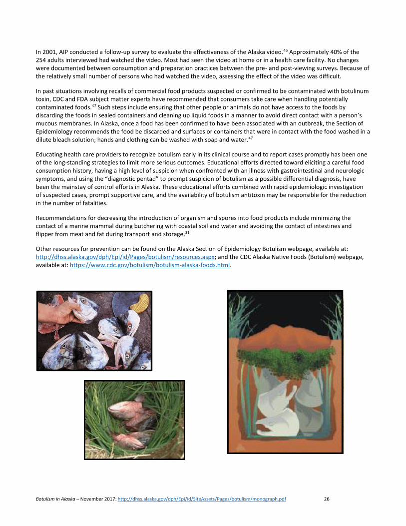
In addition to inadvertent spoilage, many traditional methods of food preparation lend themselves to botulinum toxin information. Traditional “stink” foods such as aged salmon eggs (stink eggs) or salmon heads (stink heads) are prepared by burial in moss-lined pits or barrels in the ground. Nelson described the process he observed during a visit to the coastal villages of northwest Alaska in 1877–1881:13
In the district between the Yukon and Kuskokwim, the heads of king salmon, taken in the summer, are
placed in small pits in the ground surrounded by straw and covered with turf. They are kept there during
the summer and in the autumn have decayed until even the bones have become the same consistency as
the general mass. They are taken out and kneaded in a wooden tray until they form a pasty compound
and are eaten as a favorite dish by some of the people.

Botulism in Alaska – November 2017: http://dhss.alaska.gov/dph/Epi/id/SiteAssets/Pages/botulism/monograph.pdf 6
The process described by Nelson has changed somewhat. Now, aging may be carried out in a barrel, a plastic or glass jar, or a plastic bag. These containers may increase the risk of botulinum toxin formation because most can be easily sealed and/or brought indoors, thereby increasing the likelihood of anaerobic conditions and warmer temperatures. Some foods are aged in a seal skin or fish skin bag or “poke,” which is either buried or hung up. If salmon eggs are aged in this manner, they can be left until they dry out somewhat and form a “cheese” that is firm on the outside and soft in the center.
Toxin production is temperature-dependent, and is less likely to occur at the lower temperatures that were usually attained during traditional aging, i.e., outdoors in moss-lined pits. For the clostridial organisms causing most types of botulism in Alaska, optimal growth occurs around 25°C and minimal or no growth at 2.5°C.23 Aging now may be done indoors, or in a container above ground and in the sun, which produces warmer temperatures that make aging more rapid and production of botulinum toxin more likely. In one experiment, botulinum toxin was detected in salmon heads that had been aged in a sealed plastic container kept underground for 17 days, but not in salmon heads aged in a grass-lined pit for the same length of time (personal communication 2011, Dr. Thomas Hennessy, CDC Arctic Investigations Program).
Although no commercial products have been associated with illness among Alaskans, fish products from Alaska have been implicated in cases of botulism elsewhere in the world. In 1978, four cases of botulism (two ultimately fatal) were reported in Birmingham, England, associated with consumption of canned Alaska salmon.24 Additionally in 1982, the U. S. Food and Drug Administration (FDA) was notified of two cases of botulism (one ultimately fatal) in Belgium that prompted a massive recall of canned Alaska salmon.25

Botulism in Alaska – November 2017: http://dhss.alaska.gov/dph/Epi/id/SiteAssets/Pages/botulism/monograph.pdf 7
SURVEILLANCE DATA
FOODBORNE BOTULISM CASES, 1950–2016
During 1950–2016, 366 cases of foodborne botulism were reported to the Alaska Section of Epidemiology (Figure 1). Of these, 303 (83%) were classified as confirmed and 63 (17%) as probable (Table 1). An outbreak was defined as the occurrence of botulism among one or more persons who had eaten a common food.
FIGURE 1. CONFIRMED AND PROBABLE CASES OF BOTULISM REPORTED DURING 1950–2016
(LOWER PANEL) AND NUMBER OF THOSE CASES THAT RESULTED IN FATALITIES (UPPER PANEL)
TABLE 1. CASE DEFINITIONS OF CONFIRMED AND PROBABLE BOTULISM
Category Specifics Case Status
Presence of Symptoms
Any person in Alaska with a compatible illness having one or more of the symptoms below following consumption of food frequently associated with botulism:
Gastrointestinal urinary system: Abdominal pain, diarrhea, intestinal ileus, nausea, urinary retention, vomiting
Neurologic system: Blurry vision, decreased gag reflex, dilated or unreactive pupils, diplopia, dry mouth, dysphagia
Muscular system: Dyspnea (without typical signs, such as gasping), fatigue, respiratory muscle paralysis, symmetrical skeletal muscle weakness
Probable Confirmed (patient has
symptoms and test results of at least one human or food sample
positive) Positive Test Result from Human or Food Sample
The identification of botulinum toxin in an implicated food; or in serum, stool, gastric aspirate or vomitus collected from the person; and/or
The isolation of Clostridium botulinum organism from the person’s stool or gastric aspirate/vomitus; and/or
A history of eating the same implicated food as a person meeting one of the first two conditions.
--

Botulism in Alaska – November 2017: http://dhss.alaska.gov/dph/Epi/id/SiteAssets/Pages/botulism/monograph.pdf 8
During 1950–2016, the mean annual number of cases was 5.5 (median: 5.0, range: 0–31); the mean annual number of confirmed and probable cases per year was 4.5 and 0.9, respectively. The largest annual number of confirmed cases (N = 22) and probable cases (N = 9) occurred in 1985. Annual case counts have generally declined steadily since 1985 (Figure 1). Of the 366 cases, 22 (6%) resulted in death. The mean annual number of deaths was 0.3 (median: 0) per year. The highest number of deaths in one year was three (in 1990, Figure 1).
During 1950–2016, 200 distinct outbreaks of foodborne botulism (one or more cases linked to a common exposure) were identified. Over this period, the mean annual number of outbreaks was 3.0 (median = 2 outbreaks per year) per year. The highest number of identified outbreaks that occurred in one year was 14 (in 1997, Figure 2). Based on a 7-year moving average, annual outbreak counts were low and relatively stable until about 1968, rose consistently until about 1994, and then declined again to intermediate levels (Figure 2).
FIGURE 2. NUMBER OF OUTBREAKS OF BOTULISM DURING 1950–2016 (INSET PANEL SHOWS 7-
YEAR MOVING AVERAGE CALCULATED FROM ANNUAL VALUES)
During 1950–2016, race was recorded in 359/366 (98%) cases; all cases occurred in Alaska Native people; the incidence was 7.2 cases per 100,000 Alaska Native people, and the case fatality rate was 0.06 (Table 2). When only the 303 confirmed cases of foodborne botulism during this time period were considered, the incidence was 5.9 cases per 100,000 Alaska Native people, and the case fatality rate was 0.07 (Table 3).
A review of the 14 fatal cases that occurred during 1970–2007 revealed that significant predictors of death were a) an initial diagnosis other than botulism (thus resulting in no administration of antitoxin or mechanical ventilation), and b) botulism due to toxin type A.26

Botulism in Alaska – November 2017: http://dhss.alaska.gov/dph/Epi/id/SiteAssets/Pages/botulism/monograph.pdf 9
TABLE 2. NUMBER OF CASES, INCIDENCE, NUMBER OF DEATHS, AND CASE FATALITY RATES BY 5-
YEAR* INTERVALS OF CONFIRMED AND PROBABLE BOTULISM CASES — ALASKA, 1950–2016
Intervals (years) Number of cases† Incidence per 100,000 Alaska
Natives‡ Number of deaths† Case fatality rate
1950–1954 6 3.4 1 0.17
1955–1959 8 4.0 4 0.50
1960–1964 3 1.3 2 0.67
1965–1969 2 0.8 0 0.00
1970–1974 20 7.5 2 0.10
1975–1979 30 10.1 5 0.17
1980–1984 23 6.7 0 0.00
1985–1989 59 14.9 2 0.03
1990–1994 61 13.3 4 0.07
1995–1999 48 9.5 0 0.00
2000–2004 31 5.6 0 0.00
2005–2009 32 5.5 1 0.03
2010–2016* 43 5.0 1 0.02
1950–2016 366 7.2 22 0.06
*The final interval, 2010–2016, was 7 years. †From all confirmed and probable cases. ‡Population estimates used to calculate incidence per 100,000 Alaska Native people obtained from four sources. Annual estimates for years 1950–1971 from Rogers, GW. 1971. Alaska Native population trends and vital statistics, 1950–1985, ISEGR Research Note, University of Alaska, Fairbanks. 19pp. Annual estimates for years 1972–1979 we estimated via back calculation using incidence values reported in Table 3 in Castrodale, L. 2012. Botulism in Alaska, 2011 Update, Alaska Department of Health and Social Services, Anchorage. 28pp. Annual estimates for years 1980–2015 are population bridged estimates from the Alaska Department of Labor and Workforce Development (shared by E. Hunsinger, State Demographer, on 2June2017). Annual estimate for 2015 was used for 2016 as data were not available.
TABLE 3. NUMBER OF CASES, INCIDENCE, NUMBER OF DEATHS, AND CASE FATALITY RATES BY 5-
YEAR* INTERVALS OF CONFIRMED BOTULISM CASES — ALASKA, 1950–2016
Intervals (years) Number of cases† Incidence per 100,000 Alaska
Natives‡ Number of deaths† Case fatality rate
1950–1954 6 3.4 1 0.17
1955–1959 7 3.5 3 0.43
1960–1964 3 1.3 2 0.67
1965–1969 2 0.8 0 0.00
1970–1974 15 5.7 1 0.07
1975–1979 28 9.4 5 0.18
1980–1984 19 5.5 0 0.00
1985–1989 50 12.6 2 0.04
1990–1994 55 12.0 4 0.07
1995–1999 43 8.5 0 0.00
2000–2004 23 4.2 0 0.00
2005–2009 24 4.2 1 0.04
2010–2016* 28 3.2 1 0.04
1950–2016 303 5.9 20 0.07
*The final interval, 2010–2016, was 7 years. †From all confirmed cases. ‡Population estimates used to calculate incidence per 100,000 Alaska Native people obtained from four sources. Annual estimates for years 1950–1971 from Rogers, GW. 1971. Alaska Native population trends and vital statistics, 1950–1985, ISEGR Research Note, University of Alaska, Fairbanks. 19pp. Annual estimates for years 1972–1979 we estimated via back calculation using incidence values reported in Table 3 in Castrodale, L. 2012. Botulism in Alaska, 2011 Update, Alaska Department of Health and Social Services, Anchorage. 28pp. Annual estimates for years 1980–2015 are population bridged estimates from the Alaska Department of Labor and Workforce Development (shared by E. Hunsinger, State Demographer, on 2June2017). Annual estimate for 2015 was used for 2016 as data were not available.

Botulism in Alaska – November 2017: http://dhss.alaska.gov/dph/Epi/id/SiteAssets/Pages/botulism/monograph.pdf 10
During 1950–2016, monthly case counts of foodborne botulism exhibited pronounced seasonality. Cumulative case counts by month peaked in July (N = 84 cases, 71 confirmed and 13 probable), gradually declined to a low in April (N = 8 cases, 4 confirmed and 4 probable), and then increased slightly in May and June (Figure 3, left panel). A qualitatively similar pattern was observed in the cumulative outbreak counts by month, which were also highest in July (N = 40) and lowest in April (N = 6; Figure 3B, right panel).
FIGURE 3. SUM OF CONFIRMED AND PROBABLE CASES (LEFT) AND NUMBER OF OUTBREAKS
(RIGHT) OF BOTULISM REPORTED IN ALASKA DURING 1950–2016, BY MONTH OF THE YEAR
Of all confirmed and probable cases, 61% (N = 224) were in females and 39% (N = 142) were in males. The mean age of female cases was 45.2 years (median: 45, range: 3–93) compared to a mean age of 47.1 years for male cases (median: 46, range: 6–85, with three cases lacking age information). The distribution of case counts among 10-year age intervals was broader and slightly right skewed for female cases compared to male cases (Figure 4). The highest percentage of female cases (21%) was associated with the 30–39 year age interval (N = 46 cases); the highest percentage of male cases (24%) was associated with the 40–49 year age interval (N = 34 cases).
FIGURE 4. SUM OF CONFIRMED AND PROBABLE CASES OF BOTULISM REPORTED IN ALASKA
DURING 1950–2016 BY AGE AND SEX. INSERT PIE CHART SHOWS PERCENTAGE OF CASES BY SEX

Botulism in Alaska – November 2017: http://dhss.alaska.gov/dph/Epi/id/SiteAssets/Pages/botulism/monograph.pdf 11
Analysis of the age of the patient at time of illness onset, by year of illness onset using linear regression demonstrated a statistically significant increase in case age through time (F1,363 = 17.8, P<0.0001, Figure 5). Based on the best-fit line, the age of cases as predicted by the best-fit line rose from 41.7 years in 1980 to 52.2 years in 2016. Predicted ages of cases reported during 1950–1970 were less reliable due to small sample sizes and are not reported. In general, >50% of cases were 40–70 years old, which is not surprising as previous research has shown that the proportion of persons eating traditional foods in Alaska increases with increased age.27
FIGURE 5. LINEAR REGRESSION OF CONFIRMED AND PROBABLE CASE-PATIENTS’ AGE BY YEAR
OF ILLNESS ONSET

Botulism in Alaska – November 2017: http://dhss.alaska.gov/dph/Epi/id/SiteAssets/Pages/botulism/monograph.pdf 12
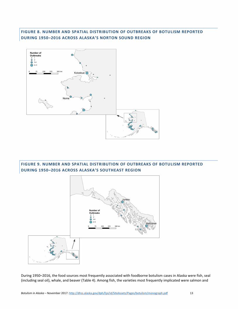
Foodborne botulism outbreaks have occurred across much of Alaska, but with greatest frequency in coastal communities in western and southeastern Alaska (Figure 6-9).
FIGURE 6. NUMBER AND SPATIAL DISTRIBUTION OF OUTBREAKS OF BOTULISM REPORTED
DURING 1950–2016 THROUGHOUT ALASKA
FIGURE 7. NUMBER AND SPATIAL DISTRIBUTION OF OUTBREAKS OF BOTULISM REPORTED
DURING 1950–2016 ACROSS ALASKA’S YUKON-KUSKOKWIM REGION
Botulism in Alaska – November 2017: http://dhss.alaska.gov/dph/Epi/id/SiteAssets/Pages/botulism/monograph.pdf 13
FIGURE 8. NUMBER AND SPATIAL DISTRIBUTION OF OUTBREAKS OF BOTULISM REPORTED
DURING 1950–2016 ACROSS ALASKA’S NORTON SOUND REGION
FIGURE 9. NUMBER AND SPATIAL DISTRIBUTION OF OUTBREAKS OF BOTULISM REPORTED
DURING 1950–2016 ACROSS ALASKA’S SOUTHEAST REGION
During 1950–2016, the food sources most frequently associated with foodborne botulism cases in Alaska were fish, seal (including seal oil), whale, and beaver (Table 4). Among fish, the varieties most frequently implicated were salmon and

Botulism in Alaska – November 2017: http://dhss.alaska.gov/dph/Epi/id/SiteAssets/Pages/botulism/monograph.pdf 14
whitefish; among whales, the species most frequently implicated were beluga and gray. The food preparation methods most frequently associated with cases of foodborne botulism reported were putrefaction (or “aging”) and rendering (Table 4).
TABLE 4. NUMBER OF OUTBREAKS AND CASES OF BOTULISM REPORTED DURING 1950–2016 BY
FOOD SOURCES AND PREPARATION METHODS
Associated food, source Number of outbreaks Number of cases
Fish 87 165
Salmon 32 66
Whitefish 17 31
Other 6 13
Unknown 32 55
Seal (including seal oil) 66 102
Whale 21 50
Beluga 10 25
Gray 2 13
Unknown 9 12
Beaver 11 28
Multiple, undetermined 10 15
Walrus 2 2
Unknown 3 4
Associated food preparation method Number of outbreaks Number of cases
Putrefaction (“aging”) 82 147
Rendering (oil) 20 36
Drying 6 16
Other 13 17
Unknown 79 150
Total 200 366
During 1950–2016, the symptoms most frequently reported among patients with confirmed and probable botulism were nausea/vomiting and dry mouth (Table 5). Because the presence or absence of symptoms was not always recorded in patient records or outbreak investigation reports, the frequency with which some symptoms were experienced could not be estimated with confidence. Of the 366 cases, 246 (67%) received botulism antitoxin as part of their clinical care, 305 (83%) were hospitalized, and 91 (25%) required endotracheal intubation for airway support (Table 5).

Botulism in Alaska – November 2017: http://dhss.alaska.gov/dph/Epi/id/SiteAssets/Pages/botulism/monograph.pdf 15
TABLE 5. NUMBER AND PERCENT OF CONFIRMED AND PROBABLE CASES OF BOTULISM
REPORTED DURING 1950–2016 (N = 366) BY SYMPTOMS AND RECEIPT OF CLINICAL CARE
Number (percent) of cases
Symptoms Yes No Unknown
Nausea/vomiting 291 (80%) 42 (11%) 33 (9%)
Dry mouth 289 (79%) 30 (8%) 47 (13%)
Double vision (diplopia) 151 (41%) 142 (39%) 73 (20%)
Dilated / fixed pupils 185 (51%) 98 (27%) 83 (23%)
Trouble breathing (dyspnea) 175 (48%) 104 (28%) 87 (24%)
Difficulty swallowing (dysphagia) 172 (47%) 106 (29%) 88 (24%)
Constipation 88 (24%) 146 (40%) 132 (36%)
Blurred vision 182 (50%) 37 (10%) 147 (40%)
Diarrhea 124 (34%) 68 (19%) 174 (48%)
Urinary retention 83 (23%) 88 (24%) 195 (53%)
Drooping eyelids (ptosis) 69 (19%) 94 (26%) 203 (55%)
Hoarseness 64 (17%) 69 (19%) 233 (64%)
Slurred speech (dysarthria) 60 (16%) 72 (20%) 234 (64%)
Clinical care
Received antitoxin 246 (67%) 86 (23%) 34 (9%)
Hospitalized 305 (83%) 36 (10%) 25 (7%)
Intubated 91 (25%) 216 (59%) 59 (16%)
Of the 303 cases and 157 outbreaks associated with one or more samples that tested positive for a specific botulism toxin, 249 (82%) of the former and 132 (84%) of the latter were associated with toxin type E (Figure 10). Toxin type B was the second most frequently detected toxin type with 38 (13%) of toxin-positive cases and 17 (11%) of toxin-positive outbreaks), followed by toxin type A with 10 and 5 (each 3%) of both toxin-positive cases and outbreaks respectively. One or more samples associated with 48 cases and 29 outbreaks tested negative for evidence of botulism toxin or Clostridium spp.
FIGURE 10. NUMBER OF CONFIRMED AND PROBABLE CASES AND OUTBREAKS OF BOTULISM
REPORTED DURING 1950–2016 BY TOXIN TYPES

Botulism in Alaska – November 2017: http://dhss.alaska.gov/dph/Epi/id/SiteAssets/Pages/botulism/monograph.pdf 16
BOTULISM IN THE ENVIRONMENT
In the 1970s, several studies documented the presence of C. botulinum in the environment. Miller et al. demonstrated type E botulism toxicity in enrichment cultures in 17 of 23 beach soil samples collected in the Kotzebue region.28 Other investigators detected low-level intrinsic contamination of Alaska salmon with type E spores.290 Among 589 pink, sockeye, chinook, and chum salmon collected from Bristol Bay, Southeastern Alaska, Kodiak, and the Yukon River, six (1%) had gill specimens yielding positive cultures for type E toxin. None of the 494 viscera specimens were positive.
The most extensive published environmental survey for C. botulinum in Alaska was conducted by Miller.30 Samples of beach soil, ocean water and sediments, salmon, and marine mammals were collected from 23 sites in both interior and coastal areas; type E spores were widely distributed. No other type of C. botulinum was identified and, with one exception, no specimens from north of Point Hope were positive. Although no more recent surveys have been conducted, it is assumed that C. botulinum spores are widely distributed in the Alaska environment.
A 2017 paper from Canada explored how specific harvesting practices of seals in a coastal environment of Nunavik may impact the likelihood of meat products to contain C. botulinum spores.31 Food products were tested after aging for toxin presence and compared to spores and organism found in the environment. While sometimes food and environment toxin types matched molecularly, there was also high biodiversity demonstrated in toxin types found in seawater and shoreline soil isolates.
Die-offs from botulism among bird populations in the U.S. and Canada have occurred sporadically during the summer months for many years. Birds ingest botulinum toxin present in decomposing vegetation or invertebrates that have already accumulated toxin. C. botulinum can be found in many different natural environments. However, the majority of birds affected by botulism are waterbirds (or waterfowl). Avian botulism is usually associated with toxin type C and sometimes type E. Humans appear to be relatively resistant to toxin type C.32 Since 1980, only one outbreak in 1999 has been recorded among Alaska birds (personal communication 2004, Dr. Kimberlee Beckmen, Alaska Department of Fish and Game). The die-off occurred in Haines and involved more than a dozen birds of several different species, including five trumpeter swans and two golden eyes. Blood samples from two of the swans and a goldeneye tested positive for botulinum toxin type E. No human illness was associated with this incident.

Botulism in Alaska – November 2017: http://dhss.alaska.gov/dph/Epi/id/SiteAssets/Pages/botulism/monograph.pdf 17
CLINICAL CONSIDERATIONS
INITIAL PRESENTATION AND EVALUATION
Because preliminary laboratory testing results for botulinum toxin takes 5–7 days, the initial diagnosis depends on accurate and rapid clinical assessment. A careful history often reveals recent consumption of traditional Alaska Native foods, particularly aged foods. The incubation period, or interval from consumption of contaminated food to illness onset, is typically 12–48 hours. Longer incubation periods out to 84 hours have been recorded for some patients who reported vague symptoms and did not have laboratory-confirmed specimens, but were counted as cases given their exposure to toxin-containing food products. Severely affected patients may have a more rapid onset (as short as 6 hours) and although unusual, incubation periods as long as 10 days have been reported anecdotally.
Botulinum toxin acts at cholinergic neuromuscular junctions by inhibiting the release of acetylcholine from presynaptic motor neurons. The action is believed to be irreversible. Both autonomic and voluntary motor activities are affected and molecular differences in toxin types may result in different signs and symptoms. The salient clinical features of botulism can be grouped into three major areas: gastrointestinal/urinary, neurologic, and muscular (Table 6). A detailed list of possible symptoms can be found on the botulism case report form (available with other botulism resources at: http://dhss.alaska.gov/dph/Epi/id/Pages/botulism/resources.aspx ).
TABLE 6. SIGNS AND SYMPTOMS OF BOTULISM BY SYSTEM
System Sign/Symptom
Gastrointestinal/ Urinary
Abdominal pain
Diarrhea
Intestinal ileus
Nausea
Urinary retention
Vomiting
Neurologic
Blurry vision
Decreased gag reflex
Dilated or unreactive pupils
Diplopia
Dry mouth
Dysphagia
Muscular
Dyspnea (without typical signs, such as gasping)
Fatigue
Respiratory muscle paralysis
Symmetrical skeletal muscle weakness
A. GASTROINTESTINAL/URINARY
Gastrointestinal symptoms are usually the initial manifestations of botulism; however, they lack diagnostic specificity unless associated with other findings. Nausea, vomiting, diarrhea, and abdominal pain may be present initially or appear within 2–3 days of illness onset. The origin of these symptoms is not completely clear, but may be secondary to toxin-induced intestinal ileus. Ileus is sometimes severe and relatively long-lasting (i.e., more than a week). Aged foods might also contain additional bacteria or substances that could cause acute onset of vomiting and diarrhea within hours of consumption, and which may resolve quickly. A diagnosis of botulism is sometimes ruled out in these patients; however, it is important to consider the possibility that two disease processes are associated with the same meal. Urinary retention, presumably caused by detrusor weakness, is often present; however, early in the course of the illness, it is frequently asymptomatic.

Botulism in Alaska – November 2017: http://dhss.alaska.gov/dph/Epi/id/SiteAssets/Pages/botulism/monograph.pdf 18
B. NEUROLOGIC
When the effects of the cholinergic blockade are observed, the diagnosis of botulism must be seriously considered, especially in an Alaska Native patient with gastrointestinal symptoms. Dryness of oral mucous membranes may be extreme and can lead to fissuring of the tongue and severe pharyngeal pain. In the past, the pharyngeal presentation of botulism was confused with diphtheria. Ocular findings are classic: diplopia, blurry vision, and fixed or dilated pupils. Ptosis is commonly present. The absence of ocular findings does not rule out the diagnosis of botulism. However, the absence of any objective signs of cranial nerve deficits makes botulism extremely unlikely; typical deficits are listed in Table 7. The progressive paralysis typically descends, affecting the cranial nerves first, then the neck, upper arms, trunk, and diaphragm, and finally the hands and legs. The fingers may be affected last. Neurologic findings are typically bilateral. Although asymmetry of certain deficits may occur, truly unilateral deficits are uncommon. Botulism can also markedly impair control of heart rate and blood pressure, and cause bradycardia and hypotension.33
TABLE 7. TYPICAL MANIFESTATIONS OF CRANIAL NERVE DEFICITS RESULTING FROM BOTULISM*
Cranial Nerve Finding With Botulism
III – Oculomotor Can’t move eyes left and right, eyelids droop (ptosis)
IV – Trochlear Can’t look downward symmetrically
V – Trigeminal Can’t bite down
VI – Abducens Can’t look outward
VII – Facial Can’t close eyes against force, purse lips or smile
X – Vagus Diminished gag reflex and difficulty swallowing, saying “Ah”
XI – Accessory Diminished strength trapezius and sternocleidomastoid
XII – Hypoglossal Difficulty moving tongue side to side
*Adapted from Table 4.11
C. MUSCULAR
Skeletal muscle weakness, especially associated with fatigue, shoulder, neck or truncal weakness, or dyspnea, is an ominous sign. Because the muscles of respiration are weakened, typical signs of dyspnea such as gasping, vigorous chest motions or use of accessory muscles of respiration are usually absent. Precipitous deterioration of respiratory reserve with concomitant respiratory arrest has caused almost all of the early deaths from botulism and is not necessarily preceded by other complaints. Often a patient’s paralysis prevents demonstration of agitation or restlessness, so he or she may appear to be resting comfortably. It is imperative that respiratory reserve be assessed and followed diligently. Measurement of forced vital capacity (FVC) should be sufficient to indicate the degree of respiratory compromise and is a convenient index to follow for signs of deterioration.
Assessing changes to FVC via spirometry or other formal pulmonary function testing is an objective method of documenting diminishing respiratory capacity and muscular weakness. Other quick and more subjective tests can also provide evidence of muscular impairment. For example, serial counts of how many times a patient can successively stand up and sit down, or the number of stairs they can walk up and down before becoming fatigued can provide evidence of progression of muscular weakness. This information can be used together with other signs and symptoms to support a clinical diagnosis of botulism.
Knowledge of findings that would be unchanged by botulism may be helpful in establishing or ruling out the diagnosis. Body temperature, orientation to person, place and time, sensory examination, and deep tendon reflexes (if the patient is not completely paralyzed) should all be normal. Rare exceptions have occurred. Even if signs or symptoms not usually associated with botulism are present, clinicians may still need to consider botulism in the differential diagnosis, especially if other findings are suggestive.
The differential diagnosis of botulism generally involves consideration of rare conditions or unusual presentations of common problems, such as stroke (Table 8). It is often best to pursue a diagnosis of botulism, perhaps in parallel with
Botulism in Alaska – November 2017: http://dhss.alaska.gov/dph/Epi/id/SiteAssets/Pages/botulism/monograph.pdf 19
others, until the diagnosis is clear, particularly if the patient is an Alaska Native who has consumed traditional foods during the week before onset of symptoms.
Laboratory data from electromyography, nerve conduction studies, cerebrospinal fluid analysis, or Tensilon® testing are more helpful for diagnosing other conditions than for establishing the diagnosis of botulism. Occasionally an electromyogram will show convincing post-tetanic potentiation, which is almost specific for botulism. Cerebrospinal fluid and nerve conduction studies should be normal in patients with botulism.
Past reports have suggested that if a patient has three or more signs or symptoms in a “diagnostic pentad” and a history of consuming traditional Alaska Native food, botulism should be strongly suspected (Table 9).34-35 The term “diagnostic pentad,” however, can be misleading because when pentad symptoms are present, they are suggestive, but not necessarily diagnostic, of botulism. Also listed in Table 9 is a botulism “clinical paradigm” that focuses on body systems (i.e., gastrointestinal, neurologic, muscular) and may be a more useful screening tool in assessing suspected cases of botulism compared with the pentad. Similarly, the pentad may be more meaningful when signs or symptoms are considered with respect to body systems (e.g., dry mouth from cholinergic blockade, as opposed to resulting from repeated vomiting and subsequent dehydration).
Neither of these approaches have been rigorously tested, but both have been found useful by health care providers experienced in the diagnosis of botulism in Alaska and may help trigger suspicion of botulism. Regardless of the approach, all relevant clinical and exposure information should be considered when assessing whether a patient might have an illness compatible with a diagnosis of botulism.
TABLE 8. DIFFERENTIAL DIAGNOSIS FOR BOTULISM
Condition Points of Differentiation from Botulism
Diphtheria*
Cardiac conduction abnormalities
Cervical adenopathy
Culture
Fever
Typical pharyngeal or nasal mucosal lesions
Drug ingestion/Poisoning Central nervous system abnormalities
Drug levels
History
Gastroenteritis Lack of autonomic, ocular or muscular findings
Guillain-Barre syndrome
Abnormal nerve conduction
Absent deep tendon reflexes
Cerebrospinal fluid protein elevated
Sensory findings
Myasthenia gravis Response to Tensilon® testing
Paralytic shellfish Poisoning* History
Poliomyelitis*
Abnormal cerebrospinal fluid
Muscle denervation findings
Presence of sensory findings
Stroke Absence of gastrointestinal and autonomic findings
Unilateral findings
*Note: in addition to botulism, suspected or confirmed cases of these conditions must also be reported to the Alaska Section of Epidemiology: see http://dhss.alaska.gov/dph/Epi/Pages/pubs/conditions/default.aspx

Botulism in Alaska – November 2017: http://dhss.alaska.gov/dph/Epi/id/SiteAssets/Pages/botulism/monograph.pdf 20
TABLE 9. SIGNS AND SYMPTOMS PROFILES SUGGESTIVE OF BOTULISM
Botulism should be considered in any patient with a history of recent consumption of traditional Alaska Native food and either of the following symptom profiles:
A. At least three of the five signs or symptoms in the “diagnostic pentad”
Dilated or fixed pupils
Diplopia
Dry throat
Dysphagia
Nausea or vomiting
B. Any elements of a clinical botulism paradigm
Cranial nerve palsy with no apparent cause**
Descending symmetrical paralysis or weakness with no apparent cause
Gastrointestinal symptoms with autonomic or neurologic abnormality*
*Autonomic involvement includes evidence of hypotension.
**See Table 7, page 18.
HOSPITAL COURSE AND TREATMENT
Convalescence after foodborne botulism can be prolonged, and few reports are available in the medical literature on long-term sequelae among botulism survivors.36 Health care providers should be aware that the clinical course and recovery may be quite different for persons with botulism acquired from consumption of home-canned products or other exposures. The clinical course and appropriate management of any botulism patient needs to be tailored to current circumstances. As such, clinicians with experience caring for botulism patients in Alaska may be useful resources in managing individual patients.
The most urgent clinical concern for the patient suspected of having botulism is assessment of respiratory reserve. Most patients will require frequent (at least hourly at first) determination of forced vital capacity or an equivalent measure. Any significant decline in respiratory function should prompt consideration of endotracheal intubation and assisted ventilation. For patients in an outlying hospital requiring transfer for management of respiratory insufficiency, placement of endotracheal and nasogastric tubes should be strongly considered before transfer.
ANTITOXIN USE AND CLINICAL MANAGEMENT
Although the primary treatment for botulism is supportive care, antitoxin is available from the U.S. Centers for Disease Control and Prevention (CDC) to prevent further binding of botulinum toxin to receptors at the neuromuscular junction. Antitoxin is indicated for any person suspected of having botulism intoxication. Before 1999, an equine trivalent antitoxin was used. From 1999–2010, botulinum antitoxin was available in two formulations: bivalent type A/B and type E. Antitoxin type E was considered an Investigational New Drug, which meant that specific guidelines and protocols had to be followed during and after administration.
In March 2010, CDC began to supply the Alaska Section of Epidemiology with a new heptavalent botulinum antitoxin (HBAT) produced by Cangene Corporation to replace the types A/B and E antitoxin products.37 HBAT contains antibodies specific for seven toxin types (A–G). HBAT was an Investigational New Drug until March 2015, when it received FDA-approval and was packaged as BAT®. Post-marketing surveillance paperwork was required by the FDA and implemented by the manufacturer (Cangene, dba Emergent BioSolutions, Inc.); however, the surveillance paperwork was no longer required after June 2017.
Antitoxin kits are packaged by the Alaska Section of Epidemiology and supplied to certain hospital pharmacies located throughout the state. The Section of Epidemiology has additional kits stocked in Anchorage to be sent as needed to other locations.
In Alaska, serum sickness and anaphylaxis, although reported elsewhere, have not been documented following administration of antitoxin. Before 1999, there was only one documented case of a hypersensitivity reaction following administration of antitoxin in Alaska. No adverse reactions were reported from 1999–2010 when bivalent type A/B and type E antitoxin formulations were administered. Additionally, no adverse reactions have been reported to date in Alaska from the use of HBAT.
Botulism in Alaska – November 2017: http://dhss.alaska.gov/dph/Epi/id/SiteAssets/Pages/botulism/monograph.pdf 21
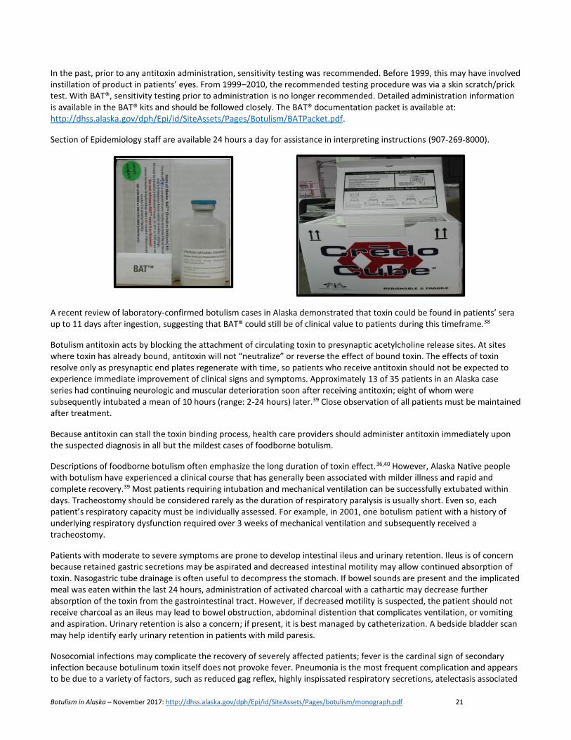
In the past, prior to any antitoxin administration, sensitivity testing was recommended. Before 1999, this may have involved instillation of product in patients’ eyes. From 1999–2010, the recommended testing procedure was via a skin scratch/prick test. With BAT®, sensitivity testing prior to administration is no longer recommended. Detailed administration information is available in the BAT® kits and should be followed closely. The BAT® documentation packet is available at: http://dhss.alaska.gov/dph/Epi/id/SiteAssets/Pages/Botulism/BATPacket.pdf.
Section of Epidemiology staff are available 24 hours a day for assistance in interpreting instructions (907-269-8000).
A recent review of laboratory-confirmed botulism cases in Alaska demonstrated that toxin could be found in patients’ sera up to 11 days after ingestion, suggesting that BAT® could still be of clinical value to patients during this timeframe.38
Botulism antitoxin acts by blocking the attachment of circulating toxin to presynaptic acetylcholine release sites. At sites where toxin has already bound, antitoxin will not “neutralize” or reverse the effect of bound toxin. The effects of toxin resolve only as presynaptic end plates regenerate with time, so patients who receive antitoxin should not be expected to experience immediate improvement of clinical signs and symptoms. Approximately 13 of 35 patients in an Alaska case series had continuing neurologic and muscular deterioration soon after receiving antitoxin; eight of whom were subsequently intubated a mean of 10 hours (range: 2-24 hours) later.39 Close observation of all patients must be maintained after treatment.
Because antitoxin can stall the toxin binding process, health care providers should administer antitoxin immediately upon the suspected diagnosis in all but the mildest cases of foodborne botulism.
Descriptions of foodborne botulism often emphasize the long duration of toxin effect.36,40 However, Alaska Native people with botulism have experienced a clinical course that has generally been associated with milder illness and rapid and complete recovery.39 Most patients requiring intubation and mechanical ventilation can be successfully extubated within days. Tracheostomy should be considered rarely as the duration of respiratory paralysis is usually short. Even so, each patient’s respiratory capacity must be individually assessed. For example, in 2001, one botulism patient with a history of underlying respiratory dysfunction required over 3 weeks of mechanical ventilation and subsequently received a tracheostomy.
Patients with moderate to severe symptoms are prone to develop intestinal ileus and urinary retention. Ileus is of concern because retained gastric secretions may be aspirated and decreased intestinal motility may allow continued absorption of toxin. Nasogastric tube drainage is often useful to decompress the stomach. If bowel sounds are present and the implicated meal was eaten within the last 24 hours, administration of activated charcoal with a cathartic may decrease further absorption of the toxin from the gastrointestinal tract. However, if decreased motility is suspected, the patient should not receive charcoal as an ileus may lead to bowel obstruction, abdominal distention that complicates ventilation, or vomiting and aspiration. Urinary retention is also a concern; if present, it is best managed by catheterization. A bedside bladder scan may help identify early urinary retention in patients with mild paresis.
Nosocomial infections may complicate the recovery of severely affected patients; fever is the cardinal sign of secondary infection because botulinum toxin itself does not provoke fever. Pneumonia is the most frequent complication and appears to be due to a variety of factors, such as reduced gag reflex, highly inspissated respiratory secretions, atelectasis associated

Botulism in Alaska – November 2017: http://dhss.alaska.gov/dph/Epi/id/SiteAssets/Pages/botulism/monograph.pdf 22
with low tidal volumes, and aspiration of pharyngeal or gastric secretions due to paralysis or weakness. Protection of the airway, high environmental humidity, adequate lung expansion, and use of mucolytic agents may all help to reduce pulmonary infection. Urinary tract infections have been reported but may be related to catheter use. Bed sores and secondary skin infection may occur if a paralyzed and intubated patient is not turned frequently and inspected for early skin breakdown.
RECOVERY
It is important for health care providers caring for a completely paralyzed patient to remember that the person is fully awake. The illness, procedures, and medical routines should be explained with recognition that the patient is conscious. Health care providers should provide appropriate pain control and sedation for intubated patients who are otherwise alert. Patients can have excellent recall for events and conversations heard during total paralysis.
Alaska patients generally have rapid recovery of respiratory function, but may have lingering ocular or intestinal symptoms. Persistent ileus has delayed oral feeding in some patients for several weeks and necessitated total parenteral nutrition. The risk of aspiration of gastric contents exists until the gag reflex has clearly returned and ileus has resolved. If any concern about swallowing ability is present, it is reasonable to conduct either a swallowing evaluation, similar to those used in stroke assessment, or a radiologic evaluation of swallowing prior to beginning oral feeding. Complete resolution of all effects of botulinum toxin is expected for most botulism patients within 1–2 months.
There is no evidence to suggest that having a history of botulism intoxication mitigates the course of a subsequent illness; several previous botulism patients in Alaska have experienced another episode of botulism following consumption of traditionally prepared Alaska Native foods.41 This has also been documented in Canada.42 Based on evidence from a survey of botulism knowledge among Alaska Natives, almost half of the respondents believed that there was some form of immunity to botulism.27 Health care providers should ensure that current patients are aware they may contract botulism in the future if they are exposed again.

Botulism in Alaska – November 2017: http://dhss.alaska.gov/dph/Epi/id/SiteAssets/Pages/botulism/monograph.pdf 23
LABORATORY EVALUATION
Botulism is detected in the laboratory by identifying botulinum neurotoxin, or neurotoxin-producing Clostridium botulinum bacteria, in clinical materials or remnants of suspect food consumed.
Suitable specimen types for foodborne outbreak investigation include serum, feces, vomitus, gastric contents, and suspected food; for infants with botulism, feces and serum; and for wound infections, serum, feces, exudates, debrided tissue, or swab samples from wounds (Table 10).
TABLE 10. BOTULISM SPECIMEN COLLECTION GUIDE
Botulism Type
Specimen Amount Required
Storage Conditions
Transport Conditions
Special Considerations
Foodborne
Serum 5–10 ml Refrigerate Cold-pack Collect sample before administration of BAT®
Gastric contents/vomit
20 ml Refrigerate Cold-pack
Feces (stool) 10–50 g Refrigerate Cold-pack Rectal swab is not acceptable
Suspect food 50 g Refrigerate Cold-pack Leave food in original containers if possible, or place in sterile unbreakable, leak proof containers
Infant
Feces (stool) ≥ 0.2 g Refrigerate Cold-pack Diapers are acceptable, but not preferred.
Enema ≥ 5 ml Refrigerate Cold-pack Collect with non-bacteriostatic water; do not use glycerin suppository
Serum ≥ 0.2 ml Refrigerate Cold-pack Only collect by request
Wound
Serum 5–10 ml Refrigerate Cold-pack Collect sample before administration of BAT®
Feces (stool) 10–50 g Refrigerate Cold-pack Rectal swab is not acceptable
Culture isolate Anaerobic transport media
Room temp Ambient
Wound exudates/debrided tissue
Anaerobic transport media
Room temp Ambient
Gastric aspirates and fecal (stool) specimens often yield positive results among persons with laboratory-confirmed botulism. An ileus resulting from botulism intoxication will slow the transit of contaminated food, which may delay the

Botulism in Alaska – November 2017: http://dhss.alaska.gov/dph/Epi/id/SiteAssets/Pages/botulism/monograph.pdf 24
passage of stool. Although it may be some days after serum is collected, submission of stool, once it can be collected from the patient, is highly recommended.
All samples must be clearly labeled, placed in a leak-proof container or plastic bag, and shipped according to current shipping guidelines (submission details are available at: http://dhss.alaska.gov/dph/Labs/Documents/LaboratoryTests.pdf). Arrangements for testing are handled by the Alaska Division of Public Health, Section of Epidemiology. For consultation and more information about specimen collection and handling, call the Section of Epidemiology at (907) 269-8000, or after-hours (800) 478-0084. Send samples to:
Alaska State Public Health Laboratory Special Pathogens Branch 5455 Dr. Martin Luther King Jr. Ave. Anchorage, AK 99507 Phone 907-334-2100 http://dhss.alaska.gov/dph/Labs/Pages/default.aspx/
Laboratory test methods include analysis of all sample types for the presence of botulinum toxin; non-serum samples are further evaluated for presence of Clostridium botulinum organism by culture.
Because preliminary results may not be available for up to 7 days, laboratory testing is not useful for immediate patient care management, but is very helpful in corroborating the diagnosis of botulism.
Laboratory results will include analyses performed and an overall final interpretation (Table 11); results should be correlated with clinical history to confirm as a case of botulism.
TABLE 11. LABORATORY RESULTS INTERPRETATION BY TEST METHOD AND SAMPLE TYPE
Sample Type Test Method Final Result Potential Interpretation Outcomes
Serum Botulinum Toxin
1. No botulinum neurotoxin identified in sample
2. Positive: Botulinum neurotoxin identified in sample (type specified)
3. Inconclusive for botulinum neurotoxin
Non-serum
Botulinum Toxin 1. Negative for Clostridium botulinum bacteria and botulinum neurotoxin
2. Positive: Botulinum neurotoxin identified in sample (type specified)
Culture for isolation of C. botulinum bacteria
1. Positive: Clostridium botulinum neurotoxin-producing bacteria identified in sample
(type specified)
2. Confirmed Positive: Both Clostridium botulinum neurotoxin-producing bacteria and
botulinum neurotoxin identified in sample (type specified)

Botulism in Alaska – November 2017: http://dhss.alaska.gov/dph/Epi/id/SiteAssets/Pages/botulism/monograph.pdf 25
PREVENTION
Strategies for controlling foodborne botulism fall largely into two approaches: (1) reducing contamination of food with C. botulinum spores and preventing toxin production in food; and (2) early identification of botulism cases to minimize additional exposures to implicated foods. Reducing contamination and preventing toxin formation are difficult to achieve. Botulism spores, particularly type E, are ubiquitous in Alaska and traditional Alaska Native food preparation practices will not always prevent toxin production.
Shaffer et al. described Alaska Native food consumption patterns and preparation practices in the Bristol Bay region in 1987.43 In four Yupik villages, they found that aged foods were regularly prepared by 15% of high school students, 71% of the students’ parents, and 80% of the students’ grandparents. Aging practices appeared to have changed from the traditional method of using a clay pit in the ground. Only 13% of the preparers reported that they used the traditional method to age fish heads, while 42% used a wooden barrel above ground, 38% used a wooden barrel in the ground, and 8% used a plastic bucket above ground.
Another survey performed in 1999 by CDC’s Arctic Investigations Program (AIP) based in Anchorage, Alaska, assessed whether educational messages could be tailored to decrease the risk of botulism from consuming traditional foods. The survey examined the knowledge, attitudes, and practices of a sample of Alaska Natives in the Bristol Bay region, and found that knowledge and awareness of botulism was relatively high. Almost half of the 140 respondents indicated they would consider eating traditional foods that had been boiled to reduce toxin, or consider not eating foods that had been prepared without the use of a refrigerator, or by methods that allow for an anaerobic environment.27
The results from the 1999 AIP survey were used to create an educational video in 2000 (available online at: https://vimeo.com/105157044).44 In conjunction with the video, safe food preparation steps were developed (Table 12). This video was then distributed to all rural schools and medical facilities in Alaska in the spring of 2000. Approximately a decade earlier, a similar video was produced by the Inuvik Regional Health Board in the Northwest Territories, Canada.45 A follow-up survey for the Canadian video was not done.
TABLE 12. HOW TO PROTECT YOUR FAMILY FROM BOTULISM: FIVE FOOD SAFETY STEPS*
The following five food safety steps are recommended for persons who prepare or eat traditional aged foods: 1. Try to use traditional methods for preparing Alaska Native foods as these may decrease the likelihood the food
will become contaminated botulism bacteria. Plastic, glass, or sealed plastic bags do not allow air to reach the food and can promote the growth of C. botulinum bacteria. Use salt to preserve dried fish and to also discourage growth of C. botulinum bacteria.
2. Age food at a cold temperature, ideally below 36° Fahrenheit (or 2° Celsius). This will also discourage the growth of C. botulinum bacteria.
3. Before preparing food, wash your hands, your containers, and your food. 4. Cook your food before eating it. Heat destroys botulinum toxin and may be the best way to reduce the risk of
getting botulism after eating aged foods. 5. When in doubt, throw it out! Don’t take the risk of getting botulism if you don’t know how the food was
prepared. Botulinum toxin is so deadly, even a small taste can make you ill.
*Adapted from CDC: “A Helping Hand: Keeping Your Family Safe from Botulism” (the video is available on-line at: https://vimeo.com/105157044)
Botulism in Alaska – November 2017: http://dhss.alaska.gov/dph/Epi/id/SiteAssets/Pages/botulism/monograph.pdf 26
In 2001, AIP conducted a follow-up survey to evaluate the effectiveness of the Alaska video.46 Approximately 40% of the 254 adults interviewed had watched the video. Most had seen the video at home or in a health care facility. No changes were documented between consumption and preparation practices between the pre- and post-viewing surveys. Because of the relatively small number of persons who had watched the video, assessing the effect of the video was difficult.
In past situations involving recalls of commercial food products suspected or confirmed to be contaminated with botulinum toxin, CDC and FDA subject matter experts have recommended that consumers take care when handling potentially contaminated foods.47 Such steps include ensuring that other people or animals do not have access to the foods by discarding the foods in sealed containers and cleaning up liquid foods in a manner to avoid direct contact with a person’s mucous membranes. In Alaska, once a food has been confirmed to have been associated with an outbreak, the Section of Epidemiology recommends the food be discarded and surfaces or containers that were in contact with the food washed in a dilute bleach solution; hands and clothing can be washed with soap and water.47
Educating health care providers to recognize botulism early in its clinical course and to report cases promptly has been one of the long-standing strategies to limit more serious outcomes. Educational efforts directed toward eliciting a careful food consumption history, having a high level of suspicion when confronted with an illness with gastrointestinal and neurologic symptoms, and using the “diagnostic pentad” to prompt suspicion of botulism as a possible differential diagnosis, have been the mainstay of control efforts in Alaska. These educational efforts combined with rapid epidemiologic investigation of suspected cases, prompt supportive care, and the availability of botulism antitoxin may be responsible for the reduction in the number of fatalities.
Recommendations for decreasing the introduction of organism and spores into food products include minimizing the contact of a marine mammal during butchering with coastal soil and water and avoiding the contact of intestines and flipper from meat and fat during transport and storage.31
Other resources for prevention can be found on the Alaska Section of Epidemiology Botulism webpage, available at: http://dhss.alaska.gov/dph/Epi/id/Pages/botulism/resources.aspx; and the CDC Alaska Native Foods (Botulism) webpage, available at: https://www.cdc.gov/botulism/botulism-alaska-foods.html.

Botulism in Alaska – November 2017: http://dhss.alaska.gov/dph/Epi/id/SiteAssets/Pages/botulism/monograph.pdf 27
INFANT BOTULISM
SUMMARY OF INFANT BOTULISM IN ALASKA, 1950–2016
Infant botulism is the most commonly reported form of botulism in the United States. During 1950–2016, five cases of infant botulism were reported in Alaska (Table 13). In contrast to foodborne botulism where the toxin is ingested, infant botulism results from ingestion of C. botulinum spores with subsequent intestinal colonization and toxin production. Most infants affected by botulism are 3–20 weeks of age. In the United States, infant botulism is usually due to toxin type A or B. Additional clostridial species (e.g., C. baratti and butyricum) also produce botulinum toxins, and are known to be causative agents for infant botulism.48 Of the five infant botulism cases in Alaska, four were caused by toxin type A and one by toxin type B. The most recently reported case of infant botulism in Alaska occurred in 2015.
TABLE 13. SUMMARY OF INFANT BOTULISM CASES REPORTED IN ALASKA DURING 1950–2016
Year Demographics Toxin type Presenting symptoms Clinical course
1982 • 6-month old white female
A Unknown* Unknown*
• Anchorage/Mat-Su region • Unknown breastfeeding status
1987
• 3-month old female of unknown race
B
9-day history of constipation, increasing lethargy and decreasing muscular tone; 3-day history of decreased suck reflex, inability to swallow, droopy head and weak cry
6-day hospitalization** • Gulf Coast region • Mostly breastfed; some water and
formula
1992 • 5-month old white male
A ~5-7 day history of increasing weakness and inability to suck
10-day hospitalization† • Southwest region • Breastfed; rice cereal
2009
• 6-month old Alaska Native female
A
3-week history of constipation; 1-week history of febrile respiratory illness, hypotonia appreciated at follow-up visit
Given BabyBIG®; 7-day hospitalization‡ • Interior region
• Breastfed
2015
• 5-month old Alaska Native female
A
3-day history of hypotonia, poor eating; 1-week of nutritional support, no invasive respiratory procedures
Given BabyBIG®; 12-day hospitalization • Anchorage/Mat-Su region
• Breast fed; commercially canned baby food
*Patient backloaded in 1987 after review of laboratory data at CDC.
**See Section of Epidemiology Bulletin, http://www.epi.alaska.gov/bulletins/docs/b1987_10.pdf †See Section of Epidemiology Bulletin, http://www.epi.alaska.gov/bulletins/docs/b1992_03.pdf ‡See Section of Epidemiology Bulletin, http://www.epi.alaska.gov/bulletins/docs/b2009_17.pdf
The first symptom of infant botulism is often constipation, followed in several days by progressive muscular weakness, poor sucking reflex, weak cry, and difficulty swallowing. Respiratory arrest occurs in approximately half of affected infants; infants may present with apnea, or become apneic, during a diagnostic procedure. Simple tests are available to help support a diagnosis of infant botulism (Table 14).49 Patients are usually afebrile and have normal cerebrospinal fluid. Electromyography may be helpful in differentiating botulism from other causes of neuromuscular disease. Diagnosis is made by demonstration of botulinum toxin in stool and supported by a positive stool culture. It is unusual to find toxin in serum.

Botulism in Alaska – November 2017: http://dhss.alaska.gov/dph/Epi/id/SiteAssets/Pages/botulism/monograph.pdf 28
TABLE 14. TESTS TO AID IN THE DIAGNOSIS OF INFANT BOTULISM4 9
Test Findings
1. Take the patient to a dark room. Shine a bright light into the infant’s eye; note quickness of pupillary constriction. Remove the light when constriction is maximal; let the pupil dilate again. Immediately repeat, continuing for 2–3 minutes.
The initially brisk pupillary constriction may become sluggish and unable to constrict maximally (fatigability with repetitive muscle activity is the clinical hallmark of botulism).
2. Shine a bright light onto fovea, keeping it there for 1–3 minutes, even if the infant tries to deviate the eyes.
Latent ophthalmoplegia may be elicited, and/ or purposeful efforts to avoid the light may diminish. Also observe for initial squirming of the extremities that may diminish due to fatigability.
3. Place a clean fifth finger in the infant’s mouth, taking care not to obstruct the airway. Note the strength and duration of the reflex sucking.
The suck is weak and poorly sustained. The gag reflex strength also may be quickly checked (if the infant has not been fed recently). Examination may show a decreased gag reflex; cranial nerve involvement including ptosis, ophthalmoplegia, and facial nerve palsy; mydriasis; and areflexia or generalized hypotonia.
Therapy for infant botulism is primarily supportive; mechanical ventilation can be lifesaving. BabyBIG® is used to shorten hospital stays and reduce complications. Antimicrobial medications, particularly aminoglycosides, have been reported to increase the incidence of respiratory paralysis.50 Prompt diagnosis and treatment of infant botulism with human-derived Botulism Immune Globulin Intravenous (BabyBIG®) might reduce the length of time needed for recovery. BabyBIG® can be obtained from the California Department of Public Health, Infant Botulism Treatment and Prevention Program (IBTPP, 510-231-7600; see also www.infantbotulism.org). BabyBIG® should be requested from the IBTPP pediatrician on-call without awaiting laboratory confirmation. BabyBIG® was shown to be most effective if given within 7 days of hospital admission.51 FDA approval of BabyBIG® was based upon studies that its use produced a statistically signification reduction in the durations of hospital stay, mechanical ventilation, and tube feeding among those infants with types A or B botulism.
Since 2008, BabyBIG® has been administered to three Alaska infants. For one infant, botulism was one of numerous diagnoses being considered based on clinical findings and a preliminary result of nonspecific toxicity from a stool specimen. In consultation with IBTPP physicians, BabyBIG® was recommended and administered. Unfortunately, this infant was ultimately diagnosed with spinal muscular atrophy. BabyBIG® was also administered, after consultation with IBTPP physicians, on the infants diagnosed with infant botulism in 2009 and 2015.
Antitoxin, such that is used in cases of foodborne botulism, has not been shown to affect the outcome of infant botulism. However, depending on the type of botulism (i.e., not A or B) the use of BAT® may be considered in consultation with the Section of Epidemiology and CDC.
Regarding risk factors, in a study conducted outside of Alaska, affected infants had higher birthweights, their mothers tended to be Caucasian, and they were more commonly breast-fed.52 Approximately 20% of the infant botulism cases reported nationally to CDC have been associated with the ingestion of honey. The sources for the other cases are unknown, but hypotheses include soil, household dust, and other foods. Honey should not be fed to infants <1 year of age. No other specific prevention measures exist. The source of botulism was unknown for all of the Alaska cases to date.

Botulism in Alaska – November 2017: http://dhss.alaska.gov/dph/Epi/id/SiteAssets/Pages/botulism/monograph.pdf 29
OTHER FORMS OF BOTULISM
WOUND BOTULISM
Wound botulism occurs after Clostridium botulinum spores have been introduced into a wound and begin to produce toxin, which in turn causes signs and symptoms of a symmetric descending paralysis. Case reports of wound botulism were quite rare until 1982. From 1943 to 1982, 27 cases of wound botulism in the United States were reported to CDC.53 In 1982, New York reported the first case of wound botulism associated with injection drug use. Since then, other clusters of wound botulism have been described among injection drug users, primarily in western states, e.g., California and Washington.
Among injection drug users, C. botulinum spores are introduced subcutaneously, either directly via contaminated drugs or indirectly by injecting through insufficiently disinfected skin. Spores, unlike botulinum toxin, are not inactivated by heat. Therefore, heating heroin mixtures does not guard against wound botulism. Wound botulism has also been documented among intranasal cocaine users, who may have wounds or skin breaks that allow for spore germination in the paranasal sinuses or nasal septa.
Wound botulism has never been documented in Alaska. Even so, health care providers who see patients with clinical signs, such as a descending paralysis or severe weakness and a history of injection drug use or infected wounds, should consider a diagnosis of botulism.
BIOTERRORISM CONSIDERATIONS
Botulinum toxin poses a major bioweapons threat because of its extreme potency and lethality; its ease of production, transport and misuse; and the potential need for prolonged intensive care in affected persons.54 A number of nations or states named by the U.S. State Department as “state sponsors of terrorism” have developed, or are developing, botulinum toxin as a biological weapon.
A deliberate aerosol or foodborne release of botulinum toxin could be detected by several features which include a large number of acute cases presenting all at once, cases involving an uncommon toxin type (C, D, F, or G), patients with a common geographic factor without a common dietary exposure, or multiple simultaneous outbreaks without a common source.
As for all cases of suspected botulism, health care providers who suspect an intentional aerosol or foodborne release of toxin as the source of a patient’s symptoms should contact the Section of Epidemiology immediately. Call (907) 269-8000 Monday–Friday 8am–5pm, or after-hours (800) 478-0084.

Botulism in Alaska – November 2017: http://dhss.alaska.gov/dph/Epi/id/SiteAssets/Pages/botulism/monograph.pdf 30
REFERENCES
1. Kerner CAJ. Neue Beobachtungen ueber die in
Wurttembergso haufig vorfallenden todlichen Vergiftungen durch in den Genuss geraucherter Wurste, Tubingen. 1829; cited in Dickson 1918.
2. Van Ermengem E. Ueber einen neuen anaeroben Bacillus und seine Beziehungen zum Botulismus. Ztsch Hyg Infekt 1897;26:1–56.
3. Landmann G. Ueber die Ursache der Darmstadter Bohnenvergiftung. Hyg Rundschau 1904;10:449–52.
4. Bengston IA. Preliminary note on a toxin-producing anaerobe isolated from the larvae of Lucilla caesar. Public Health Rep 1922;37:164–70.
5. Seddon HE. Bulbar paralysis in cattle due to the action of a toxicogenic bacillus, with a discussion on the relationship of the condition to forage poisoning (botulism). J Comp Pathol Therap 1922;35:147–90.
6. Robinson EM. Notes on botulism in domesticated animals, 15 Ann Rept Dir Vet Services, Union of South Africa, 97–110, 1929.
7. Gunnison JB, Cummings JR, Meyer KF. Clostridium botulinum type E. Proc Soc Exp Biol Med 1936-1937;35:278–80.
8. Moller V, Scheibel I. Preliminary report on the isolation of an apparently new type of Cl. botulinum. Acta Pathol Microbiol Scand 1960;48:80.
9. Gimenez DF, Ciccarelli AS. New strains of Clostridium botulinum subtype Af. Zentrabl Bakteriol [Orig A] 1978;24(2):215–20.
10. Dolman CE. Type E botulism: a hazard of the North. Arctic 1960;13:230–56.
11. Horowitz BZ. Type E botulism. Clin Toxicol 2010;48(9):880–95.
12. Stefansson V. The Stefansson-Anderson Arctic expedition: Preliminary ethnological report. Anthropol Papers Am Mus Nat Hist 1914;14:449.
13. Nelson EW. The Eskimo About Bering Strait. New York: Johnson Reprint Corp, 1971.
14. Dolman CE. Human botulism in Canada (1919-1973). Can Med Assoc J 1974;110(2):191–7.
15. Muller J, Thomsen BF. An outbreak of type E botulism in West-Greenland. Nord Vet Med 1968;20:485.
16. Rabeau ES. Botulism in Arctic Alaska. Report of 13 cases with 5 fatalities. Alaska Med 1959;1:6–9.
17. Geiges ML. The history of botulism. Curr Prob Derm 2002;30:77–93.
18. LeClair D, Fung J, Isaac-Renton JL, et al. Foodborne botulism in Canada. Emerg Inf Dis 2013;19(6):961–68.
19. MacDonald KL, Cohen ML, Blake PA. The changing epidemiology of adult botulism in the United States. Am J Epidemiol 1986;124(5):794–99.
20. Sobel J, Tucker N, Sulka A, et al. Foodborne botulism in the United States, 1990–2000. Emerg Infect Dis 2004;10(9):1606–11.
21. Austin JW, Leclair D. Botulism in the North: a disease without borders. Clin Infect Dis 2011;52(5):593–94.
22. Zottola EA, Zoltai PT. A Preliminary Report on Research Concerning Native Alaska Foods, Methods of Preparation, Preservation and the Effect of These Methods on Their Nutritional Quality and Safety, Department of Food Sciences and Nutrition, Agricultural Extension Service, University of Minnesota, St. Paul; 1981.
23. Peck MW. Biology and genomic analysis of Clostridium botulinum. Adv Microb Physiol 2009;55:183-265, 320.
24. Ball AP, Hopkinson RB, Farrell ID, et al. Human botulism caused by Clostridium botulinum type E: The Birmingham outbreak. Q J Med 1979;48(191):473–91.
25. Hayes Jr AH. The Food and Drug Administration’s role in the canned salmon recalls of 1982. Public Health Rep 1983;98(5):412–15.
26. Fagan RP, McLaughlin JB, Castrodale LJ, et al. Endemic foodborne botulism among Alaska Native persons — Alaska, 1947–2007. Clin Infect Dis 2011; 52(5): 585–92.
27. Chiou LA, Hennessy TW, Horn A, et al. Botulism among Alaska Natives in the Bristol Bay area of southwest Alaska: a survey of knowledge, attitudes, and practices related to fermented foods known to cause botulism. Int J Circumpolar Health 2002; 61(1):50–60.
28. Miller LG, Clark PS, Kunkle GA. Possible origin of Clostridium botulinum contamination of Eskimo foods in northwestern Alaska. Appl Microbiol 1972;23(2):427–28.
29. Houghtby GA, Kaysner CA. Incidence of Clostridium botulinum type E in Alaskan salmon. Appl Microbiol 1969;18(5):950–51.
30. Miller LG. Observations on the distribution and ecology of Clostridium botulinum type E in Alaska. Can J Microbiol 1975;21(6):920–26.

Botulism in Alaska – November 2017: http://dhss.alaska.gov/dph/Epi/id/SiteAssets/Pages/botulism/monograph.pdf 31
31. LeClair D, Farber JM, Pagotto F, Suppa S, Doidge B, Austin JW. Tracking sources of Clostridium botulinum type E contamination in seal meat. Int J Circum Health 2017;76(1):1–7.
32. Avian Botulism. National Wildlife Health Center, United States Geological Survey, 2001. Available at http://www.nwhc.usgs.gov/disease_information/avian_botulism/index.jsp
33. Topakian R, Heibl C, Stieglbauer K, et al. Quantitative autonomic testing in the management of botulism. J Neurol 2009;256(5):803-9.
34. Eisenberg MS, Bender TR. Botulism in Alaska, 1947 through 1974. Early detection of cases and investigation of outbreaks as a means of reducing mortality. JAMA 1976;235(1):35–38.
35. Wainwright RB, Heyward WL, Middaugh JP, et al. Foodborne botulism in Alaska, 1947-1985: epidemiology and clinical findings. J Infect Dis 1988;157(6):1158–62.
36. Gottlieb SL, Kretsinger K, Tarkhashvili N, et al. Long-term outcomes of 217 botulism cases in the Republic of Georgia. Clin Inf Dis 2007;45:174–80.
37. Section of Epidemiology Bulletin. New Recommendations for Use of Heptavalent Botulinum Antitoxin (H-BAT). No. 5, March 3, 2010. Available at http://www.epi.alaska.gov/bulletins/docs/b2010_05.pdf
38. Fagan RP, McLaughlin JB, Middaugh JP. Persistence of botulinum toxin in patients’ serum: Alaska, 1959-2007. J Infect Dis 2009;199(7):1029–31.
39. Barrett DH. Endemic food-borne botulism: clinical experience, 1973-1986 at Alaska Native Medical Center. Alaska Med 1991;33(3):101–8.
40. St. Louis ME. Botulism. In: Evans AS, Brachman PS, eds. Bacterial Infections of Humans: Bacteriology and Control. New York: Plenum Publ. Corp, 2nd edition, 1991:115–31.
41. CDC. Botulism outbreak associated with eating fermented food — Alaska, 2001. MMWR Morb Mort Wkly Rep 2001;50(32):680–82.
42. Proulx JF, Milor-Roy V, Austin J. Four outbreaks of botulism in Ungava Bay, Nunavik, Quebec. Can Commun Dis Rep 1997:23(4):30–32.
43. Shaffer N, Wainwright RB, Middaugh JP, et al. Botulism among Alaska Natives. The role of changing food preparation and consumption practices. West J Med 1990;15(4):390–93.
44. Arctic Investigations Program. A helping hand: keeping your family safe from botulism. [Video]. National Center for Infectious Diseases, Centers for Disease Control and
Prevention, 2000. Copies available at CDC/AIP, 4055 Tudor Centre Dr, Anchorage, AK, 99508.
45. Botulism: An arctic perspective. [Video]. Environmental Health Division of the Inuvik Regional Health Board. Inuvialuit Communications Society, Inuvik, Northwest Territories, Canada, 1990.
46. Dentinger C, Horn A, Chiou L, et al. An evaluation of an educational videotape to prevent botulism among Alaska Natives. [Abstract]. Presented at the 3rd International Conference on Emerging Infectious Diseases, Atlanta, Georgia, March 2002.
47. CDC. Home-Canned Foods: Protect Yourself from Botulism. May 2017. Available at: https://www.cdc.gov/botulism/consumer.html
48. Fox CK, Keet CA, Strober JB. Recent advances in infant botulism. Pediatric Neurol 2005;32:149–54.
49. Arnon SS. Infant botulism [Chapter 153]. In Feigin RD, Cherry JD, Demmler GJ, Kaplan SL, eds. Textbook of Pediatric Infectious Diseases, Fifth Edition. WB Saunders, Philadelphia, 2004. Available at: http://www.infantbotulism.org/readings/ibchap.pdf
50. L’Hommedieu C, Stough R, Brown L, et al. Potentiation of neuromuscular weakness in infant botulism by aminoglycosides. J Pediatr 1979;95(6):1065–70.
51. Arnon SS, Schechter R, Maslanka SE, Jewell NP, Hatheway CL. Human botulism immune globulin for the treatment of infant botulism. New Eng J Med 2006;354(5):462–71.
52. Spika JS, Shaffer N, Hargrett-Bean N, et al. Risk factors for infant botulism in the United States. Am J Dis Child 1989;143(7):828–32.
53. CDC. Wound botulism associated with parenteral cocaine abuse — New York City. MMWR Morb Mort Wkly Rep 1982;31(7);87–88.
54. Arnon SS, Schechter R, Inglesby TV, et al. Botulinum toxin as a biological weapon: medical and public health management. JAMA 2001;285(8):1059–70.

Botulism in Alaska – November 2017: http://dhss.alaska.gov/dph/Epi/id/SiteAssets/Pages/botulism/monograph.pdf 32
ADDITIONAL READING
Alaska Section of Epidemiology Bulletins written about “Botulism”. Available at: http://epibulletins.dhss.alaska.gov/Bulletin/DisplayClassificationBulletins/122
Arnariak C, Andrews J. How to make stinkyhead. Ak’a Tamaani 1980;1:11.
Arnariak C, Andrews J. Dry fish for winter. Ak’a Tamaani 1980;1:18.
Arnon SS, Damus K, Chin J. Infant botulism: epidemiology and relation to sudden infant death syndrome. Epidemiol Rev 1981;3:45–66.
Austin J, Leclair D. Botulism in the north: a disease without borders. Clin Infect Dis 2011;52(5):593–94.
Barrett DH, Eisenberg MS, Bender TR, et al. Type A and type B botulism in the North: first reported cases due to toxin other than type E in Alaskan Inuit. Can Med Assoc J 1977;117(5):483–89.
Beller M, Middaugh JP. Repeated type E botulism in an Alaskan Eskimo. N Engl J Med 1990;322(12):855.
Castrodale L, Beller M. Outbreak of botulism associated with fermented beaver — Alaska, 2001. Am J Epid 2001;153(11):S273. [Abstract #1024]. Presented at the 2001 Congress of Epidemiology, Toronto, Canada, June 2001.
CDC. Botulism in the United States, 1899–1977. U.S. Department of Health, Education, and Welfare, 1979.
CDC. Botulism in the United States, 1899–1996. Handbook for Epidemiologists, Clinicians, and Laboratory Workers, Atlanta, GA. Centers for Disease Control and Prevention, 1998.
CDC. Outbreak of botulism type E associated with eating a beached whale — western Alaska, July 2002. MMWR Morb Mort Wkly Rpt 2003;52(2):24–26.
CDC. Infant botulism — New York City, 2001-2002. MMWR Morb Mort Wkly Rep 2003;52(2):21–24.
Chiou L, Hennessy T, Horn A, et al. A survey of knowledge, attitudes and practices related to fermented foods known to cause botulism among Alaska Natives of southwest Alaska. [Abstract]. Presented at the 2nd International Conference onEmerging Infectious Diseases, Atlanta, Georgia, July 2000.
Davis LE. Botulism. Curr Treat Options Neurol 2003; 5(1)23–31.
Dawar M, Moody L, Martin JD, et al. Two outbreaks of botulism associated with fermented salmon roe — British Columbia, August 2001. Can Commun Dis Rep 2002:28(6):45–49.
Dickson EC. Botulism: a clinical and experimental study. Rockefeller Inst Med Res Monogr No. 8, 1918.
Dodds K, Davidson C, Reason J. Botulism in Canada — summary for 1989. Can Dis Wkly Rep 1990;16(19):89–90.
Dolman CE. Human botulism in Canada. Can Med Assoc J 1953;68:538–43.
Eisenberg MS, Bender TR. Plastic bags and botulism; a new twist on an old hazard of the North. Alaska Med 1976;18(14):47–49.
Fenicia L, DaDalt L, Anniballi F, et al. A case of infant botulism due to neurotoxigenic Clostridium butyricum type E associated with Clostridium difficile colitis. Eur J Clin Microbiol Infect Dis 2002;21:736–78.
Hatheway CL. Toxigenic clostridia. Clin Microbiol Rev 1990;3(1):66–98.
Hatheway CL. Botulism: the present status of the disease. Curr Top Microbiol Immunol 1995;195:55–75.
Hatheway CL, Snyder JD, Seals JE, et al. Antitoxin levels in botulism patients treated with trivalent equine botulism antitoxin to toxin types A, B, and E. J Infect Dis 1984;3(150):407–12.
Kern Hansen P, Bennike T. Botulismus in Greenland Eskimos. Proceedings of 5th International Symposium on Circumpolar Health 1981; Copenhagen, Denmark, 438.
Lockuk M. Fish eggs. Ak’a Tamaani 1980;1:23.
McCurdy DM, Krishnan C, Hauschild AH. Infant botulism in Canada. Can Med Assoc J 1981;125(7):741–3.
McLaughlin JB, Sobel J, Lynn T, et al. Botulism type E outbreak associated with eating a beached whale, Alaska. Emerg Infect Dis 2004;10(9):1685–87.
Midura TF. Update: infant botulism. Clin Microbiol Rev 1996;9(2):119–25.
Morris JG Jr., Snyder JD, Wilson R, et al. Infant botulism in the United States: an epidemiologic study of cases occurring outside of California. Am J Public Health 1983;73(12):1385–88.
Nobmann ED. Assessment of current dietary intakes of Alaska Native adults: final report. Alaska Area Native Health Service, Anchorage; 1989.
Peterson DR, Eklund MW, Chinn NM. The sudden infant death syndrome and infant botulism. Rev Infect Dis 1979;1(4):630–36.
Philipsen EK, Jensen TH, Andersen LW. [Botulism. A review of patients treated 1943-83 in the Department of Epidemic

Botulism in Alaska – November 2017: http://dhss.alaska.gov/dph/Epi/id/SiteAssets/Pages/botulism/monograph.pdf 33
Diseases, Blegdamshospitalet/Rigshospitalet, Copenhagen]. Ugeskr Laeger 1986;148(6):313–16.
Polo JM, Martin J, Berciano J. Botulism and pregnancy. Lancet 1996;348(9021):195.
Robin L, Herman D, Redett R. Botulism in a pregnant woman. N Engl J Med 1996;335(11):823–24.
Sanders AB, Seifert S, Kobernick M. Botulism. J Fam Pract 1983;16(5):987–8, 993–4, 999–1000.
Smith LD. Botulism: the organism, its toxin, the disease. Springfield, IL: CC Thomas, 1977.

Botulism in Alaska – November 2017: http://dhss.alaska.gov/dph/Epi/id/SiteAssets/Pages/botulism/monograph.pdf 34
ACKNOWLEDGEMENTS
2017 UPDATE:
Data Compilation: Yuri Springer, PhD and Sarah Mixson, Alaska Section of Epidemiology
Acknowledgements: In addition to those mentioned below, botulism cases have been investigated by Yuri Springer, PhD, Brian Yablon, MD, and Kim Porter, PhD, MSPH (EIS officers assigned to the Alaska Section of Epidemiology); Peggy Cobey, ANP, MPH, Michael Cooper, MD, MS, and Catherine Ducasse, RN, BSN, of the Alaska Division of Public Health. Testing was performed at the Alaska State Public Health Laboratory by June Pounder PhD, John Laurance, Janet Fahrni.
2011 UPDATE:
Editor: Louisa Castrodale, DVM, MPH, Alaska Section of Epidemiology
Acknowledgements: In addition to those mentioned below, botulism cases have been investigated by Ryan Fagan, MD, MPH&TM and Tracie Gardner, PhD, (EIS officers assigned to the Alaska Section of Epidemiology); Donna Fearey, ANP, MS, Ginger Provo, RN, BSN, Barbara Smith, RN, BSN, and Kimberly Spink, RN, BSN, of the Alaska Division of Public Health. Testing was performed at the Alaska State Public Health Laboratory by Stephanie Massay MPH, MT(ASCP); Tricia Franklin MPH, MT(ASCP); and Bonnie Bond MS, MT. For graphic design and layout, we thank Regina Moran, Alaska Department of Health and Social Services, Public Information Office. For providing critical review and helpful comments, we thank B. Zane Horowitz, MD, FACMT, Oregon Poison Center, and Ryan Fagan, MD, MPH&TM, CDC. Figure 3 map created by Amy Krueger, MPH, CDC.
2005 UPDATE:
Editor: Louisa Castrodale, DVM, MPH, Alaska Section of Epidemiology
Acknowledgements: In addition to those mentioned below, botulism cases have been investigated by Louisa Castrodale DVM, MPH; Joe McLaughlin, MD, MPH&TM; and Marc Chimonas, MD, MPH, (EIS officers assigned to the Alaska Section of Epidemiology); Gail Bernth, MS, ANP; Ann Marie Bailey, RN, MS; Julie Serstad, RN, MSN; Susan Keady, RN, MS; and Karen Martinek, RN, MPH, of the Alaska Division of Public Health. Specimens were processed by Bernd Jilly, PhD, MT(ASCP), HCLD (ABB); Gregg Herriford; Stephanie Massay, MT(ASCP); and Bonnie Bond, MS, MT, Alaska State Public Health Laboratory. Testing performed at the National Botulism Surveillance and Reference Laboratory, CDC, Atlanta, GA, with thanks to Susan Maslanka, PhD and Jim McGee. Graphic design and layout was adapted and revised by the Public Information and Publications Office of the Department of Health and Social Services. The CDC’s Arctic Investigation Program, especially Thomas Hennessy, MD, MPH, contributed important information about botulism prevention efforts.
1998 UPDATE:
Editor: Michael Beller, MD, MPH, Alaska Section of Epidemiology
Acknowledgements: In addition to those mentioned below, botulism cases have been investigated by Michael Landen, MD, MPH; Tracey Lynn, DVM, MS; and Laura Robin, DO, MPH (EIS officers assigned to the Alaska Division of Public Health); and Barbara McCumber, FNP; Gerri Yett, BSN; and Beth Funk, MD, MPH, of the Alaska Division of Public Health, Section of Epidemiology. Specimens were processed by Rose Tanaka, Alaska State Public Health Laboratory and tested by Susan Maslanka, PhD, CDC. Graphic design and layout was adapted by Dave Worrell, Publications Technician II, Section of Epidemiology. The authors note, with great sadness, that Charles Hatheway, PhD, died on January 7, 1998.
1993 EDITION:
Authors: Michael Beller, MD, MPH, Alaska Section of Epidemiology; Brad Gessner, MD, Former affiliation: Epidemic Intelligence Service Officer, U.S. CDC; Robert Wainwright, MD, DTM &H, Former affiliation: Arctic Investigations Program, U.S. CDC; David Barrett, MD, Alaska Native Medical Center, Indian Health Service
Acknowledgements: For their investigations of botulism cases, we would like to thank the following Epidemic Intelligence Service (EIS) officers assigned to Alaska by the U.S. Centers for Disease Control and Prevention (CDC): Michael Beller, MD, MPH; Thomas Bender, MD, MPH; John Burks, MD; Mickey Eisenberg, MD, PhD, MPH; Brad Gessner, MD; William Heyward, MD, MPH; Gary Hlady, MD, MPH; Stephen Jones, MD, MPH; Thomas Kosatsky, MD, MPH; Carl Li, MD, MPH; John Middaugh, MD; Michael Moser, MD, MPH; Charles Ryan, MD; Paul Steer, MD; and Thad Woodard, MD. Botulism investigations were also conducted by Sue Anne Jenkerson, RNC, MSN, FNP; Mindy Schloss, RN, MPH; and Michael Jones, MD, of the Alaska Division of Public Health. Charles Hatheway, PhD, and Annette Harpster, RN, of the CDC have made important contributions to botulism investigations. For their assistance in reviewing the monograph for content and style, we thank Michael Jones, MD; Sue Anne Jenkerson, RNC, MSN, FNP; Rob Tauxe, MD, MPH; and Roy Baron, MD, MPH. For graphic design and layout, we thank Valerie Hendrickson, Publications Technician, Section of Epidemiology. For word processing, we thank Cassandra Kunkel.