breast james taclin c. banez m.d., fpsgs, fpcs. anatomy: boundaries boundaries arterial blood supply...
TRANSCRIPT

BREASTBREASTJames Taclin C. Banez M.D., FPSGS, James Taclin C. Banez M.D., FPSGS,
FPCSFPCS

ANATOMY:ANATOMY: BoundariesBoundaries Arterial blood supplyArterial blood supply Lymphatic drainageLymphatic drainage
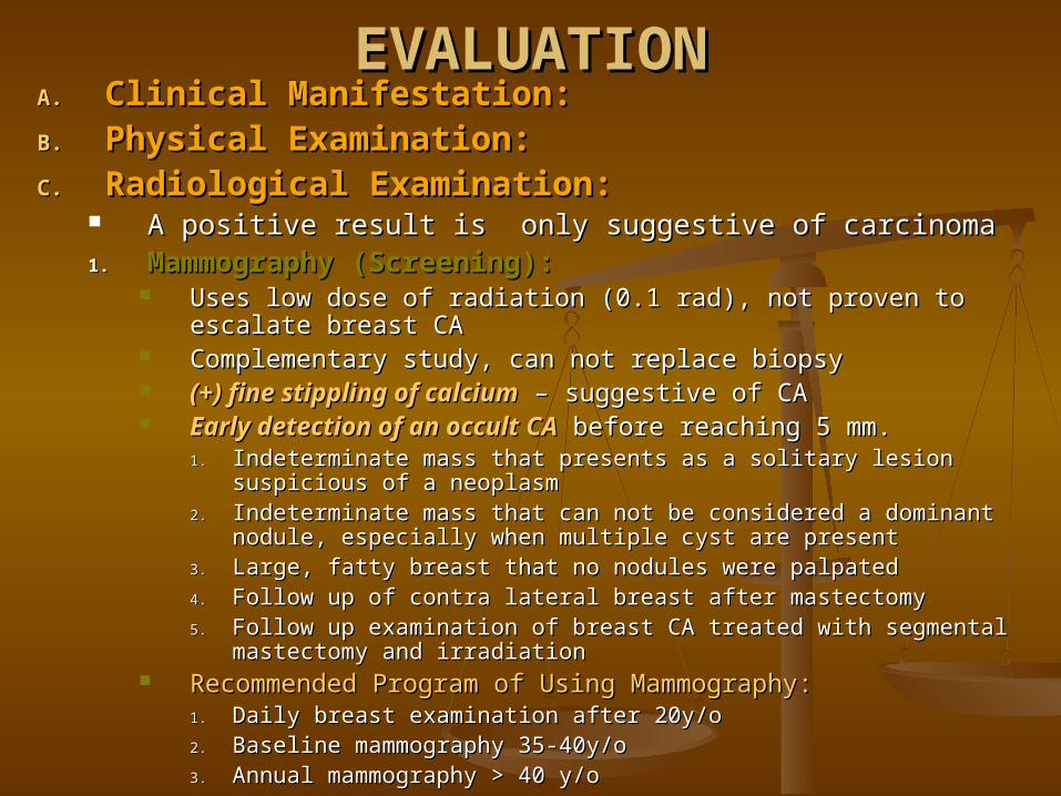
EVALUATIONEVALUATIONA.A. Clinical Manifestation:Clinical Manifestation:B.B. Physical Examination:Physical Examination:C.C. Radiological Examination:Radiological Examination:
A positive result is only suggestive of carcinomaA positive result is only suggestive of carcinoma1.1. Mammography (Screening):Mammography (Screening):
Uses low dose of radiation (0.1 rad), not proven to escalate Uses low dose of radiation (0.1 rad), not proven to escalate breast CAbreast CA
Complementary study, can not replace biopsyComplementary study, can not replace biopsy (+) fine stippling of calcium(+) fine stippling of calcium – suggestive of CA– suggestive of CA Early detection of an occultEarly detection of an occult CACA before reaching 5 mm. before reaching 5 mm.
1.1. Indeterminate mass that presents as a solitary lesion suspicious of a Indeterminate mass that presents as a solitary lesion suspicious of a neoplasmneoplasm
2.2. Indeterminate mass that can not be considered a dominant nodule, Indeterminate mass that can not be considered a dominant nodule, especially when multiple cyst are presentespecially when multiple cyst are present
3.3. Large, fatty breast that no nodules were palpatedLarge, fatty breast that no nodules were palpated4.4. Follow up of contra lateral breast after mastectomyFollow up of contra lateral breast after mastectomy5.5. Follow up examination of breast CA treated with segmental Follow up examination of breast CA treated with segmental
mastectomy and irradiationmastectomy and irradiation Recommended Program of Using Mammography:Recommended Program of Using Mammography:
1.1. Daily breast examination after 20y/oDaily breast examination after 20y/o2.2. Baseline mammography 35-40y/oBaseline mammography 35-40y/o3.3. Annual mammography > 40 y/oAnnual mammography > 40 y/o

EVALUATIONEVALUATIONC.C. Radiological Examination:Radiological Examination:
2.2. Computed Tomography or Magnetic Resonant Computed Tomography or Magnetic Resonant Imaging:Imaging:
To expensiveTo expensive For detection of vertebral metastasisFor detection of vertebral metastasis
3.3. UltrasonographyUltrasonography No radiation exposureNo radiation exposure Can differentiate cystic lesions from solid massCan differentiate cystic lesions from solid mass Can not detect less than 5mm.Can not detect less than 5mm.
4.4. Interventional Technique:Interventional Technique:Ductography:Ductography:
Inject radio-opaque contrast media into the mammary ductInject radio-opaque contrast media into the mammary duct
D.D. Biopsy: Biopsy: positive result is positive result is diagnosticdiagnostic1.1. Excision biopsyExcision biopsy2.2. Incision biopsyIncision biopsy3.3. True-cut or core biopsy (Vim-Silverman)True-cut or core biopsy (Vim-Silverman)4.4. Fine needle biopsyFine needle biopsy

BENIGN LESIONS OF THE BREASTBENIGN LESIONS OF THE BREAST1.1. Non-proliferative lesions:Non-proliferative lesions:
a.a. Chronic Cystic Mastitis Chronic Cystic Mastitis (Fibrocystic disease, (Fibrocystic disease, fibroadenosis, Schimmelbuschs’ dse.)fibroadenosis, Schimmelbuschs’ dse.)
most common breast lesion (30-40y/o)most common breast lesion (30-40y/o) Hormonal imbalance (exact etiology - ?)Hormonal imbalance (exact etiology - ?)
Increase estrogen production – producing exaggerated responsesIncrease estrogen production – producing exaggerated responses Some parts of the breast is hyper-reactingSome parts of the breast is hyper-reacting
Manifestations:Manifestations:1.1. Unilateral / BilateralUnilateral / Bilateral2.2. Rubbery in consistency, not encapsulatedRubbery in consistency, not encapsulated3.3. Size changes / can be tender ---> related to menstrual cycleSize changes / can be tender ---> related to menstrual cycle4.4. 15% presents a nipple discharge15% presents a nipple discharge5.5. (-) risk factor of carcinoma degeneration(-) risk factor of carcinoma degeneration6.6. Co-exist w/ breast carcinoma Co-exist w/ breast carcinoma (mammography is suggested)(mammography is suggested)
Schmmelbusch disease:Schmmelbusch disease: classic diffuse cystic diseaseclassic diffuse cystic disease Bloodgood cyst:Bloodgood cyst: single, tense, large blue domed cyst single, tense, large blue domed cyst Treatment:Treatment:
Conservative for small and not very painful and tender lesionsConservative for small and not very painful and tender lesions Danazol – alleviate mod to severe painful & tenderDanazol – alleviate mod to severe painful & tender
- synthetic FSH and LH analog- synthetic FSH and LH analog - Suppresses FSH and LH- Suppresses FSH and LH - 100 – 400mg- 100 – 400mg
Surgery for Bloodgood cystSurgery for Bloodgood cyst

BENIGN LESIONS OF THE BREASTBENIGN LESIONS OF THE BREAST
2.2. Fibroadenoma:Fibroadenoma: Well circumscribed lesion, movable, Well circumscribed lesion, movable,
smooth, lobulated, encapsulated, smooth, lobulated, encapsulated, painless, not associated w/ nipple painless, not associated w/ nipple dischargedischarge
Etiology (?), could also be due to Etiology (?), could also be due to hormonal imbalancehormonal imbalance
Size does not regress after Size does not regress after menstruationmenstruation
Treatment:Treatment: Excision biopsy (rule out malignancy)Excision biopsy (rule out malignancy)

BENIGN LESIONS OF THE BREASTBENIGN LESIONS OF THE BREAST3.3. Intra-ductal Papilloma:Intra-ductal Papilloma:
Proliferation of the ductal epithelium; 75% Proliferation of the ductal epithelium; 75% occurs beneath the epitheliumoccurs beneath the epithelium
Commonly causes Commonly causes Bloody Nipple DischargeBloody Nipple Discharge Palpable mass – 95% is intra-ductal papillomaPalpable mass – 95% is intra-ductal papilloma Non-palpable mass – possibility of malignancy is Non-palpable mass – possibility of malignancy is
increased: (Ductography)increased: (Ductography)
a.a. Paget disease of the nipple Paget disease of the nipple
b.b. Adenoma of the nippleAdenoma of the nipple
c.c. Deep lying carcinoma w/ ductal invasionDeep lying carcinoma w/ ductal invasion Treatment:Treatment:
Excision of a palpable mass by biopsyExcision of a palpable mass by biopsy Non-palpable mass --> do wedge resection of the Non-palpable mass --> do wedge resection of the
nipple/areola based on ductographic result or PE nipple/areola based on ductographic result or PE (+) bloody discharge(+) bloody discharge

BENIGN LESIONS OF THE BREASTBENIGN LESIONS OF THE BREAST4.4. Phyllodes TumorPhyllodes Tumor
Diagnostic problem separating it from Diagnostic problem separating it from fibroadenomafibroadenoma and it’s rare variant that is malignant, and it’s rare variant that is malignant, sarcomasarcoma
Bulk of the mass is made up of connective tissue, with Bulk of the mass is made up of connective tissue, with mixed areas of gelatinous, edematous areas. Cystic mixed areas of gelatinous, edematous areas. Cystic areas are due to necrosis and infarct degenerationsareas are due to necrosis and infarct degenerations
Phyllodes has greater activity and cellular component Phyllodes has greater activity and cellular component than fibroadenoma (3mitoses/hpf); while malignant than fibroadenoma (3mitoses/hpf); while malignant component has mitotic figure.component has mitotic figure.
80% are benign, usually large bulky lesions 80% are benign, usually large bulky lesions (tear drop (tear drop appearance)appearance) Malignant component is dependent on:Malignant component is dependent on:
a.a. Number of mitotic figures/hpfNumber of mitotic figures/hpfb.b. Vascular invasionVascular invasionc.c. Lymphatic invasionsLymphatic invasionsd.d. Distant metastasisDistant metastasis
Treatment:Treatment: Excision biopsy:Excision biopsy:
Benign – no further treatment, observeBenign – no further treatment, observe Malignant – total mastectomy / MRMMalignant – total mastectomy / MRM

BENIGN LESIONS OF THE BREASTBENIGN LESIONS OF THE BREAST
5.5. Mammary Duct Ectasia Mammary Duct Ectasia (Plasma cell (Plasma cell mastitis, Comedomasttitis & Chronic mastitis)mastitis, Comedomasttitis & Chronic mastitis)
Sub-acute inflammation of the ductal system Sub-acute inflammation of the ductal system usually beginning in the subareolar area w/ usually beginning in the subareolar area w/ ductal obstruction ductal obstruction
Usually present as a hard mass beneath or Usually present as a hard mass beneath or near areola w/ either nipple or skin retraction near areola w/ either nipple or skin retraction due to increase fibrosisdue to increase fibrosis
Appears during or after menopausal period w/ Appears during or after menopausal period w/ hx. Of difficulty of nursinghx. Of difficulty of nursing
Histologically, the duct are dilated and filled w/ Histologically, the duct are dilated and filled w/ debris and fatty material w/ atrophic debris and fatty material w/ atrophic epithelium. Sheets of plasma cells in the epithelium. Sheets of plasma cells in the periductal area.periductal area.
Treatment:Treatment: Excision biopsyExcision biopsy

BENIGN LESIONS OF THE BREASTBENIGN LESIONS OF THE BREAST
6.6. Galactocele:Galactocele: Cystic or solid mass w/ or w/o tendernessCystic or solid mass w/ or w/o tenderness Occurs during or after lactationOccurs during or after lactation Due to obstruction of a duct distended w/ milkDue to obstruction of a duct distended w/ milk Treatment:Treatment:
w/ abscess ---> incision and drain w/ abscess ---> incision and drain Solid mass ---> excison biopsySolid mass ---> excison biopsy
7.7. Fat necrosis:Fat necrosis: Present as a solid mass, usually asymptomaticPresent as a solid mass, usually asymptomatic w/ or w/o history of traumaw/ or w/o history of trauma Treatment:Treatment:
Excison biopsyExcison biopsy

BENIGN LESIONS OF THE BREASTBENIGN LESIONS OF THE BREAST
8.8. Acute Mastitis / Abscess:Acute Mastitis / Abscess: Bacterial infection usually during 1Bacterial infection usually during 1stst week of week of
lactationlactation s/sx of inflammations/sx of inflammation Treatment:Treatment:
Proper hygieneProper hygiene Cellulitis ----> antibiotis / analgesicCellulitis ----> antibiotis / analgesic Abscess ----> incision and drainAbscess ----> incision and drain

BENIGN LESIONS OF THE BREASTBENIGN LESIONS OF THE BREAST9.9. Gynecomastia:Gynecomastia:
Development of female type of breast in maleDevelopment of female type of breast in male Usually unilateral, if bilateral look for systemic causes:Usually unilateral, if bilateral look for systemic causes:
a.a. Hepatic cirrhosis (for elderly alcoholic)Hepatic cirrhosis (for elderly alcoholic)
b.b. Estrogen medication for prostatic CAEstrogen medication for prostatic CA
c.c. Tumor producing estrogen/progesteroneTumor producing estrogen/progesterone Pituitary / Adrenal / TestesPituitary / Adrenal / Testes CT scan / PECT scan / PE
Treatment:Treatment: Subcutaneous mastectomy (if other lesions, Subcutaneous mastectomy (if other lesions,
producing estrogen/progesterone, present)producing estrogen/progesterone, present) Tumor secreting estrogen ---> tx primary causeTumor secreting estrogen ---> tx primary cause

BENIGN LESIONS OF THE BREASTBENIGN LESIONS OF THE BREAST
10.10. Developmental Abnormality:Developmental Abnormality:a.a. AmastiaAmastia
b.b. PolymastiaPolymastia
c.c. AtheliaAthelia
d.d. PolytheliaPolythelia
Treatment:Treatment:
- - plastic surgeryplastic surgery

Malignant Lesions of the BreastMalignant Lesions of the Breast One of the leading cause of death from CAOne of the leading cause of death from CA Etiology:Etiology: - multifactorial- multifactorial
1.1. Sex:Sex: male : female ratio (1 : 100)male : female ratio (1 : 100)2.2. Age:Age: almost unknown for pre-pubertal age almost unknown for pre-pubertal age
20 – 40 y/o steady increase incidence20 – 40 y/o steady increase incidence 40 – 50 y/o (menopausal) plateau40 – 50 y/o (menopausal) plateau > 50 y/o higher incidence> 50 y/o higher incidence
3.3. Genetic:Genetic: Mother with carcinoma ---> (2 – 3x) daughterMother with carcinoma ---> (2 – 3x) daughter (+) family history ----> younger, bilateral(+) family history ----> younger, bilateral
4.4. Dietary influence:Dietary influence: Increase in developed countries (except) JapanIncrease in developed countries (except) Japan Increase in upper class societyIncrease in upper class society Dietary: Increase in Dietary: Increase in animal fatanimal fat

Malignant Lesions of the BreastMalignant Lesions of the Breast
5.5. Hormonal Usage:Hormonal Usage: Oral contraceptive has adverse effect if taken for Oral contraceptive has adverse effect if taken for
prolonged time at early age or when before the prolonged time at early age or when before the 11stst full term pregnancy full term pregnancy
No effect if taken 25 – 39y/oNo effect if taken 25 – 39y/o Slight increase risk if estrogen usage by peri-Slight increase risk if estrogen usage by peri-
menopausal for hormonal replacementmenopausal for hormonal replacement
6.6. Physical Stature:Physical Stature: Obesity ---> increase fat cells ----> increase Obesity ---> increase fat cells ----> increase
tissue concentrationtissue concentration

Malignant Lesions of the BreastMalignant Lesions of the Breast
6.6. Multiple primary neoplasm:Multiple primary neoplasm: Hx of primary breast CA ---> 4x fold increase of Hx of primary breast CA ---> 4x fold increase of
primary CAprimary CA Hx of primary CA of uterus and ovary ----> 1-1.5 Hx of primary CA of uterus and ovary ----> 1-1.5
riskrisk
7.7. Irradiation:Irradiation: Multiple exposureMultiple exposure Had radiotherapy for breast CA of contralateral Had radiotherapy for breast CA of contralateral
breastbreast

Malignant Lesions of the BreastMalignant Lesions of the Breast8.8. Other factorsOther factors
a.a. 11stst pregnancy – pregnancy – due to estrogendue to estrogen
a.a. Long term nursingLong term nursing > 36 months> 36 months No ovulation for 9 mos.No ovulation for 9 mos. Decrease estrogenDecrease estrogen
b.b. Age of menopauseAge of menopause Late menopause (55y/o) higher riskLate menopause (55y/o) higher risk
c.c. InfertilityInfertility Higher riskHigher risk

Risk factorRisk factor High riskHigh risk Low riskLow risk Relative Relative riskrisk
AgeAge oldold youngyoung >4.0>4.0
Socioeconomic statusSocioeconomic status highhigh lowlow 2.0 – 4.02.0 – 4.0
Marital statusMarital status Never marriedNever married Ever marriedEver married 1.1 – 1.91.1 – 1.9
Place of residencePlace of residence urbanurban ruralrural 1.1 – 1.91.1 – 1.9
Race > 45 yearsRace > 45 years
< 40 years< 40 yearswhitewhite blackblack 1.1 – 1.91.1 – 1.9
blackblack whitewhite 1.1 – 1.91.1 – 1.9
NulliparityNulliparity yesyes nono 1.1 – 1.91.1 – 1.9
Age of first full-term pregnancyAge of first full-term pregnancy > 30 y/o> 30 y/o < 20 y/o< 20 y/o 2.0 – 4.02.0 – 4.0
Oophorectomy premenopausallyOophorectomy premenopausally nono yesyes 2.0 – 4.02.0 – 4.0
Age at menopauseAge at menopause latelate earlyearly 1.1 – 1.91.1 – 1.9
Age at menarchyAge at menarchy earlyearly latelate 1.1 - 1.91.1 - 1.9
Weight, postmenopausal womenWeight, postmenopausal women heavyheavy thinthin 1.1 – 1.91.1 – 1.9
Hx of benign or cancer in one Hx of benign or cancer in one breastbreast
yesyes nono 2.0 – 4.02.0 – 4.0
Hx of breast Ca 1Hx of breast Ca 1stst degree degree relativerelative
yesyes nono 2.0 – 4.02.0 – 4.0
Mother or sister w/ hx. Of breast Mother or sister w/ hx. Of breast CACA
yesyes nono > 4.0> 4.0
Hx. Of primary ovarian or Hx. Of primary ovarian or endometrial CAendometrial CA
yesyes nono 1.1 – 9.01.1 – 9.0
Mammographic parenchymal Mammographic parenchymal patternspatterns
Dysplastic Dysplastic parenchymaparenchyma
Normal Normal parenchymaparenchyma
2.0 – 4.02.0 – 4.0
Radiation to chestRadiation to chest Large dosesLarge doses Minimal dosesMinimal doses 2.0 – 4.02.0 – 4.0
Established Risk factors For Breast cancer in Established Risk factors For Breast cancer in Females:Females:

Malignant Lesions of the BreastMalignant Lesions of the Breast
Natural historyNatural history (Schirrhous (Schirrhous adenocarcinoma)adenocarcinoma)
Doubling time (2-9mos)Doubling time (2-9mos) 1 cell ---> 30DT/5 yrs ---> 1cm. Mass/20DT 1 cell ---> 30DT/5 yrs ---> 1cm. Mass/20DT
---> increase size & fibrosis ----> dimpling ---> increase size & fibrosis ----> dimpling (retraction) ---> invade the lymphatics ---> (retraction) ---> invade the lymphatics ---> edema ----> invade regional LN/venous ----> edema ----> invade regional LN/venous ----> systemic.systemic.
Successful implantation depends on:Successful implantation depends on:1.1. Number of cellsNumber of cells
2.2. Character of cellCharacter of cell
3.3. Host resistanceHost resistance

Histological Classification of Breast CancerHistological Classification of Breast CancerCancers of the Mammary Gland can be Classified:Cancers of the Mammary Gland can be Classified:
1.1. HistogenesisHistogenesis – duct, lobule (acini)– duct, lobule (acini)
2.2. Histologic CharacteristicHistologic Characteristic – adenocarecinoma, epidermoid CA, etc. – adenocarecinoma, epidermoid CA, etc.
3.3. Gross CharacteristicGross Characteristic – Scirrhous, colloid, medullary, papillary, tubular – Scirrhous, colloid, medullary, papillary, tubular
4.4. Invasive CriteriaInvasive Criteria – Infiltrating, in-situ – Infiltrating, in-situ
Non-infiltrating (In-situ) Carcinoma of duct and lobules:Non-infiltrating (In-situ) Carcinoma of duct and lobules: Increase diagnosis due to mammographyIncrease diagnosis due to mammography DCIS : LCIS (3:1)DCIS : LCIS (3:1)
1.1. LOBULAR CARCINOMA in SITU:LOBULAR CARCINOMA in SITU: Considered as a risk factorConsidered as a risk factor Observed only in females, premenopousalObserved only in females, premenopousal No involvement of the basement membraneNo involvement of the basement membrane Tx:Tx: 1. Closed observation1. Closed observation
2. Hormonal treatment (Tamoxifen/aromatase inhibitor) for 5 years2. Hormonal treatment (Tamoxifen/aromatase inhibitor) for 5 years
3. Surgery (bilateral mastectomy) w/ immediate reconstruction3. Surgery (bilateral mastectomy) w/ immediate reconstruction

Histological Classification of Breast CancerHistological Classification of Breast Cancer
Non-infiltrating (In-situ) Carcinoma of duct and lobules:Non-infiltrating (In-situ) Carcinoma of duct and lobules:2.2. Tubular Carcinoma In Situ:Tubular Carcinoma In Situ:
Absence of invasion of surrounding stroma hence confined w/in the Absence of invasion of surrounding stroma hence confined w/in the basement membranebasement membrane
Type:Type:
1.1. PAPILLARYPAPILLARY:: Duct epithelium are thrown into papillae with loss of Duct epithelium are thrown into papillae with loss of
cohesiveness, loss of cohesiveness, disorientation of cells cohesiveness, loss of cohesiveness, disorientation of cells with pleomorphism and increase mitotic figurewith pleomorphism and increase mitotic figure
2.2. MICRO-PAPILLARY:MICRO-PAPILLARY:
3.3. SOLIDSOLID
4.4. CRIBRIFORMCRIBRIFORM
5.5. COMEDOCARCINOMA:COMEDOCARCINOMA: Hyperplasia is more extreme choking the entire duct w/ Hyperplasia is more extreme choking the entire duct w/
masses of cells developing central necrosis of cellsmasses of cells developing central necrosis of cells Most aggressiveMost aggressive
Treatment: Treatment: treated as an early cancertreated as an early cancer

Histological Classification of Breast CancerHistological Classification of Breast Cancer
Non-infiltrating (In-situ) Carcinoma of duct and lobules:Non-infiltrating (In-situ) Carcinoma of duct and lobules:
LCISLCIS DCISDCISAgeAge 44 - 4744 - 47 54 – 5854 – 58
IncidenceIncidence 2 - 5%2 - 5% 5 - 10%5 - 10%
Clinical SignsClinical Signs NoneNone Mass, Pain, Nipple Mass, Pain, Nipple dischargedischarge
Mammographic signsMammographic signs NoneNone MicrocalcificationMicrocalcification
Incidence of Synchronous Incidence of Synchronous Invasive CAInvasive CA
5%5% 2 – 46%2 – 46%
MulticentricityMulticentricity 60 – 90%60 – 90% 40 – 80%40 – 80%
BilateralityBilaterality 50 – 70%50 – 70% 10 – 20%10 – 20%
Axillary metastasisAxillary metastasis 1%1% 1 – 2%1 – 2%
Subsequent carcinomas:Subsequent carcinomas:
IncidenceIncidence
LateralityLaterality
Interval to diagnosisInterval to diagnosis
HistologyHistology
25 – 35%25 – 35%
BilateralBilateral
15 – 20 yrs15 – 20 yrs
ductalductal
25 – 70%25 – 70%
IpsilateralIpsilateral
5 – 10 yrs5 – 10 yrs
ductalductal

Histological Classification of Breast CancerHistological Classification of Breast Cancer
Infiltrating Carcinoma of the Breast:Infiltrating Carcinoma of the Breast:1.1. Paget’s disease of the nipple (1%):Paget’s disease of the nipple (1%):
Primary carcinoma of mammary duct that invaded the Primary carcinoma of mammary duct that invaded the skinskin
Chronic eczematoid lesion of the nippleChronic eczematoid lesion of the nipple Tenderness, itching, burning and intermittent bleedingTenderness, itching, burning and intermittent bleeding Palpable mass in the subareolar areaPalpable mass in the subareolar area PAGET cells:PAGET cells:
Characterictic cellsCharacterictic cells Large cell w/ clear cytoplasm and binucleatedLarge cell w/ clear cytoplasm and binucleated
80% non-infiltrating CA80% non-infiltrating CA 100% 5yr survival100% 5yr survival

Histological Classification of Breast CancerHistological Classification of Breast Cancer
2.2. Scirrhous carcinoma: Scirrhous carcinoma: (fibrocarcinoma, sclerosing (fibrocarcinoma, sclerosing CA):CA):
78% (most common)78% (most common) Increased Desmoplastic response to invading CA cells Increased Desmoplastic response to invading CA cells
(protective)(protective) Neoplastic cells are arranged in small clusters or in single Neoplastic cells are arranged in small clusters or in single
rows occupyning a space between collagen bundlesrows occupyning a space between collagen bundles Originate in the myoepithelial cells of the mammary ductOriginate in the myoepithelial cells of the mammary duct Desmoplastic ---> shortend Cooper’s ligament ---> dimpling Desmoplastic ---> shortend Cooper’s ligament ---> dimpling
over the tumorover the tumor
3.3. Medullary carcinoma:Medullary carcinoma: 2-15%2-15% Large round cancer cells arranged in broad plexiform mass Large round cancer cells arranged in broad plexiform mass
surrounded by lymphocytes and lymphatic folliclessurrounded by lymphocytes and lymphatic follicles Soft, bulky and large tumors w/ necrotic areasSoft, bulky and large tumors w/ necrotic areas 5 year survival = 85 – 90%5 year survival = 85 – 90% Good prognosisGood prognosis
Histological Classification of Breast CancerHistological Classification of Breast Cancer
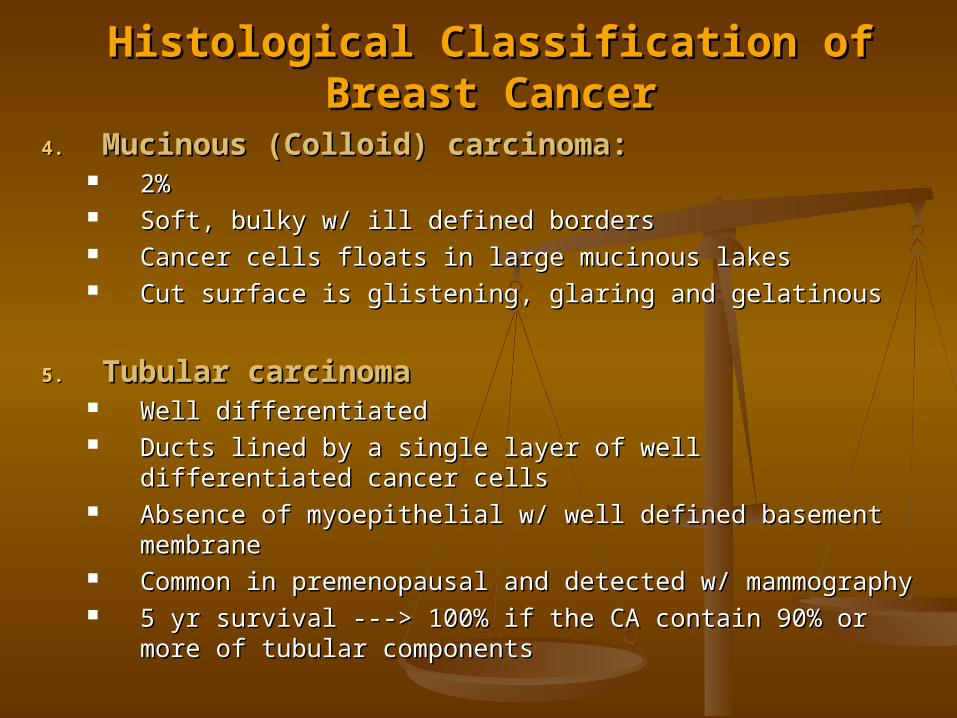
4.4. Mucinous (Colloid) carcinoma:Mucinous (Colloid) carcinoma: 2%2% Soft, bulky w/ ill defined bordersSoft, bulky w/ ill defined borders Cancer cells floats in large mucinous lakesCancer cells floats in large mucinous lakes Cut surface is glistening, glaring and gelatinousCut surface is glistening, glaring and gelatinous
5.5. Tubular carcinomaTubular carcinoma Well differentiatedWell differentiated Ducts lined by a single layer of well differentiated cancer Ducts lined by a single layer of well differentiated cancer
cellscells Absence of myoepithelial w/ well defined basement Absence of myoepithelial w/ well defined basement
membranemembrane Common in premenopausal and detected w/ Common in premenopausal and detected w/
mammographymammography 5 yr survival ---> 100% if the CA contain 90% or more of 5 yr survival ---> 100% if the CA contain 90% or more of
tubular componentstubular components
Histological Classification of Breast CancerHistological Classification of Breast Cancer
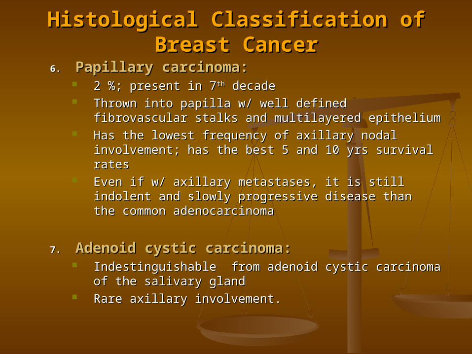
6.6. Papillary carcinoma:Papillary carcinoma: 2 %; present in 72 %; present in 7thth decade decade Thrown into papilla w/ well defined fibrovascular Thrown into papilla w/ well defined fibrovascular
stalks and multilayered epitheliumstalks and multilayered epithelium Has the lowest frequency of axillary nodal Has the lowest frequency of axillary nodal
involvement; has the best 5 and 10 yrs survival involvement; has the best 5 and 10 yrs survival ratesrates
Even if w/ axillary metastases, it is still indolent and Even if w/ axillary metastases, it is still indolent and slowly progressive disease than the common slowly progressive disease than the common adenocarcinomaadenocarcinoma
7.7. Adenoid cystic carcinoma:Adenoid cystic carcinoma: Indestinguishable Indestinguishable from adenoid cystic carcinoma of from adenoid cystic carcinoma of
the salivary glandthe salivary gland Rare axillary involvement.Rare axillary involvement.

Histological Classification of Breast CancerHistological Classification of Breast Cancer
8.8. Carcinoma of Lobular origin:Carcinoma of Lobular origin: 10% of breast CA; LCIS – 3%10% of breast CA; LCIS – 3% Small cell w/ round nucleus, inconspicuous nucleoli Small cell w/ round nucleus, inconspicuous nucleoli
and scant, indistinct cytoplasm.and scant, indistinct cytoplasm. Arises from the terminal ducts and aciniArises from the terminal ducts and acini Similar to colloid CA were mucin displaced the Similar to colloid CA were mucin displaced the
nucleus, resembling signet-ring carcinoma of the nucleus, resembling signet-ring carcinoma of the GIT.GIT.
High propensity for bilaterality (35-60%), High propensity for bilaterality (35-60%), multicentricity (88%) and multifocalitymulticentricity (88%) and multifocality
9.9. Squamous Carcinoma:Squamous Carcinoma: Metaplasia w/in the lactiferous duct systemMetaplasia w/in the lactiferous duct system Similar to epidermoid CA of the skinSimilar to epidermoid CA of the skin Metastasize thru the lymphaticMetastasize thru the lymphatic

Histological Classification of Breast CancerHistological Classification of Breast Cancer
10.10. Sarcoma of the Breast: Sarcoma of the Breast: (Fibrosarcoma, (Fibrosarcoma, liposarcom, leiomyosarcoma, malignant liposarcom, leiomyosarcoma, malignant fibrous histiocytoma, etc.)fibrous histiocytoma, etc.)
Large, painless breast mass w/ rapid growthLarge, painless breast mass w/ rapid growth Mammography ---> false (-)Mammography ---> false (-) Grossly: --> it lacks the cut gabbage surface of Grossly: --> it lacks the cut gabbage surface of
phyllodesphyllodes Histologically:Histologically:
Spindle cell neoplasm that grows expansile Spindle cell neoplasm that grows expansile and it’s margin either pushes or infiltrate and it’s margin either pushes or infiltrate adjacent structuresadjacent structures
It invades the fat and tend to intervene It invades the fat and tend to intervene between the glandular aspect of the breast between the glandular aspect of the breast parenchyma and expands the lobules and parenchyma and expands the lobules and intralobular spacesintralobular spaces
Treatment: --> total mastectomyTreatment: --> total mastectomy

Histological Classification of Breast CancerHistological Classification of Breast Cancer
11.11. Lymphoma of the Breast:Lymphoma of the Breast: Similar to other malignant lymphomaSimilar to other malignant lymphoma Mastectomy w/ axillary LN samplingMastectomy w/ axillary LN sampling Tx: radiotherapy / chemotherapyTx: radiotherapy / chemotherapy
12.12. Inflammatory Carcinoma of the BreastInflammatory Carcinoma of the Breast 1.5 – 3%1.5 – 3% Clinically: erythema, Clinically: erythema, Peau-d’ orangePeau-d’ orange, skin ridging , skin ridging
w/ or w/o a mass. Skin is warm sometimes scaly w/ or w/o a mass. Skin is warm sometimes scaly and indurated (cellulitis), nipple retract.and indurated (cellulitis), nipple retract.
Diagnosis: biopsyDiagnosis: biopsy Histologically: ---> Histologically: ---> no predominant histological no predominant histological
type.type. Subdermal lymphatic and vascular channels are Subdermal lymphatic and vascular channels are
permeated w/ highly undifferentiated tumorpermeated w/ highly undifferentiated tumor Characteristically: ---> absence of PMN and Characteristically: ---> absence of PMN and
lymphocyte near the tumorlymphocyte near the tumor Rapid growth and majority has (+) cervical LN and Rapid growth and majority has (+) cervical LN and
distant metastasisdistant metastasis

TNM Staging System for Breast CarcinomaTNM Staging System for Breast Carcinoma
Primary Tumor (T)Primary Tumor (T)TX TX – Primary tumor cannot be assessed– Primary tumor cannot be assessed
T0 T0 – No evidence of primary tumor– No evidence of primary tumor
Tis Tis – CA in situ (LCIS / DCIS), Paget’s dse of the nipple w/o tumor– CA in situ (LCIS / DCIS), Paget’s dse of the nipple w/o tumor
T1T1 – 2 cm or less – 2 cm or lessT1a T1a – 0.5 cm. or less– 0.5 cm. or lessT1b T1b - > 0.5 cm. to 1 cm.- > 0.5 cm. to 1 cm.T1c T1c - > 1cm. to 2 cm.- > 1cm. to 2 cm.
T2 T2 – 2 to 5 cm.– 2 to 5 cm.
T3 T3 - > 5 cm.- > 5 cm.
T4 T4 – – any size w/ direct extension to chest wall or skinany size w/ direct extension to chest wall or skinT4aT4a – extension to chest wall – extension to chest wallT4b T4b – edema / ulceration of the skin / satelite – edema / ulceration of the skin / satelite
nodulenoduleT4c T4c – both T4a and T4b– both T4a and T4bT4dT4d – Inflammatory carcinoma – Inflammatory carcinoma

TNM Staging System for Breast CarcinomaTNM Staging System for Breast CarcinomaRegional Lymph Nodes (N)Regional Lymph Nodes (N)
NX NX – Not assessed (previously removed)– Not assessed (previously removed)N0 N0 – No regional LN metastasis– No regional LN metastasisN1 N1 – (+) movable ipsilateral axillary LN– (+) movable ipsilateral axillary LNN2 N2 – (+) LN fixed to one another– (+) LN fixed to one anotherN3 N3 – (+) Ipsilateral INTERNAL MAMMARY LN– (+) Ipsilateral INTERNAL MAMMARY LN
Pathological Classification LN (pN):Pathological Classification LN (pN):pNX pNX – not assessed– not assessedpNO pNO – (-)– (-)pN1pN1 – (+) movable ipsilateral axillary LN – (+) movable ipsilateral axillary LN
pN1a pN1a – (+) micrometastasis (0.2 cm or less)– (+) micrometastasis (0.2 cm or less)pN1b pN1b – any larger than 0.2 cm but less than 2 cm– any larger than 0.2 cm but less than 2 cm
pN1bi pN1bi - (+) 1-3 LN- (+) 1-3 LNpN1bii pN1bii - (+) 4 or more LN- (+) 4 or more LNpN1biii pN1biii – extension of tumor beyond the – extension of tumor beyond the
capsulecapsulepN1biv pN1biv – (+) LN > than 2 cm– (+) LN > than 2 cm
pN2 pN2 – Axillary LN fixed with each other– Axillary LN fixed with each otherpN3 pN3 – (+) internal mammary LN– (+) internal mammary LN

TNM Staging System for Breast CarcinomaTNM Staging System for Breast CarcinomaDistant Metastasis (M):Distant Metastasis (M):
MX MX – not assessed– not assessedM0 M0 – (-)– (-)M1 M1 –(+) including metastasis to ipsilateral supraclavicular LN–(+) including metastasis to ipsilateral supraclavicular LN
Stage Grouping:Stage Grouping:Stage 0Stage 0 TisTis N0N0 M0M0Stage IStage I T1T1 N0N0 M0M0
Stage IIAStage IIA T0T0 N1N1 M0M0T1T1 N1aN1a M0M0T2T2 N0N0 M0M0
Stage IIBStage IIB T2T2 N1N1 M0M0T3T3 N0N0 M0M0
Stage IIIAStage IIIA T0 – T2T0 – T2 N2N2 M0M0 T3 N1-2T3 N1-2 M0M0
Stage IIIBStage IIIB T4 Any NT4 Any N M0M0 Any TAny T N3N3 M0M0
Stage IVStage IV Any T Any NAny T Any N M1M1

Survival Rates for patients w/ Breast Cancer Survival Rates for patients w/ Breast Cancer Relative to Clinical StageRelative to Clinical Stage
Clinical stagingClinical staging
(American Joint Committee)(American Joint Committee)
Crude 5-Crude 5-yr yr
survival survival (%)(%)
Range Range Survival Survival
(%)(%)
STAGE ISTAGE I Tumor < 2cm in diameter Tumor < 2cm in diameter
Nodes, if present, not felt to contain Nodes, if present, not felt to contain metastasesmetastases
w/o distant metastasesw/o distant metastases
8585 82 - 9482 - 94
STAGE IISTAGE II Tumors > 5 cm in diameter Tumors > 5 cm in diameter
Nodes, if palpable, not fixedNodes, if palpable, not fixed
w/o distant metastasisw/o distant metastasis
6666 47 – 7447 – 74
STAGE IIISTAGE III Tumor > 5cm in diameter Tumor > 5cm in diameter
Tumor any size w/ invasion of skin Tumor any size w/ invasion of skin attached to attached to
chest wallchest wall
Nodes in supraclavicular areaNodes in supraclavicular area
Without distant metastasesWithout distant metastases
4141 7 – 807 – 80
STAGE IVSTAGE IV With distant metastases With distant metastases 1010 --

Survival Rates for patients w/ Breast Cancer Survival Rates for patients w/ Breast Cancer Relative to Histologic StageRelative to Histologic Stage
Histologic Staging Histologic Staging (NSABP)(NSABP)
Crude Crude survival (%)survival (%)
5yr 10yr5yr 10yr
5-yr 5-yr Disease-Disease-
free free survival (%)survival (%)
All patientsAll patients 63.5 45.963.5 45.9 60.360.3
Negative axillary lymph nodesNegative axillary lymph nodes 78.1 64.978.1 64.9 82.382.3
Positive axillary lymph nodesPositive axillary lymph nodes 46.5 24.946.5 24.9 34.934.9
1 - 3 positive axillary lymph 1 - 3 positive axillary lymph nodesnodes
62.2 37.562.2 37.5 50.050.0
> 4 positive axillary lymph > 4 positive axillary lymph nodesnodes
32.0 13.432.0 13.4 21.121.1
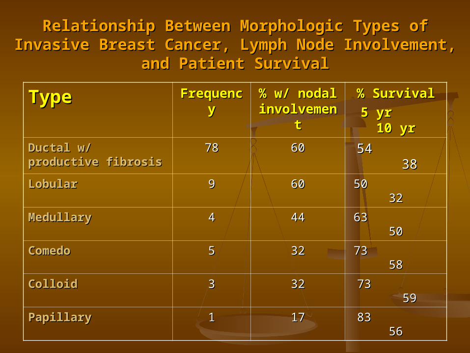
Relationship Between Morphologic Types of Invasive Relationship Between Morphologic Types of Invasive Breast Cancer, Lymph Node Involvement, and Patient Breast Cancer, Lymph Node Involvement, and Patient
SurvivalSurvival
TypeType FrequencFrequencyy
% w/ nodal % w/ nodal involvemeinvolveme
ntnt
% Survival% Survival
5 yr 10 5 yr 10 yryr
Ductal w/ productive Ductal w/ productive fibrosisfibrosis
7878 6060 54 3854 38
LobularLobular 99 6060 50 3250 32
MedullaryMedullary 44 4444 63 5063 50
ComedoComedo 55 3232 73 5873 58
ColloidColloid 33 3232 73 5973 59
PapillaryPapillary 11 1717 83 5683 56

Treatment:Treatment:1.1. Benign: Benign: hormonal, surgery (excision biopsy), antibioticshormonal, surgery (excision biopsy), antibiotics
2.2. Malignant:Malignant:
Selection of patientsSelection of patients a. stage of lesiona. stage of lesion
b. medical condition of ptb. medical condition of pt
Criteria of Inoperability / IncurabilityCriteria of Inoperability / Incurability (Haangensen)(Haangensen)a) extensive edema of the skin over the breasta) extensive edema of the skin over the breast
b) satellite nodule in the skin over the breastb) satellite nodule in the skin over the breast
c) inflammatory carcinoma of the breastc) inflammatory carcinoma of the breast
d) parasternal tumor noduled) parasternal tumor nodule
e) supraclavicular metastasise) supraclavicular metastasis
f) edema of the armf) edema of the arm
g) distant metastasisg) distant metastasis
h) Any 2 or more of the following locally advances h) Any 2 or more of the following locally advances cancercancer
i. ulceration of skini. ulceration of skin
ii. Edema of skin less 1/3ii. Edema of skin less 1/3
iii. Solid fixation of tumor to the chest walliii. Solid fixation of tumor to the chest wall
iv. Axillary LN 2 cm or moreiv. Axillary LN 2 cm or more
v. Fixation of axillary LN to skin and dep v. Fixation of axillary LN to skin and dep structurestructure
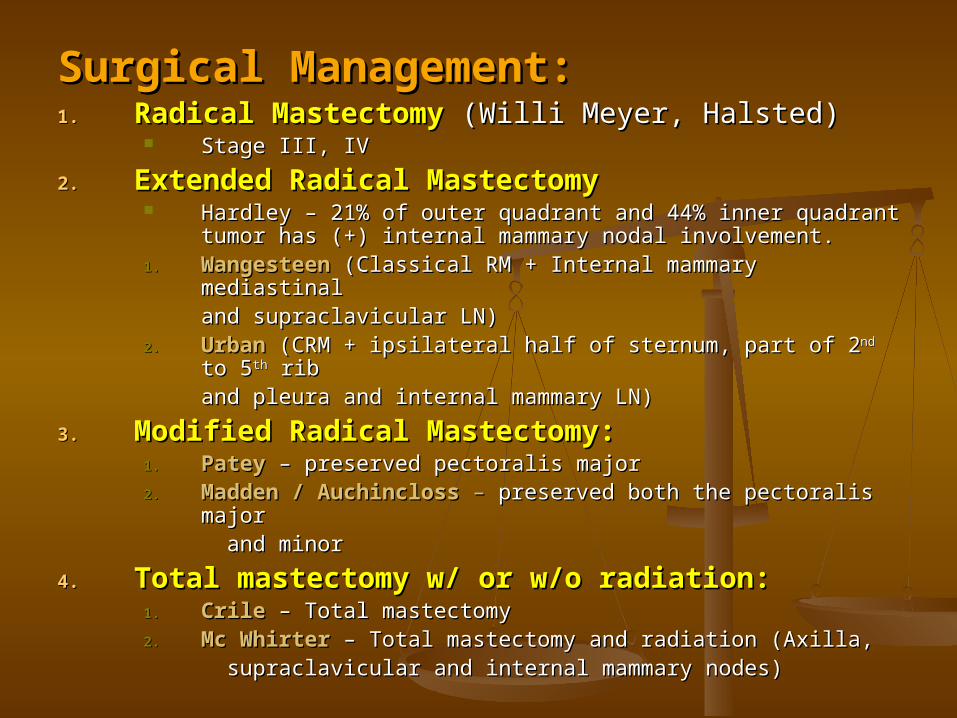
Surgical Management:Surgical Management:1.1. Radical MastectomyRadical Mastectomy (Willi Meyer, Halsted)(Willi Meyer, Halsted)
Stage III, IVStage III, IV
2.2. Extended Radical MastectomyExtended Radical Mastectomy Hardley – 21% of outer quadrant and 44% inner quadrant Hardley – 21% of outer quadrant and 44% inner quadrant
tumor has (+) internal mammary nodal involvement.tumor has (+) internal mammary nodal involvement.1.1. WangesteenWangesteen (Classical RM + Internal mammary mediastinal (Classical RM + Internal mammary mediastinal
and supraclavicular LN)and supraclavicular LN)2.2. UrbanUrban (CRM + ipsilateral half of sternum, part of 2 (CRM + ipsilateral half of sternum, part of 2ndnd to 5 to 5thth rib rib
and pleura and internal mammary LN)and pleura and internal mammary LN)
3.3. Modified Radical Mastectomy:Modified Radical Mastectomy:1.1. PateyPatey – preserved pectoralis major– preserved pectoralis major2.2. Madden / Auchincloss Madden / Auchincloss – – preserved both the pectoralis major preserved both the pectoralis major
and minorand minor
4.4. Total mastectomy w/ or w/o radiation:Total mastectomy w/ or w/o radiation:1.1. Crile Crile – Total mastectomy– Total mastectomy2.2. Mc WhirterMc Whirter – Total mastectomy and radiation (Axilla, – Total mastectomy and radiation (Axilla,
supraclavicular and internal mammary supraclavicular and internal mammary nodes)nodes)

Surgical Management:Surgical Management:5.5. Subcutaneous Mastectomy:Subcutaneous Mastectomy:
Nipple is retained / for T1sNipple is retained / for T1s
6.6. Quandrantectomy, axillary, radiotherapy (QUART)Quandrantectomy, axillary, radiotherapy (QUART) Quadrant of the breast that has the CA is resectedQuadrant of the breast that has the CA is resected
(quadrant of breast tissue, skin and superficial (quadrant of breast tissue, skin and superficial pectoralis fascia)pectoralis fascia)
Unacceptable cosmetic resultUnacceptable cosmetic result
7.7. Partial Mastectomy and Radiation:Partial Mastectomy and Radiation: Lumpectomy, segmental resection or tylectomyLumpectomy, segmental resection or tylectomy Histologically free margin of breast CA (1cm)Histologically free margin of breast CA (1cm) Advent of supervoltage radiotherapy with skin sparing effectAdvent of supervoltage radiotherapy with skin sparing effect Frozen section evaluation of marginFrozen section evaluation of margin To determine adjuvant chemotherapy adequate sampling of axillary LN To determine adjuvant chemotherapy adequate sampling of axillary LN
(level I), curvilinear incision should be done(level I), curvilinear incision should be done If LN (+) ----> adjuvant chemotherapyIf LN (+) ----> adjuvant chemotherapy
Indications for Conservative Surgery:Indications for Conservative Surgery:1.1. Small breast CA < 4cmSmall breast CA < 4cm2.2. Clinically (-) axillary LNClinically (-) axillary LN3.3. Breast volume adequate size to allow uniform dosage of Breast volume adequate size to allow uniform dosage of
irradiationirradiation4.4. Radiation therapist experience to avoid damage of retained Radiation therapist experience to avoid damage of retained
breastbreast

Radiotherapy:Radiotherapy: Local controlLocal control Pre-operative / post-operative radiationPre-operative / post-operative radiation
Chemotherapy:Chemotherapy: CMF, CAF, CA, AV, doxorubicinCMF, CAF, CA, AV, doxorubicin Side effect: nausea, vomiting, myelosuppression, alopecia, Side effect: nausea, vomiting, myelosuppression, alopecia,
thrombocytopenia, exercise intolerancethrombocytopenia, exercise intolerance
Hormonal Therapy:Hormonal Therapy: Receptor Assay (ER/PR):Receptor Assay (ER/PR):
1 gm of fresh tissue obtained by using cold scalpel and should be 1 gm of fresh tissue obtained by using cold scalpel and should be determined w/in 20-30 min.determined w/in 20-30 min.
ER (-) < 10% respond to endocrine ablation or exogenous estrogenER (-) < 10% respond to endocrine ablation or exogenous estrogenER (+) > 60% responds ER (+) > 60% responds
premenopausal – 30% (only due to masking effect of endogenous premenopausal – 30% (only due to masking effect of endogenous estrogen)estrogen)
Menopausal – 60% Menopausal – 60%
PR (+) 15% of premenopausal benefit from 15% PR (+) 15% of premenopausal benefit from 15%

Hormonal Therapy:Hormonal Therapy:1.1. Ablation:Ablation:
Oophorectomy, adrenalectomy, Oophorectomy, adrenalectomy, hypophysectomyhypophysectomy
Replaced by medical adrenelectomy, etc.Replaced by medical adrenelectomy, etc.
2.2. Anti-estrogen:Anti-estrogen:a.a. Tamoxifen Tamoxifen – a non-steroidal anti-estrogenic – a non-steroidal anti-estrogenic
compound that compete w/ estrogen at compound that compete w/ estrogen at receptor site. receptor site. Estrogen receptor assay should be determined; if Estrogen receptor assay should be determined; if
negative chance of success is very lownegative chance of success is very low
b.b. AromasinAromasinc.c. AminogluthethimideAminogluthethimide – it interferes with – it interferes with
conversion of androstinedione to estrone and conversion of androstinedione to estrone and estradiol in the peripheral tissue and inhibit the estradiol in the peripheral tissue and inhibit the conversion of cholesterol to pregnanoloneconversion of cholesterol to pregnanolone Hydrocortisone should be addedHydrocortisone should be added

Hormonal Therapy:Hormonal Therapy:
Receptor Receptor StatusStatus
PremenopausaPremenopausall
PostmenopausPostmenopausalal
ER + / PR +ER + / PR + O, TO, T
T + CTT + CTT, CTT, CT
ER + / PR -ER + / PR - OO
T---> T + CTT---> T + CTTT
T + CTT + CT
ER - / PR -ER - / PR - CTCT CTCT
ER - / PR +ER - / PR + O, TO, T
? T + CT? T + CTCTCT
T + CTT + CT
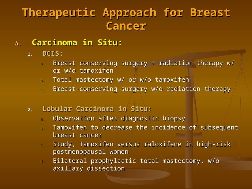
Therapeutic Approach for Breast CancerTherapeutic Approach for Breast Cancer
A.A. Carcinoma in Situ:Carcinoma in Situ:1.1. DCIS:DCIS:
a.a. Breast conserving surgery + radiation therapy w/ or w/o Breast conserving surgery + radiation therapy w/ or w/o tamoxifentamoxifen
b.b. Total mastectomy w/ or w/o tamoxifenTotal mastectomy w/ or w/o tamoxifen
c.c. Breast-conserving surgery w/o radiation therapyBreast-conserving surgery w/o radiation therapy
2.2. Lobular Carcinoma in Situ:Lobular Carcinoma in Situ:a.a. Observation after diagnostic biopsyObservation after diagnostic biopsy
b.b. Tamoxifen to decrease the incidence of subsequent breast Tamoxifen to decrease the incidence of subsequent breast cancercancer
c.c. Study, Tamoxifen versus raloxifene in high-risk Study, Tamoxifen versus raloxifene in high-risk postmenopausal womenpostmenopausal women
d.d. Bilateral prophylactic total mastectomy, w/o axillary Bilateral prophylactic total mastectomy, w/o axillary dissectiondissection

Therapeutic Approach for Breast CancerTherapeutic Approach for Breast CancerB.B. Stage I & IIStage I & II
Modified radical mastectomyModified radical mastectomy
(+) LN(+) LN (-) LN(-) LN (-) LN(-) LN
Low riskLow risk High risk High risk
Hormonal /Hormonal / observeobserve chemotherapy chemotherapy
chemotherapychemotherapy
High Risk Patients (Stage I):High Risk Patients (Stage I):A.A. Histologic criteria:Histologic criteria: 1. Poor cytologic differentiation1. Poor cytologic differentiation
2. Lymphatic permeation2. Lymphatic permeation
3. Blood vessel invasion3. Blood vessel invasion
4. Poor circumscritption4. Poor circumscritption
B.B. Rapid growth rate, by clinical history or thymidine labeling indexRapid growth rate, by clinical history or thymidine labeling index
C.C. Youth of the patientYouth of the patient
D.D. Estrogen receptor negativeEstrogen receptor negative
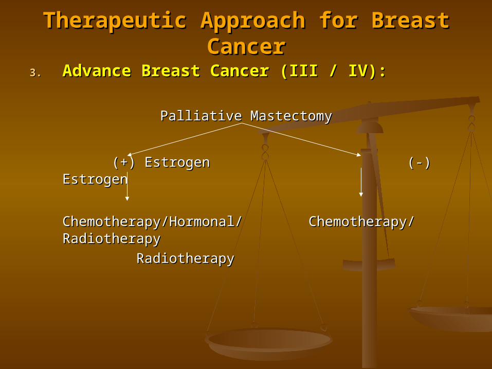
Therapeutic Approach for Breast CancerTherapeutic Approach for Breast Cancer
3.3. Advance Breast Cancer (III / IV):Advance Breast Cancer (III / IV):
Palliative MastectomyPalliative Mastectomy
(+) Estrogen(+) Estrogen (-) Estrogen(-) Estrogen
Chemotherapy/Hormonal/Chemotherapy/Hormonal/ Chemotherapy/Chemotherapy/RadiotherapyRadiotherapy
RadiotherapyRadiotherapy

Therapeutic Approach for Breast CancerTherapeutic Approach for Breast Cancer4.4. Inflammatory Breast Carcinoma:Inflammatory Breast Carcinoma:
3 – 5% 5 year survival3 – 5% 5 year survival Main role of surgery is in the diagnosisMain role of surgery is in the diagnosis Primary therapy is chemotherapy and radiotherapy and if Primary therapy is chemotherapy and radiotherapy and if
possible surgery (mastectomy).possible surgery (mastectomy).CAF ----- regression ------> extended mastectomy (level I) CAF ----- regression ------> extended mastectomy (level I) ----------> irradiation of axillary and skin flap (30% - 5 yr ----------> irradiation of axillary and skin flap (30% - 5 yr survival)survival)
5.5. Breast Cancer and Pregnancy/Lactation:Breast Cancer and Pregnancy/Lactation: The risk of aggressive and distant metastasis is profound due The risk of aggressive and distant metastasis is profound due
to high level of estrogen and progesterone secreted from the to high level of estrogen and progesterone secreted from the placenta and corpus luteum.placenta and corpus luteum.
Treat patient as if she is not pregnantTreat patient as if she is not pregnant Lactation should be suppressed promptly, even if biopsy was Lactation should be suppressed promptly, even if biopsy was
benign because milk from transected lactiferous will drain via benign because milk from transected lactiferous will drain via the biopsy sitethe biopsy site
If patient is undergoing radiotherapy and chemotherapy for If patient is undergoing radiotherapy and chemotherapy for breast CA, advice patient not to get pregnant. ( advice not to breast CA, advice patient not to get pregnant. ( advice not to use contraceptive pills).use contraceptive pills).
Treatment:Treatment: MRM / Segmental resection + radiation (after delivery)MRM / Segmental resection + radiation (after delivery) (+) axillary ---> chemotherapy is delayed on the 2(+) axillary ---> chemotherapy is delayed on the 2ndnd trimester trimester
(single agent) 11 – 12% teratogenicity on 1(single agent) 11 – 12% teratogenicity on 1stst trimester. trimester.

Therapeutic Approach for Breast CancerTherapeutic Approach for Breast Cancer
6.6. Breast Cancer in Men:Breast Cancer in Men: Factors: Factors:
a.a. Klinefelter syndromeKlinefelter syndromeb.b. Estrogen therapyEstrogen therapyc.c. Testicular feminizing syndromesTesticular feminizing syndromesd.d. IrradiationIrradiatione.e. TraumaTrauma
Age: 60-70y/oAge: 60-70y/o s/sx: breast mass, nipple retraction and/or discharge, s/sx: breast mass, nipple retraction and/or discharge,
ulceration and pain.ulceration and pain. Commonly ER positive and well differentiatedCommonly ER positive and well differentiated Prognosis is similar w/ femalePrognosis is similar w/ female Treatment:Treatment:
MRM + radiation if with ulceration and high gradeMRM + radiation if with ulceration and high grade Orchiectomy / chemotherapyOrchiectomy / chemotherapy