bridging course - week 1
TRANSCRIPT
Page 1 of 28
A-level Biology
Bridging Course - Week 1

Page 2 of 28
St Mary’s Catholic School A-level Biology Bridging Course
This bridging course will provide you with a mixture of information about A-level Biology, and what to expect from the course, as well as key work to complete. Students who are expecting to study Biology at A-level, and are likely to meet the entry requirements, must complete the bridging course fully and thoroughly, to the best of their ability. You should complete all work digitally if possible, so it is available to print and place in your file at the start of the course. You will submit it to your teacher in September. All of the work will be reviewed and selected work will be assessed, and you will be given feedback on it. This work will be signalled to you. If you do not have access to the internet, please contact the school and appropriate resources will be sent to you. If you are thinking about studying Biology at A-level you should attempt this work to see whether or not you think studying a subject like this is right for you. If you later decide to study Biology, you must ensure you complete this work in full. This work should be completed after you have read and completed the Study Skills work that all of Year 12 should complete.
Entry Requirements for Studying A-level Biology?
▪ Students who are expected to achieve at least a grade 7 in GCSE Biology Separate Science or a grade 7 in
Combined Science.
▪ Students who have enjoyed their GCSE Biology course, and who enjoy extra reading and research.
▪ Students should be competent in both Mathematics and Chemistry.
What to expect from A-level Biology.
The study of A Level Biology compliments a large number of university courses such as Medicine, Dentistry, Biomedical Science, Genetic Engineering, Environmental Science along with many others. It can also provide academic credentials for unrelated courses such as Law and Architecture. The course covers both animal, plant and environmental Biology, which will be taught through a combination of theory and practical work. This is a demanding A level, and students will need to be competent in both Maths and Chemistry.

Page 3 of 28
Course outline
The topics that you will study over the two years are as follows; Year 12
• Topic 1 - Biological molecules
• Topic 2 – Cells
• Topic 3 – Organisms exchange substances with their environment
• Topic 4 – Genetic information, variation and relationships between organisms
• Topic 5 – Energy transfers between organisms - Respiration and photosynthesis
Year 13
• Topic 5 - Energy transfers between organisms – Energy and ecosystems
• Topic 6 - Organisms respond to changes in their environment
• Topic 7 - Genetics, populations, evolution and ecosystems
• Topic 8 - The control of gene expression The following work will introduce key aspects of the Year 12 content along with some of the skills required during the A-level Biology course. This week we will be looking at cells.

Page 4 of 28
Cells
1. Cell Structure
During your GCSE course you have studied the basics of cell structure. At A-level we look at cells in much more detail. For the purpose of this course we are going to concentrate on eukaryotic cells, but the A-level course also requires knowledge of prokaryotic cells. Below is a diagram of an animal and plant cell, viewed with the level of detail you would expect to see using a transmission electron microscope. Animal Cell Plant Cell
1a. Complete the table to describe the function of the cell organelles listed (expand the cells as
necessary. Try to find detailed diagrams of each of the organelles. To help you do this you should use
the following resources;
https://www.youtube.com/watch?v=URUJD5NEXC8
https://www.youtube.com/watch?v=1Z9pqST72is

Page 5 of 28
TextBook

Page 6 of 28

Page 7 of 28

Page 8 of 28

Page 9 of 28

Page 10 of 28

Page 11 of 28
Organelle Structure and function Diagram
Cell-surface membrane
Nucleus
Mitochondria
Chloroplasts (in plants and algae)
Golgi apparatus and Golgi vesicles
Lysosomes
Ribosomes
Rough endoplasmic reticulum
Smooth endoplasmic reticulum
Cell wall (in plants, algae and fungi)
Cell vacuole (in plants)
The cell membrane

Page 12 of 28
The diagram above shows a detailed view of the cell membrane.
1b. Describe the role of the protein channels and the glycoproteins in the cell membrane
TextBook

Page 13 of 28

Page 14 of 28

Page 15 of 28
1c. Which of these organelles are linked to protein synthesis? Explain how these organelles interact with each
other to allow the cell to manufacture proteins.
Write your answer here
Write your answer here

Page 16 of 28
2. Studying Cells
Types of Microscope
Cells are microscopic, and our understanding of cells has largely developed in parallel to the development of
the microscope.
There are many different types of microscopes, but they can be divided into those that use light (light
microscopes), and those that use an electron beam (electron microscopes). You need to know the main
properties and uses of each, and understand the concept of magnification and resolution.
The images below are from a scanning electron microscope, transmission electron microscope and a light
microscope (from left to right).
The table summarises the main features of each type of microscope.
Light microscope Low magnification Maximum magnification x1500 Low resolution Colour image
Scanning electron microscope High magnification Maximum magnification x500000 High resolution 3D image Black and white image
Transmission electron microscope Highest magnification Maximum magnification x500000 Highest resolution
2D image Black and white image
2a. Use the information above to evaluate the advantages and disadvantages of the different types of
microscopes described for different functions.
Write your answer here

Page 17 of 28
200nm
Resolution and Magnification
Magnification – We increase magnification by using lenses. The more lenses we use and the more powerful
they are, the greater the magnification will be.
Eg. A light microscope with a x10 eye piece lens and a x40 objective lens will have a total magnification of 400.
10 x 40 = 400
If an object is 5mm long, and we look at an image of it which is 10mm long, the object has been magnified by 2
times.
Actual size(A) x Magnification(M) = Image size(I)
5 x 2 = 10
This formulae can also be expressed as;
Actual size = Image size
Magnification
Magnification = Image size
Actual size
You need to be able to make calculations using these 3 formulae, expressing the results in standard form in the
required unit.
Resolution
There is a limit to what you are able to see under a light microscope. Regardless of how good your microscope
is, and how powerful the lenses are, you will never be able to distinguish 2 points that are less than 200nm
apart. This is known as resolution. The resolving power is determined by the wave length of light.
The wave length of light is 200nm

Page 18 of 28
If we want to look at objects that are smaller than 200nm we need to use an electron microscope. Instead of
using beams of light an electron microscope uses an electron beam. The wave length of an electron beam is
0.2nm.
As long as the points are at least
200nm apart there is no overlap in the
waves and they will appear as 2
separate objects.
If the points are closer than 200nm the
waves will overlap and interfere with
each other, and the 2 points will
appear as a single object.

Page 19 of 28
2b. Look at the information above. What is the smallest object you could sucessfully use a light microscope to
veiw. Explain your answer.
We will concentrate on magnification calculations in the next section.
Biological Drawings
The AQA rules for biological drawings are;
Drawings should always be in pencil. Fine detail cannot be represented accurately unless the pencil has a sharp point.
The outlines of any structures should be drawn but there should be no colouring or shading. The relative sizes of the structures drawn should be accurate. Construction lines or frames could be used to solve this problem. If the relative size of any structure has been exaggerated, e.g., because an actual cell wall was too thin to be able to draw its outline using two pencil lines, a note should be added to the drawing to explain this.
If required, the drawn structures should be labelled with brief annotations about their functions or interrelationships.
The drawing should have an explanatory title and an indication of the real size of the structures drawn or of the magnification used.
Labels identify a structure or object.
Annotations are descriptive statements.
Write your answer here
Page 20 of 28
2c. In the box below produce a scientific drawing of the mitochondria shown in the photo. The drawing should be labelled and annotated. (You could produce this on a separate piece of paper, photograph it on your phone and insert the image.)
Page 21 of 28
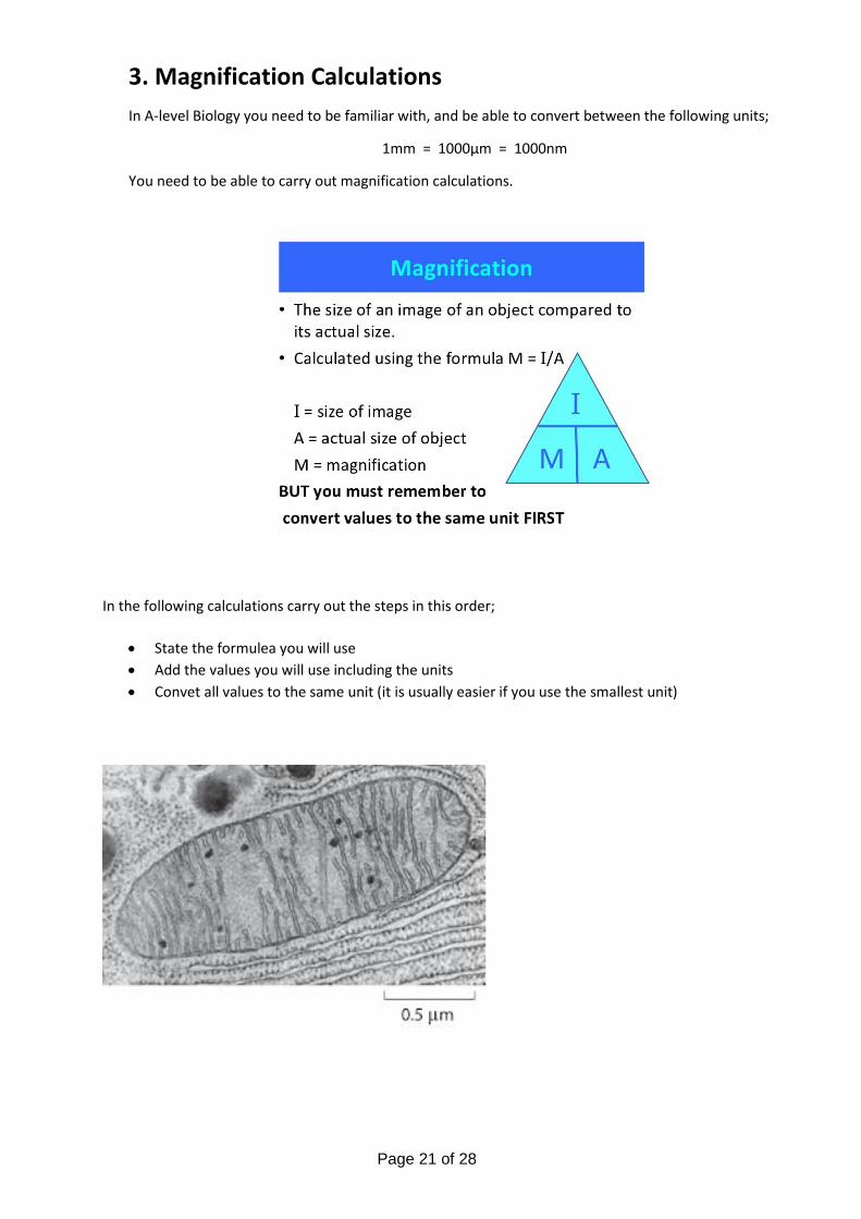
3. Magnification Calculations
In A-level Biology you need to be familiar with, and be able to convert between the following units;
1mm = 1000µm = 1000nm
You need to be able to carry out magnification calculations.
In the following calculations carry out the steps in this order;
• State the formulea you will use
• Add the values you will use including the units
• Convet all values to the same unit (it is usually easier if you use the smallest unit)

Page 22 of 28
3a. Calculate the magnification of this image.
3b. Calculate the maximum length of the mitochondria. Express your answer in µm.
Write your answer here
Write your answer here

Page 23 of 28
4. Exam Questions
The answers for Q1 and Q2 are at the end of this document. Q3, Q4 and Q5 will be marked by your teacher and
feedback given.
Q1. (a) Contrast how an optical microscope and a transmission electron microscope work
and contrast the limitations of their use when studying cells.
___________________________________________________________________
___________________________________________________________________
___________________________________________________________________
___________________________________________________________________
___________________________________________________________________
___________________________________________________________________
___________________________________________________________________
___________________________________________________________________
___________________________________________________________________
___________________________________________________________________
___________________________________________________________________
___________________________________________________________________
(6)
(Total 6 marks)

Page 24 of 28
Q2. Below is a diagram of an animal cell.
(a) Name the organelles labelled:
B _________________________________
C _________________________________
(2)
(b) Name two structures present in plant cells that are not present in animal cells.
1. _________________________________________________________________
2. _________________________________________________________________
(1)
(Total 3 marks)

Page 25 of 28
Q3.
The figure below shows a microscopic image of a plant cell.
© Science Photo Library
(a) Give the name and function of the structures labelled W and Z.
Name of W__________________________________________________________
Function of W________________________________________________________
Name of Z __________________________________________________________
Function of Z ________________________________________________________
(2)
(b) A transmission electron microscope was used to produce the image in the figure above. Explain why.
___________________________________________________________________
______________________________________________________________________________________________________________________________________
___________________________________________________________________
(2)
(c) Calculate the magnification of the image shown in the figure in part (a).
Answer = ____________________
(1)
(Total 5 marks)

Page 26 of 28
Q4. The image below shows the cell-surface membrane of a red blood cell seen with a transmission electron microscope.
(a) The cell-surface membrane can be seen with a transmission electron microscope but not with an optical microscope.
Explain why.
___________________________________________________________________
___________________________________________________________________
___________________________________________________________________
(1)
(b) No organelles are visible in the cytoplasm of this red blood cell.
Suggest why.
___________________________________________________________________
___________________________________________________________________
(1)
(c) Before the cell was examined using the electron microscope, it was stained. This stain caused parts of the structure of the cell-surface membrane to appear as two dark lines.
Suggest an explanation for the appearance of the cell-surface membrane as two dark lines.
___________________________________________________________________
______________________________________________________________________________________________________________________________________
___________________________________________________________________
(3)
(Total 5 marks)

Page 27 of 28
Q5.
The figure below shows a photograph of a chloroplast taken with an electron microscope.
© Science Photo Library
(a) Name the parts of the chloroplast labelled A and B.
Name of A ________________________________________________________
Name of B ________________________________________________________
(2)
(b) Calculate the length of the chloroplast shown in the figure above.
Answer ____________________
(1)
(c) Name two structures in a eukaryotic cell that cannot be identified using an optical microscope.
1. _________________________________________________________________
2. _________________________________________________________________
(1)
(Total 4 marks)

Page 28 of 28
Answers 1b. Channel proteins allow molecules to pass through the membrane by diffusion and active transport.
Glycoproteins act as antigens and allow cell recognition.
1c. DNA in nucleus codes for proteins, ribosomes read the code and manufacture the protein. It is modified in the endoplasmic reticulum and packaged in the golgi apparatus.
2b. Bacteria. Larger than 200nm.
3a. M=I/A I (length of scale bar)=21mm, A=0.5µm so M=21mm/0.5 µm, M=21000 µm/0.5 µm = 42000
3b. Maximum length of mitochondria on image is 85mm. A=I/M co A=85mm/42000=0.002mm = 2 µm
Exam Questions
Q1. (a) 1. TEM use electrons and optical use light;
2. TEM allows a greater resolution; 3. (So with TEM) smaller organelles / named cell structure can be observed
OR greater detail in organelles / named cell structure can be observed;
4. TEM view only dead / dehydrated specimens and optical (can) view live specimens;
5. TEM does not show colour and optical (can); 6. TEM requires thinner specimens; 7. TEM requires a more complex/time consuming preparation; 8. TEM focuses using magnets and optical uses (glass) lenses;
3. ‘clearer’ is not equivalent to ‘detail’
4. Accept ‘Only optical can view live specimens’
5. Accept ‘Only optical can show colour’
7. Accept ‘TEM requires a more difficult preparation’
Ignore references to artefacts 6 max
[6]
Q2. (a) B Golgi (body / apparatus);
C Mitochondria / mitochondrion; 2
(b) 1. Chloroplasts / plastids
2. Cell wall
3. Cell vacuole
4. Starch grains / amyloplasts;
Any 2 for 1 mark 1 max
[3]