brugia malayi is soluble and active in the absence of ... filebrugia malayi is soluble and active in...
TRANSCRIPT
1
A Hypodermally Expressed Prolyl 4-Hydroxylase from the Filarial Nematode
Brugia malayi is Soluble and Active in the Absence of Protein Disulfide Isomerase
Alan D. Winter‡, Johanna Myllyharju§ and Antony P. Page‡¶
From the ‡Wellcome Centre for Molecular Parasitology, Anderson College, University of Glasgow,
Glasgow G11 6NU, Scotland, United Kingdom and the §Collagen Research Unit, Biocenter Oulu and
Department of Medical Biochemistry and Molecular Biology, University of Oulu, FIN-90014 Oulu,
Finland
¶To whom correspondence should be addressed: Wellcome Centre for Molecular Parasitology, University
of Glasgow, Anderson College, 56 Dumbarton Road, Glasgow G11 6NU, Scotland, United Kingdom.
Telephone: (44) 141 330 3650. Fax: (44) 141 330 5422. E-mail: [email protected]
Running title: Prolyl 4-Hydroxylase in the Nematode Brugia malayi
Copyright 2002 by The American Society for Biochemistry and Molecular Biology, Inc.
JBC Papers in Press. Published on November 1, 2002 as Manuscript M210381200 by guest on January 7, 2020
http://ww
w.jbc.org/
Dow
nloaded from
2
SUMMARY
The collagen prolyl 4-hydroxylase (P4H) class of enzymes catalyze the hydroxylation of prolines
in the X-Pro-Gly repeats of collagen chains. This modification is central to the synthesis of all collagens.
Most P4Hs are α2β2 tetramers with the catalytic activity residing in the α subunits. The β subunits are
identical to the enzyme protein disulfide isomerase (PDI). The nematode cuticle is a collagenous
extracellular matrix required for maintenance of the worm body shape. Examination of the model nematode
Caenorhabditis elegans has demonstrated that its unique P4Hs are essential for viability and body
morphology. The filarial parasite Brugia malayi is a causative agent of lymphatic filariasis in humans. We
report here on the cloning and characterization of a B. malayi P4H with unusual properties. The
recombinant B. malayi α subunit, PHY-1, is a soluble and active P4H by itself, and it does not become
associated with PDI. The active enzyme form is a homotetramer with catalytic and inhibition properties
similar to those of the C. elegans P4Hs. High levels of B. malayi phy-1 transcript expression were observed
in all developmental stages examined, and its expression localized to the cuticle synthesizing hypodermal
tissue in the heterologous host C. elegans. Although active by itself, the B. malayi PHY-1 was not able to
replace enzyme function in a C. elegans P4H mutant.
by guest on January 7, 2020http://w
ww
.jbc.org/D
ownloaded from
3
INTRODUCTION
Biosynthesis of vertebrate collagens requires processing by up to eight specific intra- and
extracellular posttranslational enzymes (1). The collagen prolyl 4-hydroxylase (P4H)1 class of enzymes
(EC 1.14.11.2) catalyze the hydroxylation of prolines in the X-Pro-Gly repeats of collagen chains. This
endoplasmic reticulum (ER) resident enzyme is central to collagen synthesis, as collagen triple helices are
thermally unstable in the absence of 4-hydroxyproline residues (2,3). P4H also acts as a chaperone in the
assembly of collagen ensuring that only correctly folded collagens are released for secretion (4). In
vertebrates and Drosophila melanogaster the enzyme is an α2β2 tetramer (2,3,5,6), with hydroxylation
activity residing in the catalytic α subunits. Two α subunit isoforms, α(I) and α(II), have been
characterized in vertebrates (7,8). They become assembled into [α(I)]2β2 and [α(II)]2β2 tetramers, with
insect cell coexpression data arguing strongly against the formation of mixed α(I)α(II)β2 tetramers (8). The
β subunits of P4Hs are identical to the enzyme and chaperone protein disulfide isomerase (PDI) (EC
5.3.4.1) (9) and are required to maintain the α subunits in a catalytically active nonaggregated
conformation (10,11). The P4H is also maintained within its correct subcellular compartment by virtue of
an ER retention signal at the C-terminus of PDI (11). When expressed alone in a recombinant expression
system the α subunits are insoluble and inactive, whereas coexpression with PDI results in the formation of
an active, soluble P4H (7,12-14). The PDIs from different organisms can often substitute for the authentic
partner, such as the human PDI that can function as a β subunit in the mouse and Drosophila P4H tetramers
and in a P4H dimer with Caenorhabditis elegans PHY-1 (5,7,13).
In the model nematode C. elegans the cuticular collagen modifying function of P4H is essential
for body morphology and viability (15,16). In nematodes the exoskeleton (known as the cuticle) is an
extracellular matrix (ECM) composed of small collagen-like molecules (17). The nematode cuticle is
synthesized by the underlying hypodermal tissue and performs multiple functions including maintenance of
worm body shape. Mutations in collagens forming the cuticle, and in the enzymes involved in collagen
biosynthesis, can result in lethality and severe alterations to body shape as illustrated by the C. elegans sqt-
3 (18) and bli-4 (19) mutant phenotypes.
The P4Hs in C. elegans that are involved in the synthesis of cuticle collagens are formed from the
α subunits PHY-1 and PHY-2, and the β subunit PDI-2 (14,15). The expression, function and assembly of
by guest on January 7, 2020http://w
ww
.jbc.org/D
ownloaded from
4
these subunits have been examined in detail showing that unique P4H forms exist in C. elegans. The most
abundant form is a tetramer, this however differs from others described, in being a mixed PHY-1/PHY-
2/(PDI-2)2 tetramer (14). PHY-1 and PHY-2 can also each individually associate with PDI-2 to form dimers
(14). Such P4H forms have not been described for any other species to date. Genetic disruption of phy-1
and phy-2 simultaneously, or pdi-2 singly, results in embryonic lethality where embryos develop normally
until the first cuticle is required to maintain the elongated worm shape (15,16). The weakened cuticle is
then unable to maintain this form after which embryos collapse to a disorganized state and eventually die.
The body shape defect and reduced 4-hydroxyproline levels in the cuticle collagens of the viable phy-1 null
genetic mutant, dpy-18, underline the importance of collagen modification by P4H for nematode body
morphology (15,16).
Applying the knowledge of P4H function in C. elegans we examined a P4H in the filarial parasitic
nematode Brugia malayi. B. malayi along with B. timori and Wuchereria bancrofti are the causative agents
of lymphatic filariasis in humans, with over 120 million people infected and over 1 billion people at risk of
infection worldwide (20). Lymphatic filariasis is a debilitating disease, with approximately a third of those
infected being incapacitated and/or disfigured by the infection (21). Commercially available inhibitors of
P4H have been shown to be toxic to B. malayi adults, producing associated cuticular defects (22). These
observations and the requirement in C. elegans for P4H activity, highlight this enzyme class as a potential
drug target in the control of human and veterinary parasitic nematode infections.
In this paper we describe the identification of a P4H phy-1 gene from B. malayi, characterize the
molecular and enzymatic properties of the recombinant B. malayi P4H produced in an insect cell
expression system, and examine the expression profile and putative function of the B. malayi phy-1 gene by
heterologous expression in the model nematode C. elegans. Unusually, the B. malayi PHY-1 is a soluble
and active P4H when expressed alone in a recombinant system in the absence of PDI, and it does not
associate with PDIs from other organisms, including C. elegans. The developmental temporal expression
pattern of the B. malayi phy-1 gene was analyzed by RT-PCR using stage specific mRNA samples.
Reporter gene experiments showed that the B. malayi phy-1 promoter directs tissue-specific spatial
expression to the hypodermal cells of C. elegans.
by guest on January 7, 2020http://w
ww
.jbc.org/D
ownloaded from
5
MATERIALS AND METHODS
Nematode Strains and Culture Conditions - C. elegans strains were cultured as described
elsewhere (23). The wild type Bristol N2, CB364[dpy-18(e364)] and DR96[unc-76(e911)] C. elegans
strains were provided by the Caenorhabditis Genetics Centre. The B. malayi nematodes were provided by
Rick Maizels (University Edinburgh).
Isolation of cDNA and Genomic Clones - ESTs SW3D9CA480SK, MBAFCX8G05T3 and
MBAFCZ7H09T3 (Fig. 1A) were received from the Filarial Genome Project (FGP), subcloned and
sequenced. The primers X8G5F1, 5'-CAGTCGCTCAACACCGG-3', and BMNPHYR, 5'-
CCAATAGTATTTAAGCAC-3', were designed from the EST sequences and used to obtain a 312-bp PCR
product (Fig. 1A) from B. malayi adult-stage cDNA that was prepared as described previously (15). The
purified PCR product was labeled with α-32P dCTP and used to screen a B. malayi adult male cDNA library
SAW94NLBmAm (from Steven Williams, Filarial Genome Project, Northampton, MA). Eleven positive
clones all representing the same cDNA, named B. malayi phy-1, were identified from a total of 2 x 10⁴
recombinants. Amplification of the 5' end of the B. malayi phy-1 cDNA was performed using the Gibco
BRL Life Technologies 5' RACE System. Two 418-bp products from independent 5' RACE PCR reactions
(Fig. 1A) were sequenced, to generate a consensus 5' sequence. The primers BMPHY-1RESF(BamHI), and
BMPHY-1RESR(NotI), (for primer sequences see later) were used to generate the B. malayi phy-1 genomic
sequence from the translation start codon ATG to the stop codon TAA using B. malayi genomic DNA as a
template. Three identical full-length clones were then fully sequenced to generate a consensus genomic
sequence. Computer prediction for analysis of conceptual translation of B. malayi phy-1, protein
alignments, signal peptide analysis and predicted posttranslational modifications were performed on the
ExPASy proteomics tools database (http://www.expasy.ch).
Generation of a Recombinant Baculovirus coding for B. malayi PHY-1, and Expression and
Analysis of Recombinant Proteins in Insect Cells – The full-length coding sequence of B. malayi phy-1
cDNA was cloned from B. malayi adult stage cDNA by PCR using Pfu polymerase (Stratagene) with the
primers BMPHY-1BVF(NotI), 5'-gagcggccgcATGATAGCTACCGTGGTGTTC-3', and BMPHY-
1BVR(XbaI), 5'-gctctagaTTAAGCACTTAGATCGCCCAC-3' (artificial restriction sites in lower case and
underlined). The PCR product was cloned into a NotI-XbaI-digested transfer vector pVL1392 (Pharmingen)
by guest on January 7, 2020http://w
ww
.jbc.org/D
ownloaded from
6
and sequenced. The recombinant vector was cotransfected into Spodoptera frugiperda Sf9 cells with a
modified Autographa californica nuclear polyhedrosis virus DNA (BaculoGold, Pharmingen) by calcium-
phosphate precipitation.
Sf9 or High Five (Invitrogen) insect cells were cultured as monolayers in TNM-FH medium
(Sigma) supplemented with 10% fetal bovine serum (BioClear) or in suspension in Sf900IISFM serum-free
medium (Invitrogen). The cells were seeded at a density of 5 x 10⁶ cells/100-mm plate or 1 x 10⁶ cells/ml
and infected at a multiplicity of five with the virus coding for the B. malayi PHY-1 alone or together with
viruses coding for C. elegans PDI-1, PDI-2, or human PDI. In control experiments, the cells were
coinfected with the viruses coding for C. elegans PHY-1, PHY-2 and PDI-2, with PHY-1 and human PDI
or with the various PDI viruses alone. The cells were harvested 72 h after infection, washed with a solution
of 0.15 M NaCl and 0.02 M phosphate, pH 7.4, homogenized in a 0.1 M NaCl, 0.1 M glycine, 10 µM
dithiothreitol (DTT), 0.1% Triton X-100, and 0.01 M Tris buffer, pH 7.4, and centrifuged at 10,000 x g for
20 min. The pellets were further solubilized in 1% SDS. Aliquots of the samples were analyzed by 8%
SDS-PAGE under reducing conditions and by nonenaturing PAGE followed by Western blotting with
polyclonal antibodies against B. malayi PHY-1 (see below), C. elegans PDI-1 or PDI-2 (14) or a
monoclonal antibody against human PDI (5B5, DAKO). P4H activity was assayed by a method based on
the hydroxylation-coupled decarboxylation of 2-oxo-[1-14C]glutarate, and Km and Ki values were
determined as described previously (24). The molecular weight of the recombinant B. malayi P4H was
analyzed by applying the Triton X-100-soluble fraction of insect cells expressing B. malayi PHY-1 to a
calibrated HiPrep Sephacryl S-200 HR gel filtration column (Amersham Biosciences), equilibrated and
eluted with 0.1 M NaCl, 0.1 M glycine, 10 µM DTT, and 0.01 M Tris buffer, pH 7.4, and P4H activity in
the eluted fractions was assayed. Purified recombinant human type I P4H (12) was used as a control in the
gel filtration experiments.
Protein Analysis of B. malayi Extracts - Extracts from B. malayi were made by disrupting 100
adult females with a hand-held glass homogenizer in the following buffer; 0.1 M NaCl, 0.1 M glycine, 10
µM DTT, 0.1% Triton -X100, and 10 mM Tris buffer, pH 8.0, supplemented with the protease inhibitors; 1
mM PMSF, 1 mM EDTA, 1mM EGTA, 2 µM E64 and 0.1 µM pepstatin. The soluble extracts were
analyzed by reducing SDS-PAGE and nondenaturing PAGE in 4-12% NuPAGE Bis-Tris polyacrylamide
by guest on January 7, 2020http://w
ww
.jbc.org/D
ownloaded from
7
gels and 4-12% Tris-glycine gels (Invitrogen), respectively, followed by Western blotting. N-glycosidaseF
(PNGaseF, New England Biolabs) treatment was performed according to the manufacturers
recommendations.
Polyclonal antiserum was raised in two rabbits against a synthetic peptide corresponding to the C-
terminal region of B. malayi PHY-1. The peptide CRRPCGLSRSVEEQFVGDLSA was conjugated to
keyhole limpet heamocyanin (Sigma Genosys) via an added cysteine residue (underlined) and used for
immunization.
dpy-18 Rescue Experiments with B. malayi phy-1 - A 2.8-kb PstI-BamHI C. elegans phy-1
promoter fragment from the pPD95-03-phy-1 construct (15) was cloned into pBluescript SKM (Stratagene).
The C. elegans phy-1 3' UTR sequence was generated by PCR from C. elegans N2 genomic DNA using the
primers CEPHY-1 3'UTRF(SacI), 5'-gcggagctcCTCTAAGCATTGGTTTTCATTG-3', and CEPHY-
13'UTRR(SacI), 5'-gcggagctcACTAGGGAATTGTCGGCTGC-3' with Vent polymerase (NEB), and
cloned into the pBluescript-Cephy-1promoter construct to generate the plasmid pAW1 (Fig. 8B).
The coding sequence of B. malayi phy-1 cDNA and the genomic sequence of B. malayi phy-1
from the translation initiation codon to the stop codon were generated by PCR using the primers BMPHY-
1RESF(BamHI), 5'-gcggatccGATGATAGCTACCGTGGTGTTC-3', and BMPHY-1RESR(NotI), 5'-
gagcggccgcTTAAGCACTTAGATCGCCCAC-3', with Pfu and Pfu Turbo (Stratagene) polymerases,
respectively. The PCR products were cloned into BamHI-NotI-digested vector pAW1 to generate B. malayi
phy-1 cDNA and genomic rescue constructs (Fig. 8B). A synthetic intron (5'-
GTAAGTTTAAACTATTCGTTACTAACTAACTTTAAACATTTAAATTTTCAG -3') was inserted into
the B. malayi phy-1 cDNA rescue construct by ligating a double stranded oligo into a StuI blunt-ended
restriction site.
The B. malayi phy-1 cDNA (± a synthetic intron) and genomic rescue constructs were
microinjected into the syncytial gonad of C. elegans phy-1 null, dpy-18(e364), nematodes at concentrations
of 10 µg/ml and 100 µg/ml. A marker plasmid with a dpy-7 cuticle collagen promoter in the green
fluorescent protein (GFP) fusion vector pPD95-67 (from Iain Johnstone, University of Glasgow) was co-
injected at 5 µg/ml, and the injection mixes were made up to a final concentration of 150 µg/ml with
by guest on January 7, 2020http://w
ww
.jbc.org/D
ownloaded from

8
pBluescript SKM. Transformants were selected by GFP fluorescence, and more than five semi-stable
transmitting lines were examined for each concentration.
Developmental Timecourse RT-PCR - PCR was performed on cDNA samples generated from
daily extracts of B. malayi infected jirds, up to day 14 post infection, after which extracts from 2-4 day
intervals were taken (25). Two sets of primers were used for each PCR, BMPHY1.1IS1F, 5'-
GCTTCTGGTGTTCAACCG-3', and BMPHY1.1IS2R, 5'-GGTATGATGCTGTTTCAAG-3',
corresponding to the B. malayi phy-1, and BMTUBA, 5'-AATATGTGCCACGAGCAGTC-3', and
BMTUBB, 5'-CGGATACTCCTCACGAATTT-3', corresponding to the control B. malayi tubulin gene.
Cloning of the B. malayi phy-1 Promoter - To identify the putative promoter region, a genomic B.
malayi BAC library was screened with a 1.7-kb biotin labelled probe generated by PCR, using the primers
T7PL, 5'-CTCACTATAGGGCGAATTGG-3' (biotin labelled, New England Biolabs), and
BMPHY1.1IS3R(B), 5'-GCGTGGATGATTTGGATC-3', and a plasmid containing a T7 site and the 5'
genomic coding sequence from B. malayi phy-1 as a template. The gridded BAC filters were hybridized
using NEBlot Phototope and Phototope-star (New England Biolabs) detection kits. BLASTX analysis was
performed on the ExPASy proteomics tools database.
Construction of a B. malayi phy-1 Promoter-Reporter Plasmid - A 2.2-kb putative promoter
fragment of the B. malayi phy-1 gene was amplified from B. malayi genomic DNA using Pfu polymerase
and the primers BMPHY-1PF(SphI), 5'-ggcgcatgcGAATGAGACAATTGCACAAG-3', and BMPHY-
1PR(BamHI), 5'-ggcggatccGCTATCATCACTGGCTCTGGA-3'. This fragment, extending from –2189 to
+8 relative to the translation start site, was cloned into SphI-BamHI-digested C. elegans reporter gene
vector pPD96-04. This was microinjected into the syncytial gonad of the C. elegans strain DR96(unc-76)
together with the unc-76 rescue plasmid (p7616B); both at 100 µg/ml. Six semi-stable transgenic lines were
identified and examined for reporter gene expression by viewing GFP expression in live worms and by
sensitive staining of fixed worms for β-galactosidase activity (26). Live nematodes were transferred to 2%
agarose/0.065% sodium azide pads, and images were taken with an Axioskop 2 microscope using a
Hamamatsu digital camera and Improvision Openlab processing software.
by guest on January 7, 2020http://w
ww
.jbc.org/D
ownloaded from
9
RESULTS
Cloning of B. malayi phy-1 - Three ESTs from the B. malayi database were identified that encode
amino acid sequences homologous to C. elegans PHY-1 and PHY-2. MBAFCX8G05T3 (AA509222) and
MBAFCZ7H09T3 (AA406985) were both derived from a B. malayi adult female cDNA library, and
SW3D9CA480SK (AA585698) from a B. malayi L3 cDNA library. Sequencing of the ESTs established
that they were all derived from a single gene, termed B. malayi phy-1 (Fig. 1A). A 312-bp PCR probe was
generated from B. malayi adult stage cDNA based on the EST sequences, and used to screen a B. malayi
adult male cDNA library (Fig. 1A). Eleven positive clones were identified, full-length sequencing was
performed on two 1.6-kb clones, and both were found to represent the B. malayi phy-1 cDNA. The nine
other identified clones contained inserts with sizes ranging from 0.6 kb to 1.6 kb, and all corresponded to
the B. malayi phy-1 gene. The 1669-bp sequence generated from the library screening contained a 1515-bp
open reading frame and 154-bp of 3' UTR sequence (Fig. 1A). The 3’ untranslated region did not contain a
consensus polyadenylation signal (AATAAA). A divergent poly(A) signal sequence (GATAAA) was
however located 11-bp upstream of the poly(A) tail, representing a variant poly(A) signal sequence also
found in approximately 5% of C. elegans genes examined (27).
Comparison of amino acid sequence encoded by the 1515-bp B. malayi phy-1 cDNA sequence
with known P4H α subunits signified that the 5' coding sequence was incomplete. Additionally, according
to the signal sequence prediction program SignalP, the N terminus did not contain a characteristic signal
peptide. The 5' UTR, the transplice leader sequence and the coding sequence for 37 additional N-terminal
amino acids was obtained using the 5' RACE system for rapid amplification of cDNA ends (Fig. 1A). This
data was assembled with the 1669-bp B. malayi phy-1 sequence obtained from the cDNA libraries to give
the full-length cDNA sequence, which was confirmed by the sequencing of a full-length Pfu-generated
PCR product (Fig. 1A). The complete B. malayi phy-1 cDNA sequence contains a consensus 22-bp SL1
trans-splice leader sequence, an 8-bp 5' UTR, a single open-reading frame of 1626-bp that encodes a 541
amino acid polypeptide (AJ297845)2, and a 154-bp 3' UTR (Fig. 1A).
The 4596-bp consensus genomic sequence of the B. malayi phy-1 was determined from three
individual full-length PCR products, and contains 12 exons and 11 introns (Fig. 1B) (AJ421993)2. The
intron sizes range from 119-bp to 479-bp and have an average size of 270-bp.
by guest on January 7, 2020http://w
ww
.jbc.org/D
ownloaded from
10
Comparison of the B. malayi PHY-1 Amino Acid Sequence with Those of Other P4H α Subunits -
A signal peptide cleavage site between Ala17 and Asp18 was predicted by the SignalP program, and thus
the processed B. malayi PHY-1 consists of 524 amino acids. Highest amino acid sequence homology was
found between the processed B. malayi PHY-1 and C. elegans PHY-1 sequences (13), the identity being
59% and similarity 76%, while the identity and similarity between the B. malayi PHY-1 and C. elegans
PHY-2 (15,16) are 53% and 71%, respectively (Fig. 2). The amino acid sequence homology between the B.
malayi and C. elegans PHY polypeptides is slightly higher than that between the B. malayi PHY-1 and the
PHY-1 from a closely related filarial nematode Onchocerca volvulus (22), the B. malayi and O. volvulus
PHY-1 polypeptides being 49% identical and 70% similar. The amino acid sequence identities between the
B. malayi PHY-1 and the human α(Ι) and α(II) subunits are 45% and 44%, and similarities are 62% and
63%, respectively. The cysteine residues essential for intrachain disulfide bonding (28,29) and the active
site histidine, aspartic acid and lysine residues (29,30) are all conserved in B. malayi PHY-1 (Fig. 2). The
extended C-terminal regions present in the C. elegans and O. volvulus PHY-1 polypeptides are not found in
B. malayi PHY-1 (Fig. 2).
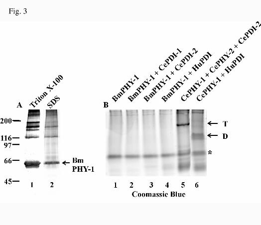
B. malayi PHY-1 is a Soluble and Active P4H when Expressed in the Absence of PDI in a
Baculovirus System - The B. malayi phy-1 cDNA was cloned into the baculovirus expression vector
pVL1392, a recombinant baculovirus was generated and used to infect insect cells. The cells were
harvested 72 h after infection, homogenized in a 0.1% Triton X-100 containing buffer, and centrifuged. The
remaining pellet was solubilized in 1% SDS, and the samples were analyzed by reducing SDS-PAGE
followed by Western blotting (Fig. 3 A). The C. elegans PHY polypeptides (13,14) and vertebrate P4H α
subunits (7,12) require association with PDI to form soluble and active P4Hs. When expressed alone in a
recombinant system, these polypeptides form inactive aggregates and 1% SDS is required for their efficient
solubilization (7,12-14). In contrast to these P4H α subunits, Western blotting showed that the majority of
the recombinant B. malayi PHY-1 was soluble in the Triton X-100 containing buffer (Fig. 3A, lane 1), and
only a minor amount formed insoluble aggregates that required 1% SDS for solubilization (Fig. 3A, lane 2).
The Triton X-100 extracts from insect cells expressing the B. malayi PHY-1 were analyzed for P4H activity
with an assay based on the hydroxylation-coupled decarboxylation of 2-oxo-[1-14C]glutarate (24), and a
significant amount of P4H activity was observed (Table I). The B. malayi PHY-1 thus differs from the C.
by guest on January 7, 2020http://w
ww
.jbc.org/D
ownloaded from

11
elegans PHY polypeptides and the vertebrate P4H α subunits, which require association with PDI to form
soluble and active P4Hs (7,12-14).
To study whether B. malayi PHY-1 has the potential to associate with PDI, the recombinant
protein was expressed in insect cells either alone, with the C. elegans β subunit PDI-2 (31), with the C.
elegans PDI-isoform PDI-1 (31), or with the human PDI (12). In control experiments the insect cells were
infected with viruses coding for the various PDIs alone, or coinfected with viruses coding for the C.
elegans PHY-1/PHY-2/(PDI-2)2 tetramer or the hybrid C. elegans PHY-1/human PDI dimer. Triton X-100-
soluble extracts of the cell lysates were analyzed by nondenaturing PAGE followed by Coomassie Blue
staining (Fig. 3B). In the control experiments, bands corresponding to the C. elegans PHY-1/PHY-2/(PDI-
2)2 tetramer (Fig. 3B, lane 5) and the C. elegans PHY-1/human PDI dimer (Fig. 3B, lane 6) were detected.
In contrast, neither a tetramer nor a dimer were detected by Coomassie Blue staining in the extracts from
cells expressing the B. malayi PHY-1 alone or in combination with different PDIs (Fig. 3B, lanes 1-4).
However, three B. malayi PHY-1 immunoreactive bands were detected by nondenaturing Western analysis
in extracts from cells expressing B. malayi PHY-1 alone (Fig. 4A, lane 1). Three bands with approximately
the same mobilities were also detected in the extracts from cells coexpressing the B. malayi PHY-1 and the
different PDIs (Fig. 4A, lanes 2-4). The mobility of the B. malayi PHY-1 upper band was similar to those of
the C. elegans PHY-1/PHY-2/(PDI-2)2 tetramer (Fig. 4A, lane 8) and the human P4H tetramer (data not
shown), and the mobility of the middle band was likewise similar to that of the PHY-1/PDI-2 dimer (Fig.
4A, lane 8). No B. malayi PHY-1 immunoreactive bands were detected in the extracts from cells expressing
the different PDIs alone (Fig. 4A, lanes 5-7). Coexpression of the recombinant B. malayi PHY-1 with the
different PDIs did not significantly increase the amount of P4H activity obtained (Table I), and Western
blotting of the nondenaturing PAGE with antibodies against the different PDIs showed that the B. malayi
PHY-1 does not associate with either the human PDI (Fig. 4B), C. elegans PDI-1 (Fig. 4C) or C. elegans
PDI-2 (Fig. 4D). This is in contrast to the vertebrate, C. elegans and Drosophila α subunits that can each
associate with orthologous P4H β subunits (5,7,13).
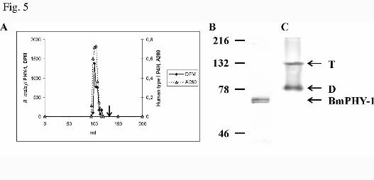
Gel filtration experiments in a calibrated HiPrep Sephacryl S-200HR column of the Triton X-100
extracts from cells expressing the recombinant B. malayi PHY-1 alone showed that P4H activity was eluted
in fractions that corresponded to a molecular weight of approximately 350,000 (Fig. 5A). Control
by guest on January 7, 2020http://w
ww
.jbc.org/D
ownloaded from
12
experiments with purified recombinant human type I P4H tetramer showed that it eluted in exactly the
same position as the recombinant B. malayi P4H (Fig. 5A). Previous gel filtration studies have also shown
that the elution position of a purified chick embryo P4H tetramer corresponds to a molecular weight of
350,000 (32) . The calculated molecular weights of a B. malayi PHY-1 tetramer and the human type I P4H
tetramer are 241,468 and 228,808, respectively. The fractions containing B. malayi P4H activity were
pooled and analyzed by reducing SDS-PAGE and nondenaturing PAGE followed by Western blotting (Fig.
5B and C). Two B. malayi PHY-1 immunoreactive bands corresponding to the nonglycosylated and
glycosylated forms (see Fig. 7) were detected in SDS-PAGE (Fig. 5B), and two bands with mobilites
similar to the human or C. elegans P4H tetramers and a C. elegans P4H dimer, respectively, were detected
in the nondenaturing PAGE (Fig. 5C). No detectable B. malayi P4H activity was eluted from the gel
filtration column in a position that corresponds to that of the C. elegans P4H dimer (Fig. 5A) (13), and
therefore our results indicate that the B. malayi PHY-1 self-associates into active P4H tetramers (Figs. 4A
and 5C), but during the nondenaturing PAGE partial dissociation of the B. malayi P4H tetramers into
dimers (Figs. 4A and 5C) and monomers (Fig. 4A) occurs.
The Km values for the cosubstrates 2-oxoglutarate, Fe2+ and ascorbate of the B. malayi PHY-1
(Table II) were very similar to those of the C. elegans PHY-1/PHY-2/(PDI-2)2 (14) and other P4Hs (13,30).
The Km value for the substrate (Pro-Pro-Gly)10 of the B. malayi PHY-1 (Table II) was slightly lower than
those reported for other P4Hs (13,14,30). The B. malayi PHY-1 was not efficiently inhibited by poly(L-
proline) (data not shown), and it thus resembles the C. elegans P4Hs (13,14) and the vertebrate type II
P4Hs (7,8). The Ki values of the B. malayi PHY-1 for the 2-oxoglutarate analogues pyridine-2,4-
dicarboxylate and pyridine-2,5-dicarboxylate (Table II) were approximately 2-fold lower than those
reported for C. elegans (13,14) and human P4Hs (3,30).
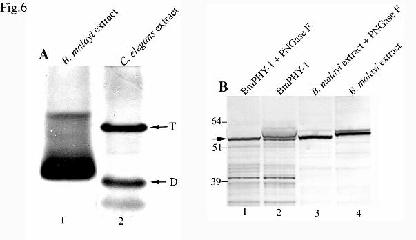
Analysis of Tissue-Extracted B. malayi PHY-1 and Native Glycosylation – Three immunoreactive
recombinant B. malayi PHY-1 bands were observed when Triton X-100 soluble extracts from insect cells
were analyzed by nondenaturing PAGE and Western blotting (Fig. 4A, lane 1). Native extracts were
prepared from the nematodes and were likewise analyzed to determine if similar PHY-1 immunoreactive
bands are found in B. malayi in vivo. Two major immunoreactive bands were observed in freshly-prepared
parasite extracts (Fig. 6A, lane 1), that have a similar native gel migration pattern to the characterized C.
by guest on January 7, 2020http://w
ww
.jbc.org/D
ownloaded from
13
elegans PHY-1/PHY-2/(PDI-2)2 tetramer and PHY-1/PDI-2 dimer from native C. elegans extracts (Fig. 6A,
lane 2).
The Proscan programme (ExPASy) predicted that B. malayi PHY-1 has two N-linked
glycosylation sites at positions 49-52 (NRSL) and 140-143 (NASG) (amino acid positions given relate to
the mature processed protein) (Fig. 2). The extracts from B. malayi nematodes and from insect cells
expressing recombinant B. malayi PHY-1 were treated with N-glycosidase F and analyzed by reducing
SDS-PAGE followed by Western blotting (Fig. 6B) to compare the glycosylation of native and recombinant
B. malayi PHY-1 polypeptides. Following glycosidase treatment of insect cell and worm extracts (Fig. 6B,
lanes 1 and 3), a single band of approximately 60 kDa was observed in both samples, the size of the
immunoreactive band being consistent with the predicted size of B. malayi PHY-1. In addition to the 60
kDa band, an additional band migrating slightly higher was detected in the untreated insect cell extract
(Fig. 6B, lane 2). The 60 kDa band was not detected in the untreated worm extract (Fig 6B, lane 4), but
instead two bands with higher mobilities were observed, representing B. malayi PHY-1 polypeptides in
which one or both glycosylation sites have been modified (Fig. 6B., lane 4). Thus, approximately half of
the recombinant B. malayi PHY-1 polypeptides expressed in insect cells remain unglycosylated, while in
the remaining polypeptides, only one of the glycosylation sites is modified. In B. malayi in vivo no
unglycosylated forms are present, and a minority of PHY-1 polypeptides are glycosylated at both sites.
Examination of the Functional Conservation Between the B. malayi and C. elegans PHY-1
Polypeptides - Phenotypic rescue of the C. elegans phy-1 null mutant [dpy-18(e364)] was attempted in
order to assess interspecies conservation of the phy-1 gene function. Attempts were made to rescue the C.
elegans mutant strain CB364[dpy18(e364)] using the B. malayi phy-1 coding sequence expressed under the
control of the C. elegans phy-1 promoter (Fig. 7). The B. malayi PHY-1 should therefore be expressed in
the relevant tissues with appropriate developmental timing and at comparable levels to that of the C.
elegans PHY-1. A vector was constructed which contained the previously defined C. elegans phy-1
promoter (15), and a splice site containing 3' UTR with the polyadenylation signal and poly-A transfer
sequences (13) (vector pAW1) (Fig. 7B). The B. malayi phy-1 coding sequence, with and without introns,
was inserted between the two C. elegans sequences (Fig. 7B), transformed into the CB364 strain, and the
by guest on January 7, 2020http://w
ww
.jbc.org/D
ownloaded from
14
ability to rescue the medium dumpy phenotype in the transformed animals carrying B. malayi PHY-1-
encoding transgene was analyzed.
Multiple semi-stable lines of nematodes transformed with 10 µg/ml and 100 µg/ml of the rescue
construct containing the B. malayi phy-1 cDNA sequence were examined and found not to rescue the dpy-
18 phenotype (data not shown). The lack of introns may result in extremely low levels or absence of
expression of the heterologous protein (33,34), and therefore an artificial intron was synthesized based on
standard C. elegans introns and inserted into the B. malayi phy-1 cDNA coding sequence (Fig. 7B).
Microinjection of this cDNA construct containing a synthetic intron at 10 µg/ml and 100 µg/ml
concentrations likewise failed to rescue the dumpy phenotype (data not shown). The ability of the genomic
B. malayi phy-1 sequence to rescue the dpy-18 phenotype was also studied. High concentrations (100
µg/ml) of this construct were however toxic, as all the transformed nematodes died during embryogenesis,
and no transformed lines could be generated. Injections at lower concentrations (10 µg/ml) yielded
transformed lines but examination of multiple lines again revealed a failure to rescue the dumpy phenotype
(data not shown).
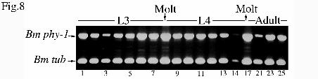
Temporal Expression Analysis of B. malayi phy-1 mRNA - Data from ESTs and library screenings
indicated that the B. malayi phy-1 mRNA was expressed in adult females, males and L3 larvae. A more
detailed analysis of the temporal expression pattern of B. malayi phy-1 in developmental stages from L3 to
adults was performed by RT-PCR using mRNA samples prepared from extracts isolated from infected
jirds; daily extracts taken up to day 14 post infection, after which extracts were taken every 2-4 days (25).
The 655-bp PCR product from B. malayi phy-1 cDNA can be distinguished from that produced by any
genomic contamination as the primers applied in this study were designed to span introns. A second set of
primers simultaneously applied in the same PCR reactions, were used to amplify a fragment of the tubulin
gene, as an internal control. The B. malayi phy-1 transcript was detectable in all L3, L4 and adult samples
analyzed (Fig. 8). Expression was likewise found in an L1 (microfilaria) cDNA sample (data not shown),
and taken together with the EST and library screening data, the RT-PCR results from stage-specific
samples show that the B. malayi phy-1 is expressed in L1 (microfilaria), at all time points examined
throughout L3 and L4 development, and in both adult sexes. Visual examination of this expression profile
reveals peaks of abundance corresponding to the L3-L4 and L4 to adult molts (Fig. 8 lanes 8 and 17).
by guest on January 7, 2020http://w
ww
.jbc.org/D
ownloaded from
15
Analysis of the Function of the B. malayi phy-1 Promoter Region - A 2.2-kb region of the putative
B. malayi phy-1 promoter sequence (AJ421994)2 was identified by screening a genomic BAC Brugia
library. This 2.2-kb region does not contain any other coding sequences as determined by BLASTX
analysis. A 3' trans-splicing acceptor site (27) is located at position –15 to –8 with respect to the translation
start codon. The 2.2-kb promoter fragment followed by the codons for the first three amino acids of B.
malayi PHY-1 was generated by PCR, and fused in frame to lacZ/GFP in the multi-intron reporter gene
vector pPD96-04. The reporter gene construct was transformed into C. elegans at 100 µg/ml and multiple
lines were examined, with expression found consistently in hypodermal cells of all lines examined (Fig.
9A-D). GFP expression was particularly prominent in the hypodermal cell nuclei hyp5, 6 and 7 of the head
(Fig. 9A), and in the pair of hyp7 cell nuclei in the tail of adult worms (Fig. 9B). Using sensitive staining
techniques for the detection of β-galactosidase activity, expression was also detected in larval stages, again
strongly in the head and tail regions (Fig. 9C and D). The lacZ expression was less pronounced in the
hypodermal cells of the body (Fig. 9C), with expression being detected in the hypodermal cell nuclei hyp4,
5, 6 and 7 of the head. With prolonged staining (overnight at room temperature) lacZ expression was also
observed in the vulval cells of the adult, which are of hypodermal origin, in the adult body wall muscle
cells, and in embryonic stages (data not shown).
DISCUSSION
In this study we have identified and characterized a P4H with unusual properties from the filarial
parasite B. malayi. The recombinant B. malayi PHY-1 assembles into an active soluble P4H tetramer when
expressed in insect cells in the absence of PDI. High-levels of B. malayi phy-1 transcript expression was
detected in all parasite stages examined. Analysis of B. malayi phy-1 promoter-reporter gene fusions
showed that the construct is expressed in hypodermal cells in a heterologous C. elegans system, thus
indicating a conservation of control elements between these species. Although B. malayi PHY-1 appears to
function as a P4H independently, without a PDI subunit, the B. malayi phy-1 was not able to replace P4H
function in a C. elegans phy-1 mutant.
The essential nature of the cuticle collagen modifying function of P4H has previously been
demonstrated in the nematode C. elegans where animals lacking or deficient in this function are non viable
by guest on January 7, 2020http://w
ww
.jbc.org/D
ownloaded from
16
or malformed (15,16,35). Chemical inhibitors of P4H have also been shown to have similar cuticle-specific
effects in both C. elegans (14, 16) and B. malayi (22). As P4H is involved in the synthesis of the nematode
cuticle, it is therefore an excellent target for parasite control by chemical inhibition.
The general structure and composition of the cuticle or exoskeleton is well conserved throughout
the nematode phyla (17). The cuticle is synthesized five times during development and is critical for many
functions such as movement and protection from the environment. In C. elegans the structure of the cuticle
has been studied in detail, demonstrating that despite overall similarity, cuticles from each developmental
stage are chemically and structurally distinct (36). Following the completion of embryonic elongation the
cuticle of the unhatched first larval stage maintains the elongated form of the worm (18). A weakened or
malformed cuticle may cause altered body morphology in viable worms or in some cases, as in the
combined disruption of the P4H α subunit encoding genes phy-1 and phy-2 in C. elegans, the resulting
cuticle is too weak for survival (15,16). The cuticle is composed predominantly of collagen, and over 150
cuticular collagen genes are present in the C. elegans genome (37). Similar large gene families are likewise
present in parasitic nematodes (17), and a representative cuticular collagen, COL-2 (38), has been cloned
from B. malayi. The size of B. malayi COL-2 (33 kDa) is similar to the small C. elegans collagens (26-35
kDa), and shows overall amino acid sequence similarity to the C. elegans cuticle collagens. Comparison at
the amino acid level, reveals 46% identity to the C. elegans group 2 (37) collagen T07H6.3. Precise
positioning of the cysteine residues, that are required for registering and stabilizing the collagen triple helix,
are likewise conserved between the B. malayi COL-2 and the C. elegans group 2 collagens. We propose
that B. malayi PHY-1 may function to modify cuticular collagens such as B. malayi COL-2, a process,
which in C. elegans is an essential feature of development.
B. malayi PHY-1 is unusual among the metazoan P4Hs in that it is a soluble and active P4H in the
absence of a PDI subunit. The function of PDI subunits in the vertebrate and D. melanogaster P4Hs, and in
the C. elegans P4Hs that are involved in the synthesis of cuticle collagens, is to keep the α, or PHY,
subunits in a catalytically active, nonaggregated conformation and to maintain the enzyme within the ER
(10,11). Active soluble monomeric P4Hs have been cloned and characterized from the Paramecium
bursaria Chlorella virus-1 (39), Arabidopsis thaliana (40), and partially purified from algae (41). It is also
possible that the C. elegans PHY-3, which is involved in the hydroxylation of collagens in early embryos,
by guest on January 7, 2020http://w
ww
.jbc.org/D
ownloaded from
17
acts as a monomeric P4H (42). Furthermore, the recently identified P4Hs that hydroxylate the hypoxia
inducible transcription factor HIFα are monomeric cytoplasmic enzymes (43,44). Interestingly, PHY-1
from the related parasite O. volvulus may require a PDI subunit to form an active P4H, but neither subunit
contains a recognizable ER retention signal (22,45). These unusual parasite P4Hs and the unique C. elegans
P4H forms suggest that nematodes have evolved specialized forms of these enzymes, possibly in order to
process the large number of collagens required to form the cuticular ECM.
When recombinant B. malayi PHY-1 was coexpressed in insect cells with PDI from other
organisms, no detectable association of the polypeptides was observed. In contrast, the C. elegans PHY-1,
and the Drosophila and vertebrate α subunits have been shown to assemble into an active P4H with PDIs
from other species (5, 7, 13). The assembly of the recombinant C. elegans PHY-2 with PDI-2 is very
inefficient, and only when it is coexpressed with C. elegans PHY-1 and PDI-2 it assembles into a fully
active mixed tetramer (14). Thus, although the recombinant B. malayi PHY-1 can function as an active P4H
by itself, it may assemble with additional, as yet unidentified assembly partners.
The B. malayi phy-1 expression constructs under the control of the C. elegans phy-1 promoter
were not able to rescue the C. elegans phy-1 null [dpy-18(e364)] mutant phenotype. A similar approach
has however been successfully applied in the rescue of the same mutant strain with a wild type copy of the
endogenous gene (15) (Fig. 7A). Inability of the B. malayi PHY-1 to functionally compensate for the lack
of C. elegans PHY-1 could be attributable to a number of causes such as low expression levels, differences
in codon usage, mis-splicing of introns or low activity of the transgenic protein. Alternatively, although the
recombinant B. malayi PHY-1 was shown to be an active P4H by itself, it may have higher activity in vivo
when assembled with an as yet unidentified partner. In support of this contention, the C. elegans PDI-2
does not associate with the B. malayi PHY-1 in a recombinant insect cell expression system (Fig. 4D).
Thus, if additional subunits are needed for B. malayi PHY-1 to rescue dpy-18, these could not be provided
by C. elegans. This possibility will be addressed following the completion of sequencing the B. malayi
genome, and all possible partners can be assessed for associations with B. malayi PHY-1. Heterologous
rescue of C. elegans mutants by B. malayi genes has not been described to date, but C. elegans rescue has
been obtained with genes from the more closely related parasitic nematode Haemonchus contortus. Using a
similar system to that employed in our study the embryonic lethal phenotype resulting from the lack of a
by guest on January 7, 2020http://w
ww
.jbc.org/D
ownloaded from
18
cathepsin L protease in C. elegans was rescued by transformation with a homologous gene from H.
contortus (46). This demonstrates the conservation of essential developmental function of these two genes
in different species. Amino acid sequence identity of mature cathepsin L proteases between C. elegans and
H. contortus is 87%, which is significantly higher than the 59% identity found between PHY-1
polypeptides from C. elegans and B. malayi. The lower identity reflects the more distant evolutionary
relationship between the latter pair of nematodes.
The B. malayi phy-1 promoter is capable of directing tissue-specific expression in C. elegans to the
cuticle synthesizing hypodermis, a location that is consistent with the proposed function of its gene product.
Appropriate tissue-specific expression of reporter genes in C. elegans has been described for promoters
from a number of genes from related parasitic nematodes (47-49), although to date, none have been
described from B. malayi. This study therefore confirms that C. elegans represents and excellent
heterologous system for the analysis of gene expression in the filarial nematodes. Although the native
localization of PHY-1 in B. malayi is not known, its function as a P4H, taken together with data from
reporter gene studies of the homologous genes from C. elegans (and the reporter-gene studies reported
here), would predict a hypodermal localization for B. malayi phy-1 expression, and is consistent with a role
in cuticle collagen modification. The secondary staining of muscle cells and vulval cells is also identical to
that found for the C. elegans homologues phy-1 and phy-2, which are both expressed in vulval cells with
additional muscle cell staining found for phy-2 (15). Like the C. elegans homologues, the B. malayi phy-1
transcript was strongly expressed throughout larval development, with greatest peaks of abundance
consistent with a role in the molting cycle and cuticle synthesis.
The essential nature of P4H in the development and body morphology in C. elegans has been
demonstrated both by genetic and RNAi analyses and by chemical inhibition studies. The extrapolation of
the knowledge gained in C. elegans and other species, along with the emerging use of C. elegans as a
surrogate system for expression of foreign proteins, provides powerful approaches for further
characterization of parasite P4Hs and assessment of their potential as targets for chemical control.
Acknowledgements- We thank the members of the Filarial Genome Project for supplying the B. malayi EST
and cDNA libraries used in this study. Rick Maizels and Yvonne Harcus are thanked for the gift of B.
by guest on January 7, 2020http://w
ww
.jbc.org/D
ownloaded from
19
malayi parasites, and Bill Gregory for the stage-specific cDNA samples (all from The Institute of Cell,
Animal and Population Biology, Edinburgh). BAC library screening and clone isolation was performed
using reagents supplied by Mark Blaxter and Jennifer Daub (The Institute of Cell, Animal and Population
Biology, Edinburgh), with their kind assistance. Iain Johnstone (Wellcome Center for Molecular
Parasitology, Glasgow) provided the dpy-7-GFP marker and Andrew Fire (Carnegie Institution of
Washington, Baltimore) the pPD vector and intron insertion protocol. The Caenorhabditis elegans Genetics
Centre provided some nematode strains used in this work. We thank Tanja Aatsinki, Raija Juntunen and
Eeva Lehtimäki for expert technical assistance. This work was supported by grants from the Medical
Research Council of Great Britain (APP), and the Health Science Council of the Academy of Finland
(200471) (JM) and the Finnish Centre of Excellence Programme 2000-2005 (44843) (JM).
REFERENCES
1. Myllyharju, J., and Kivirikko, K. I. (2001) Ann. Med. 33, 7-212. Kivirikko, K. I., and Myllyharju, J. (1998) Matrix Biol. 16, 357-3683. Kivirikko, K. I., and Pihlajaniemi, T. (1998) Adv. Enzymol. Related Areas Mol. Biol. 72, 325-4004. Walmsley, A. R., Batten, M. R., Lad, U., and Bulleid, N. J. (1999) J. Biol. Chem. 274, 14884-
148925. Annunen, P., Koivunen, P., and Kivirikko, K. I. (1999) J. Biol. Chem. 274, 6790-67966. Myllyharju, J. (2002) Matrix Biol. in press7. Helaakoski, T., Annunen, P., Vuori, K., MacNeil, I. A., Pihlajaniemi, T., and Kivirikko, K. I.
(1995) Proc. Natl. Acad. Sci. U.S.A. 92, 4427-44318. Annunen, P., Helaakoski, T., Myllyharju, J., Veijola, J., Pihlajaniemi, T., and Kivirikko, K. I.
(1997) J. Biol. Chem. 272, 17342-173489. Pihlajaniemi, T., Helaakoski, T., Tasanen, K., Myllylä, R., Huhtala, M.-L., Koivu, J., and
Kivirikko, K. I. (1987) EMBO J. 6, 643-64910. John, D. C. A., Grant, M. E., and Bulleid, N. J. (1993) EMBO J. 12, 1587-159511. Vuori, K., Pihlajaniemi, T., Myllylä, R., and Kivirikko, K. I. (1992) EMBO J. 11, 4213-421712. Vuori, K., Pihlajaniemi, T., Marttila, M., and Kivirikko, K. I. (1992) Proc. Natl. Acad. Sci. U.S.A.
89, 7467-747013. Veijola, J., Koivunen, P., Annunen, P., Pihlajaniemi, T., and Kivirikko, K. I. (1994) J. Biol. Chem.
269, 26746-2675314. Myllyharju, J., Kukkola, L., Winter, A. D., and Page, A. P. (2002) J. Biol. Chem. 277, 29187-
2919615. Winter, A. D., and Page, A. P. (2000) Mol. Cell. Biol. 20, 4084-409316. Friedman, L., Higgin, J. J., Moulder, G., Barstead, R., Raines, R. T., and Kimble, J. (2000) Proc.
Natl. Acad. Sci. U.S.A. 97, 4736-474117. Page, A. P. (2001) in Parasitic Nematodes (Kennedy, M.W. and Harnett, W., eds) pp. 167-193,
CABI Press, Wallingford, Oxon, UK18. Priess, J. R., and Hirsh, D. I. (1986) Dev. Biol. 117, 156-17319. Thacker, C., Peters, K., Srayko, M., and Rose, A. M. (1995) Genes Dev. 9, 956-97120. W.H.O. (2000) Fact sheet no. 10221. Ottesen, E. A., Duke, B. O. L., Karam, M., and Behbehani, K. (1997) Bull. W.H.O. 75, 491-503
by guest on January 7, 2020http://w
ww
.jbc.org/D
ownloaded from
20
22. Merriweather, A., Guenzler, V., Brenner, M., and Unnasch, T. R. (2001) Mol. Biochem. Parasitol.116, 185-197
23. Brenner, S. (1974) Genet. 77, 71-9424. Kivirikko, K. I., and Myllylä, R. (1982) Methods Enzymol. 82, 245-30425. Gregory, W. F., Atmadja, A. K., Allen, J. E., and Maizels, R. M. (2000) Infect. Immun. 68, 4174-
417926. Fire, A. (1992) Genet. Anal. Biomol. Eng. 9, 151-15827. Blumenthal, T., and Steward, K. (1997) in C. elegans II (Riddle, D. L., Blumenthal, T., Meyer, B.
J., Priess, J. R., eds) pp. 117-145, Cold Spring Harbor Laboratory Press, Cold Spring Harbor, NY28. John, D. C. A., and Bulleid, N. J. (1994) Biochem. 33, 14018-1402529. Lamberg, A., Pihlajaniemi, T., and Kivirikko, K. I. (1995) J. Biol. Chem. 270, 9926-993130. Myllyharju, J., and Kivirikko, K. I. (1997) EMBO J. 16, 1173-118031. Veijola, J., Annunen, P., Koivunen, P., Page, A. P., Philajaniemi, T., and Kivirikko, K. I. (1996)
Biochem. J. 317, 721-72932. Tuderman, L., Kuutti, E.-R., and Kivirikko, K. I. (1975) Eur. J. Biochem. 52, 9-1633. Brinster, R. L., Allen, J. M., Behringer, R. R., Gelinas, R. E., and Palmiter, R. D. (1988) Proc.
Natl. Acad. Sci. U.S.A. 85, 836-84034. Buchman, A. R., and Berg, P. (1988) Mol. Cell. Biol. 8, 4395-440535. Hill, K. L., Harfe, B. D., Dobbins, C. A., and L'Hernault, S. W. (2000) Genet. 155, 1139-114836. Cox, G. N., Staprans, S., and Edgar, R. S. (1981) Dev. Biol. 86, 456-47037. Johnstone, I. L. (2000) Trends Genet. 16, 21-2738. Scott, A. L., Yenbutr, P., Eisinger, S. W., and Raghavan, N. (1995) Mol. Biochem. Parasitol. 70,
221-22539. Eriksson, M., Myllyharju, J., Tu, H., Hellman, M., and Kivirikko, K. I. (1999) J. Biol. Chem. 274,
22131-2213440. Hieta, R., and Myllyharju, J. (2002) J. Biol. Chem. 277, 23965–2397141. Kaska, D. D., Myllylä, R., Gunzler, V., Gibor, A., and Kivirikko, K. I. (1988) Biochem. J. 256,
257-26342. Riihimaa, P., Nissi, R., Page, A. P., Winter, A. D., Keskiaho, K., Kivirikko, K. I., and Myllyharju,
J. (2002) J. Biol. Chem. 277, 18238–1824343. Epstein, A. C. R., Gleadle, J. M., McNeill, L. A., Hewitson, K. S., O'Rourke, J., Mole, D. R.,
Mukherji, M., Metzen, E., Wilson, M. I., Dhanda, A., Tian, Y. M., Masson, N., Hamilton, D. L.,Jaakkola, P., Barstead, R., Hodgkin, J., Maxwell, P. H., Pugh, C. W., Schofield, C. J., andRatcliffe, P. J. (2001) Cell 107, 43-54
44. Bruick, R. K., and McKnight, S. L. (2001) Science 294, 1337-134045. Wilson, W. R., Tuan, R. S., Shepley, K. J., Freedman, D. O., Greene, B. M., Awadzi, K., and
Unnasch, T. R. (1994) Mol. Biochem. Parasitol. 68, 103-11746.46. Britton, C., and Murray, L. (2002) Mol. Biochem. Parasitol. 122, 21-3347. Krause, S., Sommer, A., Fischer, P., Brophy, P. M., Walter, R. D., and Liebau, E. (2001) Mol.
Biochem. Parasitol. 117, 145-15448. Qin, L., Smant, G., Stokkermans, J., Bakker, J., Schots, A., and Helder, J. (1998) Mol. Biochem.
Parasitol. 96, 59-6749. Britton, C., Redmond, D. L., Knox, D. P., McKerrow, J. H., and Barry, J. D. (1999) Mol. Biochem.
Parasitol. 103, 171-181
by guest on January 7, 2020http://w
ww
.jbc.org/D
ownloaded from
21
FOOTNOTES
1The abbreviations used are: P4H, prolyl 4-hydroxylase; PHY, prolyl 4-hydroxylase α subunit; PDI, protein
disulfide isomerase; UTR, untranslated region; GFP, green fluorescent protein; lacZ, β-galactosidase
encoding gene; ECM, extracellular matrix; ER, endoplasmic reticulum; hyp, hypodermal; EST, expressed
sequence tag; BAC, bacterial artificial chromosome.
2The cDNA and genomic sequences described in this paper have been deposited in the Genbank database
under the accession numbers AJ297845, AJ421993 and AJ421994.
by guest on January 7, 2020http://w
ww
.jbc.org/D
ownloaded from

22
FIGURE LEGENDS
FIG. 1. Cloning and gene structure of B. malayi phy-1. A, Cloning of the full-length cDNA sequence of
B. malayi phy-1. The SL1 trans-spliced leader, 5' UTR and 3' UTR are depicted as open boxes, and the
coding sequence by a filled box. ATG and TAA indicate the positions of the translational start and stop
codons, respectively. The relative positions of the ESTs, the probe used for library screens, and the largest
phage clone isolated are indicated. The full-length coding cDNA sequence derived from phage clones and
5' RACE data were confirmed by sequencing a full-length PCR product. B, Gene structure of B. malayi
phy-1. The 5' UTR and intronic regions are represented by lines and exons depicted as filled boxes. The 3'
UTR is shown as an open box and the poly-adenyation site is indicated. The sizes of exons and introns are
given in base pairs above the exon boxes and below the intron lines, respectively. The exons are numbered
with roman numerals.
FIG. 2. Amino acid alignments of B. malayi PHY-1 with C. elegans, human and O. volvulus P4H αααα
subunits. Alignment of B. malayi (Bm), C. elegans (Ce), O. volvulus (Ov) PHY polypeptides and human α
subunits using ClustalW and Boxshade. Gaps (-) were introduced for maximal alignment and signal
peptides were removed, therefore numbering refers to the mature processed proteins. The N-glycosylation
sites predicted by PROSITE for B. malayi PHY-1 are indicated with (++++). The cysteine residues (C)
required for intrachain disulfide bonding (28,29), and the catalytically critical aspartate (D), histidine (H)
and lysine (K) residues (29,30) identified by site-directed mutagenesis studies on the human P4H α(I)
subunit are indicated with (*) and are all conserved in the B. malayi PHY-1. Accession numbers: B. malayi
PHY-1 (AJ297845), C. elegans PHY-1 (Z81134), C. elegans PHY-2 (Z69637), human α(I) subunit
(M24486), human α(II) subunit (U90441), O. volvulus PHY-1 (AF369787).
FIG. 3. Analysis of the expression of recombinant B. malayi PHY-1 in insect cells (A) and
coexpression of B. malayi PHY-1 with various PDIs (B). A, Insect cells were infected with a recombinant
baculovirus coding for the B. malayi PHY-1 polypeptide, harvested 72 h after infection, homogenized in a
Triton X-100-containing buffer, and centrifuged. The remaining pellet was solubilized in 1% SDS. The
by guest on January 7, 2020http://w
ww
.jbc.org/D
ownloaded from

23
fractions were analyzed by 8% SDS-PAGE under reducing conditions followed by Western blotting using
an antibody against B. malayi PHY-1. B, Insect cells were infected with the virus coding for the B. malayi
PHY-1 alone (lane 1) or together with viruses coding for C. elegans PDI-1 (lane 2), PDI-2 (lane 3) or
human PDI (lane 4). In control experiments, insect cells were coinfected with viruses coding for C. elegans
PHY-1, PHY-2 and PDI-2 (lane 5), and C. elegans PHY-1 and human PDI (lane 6). Cells were harvested
and homogenized as above, and the Triton X-100-soluble fractions were analyzed by nondenaturing PAGE
followed by Coomassie Blue staining. The C. elegans PHY-1/PHY-2/(PDI-2)2 tetramer (T) and the C.
elegans PHY-1/human PDI dimer (D) are indicated by arrows. The band indicated by an asterisk is also
found in extracts from cells infected with a wild-type baculovirus (not shown), and the faint bands below it
represents the non-associated PDI subunits.
FIG. 4. Analysis of the assembly of B. malayi PHY-1 with various PDIs by nondenaturing PAGE and
Western blotting. Recombinant B. malayi PHY-1 was expressed in insect cells alone (lanes 1) or together
with human PDI (lanes 2), C. elegans PDI-1 (lanes 3) or PDI-2 (lanes 4). In control experiments, the
human PDI (A and B, lanes 5), C. elegans PDI-1 (A, lane 6 and C, lane 5) and PDI-2 (A, lane 7 and D, lane
5) were expressed alone, or the cells were coinfected with viruses coding for the C. elegans PHY-1, PHY-2
and PDI-2 (A, lane 8). The cells were harvested and homogenized as described in the legend to Fig. 3.
Triton X-100-soluble fractions were analyzed by nondenaturing 8% PAGE followed by Western blotting
using antibodies against B. malayi PHY-1 (A, lanes 1-7), C. elegans PHY-1 (A, lane 8), human PDI (B), C.
elegans PDI-1 (C) and C. elegans PDI-2 (D). The positions of the C. elegans PHY-1/PHY-2/(PDI-2)2
tetramer (T) and the PHY-1/PDI-2 dimer (D) are indicated by arrows.
FIG. 5. Gel filtration analysis of a Triton X-100-soluble fraction from insect cells expressing
recombinant B. malayi PHY-1. A, A Triton X-100-soluble fraction from insect cells expressing
recombinant B. malayi PHY-1 was applied to a HiPrep Sephacryl S-200 HR column and the eluted
fractions were assayed for P4H activity (♦). In a control experiment, purified recombinant human type
I P4H was applied to the column and absorbance at 280 nm (----∆----) in the eluted fractions was measured.
The arrow indicates the elution position of the C. elegans PHY-1/human PDI dimer. B and C, The gel
by guest on January 7, 2020http://w
ww
.jbc.org/D
ownloaded from
24
filtration fractions containing B. malayi P4H activity were pooled, concentrated, and analyzed by 8% SDS-
PAGE under reducing conditions (B) and nondenaturing 8% PAGE (C) followed by Western blotting using
an antibody against B. malayi PHY-1. The positions of the B. malayi PHY-1 polypeptide and the B. malayi
PHY-1 tetramers (T) and dimers (D) are indicated by arrows.
FIG. 6. Analysis of tissue-extracted B. malayi PHY-1 and determination of native glycosylation forms.
A, B. malayi protein extracts were made from the parasites and analyzed by nondenaturing gradient (4-
12%) PAGE followed by Western blotting using a B. malayi PHY-1 antibody (lane 1). C. elegans protein
extracts were separated on the same gel, blotted and probed with an anti-C. elegans PHY-1 antibody (lane
2). Arrows represent the C. elegans PHY-1-PHY-2/PDI-2)2 tetramer (T) and PHY-1/PDI-2 dimer (D), with
B. malayi PHY-1 immunoreactive bands showing comparable sizes to the characterized C. elegans tetramer
and dimer. B, Extracts from insect cells expressing the recombinant B. malayi PHY-1 and the B. malayi
extracts were incubated either in the absence (lanes 2 and 4) or presence (lanes 1 and 3) of PNGase F N-
glycanase. The samples were analyzed by denaturing (4-12% gradient) SDS-PAGE followed by Western
blotting with an anti-B. malayi PHY-1 antibody. The arrow indicates the 60 kDa deglycosylated B. malayi
PHY-1.
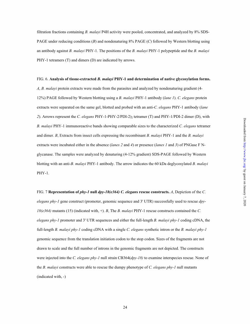
FIG. 7 Representation of phy-1 null dpy-18(e364) C. elegans rescue constructs. A, Depiction of the C.
elegans phy-1 gene construct (promoter, genomic sequence and 3' UTR) successfully used to rescue dpy-
18(e364) mutants (15) (indicated with, +). B, The B. malayi PHY-1 rescue constructs contained the C.
elegans phy-1 promoter and 3' UTR sequences and either the full-length B. malayi phy-1 coding cDNA, the
full-length B. malayi phy-1 coding cDNA with a single C. elegans synthetic intron or the B. malayi phy-1
genomic sequence from the translation initiation codon to the stop codon. Sizes of the fragments are not
drawn to scale and the full number of introns in the genomic fragments are not depicted. The constructs
were injected into the C. elegans phy-1 null strain CB364(dpy-18) to examine interspecies rescue. None of
the B. malayi constructs were able to rescue the dumpy phenotype of C. elegans phy-1 null mutants
(indicated with, -)
by guest on January 7, 2020http://w
ww
.jbc.org/D
ownloaded from
25
FIG. 8. Developmental expression analysis of B. malayi phy-1. Expression of the B. malayi phy-1
transcript was compared to the tubulin transcript through L3 to L4 and L4 to adult molts. PCR reactions
were performed on cDNA samples using primer pairs from both genes simultaneously. B. malayi extracts
were prepared from daily samples of infected jirds through L3 and L4, then at longer intervals after day 14.
Numbers below the figure refer to days post infection.
FIG. 9. Expression of reporter genes from B. malayi phy-1 promoter in a heterologous expression
system. Transgenic expression patterns in C. elegans of a reporter gene construct in which the B. malayi
promoter directs tissue-specific expression of lacZ and GFP. Panel A is a merge of DIC and UV images
showing expression of GFP in the hypodermal cells hyp5, 6 and 7 in the head. Panel B is a merge showing
expression of GFP in the hyp7 cells of the tail. Panels C-D show lacZ staining, with panel C showing the
expression in the hypodermal cells hyp4, 5, 6 and 7 of the head and body; panel D depicts reporter gene
expression in the hyp7 cells of the tail (compare to panel B).
by guest on January 7, 2020http://w
ww
.jbc.org/D
ownloaded from
26
Table I
P4H activity in Triton X-100 extracts of insect cells expressing various PDIs, B. malayi PHY-1 alone or
with various PDIs or C. elegans PHY-1/PHY-2/(PDI-2)2
Values are given in dpm/50 µl of Triton X-100 extractable cell protein, mean ± SD for at least four
experiments.
Polypeptides expressed P4H activity
dpm/50 µl
C. elegans PDI-1, PDI-2 or human PDI 500 ± 70
B. malayi PHY-1 3920 ± 1070
B. malayi PHY-1 and C. elegans PDI-1 3640 ± 710
B. malayi PHY-1 and C. elegans PDI-2 4210 ± 770
B. malayi PHY-1 and human PDI 4710 ± 1210
C. elegans PHY-1/PHY-2/(PDI-2)2 8650 ± 1970
by guest on January 7, 2020http://w
ww
.jbc.org/D
ownloaded from
27
TABLE II
Km values of the B. malayi PHY-1 and the C. elegans PHY-1/PHY-2/(PDI-2)2 tetramer for
cosubstrates and the peptide substrate and Ki values for two competitive inhibitors with respect to 2-
oxoglutarate
Cosubstrate, substrate, Km or Ki
or inhibitor Constant Bm PHY-1 Ce PHY-1/PHY-2/(PDI-2)2
µM
2-Oxoglutarate Km 60 80a
Fe2+ Km 0.5 2a
Ascorbate Km 400 350a
(Pro-Pro-Gly)10 Km 7 20a
Pyridine-2,4-dicarboxylate Ki 0.5 1a
Pyridine-2,5-dicarboxylate Ki 0.35 1a
aRef. 14.
by guest on January 7, 2020http://w
ww
.jbc.org/D
ownloaded from
ATAA
SL1Full length cDNA 1626 bp
3’ UTR
ATG
5’ RACE
MBAFCX8G05T3
PCR probe
SW3D9CA480SK
MBAFCZ7H09T3
Full length PCR
5’ UTR
B
Poly-A
II III IV V VI
VII VIII
IX X XI XII
ATG
5’
SL1
+1 +4596+1000 +2000 +3000 +4000
167 211 118 216 185 171 71 100 54 66 69 198
175 212 354 218 479 203 382 337 119 282 209
154
I
Base
3’
TAA
Phage clone
Fig. 1
by guest on January 7, 2020http://w
ww
.jbc.org/D
ownloaded from

++++BmPHY-1 1 --DLFTSIAEMELLLEADKRIPDLLDMYIERFQQRLDQIRQLSVGKKQLGNRSLGNDIRLCePHY-1 1 --DLFTSIADMQNLLETERNIPKILDKYIHDEEERLVQLKKLSEEYSKKNEISIENGLKDOvPHY-1 1 --EFYSSLASLKVIFEAERNISVIINGYVEKELERLDYLKKFAQEVQEHNDKAIRDGEEACePHY-2 1 --DLFTAIADLQHMLGAEKDVTTIIDQYIEAERARLDDLRRYAHEYVHRNAHAESVGPEFHumanαI 1 HPGFFTSIGQMTDLIHTEKDLVTSLKDYIKAEEDKLEQIKKWAEKLDRLTSTATKDPEGFHumanαII 1 --EFFTSIGHMTDLIYAEKELVQSLKEYILVEEAKLSKIKSWANKMEALTSKSAADAEGY
BmPHY-1 59 LSNPVSAYLLIKRLIEEWDDIKRLAGSDIGEELLKEISELRAMNYVKNPTTEDLVGAAIACePHY-1 59 ITNPINAFLLIKRKIFDWKEIESKMNANKAGNVVSSITDDSYG--VRYPTADDLSGAAIGOvPHY-1 59 IRHPINAFLLIKGMITDWNKIVKIMRSNSADDVIRNVTRHQDIKCINYPTEEDLIGAATGCePHY-2 59 VTNPINAYLLIKRLTTEWKKVENIMLNNKASTFLKNITDNRVRSEVKFPGEEDLSGAATAHumanαI 61 VGHPVNAFKLMKRLNTEWSELENLVLKDMSDGFISNLTIQRPV----LSNDEDQVGAAKAHumanαII 59 LAHPVNAYKLVKRLNTDWPALEDLVLQDSAAGFIANLSVQRQF----FPTDEDEIGAAKA
++++BmPHY-1 119 LLRLQDTYRLNVKEIADGKILNASG-VQPFTARDCFDIGRAAYNVNDYYHTLIWMEEAQECePHY-1 117 LLRLQDTYRLDTKDLADGKIYADQG-NYTFSAKDCFEIARAAYNEHDFYHTVMWMEEAQROvPHY-1 119 LLRLQDTFQMDTKDIADGKISNSQMRTVALTAEDCLEIGRAAYNAYDYYHTILWMREALECePHY-2 119 LLRLQDTYSLDTLDLSNG-IIGGEKVSNKLSGHDTFEVGRSAYNQKDYYHCLMWMQVALVHumanαI 117 LLRLQDTYNLDTDTISKGNLPGVKH-KSFLTAEDCFELGKVAYTEADYYHTELWMEQALRHumanαII 115 LMRLQDTYRLDPGTISRGELPGTKY-QAMLSVDDCFGMGRSAYNEGDYYHTVLWMEQVLK
BmPHY-1 178 RLRDEAPHETVQLKEILEYLAFALFKQGNLKRALLLTEQLHTIDPNHPRAKNNIKWYEDLCePHY-1 176 RLGDEV-EPTVEVEDILEYLAFALYKQNNLKHALKLTEELYKMNPTHPRAKGNVKWYEDLOvPHY-1 179 RLEKER-VPTANLEDILEYLAFSQYKQGNLKRALLLTDELYRINPDHPRAKDNVKEYEYLCePHY-2 178 KIENEN-PPTIEEWEILEYLAYSLYQQGNVRRALSLTKRLAKIAPNHPRAKGNVKWYEDMHumanαI 176 QLDEGE-ISTIDKVSVLDYLSYAVYQQGDLDKALLLTKKLLELDPEHQRANGNLKYFEYIHumanαII 174 QLDAGE-EATTTKSQVLDYLSYAVFQLGDLHRALELTRRLLSLDPSHERAGGNLRYFEQL
* *BmPHY-1 238 LAEEG----LKPIDYRRNIPPVTNPRPTTGLETAEHDIYEALCRNEIP-VSIKVTSKLYCCePHY-1 235 LEQEG----VRRSDMRKNLPPIQNRRPDSVLGNTERTMYEALCRNEVP-VSQKDISRLYCOvPHY-1 238 LKNNE----VQRIDLWRKTFPINNMRNDNEFDEGIKLIYEALCRREVP-VNTKVQSQLYCCePHY-2 237 LQGK---------DMVGDLPPIVNKRVEYD-GIVERDAYEALCRGEIPPVEPKWKNKLRCHumanαI 235 MAKEKDVNKSASDDQSDQKTTPK-KKGVAVDYLPERQKYEMLCRGEGIKMTPRRQKKLFCHumanαII 233 LEEER--EKTLTNQTEAELATPEGIYERPVDYLPERDVYESLCRGEGVKLTPRRQKRLFC
BmPHY-1 293 YYKMDR--PFLRLAPFKVEILRFNPLAVLFRDVITDEEVTMIQMLATPRLRRATVQNSITCePHY-1 290 YYKRDR--PFLVYAPIKVEIKRFNPLAVLFKDVISDDEVAAIQELAKPKLARATVHDSVTOvPHY-1 293 YYKTDR--PYLRLAPFKVEIVRQNPLNVLFYGIISDEQARIIQMLAVPKLNGSRIYNDLTCePHY-2 287 YLKRDK--PFLKLAPIKVEILRFDPLAVLFKNVIHDSEIEVIKELASPKLKRATVQNSKTHumanαI 294 RYHDGNRNPKFILAPAKQEDEWDKPRIIRFHDIISDAEIEIVKDLAKPRLSRATVHDPETHumanαII 291 RYHHGNRAPQLLIAPFKEEDEWDSPHIVRYYDVMSDEEIERIKEIAKPKLARATVRDPKT
*BmPHY-1 351 GELETASYRTSKSAWLKDEEHEVVHRINKRIDLMTNLEQETSEELQVGNYGIGGHYDPHFCePHY-1 348 GKLVTATYRISKSAWLKEWEGDVVETVNKRIGYMTNLEMETAEELQIANYGIGGHYDPHFOvPHY-1 351 GSFELPSFRILKSARLRSTEYETVKRIDKRLELATNLEIETAEDLAVLNYGIGGQFEPHFCePHY-2 345 GELEHATYRISKSAWLKGDLDPVIDRVNRRIEDFTNLNQATSEELQVANYGLGGHYDPHFHumanαI 354 GKLTTAQYRVSKSAWLSGYENPVVSRINMRIQDLTGLDVSTAEELQVANYGVGGQYEPHFHumanαII 351 GVLTVASYRVSKSSWLEEDDDPVVARVNRRMQHITGLTVKTAELLQVANYGVGGQYEPHF
*BmPHY-1 411 DFARREEVNAFQSLNTGNRLATLLFYMTQPESGGATVFT-EVKTTVMPSKNDALFWYNLLCePHY-1 408 DHAKKEESKSFESLGTGNRIATVLFYMSQPSHGGGTVFT-EAKSTILPTKNDALFWYNLYOvPHY-1 411 DCALKGD-QCFEKLGTGNRIATFLIYLTEPEIGGRTVFTSNLKISVPCVKNAALFWYNLMCePHY-2 405 DFARKEEKNAFKTLNTGNRIATVLFYMSQPERGGATVFN-HLGTAVFPSKNDALFWYNLRHumanαI 414 DFARKDEPDAFKELGTGNRIATWLFYMSDVSAGGATVFP-EVGASVWPKKGTAVFWYNLFHumanαII 411 DFSRNDERDTFKHLGTGNRVATFLNYMSDVEAGGATVFP-DLGAAIWPKKGTAVFWYNLL
* * * * *BmPHY-1 470 RSGEGDLRTRHAACPVLTGTKWVSNKWIHERGQEFRRPCGLSRSVEEQFVGDLSA-----CePHY-1 467 KQGDGNPDTRHAACPVLVGIKWVSNKWIHEKGNEFRRPCGLKSSDYERFVGDLGYGPEPROvPHY-1 470 RNGEVDTRSLHAACPVATGIKWTANKWFHERGQEWRRPCGLNQFDQERYVGDLGT-PEPKCePHY-2 464 RDGEGDLRTRHAACPVLLGVKWVSNKWIHEKGQEFTRPCGLEEEVQENFIGDLSPYANDPHumanαI 473 ASGEGDYSTRHAACPVLVGNKWVSNKWLHERGQEFRRPCTLSELE---------------HumanαII 470 RSGEGDYRTRHAACPVLVGCKWVSNKWFHERGQEFLRPCGSTEVD---------------
BmPHY-1 --------------------CePHY-1 527 NAPNVSPNLAKDVWETL---OvPHY-1 529 HLINIRSETKKFKKMKKKNYCePHY-2 524 --------------------HumanαI --------------------HumanαII --------------------
by guest on January 7, 2020http://w
ww
.jbc.org/D
ownloaded from


Not I Sal I
Ce phy-1 promoter, genomic sequence and 3’UTR
C. elegans dpy-18 rescue constructA
Bam HI Not I
Bm phy-1 cDNA
Not IBam HIBm phy-1 cDNA
+ Ce synthetic intron
Bam HI Not IBm phy-1 genomic
sequence
Bam HI
Ce phy-1 promoter
Pst I Not I
Ce phy-1 3’UTR
Sac I Sac I
B. malayi dpy-18 rescue constructs B
(+)
(-)
(-)
(-)
Vector pAW 1
Fig. 7
by guest on January 7, 2020http://w
ww
.jbc.org/D
ownloaded from

Alan D. Winter, Johanna Myllyharju and Antony P. Pagemalayi is soluble and active in the absence of protein disulfide isomerase
A hypodermally expressed prolyl 4-hydroxylase from the filarial nematode brugia
published online November 1, 2002J. Biol. Chem.
10.1074/jbc.M210381200Access the most updated version of this article at doi:
Alerts:
When a correction for this article is posted•
When this article is cited•
to choose from all of JBC's e-mail alertsClick here
by guest on January 7, 2020http://w
ww
.jbc.org/D
ownloaded from