burns james taclin c. banez m.d., fpsgs, fpcs. etiology cutaneous burns caused by application of:...
TRANSCRIPT
BURNSBURNSJames Taclin C. Banez M.D., FPSGS, FPCSJames Taclin C. Banez M.D., FPSGS, FPCS
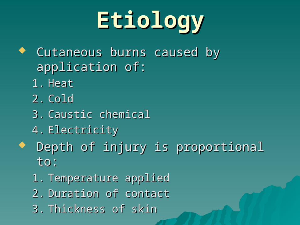
EtiologyEtiology Cutaneous burns caused by Cutaneous burns caused by
application of:application of:1.1. HeatHeat
2.2. ColdCold
3.3. Caustic chemicalCaustic chemical
4.4. ElectricityElectricity Depth of injury is proportional to:Depth of injury is proportional to:
1.1. Temperature appliedTemperature applied
2.2. Duration of contactDuration of contact
3.3. Thickness of skinThickness of skin

EtiologyEtiology1.1. Scald Burns:Scald Burns:
Caused by hot water (most common Caused by hot water (most common burn in civilian practice)burn in civilian practice)
140F (60C) ---> deep partial of full 140F (60C) ---> deep partial of full thickness burns in 3 secs.thickness burns in 3 secs.
Exposed areas of skin burned less than Exposed areas of skin burned less than clothed areasclothed areas
Children and elderly has thinner skin Children and elderly has thinner skin Immersion has longer contactImmersion has longer contact Scald burns from grease or hot oil are Scald burns from grease or hot oil are
deep burns (400F/200C).deep burns (400F/200C).
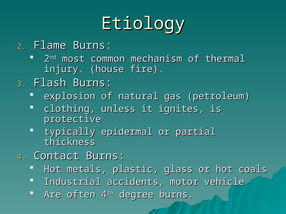
EtiologyEtiology2.2. Flame Burns:Flame Burns:
22ndnd most common mechanism of most common mechanism of thermal injury. (house fire).thermal injury. (house fire).
3.3. Flash Burns:Flash Burns: explosion of natural gas (petroleum)explosion of natural gas (petroleum) clothing, unless it ignites, is protectiveclothing, unless it ignites, is protective typically epidermal or partial thicknesstypically epidermal or partial thickness
4.4. Contact Burns:Contact Burns: Hot metals, plastic, glass or hot coalsHot metals, plastic, glass or hot coals Industrial accidents, motor vehicleIndustrial accidents, motor vehicle Are often 4Are often 4thth degree burns. degree burns.
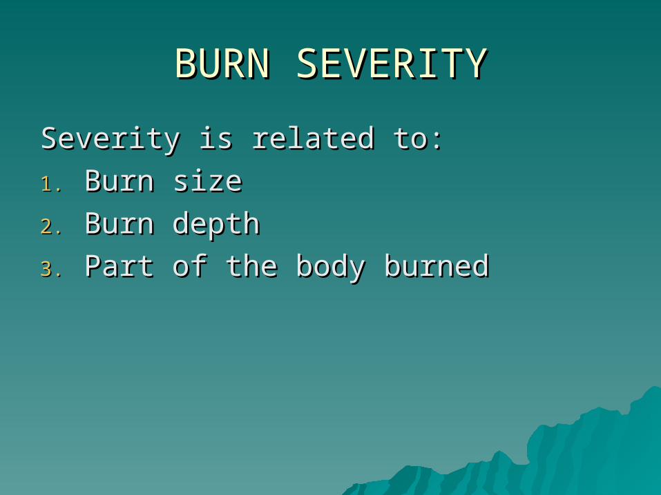
BURN SEVERITYBURN SEVERITY
Severity is related to:Severity is related to:
1.1. Burn sizeBurn size
2.2. Burn depthBurn depth
3.3. Part of the body burnedPart of the body burned
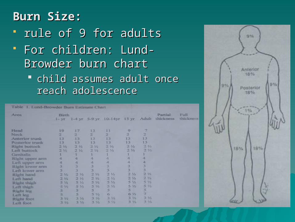
Burn Size:Burn Size: rule of 9 for adultsrule of 9 for adults For children: Lund-Browder For children: Lund-Browder
burn chartburn chart child assumes adult once child assumes adult once
reach adolescence reach adolescence
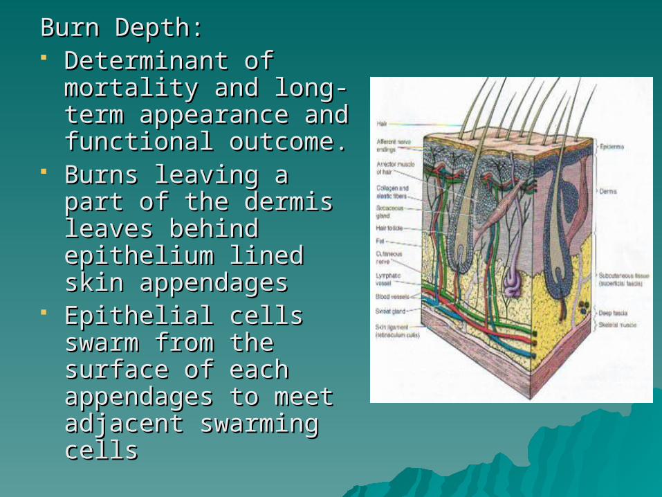
Burn Depth:Burn Depth: Determinant of Determinant of
mortality and long-mortality and long-term appearance and term appearance and functional outcome.functional outcome.
Burns leaving a part of Burns leaving a part of the dermis leaves the dermis leaves behind epithelium behind epithelium lined skin appendageslined skin appendages
Epithelial cells swarm Epithelial cells swarm from the surface of from the surface of each appendages to each appendages to meet adjacent meet adjacent swarming cellsswarming cells

The thickness of skin varies with age, The thickness of skin varies with age, sex and area of the body:sex and area of the body: Epidermis is thickest (0.5cm) on palm & Epidermis is thickest (0.5cm) on palm &
solesole Dermis varies from 1mm on the eyelids Dermis varies from 1mm on the eyelids
and genitalia to more than 5mm at the and genitalia to more than 5mm at the back.back.
Dermal atrophy >50y/o and skin Dermal atrophy >50y/o and skin appendages are less active.appendages are less active.
Burn depth is dependent on:Burn depth is dependent on:
1.1. Temperature of the burn sourceTemperature of the burn source
2.2. Thickness of the skinThickness of the skin
3.3. Duration of contactDuration of contact
4.4. Heat-dissipating capability of the Heat-dissipating capability of the skin (blood flow).skin (blood flow).

Classification of Burn According Classification of Burn According to Increasing Depthto Increasing Depth
A.A. Shallow Burns:Shallow Burns:1.1. Epidermal Burn: Epidermal Burn:
(1(1stst degree) degree) No blisterNo blister Erythema due to Erythema due to
dermal vasodilationdermal vasodilation Quite painfulQuite painful 44thth day injured day injured
epithelium epithelium desquamate – desquamate – peeling (sunburn)peeling (sunburn)

Classification of Burn According Classification of Burn According to Increasing Depthto Increasing Depth
A.A. Shallow Burns:Shallow Burns:2.2. Superficial Partial-thickness:Superficial Partial-thickness:
Second degreeSecond degree Includes upper layer of dermis Includes upper layer of dermis Characteristically forms Characteristically forms
blisters (fluid accumulation blisters (fluid accumulation between epidermis and between epidermis and dermis). dermis).
If blisters are removed the If blisters are removed the wound is pink, wet, painful and wound is pink, wet, painful and blanch w/ pressureblanch w/ pressure
Heal spontaneously < 3wks Heal spontaneously < 3wks w/o functional impairmentw/o functional impairment
Rarely cause hypertrophic Rarely cause hypertrophic scarring, but never completely scarring, but never completely match color of surrounding match color of surrounding normal skinnormal skin

Classification of Burn According Classification of Burn According to Increasing Depthto Increasing Depth
B.B. Deep Burns:Deep Burns:1.1. Deep Partial Thickness: (second Deep Partial Thickness: (second
degree)degree) Extends into the reticular layer of Extends into the reticular layer of
the dermisthe dermis w/ blisters but wound surface w/ blisters but wound surface
mottled pink and white colormottled pink and white color Complains of discomfort rather Complains of discomfort rather
than painthan pain Pressure applied --> capillary refill Pressure applied --> capillary refill
is slow or absent.is slow or absent. 22ndnd day wound is white and dry day wound is white and dry If not excised & grafted heals in 3 If not excised & grafted heals in 3
to 9 wks w/ scarring to 9 wks w/ scarring Joint function can be impairedJoint function can be impaired
Classification of Burn According Classification of Burn According to Increasing Depthto Increasing Depth
B.B. Deep Burns:Deep Burns:2.2. Full Thickness: (third degree)Full Thickness: (third degree)
All layers of the dermisAll layers of the dermis Appear white, cherry red or black and may or may not Appear white, cherry red or black and may or may not
have deep blistershave deep blisters Leathery, firm and depressed compared w/ adjoining Leathery, firm and depressed compared w/ adjoining
normal skinnormal skin InsensateInsensate Develop a classic burn eschar, an intact dead and Develop a classic burn eschar, an intact dead and
denatured dermis that separates after days or wks.denatured dermis that separates after days or wks. Heal only by wound contracture, epithelialization from Heal only by wound contracture, epithelialization from
the wound margin or skin graftingthe wound margin or skin grafting
Classification of Burn According Classification of Burn According to Increasing Depthto Increasing Depth
B.B. Deep Burns:Deep Burns:3.3. Fourth Degree:Fourth Degree:
Involves also the subcutaneous fat and Involves also the subcutaneous fat and deeper structuresdeeper structures
Almost always have charred appearanceAlmost always have charred appearance Electrical burns, contact burns, immersion Electrical burns, contact burns, immersion
burns and patients who are unconscious at burns and patients who are unconscious at time of burntime of burn

Those that will heal w/in 3wks are Those that will heal w/in 3wks are better treated by nonoperative better treated by nonoperative wound care:wound care: Shallow burnsShallow burns
State-of-the-art burn care involves State-of-the-art burn care involves early excision and grafting (E&G) early excision and grafting (E&G) of all burns that will not heal w/in of all burns that will not heal w/in 3wks:3wks: Deep burnsDeep burns

Assessment of Burn DepthAssessment of Burn Depth
Standard technique for determining burn Standard technique for determining burn depth: depth: clinical observation of the clinical observation of the woundwound
Other techniques to qualify burn depth:Other techniques to qualify burn depth:1.1. Ability to detect dead cells or denatured Ability to detect dead cells or denatured
collagen (biopsy, ultrasound, vital dyes)collagen (biopsy, ultrasound, vital dyes)2.2. Assessment of changes in blood flow Assessment of changes in blood flow
(fluorometry, laser doppler & thermography)(fluorometry, laser doppler & thermography)3.3. Analysis of the color of the wound (light Analysis of the color of the wound (light
reflectance methods)reflectance methods)4.4. Evaluation physical changes, such as edema Evaluation physical changes, such as edema
(MRI)(MRI)

Physiologic Response to Burn InjuryPhysiologic Response to Burn Injury Burn – inflammatory process Burn – inflammatory process
involving the entire organisminvolving the entire organism Systemic Inflammatory Response Systemic Inflammatory Response
Syndrome (SIRS):Syndrome (SIRS): Alterations of the metabolic, Alterations of the metabolic,
cardiovascular, gastrointestinal and cardiovascular, gastrointestinal and coagulation systemscoagulation systems
Resulting to hypermetabolism, Resulting to hypermetabolism, increased cellular, endothelial and increased cellular, endothelial and epithelial permeabilityepithelial permeability
Often extensive microthrombosis Often extensive microthrombosis

Physiologic Response to Burn InjuryPhysiologic Response to Burn Injury
Burn Shock:Burn Shock: Complex process of circulatory and Complex process of circulatory and
microcirculatory dysfunction that is not easily microcirculatory dysfunction that is not easily or fully repaired by fluid resuscitation.or fully repaired by fluid resuscitation.
Tissue trauma & hypovolemic shock releases Tissue trauma & hypovolemic shock releases local & systemic mediators ---> increase local & systemic mediators ---> increase vascular permeability and microvascular vascular permeability and microvascular hydrostatic pressure --->burn edema.hydrostatic pressure --->burn edema.
1.1. Histamine – causes increased vascular permeability Histamine – causes increased vascular permeability by disrupting venular endothelial tight junction--> by disrupting venular endothelial tight junction--> egress of fluids and proteinsegress of fluids and proteins Released by masts cells in burned skinReleased by masts cells in burned skin
2.2. Serotinin – increases vascular resistance amplifying Serotinin – increases vascular resistance amplifying vascular effects of NE, histamine, angiotensin II vascular effects of NE, histamine, angiotensin II
Physiologic Response to Burn InjuryPhysiologic Response to Burn Injury
Burn Shock:Burn Shock:3.3. Eicosanoids – vasoactive products of Eicosanoids – vasoactive products of
arachidonic acid metabolismarachidonic acid metabolism Released in burn tissue; produced edemaReleased in burn tissue; produced edema Increases the level of Prostaglandin causing Increases the level of Prostaglandin causing
vasodilatation leading to increase bld flow and vasodilatation leading to increase bld flow and hydrostatic pressure ---> edemahydrostatic pressure ---> edema
4.4. Kinins (bradykinins) – inc. premeabilityKinins (bradykinins) – inc. premeability
There is hypercoagulable and There is hypercoagulable and hyperfibrinolytic statehyperfibrinolytic state
Pathophysiology of Burn Shock:Pathophysiology of Burn Shock:A.A. Hypovolemic etiology:Hypovolemic etiology:
− Decreased cardiac outputDecreased cardiac output− Increased extracellular fluidsIncreased extracellular fluids− Decreased plasma volumeDecreased plasma volume− OliguriaOliguria
In burn shock, resuscitation is In burn shock, resuscitation is complicated by obligatory burn edemacomplicated by obligatory burn edema
Maximal edema formations occurs Maximal edema formations occurs between: - 8-12hrs in small burnsbetween: - 8-12hrs in small burns
- 12-24hrs in major - 12-24hrs in major burnsburns

Pathophysiology of Burn Pathophysiology of Burn ShockShock::
B.B. Changes in cellular level:Changes in cellular level:− >30% burn; cell transmembrane potential >30% burn; cell transmembrane potential
----> Na-K ATPase----> Na-K ATPase− Due to defective ATP metabolismDue to defective ATP metabolism

Physiologic Response to Burn InjuryPhysiologic Response to Burn Injury
Metabolic Response to Burn InjuryMetabolic Response to Burn InjuryA.A. Hypermetabolism:Hypermetabolism:
> 100% REE (1.3x BMR) due to:> 100% REE (1.3x BMR) due to:1.1. Increased heat loss from the burn woundsIncreased heat loss from the burn wounds
Due to increased blood flow and skin lossDue to increased blood flow and skin loss
2.2. Increased beta-adrenergic stimulationIncreased beta-adrenergic stimulation Increased catabolism of CHO, Lipids and Increased catabolism of CHO, Lipids and
CHONCHON
B.B. Neuroendocrine Response:Neuroendocrine Response: Catecholamine massively elevated and major Catecholamine massively elevated and major
endocrine mediator of hypermetabolism in endocrine mediator of hypermetabolism in burnburn
Giving propranolol – diminish REE and O2 Giving propranolol – diminish REE and O2 consumptionconsumption

Emergency CareEmergency Care
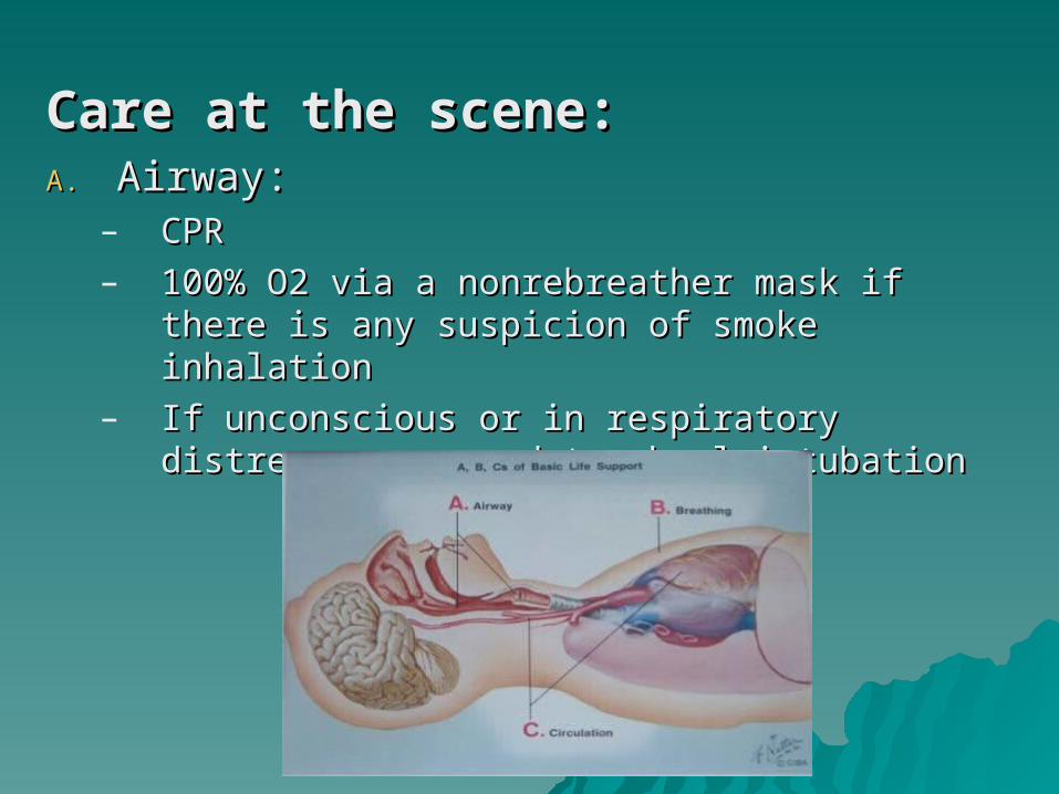
Care at the scene:Care at the scene:A.A. Airway:Airway:
– CPRCPR– 100% O2 via a nonrebreather mask if there is 100% O2 via a nonrebreather mask if there is
any suspicion of smoke inhalationany suspicion of smoke inhalation– If unconscious or in respiratory distress ----> If unconscious or in respiratory distress ---->
endotracheal intubationendotracheal intubation

Care at the scene:Care at the scene:B.B. Other injuries and transport:Other injuries and transport:
– assess other injuries; transport to assess other injuries; transport to nearest hospitalnearest hospital
– kept flat and warm and wrapped in kept flat and warm and wrapped in clean sheet and blanket, NPOclean sheet and blanket, NPO
– IVF = lactated Ringer’s sol. at rate of IVF = lactated Ringer’s sol. at rate of 1liter/hr. in case of severe burns1liter/hr. in case of severe burns
– Constricting clothing and jewelry shd Constricting clothing and jewelry shd be removed from burned parts due to be removed from burned parts due to local swelling beginslocal swelling begins
Care at the scene:Care at the scene:C.C. Cold Application:Cold Application:
– Cooling cannot reduce skin Cooling cannot reduce skin temperature enough to prevent further temperature enough to prevent further tissue damagetissue damage
– Cooling delays edema formation by Cooling delays edema formation by reducing initial thromboxane reducing initial thromboxane productionproduction
– Ice or cold water:Ice or cold water: Never be used for it can lead to systemic Never be used for it can lead to systemic
hypothermia and associated cutaneous hypothermia and associated cutaneous vasoconstriction can extend thermal vasoconstriction can extend thermal damagedamage

Emergency CareEmergency Care
Emergency Room Care:Emergency Room Care: Primary rule for emergency Primary rule for emergency
physician is to ignore the burnphysician is to ignore the burn ABC: Search for other life-ABC: Search for other life-
threatening injuriesthreatening injuries
Emergency Room Care:Emergency Room Care:1.1. Emergency assessment of inhalation Emergency assessment of inhalation
injury:injury:– Suspected in flame burns or anyone Suspected in flame burns or anyone
burned in an enclosed spaceburned in an enclosed space– Rescuers are the most important Rescuers are the most important
historians historians – Signs of potentially serious airway injurySigns of potentially serious airway injury
a.a. Hoarseness & expiratory wheezes Hoarseness & expiratory wheezes
b.b. Copious mucus production and carbonaceous Copious mucus production and carbonaceous sputumsputum
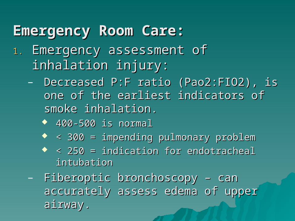
Emergency Room Care:Emergency Room Care:1.1. Emergency assessment of inhalation Emergency assessment of inhalation
injury:injury:– Decreased P:F ratio (Pao2:FIO2), is one of Decreased P:F ratio (Pao2:FIO2), is one of
the earliest indicators of smoke the earliest indicators of smoke inhalation.inhalation.
400-500 is normal400-500 is normal < 300 = impending pulmonary problem< 300 = impending pulmonary problem < 250 = indication for endotracheal < 250 = indication for endotracheal
intubationintubation
– Fiberoptic bronchoscopy – can accurately Fiberoptic bronchoscopy – can accurately assess edema of upper airway.assess edema of upper airway.

Emergency Room Care:Emergency Room Care:2.2. Fluid Resuscitation:Fluid Resuscitation:
– IV LR 1L/hr in adultIV LR 1L/hr in adult
20ml/kg/hr in children20ml/kg/hr in children– Foley catheter – UO/hr.:Foley catheter – UO/hr.:
30ml/hr in adults30ml/hr in adults 1ml/kg/hr in children1ml/kg/hr in children
– After ascertaining the extent of the burn After ascertaining the extent of the burn estimate the fluid needs:estimate the fluid needs:
Parkland formula: 4ml/kg/%burn (LR) Parkland formula: 4ml/kg/%burn (LR) Modified Brooke: 2ml/kg/%burn (LR) Modified Brooke: 2ml/kg/%burn (LR)
– <50% TBSA <50% TBSA use 2 large bore IV line use 2 large bore IV line >50% or other medical problems >50% or other medical problems CVP CVP
>65% TBSA--> burn center >65% TBSA--> burn center – Use upper extremities as portals for IV Use upper extremities as portals for IV

Other formulas estimating adult Other formulas estimating adult burn pt resuscitation fluid needs:burn pt resuscitation fluid needs:
Use colloid formulas:Use colloid formulas:1.1. Evans:Evans:
– NSS NSS 1ml/kg/%burn1ml/kg/%burn
– 1ml/kg/%burn1ml/kg/%burn– 2000ml D5W2000ml D5W
2.2. Brooke:Brooke:– LR LR
1.5ml/kg/%burn1.5ml/kg/%burn– 0.5ml/kg0.5ml/kg– 2000ml D5W2000ml D5W
3.3. Slater:Slater:– LR 2L/24hLR 2L/24h– Fresh frozen Fresh frozen
plasma plasma 75ml/kg/24hr75ml/kg/24hr
Colloid generates Colloid generates inward oncotic inward oncotic force to force to counteracts counteracts outward outward intravascular intravascular hydrostatic force.hydrostatic force.

Emergency Room Care:Emergency Room Care:3.3. Tetanus prophylaxis: active/passiveTetanus prophylaxis: active/passive
4.4. Gastric decompression: NGTGastric decompression: NGT– enteral feeding ---> reduce Curling enteral feeding ---> reduce Curling
ulcers, prevents ileus and blunt ulcers, prevents ileus and blunt catabolism catabolism
5.5. Pain control: IV opiatesPain control: IV opiates
6.6. Psychosocial carePsychosocial care

Care of Burn Wound:Care of Burn Wound:– In transferring a pt, wound is minimally In transferring a pt, wound is minimally
dressed in gauze; pulses distal to dressed in gauze; pulses distal to circumferential deep burns should be circumferential deep burns should be monitoredmonitored
– Escharotomy:Escharotomy: Edema formation--> vascular Edema formation--> vascular
compromise---->permanent neuromuscular compromise---->permanent neuromuscular & vascular deficits.& vascular deficits.– Skin color, sensation, capillary refill and Skin color, sensation, capillary refill and
peripheral pulses assessed hourlyperipheral pulses assessed hourly S/Sx warrants escharotomy:S/Sx warrants escharotomy:
1.1. CyanosisCyanosis2.2. Deep tissue painDeep tissue pain3.3. Progressive paresthesiaProgressive paresthesia4.4. Progressive decrease or absence of pulsesProgressive decrease or absence of pulses5.5. Sensation of cold extremitiesSensation of cold extremities

Respiratory distress Respiratory distress due to deep due to deep circumferential burn circumferential burn wound of the thoraxwound of the thorax
Local anesthesia is not Local anesthesia is not needed; but IV opiates needed; but IV opiates or anxiolytics is given.or anxiolytics is given.

INHALATION INHALATION INJURYINJURY
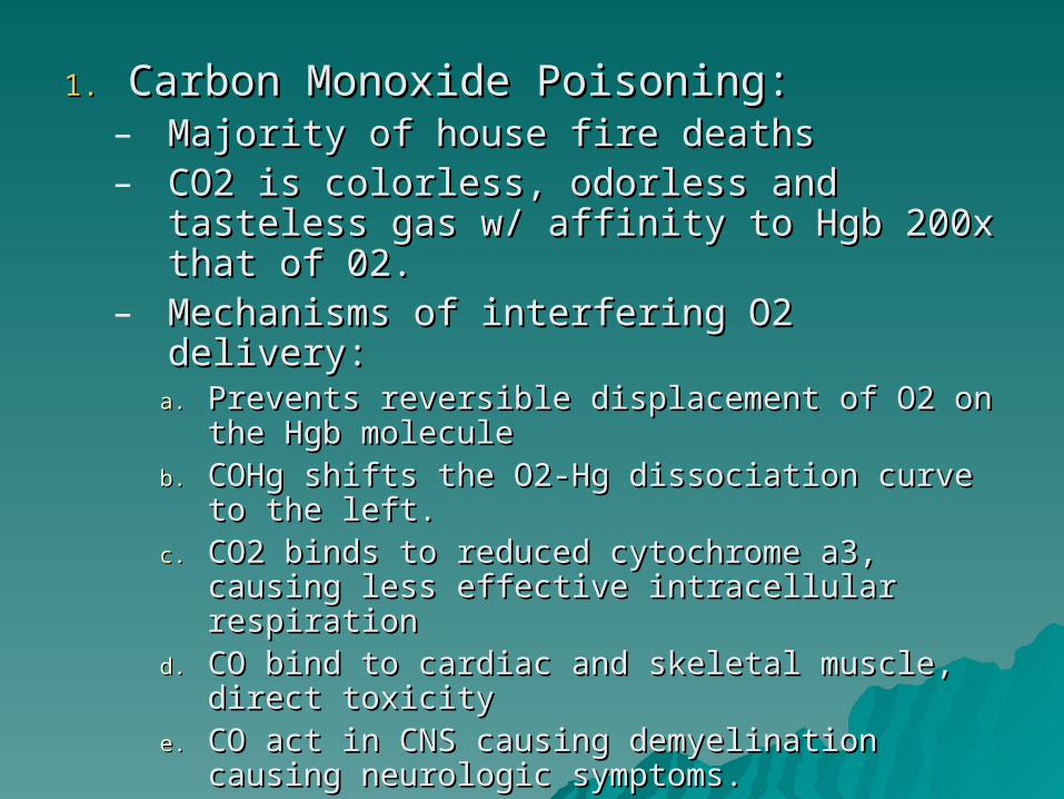
1.1. Carbon Monoxide Poisoning:Carbon Monoxide Poisoning:– Majority of house fire deathsMajority of house fire deaths– CO2 is colorless, odorless and tasteless CO2 is colorless, odorless and tasteless
gas w/ affinity to Hgb 200x that of 02.gas w/ affinity to Hgb 200x that of 02.– Mechanisms of interfering O2 delivery:Mechanisms of interfering O2 delivery:
a.a. Prevents reversible displacement of O2 on Prevents reversible displacement of O2 on the Hgb moleculethe Hgb molecule
b.b. COHg shifts the O2-Hg dissociation curve COHg shifts the O2-Hg dissociation curve to the left.to the left.
c.c. CO2 binds to reduced cytochrome a3, CO2 binds to reduced cytochrome a3, causing less effective intracellular causing less effective intracellular respirationrespiration
d.d. CO bind to cardiac and skeletal muscle, CO bind to cardiac and skeletal muscle, direct toxicitydirect toxicity
e.e. CO act in CNS causing demyelination CO act in CNS causing demyelination causing neurologic symptoms.causing neurologic symptoms.

1.1. Carbon Monoxide Poisoning:Carbon Monoxide Poisoning:– Symptoms:Symptoms:
a.a. Headache, N/V, loss of manual dexterityHeadache, N/V, loss of manual dexterity
b.b. Weak, confused and lethargicWeak, confused and lethargic
c.c. ComaComa
− CO reversibly bound to the heme:CO reversibly bound to the heme:− Room temp = half-life (tRoom temp = half-life (t1/21/2) of COHb is 4hrs) of COHb is 4hrs− 100% O2 = t100% O2 = t1/21/2 is reduced to 45-60mins is reduced to 45-60mins− Hyperbaric O2 at 2atm. = tHyperbaric O2 at 2atm. = t1/21/2 30mins 30mins− Hyperbaric )2 at 3atm. = tHyperbaric )2 at 3atm. = t1/21/2 15-20min 15-20min
− Tx: 100% oxygen via a nonbreather face Tx: 100% oxygen via a nonbreather face maskmask
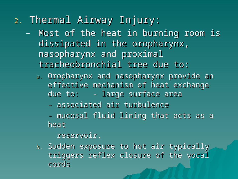
2.2. Thermal Airway Injury:Thermal Airway Injury:– Most of the heat in burning room is Most of the heat in burning room is
dissipated in the oropharynx, dissipated in the oropharynx, nasopharynx and proximal nasopharynx and proximal tracheobronchial tree due to:tracheobronchial tree due to:
a.a. Oropharynx and nasopharynx provide an Oropharynx and nasopharynx provide an effective mechanism of heat exchange due effective mechanism of heat exchange due to:to: - large surface area- large surface area
- associated air turbulence- associated air turbulence
- mucosal fluid lining that acts - mucosal fluid lining that acts as a heat as a heat
reservoir.reservoir.
b.b. Sudden exposure to hot air typically Sudden exposure to hot air typically triggers reflex closure of the vocal cordstriggers reflex closure of the vocal cords
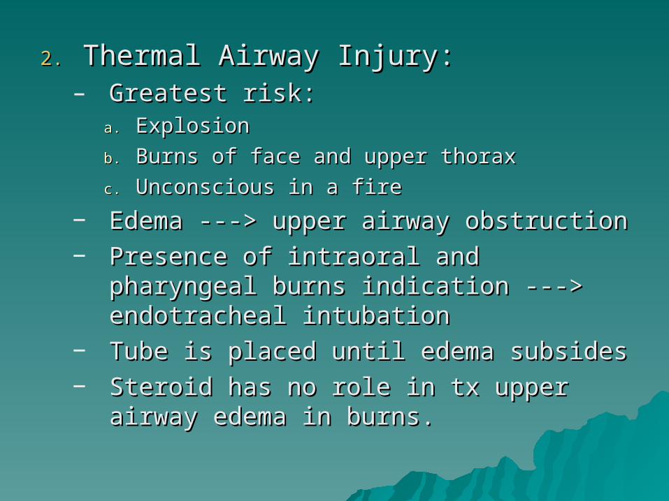
2.2. Thermal Airway Injury:Thermal Airway Injury:– Greatest risk:Greatest risk:
a.a. ExplosionExplosion
b.b. Burns of face and upper thoraxBurns of face and upper thorax
c.c. Unconscious in a fireUnconscious in a fire
− Edema ---> upper airway obstructionEdema ---> upper airway obstruction− Presence of intraoral and pharyngeal Presence of intraoral and pharyngeal
burns indication ---> endotracheal burns indication ---> endotracheal intubationintubation
− Tube is placed until edema subsidesTube is placed until edema subsides− Steroid has no role in tx upper airway Steroid has no role in tx upper airway
edema in burns.edema in burns.

3.3. Smoke Inhalation:Smoke Inhalation:– Product of combustion (hydrogen cyanide)Product of combustion (hydrogen cyanide)
Wheezing and air hunger early manifestations Wheezing and air hunger early manifestations of inhalation injury:of inhalation injury:
Classical findings in pulmonary function test:Classical findings in pulmonary function test:1.1. Decreased functional residual capacityDecreased functional residual capacity2.2. Decreased vital capacityDecreased vital capacity3.3. Evidence of obstructive disease w/ reduction in flow Evidence of obstructive disease w/ reduction in flow
ratesrates4.4. Increase in dead spaceIncrease in dead space5.5. Rapid decrease in complianceRapid decrease in compliance
Concomitant cutaneous burn releases Concomitant cutaneous burn releases mediators (Prostaglandin, thromboxane, and mediators (Prostaglandin, thromboxane, and reactive O2 intermediates) that can aggravate reactive O2 intermediates) that can aggravate pulmonary injry.pulmonary injry.
w/o associated cutaneous injury, mortality from w/o associated cutaneous injury, mortality from isolated inhalation injury is quite low.isolated inhalation injury is quite low.
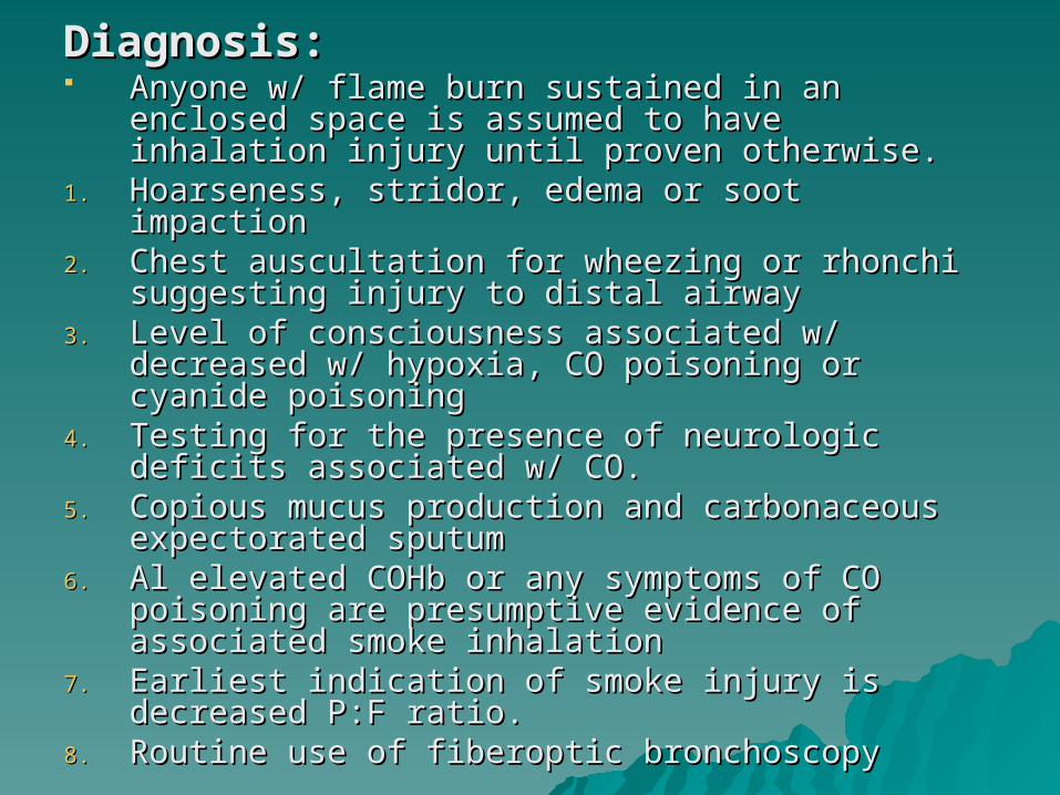
Diagnosis:Diagnosis: Anyone w/ flame burn sustained in an enclosed Anyone w/ flame burn sustained in an enclosed
space is assumed to have inhalation injury until space is assumed to have inhalation injury until proven otherwise.proven otherwise.
1.1. Hoarseness, stridor, edema or soot impactionHoarseness, stridor, edema or soot impaction2.2. Chest auscultation for wheezing or rhonchi Chest auscultation for wheezing or rhonchi
suggesting injury to distal airwaysuggesting injury to distal airway3.3. Level of consciousness associated w/ decreased Level of consciousness associated w/ decreased
w/ hypoxia, CO poisoning or cyanide poisoningw/ hypoxia, CO poisoning or cyanide poisoning4.4. Testing for the presence of neurologic deficits Testing for the presence of neurologic deficits
associated w/ CO. associated w/ CO. 5.5. Copious mucus production and carbonaceous Copious mucus production and carbonaceous
expectorated sputumexpectorated sputum6.6. Al elevated COHb or any symptoms of CO Al elevated COHb or any symptoms of CO
poisoning are presumptive evidence of poisoning are presumptive evidence of associated smoke inhalationassociated smoke inhalation
7.7. Earliest indication of smoke injury is decreased Earliest indication of smoke injury is decreased P:F ratio.P:F ratio.
8.8. Routine use of fiberoptic bronchoscopyRoutine use of fiberoptic bronchoscopy
Treatment:Treatment:
A.A. Upper Airway:Upper Airway:– Rapid endotracheal intubation due to Rapid endotracheal intubation due to
oropharyngeal/laryngeal edema.oropharyngeal/laryngeal edema.– Ability of adult pt to breathe around Ability of adult pt to breathe around
the tube w/ the cuff deflated is an the tube w/ the cuff deflated is an indication for tube removal.indication for tube removal.
– Tx of postextubation stridor:Tx of postextubation stridor: Administration of nebulized racemic Administration of nebulized racemic
epinephrine and helium-oxygen mixturesepinephrine and helium-oxygen mixtures Steroids are never used.Steroids are never used.

Treatment:Treatment:
B.B. Lower Airway and alveoli:Lower Airway and alveoli:– Overall treatment for smoke inhalation Overall treatment for smoke inhalation
is supportive w/ the goal being is supportive w/ the goal being maintenance of adequate oxygenation maintenance of adequate oxygenation and ventilation until the lungs heal.and ventilation until the lungs heal.
– Mild cases are treated w/ humidified Mild cases are treated w/ humidified O2, vigorous pulmonary toilet, and O2, vigorous pulmonary toilet, and bronchodilators as needed.bronchodilators as needed.
– If w/ atelectasis, positive end-If w/ atelectasis, positive end-expiratory pressure is useful to expiratory pressure is useful to increase FRC, increasing oxygenation.increase FRC, increasing oxygenation.

Treatment:Treatment:B.B. Lower Airway and alveoli:Lower Airway and alveoli:
– High frequency percussive ventilation (HFPV) High frequency percussive ventilation (HFPV) provide superior oxygenation at a lower Fprovide superior oxygenation at a lower FIO2IO2, , w/ lower airway pressures than conventional w/ lower airway pressures than conventional mechanical ventilation. This enhanced mechanical ventilation. This enhanced clearance of bronchial secretionsclearance of bronchial secretions
– Cricothyroidotomy for danger imminent Cricothyroidotomy for danger imminent obstruction and unsuccessful endotracheal obstruction and unsuccessful endotracheal intubation. This is later converted to intubation. This is later converted to tracheostomytracheostomy
– Prophylactic antibiotics are indicated w/ Prophylactic antibiotics are indicated w/ inhhalation injury, which is a chemical inhhalation injury, which is a chemical pneumonitis.pneumonitis.
– Steroids are contraindicated due to septic Steroids are contraindicated due to septic complications and higher mortality ratecomplications and higher mortality rate

WOUND WOUND MANAGEMENTMANAGEMENT
Shallow burns healed w/in 3 wks while Shallow burns healed w/in 3 wks while deep burns healed over many weeks if deep burns healed over many weeks if infection was preventedinfection was prevented
For deeper wounds rather waiting for For deeper wounds rather waiting for spontaneous separation, the eschar is spontaneous separation, the eschar is surgically removed and closed via grafting surgically removed and closed via grafting techniques and/or immediate flap techniques and/or immediate flap procedures (E&G)procedures (E&G)
Early E&G has reduced burn mortality Early E&G has reduced burn mortality more than any other intervention. It more than any other intervention. It decreases the number of painful decreases the number of painful debridements required. debridements required. Immunosuppression and hypermetabolism Immunosuppression and hypermetabolism associated w/ burns is amelioratedassociated w/ burns is ameliorated
NOTE:NOTE:
His notes only cover up to page 204 His notes only cover up to page 204 in schwartz. in schwartz.
READ!!! Pg. 205-217READ!!! Pg. 205-217 Good luckGood luck