canadian diabetes clinical guidelines 2008
DESCRIPTION
GUIA DIABETES CANADIENSE 2008TRANSCRIPT
A Publication of the Professional Sections of the Canadian Diabetes Association
Pu
blic
atio
n M
ail A
gre
emen
t #4
0063
447
Can
ada
Po
st: P
leas
e re
turn
un
del
iver
able
ad
dre
ss b
lock
s to
Can
adia
n D
iab
etes
Ass
oci
atio
n,
522
Un
iver
sity
Ave
nu
e, S
uit
e 14
00, T
oro
nto
ON
M5G
2R
5
Canadian Journal of Diabetes
September 2008 | Volume 32 | Supplement 1
Canadian Diabetes Association 2008 Clinical Practice Guidelines for the Prevention and Management of Diabetes in Canada


A Publication of the Professional Sections of the Canadian Diabetes Association
Canadian Journal of Diabetes
September 2008 | Volume 32 | Supplement 1
Canadian Diabetes Association 2008 Clinical Practice Guidelines for the Prevention and Management of Diabetes in Canada
ii
2008 CLINICAL PRACTICE GUIDELINES
TABLE OF CONTENTS
Canadian Diabetes Association 2008 Clinical Practice Guidelines for the Preventionand Management of Diabetes in Canada
Notes to Readers. . . . . . . . . . . . . . . . . . . . . . . . . . . . . . . . . . . . . . . . . . . . . . . . . . . . . . . . . . . . . . . . . . . iv
2008 Clinical Practice Guidelines Committees . . . . . . . . . . . . . . . . . . . . . . . . . . . . . . . . . . . . . . . . . . . . v
Acknowledgements. . . . . . . . . . . . . . . . . . . . . . . . . . . . . . . . . . . . . . . . . . . . . . . . . . . . . . . . . . . . . . . . . x
Introduction . . . . . . . . . . . . . . . . . . . . . . . . . . . . . . . . . . . . . . . . . . . . . . . . . . . . . . . . . . . . . . . . . . . . . S1
Methods . . . . . . . . . . . . . . . . . . . . . . . . . . . . . . . . . . . . . . . . . . . . . . . . . . . . . . . . . . . . . . . . . . . . . . . . . S5
Definition, Classification and Diagnosis of Diabetes and Other Dysglycemic Categories . . . . . . . . S10
Screening for Type 1 and Type 2 Diabetes . . . . . . . . . . . . . . . . . . . . . . . . . . . . . . . . . . . . . . . . . . . . . . S14
Prevention of Diabetes . . . . . . . . . . . . . . . . . . . . . . . . . . . . . . . . . . . . . . . . . . . . . . . . . . . . . . . . . . . . S17
Management
Organization of Diabetes Care . . . . . . . . . . . . . . . . . . . . . . . . . . . . . . . . . . . . . . . . . . . . . . . . . . . . . . . . . S20
Self-management Education. . . . . . . . . . . . . . . . . . . . . . . . . . . . . . . . . . . . . . . . . . . . . . . . . . . . . . . . . . . S25
Targets for Glycemic Control . . . . . . . . . . . . . . . . . . . . . . . . . . . . . . . . . . . . . . . . . . . . . . . . . . . . . . . . . . S29
Monitoring Glycemic Control . . . . . . . . . . . . . . . . . . . . . . . . . . . . . . . . . . . . . . . . . . . . . . . . . . . . . . . . . . S32
Physical Activity and Diabetes . . . . . . . . . . . . . . . . . . . . . . . . . . . . . . . . . . . . . . . . . . . . . . . . . . . . . . . . . S37
Nutrition Therapy. . . . . . . . . . . . . . . . . . . . . . . . . . . . . . . . . . . . . . . . . . . . . . . . . . . . . . . . . . . . . . . . . S40
Insulin Therapy in Type 1 Diabetes. . . . . . . . . . . . . . . . . . . . . . . . . . . . . . . . . . . . . . . . . . . . . . . . . . . . . . . S46
Pharmacologic Management of Type 2 Diabetes . . . . . . . . . . . . . . . . . . . . . . . . . . . . . . . . . . . . . . . . . . . . . . . S53
Hypoglycemia . . . . . . . . . . . . . . . . . . . . . . . . . . . . . . . . . . . . . . . . . . . . . . . . . . . . . . . . . . . . . . . . . . . S62
Hyperglycemic Emergencies in Adults . . . . . . . . . . . . . . . . . . . . . . . . . . . . . . . . . . . . . . . . . . . . . . . . . . . . . S65
In-hospital Management of Diabetes . . . . . . . . . . . . . . . . . . . . . . . . . . . . . . . . . . . . . . . . . . . . . . . . . . . . . S71
Management of Obesity in Diabetes . . . . . . . . . . . . . . . . . . . . . . . . . . . . . . . . . . . . . . . . . . . . . . . . . . . . . . S77
Psychological Aspects of Diabetes. . . . . . . . . . . . . . . . . . . . . . . . . . . . . . . . . . . . . . . . . . . . . . . . . . . . . . . . S82
Influenza and Pneumococcal Immunization . . . . . . . . . . . . . . . . . . . . . . . . . . . . . . . . . . . . . . . . . . . . . . . . . S86
Pancreas and Islet Transplantation . . . . . . . . . . . . . . . . . . . . . . . . . . . . . . . . . . . . . . . . . . . . . . . . . . . . . . . S88
Complementary and Alternative Medicine in the Management of Diabetes . . . . . . . . . . . . . . . . . . . . . . . . . . . . . . S91
Macrovascular and Microvascular Complications
Identification of Individuals at High Risk of Coronary Events . . . . . . . . . . . . . . . . . . . . . . . . . . . . . . . . . . . . . . S95
Screening for the Presence of Coronary Artery Disease . . . . . . . . . . . . . . . . . . . . . . . . . . . . . . . . . . . . . . . . . . . S99
Vascular Protection in People With Diabetes . . . . . . . . . . . . . . . . . . . . . . . . . . . . . . . . . . . . . . . . . . . . . . . . S102
Dyslipidemia . . . . . . . . . . . . . . . . . . . . . . . . . . . . . . . . . . . . . . . . . . . . . . . . . . . . . . . . . . . . . . . . . . . S107
Treatment of Hypertension. . . . . . . . . . . . . . . . . . . . . . . . . . . . . . . . . . . . . . . . . . . . . . . . . . . . . . . . . . . S115
Management of Acute Coronary Syndromes. . . . . . . . . . . . . . . . . . . . . . . . . . . . . . . . . . . . . . . . . . . . . . . . . S119

iii
Treatment of Diabetes in People With Heart Failure . . . . . . . . . . . . . . . . . . . . . . . . . . . . . . . . . . . . . . . . . . . S123
Chronic Kidney Disease in Diabetes . . . . . . . . . . . . . . . . . . . . . . . . . . . . . . . . . . . . . . . . . . . . . . . . . . . . . S126
Retinopathy. . . . . . . . . . . . . . . . . . . . . . . . . . . . . . . . . . . . . . . . . . . . . . . . . . . . . . . . . . . . . . . . . . . . S134
Neuropathy . . . . . . . . . . . . . . . . . . . . . . . . . . . . . . . . . . . . . . . . . . . . . . . . . . . . . . . . . . . . . . . . . . . . S140
Foot Care . . . . . . . . . . . . . . . . . . . . . . . . . . . . . . . . . . . . . . . . . . . . . . . . . . . . . . . . . . . . . . . . . . . . . S143
Erectile Dysfunction. . . . . . . . . . . . . . . . . . . . . . . . . . . . . . . . . . . . . . . . . . . . . . . . . . . . . . . . . . . . . . . S147
Diabetes in Children
Type 1 Diabetes in Children and Adolescents. . . . . . . . . . . . . . . . . . . . . . . . . . . . . . . . . . . . . . . . . . . . . . . . S150
Type 2 Diabetes in Children and Adolescents. . . . . . . . . . . . . . . . . . . . . . . . . . . . . . . . . . . . . . . . . . . . . . . . S162
Diabetes in Special Populations
Diabetes and Pregnancy . . . . . . . . . . . . . . . . . . . . . . . . . . . . . . . . . . . . . . . . . . . . . . . . . . . . . . . . . . . . S168
Diabetes in the Elderly . . . . . . . . . . . . . . . . . . . . . . . . . . . . . . . . . . . . . . . . . . . . . . . . . . . . . . . . . . . . . S181
Type 2 Diabetes in Aboriginal Peoples . . . . . . . . . . . . . . . . . . . . . . . . . . . . . . . . . . . . . . . . . . . . . . . . . . . . S187
Type 2 Diabetes in High-risk Ethnic Populations . . . . . . . . . . . . . . . . . . . . . . . . . . . . . . . . . . . . . . . . . . . . . S191
Appendices
Appendix 1. Etiologic Classification of Diabetes Mellitus . . . . . . . . . . . . . . . . . . . . . . . . . . . . . . . . . . . . . . . . S194
Appendix 2. Sample Diabetes Patient Care Flow Sheet for Adults . . . . . . . . . . . . . . . . . . . . . . . . . . . . . . . . . . . S195
Appendix 3. Examples of Insulin Initiation and Titration Regimens in People With Type 2 Diabetes . . . . . . . . . . . . . . S197
Appendix 4. Rapid Screening for Diabetic Neuropathy . . . . . . . . . . . . . . . . . . . . . . . . . . . . . . . . . . . . . . . . . S199
Appendix 5. Diabetes and Foot Care:A Patient’s Checklist. . . . . . . . . . . . . . . . . . . . . . . . . . . . . . . . . . . . . . . . S200
Appendix 6. Diabetic Foot Ulcers: Essentials of Management . . . . . . . . . . . . . . . . . . . . . . . . . . . . . . . . . . . . . . S201
iv
2008 CLINICAL PRACTICE GUIDELINES
NOTES TO READERS
OverviewThe Canadian Diabetes Association 2008 Clinical Practice Guidelines for the Prevention and Management of Diabetes inCanada are intended to guide practice and are not intended to serve as a comprehensive text of diabetes management,nor are they intended to set criteria for research protocols.These guidelines are intended to inform general patterns ofcare.These guidelines are also intended to enhance diabetes prevention efforts in Canada and to reduce the burden ofdiabetes complications in people living with this disease.
As per the Canadian Medical Association Handbook on Clinical Practice Guidelines (Davis D, et al. Ottawa, ON: CanadianMedical Association; 2007), guidelines should not be used as a legal resource in malpractice cases as “their more generalnature renders them insensitive to the particular circumstances of the individual cases.” Healthcare professionals mustconsider the needs, values and preferences of individual patients, use clinical judgement, and work with available humanand healthcare service resources in their settings.These guidelines were developed using the best available evidence. It isincumbent upon healthcare professionals to stay current in this rapidly changing field.
Unless otherwise specified, these guidelines pertain to the care of adults with diabetes.Two chapters – “Type 1 Diabetesin Children and Adolescents” and “Type 2 Diabetes in Children and Adolescents” – are included to highlight aspects ofcare that must be tailored to the pediatric population.
Suggested CitationCanadian Diabetes Association Clinical Practice Guidelines Expert Committee. Canadian Diabetes Association 2008 clinical practice guidelines for the prevention and management of diabetes in Canada. Can J Diabetes. 2008;32(suppl 1):S1-S201.
Reproduction of the GuidelinesReproduction of the Canadian Diabetes Association 2008 Clinical Practice Guidelines for the Prevention andManagement of Diabetes in Canada in whole or in part is prohibited without written consent of the publisher.
Extra CopiesCopies of this document may be ordered, for a nominal fee, from the Canadian Diabetes Association. Please dial 1-800-BANTING or visit orders.diabetes.ca.
WebsiteThese guidelines are available at http://www.diabetes.ca.

Lori D. Berard RN CDEAdvisor Nurse Manager, Health Sciences Centre, Diabetes Research
Group,Winnipeg, Manitoba
Gillian Booth MD MSc FRCPCSub-group Chair, MethodsDivision of Endocrinology and Metabolism and Li Ka Shing
Institute, St. Michael’s Hospital and University ofToronto,Toronto, Ontario
Alice Y.Y. Cheng MD FRCPCAdvisorDivision of Endocrinology and Metabolism,
St. Michael’s Hospital and Credit Valley Hospital,University of Toronto,Toronto, Ontario
Maureen Clement MD CCFPAdvisorMedical Director, Diabetes Education,
Vernon Jubilee Hospital; Assistant Clinical Professor,University of British Columbia,Vernon, British Columbia
Keith Dawson MD PhD FRCPCAdvisorProfessor of Medicine (Emeritus), University
of British Columbia,Vancouver, British Columbia
Amir Hanna MB BCh FRCPCSub-group Chair, ManagementProfessor Emeritus, Department of Medicine, University of
Toronto, and St. Michael’s Hospital,Toronto, Ontario
William Harper MD FRCPCAdvisorAssociate Professor of Medicine, McMaster University,
Hamilton, Ontario
2008 Clinical Practice Guidelines CommitteesThe Canadian Diabetes Association 2008 Clinical Practice Guidelines for the Prevention and Management ofDiabetes in Canada were developed under the auspices of the Clinical & Scientific Section of the Canadian DiabetesAssociation.The following committee members contributed to these guidelines. Committee members were volunteers andreceived no remuneration or honoraria for their participation.
Sarah Capes MD MSc FRCPCStaff,Vancouver Island Health Authority,Victoria,
British Columbia
Karie Quinn RD CDEGrand Prairie, Alberta
Vincent Woo MD FRCPC (Chair)Health Sciences Centre, University of Manitoba,
Winnipeg, Manitoba
Executive Committee
Steering Committee
Stewart B. Harris MD MPH FCFP FACPMAdvisorProfessor of Medicine, Department of Family Medicine,
University of Western Ontario, London, Ontario
Robyn Houlden MD FRCPCSub-group Chair, ManagementProfessor, Division of Endocrinology,
Queen’s University, Kingston, Ontario
Dereck Hunt MD MSc FRCPCMethodsAssociate Professor, McMaster University,
Hamilton, Ontario
Helen Jones RN MSN CDEAdvisorClinical Nurse Specialist/Manager, Leadership
Sinai Centre for Diabetes, Mount Sinai Hospital and University of Toronto,Toronto, Ontario
Margaret L. Lawson MSc MD FRCPCSub-group Chair, Diabetes in Children and AdolescentsAssociate Professor of Pediatrics, University of Ottawa;
Division of Endocrinology and Metabolism, Children’sHospital of Eastern Ontario, Ottawa, Ontario
v

Lawrence A. Leiter MD FRCPC FACPSub-group Chair, Macrovascular ComplicationsProfessor of Medicine and Nutritional Sciences, University
of Toronto; Head, Division of Endocrinology andMetabolism, St. Michael’s Hospital,Toronto, Ontario
David M.Thompson MD FRCPCSub-group Chair, Diabetes and Pregnancy
Diabetes in Pregnancy Clinic, BC Women’s Hospital,Vancouver, British Columbia
Filiberto Altomare MD FRCSCDepartment of Ophthalmology and Vision Sciences,
St. Michael’s Hospital, University of Toronto,Toronto, Ontario
J. Malcolm O. Arnold MD FRCPC FRCP FACCProfessor of Medicine, Physiology, Pharmacology,
University of Western Ontario, London Health SciencesCentre, London, Ontario
Nicole Aylward RD CDEWinnipeg, Manitoba
Richard Bebb MD ABIM FRCPCClinical Assistant Professor, University of British Columbia,
Vancouver, British Columbia
Ian Blumer MD FRCPCSub-group Chair, Dissemination CommitteeCharles H. Best Diabetes Centre, Ajax, Ontario
Keith Bowering MD FRCPC FACPClinical Professor of Medicine, Division of Endocrinology,
University of Alberta, Edmonton, Alberta
Shelley R. Boyd MD FRCSC DABOClinician-Scientist, Assistant Professor, Department of
Ophthalmology & Vision Sciences, St. Michael’s Hospital,University of Toronto,Toronto, Ontario
Sharon Brez RN BScN MA(Ed) CDEAdvanced Practice Nurse, Endocrinology and Metabolism,
The Ottawa Hospital, Ottawa, Ontario
Vera Bril MD FRCPCProfessor of Medicine, Head, Division of Neurology,
University Health Network / Mount Sinai Hospital,University Health Network, University of Toronto,Toronto, Ontario
Expert Committee
Ehud Ur MB FRCPSub-group Chair, Definition, Classification,Diagnosis of Diabetes and Other Dysglycemic CategoriesProfessor of Medicine, University of British Columbia,
Vancouver, British Columbia
Jean-François Yale MD CSPQSub-group Chair, Microvascular ComplicationsProfessor of Medicine, McGill University, Montreal,
Quebec
Gerald Brock MD FRCPCProfessor of Surgery, University of Western Ontario,
London, Ontario
Jean-Louis Chiasson MDProfessor of Medicine, Université de Montréal,
Montreal, Quebec
Bruce Culleton MD FRCPCClinical Associate Professor, Foothills Hospital,
Calgary, Alberta
David Dannenbaum MD CCFPMedical Advisor, Chronic Disease Prevention,
Cree Board of Health and Social Services of James Bay,Montreal, Quebec
Jean-Pierre Després PhDDirector of Research, Cardiology,
Hôpital Laval Research Centre, Quebec, Quebec
Denis Drouin MDConsultant in Public Health, Clinical Professor of Family
Medicine, Laval University Faculty of Medicine, AssociateDirector of the Continuing Professional DevelopmentCentre, Quebec, Quebec
Peggy Dunbar MEd PDt CDECoordinator, Diabetes Care Program of Nova Scotia,
Halifax, Nova Scotia
Alun Edwards MB MRCP(UK) FRCPCAssociate Professor and Head, Division of Endocrinology
and Metabolism, University of Calgary and CalgaryHealth Region, Calgary, Alberta
Jean-Marie Ekoé MD CSPQProfessor of Medicine, Endocrinology, Metabolism,
Nutrition, CHUM, Hôtel-Dieu Hospital,Montreal, Quebec
vi
2008 CLINICAL PRACTICE GUIDELINES

Denice Feig MD MSc FRCPCAssociate Professor, University of Toronto, Staff
Endocrinologist, Mount Sinai Hospital,Toronto, Ontario
David Fitchett MD FRCPCAssociate Professor of Medicine, University of Toronto,
Toronto, Ontario
Jacques Genest MD FRCPCMontreal, Quebec
Jeannette Goguen MD FRCPCAssistant Professor, University of Toronto, St. Michael’s
Hospital,Toronto, Ontario
Réjeanne Gougeon PhDAssociate Professor, McGill Nutrition and Food Science
Centre, Montreal, Quebec
Peter Hall PhDAssistant Professor, University of Waterloo,Waterloo,
Ontario
Elisabeth Harvey RNEC MScNLondon, Ontario
Michael D. Hill MDUniversity of Calgary, Calgary, Alberta
Maryann Hopkins BSP CDEClinical Pharmacist,The Ottawa Hospital, Ottawa, Ontario
S. Ali Imran MBBS FRCP(Edin) FRCPCHalifax, Nova Scotia
Jacqueline James MD MEd FRCPCAssociate Professor of Medicine, Division of Endocrinology
and Metabolism, Mount Sinai Hospital,Toronto, Ontario
Jeffrey A. Johnson PhDDepartment of Public Health Sciences, School of Public
Health, University of Alberta, Edmonton, Alberta
Tina Kader MD FRCPC CDEAssistant Professor Endocrinology and Internal Medicine,
Jewish General Hospital, Montreal, Quebec
Timothy P. Kalla BSc DPM FACFASClinical Instructor, University of British Columbia; BC’s
Foot & Ankle Clinic, Providence Health Care,Vancouver,British Columbia
Erin Keely MD FRCPCChief, Division of Endocrinology and Metabolism, Ottawa
Hospital; Professor, University of Ottawa, Department ofMedicine / Obstetrics and Gynecology, Ottawa, Ontario
Glen Kenny PhDFull Professor and University Research Chair, University
of Ottawa, Faculty of Health Sciences School of HumanKinetics, Affiliate Investigator, Ottawa Health ResearchInstitute, Associate Investigator: Institute of PopulationHealth, Ottawa, Ontario
Sharon E. Kozak BSNVancouver, British Columbia
Maria Kraw MD MHSc FRCPCAssistant Professor, St. Michael’s Hospital,Toronto, Ontario
Pierre LaRochelle MD PhD FRCPCDirector, Clinical Research, Institut de Recherches
Cliniques de Montreal; Professor, Department ofPharmacology, Université de Montréal; Chief, InternalMedicine Service, Centre Hospitalier de l’Université deMontréal, Montreal, Quebec
David Lau MD PhD FRCPCDepartments of Medicine, Biochemistry and Molecular
Biology and Julia McFarlane Diabetes Research Centre,University of Calgary, Calgary, Alberta
Gary Lewis MD FRCPCProfessor, Departments of Medicine and Physiology,
University of Toronto,Toronto General Hospital,Toronto, Ontario
Sora Ludwig MD FRCPCAssociate Professor, Section of Endocrinology and
Metabolism, Department of Internal Medicine, Universityof Manitoba, Medical Advisor, Diabetes and ChronicDiseases Branch, Manitoba Health,Winnipeg, Manitoba
Lori MacCallum BScPharm PharmDAssistant Professor, Leslie Dan Faculty of Pharmacy,
University of Toronto, Clinical Pharmacy Specialist,Diabetes Comprehensive Care Program, St. Michael’sHospital,Toronto, Ontario
Charlotte McDonald MD FRCPCAssistant Professor, University of Western Ontario,
London, Ontario
vii

Philip McFarlane MD PhD FRCPCMedical Director, Home Dialysis, Division of Nephrology,
St. Michael’s Hospital, University of Toronto,Toronto, Ontario
Ruth McPherson MD PhD FRCPCProfessor, Department of Medicine, University of Ottawa
Heart Institute, Ottawa, Ontario
Graydon Meneilly MD FRCPC FACPProfessor and Head, Department of Medicine, University
of British Columbia,Vancouver, British Columbia
Beth Mitchell PhDDirector, Mental Health Program, London Health Sciences
Centre, London, Ontario
Heather Nichol RN MScN CDEClinical Nurse Specialist, British Columbia’s Children’s
Hospital,Vancouver, British Columbia
Paul Oh MD MSc FRCPCToronto Rehabilitation Institute,Toronto, Ontario
Danièle Pacaud MD FRCPCPediatric Endocrinologist, Associate Professor, Department
of Pediatrics, University of Calgary, Calgary, Alberta
Constadina Panagiotopoulos MD FRCPCAssistant Professor, University of British Columbia,
Vancouver, British Columbia
Breay W. Paty MD FRCPCClinical Associate Professor, University of British Columbia,
Vancouver, British Columbia
Bruce A. Perkins MD MPH FRCPCAssistant Professor, Department of Medicine (Division of
Endocrinology), University Health Network and MountSinai Hospital, University of Toronto,Toronto, Ontario
Ronald C. Plotnikoff PhDEdmonton, Alberta
Paul Poirier MD PhD FRCPC FACC FAHADirector of Cardiac Prevention/Rehabilitation Program,
Institut de cardiologie et de pneumologie de l’HôpitalLaval, Quebec City, Quebec
Denis Prud’homme MD MScOttawa, Ontario
Simon Rabkin MD FRCPC FACCProfessor of Medicine, University of British Columbia,
Vancouver, British Columbia
Tom Ransom MD MSc FRCPCHalifax, Nova Scotia
Cindy Jo Richardson MD FRCPCAssistant Professor, Section of Endocrinology and
Metabolism, University of Manitoba,Winnipeg, Manitoba
Michael C. Riddell PhDAssociate Professor, School of Kinesiology and Health
Science, Faculty of Science and Engineering,YorkUniversity,Toronto, Ontario
Stuart Ross MBCLB RACP FRCPCClinical Professor of Medicine, University of Calgary,
Calgary, Alberta
Richard Rowe MBBS MAEd FRCPCProfessor of Medicine, Section of Endocrinology and
Metabolism, University of Manitoba,Winnipeg, Manitoba
Edmond A. Ryan MD FRCPCProfessor, University of Alberta, Edmonton, Alberta
Elizabeth Sellers MD MSc FRCPCAssociate Professor, Pediatrics and Child Health, University
of Manitoba,Winnipeg, Manitoba
Ronald Sigal MD MPH FRCPCAssociate Professor of Medicine, Cardiac Sciences,
Kinesiology and Community Health Sciences,University of Calgary, Calgary, Alberta
Barry Simon MD FRCPAssistant Professor of Psychiatry, University of Toronto,
Mount Sinai Hospital,Toronto, Ontario
Scot Simpson BSP PharmD MScAssistant Professor, Faculty of Pharmacy and
Pharmaceutical Sciences, University of Alberta,Edmonton, Alberta
Parmjit Sohal MD PhD CCFPSurrey, British Columbia
George Steiner MD FRCPCToronto General Hospital, University of Toronto,Toronto,
Ontario
viii
2008 CLINICAL PRACTICE GUIDELINES

Daniel Tessier MD MScProfessor, Geriatrics, Department of Medicine, Faculty of
Medicine, Sherbrooke University, Sherbrooke, Quebec
Sheldon Tobe MD FRCPCDivision of Nephrology, Sunnybrook Health Sciences
Centre, University of Toronto,Toronto, Ontario
Guy Tremblay MD FRCPCProfesseur de clinique Médecine, Université Laval, Quebec,
Quebec
Diane Wherrett MD FRCPCAssistant Professor, Hospital for Sick Children and
University of Toronto,Toronto, Ontario
Dana Whitham MSc RD CDEClinical Dietitian, St. Michael’s Hospital,Toronto, Ontario
Jay Wortman MDSenior Medical Advisor, First Nations and Inuit Health,
Health Canada,Vancouver, British Columbia
Donna Lillie RN BASenior Vice President, Research, Professional Education
& Government Affairs, Canadian Diabetes Association,Toronto, Ontario
Fiona Hendry BADirector, Publications & Literature, Research, Professional
Education & Government Affairs Department, CanadianDiabetes Association,Toronto, Ontario
Patti Sayle BAPublications Coordinator, Research, Professional Education
& Government Affairs Department, Canadian DiabetesAssociation,Toronto, Ontario
Staff
Elizabeth Neilly BACoordinator, Administrative Services, Research,
Professional Education & Government AffairsDepartment, Canadian Diabetes Association,Toronto, Ontario
Karen Philp DPhil (Oxon)Vice President, Public Policy & Government Relations,
Canadian Diabetes Association,Toronto, Ontario
ix
Cindy Campbell BAExecutive EditorToronto, Ontario
Cynthia N. Lank BScConsulting EditorHalifax, Nova Scotia
Angela Eady MLSMedical LibrarianHamilton, Ontario
Comet art + designArt DirectionToronto, Ontario
Consultants
Jeanne McKane MLitt ELSCopy Editor, ProofreaderToronto, Ontario
Joanne Auchinachie BA ProofreaderBrantford, Ontario
Ruth Hanley BJProofreaderToronto, Ontario
Primary SponsorsGlaxoSmithKline Inc.Novo Nordisk Canada Inc.sanofi-aventis Canada Inc.Servier Canada Inc.
Secondary SponsorsAstraZeneca Canada Inc.Bayer Inc.Eli Lilly Canada Inc.Merck Frosst Canada Ltd.Pfizer Canada Inc.Hoffmann-La Roche Ltd.
Acknowledgements
Financial assistance for the Canadian Diabetes Association 2008 Clinical Practice Guidelines for the Prevention andManagement of Diabetes in Canada was generously provided by the following sponsors, in the form of unrestrictededucational grants.
Sponsors were not involved in any aspect of guidelines development, literature interpretation, the decision to publish,or any other aspect of publication of the guidelines.
x
2008 CLINICAL PRACTICE GUIDELINES
xi


S1
INT
RO
DU
CT
ION
Since the publication of the 1998 Clinical Practice Guidelinesfor the Management of Diabetes in Canada, the Clinical &Scientific Section of the Canadian Diabetes Association haspublished comprehensive, evidence-based recommendationsfor healthcare professionals to consider in the management oftheir patients living with diabetes. In the 2003 ClinicalPractice Guidelines for the Prevention and Management ofDiabetes in Canada, the evidence from the 1998 recommen-dations was completely reviewed, and recommendations onthe prevention of type 2 diabetes were enhanced. In develop-ing the 2008 Clinical Practice Guidelines for the Preventionand Management of Diabetes in Canada, volunteers from theClinical Practice Guidelines Expert Committee assessed thepeer-reviewed evidence published since 2003 relevant to theprevention and management of diabetes, and then incorporat-ed the evidence into revised diagnostic, prognostic and thera-peutic recommendations for the care of Canadians living withdiabetes, as well as recommendations for preventive measuresfor populations at high risk of developing type 2 diabetes.
A number of important changes have occurred in thedevelopment of the 2008 clinical practice guidelines. TheExpert Committee has been expanded to include 76 volun-teers, representing a broader variety of healthcare profession-als from across Canada. Expert Committee members bringexpertise from diverse practice settings, including multiplespecialists, family physicians, nurses, dietitians, pharmacistsand other healthcare professionals.
In addition to updating previous chapters, a number ofnew chapters have been added to the 2008 guidelines,widening their scope to other areas of diabetes care and com-plications. It is hoped that primary care physicians and otherhealthcare professionals who care for people with diabetes orthose at risk of type 2 diabetes will continue to find the evi-dence compiled in these guidelines a vital aid and resource intheir efforts. It is our hope that, ultimately, these guidelineswill lead to improved quality of care, reduced morbidity andmortality from diabetes and its complications, and a betterquality of life for people living with this chronic disease.
UPDATESIn the past, full updates of these guidelines have occurredevery 5 years. However, chapter updates and positionstatements are produced on an “as needed” basis. Theseupdates are posted on the Canadian Diabetes Association
website at http://www.diabetes.ca and published in theCanadian Journal of Diabetes.
PATIENT ISSUESPeople with diabetes are a diverse and heterogeneous group,and it must therefore be emphasized that treatment decisionsmust be individualized. Guidelines are meant to aid in deci-sion making, but the therapeutic decisions are made at thelevel of the patient-physician relationship. Evidence-basedguidelines try to weigh the benefit and harm of various treat-ments; however, patient preferences are not always includedin clinical research, although quality-of-life assessments arebecoming standard practice. It is important to remind health-care professionals about the need to incorporate patient val-ues and preferences into decision making (1).
THE CHALLENGE OF DIABETES Diabetes is a serious condition with potentially devastatingcomplications that affects all age groups worldwide. In 1985,an estimated 30 million people around the world were diag-nosed with diabetes; in 2000, that figure rose to over 150 mil-lion, and it is projected to rise further to 380 million by 2025(2). The International Diabetes Federation states that “everyten seconds, two people are diagnosed with diabetes some-where in this world,” and given the current trend, more peo-ple will have diabetes in 2025 than the current populations ofthe United States, Canada and Australia combined (3).
The impact of diabetes is felt in both developed and devel-oping countries. For this reason, the 61st session of theUnited Nations General Assembly passed a resolution in2007 recognizing November 14th as World Diabetes Day,and it encouraged all member states to develop nationalstrategies and policies for the prevention, treatment and careof people with diabetes.
The impact of diabetes is also felt in Canada, where 1.8million adult Canadians – 5.5% of the population – had diag-nosed diabetes in 2005 (4). That is an increase from 1998,when the physician-diagnosed prevalence of diabetes inCanada was 4.8% (1 054 000 adult Canadians). Diagnoseddiabetes has grown 70% since the publication of the 1998Canadian Diabetes Association clinical practice guidelines.This number will continue to grow given Canada’s demo-graphic trends. An aging population, increasing immigrationfrom high-risk populations and growth in the Aboriginal
IntroductionCanadian Diabetes Association Clinical Practice Guidelines Expert CommitteeThe initial draft of this chapter was prepared by Vincent Woo MD FRCPC

S2
2008 CLINICAL PRACTICE GUIDELINES
population will increase the burden of diabetes over the next10 years. Researchers project an increase of diagnosed dia-betes in Canada to 2.4 million by the year 2016 (5).
The rate of diagnosed diabetes contributes significantly tocomorbidity and diabetes complication rates. Diabetes is theleading cause of blindness, end-stage renal failure and non-traumatic amputation in Canadian adults. Cardiovascular dis-ease, the leading cause of death in individuals with diabetes,occurs 2- to 4-fold more often compared to people withoutdiabetes.Approximately one-quarter of Canadians living withdiabetes are also diagnosed with depression, and the combi-nation of diabetes and depression is associated with poorcompliance with treatment and increased healthcare costs(6,7). Eleven percent of Canadians living with diabetes alsohave 3 or more chronic health conditions, and compared tothe general population, they are 4 times more likely to beadmitted to a hospital or a nursing home, 7 times more like-ly to need home care and 3 to 5 times more likely to see ahealthcare provider (8).
Diabetes and its complications increase costs and servicepressures on Canada’s publicly funded healthcare system.Because of poor compliance to evidence-based recommend-ed management regimens, diabetes and its complicationssignificantly contribute to the cost of primary healthcare,and add to waiting times for treatment in emergency depart-ments and surgeries. Research indicates that 280 330 admis-sions into Canadian acute care hospitals in 2006 – or 10% ofall such admissions – were related to diabetes or its compli-cations (9,10).
Caution is required when identifying direct, indirect andinduced costs for treating diabetes, given the differing esti-mates by different researchers (11-15). Nonetheless, in 2005,federal, provincial and territorial governments spent an esti-mated $5.6 billion to treat people with diabetes and its com-plications within the acute healthcare system (5).This amount,equal to 10% of the annual cost of Canada’s healthcare system,includes the cost of hospitalization for surgical and emergencycare, in-hospital medications, devices and supplies, as well asphysician and specialist visits. It does not include the costs ofrehabilitation after major surgery or amputation, or the per-sonal costs to the individual and family (e.g. a parent’s inabili-ty to pay for a child’s higher education).
Moreover, the trend of increased hospitalization has goneunchecked in the last 5 years. In Ontario, for example,research shows that little has changed in the rate of complica-tions due to diabetes. Data analysis shows that approximately4% of newly diagnosed diabetes patients end up in an emer-gency department or hospital for acute complications of theircondition (16).The lack of change in the rate of complicationssuggests that despite the increasing evidence about the impor-tance of managing diabetes effectively, little progress has beenmade in ensuring that people living with diabetes get the rec-ommended care, education and management required tolower their risk of developing complications.
PREVENTION OF TYPE 2 DIABETESPrevention of type 1 diabetes has not yet been successful;however, the evidence indicates that preventing or delayingthe onset of type 2 diabetes results in significant health ben-efits, including lower rates of cardiovascular disease and renalfailure; ~30 to 60% of type 2 diabetes may be preventedthrough early lifestyle or medication intervention (3).
The modifiable risk factors for type 2 diabetes are wellknown. By 2011, more than 50% of Canadians will be over40 years of age and at risk for type 2 diabetes. Our lifestylestoday contribute to unhealthy eating and physical inactivity.In 2005, 2 of 3 Canadian adults and nearly 1 of 3 childrenaged 12 to 17 years were overweight or obese (17), and aretherefore at high risk of developing type 2 diabetes.
The Diabetes Prevention Program found that people atrisk of developing type 2 diabetes were able to cut their riskby 58% with moderate physical activity (30 minutes a day)and weight loss (5 to 7% of body weight, or about 15 lb). Forpeople over age 60, the risk was cut by almost 71% (18).
There remains an urgent and increasing need for govern-ments to invest in research to define effective strategies andprograms to prevent and treat obesity and to encouragephysical activity. Health promotion and disease preventionstrategies should be tailored to specific populations, andshould include policies aimed at addressing poverty andother systemic barriers to health.
ADVOCACY AND OPTIMAL CAREEffective diabetes care is supported by evidence-based clini-cal practice guidelines; regular monitoring of blood glucose,blood pressure and cholesterol levels; and ongoing feedbackamong all members of the diabetes health team to lower therisk and potential impact of serious complications for indi-viduals with diabetes. Government investments in chronicdisease management approaches offer an interdisciplinaryapproach recommended for effective diabetes care. A teamof healthcare professionals – including physicians, nurses,diabetes educators, pharmacists and other healthcare expertswho work together with the individual living with diabetes –is the recommended approach to achieve optimal care.
One of the key challenges of the chronic disease manage-ment approach for individuals living with diabetes is thegreater level of self-management required in order for thisapproach to be effective. People with diabetes are asked tohave the skills and abilities to reduce the physical and emo-tional impact of their disease, with or without the collabora-tion of their healthcare team. There is no question thatself-management skills complement the expertise and careprovided by members of the diabetes health team; however,the chronic disease management model is a paradigm shiftfrom the traditional primary or acute care model. Peoplewith diabetes require training in goal setting, problem solv-ing and planning skills, all of which are critical componentsof self-management. They also need access to a broad range

S3
INT
RO
DU
CT
ION
of tools, including medications, devices and supplies to helpthem achieve the recommended blood glucose, cholesteroland blood pressure targets. Health outcomes depend onmanaging the disease effectively, and without access to thenecessary tools and strategies, Canadians living with diabeteswill not be able to achieve optimal results.
All levels of government should commit to a strategy thatensures that the personal cost of managing diabetes and itscomplications will not be a barrier to the effective manage-ment of this chronic disease. More than ever, Canada needsto shift to an evidence-based model of managing diabetes.With healthcare sustainability remaining at the top of theCanadian political agenda, all levels of government requirejustification for healthcare expenditures, and evidence-basedguidelines can be used to make funding decisions thatimprove cost and efficiency in healthcare delivery.
RESEARCHCanada continues to be a world leader in diabetes research.This research is essential for continued improvement in thelives of people with diabetes. Regulatory agencies should notapply these guidelines in a rigid way with regards to clinicalresearch in diabetes. There are already many safeguards inplace to protect clinical trial subjects, including ethics reviewboards and the integrity of Canadian researchers. It is sug-gested that study protocols can include guideline recommen-dations, but individual decisions belong in the domain of thepatient-physician relationship. The merits of each researchstudy must be assessed individually so as not to block orrestrict the pursuit of new information. The CanadianDiabetes Association welcomes the opportunity to work withregulatory agencies to enhance research in Canada and ulti-mately improve the care of people with diabetes.
DISSEMINATION AND IMPLEMENTATIONThe challenges of effective dissemination and implementa-tion of the 2 previous clinical practice guidelines wereassessed prior to the launch of the 2008 Clinical PracticeGuidelines for the Prevention and Management of Diabetesin Canada. In response, strategies were developed toincrease practitioner implementation and to improvepatient care and health outcomes. The Expert Committeeestablished a Dissemination & Implementation Committeewith the mandate to develop a strategic plan to be imple-mented at the launch of the guidelines. More than 80 vol-unteers from across Canada were involved in creating a3-year plan to translate the evidence compiled in the guide-lines into community practice.The guidelines will continueto be available on the web, and summary articles will beplaced in journals and newsletters. In addition, key mes-sages and tools supporting specific themes from the guide-lines will be highlighted in focused awareness campaignsover the next 3 years. Primary care physicians, healthcareproviders, government officials, Canadians living with
diabetes and the general public continue to be the audiencesfor these campaigns.
CONCLUSIONDiabetes is a complex and complicated disease. The bur-geoning evidence on new technologies and therapeutic treat-ments is rapidly expanding our knowledge and ability tomanage diabetes and its complications; at the same time,however, it is challenging physicians and other healthcareprofessionals who care for people with diabetes.
These 2008 clinical practice guidelines are evidence-based recommendations that provide a useful reference toolto help healthcare professionals translate the best availableevidence into practice. A cost-benefit analysis of the 2008recommendations is not included. The most effective thera-pies may not be the most cost-effective ones.The hope is thatthese guidelines will provide government officials with theevidence they need when rationalizing access to healthcare sothat the potentially beneficial health outcomes are maxi-mized for people living with diabetes. Moreover, the issue ofevidence-based versus cost-effective healthcare is an ethicaldebate that should involve all citizens, because the outcomeof this debate ultimately impacts every Canadian.
Physicians, other healthcare professionals and generalreaders are encouraged to judge independently the value ofthe diagnostic, prognostic and therapeutic recommendationspublished in the 2008 Clinical Practice Guidelines for thePrevention and Management of Diabetes in Canada. By doingso, they will remain current in this ever-changing field.
REFERENCES1. McCormack JP, Loewen P. Adding “value” to clinical practice
guidelines. Can Fam Physician. 2007;53:1326-1327.2. Clinical Guidelines Task Force. Guide for Guidelines: A Guide for
Clinical Guideline Development. Brussels, Belgium: InternationalDiabetes Federation; 2003. Available at: http://www.idf.org/webdata/docs/Guide%20for%20Guidelines.pdf. AccessedSeptember 1, 2008.
3. United for Diabetes Campaign: Key Messages. Brussels, Belgium:International Diabetes Federation; 2007. Available at:http://www.unitefordiabetes.org/assets/files/UNR_key_messages_20060828.pdf.Accessed September 1, 2008.
4. National Diabetes Fact Sheet; Canada 2007. Public Health Agencyof Canada website. Available at: http://www.phac-aspc.gc.ca/ccdpc-cpcmc/diabetes-diabete/english/pubs/ndfs-fnrd07-eng.html. Accessed September 1, 2008.
5. Ohinmaa A, Jacobs P, Simpson S, et al.The projection of preva-lence and cost of diabetes in Canada: 2000 to 2016. Can JDiabetes. 2004;28:116-123.
6. Egede LE. Effect of depression on work loss and disability beddays in individuals with diabetes. Diabetes Care. 2004;27:1751-1753.
7. Brown LC, Svenson LW, Beck CA. Diabetes and mental healthdisorders in Alberta. In: Alberta Diabetes Atlas 2007. Edmonton,
S4
2008 CLINICAL PRACTICE GUIDELINES
AB: Institute for Health Economics; 2007:113-126.8. Why Health Care Reform Matters. Ottawa, ON: Health Council of
Canada; 2007.9. Highlights 2006–2007: Inpatient Hospitalizations and Emergency
Department Visits. Ottawa, ON: Canadian Institute for HealthInformation; 2007.
10. Hux JE, Booth GL, Slaughter PM, et al, eds. Diabetes in Ontario.An ICES Practice Atlas. Toronto, ON: Institute for ClinicalEvaluative Sciences; 2003.
11. Dawson KG, Gomes D, Gerstein H, et al.The economic cost ofdiabetes in Canada, 1998. Diabetes Care. 2002;25:1303-1307.
12. O’Brien JA, Caro I, Getsios D, et al. Diabetes in Canada: directmedical costs of major macrovascular complications. ValueHealth. 2001;4:258-265.
13. Pagano E, Brunetti M, Tediosi F, et al. Costs of diabetes. Amethodological analysis of the literature. Pharmacoeconomics.1999;15:583-595.
14. Ray JA,Valentine WJ, Secnik K, et al. Review of the cost of dia-betes complications in Australia, Canada, France, Germany, Italyand Spain. Curr Med Res Opin. 2005;21:1617-1629.
15. Simpson SH, Corabian P, Jacobs P, et al. The cost of majorcomorbidity in people with diabetes mellitus. CMAJ. 2003;168:1661-1667.
16. 2008 Report on Ontario’s Health System. Toronto, ON: OntarioHealth Quality Council; 2008.
17. Shields M, Tjepkema M. Nutrition: Findings from the CanadianCommunity Health Survey. Ottawa, ON: Statistics Canada; 2005.
18. The Diabetes Prevention Program Research Group. Reductionin the incidence of type 2 diabetes with lifestyle intervention ormetformin. New Engl Med J. 2002;346:393-403.
S5
ME
TH
OD
S
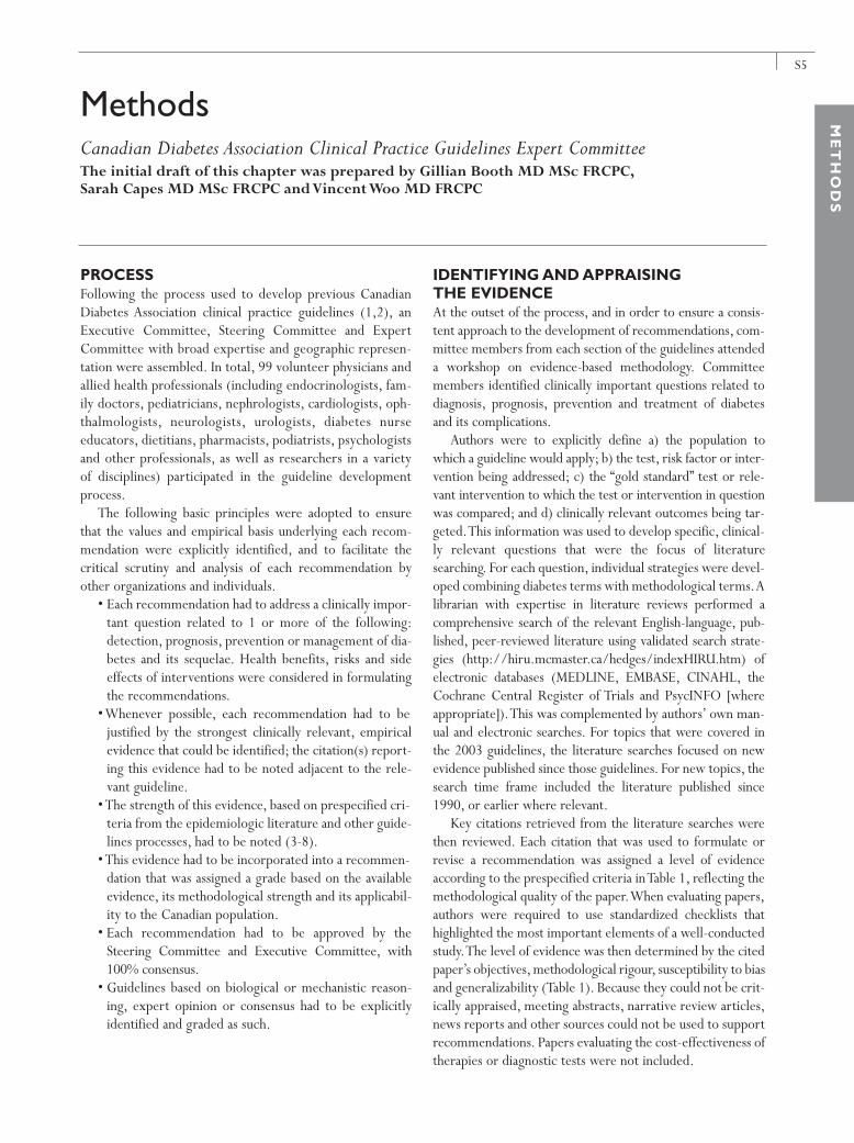
PROCESSFollowing the process used to develop previous CanadianDiabetes Association clinical practice guidelines (1,2), anExecutive Committee, Steering Committee and ExpertCommittee with broad expertise and geographic represen-tation were assembled. In total, 99 volunteer physicians andallied health professionals (including endocrinologists, fam-ily doctors, pediatricians, nephrologists, cardiologists, oph-thalmologists, neurologists, urologists, diabetes nurseeducators, dietitians, pharmacists, podiatrists, psychologistsand other professionals, as well as researchers in a variety of disciplines) participated in the guideline developmentprocess.
The following basic principles were adopted to ensurethat the values and empirical basis underlying each recom-mendation were explicitly identified, and to facilitate thecritical scrutiny and analysis of each recommendation byother organizations and individuals.
• Each recommendation had to address a clinically impor-tant question related to 1 or more of the following:detection, prognosis, prevention or management of dia-betes and its sequelae. Health benefits, risks and sideeffects of interventions were considered in formulatingthe recommendations.
•Whenever possible, each recommendation had to bejustified by the strongest clinically relevant, empiricalevidence that could be identified; the citation(s) report-ing this evidence had to be noted adjacent to the rele-vant guideline.
• The strength of this evidence, based on prespecified cri-teria from the epidemiologic literature and other guide-lines processes, had to be noted (3-8).
• This evidence had to be incorporated into a recommen-dation that was assigned a grade based on the availableevidence, its methodological strength and its applicabil-ity to the Canadian population.
• Each recommendation had to be approved by theSteering Committee and Executive Committee, with100% consensus.
• Guidelines based on biological or mechanistic reason-ing, expert opinion or consensus had to be explicitlyidentified and graded as such.
IDENTIFYING AND APPRAISING THE EVIDENCEAt the outset of the process, and in order to ensure a consis-tent approach to the development of recommendations, com-mittee members from each section of the guidelines attendeda workshop on evidence-based methodology. Committeemembers identified clinically important questions related todiagnosis, prognosis, prevention and treatment of diabetesand its complications.
Authors were to explicitly define a) the population towhich a guideline would apply; b) the test, risk factor or inter-vention being addressed; c) the “gold standard” test or rele-vant intervention to which the test or intervention in questionwas compared; and d) clinically relevant outcomes being tar-geted.This information was used to develop specific, clinical-ly relevant questions that were the focus of literaturesearching. For each question, individual strategies were devel-oped combining diabetes terms with methodological terms.Alibrarian with expertise in literature reviews performed acomprehensive search of the relevant English-language, pub-lished, peer-reviewed literature using validated search strate-gies (http://hiru.mcmaster.ca/hedges/indexHIRU.htm) ofelectronic databases (MEDLINE, EMBASE, CINAHL, theCochrane Central Register of Trials and PsycINFO [whereappropriate]).This was complemented by authors’ own man-ual and electronic searches. For topics that were covered inthe 2003 guidelines, the literature searches focused on newevidence published since those guidelines. For new topics, thesearch time frame included the literature published since1990, or earlier where relevant.
Key citations retrieved from the literature searches werethen reviewed. Each citation that was used to formulate orrevise a recommendation was assigned a level of evidenceaccording to the prespecified criteria in Table 1, reflecting themethodological quality of the paper.When evaluating papers,authors were required to use standardized checklists thathighlighted the most important elements of a well-conductedstudy.The level of evidence was then determined by the citedpaper’s objectives, methodological rigour, susceptibility to biasand generalizability (Table 1). Because they could not be crit-ically appraised, meeting abstracts, narrative review articles,news reports and other sources could not be used to supportrecommendations. Papers evaluating the cost-effectiveness oftherapies or diagnostic tests were not included.
MethodsCanadian Diabetes Association Clinical Practice Guidelines Expert CommitteeThe initial draft of this chapter was prepared by Gillian Booth MD MSc FRCPC,Sarah Capes MD MSc FRCPC and Vincent Woo MD FRCPC

S6
2008 CLINICAL PRACTICE GUIDELINES
A number of considerations were made when evaluatingthe evidence within a given area. For example, people withdiabetes are at high risk for several sequelae that are notexclusive to diabetes (e.g. cardiovascular diseases, renal fail-ure and erectile dysfunction). As such, some evidence relat-ing to these problems was identified that either excluded, didnot report on, or did not focus on people with diabetes.
Whenever such evidence was identified, a level was assignedusing the approach described above. Higher levels wereassigned if a) people with diabetes comprised a predefinedsubgroup; b) the results in the diabetes subgroup wereunlikely to have occurred by chance; and c) the evidence wasgenerated in response to questions that were formulatedprior to the analysis of the results.
Table 1. Criteria for assigning levels of evidence to the published studies
Level Criteria
Studies of diagnosis
Level 1 a) Independent interpretation of test results (without knowledge of the result of the diagnostic or gold standard)
b) Independent interpretation of the diagnostic standard (without knowledge of the test result)
c) Selection of people suspected (but not known) to have the disorder
d) Reproducible description of both the test and diagnostic standard
e) At least 50 patients with and 50 patients without the disorder
Level 2 Meets 4 of the Level 1 criteria
Level 3 Meets 3 of the Level 1 criteria
Level 4 Meets 1 or 2 of the Level 1 criteria
Studies of treatment and prevention
Level 1A Systematic overview or meta-analysis of high-quality RCTs
a) Comprehensive search for evidence
b) Authors avoided bias in selecting articles for inclusion
c) Authors assessed each article for validity
d) Reports clear conclusions that are supported by the data and appropriate analyses
OR
Appropriately designed RCT with adequate power to answer the question posed by the investigators
a) Patients were randomly allocated to treatment groups
b) Follow-up at least 80% complete
c) Patients and investigators were blinded to the treatment*
d) Patients were analyzed in the treatment groups to which they were assigned
e) The sample size was large enough to detect the outcome of interest
Level 1B Nonrandomized clinical trial or cohort study with indisputable results
Level 2 RCT or systematic overview that does not meet Level 1 criteria
Level 3 Nonrandomized clinical trial or cohort study
Level 4 Other
Studies of prognosis
Level 1 a) Inception cohort of patients with the condition of interest, but free of the outcome of interest
b) Reproducible inclusion/exclusion criteria
c) Follow-up of at least 80% of subjects
d) Statistical adjustment for extraneous prognostic factors (confounders)
e) Reproducible description of outcome measures
Level 2 Meets criterion a) above, plus 3 of the other 4 criteria
Level 3 Meets criterion a) above, plus 2 of the other criteria
Level 4 Meets criterion a) above, plus 1 of the other criteria
*In cases where such blinding was not possible or was impractical (e.g. intensive vs. conventional insulin therapy), the blinding ofindividuals who assessed and adjudicated study outcomes was felt to be sufficient
RCT = randomized controlled trial

S7
ME
TH
OD
S
GUIDELINE DEVELOPMENT Expert Committee members evaluated the relevant litera-ture, and guidelines were developed and initially reviewedby the Expert Committee. In the absence of new evidencesince the publication of the 2003 clinical practice guidelines,recommendations from the 2003 document were notchanged.
The studies used to develop and support each recom-mendation are cited beside the level of evidence. In somecases, each of the citations that supported a recommendationwere not assigned the same level of evidence, but rather wereof varying levels of evidence. In those circumstances, all rel-evant studies were cited, regardless of the grading assigned tothe recommendation. The final grading depended on theoverall evidence available, including the relative strengths ofthe studies from a methodological perspective and the stud-ies’ findings. Further details on the grading process aredescribed below.
Finally, several treatment recommendations were basedon evidence generated from the use of 1 therapeutic agentfrom a given class (e.g. 1 of the “statins”).Whenever evidencerelating to 1 or more agents from a recognized class of agentswas available, the recommendation was written so as to berelevant to the class, but specifically studied therapeuticagents were identified within the recommendation and/orcited reference(s). Only medications with Health CanadaNotice of Compliance granted by February 18, 2008, wereincluded in the recommendations.
GRADING THE RECOMMENDATIONSAfter formulating new recommendations or modifying exist-ing ones based on new evidence, each recommendation wasassigned a grade from A through D (Table 2).The highest pos-sible grade that a recommendation could have was based onthe level of evidence. However, the assigned grading was low-ered in some cases; for example, if the evidence was foundnot to be applicable to the Canadian population, or if based onthe consensus of the Steering and Executive Committees,there were additional concerns regarding the recommenda-tion. In some situations, the grading was also lowered for sub-
groups that were not well represented in the study, or inwhom the beneficial effect of an intervention was less clear.Thus, a recommendation based on Level 1 evidence, deemedto be very applicable to Canadians and supported by strongconsensus, was assigned a grade of A. A recommendation notdeemed to be applicable to Canadians, or judged to requirefurther supporting evidence, was assigned a lower grade.Where available, the number of patients that would need tobe treated in order to prevent 1 clinical event (number need-ed to treat [NNT]) or to cause an adverse event (numberneeded to harm [NNH]) was considered in assessing theimpact of a particular intervention.The degree to which evi-dence derived from other populations was felt to be relevantto diabetes was also reflected in the wording and grading ofthe recommendation. Finally, in the absence of Level 1, 2 or3 supporting evidence, or if the recommendation was basedon the consensus of the Steering and Executive Committees,the highest grade that could be assigned was D.
INTERPRETING THE ASSIGNED GRADE OFA RECOMMENDATION The grade assigned to each recommendation is closely linkedto the methodological rigour and robustness of the relevantclinical research. Therefore, as noted above, a high gradereflects a high degree of confidence that following the recom-mendation will lead to the desired outcome. Similarly, alower grade reflects weaker evidence, and a greater possibili-ty that the recommendation will change when more evidenceis generated in the future. Of note, the assigned grade con-tains no subjective information regarding the importance ofthe recommendation or how strongly members of the com-mittee felt about it; it contains information regarding only theevidence upon which the recommendation is based. Thus,many Grade D recommendations were deemed to be veryimportant to the contemporary management of diabetes,based on clinical experience, case series, physiological evi-dence and current concepts of disease pathophysiology.However, the paucity of clinical evidence addressing the areasof therapy, prevention, diagnosis or prognosis precluded theassignment of a higher grade.
Clearly, clinicians need to base clinical decisions on thebest available relevant evidence that addresses clinical situa-tions. However, they are also frequently faced with having toact in the absence of clinical evidence, and there are many sit-uations where good clinical evidence may be impossible,impractical or too expensive to generate (which implies thatit would be impossible to develop Grade A recommenda-tions). For example, it took the United Kingdom ProspectiveDiabetes Study (UKPDS) Group >20 years to collect andpublish Level 1 evidence leading to a Grade A recommenda-tion in support of the role of tight glycemic control to reducemicrovascular disease in people with type 2 diabetes. Prior tothe publication of the UKPDS results, the recommendationfor glycemic control to prevent microvascular consequences
Table 2. Criteria for assigning grades ofrecommendations for clinical practice
Grade Criteria
Grade A The best evidence was atLevel 1
Grade B The best evidence was atLevel 2
Grade C The best evidence was atLevel 3
Grade D The best evidence was atLevel 4 or consensus

S8
2008 CLINICAL PRACTICE GUIDELINES
was a Grade B recommendation (1).Varying grades of recommendations, therefore, reflect
varying degrees of certainty regarding the strength of infer-ence that can be drawn from the evidence in support of therecommendation. Therefore, these evidence-based guide-lines and their graded recommendations are designed to sat-isfy 2 important needs: 1) the explicit identification of thebest research upon which the recommendation is based, andan assessment of its scientific relevance and quality (capturedby the assignment of a level of evidence to each citation); and2) the explicit assignment of strength of the recommendationbased on this evidence (captured by the grade). In this way,they provide a convenient summary of the evidence to facil-itate clinicians’ task of “weighting” and incorporating ever-increasing evidence into their daily clinical decision-making.They also facilitate the ability of clinicians, healthcare plan-ners, healthcare providers and society in general to criticallyexamine any recommendation and arrive at their own con-clusions regarding its appropriateness.Thus, these guidelinesfacilitate their own scrutiny by others according to the sameprinciples that they use to scrutinize the literature.
It is important to note that the system chosen for grad-ing recommendations differs from the approach used insome other guideline documents, such as the one pertainingto the periodic health examination in Canada, in whichharmful practices were assigned a grade of D (8). In thisCanadian Diabetes Association guidelines document, recom-mendation to avoid any harmful practices would be gradedin the same manner as all other recommendations. However,it should be noted that the authors of these guidelinesfocused on clinical practices that were thought to be poten-tially beneficial, and did not seek out evidence regarding theharmfulness of interventions.
EXTERNAL PEER REVIEW AND INDEPEN-DENT METHODOLOGICAL REVIEW In July 2007, a draft document was circulated nationally andinternationally for review by numerous stakeholders andexperts in relevant fields.This input was then considered bythe Executive and Steering Committees and revisions weremade accordingly. Subsequently, a panel of 6 methodologists,who were not directly involved with the initial review andassessment of the evidence, independently reviewed eachrecommendation, its assigned grade and supportive citations.Based on this review, the wording, assigned level of evidenceand grade of each recommendation were reassessed andmodified as necessary. Revised recommendations werereviewed and approved by the Executive and SteeringCommittees. Selected recommendations were presented at apublic forum at the Canadian Diabetes Association/CanadianSociety of Endocrinology and Metabolism ProfessionalConference and Annual Meetings in Vancouver, BritishColumbia, in October 2007.
DISCLOSURE OF DUALITY OF INTERESTCommittee members were volunteers and received noremuneration or honoraria for their participation. Membersof all committees signed an annual duality of interest formlisting all financial interests or relationships with manufac-turer(s) of any commercial product(s) and/or provider(s) ofcommercial services. A full list of committee member dis-closures is available online at http://www.diabetes.ca.Dualities of interest were also discussed during deliberationswhere relevant. In the case of a potential duality or outrightconflict of interest, committee members removed them-selves from discussions. Funding for the development of theguidelines was provided by the Canadian DiabetesAssociation and through unrestricted educational grants pro-vided by the companies listed in the acknowledgements sec-tion (p. x).These companies were not involved in any aspectof guideline development, literature interpretation, the deci-sion to publish or any other aspect related to the publicationof these guidelines, and did not have access to guidelinemeetings, guideline drafts or committee deliberations.
GUIDELINE UPDATESA process to update the full guidelines will commence with-in 5 years. Updates to individual chapters may be publishedsooner in the event of significant changes in evidence sup-porting the recommendations.
OTHER RELEVANT GUIDELINESIntroduction, p. S1.
ACKNOWLEDGEMENTSThe clinical practice guidelines Expert Committee thanksthe following individuals, who conducted the independentmethodological review:
Gillian Booth MD MSc FRCPC (Chair)Assistant Professor of Medicine, University of Toronto,Division of Endocrinology and Metabolism and Li Ka ShingInstitute, St. Michael’s Hospital,Toronto, Ontario
Denice Feig MD MSc FRCPSAssociate Professor of Medicine, University of Toronto,Toronto, Staff Endocrinologist, Mount Sinai Hospital,Toronto, Ontario
Dereck Hunt MD MSc FRCPSAssociate Professor, General Internal Medicine/Endocrinology, McMaster University, Hamilton, Ontario
Charlotte McDonald MD MSc FRCPSAssistant Professor, University of Western Ontario, London,Ontario

S9
ME
TH
OD
S
Zubin Punthakee MD MSc FRCPS ABIMAssistant Professor, Department of Medicine andDepartment of Pediatrics, Division of Endocrinology andMetabolism, McMaster University, Hamilton, Ontario
Joel Ray MD MSc FRCPCAssistant Professor, Department of Medicine, Divisions ofGeneral Internal Medicine and Endocrinology Metabolism,the Department of Obstetrics and Gynecology, and theDepartment of Health Policy Management and Evaluation,St. Michael’s Hospital,Toronto, Ontario
REFERENCES1. Meltzer S, Leiter L, Daneman D, et al. 1998 clinical practice
guidelines for the management of diabetes in Canada. CMAJ.1998;159(suppl 8):S1-S29.
2. Canadian Diabetes Association Clinical Practice GuidelineExpert Committee. Canadian Diabetes Association 2003 clini-cal practice guidelines for the prevention and management ofdiabetes in Canada. Can J Diabetes. 2003;27(suppl 2):S1-S152.
3. Straus SE, McAlister FA. What is the prognosis? In: GersteinHC, Haynes RB, eds. Evidence-based Diabetes Care. Hamilton,ON: BC Decker Inc.;2001:6-12.
4. American Medical Association. Users’Guides to the Medical Literature:Essentials of Evidence-based Clinical Practice. Chicago, IL: AmericanMedical Association; 2001.
5. Jaeschke R, Guyatt GH. How should diagnostic tests be chosenand used? In: Gerstein HC, Haynes RB, eds. Evidence-basedDiabetes Care. Hamilton, ON: BC Decker Inc.; 2001:13-23.
6. Holbrook AM, Clarke J-A, Raymond C, et al. How should aparticular problem be managed? Incorporating evidence abouttherapies into practice. In: Gerstein HC, Haynes RB, eds.Evidence-based Diabetes Care. Hamilton, ON: BC Decker Inc.;2001:24-47.
7. Harris SB, Webster-Bogaert SM. Evidence-based clinical prac-tice guidelines. In: Gerstein HC, Haynes RB, eds. Evidence-basedDiabetes Care. Hamilton, ON: BC Decker Inc.; 2001:48-61.
8. Goldbloom R, Battista RN. The periodic health examination:1. Introduction. CMAJ. 1986;134:721-723.
S10
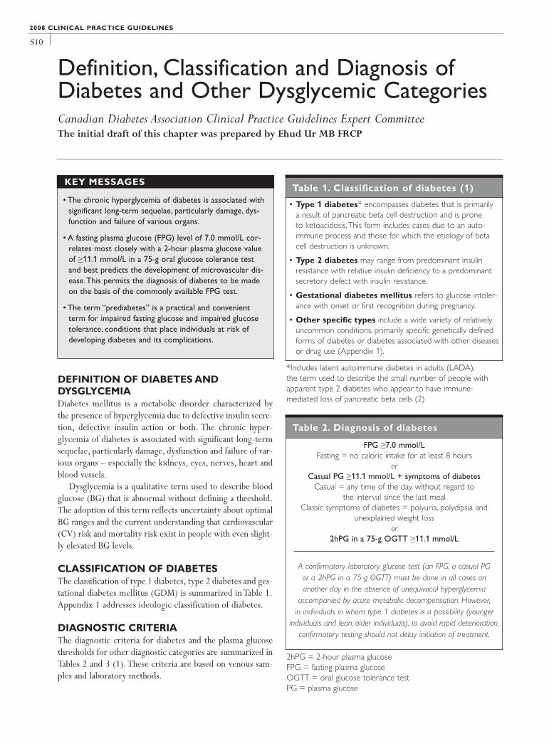
DEFINITION OF DIABETES AND DYSGLYCEMIADiabetes mellitus is a metabolic disorder characterized bythe presence of hyperglycemia due to defective insulin secre-tion, defective insulin action or both. The chronic hyper-glycemia of diabetes is associated with significant long-termsequelae, particularly damage, dysfunction and failure of var-ious organs – especially the kidneys, eyes, nerves, heart andblood vessels.
Dysglycemia is a qualitative term used to describe bloodglucose (BG) that is abnormal without defining a threshold.The adoption of this term reflects uncertainty about optimalBG ranges and the current understanding that cardiovascular(CV) risk and mortality risk exist in people with even slight-ly elevated BG levels.
CLASSIFICATION OF DIABETESThe classification of type 1 diabetes, type 2 diabetes and ges-tational diabetes mellitus (GDM) is summarized in Table 1.Appendix 1 addresses ideologic classification of diabetes.
DIAGNOSTIC CRITERIAThe diagnostic criteria for diabetes and the plasma glucosethresholds for other diagnostic categories are summarized inTables 2 and 3 (1). These criteria are based on venous sam-ples and laboratory methods.
Definition, Classification and Diagnosis ofDiabetes and Other Dysglycemic CategoriesCanadian Diabetes Association Clinical Practice Guidelines Expert CommitteeThe initial draft of this chapter was prepared by Ehud Ur MB FRCP
2008 CLINICAL PRACTICE GUIDELINES
• The chronic hyperglycemia of diabetes is associated withsignificant long-term sequelae, particularly damage, dys-function and failure of various organs.
• A fasting plasma glucose (FPG) level of 7.0 mmol/L cor-relates most closely with a 2-hour plasma glucose valueof ≥11.1 mmol/L in a 75-g oral glucose tolerance testand best predicts the development of microvascular dis-ease.This permits the diagnosis of diabetes to be madeon the basis of the commonly available FPG test.
• The term “prediabetes” is a practical and convenientterm for impaired fasting glucose and impaired glucosetolerance, conditions that place individuals at risk ofdeveloping diabetes and its complications.
KEY MESSAGESTable 1. Classification of diabetes (1)
• Type 1 diabetes* encompasses diabetes that is primarily a result of pancreatic beta cell destruction and is prone to ketoacidosis.This form includes cases due to an auto-immune process and those for which the etiology of betacell destruction is unknown.
• Type 2 diabetes may range from predominant insulinresistance with relative insulin deficiency to a predominantsecretory defect with insulin resistance.
• Gestational diabetes mellitus refers to glucose intoler-ance with onset or first recognition during pregnancy.
• Other specific types include a wide variety of relativelyuncommon conditions, primarily specific genetically definedforms of diabetes or diabetes associated with other diseasesor drug use (Appendix 1).
*Includes latent autoimmune diabetes in adults (LADA),the term used to describe the small number of people withapparent type 2 diabetes who appear to have immune-mediated loss of pancreatic beta cells (2)
Table 2. Diagnosis of diabetes
FPG ≥7.0 mmol/LFasting = no caloric intake for at least 8 hours
orCasual PG ≥11.1 mmol/L + symptoms of diabetes
Casual = any time of the day, without regard to the interval since the last meal
Classic symptoms of diabetes = polyuria, polydipsia and unexplained weight loss
or2hPG in a 75-g OGTT ≥11.1 mmol/L
A confirmatory laboratory glucose test (an FPG, a casual PG or a 2hPG in a 75-g OGTT) must be done in all cases on another day in the absence of unequivocal hyperglycemia
accompanied by acute metabolic decompensation. However,in individuals in whom type 1 diabetes is a possibility (younger
individuals and lean, older individuals), to avoid rapid deterioration,confirmatory testing should not delay initiation of treatment.
2hPG = 2-hour plasma glucoseFPG = fasting plasma glucoseOGTT = oral glucose tolerance testPG = plasma glucose

S11
DiabetesA fasting plasma glucose (FPG) level of 7.0 mmol/L corre-lates most closely with a 2-hour plasma glucose (2hPG)value of ≥11.1 mmol/L in a 75-g oral glucose tolerance test(OGTT) and best predicts the development of microvascu-lar disease (1). This permits the diagnosis of diabetes to bemade on the basis of the commonly available FPG test.Although the frequency distributions of glycated hemoglo-bin (A1C) levels in some studies have characteristics similarto those obtained from FPG and 2hPG tests, the lack of stan-dardization of the A1C test precludes its use in the diagnosisof diabetes.
PrediabetesElevated BG levels below the threshold for diabetes alsohave clinical consequences.The term “prediabetes” is a prac-tical and convenient term for impaired fasting glucose (IFG)and impaired glucose tolerance (IGT) (Table 3), conditionsthat place individuals at risk of developing diabetes and itscomplications. It is important to stress that not all individu-als with prediabetes will necessarily progress to diabetes.Indeed, a significant proportion of people who are diag-nosed with IFG or IGT will revert to normoglycemia.People with prediabetes, particularly in the context of themetabolic syndrome (see below), would benefit from CVrisk factor modification.
While people with IFG or IGT do not have the diabetes-associated risk for microvascular disease, they are at higherrisk for the development of diabetes and CVD (3). IGT ismore strongly associated with CVD outcomes. However,individuals identified as having both IFG and IGT are at high-
er risk for diabetes as well as CVD. Lifestyle interventionshave been shown to be highly effective in delaying or pre-venting the onset of diabetes in people with IGT (4,5).Studies have not yet been done to examine CVD and totalmortality.
There is no worldwide consensus on the definition of IFG(6,7).While the Canadian Diabetes Association continues todefine IFG as an FPG value of 6.1 to 6.9 mmol/L (7), a num-ber of limitations have been identified with regards to theexisting lower limit of 6.1 mmol/L. These include subopti-mal sensitivity for undiagnosed diabetes and IGT, and poten-tial instability on retesting (due to the narrowness of thediagnostic range). For those individuals with an FPG valuebetween 5.6 and 6.0 mmol/L and ≥1 risk factors for dia-betes, consideration should be given to performing a 75-gOGTT (6-10).
Metabolic syndrome Dysglycemia and type 2 diabetes are often manifestations ofa much broader underlying disorder (11,12), including themetabolic syndrome – a highly prevalent, multifaceted con-dition characterized by a distinctive constellation of abnor-malities that include abdominal obesity, hypertension,dyslipidemia, insulin resistance and dysglycemia. Individualswith the metabolic syndrome are at significant risk of devel-oping diabetes and CVD. Evidence now exists to support anaggressive approach to identifying people with the metabol-ic syndrome and treating not only the hyperglycemia but alsothe associated CV risk factors, such as hypertension, dyslipi-demia and abdominal obesity, in the hope of significantlyreducing CV morbidity and mortality.
A lack of consensus exists regarding the operational definitions of the metabolic syndrome. In 1998, the WorldHealth Organization (13) proposed a unifying definition thatincludes identification of the presence of insulin resistance.The United States (US) Expert Panel on Detection,Evaluation, and Treatment of High Blood Cholesterol inAdults (Adult Treatment Panel III [ATP III]) provided anoperational definition based on ≥3 criteria that does notrequire a measure of insulin resistance (14,15). In theInternational Diabetes Federation (IDF) definition, the pres-ence of abdominal obesity is a requisite risk factor. The IDFdefinition also provides ethnic-specific values for waist cir-cumference (16). Table 4 presents the definitions of meta-bolic syndrome proposed by these 3 organizations. Datafrom the Third National Health and Nutrition Survey, whichemployed the 2001 ATP III criteria (15), showed that theoverall prevalence of the metabolic syndrome in the US wasapproximately 20 to 25% (17).
DE
FIN
ITIO
NA
ND
DIA
GN
OS
IS
Table 3. PG levels for diagnosis of IFG,IGT and diabetes
FPG (mmol/L)
2hPG in the75-g OGTT(mmol/L)
IFG 6.1–6.9 NA
IFG (isolated) 6.1–6.9 and <7.8
IGT (isolated) <6.1 and 7.8–11.0
IFG and IGT 6.1–6.9 and 7.8–11.0
Diabetes ≥7.0 or ≥11.1
2hPG = 2-hour plasma glucoseFPG = fasting plasma glucoseIFG = impaired fasting glucoseIGT = impaired glucose toleranceOGTT = oral glucose tolerance testNA = not applicablePG = plasma glucose

S12
OTHER RELEVANT GUIDELINES Screening for Type 1 and Type 2 Diabetes, p. S14Prevention of Diabetes, p. S17Type 1 Diabetes in Children and Adolescents, p. S150Type 2 Diabetes in Children and Adolescents, p. S162
RELEVANT APPENDIXAppendix 1. Etiologic Classification of Diabetes Mellitus
REFERENCES1. American Diabetes Association. Report of the Expert
Committee on the Diagnosis and Classification of DiabetesMellitus. Diabetes Care. 2008;31(suppl 1):S55-S60.
2. Turner R, Stratton I, Horton V, et al. UKPDS 25: autoantibodiesto islet-cell cytoplasm and glutamic acid decarboxylase for pre-diction of insulin requirement in type 2 diabetes. UK ProspectiveDiabetes Study Group. Lancet. 1997;350:1288-1293.
3. Coutinho M, Gerstein HC,Wang Y, et al.The relationship between
2008 CLINICAL PRACTICE GUIDELINES
Table 4. Definitions of the metabolic syndrome
WHO (13) NCEP ATP III2001 (14) 2004 (15)
IDF (16)
Diagnosticcriteria
Diabetes, IFG, IGT orinsulin resistance (assessedby clamp studies) plus ≥2other risk determinants are present
≥3 risk determinants are present Central obesity (using ethnic-specific values)plus ≥2 other risk determinants are present(if BMI is >30 kg/m2, central obesity can beassumed and WC does not need to bemeasured)
BG Diabetes, IFG, IGT orinsulin resistance
FPG ≥6.1 mmol/L FPG ≥5.6 mmol/L FPG ≥5.6 mmol/L (or previously diagnosedtype 2 diabetes)
BP ≥140/90 mm Hg ≥130/85 mm Hg ≥130/85 mm Hg (or receiving treatment forpreviously diagnosed hypertension)
TG ≥1.7 mmol/L ≥1.7 mmol/L ≥1.7 mmol/L (or receiving treatment)
HDL-C <0.9 mmol/L (men)<1.0 mmol/L (women)
<1.0 mmol/L (men)<1.3 mmol/L (women)
<1.0 mmol/L (men)<1.3 mmol/L (women)(or receiving treatment)
Abdominalobesity
Waist-to-hip ratio:>0.90 (men)>0.85 (women)
WC:>102 cm (men)>88 cm (women)
Europids / Sub-Saharan Africans / EasternMediterranean and Middle East (Arab) populations:WC ≥94 cm (men)WC ≥80 cm (women)
South Asian / Malaysian / Asian / Indian /Chinese / Japanese / Ethnic South andCentral American populations:WC ≥90 cm (men)WC ≥80 cm (women)
Kidneyfunction
Urinary albumin excretionrate >20 µg/minorACR ≥30 mg/g
NA NA
ACR = albumin to creatinine ratioBG = blood glucoseBMI = body mass indexBP = blood pressureFPG = fasting plasma glucoseHDL-C = high-density lipoprotein cholesterolIDF = International Diabetes FederationIFG = impaired fasting glucoseIGT = impaired glucose tolerance
NA = not applicableNCEP ATP III = National Cholesterol Education
Program Adult Treatment Panel III TG = triglyceridesWC = waist circumferenceWHO = World Health Organization

S13
glucose and incident cardiovascular events. A meta-regressionanalysis of published data from 20 studies of 95,783 individualsfollowed for 12.4 years. Diabetes Care.1999;22:233-240.
4. Tuomilehto J, Lindström J, Eriksson JG, et al; Finnish DiabetesPrevention Study Group. Prevention of type 2 diabetes melli-tus by changes in lifestyle among subjects with impaired glu-cose tolerance. N Engl J Med. 2001;344:1343-1350.
5. Knowler WC, Barrett-Connor E, Fowler SE, et al; DiabetesPrevention Program Research Group. Reduction in the inci-dence of type 2 diabetes with lifestyle intervention or met-formin. N Engl J Med. 2002;346:393-403.
6. Shaw JE, Zimmet PZ, Alberti KG. Point: Impaired fasting glu-cose: the case for the new American Diabetes Association cri-terion. Diabetes Care. 2006;29:1170-1172.
7. Forouhi NG, Balkau B, Borch-Johnsen K, et al; EDEG. Thethreshold for diagnosing impaired fasting glucose: a positionstatement by the European Diabetes Epidemiology Group.Diabetologia. 2006;49:822-827.
8. Shaw JE, Zimmet PZ, Hodge AM, et al. Impaired fasting glu-cose: how low should it go? Diabetes Care. 2000;23:34-39.
9. Ko GT, Chan JC,Yeung VT, et al. Combined use of a fastingplasma glucose concentration and HbA1C or fructosaminepredicts the likelihood of having diabetes in high-risk subjects.Diabetes Care. 1998;21:1221-1225.
10. Tirosh A, Shai I, Tekes-Manova D, et al; Israeli DiabetesResearch Group. Normal fasting plasma glucose levels and type2 diabetes in young men. N Engl J Med. 2005;353:1454-1462.
11. Zimmet PZ. Diabetes epidemiology as a tool to trigger dia-betes research and care. Diabetologia. 1999;42:499-518.
12. Reaven GM. Banting lecture 1988. Role of insulin resistance inhuman disease. Diabetes. 1988;37:1595-1607.
13. Alberti KG, Zimmet PZ. Definition, diagnosis and classifica-tion of diabetes mellitus and its complications. Part 1: diagno-sis and classification of diabetes mellitus provisional report ofa WHO consultation. Diabet Med. 1998;15:539-553.
14. Expert Panel on Detection, Evaluation, and Treatment of HighBlood Cholesterol in Adults. Executive Summary of The ThirdReport of The National Cholesterol Education Program(NCEP) Expert Panel on Detection, Evaluation, and Treatmentof High Blood Cholesterol in Adults (Adult Treatment PanelIII). JAMA. 2001;285:2486-2497.
15. Grundy SM, Cleeman JI, Daniels SR, et al; American HeartAssociation; National Heart, Lung, and Blood Institute.Diagnosis and management of the metabolic syndrome: anAmerican Heart Association/National Heart, Lung, and BloodInstitute Scientific Statement. Circulation. 2005;112:2735-2752.
16. International Diabetes Federation. The IDF ConsensusWorldwide Definition of the Metabolic Syndrome. Brussels: IDFCommunications; 2006.Available at: http://www.idf.org/webdata/docs/IDF_Meta_def_final.pdf.Accessed September 1, 2008.
17. Ford ES, Giles WH, Dietz WH. Prevalence of the metabolicsyndrome among US adults: findings from the third nationalHealth and Nutrition Examination Survey. JAMA. 2002;287:356-359.
DE
FIN
ITIO
NA
ND
DIA
GN
OS
IS

S14
SCREENING FOR TYPE 1 DIABETESType 1 diabetes mellitus is primarily a result of pancreaticbeta cell destruction due to an immune-mediated processthat is likely incited by environmental factors in geneticallypredisposed individuals. An individual’s risk of developingtype 1 diabetes can be estimated by considering family histo-ry of type 1 diabetes with attention to age of onset and sexof the affected family members (1) and profiling immunityand genetic markers (2). The loss of pancreatic beta cells inthe development of type 1 diabetes passes through a subclin-ical prodrome that can be detected reliably in first- and sec-ond-degree relatives of persons with type 1 diabetes by thepresence of pancreatic islet autoantibodies in their sera (3).Given that the various serologic markers are not universallyavailable, and in the absence of evidence for interventions toprevent or delay type 1 diabetes, no recommendations forscreening for type 1 diabetes can be made.
SCREENING FOR TYPE 2 DIABETESAdultsUndiagnosed type 2 diabetes may occur in >2.8% of the gen-eral adult population (4), with the number increasing to>10% in some populations (5,6).Tests for hyperglycemia canidentify these individuals, many of whom will have or will beat risk for preventable diabetes complications (5,6).Althoughthe relatively low prevalence of diabetes in the general popu-lation makes it unlikely that mass screening will be cost-effec-tive, testing for diabetes in people with risk factors for type 2diabetes or with diabetes-associated conditions is likely to
result in more benefit than harm and will lead to overall costsavings (7,8). Routine testing for type 2 diabetes is, therefore,justifiable in some but not all settings (9). Screening individ-uals as early as age 40 in family physicians’ offices has provedto be useful in detecting unrecognized diabetes (10).
While fasting plasma glucose (FPG) is the recommendedscreening test, a 2-hour plasma glucose (2hPG) in a 75-g oralglucose tolerance test (OGTT) is indicated when the FPG is6.1 to 6.9 mmol/L (11) and may be indicated when the FPGis 5.6 to 6.0 mmol/L and suspicion of type 2 diabetes orimpaired glucose tolerance (IGT) is high (e.g. for individualswith risk factors listed in Table 1); see Figure 1.
As people with impaired fasting glucose (IFG) or IGT areat increased risk of developing type 2 diabetes and have anincreased risk of macrovascular complications, the diagnosisof IGT, particularly in apparently healthy people, has impor-
Screening for Type 1 and Type 2 DiabetesCanadian Diabetes Association Clinical Practice Guidelines Expert CommitteeThe initial draft of this chapter was prepared by Ehud Ur MB FRCP, Jean-Louis Chiasson MD,Tom Ransom MD MSc FRCPC and Richard Rowe MBBS MAEd FRCPC
2008 CLINICAL PRACTICE GUIDELINES
• In the absence of evidence for interventions to preventor delay type 1 diabetes, screening for type 1 diabetes is not recommended.
• Screening for type 2 diabetes using a fasting plasma glu-cose (FPG) should be performed every 3 years in indi-viduals ≥40 years of age.
• While the FPG is the recommended screening test, a 2-hour plasma glucose in a 75-g oral glucose tolerancetest is indicated when the FPG is 6.1 to 6.9 mmol/L andmay be indicated when FPG is 5.6 to 6.0 mmol/L andsuspicion of type 2 diabetes or impaired glucose toler-ance is high (e.g. for individuals with risk factors).
KEY MESSAGES
Table 1. Risk factors for type 2 diabetes
• Age ≥40 years• First-degree relative with type 2 diabetes• Member of high-risk population (e.g. people of Aboriginal,
Hispanic, South Asian, Asian or African descent)• History of IGT or IFG*• Presence of complications associated with diabetes• Vascular disease (coronary, cerebrovascular or peripheral)*• History of gestational diabetes mellitus• History of delivery of a macrosomic infant• Hypertension* • Dyslipidemia*• Overweight* • Abdominal obesity*• Polycystic ovary syndrome*• Acanthosis nigricans*• Schizophrenia†
• Other (see Appendix 1)
*Associated with insulin resistance† The incidence of type 2 diabetes is at least 3 times higher in people with schizophrenia than in the general population(12,13). Using data collected in 1991, the prevalence of dia-betes was assessed in >20 000 individuals diagnosed with schizophrenia.The rate of diagnosed diabetes was 9 to 14%,exceeding rates for the general population prior to the widespread use of new antipsychotic drugs (14)
IFG = impaired fasting glucoseIGT = impaired glucose tolerance

S15
tant prognostic implications (15). Classifying individualswith IFG and/or IGT, particularly in the context of the meta-bolic syndrome, identifies people who would benefit fromcardiovascular risk factor reduction.
Risk scoresA number of risk scores based on clinical characteristics havebeen developed to identify individuals at high risk of havingundiagnosed diabetes. However, the impact of known risk
SC
RE
EN
ING
FO
RT
YP
E1
AN
DT
YP
E2
Figure 1. Screening for type 2 diabetes in adults
Screen every 3 years in individuals ≥40 years of age
Screen earlier and/or more frequently in people with additional risk factors for diabetes (see Table 1)
Fasting value≥7.0 mmol/L or 2-h value≥11.1 mmol/L
Normal*Rescreen as recom-mended
FPG
<5.6 mmol/L
Diabetes‡
*If, despite a normal fasting value, an OGTT is subsequently performed and the 2hPG value is 7.8–11.0 mmol/L, a diagnosis of iso-lated IGT is made†Prediabetes = isolated IFG, isolated IGT, IFG and IGT (see Table 3 in “Definition, Classification and Diagnosis of Diabetes and OtherDysglycemic Categories,” p. S10)‡A confirmatory laboratory glucose test (either an FPG, a casual PG or a 2hPG in a 75-g OGTT) must be done on another day inall cases in the absence of unequivocal hyperglycemia accompanied by acute metabolic decompensation
2hPG = 2-hour plasma glucoseFPG = fasting plasma glucoseIFG = impaired fasting glucose
IGT = impaired glucose toleranceOGTT = oral glucose tolerance test PG = plasma glucose
5.6–6.0 mmol/L 6.1–6.9 mmol/L
If ≥1 risk factors,consider 75-g OGTT
Fasting value<6.1 mmol/Land 2-h value
7.8–11.0 mmol/L
Fasting value6.1–6.9 mmol/Land 2-h value<7.8 mmol/L
Fasting value6.1–6.9 mmol/Land 2-h value
7.8–11.0 mmol/L
IsolatedIGT
IsolatedIFG
Prediabetes†
Rescreen more oftenAt risk
Rescreen more often
IFG andIGT
Fasting value<6.1 mmol/Land 2-h value<7.8 mmol/L
≥7.0 mmol/L
No risk factors

S16
factors on having undiagnosed type 2 diabetes differs betweenpopulations of different ethnic origins, and risk scores devel-oped in Caucasian populations cannot be applied to popula-tions of other ethnic groups (16).
OTHER RELEVANT GUIDELINESDefinition, Classification and Diagnosis of Diabetes
and Other Dysglycemic Categories, p. S10Prevention of Diabetes, p. S17Type 1 Diabetes in Children and Adolescents, p. S150Type 2 Diabetes in Children and Adolescents, p. S162
RELEVANT APPENDIXAppendix 1. Etiologic Classification of Diabetes Mellitus
REFERENCES1. Harjutsalo V, Reunanen A,Tuomilehto J. Differential transmis-
sion of type 1 diabetes from diabetic fathers and mothers totheir offspring. Diabetes. 2006;55:1517-1524.
2. Decochez K, Truyen I, van der Auwera B, et al; BelgianDiabetes Registry. Combined positivity for HLA DQ2/DQ8and IA-2 antibodies defines population at high risk of develop-ing type 1 diabetes. Diabetologia. 2005;48:687-694.
3. Bingley PJ. Interactions of age, islet cell antibodies, insulinautoantibodies, and first-phase insulin response in predictingrisk of progression to IDDM in ICA+ relatives: the ICARUSdata set. Islet Cell Antibody Register Users Study Diabetes. 1996;45:1720-1728.
4. Cowie CC, Rust KF, Byrd-Holt DD, et al. Prevalence of dia-betes and impaired fasting glucose in adults in the U.S. popu-lation: National Health And Nutrition Examination Survey1999-2002. Diabetes Care. 2006;29:1263-1268.
5. Rolka DB, Narayan KM, Thompson TJ, et al. Performance ofrecommended screening tests for undiagnosed diabetes anddysglycemia. Diabetes Care. 2001;24:1899-1903.
6. Rathmann W, Haastert B, Icks A, et al. High prevalence ofundiagnosed diabetes mellitus in Southern Germany: targetpopulations for efficient screening. The KORA survey 2000.Diabetologia. 2003;46:182-189.
7. Raikou M, McGuire A.The economics of screening and treat-ment in type 2 diabetes mellitus. Pharmacoeconomics. 2003;21:543-564.
8. The cost-effectiveness of screening for type 2 diabetes. CDCDiabetes Cost-Effectiveness Study Group, Centers for DiseaseControl and Prevention. JAMA. 1998;280:1757-1763.
9. Knowler WC. Screening for NIDDM. Opportunities fordetection, treatment, and prevention. Diabetes Care. 1994;17:445-450.
10. Leiter LA, Barr A, Bélanger A, et al; Diabetes Screening inCanada (DIASCAN) Study. Diabetes Screening in Canada(DIASCAN) Study: prevalence of undiagnosed diabetes andglucose intolerance in family physician offices. Diabetes Care.2001;24:1038-1043.
11. Saydah SH, Byrd-Holt D, Harris MI. Projected impact ofimplementing the results of the Diabetes Prevention Programin the U.S. population. Diabetes Care. 2002;25:1940-1945.
12. McKee HA, D’Arcy PF,Wilson PJ. Diabetes and schizophrenia— a preliminary study. J Clin Hosp Pharm. 1986;11:297-299.
13. Mukherjee S, Decina P, Bocola V, et al. Diabetes mellitus inschizophrenic patients. Compr Psychiatry. 1996;37:68-73.
14. Dixon L, Weiden P, Delahanty J, et al. Prevalence and corre-lates of diabetes in national schizophrenia samples. SchizophrBull. 2000;26:903-912.
15. Hu G, Qiao Q, Tuomilehto J, et al; DECODE Study Group.Prevalence of the metabolic syndrome and its relation to all-cause and cardiovascular mortality in nondiabetic Europeanmen and women. Arch Intern Med. 2004;164:1066-1076.
16. Glumer C,Vistisen D, Borch-Johnsen K, et al for the Detect 2Collaboration. Risk scores for type 2 diabetes can be applied insome populations but not all. Diabetes Care. 2006;29:410-414.
2008 CLINICAL PRACTICE GUIDELINES
1. All individuals should be evaluated annually for type 2diabetes risk on the basis of demographic and clinicalcriteria [Grade D, Consensus].
2. Screening for diabetes using an FPG should be performedevery 3 years in individuals ≥40 years of age [Grade D,Consensus]. More frequent and/or earlier testing witheither an FPG or a 2hPG in a 75-g OGTT should be con-sidered in people with additional risk factors for diabetes[Grade D, Consensus].These risk factors include:
• First-degree relative with type 2 diabetes• Member of high-risk population (e.g. people of
Aboriginal, Hispanic,Asian, South Asian or Africandescent)
• History of IGT or IFG• Presence of complications associated with diabetes • Vascular disease (coronary, cerebrovascular or
peripheral)• History of gestational diabetes mellitus• History of delivery of a macrosomic infant• Hypertension • Dyslipidemia• Overweight • Abdominal obesity• Polycystic ovary syndrome• Acanthosis nigricans• Schizophrenia• Other risk factors (see Appendix 1)
3.Testing with a 2hPG in a 75-g OGTT should be under-taken in individuals with an FPG of 6.1 to 6.9 mmol/L in order to identify individuals with IGT or diabetes[Grade D, Consensus].
4.Testing with a 2hPG in a 75-g OGTT may be undertakenin individuals with an FPG of 5.6 to 6.0 mmol/L and ≥1risk factors in order to identify individuals with IGT ordiabetes [Grade D, Consensus].
RECOMMENDATIONS

S17
PR
EV
EN
TIO
NO
FD
IAB
ET
ES
PREVENTION OF TYPE 1 DIABETESTwo major trials of interventions to prevent or delay theonset of type 1 diabetes have recently been completed. TheEuropean Nicotinamide Diabetes Intervention Trial(ENDIT), a randomized, double-blind, placebo-controlledtrial of high-dose nicotinamide therapy, recruited first-degree relatives of people who were >20 years old whendiagnosed with type 1 diabetes, islet cell antibody-positive,>40 years of age and had a normal oral glucose tolerance test(OGTT) result. Although nicotinamide had proved protec-tive in animal studies, no effect was observed in the ENDITstudy during the 5-year trial period (1).
The Diabetes Prevention Trial–Type 1 (DPT-1) studiedthe efficacy of low-dose insulin injections in high-risk(>50%) first-degree relatives of subjects with type 1 dia-betes. Overall, the insulin treatments had no effect (2), butin a subset of participants with high levels of insulin auto-antibodies, a delay, and perhaps a reduction, in the incidenceof type 1 diabetes was observed (3).
As safe and effective preventive therapies for type 1 dia-betes have not yet been identified, any attempts to preventtype 1 diabetes should be undertaken only within the con-fines of formal research protocols.
PREVENTION OF TYPE 2 DIABETESPreventing type 2 diabetes would result in significant publichealth benefits, including lower rates of cardiovascular dis-ease (CVD), renal failure, blindness and premature mortali-ty. An epidemiologic analysis projected that if all diabetes
could be avoided in white American males through effectiveprimary prevention, the risk of all-cause and cardiovascularmortality in the entire population could be reduced by up to6.2 and 9.0%, respectively (4). Recent data from the USindicate that 28% of cardiovascular expenditures are attrib-utable to diabetes (5).
Primary approaches to preventing diabetes in a popula-tion include the following: 1) programs targeting high-riskindividuals in the community (such as those with impairedglucose tolerance [IGT] or obesity); 2) programs targetinghigh-risk subgroups of the population, such as high-risk eth-nic groups; and 3) programs for the general population, suchas those designed to promote physical activity and healthyeating in adults or children (6-8).
Prospective cohort studies have identified historical,physical and biochemical variables associated with the subse-quent development of type 2 diabetes. These include olderage, certain ethnic backgrounds, obesity (especially abdomi-nal obesity), physical inactivity, history of gestational dia-betes mellitus, overt coronary artery disease, high fastinginsulin levels and IGT (9-11).
Results of large, well-designed studies assessing lifestyleand pharmacologic interventions in adults to prevent theprogression from IGT to diabetes have been published.
Changes in lifestyle were assessed in the Finnish DiabetesPrevention Study (DPS) (12) and the Diabetes PreventionProgram (DPP) (13). Dietary modification that targeted alow-calorie, low-fat, low-saturated fat, high-fibre diet andmoderate-intensity physical activity of at least 150 minutesper week resulted in loss of approximately 5% of initial bodyweight. In both studies, the risk reduction for diabetes was58% at 4 years. These studies included comprehensive, sus-tained programs to achieve these outcomes.
In another lifestyle intervention trial (14), 458 Japanesemales with IGT were randomly assigned in a 4:1 ratio to astandard intervention (n=356) or an intensive intervention(n=102) and followed for 4 years. Intensive treatment wasassociated with a 67.4% reduction in risk of diabetes(p<0.001). IGT and diabetes were diagnosed using a 100-gOGTT and the following diagnostic criteria: IGT = 2-hourplasma glucose (2hPG) 8.8–13.1 mmol/L; diabetes = 2hPG≥13.2 mmol/L.These levels have been shown to correspondto the WHO diagnostic criteria using a 75-g OGTT (15,16).
Metformin was used in a second arm of the DPP (13).
Prevention of DiabetesCanadian Diabetes Association Clinical Practice Guidelines Expert CommitteeThe initial draft of this chapter was prepared by Ehud Ur MB FRCP, Jean-Louis Chiasson MD,Tom Ransom MD MSc FRCPC and Richard Rowe MBBS MAEd FRCPC
• As safe and effective preventive therapies for type 1 dia-betes have not yet been identified, any attempts to pre-vent type 1 diabetes should be undertaken only withinthe confines of formal research protocols.
• Intensive and structured lifestyle modification that resultsin loss of approximately 5% of initial body weight canreduce the risk of progression from impaired glucosetolerance to type 2 diabetes by almost 60%.
• Progression from prediabetes to type 2 diabetes can alsobe reduced by pharmacologic therapy with metformin(~30% reduction), acarbose (~30% reduction) and thiazo-lidinedione (~60% reduction).
KEY MESSAGES

S18
2008 CLINICAL PRACTICE GUIDELINES
A dosage of 850 mg BID for an average of 2.8 years signifi-cantly decreased progression to diabetes by 31%. In the DPPpopulation, metformin did not have any significant effect inthe older age group (≥60 years) and in less obese (body massindex [BMI] <35 kg/m2) subjects. To determine whetherthe observed benefit was a transient pharmacologic effect ormore sustained, a repeat OGTT was undertaken after a shortwashout period.The results of this study suggested that 26%of the diabetes prevention effect could be accounted for bythe pharmacologic action of metformin (which did not per-sist when the drug was stopped).After the washout, the inci-dence of diabetes was still reduced by 25% (17). The DPPResearch Group recently published the results from thetroglitazone arm, which was part of the original protocol(18). The drug was discontinued after a mean follow-up of0.9 year due to liver toxicity. Troglitazone 400 mg ODresulted in a relative risk reduction of 75% (p=0.02) duringthe short period of time. This effect was not sustained afterdiscontinuation of troglitazone.
The Study to Prevent Non Insulin Dependent Diabetes(STOP-NIDDM) used acarbose at a dosage of 100 mg TID ina 5-year study with a mean follow-up of 3.3 years (19).Overall, there was a 25% reduction in the risk of progressionto diabetes when the diagnosis was based on 1 OGGT and a36% reduction in the risk of progression to diabetes whenthe diagnosis was based on 2 consecutive OGTTs.This ben-eficial effect was not affected by age or BMI. However, whenthe drug was discontinued, the effect of acarbose did not per-sist (19). In this IGT population, acarbose treatment was alsoassociated with a 49% reduction in CV events (p=0.032) anda 50% reduction in the progression of carotid intima-mediathickness (20,21).
The Xenical in the Prevention of Diabetes in ObeseSubjects (XENDOS) study (22) examined the effect of orlis-tat in combination with an intensive lifestyle modificationprogram (diet and exercise) on the prevention of diabetes in3305 obese individuals. Subjects were randomized to orlistat120 mg or placebo TID with meals for 4 years. Weight losswas observed in both groups, but the orlistat group lost sig-nificantly more (5.8 vs. 3 kg, p<0.001). Compared to place-bo, orlistat treatment was associated with a further 37%reduction in the incidence of diabetes. However, 2 importantmethodological limitations affect the interpretation of theseresults. First, there was a very high dropout rate – 48% in theorlistat group and 66% in the placebo group. Second, the lastobservation carried forward was used for analysis, which isgenerally not favoured for prevention or survival studies.Nonetheless, the significant weight loss would be expectedto decrease the risk of diabetes as already shown in the DPSand the DPP.
Most recently the Diabetes Reduction Assessment withRamipril and Rosiglitazone Medication (DREAM) trial(23,24) randomized 5269 subjects with IGT and/orimpaired fasting glucose (IFG), in a 2x2 factorial fashion, to
ramipril (15 mg/day) and/or rosiglitazone (8 mg/day) vs.placebo. Eligible subjects were ≥30 years old and not knownto have CVD.The primary outcome of DREAM was a com-posite of development of diabetes or death. The conclusionof the DREAM investigators was that the “results suggest aneffect of ramipril on glucose metabolism, a finding that isconsistent with other reports. For now, the routine use oframipril for the express purpose of preventing diabetes isnot indicated.” Treatment with rosiglitazone resulted in a60% reduction in the primary composite outcome of dia-betes or death (HR 0.40, 95% CI, 0.35–0.46), primarily dueto a 62% relative reduction in the risk of progression to dia-betes (HR 0.38, 95% CI, 0.33–0.44).Although the trial wasnot powered to provide a definitive estimate of the effect ofrosiglitazone on CV outcomes, there was a trend toward anincrease in risk of the CV composite outcome with rosigli-tazone (HR 1.37, 95% CI, 0.97–1.94) driven primarily bya significant increase in nonfatal congestive heart failure(HR 7.03, 95% CI, 1.60–30.9, p=0.01). The final conclu-sion of the DREAM investigators was that “further work isneeded to determine whether the beneficial effects seenwith rosiglitazone will lead to a reduction in cardiovascular,renal, retinal, or other serious health consequences.”
REFERENCES1. Gale EA, Bingley PJ, Emmett CL, et al; European
Nicotinamide Diabetes Intervention Trial (ENDIT) Group.European Nicotinamide Diabetes Intervention Trial (ENDIT):a randomized controlled trial of intervention before the onsetof type 1 diabetes. Lancet. 2004;363:925-931.
2. Diabetes Prevention Trial — Type 1 Diabetes Study Group.Effects of insulin in relatives of patients with type 1 diabetesmellitus. N Engl J Med. 2002;346:1685-1691.
3. Skyler JS, Krischer JP,Wolfsdorf J, et al. Effects of oral insulinin relatives of patients with type 1 diabetes: the DiabetesPrevention Trial–Type 1. Diabetes Care. 2005;28:1068-1076.
4. Narayan KM,Thompson TJ, Boyle JP, et al.The use of popula-tion attributable risk to estimate the impact of prevention and
1.A structured program of lifestyle modification thatincludes moderate weight loss and regular physical activ-ity should be implemented to reduce the risk of type 2diabetes in individuals with IGT [Grade A, Level 1A(12,13)] and IFG [Grade D, Consensus].
2. In individuals with IGT, pharmacologic therapy with a biguanide (metformin) [Grade A, Level 1A (13)] or an alpha-glucosidase inhibitor [Grade A, Level 1A (19)]should be considered to reduce the risk of type 2 dia-betes. In individuals with IGT and/or IFG and no knowncardiovascular disease, treatment with a thiazolidine-dione could be considered to reduce the risk of type 2diabetes [Grade A, Level 1A (23)].
RECOMMENDATIONS
S19
PR
EV
EN
TIO
NO
FD
IAB
ET
ES
early detection of type 2 diabetes on population-wide mortal-ity risk in US males. Health Care Manag Sci. 1999;2:223-227.
5. American Diabetes Association. Economic costs of diabetes inthe US in 2007. Diabetes Care. 2008;31:596-615.
6. Micucci S, Thomas H, Vohra J. The effectiveness of school-based strategies for the primary prevention of obesity and forpromoting physical activity and/or nutrition, the major mod-ifiable risk factors for type 2 diabetes: a review of reviews.Hamilton, ON: Public Health Research, Education andDevelopment Program, Ministry of Health and Long-TermCare; 2002. Available at: http://old.hamilton.ca/phcs/ephpp/Research/Full-Reviews/Diabetes-Review.pdf. AccessedSeptember 1, 2008.
7. Daniel M, Green LW, Marion SA, et al. Effectiveness of com-munity-directed diabetes prevention and control in a ruralAboriginal population in British Columbia, Canada. Soc SciMed. 1999;48:815-832.
8. Simmons D, Voyle J, Swinburn B, et al. Community-basedapproaches for the primary prevention of non-insulin-depend-ent diabetes mellitus. Diabet Med. 1997;14:519-526.
9. Charles MA, Fontbonne A, Thibult N, et al. Risk factors forNIDDM in white population. Paris prospective study. Diabetes.1991;40:796-799.
10. Eastman RC, Cowie CC, Harris MI. Undiagnosed diabetes orimpaired glucose tolerance and cardiovascular risk. DiabetesCare. 1997;20:127-128.
11. Tuomilehto J, Knowler WC, Zimmet P. Primary prevention ofnon-insulin-dependent diabetes mellitus. Diabetes Metab Rev.1992;8:339-353.
12. Tuomilehto J, Lindström J, Eriksson JG, et al; Finnish DiabetesPrevention Study Group. Prevention of type 2 diabetes melli-tus by changes in lifestyle among subjects with impaired glu-cose tolerance. N Engl J Med. 2001;344:1343-1350.
13. Knowler WC, Barrett-Connor E, Fowler SE, et al; DiabetesPrevention Program Research Group. Reduction in the inci-dence of type 2 diabetes with lifestyle intervention or met-formin. N Engl J Med. 2002;346:393-403.
14. Kosaka K, Noda M, Kuzuya T. Prevention of type 2 diabetes bylifestyle intervention: a Japanese trial in IGT males. DiabetesRes Clin Pract. 2005;67:152-162.
15. Kosaka K. Diagnostic criteria for diabetes mellitus in Japan —from a report of the Japan Diabetes Society (JDS) Committeeon the Diagnosis of Diabetes Mellitus, 1982. Diabetes Res ClinPract. 1994;24(suppl):S59-S62.
16. WHO Expert Committee on Diabetes Mellitus. 2nd report.WHO Technical Report Series No.646. SFr 5. Geneva, Switzerland:World Health Organization;1980.
17. Diabetes Prevention Program Research Group. Effects ofwithdrawal from metformin on the development of diabetes inthe Diabetes Prevention Program. Diabetes Care. 2003;26:977-980.
18. Knowler WC, Hamman RF, Edelstein SL, et al; DiabetesPrevention Program Research Group. Prevention of type 2diabetes with troglitazone in the Diabetes Prevention
Program. Diabetes. 2005;54:1150-1156.19. Chiasson JL, Josse RG, Gomis R, et al; STOP-NIDDM Trial
Research Group. Acarbose for prevention of type 2 diabetesmellitus: the STOP-NIDDM randomised trial. Lancet. 2002;359:2072-2077.
20. Chiasson JL, Josse RG, Gomis R, et al; STOP-NIDDM TrialResearch Group. Acarbose treatment and the risk of cardio-vascular disease and hypertension in patients with impairedglucose tolerance: the STOP-NIDDM Trial. JAMA. 2003;290:486-494.
21. Hanefeld M, Chiasson JL, Koehler C, et al. Acarbose slowsprogression of intima-media thickness of the carotid arteries insubjects with impaired glucose tolerance. Stroke. 2004;35:1073-1078.
22. Torgerson JS, Hauptman J, Boldrin MN, et al. XENical in theprevention of diabetes in obese subjects (XENDOS) study: arandomized study of orlistat as an adjunct to lifestyle changesfor the prevention of type 2 diabetes in obese patients. DiabetesCare. 2004;27:155-161.
23. DREAM (Diabetes Reduction Assessment with ramipril androsiglitazone Medication) Trial Investigators, Gerstein HC,Yusuf S, Bosch J, et al. Effect of rosiglitazone on the frequencyof diabetes in patients with impaired glucose tolerance orimpaired fasting glucose: a randomized controlled trial. Lancet.2006;368:1096-1105.
24. DREAM Trial Investigators, Bosch J, Yusuf S, Gerstein HC.Effect of ramipril on the incidence of diabetes. N Engl J Med.2006;355:1551-1562.

S20
INTRODUCTIONDiabetes care depends upon the daily commitment of theperson with diabetes to self-management practices with thesupport of an integrated diabetes healthcare (DHC) team(1-3). Multifaceted interventions by a wide array of health-care providers within the DHC team are needed to improvemanagement, and should be supported by organizationalinterventions that promote regular diabetes monitoring and recall (4). Diabetes care should be founded on evi-dence-based clinical practice guidelines and be continuous,planned and equitable in terms of access. Diabetes programsand services should be community-based, culturally andsocially appropriate, and respectful of age, gender andsocioeconomic conditions.
DHC TEAM The DHC team should be multi- and interdisciplinary. Itshould establish and sustain a communication networkamong the health and community systems needed in thelong-term care of the person with diabetes (1-3,5-7). Theperson with diabetes and his or her family are central mem-bers of the DHC team. Family support has been shown tobenefit the person with diabetes (8).
The core DHC team includes the family physician and/orspecialist, and the diabetes educators (nurse and dietitian)(3,5-7). The membership of the team is extensive andincludes numerous disciplines. A variety of individual andcommunity healthcare supports, in particular psychologicalsupport, can improve glycemic control when part of usual
diabetes care (9). Flexibility in the operation of the DHCteam is important. Changes in the core team, such as addinga team member, active participation by >1 discipline, androle expansion, have been shown to be associated withimproved clinical outcomes (10,11).
The DHC team provides comprehensive, shared care thatis collaborative in nature. This approach has been shown toincrease the commitment and participation of the personwith diabetes, and recognizes and enhances the role andpractices of all members of the team (12-15).
The family physician’s role is unique as the first, and attimes, the principal medical contact for the person with dia-betes. Family physicians can provide continuity of care forthe person with diabetes, and provide care in the context ofthe family unit (16). This unique provider relationship canalso provide opportunities to assist other family memberswho may be at risk for developing type 2 diabetes.
In some circumstances, this role may be shared with orassumed by a diabetes specialist (4,17,18). Studies suggestthat diabetes-related outcomes are improved if medical careprovided by the family physician is influenced by a diabetesspecialist (18).This influence can vary from indirect input bythe specialist as an opinion leader to direct involvement aspart of a collaborative care model (4,19). Other effectiveinterventions include the opportunity for input into quality-improvement working groups and direct feedback onprocesses and outcomes (20).
SELF-MANAGEMENTDiabetes self-management is most effective when ongoingdiabetes education and comprehensive care occur together(21-23). Effective diabetes self-management programs havebeen demonstrated to improve glycated hemoglobin (A1C)values (23-25).
Diabetes education must support self-managementthrough approaches that promote informed, independentdecisions relating to the individual’s diabetes management.These approaches have been shown to improve patientadherence to treatment recommendations (26). Self-man-agement education should include problem-solving,goal-setting and active participation in decision-making.This includes supporting the learner in interpreting andacting on the results of self-monitoring of blood glucose;making informed management decisions about insulin,
Organization of Diabetes CareCanadian Diabetes Association Clinical Practice Guidelines Expert CommitteeThe initial draft of this chapter was prepared by Sora Ludwig MD FRCPC,Maureen Clement MD CCFP, Peggy Dunbar MEd PDt CDE and Jeffrey A. Johnson PhD
2008 CLINICAL PRACTICE GUIDELINES
• Diabetes care depends upon the daily commitment ofthe person with diabetes to self-management practiceswith the support of an integrated diabetes healthcare(DHC) team.
• The DHC team should be multi- and interdisciplinary,and should establish and sustain a communication net-work among the health and community systems neededin the long-term care of the person with diabetes.
• Diabetes care should be systematic and, when possible,should incorporate organizational interventions such aselectronic databases, automatic reminders for the patient and DHC team, to enable timely feedback.
KEY MESSAGES

S21
medication, nutrition, physical activity and other lifestyleissues; and including daily preventive practices such asgood foot care.
The timing of referrals for self-management educationshould be based on the severity of presenting symptoms, thedegree of metabolic control and the individual’s understand-ing of immediate survival and safety skills and long-termmanagement practices. Regular reinforcement through dia-betes self-management education should be integrated intostandard diabetes care (21,23).
Didactic programs alone should not be supported (27-28). In type 2 diabetes, group education has been shown tobe as effective as individual education and promotes efficien-cy in delivery of diabetes self-management education programs (10,29,30). Ongoing rather than time-limited dia-betes education sessions are beneficial in the long-term man-agement of all forms of diabetes (31).
ORGANIZATIONAL INTERVENTIONSA number of organizational interventions have been shownto improve the efficiency and effectiveness of the DHC team.
The DHC team should work within a structure that pro-vides reminders and recall for diabetes metabolic control andcomplications risk assessment (3,32-35). Several studies haveshown that the establishment of centralized computerizedsystems to monitor and remind both the person and theDHC team about appointments, investigations and interven-tions (including management changes and/or referrals)improves the diabetes care (35-37). Technological interven-tions including telemedicine are successful when the systemsare designed to initiate timely actions (e.g. medicationdosage changes in response to metabolic control markers)(38,39). Telephone feedback can be successful when theadvice is individualized and specific (40). Internet-based pro-grams, even with supports, have had mixed results (41).
Management systems with a population approach havebeen shown to have a positive impact on evidence-based care(42). Population-level clinical registries take an overviewperspective to help deliver and monitor patient care, andallow an individual team member or the entire DHC team toassess key elements of care for a large group of patients.Thisapproach can lead to both efficiencies in the use of existingresources and improvements in the overall level of care for agiven patient population (42,43).
Case management or care coordination across a numberof disciplines (primarily nursing, but also pharmacy and oth-ers) has been shown to improve the delivery of care.The roleof diabetes case managers is most effective when integratedas part of a collaborative team (i.e. DHC team) and wherethe role of the team members is enhanced by focusing on thespecific expertise of the discipline involved (e.g. a pharma-cist’s advice on medication adverse effects or interactions; anurse educator’s recommendations on medication selectionand/or dosage adjustments) (44,45). Case management may
also improve clinical outcomes through the additional use oftreatment algorithms and information systems (11,22,25,44-54). Case management is particularly successful whenmedication changes can be made in a timely fashion withoutthe delay of waiting for physician approval (11).
DIABETES SYSTEMS ORGANIZATIONDiabetes has often been identified as the model for chronicdisease management. Successful management of chronic dis-ease requires more than the implementation of evidence-based clinical practice guidelines. It requires reframingexisting community and healthcare systems. Unlike theapproaches used to manage acute episodic illness, approach-es to chronic disease require significant investment to createand support patients who are informed and engaged in theircare and motivated practice teams (55-57). Innovativehealthcare policy and delivery system redesign are requiredto fully support chronic disease management.
The United Kingdom, Australia and New Zealand havetaken the lead in adopting models of chronic care (58-60). InCanada, British Columbia and Ontario have embraced anexpanded chronic-care model that includes health promotionand prevention (49,61,62). Other provinces and territoriesare in various stages of discussion or adoption of chronic caremodels as a springboard for continued work within the pri-mary-care and acute-care sectors.
Chronic disease management is usually framed within thecontext of the Chronic Care Model (CCM) (63-65).Adaptations of this model are reflected in the World Health Organization (WHO) Innovative Care for ChronicConditions Framework and the Continuous Chronic CareModel (66). The CCM is a multifaceted, interdependentframework to improve healthcare delivery (55-57). It recog-nizes that the conventional acute healthcare delivery modelmust change to meet the needs of those with chronic illnesswithin a system that is more inclusive and addresses health-care from prevention to advanced management. The CCMidentifies 6 interrelated components that are key to improv-ing care (55): community resources and policy; health sys-tem organization of healthcare; self-management support;delivery-system design; decision support; and clinical infor-mation systems.
The CCM should be used as framework for continuousquality improvement. The effectiveness of the implementa-tion of the CCM in primary-care settings in a multilevel,cluster-design randomized controlled trial (67) showed amean decline in A1C of 0.6% (p=0.008). Other studies haveexamined the application of the CCM approach from localcommunity health centres to a broader application in health-care organizations and governmental jurisdictions. Thesestudies support the broad application of the principles of theCCM, as well as implementation of specific aspects such asthe use of self-management support and delivery systemredesign (59,68,69).
MA
NA
GE
ME
NT
S22
OTHER RELEVANT GUIDELINESSelf-management Education, p. S25Type 1 Diabetes in Children and Adolescents, p. S150Type 2 Diabetes in Children and Adolescents, p. S162Diabetes and Pregnancy, p. S168
REFERENCES1. Brown SA. Effects of educational interventions in diabetes
care: a meta-analysis of findings. Nurs Res. 1988;37:223-230.2. Brown SA. Meta-analysis of diabetes patient education
research: variations in intervention effects across studies. ResNurs Health. 1992;15:409-419.
3. Renders CM, Valk GD, Griffin SJ, et al. Interventions toimprove the management of diabetes in primary care, outpa-tient, and community settings: a systemic review. Diabetes Care.2001;24:1821-1833.
4. Renders CM,Valk GD, de Sonnaville JJ, et al. Quality of carefor patients with type 2 diabetes mellitus: a long-term com-parison of two quality improvement programmes in theNetherlands. Diabet Med. 2003;20:846-852.
5. Funnell MM. Integrated approaches to the management ofNIDDM patients. Diabetes Spectrum. 1996;9:55-59.
6. Dunn SM, Hoskins PL, Constantino M, et al. Diabetic man-
agement: the role of the diabetes center. Diabetes Rev. 1994;2:389-402.
7. Clement S. Diabetes self-management education. DiabetesCare. 1995;18:1204-1214.
8. Armour TA, Norris SL, Jack Jr L, et al. The effectiveness offamily interventions in people with diabetes mellitus: a sys-tematic review. Diabet Med. 2005;22:1295-1305.
9. Ismail K, Winkley K, Rabe-Hesketh S. Systemic review andmeta-analysis of randomized controlled trial of psychologicalinterventions to improve glycaemic control in patients withtype 2 diabetes. Lancet. 2004;363:1589-1597.
10. Van Dam HA, van der Horst F, van den Borne B, et al.Provider-patient interaction in diabetes care: effects on patientself-care and outcomes.A systematic review. Patient Educ Couns.2003;51:17-28.
11. Shojania KG, Ranjii SR, McDonald KM, et al. Effects of quali-ty improvement strategies for type 2 diabetes on glycemiccontrol: a meta-regression analysis. JAMA. 2006;296:427-440.
12. Greenhalgh PM. Shared care for diabetes. A systemic review.Occas Pap R Coll Gen Pract. 1994;67:1-35.
13. Hoskins PL, Fowler PM, Constantino M, et al. Sharing the careof diabetic patients between hospital and general practitioners:does it work? Diabet Med. 1993;10:81-86.
14. Hurwirz B, Goodman C,Yudkin J. Prompting the clinical careof non-insulin dependent (type II) diabetic patients in an innercity area: one model of community care. BMJ. 1993;306:624-630.
15. Griffin S. Diabetes care in general practice: a meta-analysis ofrandomized control trials. BMJ. 1998;317:390-396.
16. Cabana MD, Jee SH. Does continuity of care improve patientoutcomes? J Fam Pract. 2004;53:974-980.
17. Zgibor JC, Songer TJ, Kelsey SF, et al. Influence of health careproviders on the development of diabetes complications: longterm follow-up from the Pittsburgh Epidemiology of DiabetesComplications Study. Diabetes Care. 2003;25:1584-1590.
18. Shah BR, Hux JE, Laupacis A, et al. Diabetic patients with aprior specialist care have better glycaemic control than thosewith prior primary care. J Evaluation Clin Prac. 2005;11:568-575.
19. Abrahamian H, Schueller A, Mauler H, et al. Transfer ofknowledge from the specialist to the generalist by videocon-ferencing: effect on diabetes care. J Telemed Telecare. 2002;8:350-355.
20. Ziemer DC, Doyle JP, Barnes CS, et al. An intervention toovercome clinical inertia and improve diabetes mellitus con-trol in a primary care setting. Improving primary care ofAfrican Americans with diabetes (IPCAAD) 8. Arch Intern Med.2006;166:507-513.
21. Goudswaard AN, Stolk RP, Zuithoff NP, et al. Long-termeffects of self-management education for patients with Type 2diabetes taking maximal oral hypoglycemic therapy: a ran-domized trial in primary care. Diabet Med. 2004;21:491-496.
22. Loveman E, Cave C, Green C, et al.The clinical and cost-effec-tiveness of patient education models for diabetes: a systematic
2008 CLINICAL PRACTICE GUIDELINES
1. Diabetes care should be organized around the personwith diabetes using a multi- and interdisciplinary DHCteam approach centred on self-care management [Grade B, Level 2 (3,11,23,24)].
2. Diabetes care should be systematic and incorporateorganizational interventions such as electronic databasesand clinical flow charts with automatic reminders forthe patient and DHC team, to enable timely feedbackfor management changes [Grade B, Level 2 (3,11,35,36)].
3.The DHC team should facilitate the transfer of informa-tion among all members of the team as appropriate toensure continuity of care and knowledge transfer [Grade B, Level 2 (11,70,71)].
4. Members of the DHC team should receive support andeducation, which can vary from indirect input to directinvolvement from a diabetes specialist as part of a col-laborative care model [Grade C, Level 3 (4,11,17-19)].
5.The role of DHC team members, including nurse educa-tors [Grade B, Level 2 (11,44,51)], pharmacists [Grade B,Level 2 (11,44)] and dietitians [Grade B, Level 2 (51)],should be enhanced in cooperation with the physicianto improve coordination of care.The DHC team shouldfacilitate and/or implement timely diabetes managementchanges without unnecessary delay [Grade B, Level 2 (3)].
6. Case management or care coordination by health pro-fessionals with specialized training in diabetes should beconsidered for those individuals with difficult-to-managediabetes [Grade B, Level 2 (11,50)].
RECOMMENDATIONS

S23
review and economic evaluation. Health Technol Assess. 2003;7:1-190.
23. Norris SL, Lau J, Smith J, et al. Self-management education foradults with type 2 diabetes: a meta-analysis of the effect onglycemic control. Diabetes Care. 2002;25:1159-1171.
24. Chodosh J, Morton SC, Mojica W, et al. Meta-analysis: chron-ic disease self-management programs for older adults. AnnIntern Med. 2005;143:427-438.
25. Gary T, Genkinger J, Guallar E, et al. Meta-analysis of ran-domized educational and behavioral interventions in type 2diabetes. Diabetes Educ. 2003;29:488-501.
26. Vermeire E, Wens J, VanRoyen P, et al. Interventions forimproving adherence to treatment recommendations withpeople with type 2 diabetes mellitus. Cochrane Database SystRev. 2005;(2):CD003638.
27. Williams GC, McGregor H, Zeldman A, et al. Promotingglycemic control through diabetes self-management: evaluat-ing a patient activation intervention. Patient Educ Couns.2005;56:28-34.
28. Steed L, Lankester J, Barnard M, et al. Evaluation of the UCLdiabetes self-management programme (UCL-DSMP): a ran-domized controlled trial. J Health Psychol. 2005;10;261-276.
29. Sarkadi A, Rosenqvist U. Experience-based group education intype 2 diabetes: a randomized trial. Patient Educ Couns. 2004;53:291-298.
30. Rickheim R, Weaver T, Flader JL, et al. Assessment of groupverus individual diabetes education: a randomized study.Diabetes Care. 2002;25:269-274.
31. Brown SA, Garcia AA, Winchell M. Reaching underservedpopulations and cultural competence in diabetes education.Curr Diab Rep. 2002;2:166-176.
32. Brown JB, Nichols GA, Glauber HS. Case-control study of 10years of comprehensive diabetes care. West J Med. 2000;172:85-90.
33. Wagner EH, Grotahus LC, Sandhu, N et al. Chronic care clin-ics for diabetes in primary care: a system-wide randomizedtrial. Diabetes Care. 2001;24:695-700.
34. Balas EA, Krishna S, Kretschmer RA, et al. Computerizedknowledge management in diabetes care. Med Care. 2004;42:610-621.
35. Stroebel RJ, Scheitel SM, Fitz JS, et al. A randomized trial ofthree diabetes registry implementation strategies in a commu-nity internal medicine practice. Jt Comm J Qual Improv. 2002;28:441-450.
36. Sequist TD, Gandhi TK, Karson AS, et al.A randomized trial ofelectronic clinical reminders to improve quality of care for dia-betes and coronary artery disease. J Am Med Inform Assoc. 2005;12:431.
37. Bellazzi R, Arcelloni M, Bensa G, et al. Design, methods andevaluation directions of a multi-access service for the manage-ment of diabetes mellitus patients. Diabetes Technol Ther. 2003;5:621-629.
38. Izquierdo RE, Knudson PE, Meyer S, et al. A comparison ofdiabetes education administered through telemedicine versus
in person. Diabetes Care. 2003:26:1002-2007.39. Farmer A, Gibson OJ,Tarassenko L, et al. A systematic review
of telemedicine interventions to support blood glucose self-monitoring in diabetes. Diabet Med. 2005;22:1372-1378.
40. Bergenstal RM, Andersons RL, Bina DM, et al. Impact ofmodem-transferred blood glucose data on clinician work effi-ciency and patient glycemic control. Diabetes Technol Ther.2005;7:241-247.
41. Glasgow RE, Boles SM, McKay HG, et al.The D-Net diabetesself-management program: long-term implementation, out-comes, and generalization results. Prev Med. 2003;36:410-419.
42. Grant RW, Hamrick HE, Sullivan CM, et al. Impact of popula-tion management with direct physician feedback on care ofpatients with type 2 diabetes. Diabetes Care. 2003;26:2275-2280.
43. Grant RW, Cagliero E, Sullivan CM, et al. A controlled trial ofpopulation management: diabetes mellitus: putting evidenceinto practice (DM-PEP). Diabetes Care. 2004;27:2299-2305.
44. Choe HM, Mitrovich S, Dubay D, et al. Proactive case man-agement of high-risk patients with type 2 diabetes by a clinicalpharmacist: a randomized trial. Am J Manag Care.2005;11:253-260.
45. Blenkinsopp A, Hassey A. Effectiveness and acceptability ofcommunity pharmacy-based interventions in type 2 diabetes:a critical review of intervention design, pharmacist and patientperspectives. Int J Pharmacy Pract. 2005;13;231-240.
46. Taylor CB, Miller NH, Reilly KR, et al. Evaluation of a nurse-care management system to improve outcomes in patientswith complicated diabetes. Diabetes Care. 2003;26:1058-1063.
47. Herrin J, Nicewander DA, Hollander PA, et al. Effectivenessof diabetes resource nurse case management and physicianprofiling in a fee-for-service setting: a cluster randomizedtrial. Proc (Bayl Univ Med Cent). 2006;19:95-102.
48. Davidson MB. Effect of nurse-directed diabetes care in aminority population. Diabetes Care. 2003;26:2281-2287.
49. Wong J, Gilber J, Kilburn L. Seeking Program Sustainability in ChronicDisease Management:The Ontario Experience. Toronto, Ontario:TheChange Foundation; 2004. Available at: http://www.change foundation.com.Accessed September 1, 2008.
50. Rothman RL, Malone R, Bryant B, et al.A randomized trial ofa primary care-based disease management program to improvecardiovascular risk factors and glycated hemoglobin levels inpatients with diabetes. Am J Med. 2005;188:276-284.
51. Wolf AM, Conaway MR, Crowther JQ, et al. Translatinglifestyle intervention to practice in obese patients with type 2diabetes Improving Control with Activity and Nutrition(ICAN) study. Diabetes Care. 2004;27:1570-1576.
52. Gabbay RA, Lendel I, Saleem TM, et al. Nurse case manage-ment improves blood pressure, emotional distress and diabetescomplication screening. Diabetes Res Clin Pract. 2006;71:28-35.
53. Maljanian R, Grey N, Staff I, et al. Improved diabetes controlthrough a provider-based disease management program. DisManag Health Outcomes. 2002;10:1-8.
54. Fanning MF, Oakes DW. A tool for quantifying organizational
MA
NA
GE
ME
NT

S24
support for evidence-based practice change. J Nurs Care Qual.2006;21:110-113.
55. Bodenheimer T,Wagner EH, Grumbach K. Improving primarycare for patients with chronic illness. JAMA. 2002;288:1775-1779.
56. Bodenheimer T,Wagner EH, Grumbach K. Improving primarycare for patients with chronic illness: the chronic care model,part 2. JAMA. 2002;288:1909-1914.
57. Bodenheimer T, Wang MC, Rundall TG, et al. What are thefacilitators and barriers in physician organizations’ use of caremanagement process? Jt Comm J Qual Saf. 2004;30:505-514.
58. Battersby M. Health reform through coordinated care: SAHealthPlus. BMJ. 2005;330:662-665.
59. Singh D, Surrey and Sussex Primary Care Trust Alliance.Transforming chronic care: a systematic review of the evi-dence. Evid Based Cardiovasc Med. 2005;8:91-94.
60. Wellingham J, Tracey J, Rea H, et al. The development andimplementation of the Chronic Care Management Program inCounties Manukau. N Z Med J. 2003;116:U327.
61. Government of British Columbia, Ministry of Health website.Available at: http://www.healthservices.gov.bc.ca/cdm.Accessed September 1, 2008.
62. Barr VJ, Robinson S, Marin-Link B, et al. The expandedChronic Care Model: an integration of concepts and strategiesfrom population health promotion and the chronic caremodel. Hosp Q. 2003;7:73-82.
63. Wagner EH, Austin BT, Von Korff M. Organizing care forpatients with chronic illness. Milbank Q. 1996;74:511-544.
64. Groves T,Wagner EH. High quality care for people with chron-ic diseases. BMJ. 2005;330:609-610.
65. Baquet CR, Carter-Pokras O, Bengen-Seltzer B. Healthcaredisparities and models for change. Am J Manag Care. 2004;10:SP5-SP11.
66. World Health Organization. Innovative Care for ChronicConditions: Building Blocks for Action. Global Report. Geneva,Switzerland: World Health Organization; 2002. Available at:http://www.who.int/diabetesactiononline/about/icccglobalreport.pdf. Accessed September 1, 2008.
67. Piatt GA, Orchard TJ, Emerson S, et al.Translating the chroniccare model into the community. Diabetes Care.2006;29:811-817.
68. Tsai AC, Morton SC, Mangione CM, et al. A meta-analysis ofinterventions to improve care for chronic illnesses. Am J ManagCare. 2005;11:478-488.
69. Ansari Z, Ackland MJ, Carson NJ, et al. Small area analysis ofdiabetes complications: opportunities for targeting publichealth and health services interventions. Aust J Primary Health.2005;11:72-78.
70. Smith S, Bury G, O’Leary M, et al. The North Dublin ran-domized controlled trial of structured diabetes shared care.Fam Pract. 2004;21:39-45.
71. Diabetes Integrated Care Evaluation Team. Integrated care fordiabetes: clinical, psychological, and economic evaluation.BMJ. 1994;308:1208-1212.
2008 CLINICAL PRACTICE GUIDELINES

MA
NA
GE
ME
NT
S25
INTRODUCTIONThe objectives of diabetes self-management education (SME)are to increase the individual’s involvement in, confidencewith and motivation for control of their diabetes, its treat-ment and its effect on their lives (1).The term “SME,” ratherthan “diabetes education,” emphasizes the importance ofincluding a variety of client-centred strategies and interven-tions that address the physical, psychological and social man-agement of living with a chronic illness.
SME goes beyond a focus on adherence to guidelines andtreatment prescriptions; it incorporates didactic and non-didactic (e.g. active, participatory) education, as well as social,behavioural and psychological interventions (2).
ELEMENTS OF SMESME, which includes skills training, coping strategies, prob-lem-solving and case management, has been demonstrated toimprove the individual’s ability to engage in effective self-care, lower glycated hemoglobin (A1C) levels and enhancequality of life (3-6). The essential components of SME arehypothesized to include: education tailored to individual needs
and circumstances; a group setting with others who share thesame condition; feedback following an intervention; psycho-logical emphasis in the intervention; and involvement of med-ical providers in providing the intervention (4). Long-termeducation with scheduled follow-up has also been shown toenhance the effect of education on glycemic control (7).Didactic programs alone are not advocated (3). Motivationalinterviewing, added to a behaviour-change program, may havegreater impact (1,8).
The content and skill training components of SME pro-grams must be individualized according to the type of diabetes, current state of metabolic stability, treatment rec-ommendations, learning ability, ability to change, resourcesand motivation. Education should be offered in a timely andneeds-based manner (5,9,10). Interventions that includeface-to-face delivery, a cognitive-reframing teaching methodand practical application content are more likely to improveglycemic control (9).The following basic knowledge areas aregenerally accepted as essential to an SME program (11,12),and each topic should include a problem-solving component;monitoring of relevant health parameters; healthy eating;physical activity; pharmacotherapy; hypo- and hyperglycemiaprevention and management; and prevention and surveillanceof complications and comorbid conditions. Suggested learningobjectives for each topic area have been developed at basic,intermediate and advanced levels (Table 1) (11).
Skill training during SME should include self-monitoring ofblood glucose (SMBG), making dietary choices, incorporatingan exercise regimen, using medications as recommended andpossible medication adjustment (5,9,10). For example, indi-viduals with diabetes should be taught to interpret their ownblood glucose (BG) meter results and make appropriatechanges (5,13). Additional information regarding dietarychoices, physical activity and BG levels before and after mealsis frequently required to guide treatment decisions (13).
Self-management EducationCanadian Diabetes Association Clinical Practice Guidelines Expert CommitteeThe initial draft of this chapter was prepared by Helen Jones RN MSN CDE,Lori D. Berard RN CDE and Heather Nichol RN MScN CDE
• Self-management education (SME) that incorporatesknowledge and skills development, as well as cognitive-behavioural interventions, should be implemented for all individuals with diabetes.
• The content of SME programs must be individualizedaccording to the individual’s type of diabetes, current state ofmetabolic stability, treatment recommendations, readinessfor change, learning style, ability, resources and motivation.
• SME is a fundamental component of diabetes care and is most effective when ongoing diabetes education andcomprehensive healthcare occur together.
KEY MESSAGES
Table 1. Levels of learning (11)
Survival/basic level
• The knowledge, skills and motivation required for self-care to prevent, identify and treat the acuteshort-term complications of hyperglycemia or severe hypoglycemia
• The person may or may not wish and/or need or be able to progress beyond this level
Intermediate level • The knowledge, skills and motivation required for self-care to achieve recommended metabolic con-trol, reduce the risk of long-term complications and facilitate the adjustment to living with diabetes
Advanced level • The knowledge, skills and motivation required for self-care to support intensive diabetes managementfor optimal metabolic control, and full integration of care into the individual’s life activities and goals

S26
2008 CLINICAL PRACTICE GUIDELINES
Interventions should focus on medications (including regimenchanges and adherence), SMBG and physical activity to reduceA1C (10). For individuals with type 1 diabetes, educationoffered as part of intensified treatment interventions can resultin long-lasting improvement in metabolic control and reduc-tion in complications (14). Education for flexible insulin man-agement and dietary freedom has been shown to improvequality of life as well as glycemic control (15,16).
EMPOWERMENTEmpowerment is an essential psychological component ofSME (17). To implement interventions using an empower-ment approach and ensure informed decision making, the edu-cator should engage in the following behaviours: demonstrateacceptance (respect) for the individual’s perspectives; explorethe affective or emotional aspect of an issue; work in analliance or partnership with the individual; and facilitate activeparticipation of all parties in the education process (18).
Approaches that increase an individual’s participation andcollaboration in decision making regarding care and educa-tion have been shown to be more effective than a didacticapproach in enhancing psychological adjustment to diabetesand potentially preventing psychological distress (5,18-20).
SUPPORT SYSTEMSEvidence suggests that including family members (parents,spouses, significant others) in educational interventions is ben-eficial for both children and adults in improving diabetes-related knowledge and glycemic control (20). Interventionsthat target families’ ability to cope with stress or diabetes-related conflict are effective (20). Peer programs gearedtoward developing self-efficacy (i.e. self-confidence in one’sability to carry out a behaviour), sometimes referred to as“self-management” programs within the Chronic DiseaseModel, have demonstrated small improvements in psycholog-ical outcomes (21).
Figure 1. Process of teaching people to manage their diabetes (adapted from 28)
Short-term outcomes• Glycemic, BP and lipid control• Weight• Quality of life• Attendance at healthcare
provider appointments
Long-term outcomes• Morbidity• Mortality• Quality of life
Self-management educationIncorporate didactic, cognitive, behavioural and socialinterventions that include:• Goal-setting• Problem-solving• Other motivational strategies
• Knowledge• Psychomotor skills
Psychosocial mediators• Motivation (beliefs, attitudes)• Coping skills
Healthy self-management behaviours• Diet• SMBG• Medications• Physical activity• Smoking cessation
BP = blood pressureSMBG = self-monitoring of blood glucose
S27
MA
NA
GE
ME
NT
EDUCATIONAL SETTINGSSME conducted in community gathering places and groupeducation settings has been shown to be effective in improv-ing glycemic control in type 2 diabetes and promoting effi-ciencies in delivery of diabetes self-management programs(22,23). SME in home settings is also effective for adoles-cents with type 1 diabetes (9).
METHODS OF DELIVERYDisease-specific chronic disease management models havedemonstrated positive outcomes (4). Improved outcomes arealso associated with integrated care, which includes case man-agement (24,25). Diabetes self-management is most effectivewhen ongoing diabetes education and comprehensive health-care occur together (5,14). Interactive health communica-tions (computer-based information packages combined witheither social, decision or behaviour-change support) have alargely positive effect on users and support improved behav-iour and clinical outcomes (26,27).
CONCLUSIONWhile further study is required to define its most effective ele-ments, SME is widely accepted as being essential in enhancingknowledge, skills and subsequent behavioural change. It hasbeen shown to result in improved ability to handle the physi-cal and emotional demands of self-care and in improved short-and long-term clinical outcomes (1-6,28).The key elements ofeffective SME are summarized in Figure 1.
OTHER RELEVANT GUIDELINESOrganization of Diabetes Care, p. S20Monitoring Glycemic Control, p. S32Psychological Aspects of Diabetes, p. S82Type 1 Diabetes in Children and Adolescents, p. S150
REFERENCES1. Newman S, Steed L, Mulligan K. Self-management interven-
tions for chronic illness. Lancet. 2004;364:1523-1537.2. Ismail K, Winkley K, Rae-Hesketh S. Systematic review and
meta-analysis of randomized controlled trials of psychologicalinterventions to improve glycaemic control in patients withtype 2 diabetes. Lancet. 2004;363:1589-1597.
3. Steed L, Cooke D, Newman S. A systematic review of psy-chosocial outcomes following education, self-management andpsychological interventions in diabetes mellitus. Patient EducCouns. 2003;51:5-15.
4. Chodosh J, Morton SC, Mojica W, et al. Meta-analysis: chron-ic diseases self-management programs for older adults. AnnIntern Med. 2005;143:427-458.
5. Norris SL, Engelgau MM, Narayan KMV. Effectiveness of self-management training in type 2 diabetes: a systematic review ofrandomized controlled trials. Diabetes Care. 2001;24:561-587.
6. Norris SL, Lau J, Smith CH, et al. Self-management educationfor adults with type 2 diabetes: a meta-analysis of the effect onglycemic control. Diabetes Care. 2002;25:1159-1171.
7. Brown S, Blozis S, Kouzekanani K, et al. Dosage effects of self-management education for Mexican-Americans. Diabetes Care.2005;28:527-532.
8. Channon SJ, Huws-Thomas MV, Rollnick S, et al. A multicen-ter randomized controlled trial of motivational interviewing inteenagers with diabetes. Diabetes Care. 2007;30:1390-1395.
9. Ellis S, Speroff T, Dittus R, et al. Diabetes patient education: ameta analysis and meta-regression. Patient Educ Couns. 2004;5297-105.
10. Gary T, Genkinger J, Guallar E, et al. Meta-analysis of ran-domized educational and behavioral interventions in type 2diabetes. Diabetes Educ. 2003;29:488-501.
11. Canadian Diabetes Association. Jones H, ed. Building Competencyin Diabetes Education: The Essentials. Toronto, ON: CanadianDiabetes Association; 2004.
12. American Association of Diabetes Educators. Standards foroutcome measures of diabetes self-management. Diabetes Educ.2003;29:804-816.
13. Bergenstal R, Gavin JR 3rd, on behalf of the Global ConsensusConference on Glucose Monitoring Panel. The role of self-monitoring of blood glucose in the care of people with dia-betes: report of a global consensus conference. Am J Med.2005;118(suppl 9A):1S-6S.
14. Loveman E, Cave C, Green C, et al.The clinical and cost-effec-tiveness of patient education models for diabetes: a systematicreview and economic evaluation. Health Technol Assess. 2003;7:1-190.
15. Funnell M, Nwankwo R, Gillard ML, et al. Implementing an
1. People with diabetes should be offered timely diabeteseducation that is tailored to enhance self-care practicesand behaviours [Grade A, Level 1A (5,9)].
2.All people with diabetes who are able should be taughthow to self-manage their diabetes, including SMBG[Grade A, Level 1A (5)].
3. Self-management education that incorporates cognitivebehavioural interventions such as problem-solving, goal-setting and self-monitoring of health parameters shouldbe implemented in addition to didactic education pro-gramming for all individuals with diabetes [Grade B,Level 2 (3,9)].
4. Interventions that increase patients’ participation andcollaboration in healthcare decision-making should beused by providers [Grade B, Level 2 (5)].
5. SME interventions should be offered in small groupand/or one-on-one settings, as both are effective forpeople with type 2 diabetes [Grade A, Level 1A (22,23)].
6. Interventions that target families’ ability to cope withstress or diabetes-related conflict should be consideredin education interventions when indicated [Grade B,Level 2 (20)].
RECOMMENDATIONS

S28
2008 CLINICAL PRACTICE GUIDELINES
empowerment-based diabetes self-management educationprogram. Diabetes Educ. 2005;31:53-56.
16. DAFNE Study Group. Training in flexible, intensive insulinmanagement to enable dietary freedom in people with type 1diabetes: dose adjustment for normal eating (DAFNE) ran-domized controlled trial. BMJ. 2002;325:746.
17. Skinner TC, Cradock S. Empowerment: what about the evi-dence? Practical Diabetes Int. 2000;17:91-95.
18. Greenfield S, Kaplan SH,Ware JE Jr, et al. Patients’ participa-tion in medical care: effects on blood sugar control and quali-ty of life in diabetes. J Gen Intern Med. 1988;3:448-457.
19. Gage H, Hampson S, Skinner TC, et al. Educational and psy-chosocial programmes for adolescents with diabetes:approaches, outcomes and cost-effectiveness. Patient EducCouns. 2004;53:333-346.
20. Armour TA, Norris SL, Jack L Jr, et al. The effectiveness offamily interventions in people with diabetes mellitus: a sys-tematic review. Diabet Med. 2005;22:1295-1305.
21. Lorig KR, Sobel DS, Stewart AL, et al. Evidence suggestingthat a chronic disease self-management program can improvehealth status while reducing hospitalization: a randomizedtrial. Med Care. 1999;37:5-14.
22. Rickheim PL, Weaver TW, Flader JL, et al. Assessment ofgroup versus individual diabetes education: a randomizedstudy. Diabetes Care. 2002;25:269-274.
23. Deakin T, McShane CE, Cade JE, et al. Group based trainingfor self-management strategies in people with type 2 diabetesmellitus. Cochrane Database Syst Rev. 2005;(2):CD003417.
24. Shojania KG, Ranjii SR, McDonald KM, et al. Effects of quali-ty improvement strategies for type 2 diabetes on glycemic con-trol; a meta-regression analysis. JAMA. 2006;296:427-440.
25. Rothman RL, Malone R, Bryant B, et al.A randomized trial ofa primary care-based disease management program to improvecardiovascular risk factors and glycated hemoglobin levels inpatients with diabetes. Am J Med. 2005;118:276-284.
26. Murray E, Burns J, See TS, et al. Interactive health communi-cation applications for people with chronic disease. CochraneDatabase Syst Rev. 2005;(4):CD004274.
27. Jones H, Edwards L,Vallis TM, et al. Changes in diabetes self-care behaviors make a difference in glycemic control: theDiabetes Stages of Change (DiSC) study. Diabetes Care. 2003;26:732-737.
28. Norris SL, Nichols PJ, Caspersen CJ, et al. Increasing diabetesself-management education in community settings: a system-atic review. Am J Prev Med. 2002;22:39-66.
S29
MA
NA
GE
ME
NT
RELATIONSHIP BETWEEN BLOOD GLUCOSE LEVELS AND COMPLICATIONSOF DIABETESOptimal glycemic control is fundamental to the managementof diabetes. There is compelling evidence that improvedglycemic control reduces risks of microvascular complica-tions in both type 1 and type 2 diabetes (1-4). There is alsoevidence in patients with type 1 diabetes that improvedglycemic control reduces the risk of cardiovascular disease(CVD) (5). However, similar benefit of improved glycemiccontrol on macrovascular complications in people with type2 diabetes has not been demonstrated through randomizedcontrolled trials (4,6). In epidemiologic analyses, glycatedhemoglobin (A1C) levels >7.0% are associated with a signif-icantly increased risk of both microvascular and macrovascu-lar complications, regardless of underlying treatment (3,7-9).The data from the Diabetes Control and Complications Trial(DCCT) (7) and the United Kingdom Prospective DiabetesStudy (UKPDS) (8) demonstrated a continuous relationshipbetween A1C and diabetes complications, with no apparentthreshold of benefit. In the DCCT, a 10% reduction in A1C(e.g. from 8.0 to 7.2%) was associated with a 40 to 50%lower risk of retinopathy progression, although the absolutereduction in risk was substantially less at lower A1C levels(7). In the subsequent prospective follow-up of the DCCTcohort over 11 years, the risk of CVD and death from CVcauses was reduced by 42 to 57% in the intensive insulin ther-apy group (5). In the UKPDS, this relationship was directlylinear, with each 1.0% (absolute) reduction in mean A1Cassociated with a 37% decline in the risk of microvascularcomplications, a 14% lower rate of myocardial infarction
(MI) and fewer deaths from diabetes or any cause (8).Both fasting plasma glucose (FPG) and postprandial PG
levels correlate with the risk of complications. The analysesfrom the DCCT indicated that mean capillary glucose levels(based on both pre- and postprandial measurements) are alsodirectly correlated to the risk of complications (10). FPG isdirectly related to CV events, with the increase in risk appar-ent even at PG levels that are within the normal range forpeople without diabetes (11). In a meta-analysis of 38prospective studies, an FPG of >5.5 mmol/L was associatedwith an increased risk of CV events (12).
Postprandial hyperglycemia is a powerful predictor ofadverse outcomes.The Diabetes Epidemiology: CollaborativeAnalysis of Diagnostic Criteria in Europe (DECODE) studyfound the 2-hour postchallenge PG to be a better predictor ofCVD and all-cause mortality than FPG (13).This associationbetween CV disease and 2-hour postprandial PG appears tobe linear without a threshold (12,13). In another study, a 2-hour postprandial PG level >7.8 mmol/L was associatedwith an increase in all-cause mortality (14).The data from theStudy to Prevent Non-Insulin Dependent Diabetes Mellitus(STOP-NIDDM) also suggest that targeting postprandial PGwith acarbose may reduce the risk of CV outcomes (15).There is also a strong association between postprandial hyper-glycemia and microvascular complications. In a prospectiveobservational study, postprandial hyperglycemia was found tobe a better predictor of diabetic retinopathy than A1C (16).Similarly, in the Kumamoto study, the risk of microvascularcomplications increased with 2-hour postprandial PG levels>10.0 mmol/L (2). Additionally, the Diabetes InterventionStudy found that in patients with type 2 diabetes, a 1-hourpostprandial PG level ≤8.0 mmol/L conferred the lowest riskof MI or death, while levels >10.0 mmol/L were associatedwith the highest risk (17).
Despite the association between PG and CVD, 2 large,randomized, controlled, multicentre trials, the Action toControl Cardiovascular Risk in Diabetes (ACCORD) trial(5) and the Action in Diabetes and Vascular Disease: Preteraxand Diamicron MR Controlled Evaluation (ADVANCE) trial(4) have shown that intensive glucose lowering in type 2 dia-betes does not reduce major CV events.
The ACCORD trial recruited individuals with type 2 diabeteswho were between the ages of 40 and 79 years and had CVD,or were between the ages of 55 and 79 years and had evidence
Targets for Glycemic ControlCanadian Diabetes Association Clinical Practice Guidelines Expert CommitteeThe initial draft of this chapter was prepared by S. Ali Imran MBBS FRCP(Edin) FRCPC and Stuart A. Ross MB ChB FRCPC FRACP
• Optimal glycemic control is fundamental to the manage-ment of diabetes.
• Both fasting and postprandial plasma glucose levels correlate with the risk of complications and contributeto the measured glycated hemoglobin value.
• When setting treatment goals and strategies, considera-tion must be given to individual risk factors such as age, prognosis, presence of diabetes complications orcomorbidities, and their risk for and ability to perceivehypoglycemia.
KEY MESSAGES

S30
2008 CLINICAL PRACTICE GUIDELINES
of significant atherosclerosis, albuminuria, left ventricularhypertrophy or at least 2 additional risk factors for CVD(obesity, hypertension, dyslipidemia or current status as asmoker).At baseline, mean age was 62.2 years, median dura-tion of diabetes was 10 years and mean A1C was 8.3%. Oneof the major arms of the trial was to determine whether anintensive PG-lowering approach aimed at achieving A1C lev-els <6.0% would reduce CV events compared to a more con-ventional approach, aiming at achieving an A1C between 7.0and 7.9%. After a mean 3.5 years of follow-up, the intensivetreatment arm was halted because of safety concerns. Theincidence of death was 11 per 1000 per year in the conven-tional treatment group (median achieved A1C of 7.5%) vs. 14per 1000 per year in the intensive treatment group (medianachieved A1C of 6.4%). Furthermore, intensive treatmentwas also associated with a significantly higher risk of severehypoglycemia requiring medical assistance (3.1% in theintensive treatment group vs. 1.4% in the conventional treat-ment group) and weight gain.At the same time, there was evi-dence of a nonsignificant 10% reduction in the primarycomposite endpoint of nonfatal MI, stroke or CV death.TheADVANCE trial is a similar trial that enrolled individuals withtype 2 diabetes who were at least 55 years of age and had ahistory of major macrovascular or microvascular disease or atleast 1 other risk factor for vascular disease.At baseline, meanage was 66 years, mean duration of diabetes was 8 years andmean A1C was 7.48%. Intensive control with gliclazide(modified release) based therapy (median achieved A1C of6.5%) vs. the conventional treatment (which did not use gli-clazide-based treatment) (median achieved A1C of 7.3%)decreased nephropathy by 21% but did not decrease CVevents. Similar to the ACCORD study, weight gain and severehypoglycemia occurred more frequently in the intensivetreatment group. The risk of hypoglycemia was 2.7% in theintensive treatment group, compared to 1.5% in the standardgroup. However, there was no increased risk of death in theintensively controlled group in the ADVANCE trial.
These trials suggest that in patients with type 2 diabetesand a CV risk profile similar to the ACCORD population, astrategy to target a normal A1C (i.e. <6.0%) may increasemortality. However, this risk must be balanced against thedecrease in the incidence of nephropathy shown in theADVANCE study, in which a similar population was treatedwith a strategy to target an A1C <6.5%.
Both FPG and postprandial PG values contribute to theA1C value. When the A1C values are higher (>8.5%), themajor contribution is from the FPG levels, but as the A1Cvalue approaches the target value of ≤7.0%, there is a greatercontribution from the postprandial PG values (18,19). Arecent study by Monnier and colleagues in 130 patients withtype 2 diabetes using continuous glucose monitoring demon-strated that a 2-hour postprandial PG of <8.0 mmol/L cor-relates best with an A1C of <7.0% (20). In view of this, ifA1C targets cannot be achieved with a postprandial target of
5.0 to 10.0 mmol/L, further postprandial BG lowering to5.0 to 8.0 mmol/L can be considered (20).
RISK OF HYPOGLYCEMIAWhile epidemiologic data suggest that the lowest risk ofcomplications will occur in those with normoglycemia, theabsolute benefit of lowering A1C levels from 7.0 to 6.5% isexpected to be small and must be weighed against the risk ofhypoglycemia. The hypoglycemia data from the DCCTshowed that the risk of severe hypoglycemia was 3 timeshigher among participants receiving intensive therapy (1).Similarly, intensive therapy in type 2 diabetes increases therisk of severe hypoglycemia by 2-to-3 fold, particularlyamong those using insulin(3,4,6).
GLYCEMIC TARGETSThe glycemic targets recommended for most patients withtype 1 and type 2 diabetes are listed in Table 1. However,clinical judgment is required to determine which people canreasonably and safely achieve these targets. Treatment goalsand strategies must be tailored to the patient, with consider-ation given to individual risk factors (e.g. the patient’s age,prognosis, level of glycemic control, duration of diabetes, thepresence of diabetes complications or comorbidities, andtheir risk for and ability to perceive hypoglycemia).To makethe guidelines easier to incorporate into clinical practice, asingle A1C target is provided, and PG targets have beenrounded to whole numbers.
Table 1. Recommended targets for glycemic control
A1C*(%)
FPG orpreprandialPG (mmol/L)
2-hour postprandial PG (mmol/L)
Type 1 and type 2diabetes ≤
≤7.0 4.0–7.0 5.0–10.0(5.0–8.0 if
A1C targets not being met)
*Treatment goals and strategies must be tailored to the individ-ual with diabetes, with consideration given to individual risk factors. Glycemic targets for children ≤12 years of age andpregnant women differ from these targets. See relevant guidelines for further details. An A1C of 7.0% corresponds to a laboratory value of 0.070.Where possible, Canadian laborato-ries should standardize their A1C values to Diabetes Controland Complications Trial levels (reference range: 0.040 to 0.060).However, as many laboratories continue to use a different ref-erence range, the target A1C value should be adjusted basedon the specific reference range used by the laboratory thatperformed the test. As a useful guide, an A1C target of 7.0%refers to a threshold that is approximately 15% above theupper limit of normal.
A1C = glycated hemoglobinFPG = fasting plasma glucosePG = plasma glucose

S31
MA
NA
GE
ME
NT
OTHER RELEVANT GUIDELINESMonitoring Glycemic Control, p. S32Hypoglycemia, p. S62Type 1 Diabetes in Children and Adolescents, p. S150Type 2 Diabetes in Children and Adolescents, p. S162Diabetes and Pregnancy, p. S168Diabetes in the Elderly, p. S181
REFERENCES1. The Diabetes Control and Complications Trial Research Group.
The effect of intensive treatment of diabetes on the develop-ment and progression of long-term complications in insulin-dependent diabetes mellitus. N Engl J Med. 1993;329:977-986.
2. Ohkubo Y, Kishikawa H, Araki E, et al. Intensive insulin thera-py prevents the progression of diabetic microvascular compli-cations in Japanese patients with non-insulin-dependentdiabetes mellitus: a randomized prospective 6-year study.Diabetes Res Clin Pract. 1995;28:103-117.
3. UK Prospective Diabetes Study (UKPDS) Group. Intensiveblood-glucose control with sulphonylureas or insulin comparedwith conventional treatment and risk of complications in patientswith type 2 diabetes (UKPDS 33). Lancet. 1998;352:837-853.
4. The ADVANCE Collaborative Group. Intensive blood glucosecontrol and vascular outcomes in patients with type 2 diabetes.New Engl J Med. 2008;358:2560-2572.
5. Nathan DM, Cleary PA, Backlund JY, et al. Intensive diabetestreatment and cardiovascular disease in patients with type 1diabetes. N Eng J Med. 2005;353:2643-2653.
6. The Action to Control Cardiovascular Risk in Diabetes Study
Group. Effects of intensive glucose lowering in type 2 dia-betes. New Engl J Med. 2008;358:2545-2559.
7. The Diabetes Control and Complications Trial Research Group.The relationship of glycemic exposure (HbA1c) to the risk ofdevelopment and progression of retinopathy in the DiabetesControl and Complications Trial. Diabetes. 1995;44:968-983.
8. Stratton IM, Adler AI, Neil HAW, et al. Association of gly-caemia with macrovascular and microvascular complications oftype 2 diabetes (UKPDS 35): prospective observational study.BMJ. 2000;321:405-412.
9. Standl E, Balletshofer B, Dahl B, et al. Predictors of 10-yearmacrovascular and overall mortality in patients with NIDDM:the Munich General Practitioner Project. Diabetologia. 1996;39:1540-1545.
10. Service FJ,O'Brien PC.The relation of glycaemia to the risk of devel-opment and progression of retinopathy in the Diabetic [sic] Controland Complications Trial. Diabetologia. 2001;44:1215-1220.
11. Coutinho M, Gerstein HC,Wang Y, et al.The relationship betweenglucose and incident cardiovascular events. A metaregressionanalysis of published data from 20 studies of 95,783 individualsfollowed for 12.4 years. Diabetes Care. 1999;22:233-240.
12. Levitan EB, Song Y, Ford ES, et al. Is nondiabetic hyperglycemia a riskfactor for cardiovascular disease? Arch Intern Med. 2004;164:2147-2155.
13. DECODE Study Group, European Diabetes EpidemiologyGroup. Is current definition for diabetes relevant to mortalityrisk from all causes and cardiovascular and noncardiovascularcauses? Diabetes Care. 2003;26:688-696.
14. Sorkin JD, Muller DC, Fleg JL, et al.The relation of fasting and2-h postchallenge plasma glucose to mortality: data from theBaltimore Longitudinal Study of Aging with a critical review ofthe literature. Diabetes Care. 2005;28:2626-2632.
15. Chaisson JL, Josse RG, Gomis R, et al.Acarbose treatment andthe risk of cardiovascular disease and hypertension in patientswith impaired glucose tolerance: the STOP-NIDDM Trial.JAMA. 2003;290:486-494.
16. Shiraiwa T, Kaneto H, Miyatsuka T, et al. Post prandial hyper-glycemia is an important predictor of the incidence of diabet-ic microangiopathy in Japanese type 2 diabetic patients.Biochem Biophys Res Commun. 2005;336:339-345.
17. Hanefeld M, Fischer S, Julius U, et al. Risk factors for myocar-dial infarction and death in newly detected NIDDM: theDiabetes Intervention Study, 11-year follow-up. Diabetologia.1996;39:1577-1583.
18. Monnier L, Lapinski H, Colette C. Contributions of fastingand postprandial plasma glucose increments to the overalldiurnal hyperglycemia of type 2 diabetic patients. Diabetes Care.2003;26:881-885.
19. Woerle HHJ, Neumann C, Zschau S, et al. Impact of fastingand postprandial glycemia on overall glycemic control in type2 diabetes. Importance of postprandial glycemia to achieve tar-get HbA1c levels. Diab Res Clin Pract. 2007;77:280-285.
20. Monnier L, Colette C, Dunseath GJ, et al.The loss of postpran-dial glycemic control precedes stepwise deterioration of fastingwith worsening diabetes. Diabetes Care. 2007;30:263-269.
1. Glycemic targets must be individualized; however, thera-py in most individuals with type 1 or type 2 diabetesshould be targeted to achieve an A1C ≤7.0% in order to reduce the risk of microvascular [Grade A, Level 1A (1-4)] and, in individuals with type 1 diabetes, macrovas-cular complications [Grade C, Level 3 (5)].
2. A target A1C of ≤6.5% may be considered in some patientswith type 2 diabetes to further lower the risk of nephropa-thy [Grade A Level 1A (4)], but this must be balanced againstthe risk of hypoglycemia [Grade A Level 1A (4,5)] andincreased mortality in patients who are at significantly ele-vated risk of cardiovascular disease [Grade A Level 1A (4)].
3. In order to achieve A1C of ≤7.0%, people with diabetesshould aim for:• An FPG or preprandial PG target of 4.0 to 7.0 mmol/L
[Grade B, Level 2 (1), for type 1; Grade B, Level 2 (2,3), fortype 2 diabetes]; and
• A 2-hour postprandial PG target of 5.0 to 10.0 mmol/L[Grade B, Level 2 (1), for type 1 diabetes; Grade B, Level 2(2,3), for type 2 diabetes]. If A1C targets cannot beachieved with a postprandial target of 5.0 to 10.0mmol/L, further postprandial BG lowering to 5.0 to 8.0mmol/L can be considered [Grade D, Consensus, for type 1diabetes; Grade D, Level 4 (18,19), for type 2 diabetes].
RECOMMENDATIONS

S32
GLYCATED HEMOGLOBIN TESTINGThe Diabetes Control and Complications Trial (DCCT) (1)and the United Kingdom Prospective Diabetes Study(UKPDS) (2) demonstrated that glycated hemoglobin (A1C)and the development of long-term complications are corre-lated in both type 1 and type 2 diabetes, respectively. A1C isa reliable estimate of mean plasma glucose (PG) levels overthe previous 3 to 4 months for most individuals (3). Inuncommon circumstances where the rate of red blood cellturnover is significantly shortened or extended, or the struc-ture of hemoglobin is altered,A1C may not accurately reflectglycemic status. A1C is a valuable indicator of treatmenteffectiveness and should be measured every 3 months whenglycemic targets are not being met and when diabetes thera-py is being adjusted. Testing at 6-month intervals may beconsidered in situations when glycemic targets are consis-tently achieved (4).
Currently,A1C is the preferred standard for assessing gly-cated hemoglobin, and laboratories are encouraged to useassay methods for this test that are standardized to the DCCTreference (4,5). A strong mathematical relationship betweenmean blood glucose (BG) values and A1C levels has beenidentified (6). In the future, A1C may be reported as “aver-age blood glucose” in order to assist people to better under-stand the meaning of the results of this test (7).
SELF-MONITORING OF BLOOD GLUCOSEAwareness of all measures of glycemia, including self-monitoring of blood glucose (SMBG) results and A1C, provide
the best information to assess glycemic control (4). Mostpeople with diabetes can benefit from SMBG (8,9). Potentialbenefits, which may include improvement in A1C, avoidanceand identification of hypoglycemia and increased lifestyleflexibility, are enhanced when individuals receive self-man-agement education that enables them to adjust their dietarychoices, physical activity and medication(s) in response toSMBG values (8,10-14). Effective education and implemen-tation of strategies that employ patient empowerment andbehaviour change theory may be most effective in supportingthe incorporation of SMBG into the diabetes managementroutine (10,15-18).
Frequency of SMBGThe frequency of SMBG should be determined individually,based on the type of diabetes, the treatment prescribed, theneed for information about BG levels and the individual’scapacity to use the information from testing to modifybehaviours or adjust medication.
For people with type 1 diabetes, SMBG is an essentialcomponent of daily diabetes management. In a large cohortstudy, performance of ≥3 self-tests per day was associatedwith a statistically and clinically significant 1.0% reduction inA1C levels (8).The results of multiple tests each day provideinformation that is better correlated to A1C than fastingresults alone. BG measurements taken after lunch, after sup-per and at bedtime have demonstrated the highest correla-tion to A1C (6). More frequent testing is often required toprovide the information needed to reduce hypoglycemiarisk, adjust treatment and make appropriate lifestyle choices.
The benefits and optimal frequency of SMBG in type 2diabetes are less clear than for type 1 (8,9,12,19-26).Current evidence is at times contradictory, and methodolog-ical and conceptual limitations exist in the literature. SMBGin those who are recently diagnosed, regardless of treatment,has been demonstrated to be of benefit (24). A large cohortstudy found that for people with type 2 diabetes treated withoral antihyperglycemic agents, testing at least once daily wasassociated with a 0.6% lower A1C than less frequent moni-toring (8).A more recent randomized controlled trial (RCT)of SMBG with or without instruction on how to use resultsfor diabetes self-management failed to demonstrate improve-ment in glycemic control (26). However, other adequatelypowered RCTs, large cohort studies and consensus state-
Monitoring Glycemic ControlCanadian Diabetes Association Clinical Practice Guidelines Expert CommitteeThis initial draft of this chapter was prepared by Sharon Brez RN BScN MA(Ed) CDE,Lori Berard RN CDE and Ian Blumer MD FRCPC
2008 CLINICAL PRACTICE GUIDELINES
• Glycated hemoglobin (A1C) is a valuable indicator oftreatment effectiveness, and should be measured every 3 months when glycemic targets are not being met andwhen diabetes therapy is being adjusted.
• Awareness of all measures of glycemia, including self-monitoring of blood glucose (SMBG) results and A1C,provide the best information to assess glycemic control.
• The frequency of SMBG should be determined individu-ally, based on the type of diabetes, the treatment pre-scribed, the need for information about BG levels andthe individual’s capacity to use the information from testing to modify behaviours or adjust medications.
KEY MESSAGES
S33
ments have identified benefits of more frequent testing onglycemic control, especially when this information is used tomake appropriate and timely treatment and lifestyle adjust-ments (8,15,21,22,27,28). Given current uncertaintiesregarding the benefits of SMBG for individuals with type 2diabetes not taking insulin, a well-designed RCT is needed toadequately answer this important but complex question.
For those with type 2 diabetes using insulin, frequent test-ing is also an integral component of care. In a large, nonran-domized study of individuals with stable type 2 diabetesusing insulin, testing at least 3 times a day was associatedwith improved glycemic control (28).
In people with type 2 diabetes, timing of testing shouldtake into account the potential for hypoglycemia associatedwith oral insulin secretagogues, and the fact that postprandi-al hyperglycemia is associated with increased cardiovascularrisk (29). Postprandial PG results are generally better corre-lated to A1C than tests taken at other times of the day(30,31). In people with very poor glycemic control, howev-er, fasting plasma glucose (FPG) may more strongly reflectoverall glycemia (31).
Individuals who are intensively managed with multipledaily insulin injections or continuous subcutaneous insulininfusion (CSII), with the goal of near normalization of BG lev-els, can use information obtained from preprandial and bed-time testing, as well as intermittent postprandial andnocturnal tests, to adjust insulin, dietary choices and activitylevels. Testing before and after meals is associated withimproved glycemic control compared to preprandial testingalone (32). Since nocturnal hypoglycemia may be more fre-quent in intensively managed individuals, periodic overnighttesting at a time corresponding to peak insulin action shouldbe undertaken (1,33-37).
Verification of accuracy of SMBG performance and resultsVariability exists between BG results obtained using self-mon-itoring devices and laboratory testing of PG. At BG levels>4.2 mmol/L, a difference of <20% between fingertip sam-pling of capillary BG and simultaneous venous FPG levels isconsidered acceptable (5). Less variation is recommended forBG readings ≤4.2 mmol/L (5). In order to ensure accuracy ofmeter readings, meter results should be compared with labo-ratory measurement of PG at least annually and when indica-tors of glycemic control do not match meter readings. Inaddition, as errors in testing techniques are commonlyobserved, periodic re-education on correct monitoring tech-nique may improve the accuracy of SMBG results (10,38). Inrare situations, therapeutic interventions may interfere withthe accuracy of some BG meter results. For example, icodex-trin-containing peritoneal dialysis solutions may cause falsehigh readings in some meters utilizing glucose dehydrogenasemethods.To avoid unsafe treatment decisions, care should betaken to select an appropriate meter in these situations.
Alternate site testingMeters are available that allow SMBG using blood samplesfrom sites other than the fingertip, such as the forearm, palmof the hand or thigh. Accuracy of results over a wide rangeof BG levels and during periods of rapid change in BG levelsis variable across sites. During periods of rapid change in BG levels (e.g. after meals, after exercise and during hypo-glycemia), fingertip testing has been shown to more accu-rately reflect glycemic status than forearm or thigh testing(39,40). In comparison, blood samples taken from the palmnear the base of the thumb (thenar area), demonstrate a clos-er correlation to fingertip samples at all times of day, andduring periods of rapid change in BG levels (41,42).
KETONE TESTINGKetone testing is recommended for all individuals with type1 diabetes during periods of acute illness accompanied byelevated BG, when preprandial BG levels remain elevated(>14.0 mmol/L) or when symptoms of diabetic ketoacido-sis (DKA) such as nausea, vomiting or abdominal pain arepresent (4). If all of these conditions are present in type 2diabetes, ketone testing should be considered, as DKA canalso occur in these individuals.
During DKA, the equilibrium that is usually presentbetween ketone bodies shifts toward formation of beta-hydroxybutyric acid (beta-OHB). As a result, testing meth-ods that measure blood beta-OHB levels may provide moreclinically useful information than those that measure urineacetoacetate or acetone levels. Assays that measure acetoac-etate through urine testing may not identify the onset andresolution of ketosis as quickly as those that quantify beta-OHB levels in blood, since acetoacetate or acetone canincrease as beta-OHB decreases with effective treatment(4,5). Meters that quantify beta-OHB from capillary sam-pling may be preferred for self-monitoring of ketones, asthey have been associated with earlier detection of ketosis(4,43-45) and may provide information required to preventprogression to DKA. This may be especially useful for indi-viduals with type 1 diabetes using CSII, as interruption ofinsulin delivery can result in rapid onset of DKA (46).
CONTINUOUS GLUCOSE MONITORINGSYSTEMS Continuous glucose monitoring systems (CGMS) measureglucose concentrations in the interstitial fluid. Two types ofdevices are available – newer systems that display “real time”glucose results directly on the monitoring system, and earli-er “non-real time” (i.e. retrospective) devices that do nothave this result display capability.
Real-time CGMS has been associated with positive out-comes, including improved A1C (47) and significantly reducedduration of hypoglycemia (48), hyperglycemia (48) and noc-turnal hypoglycemia (48) in insulin-treated patients. Real-timeCGMS results have been found to be closely correlated to BG
MA
NA
GE
ME
NT

S34
values, although some discordance with BG levels during peri-ods of hypoglycemia and significant hyperglycemia have beenobserved (48,49). Given the precision of current systems andthe lag between changes in BG and interstitial glucose, partic-ularly when BG levels are rapidly fluctuating (such as in thefew hours after eating), CGMS readings may not reflect simul-taneous BG values (50,51). As a result, CGMS technologiesdo not eliminate the need for capillary BG testing. Capillarytests must be performed both for the purposes of calibratingthe device and for therapeutic decision-making.
With non-real time (i.e. retrospective) CGMS, glucosereadings for intermittent time periods (usually 72 hours) arecaptured, but results are available only for retrospectiveviewing and analysis when data are downloaded to a com-puter. Non-real time (i.e. retrospective) CGMS has beenassociated with detection of unrecognized hypoglycemia inpatients with either type 1 or type 2 diabetes (52,53), detec-tion of unexpected hyperglycemia in women with gestation-al diabetes mellitus (54), reduction in the duration ofhypoglycemia in insulin-treated patients (55) and less fre-quent hypoglycemia in a pediatric, insulin-treated population(53). It is not yet clear if use of non-real time technologyreduces A1C values (49,53,55,56). Discrepancies in non-realtime CGMS accuracy have been identified (46,57-60), espe-cially during hypoglycemia (57,58) and nocturnally (59,60).
The scarcity of data (including accuracy data) presentlyavailable precludes making definitive recommendationsregarding the role of real-time CGMS in diabetes manage-ment. However, given its rapidly increasing use, it is incum-bent upon healthcare providers involved in the managementof people with diabetes (particularly type 1 diabetes) to beaware of this technology.
OTHER RELEVANT GUIDELINESSelf-management Education, p. S25Targets for Glycemic Control, p. S29Physical Activity and Diabetes, p. S37Insulin Therapy in Type 1 Diabetes, p. S46Hypoglycemia, p. S62Hyperglycemic Emergencies in Adults, p. S65Type 1 Diabetes in Children and Adolescents, p. S150Type 2 Diabetes in Children and Adolescents, p. S162Diabetes and Pregnancy, p. S168
REFERENCES1. The Diabetes Control and Complications Trial Research
Group. The effect of intensive treatment of diabetes on thedevelopment and progression of long-term complications ininsulin-dependent diabetes mellitus. N Engl J Med. 1993;329:977-986.
2. UK Prospective Diabetes Study (UKPDS) Group. Intensiveblood-glucose control with sulphonylureas or insulin com-pared with conventional treatment and risk of complications inpatients with type 2 diabetes (UKPDS 33). Lancet. 1998;352:837-853.
3. McCarter RJ, Hempe JM, Chalew SA. Mean blood glucoseand biological variation have greater influence on HbA1c lev-els than glucose instability: an analysis of data from theDiabetes Control and Complications Trial. Diabetes Care. 2006;29:352-355.
4. American Diabetes Association. Standards of medical care indiabetes – 2007. Diabetes Care. 2007;30(suppl 1):S4-S41.
5. Sacks DB, Bruns DE, Goldstein DE, et al. Guidelines and rec-ommendations for laboratory analysis in the diagnosis and man-agement of diabetes mellitus. Clin Chem. 2002;48:436-472.
2008 CLINICAL PRACTICE GUIDELINES
1. For most individuals with diabetes,A1C should be mea-sured every 3 months to ensure that glycemic goals arebeing met or maintained.Testing at least every 6 monthsmay be considered in adults during periods of treatmentand lifestyle stability when glycemic targets have beenconsistently achieved [Grade D, Consensus].
2. For individuals using insulin, SMBG should be recommend-ed as an essential part of diabetes self-management [GradeA, Level 1 (33), for type 1 diabetes; Grade C, Level 3 (8), fortype 2 diabetes] and should be undertaken at least 3 timesper day [Grade C, Level 3 (8,28)] and include both pre- andpostprandial measurements [Grade C, Level 3 (6,28,32)]. Inthose with type 2 diabetes on once-daily insulin in additionto oral antihyperglycemic agents, testing at least once a dayat variable times is recommended [Grade D, Consensus].
3. For individuals treated with oral antihyperglycemic agentsor lifestyle alone, the frequency of SMBG should be indi-vidualized depending on glycemic control and type oftherapy and should include both pre- and postprandialmeasurements [Grade D, Consensus].
4. In many situations, for all individuals with diabetes, morefrequent testing should be undertaken to provide infor-mation needed to make behavioural or treatment adjust-ments required to achieve desired glycemic targets andavoid risk of hypoglycemia [Grade D, Consensus].
5. In order to ensure accuracy of BG meter readings, meterresults should be compared with laboratory measurementof simultaneous venous FPG at least annually, and whenindicators of glycemic control do not match meter read-ings [Grade D, Consensus].
6. Individuals with type 1 diabetes should be instructed toperform ketone testing during periods of acute illnessaccompanied by elevated BG, when preprandial BG levelsremain >14.0 mmol/L or in the presence of symptoms ofDKA [Grade D, Consensus]. Blood ketone testing methodsmay be preferred over urine ketone testing, as they havebeen associated with earlier detection of ketosis andresponse to treatment [Grade B, Level 2 (44)].
RECOMMENDATIONS

S35
6. Rohlfing CL, Wiedmeyer HM, Little RR, et al. Defining therelationship between plasma glucose and HbA(1c): analysis ofglucose profiles and HbA(1c) in the Diabetes Control andComplications Trial. Diabetes Care. 2002;25:275-278.
7. Consensus statement on the worldwide standardisation of theHbA1c measurement. American Diabetes Association,European Association for the Study of Diabetes, InternationalFederation of Clinical Chemistry and Laboratory Medicine,and the International Diabetes Federation. Diabetologia. 2007;50:2042-2043.
8. Karter AJ, Ackerson LM, Darbinian JA, et al. Self-monitoringof blood glucose levels and glycemic control: the NorthernCalifornia Kaiser Permanente Diabetes Registry. Am J Med.2001;111:1-9.
9. Karter AJ, Parker MM, Moffet HH, et al. Longitudinal studyof new and prevalent use of self-monitoring of blood glucose.Diabetes Care. 2006;29:1757-1763.
10. Norris SL, Engelgau MM, Narayan KM. Effectiveness of self-management training in type 2 diabetes: a systematic review ofrandomized controlled trials. Diabetes Care. 2001;24:561-587.
11. Franciosi M, Pellegrini F, De Berardis G, et al; QuED StudyGroup.The impact of blood glucose self-monitoring on meta-bolic control and quality of life in type 2 diabetic patients: anurgent need for better educational strategies. Diabetes Care.2001;24:1870-1877.
12. Faas A, Schellevis FG, van Eijk JT. The efficacy of self-moni-toring of blood glucose in NIDDM subjects. A criteria-basedliterature review. Diabetes Care. 1997;20:1482-1486.
13. Norris SL, Lau J, Smith SJ, et al. Self-management educationfor adults with type 2 diabetes: a meta-analysis of the effect onglycemic control. Diabetes Care. 2002;25:1159-1171.
14. Polonsky WH, Earles J, Smith S, et al. Integrating medicalmanagement with diabetes self-management training: a ran-domized control trial of the Diabetes Outpatient IntensiveTreatment program. Diabetes Care. 2003;26:3048-3053.
15. Jones H, Edwards L,Vallis TM, et al; Diabetes Stages of Change(DiSC) Study. Changes in diabetes self-care behaviors make adifference in glycemic control: the Diabetes Stages of Change(DiSC) study. Diabetes Care. 2003;26:732-737.
16. Davidson J. Strategies for improving glycemic control: effec-tive use of glucose monitoring. Am J Med. 2005;118(suppl9A):27S-32S.
17. Blonde L, Karter AJ. Current evidence regarding the value ofself-monitored blood glucose testing. Am J Med. 2005;118(suppl 9A):20S-26S.
18. Schiel R,Voigt U, Ross IS, et al. Structured diabetes therapy andeducation improves the outcome of patients with insulin treat-ed diabetes mellitus. The 10 year follow-up of a prospective,population-based survey on the quality of diabetes care (theJEVIN Trial). Exp Clin Endocrinol Diabetes. 2006;114:18-27.
19. Harris MI; National Health and Nutrition Examination Survey(NHANES III). Frequency of blood glucose monitoring in rela-tion to glycemic control in patients with type 2 diabetes.Diabetes Care. 2001;24:979-982.
20. Coster S, Gulliford MC, Seed PT, et al. Self-monitoring in type2 diabetes mellitus: a meta-analysis. Diabet Med. 2000;17:755-761.
21. Welschen LM, Bloemendal E, Nijpels G, et al. Self-monitoringof blood glucose in patients with type 2 diabetes who are notusing insulin: a systematic review. Diabetes Care. 2005;28:1510-1517.
22. Welschen LM, Bloemendal E, Nijpels G, et al. Self-monitoringof blood glucose in patients with type 2 diabetes who are notusing insulin. Cochrane Database Syst Rev. 2005;(2):CD005060.
23. Davidson MB, Castellanos M, Kain D, et al. The effect of selfmonitoring of blood glucose concentrations on glycated hemo-globin levels in diabetic patients not taking insulin: a blinded,randomized trial. Am J Med. 2005;118:422-425.
24. Davis WA, Bruce DG, Davis TM. Is self-monitoring of bloodglucose appropriate for all type 2 diabetic patients? TheFremantle Diabetes Study. Diabetes Care. 2006;29:1764-1770.
25. Davis WA, Bruce DG, Davis TM. Does self-monitoring ofblood glucose improve outcome in type 2 diabetes? TheFremantle Diabetes Study. Diabetologia. 2007;50:510-515.
26. Farmer A,Wade A, Goyder E, et al. Impact of self monitoringof blood glucose in the management of patients with non-insulin treated diabetes: open parallel group randomised trial.BMJ. 2007;335:132.
27. Bergenstal RM, Gavin JR 3rd; Global Consensus Conferenceon Glucose Monitoring Panel. The role of self-monitoring ofblood glucose in the care of people with diabetes: report of aglobal consensus conference. Am J Med. 2005;118(suppl 9A):1S-6S.
28. Sheppard P, Bending JJ, Huber JW. Pre- and post-prandial cap-illary glucose self-monitoring achieves better glycaemic con-trol than pre-prandial only monitoring. A study in insulintreated diabetic patients. Practical Diabetes Int. 2005;22:15-22.
29. Leiter LA, Ceriello A, Davidson JA, et al; InternationalPrandial Glucose Regulation Study Group. Postprandial glu-cose regulation: new data and new implications. Clin Ther.2005;27(suppl B):S42-S56.
30. Avignon A, Radauceanu A, Monnier L. Nonfasting plasma glu-cose is a better marker of diabetic control than fasting plasmaglucose in type 2 diabetes. Diabetes Care. 1997;20:1822-1826.
31. Monnier L, Lapinski H, Colette C. Contributions of fastingand postprandial plasma glucose increments to the overalldiurnal hyperglycemia of type 2 diabetic patients: variationswith increasing levels of HbA(1c). Diabetes Care. 2003;26:881-885.
32. Murata GH, Shah JH, Hoffman RM, et al; Diabetes Outcomesin Veterans Study (DOVES). Intensified blood glucose moni-toring improves glycemic control in stable, insulin-treated vet-erans with type 2 diabetes: the Diabetes Outcomes in VeteransStudy (DOVES). Diabetes Care. 2003;26:1759-1763.
33. Epidemiology of severe hypoglycemia in the Diabetes Controland Complications Trial. The DCCT Research Group. Am JMed. 1991;90:450-459.
34. Gale EAM, Tattersall RB. Unrecognised nocturnal hypogly-
MA
NA
GE
ME
NT

S36
caemia in insulin-treated diabetics. Lancet. 1979;1:1049-1052.35. Beregszàszi M, Tubiana-Rufi N, Benali K, et al. Nocturnal
hypoglycemia in children and adolescents with insulin-depend-ent diabetes mellitus: prevalence and risk factors. J Pediatr.1997;131:27-33.
36. Vervoort G, Goldschmidt HM, van Doorn LG. Nocturnalblood glucose profiles in patients with type 1 diabetes mellituson multiple (> or = 4) daily insulin injection regimens. DiabetMed. 1996;13:794-799.
37. Jones TW, Porter P, Sherwin RS, et al. Decreased epinephrineresponses to hypoglycemia during sleep. N Engl J Med. 1998;338:1657-1662.
38. Bergenstal R, Pearson J, Cembrowski GS, et al. Identifyingvariables associated with inaccurate self-monitoring of bloodglucose: proposed guidelines to improve accuracy. DiabetesEduc. 2000;26:981-989.
39. Jungheim K, Koschinsky T. Glucose monitoring at the arm:risky delays of hypoglycemia and hyperglycemia detection.Diabetes Care. 2002;25:956-960.
40. Ellison JM, Stegmann JM, Colner SL, et al. Rapid changes inpostprandial blood glucose produce concentration differencesat finger, forearm, and thigh sampling sites. Diabetes Care.2002;25:961-964.
41. Bina DM, Anderson RL, Johnson ML, et al. Clinical impact ofprandial state, exercise, and site preparation on the equiva-lence of alternative-site blood glucose testing. Diabetes Care.2003;26:981-985.
42. Jungheim K, Koschinsky T. Glucose monitoring at the thenar:evaluation of upper dermal blood glucose kinetics during rapidsystemic blood glucose changes. Horm Metab Res. 2002;34:325-329.
43. Guerci B, Benichou M, Floriot M, et al. Accuracy of an elec-trochemical sensor for measuring capillary blood ketones byfingerstick samples during metabolic deterioration after con-tinuous subcutaneous insulin infusion interruption in type 1diabetic patients. Diabetes Care. 2003;26:1137-1141.
44. Bektas F, Eray O, Sari R, et al. Point of care blood ketone test-ing of diabetic patients in the emergency department. EndocrRes. 2004;30:395-402.
45. Khan AS,Talbot JA,Tieszen KL, et al. Evaluation of a bedsideblood ketone sensor: the effects of acidosis, hyperglycaemiaand acetoacetate on sensor performance. Diabet Med. 2004;21:782-785.
46. Guerci B, Floriot M, Böhme P, et al. Clinical performance ofCGMS in type 1 diabetic patients treated by continuous sub-cutaneous insulin infusion using insulin analogs. Diabetes Care.2003;26:582-589.
47. Deiss D, Bolinder J, Riveline JP, et al. Improved glycemic con-trol in poorly controlled patients with type 1 diabetes usingreal-time continuous glucose monitoring. Diabetes Care. 2006;29:2730-2732.
48. Garg S, Zisser H, Schwartz S, et al. Improvement in glycemicexcursions with a transcutaneous, real-time continuous glu-cose sensor: a randomized controlled trial. Diabetes Care.
2006;29:44-50.49. Chase HP, Roberts MD, Wightman C, et al. Use of the
GlucoWatch biographer in children with type 1 diabetes.Pediatrics. 2003;111:790-794.
50. Rebrin K, Steil GM, van Antwerp WP, et al. Subcutaneous glu-cose predicts plasma glucose independent of insulin: implica-tions for continuous monitoring. Am J Physiol. 1999;277:E561-E571.
51. Steil GM, Rebrin K, Mastrototaro J, et al. Determination ofplasma glucose during rapid glucose excursions with a subcu-taneous glucose sensor. Diabetes Technol Ther. 2003;5:27-31.
52. Chico A, Vidal-Ríos P, Subirà M, et al. The continuous glucosemonitoring system is useful for detecting unrecognized hypo-glycemias in patients with type 1 and type 2 diabetes but is notbetter than frequent capillary glucose measurements forimproving metabolic control. Diabetes Care. 2003;26:1153-1157.
53. Chase HP, Kim LM, Owen SL, et al. Continuous subcutaneousglucose monitoring in children with type 1 diabetes. Pediatrics.2001;107:222-226.
54. Bühling KJ, Kurzidim B, Wolf C, et al. Introductory experi-ence with the continuous glucose monitoring system (CGMS;Medtronic Minimed) in detecting hyperglycemia by compar-ing the self-monitoring of blood glucose (SMBG) in non-preg-nant women and in pregnant women with impaired glucosetolerance and gestational diabetes. Exp Clin Endocrinol Diabetes.2004;112:556-560.
55. Tanenberg R, Bode B, Lane W, et al. Use of the ContinuousGlucose Monitoring System to guide therapy in patients withinsulin-treated diabetes: a randomized controlled trial. MayoClin Proc. 2004;79:1521-1526.
56. Ludvigsson J, Hanas R. Continuous subcutaneous glucosemonitoring improved metabolic control in pediatric patientswith type 1 diabetes: a controlled crossover study. Pediatrics.2003;111:933-938.
57. Diabetes Research in Children Network (DirecNet) StudyGroup. Accuracy of the GlucoWatch G2 Biographer and thecontinuous glucose monitoring system during hypoglycemia:experience of the Diabetes Research in Children Network.Diabetes Care. 2004;27:722-726.
58. Diabetes Research in Children Network (DirecNet) StudyGroup.The accuracy of the CGMS in children with type 1 dia-betes: results of the Diabetes Research in Children Network(DirecNet) accuracy study. Diabetes Technol Ther.2003;5:781-789.
59. McGowan K,Thomas W, Moran A. Spurious reporting of noc-turnal hypoglycemia by CGMS in patients with tightly con-trolled type 1 diabetes. Diabetes Care. 2002;25:1499-1503.
60. Nybäck-Nakell A, von Heijne M, Adamson U, et al. Accuracyof continuous nocturnal glucose screening after 48 and 72hours in type 2 diabetes patients on combined oral and insulintherapy. Diabetes Metab. 2004;30:517-521.
2008 CLINICAL PRACTICE GUIDELINES

S37
MA
NA
GE
ME
NT
BENEFITS OF PHYSICAL ACTIVITYPhysical activity can help people with diabetes achieve a vari-ety of goals, including increased cardiorespiratory fitness,increased vigour, improved glycemic control, decreasedinsulin resistance, improved lipid profile and maintenance ofweight loss (1,2).The terms “physical activity” and “exercise”are used interchangeably in this chapter.
A systematic review and meta-analysis found that super-vised programs involving aerobic or resistance exerciseimproved glycemic control in adults with type 2 diabetes (3).In contrast, most clinical trials evaluating exercise interven-tions in people with type 1 diabetes have not demonstrated abeneficial effect of exercise on glycemic control (4).
Moderate to high levels of physical activity and cardiores-piratory fitness are associated with substantial reductions inmorbidity and mortality in both men and women and in bothtype 1 and type 2 diabetes. Large cohort studies have demon-strated that in people with type 2 diabetes, regular physicalactivity (5-7) and/or moderate to high cardiorespiratory fit-ness (8) are associated with reductions in cardiovascular andoverall mortality of 39 to 70% over 15 to 20 years of follow-up. A cohort study in people with type 1 diabetes found that7-year mortality was 50% lower in those reporting ≥2000kcal of weekly exercise (equivalent to ≥7 hours per week ofbrisk walking) compared to those reporting <1000 kcal of
physical activity per week (9).Aerobic exercise increases car-diorespiratory fitness in both type 1 and type 2 diabetes (10),and has recently been shown to limit the development ofperipheral neuropathy (11).
EXERCISE CONSIDERATIONS IN PEOPLEWITH DIABETESPeople with diabetes should be informed that regular exer-cise is a key part of their treatment plan. Before beginning aprogram of physical activity more vigorous than walking,people with diabetes should be assessed for conditions thatmight be contraindications to certain types of exercise, pre-dispose to injury or be associated with increased likelihoodof cardiovascular disease (CVD). Examples of such condi-tions would include severe autonomic neuropathy, severeperipheral neuropathy, and preproliferative or proliferativeretinopathy, all of which require treatment prior to com-mencement of vigorous exercise. An exercise electrocardio-gram (ECG) stress test should be considered for previouslysedentary individuals with diabetes at high risk for CVDwho wish to undertake exercise more vigorous than briskwalking. Previously sedentary individuals may have to grad-ually build up their amount of exercise, starting with as lit-tle as 5 to 10 minutes per day. Multiple, shorter exercisesessions (each lasting at least 10 minutes) in the course of aday should be considered, as this regimen is probably as use-ful as a single longer session of equivalent length and inten-sity (12,13).
Studies have demonstrated a role for both aerobic andresistance exercise in suitable people with diabetes (Table 1,Table 2).Walking is the most popular and most feasible typeof aerobic exercise in most overweight middle-aged and eld-erly people with diabetes. For most middle-aged individuals,moderately brisk walking on level ground would be an exam-ple of moderate aerobic exercise, while brisk walking up anincline or jogging would be vigorous aerobic exercise.Resistance exercise performed 2 or 3 times per week mayprovide benefits that complement those of aerobic training(e.g. increased strength and vigour, reduced body fat andincreased resting metabolic rate) (3,14).The studies report-ing the greatest impact of resistance exercise on glycatedhemoglobin (A1C) have had subjects progress to 3 sets (withapproximately 8 repetitions per set) of resistance-type exer-cises at relatively high intensity (i.e. the maximum weight that
Physical Activity and DiabetesCanadian Diabetes Association Clinical Practice Guidelines Expert CommitteeThe initial draft of this chapter was prepared by Ronald Sigal MD MPH FRCPC, Glen Kenny PhD,Paul Oh MD MSc FRCPC, Bruce A. Perkins MD MPH FRCPC, Ronald C. Plotnikoff PhD,Denis Prud’homme MD MSc and Michael C. Riddell PhD
• Moderate to high levels of physical activity and cardio-respiratory fitness are associated with substantial reduc-tions in morbidity and mortality in both men and womenand in both type 1 and type 2 diabetes.
• Before beginning a program of physical activity more vigorous than walking, people with diabetes should beassessed for conditions that might be contraindicationsto certain types of exercise, predispose to injury or beassociated with increased likelihood of cardiovasculardisease.
• Structured physical activity counselling by a physician orskilled healthcare personnel or case managers has beenvery effective in increasing physical activity, improvingglycemic control, reducing the need for antihyper-glycemic agents and insulin, and producing modest butsustained weight loss.
KEY MESSAGES

S38
2008 CLINICAL PRACTICE GUIDELINES
can be lifted 8 times), 3 times per week (15,16) or more (17).The effects of resistance exercise and aerobic exercise areadditive (18). Individuals who wish to begin resistance exer-cise should receive initial instruction and periodic supervisionby a qualified exercise specialist.
During and after all but the most intense exercise, bloodglucose tends to decline due to increased glucose disposaland insulin sensitivity (19). However, during and especiallyafter brief, very intense exercise (e.g. competitive track andfield, hockey, basketball, intense resistance training), bloodglucose will rise as a result of increases in glucose productionthat exceed increases in glucose disposal (20). Exercise latein the day can be associated with increased risk of overnighthypoglycemia in people with type 1 diabetes (21). In type 1diabetes, small studies have explored 3 types of strategies forthe prevention of hypoglycemia using protocols that general-ly involve postprandial exercise.These strategies include theconsumption of extra carbohydrates for exercise (22), limit-ing preprandial bolus insulin doses (23) or altering basalinsulin for insulin pump users (24). These strategies can beused alone or in combination (25).
Despite a strong body of evidence supporting the healthbenefits of lifestyle modification in people with type 2 dia-betes, application in medical care settings remains a challenge(26). Healthcare professionals can heighten awareness of theimportance of physical activity by promoting regular exerciseas a key component of therapy and identifying resources inthe community (27). Structured physical activity counsellingby a physician (28) or skilled healthcare personnel or casemanagers (29,30) has been very effective in increasing physi-cal activity, improving glycemic control (29), reducing theneed for oral antihyperglycemic agents and insulin (30), andproducing modest but sustained weight loss (31).
OTHER RELEVANT GUIDELINESMonitoring Glycemic Control, p. S32Insulin Therapy in Type 1 Diabetes, p. S46Hypoglycemia, p. S62Identification of Individuals at High Risk of Coronary Events,
p. S95Screening for the Presence of Coronary Artery Disease,
p. S99Vascular Protection in People With Diabetes, p. S102
REFERENCES1. Sigal RJ, Kenny GP, Wasserman DH, et al. Physical activity/
exercise and type 2 diabetes: a consensus statement from theAmerican Diabetes Association. Diabetes Care. 2006;29:1433-1438.
2. Wing RR, Goldstein MG, Acton KJ, et al. Behavioral scienceresearch in diabetes: lifestyle changes related to obesity, eatingbehavior, and physical activity. Diabetes Care. 2001;24:117-123.
3. Snowling NJ, Hopkins WG. Effects of different modes of exer-cise training on glucose control and risk factors for complica-tions in type 2 diabetic patients: a meta-analysis. Diabetes Care.2006;29:2518-2527.
1. People with diabetes should accumulate a minimum of150 minutes of moderate- to vigorous-intensity aerobicexercise each week, spread over at least 3 days of theweek, with no more than 2 consecutive days withoutexercise [Grade B, Level 2, for type 2 diabetes (3); Grade C,Level 3, for type 1 diabetes (9)].
2. People with diabetes (including elderly people) shouldalso be encouraged to perform resistance exercise 3times per week [Grade B, Level 2 (15,16)] in addition toaerobic exercise [Grade B, Level 2 (18)]. Initial instructionand periodic supervision by an exercise specialist arerecommended [Grade D, Consensus].
3.An exercise ECG stress test should be considered forpreviously sedentary individuals with diabetes at highrisk for CVD who wish to undertake exercise more vigorous than brisk walking [Grade D, Consensus].
RECOMMENDATIONS
*Initial instruction and periodic supervision are recommended
Table 2. Resistance exercise
Definition Recommendedfrequency
Examples
Activities thatuse muscularstrength tomove a weightor work againsta resistant load*
3 times per week• Start with 1 set of
10–15 repetitions at moderate weight
• Progress to 2 sets of 10–15 repetitions
• Progress to 3 sets of 8 repetitions atheavier weight
• Exercise withweight machines
• Weight lifting
Table 1. Aerobic exercise
Definition and recommended frequency
Intensity Examples
Rhythmic, repeatedand continuousmovements of thesame large musclegroups for at least10 minutes at a time
Recommended for a minimum of 150minutes per week(moderate intensity)
Moderate:50–70% of person’s maximumheart rate
• Biking • Brisk walking• Continuous swimming• Dancing• Raking leaves• Water aerobics
Vigorous:>70% of person’smaximumheart rate
• Brisk walking up an incline
• Jogging• Aerobics• Hockey• Basketball• Fast swimming• Fast dancing
S39
MA
NA
GE
ME
NT
4. Laaksonen DE,Atalay M, Niskanen LK, et al.Aerobic exerciseand the lipid profile in type 1 diabetic men: a randomized con-trolled trial. Med Sci Sports Exerc. 2000;32:1541-1548.
5. Hu G, Jousilahti P, Barengo NC, et al. Physical activity, cardio-vascular risk factors, and mortality among Finnish adults withdiabetes. Diabetes Care. 2005;28:799-805.
6. Hu FB, Stampfer MJ, Solomon C, et al. Physical activity andrisk for cardiovascular events in diabetic women. Ann InternMed. 2001;134:96-105.
7. Gregg EW, Gerzoff RB, Caspersen CJ, et al. Relationship ofwalking to mortality among US adults with diabetes. ArchIntern Med. 2003;163:1440-1447.
8. Church TS, LaMonte MJ, Barlow CE, et al. Cardiorespiratoryfitness and body mass index as predictors of cardiovascular dis-ease mortality among men with diabetes. Arch Intern Med.2005;165:2114-2120.
9. Moy CS, Songer TJ, LaPorte RE, et al. Insulin-dependent dia-betes mellitus, physical activity, and death. Am J Epidemiol.1993;137:74-81.
10. Nielsen PJ, Hafdahl AR, Conn VS, et al. Meta-analysis of theeffect of exercise interventions on fitness outcomes amongadults with type 1 and type 2 diabetes. Diabetes Res Clin Pract.2006;74:111-120.
11. Balducci S, Iacobellis G, Parisi L, et al. Exercise training canmodify the natural history of diabetic peripheral neuropathy.J Diabetes Complications. 2006;20:216-223.
12. Jakicic JM, Winters C, Lang W, et al. Effects of intermittentexercise and use of home exercise equipment on adherence,weight loss, and fitness in overweight women: a randomizedtrial. JAMA. 1999;282:1554-1560.
13. Murphy MH, Hardman AE. Training effects of short and longbouts of brisk walking in sedentary women. Med Sci SportsExerc. 1998;30:152-157.
14. Eves ND, Plotnikoff RC. Resistance training and type 2 dia-betes: considerations for implementation at the populationlevel. Diabetes Care. 2006;29:1933-1941.
15. Castaneda C, Layne JE, Munoz-Orians L, et al. A randomizedcontrolled trial of resistance exercise training to improveglycemic control in older adults with type 2 diabetes. DiabetesCare. 2002;25:2335-2341.
16. Dunstan DW, Daly RM, Owen N, et al. High-intensity resist-ance training improves glycemic control in older patients withtype 2 diabetes. Diabetes Care. 2002;25:1729-1736.
17. Durak EP, Jovanovic-Peterson L, Peterson CM. Randomizedcrossover study of effect of resistance training on glycemiccontrol, muscular strength, and cholesterol in type 1 diabeticmen. Diabetes Care. 1990;13:1039-1043.
18. Sigal RJ, Kenny GP, Boulé NG, et al. Effects of aerobic exer-cise training, resistance exercise, or both on glycemic controlin type 2 diabetes: a randomized trial. Ann Intern Med. 2007;147:357-369.
19. Riddell MC, Perkins BA. Type 1 diabetes and vigorous exer-cise: applications of exercise physiology to patient manage-ment. Can J Diabetes. 2006;30:63-71.
20. Sigal RJ, Purdon C, Fisher SJ, et al. Hyperinsulinemia preventsprolonged hyperglycemia after intense exercise in insulin-dependent diabetic subjects. J Clin Endocrinol Metab. 1994;79:1049-1057.
21. Tsalikian E, Mauras N, Beck RW, et al. Impact of exercise onovernight glycemic control in children with type 1 diabetesmellitus. J Pediatr. 2005;147:528-534.
22. Dubé MC, Weisnagel SJ, Prud’homme D, et al. Exercise andnewer insulins: how much glucose supplement to avoid hypo-glycemia? Med Sci Sports Exerc. 2005;37:1276-1282.
23. Rabasa-Lhoret R, Bourque J, Ducros F, et al. Guidelines forpremeal insulin dose reduction for postprandial exercise of dif-ferent intensities and durations in type 1 diabetic subjectstreated intensively with a basal-bolus insulin regimen (ultra-lente-lispro). Diabetes Care. 2001;24:625-630.
24. Sonnenberg GE, Kemmer FW, Berger. M. Exercise in type 1(insulin-dependent) diabetic patients treated with continuoussubcutaneous insulin infusion: prevention of exercise-inducedhypoglycaemia. Diabetologia. 1990;33:696-703.
25. Perkins BA, Riddell MC. Type 1 diabetes and exercise: usingthe insulin pump to maximum advantage. Can J Diabetes. 2006;30:72-79.
26. Harris SB, Petrella RJ, Leadbetter W. Lifestyle interventionsfor type 2 diabetes. Relevance for clinical practice. Can FamPhysician. 2003;49:1618-1625.
27. Eden KB, Orleans CT, Mulrow CD, et al. Does counseling byclinicians improve physical activity? A summary of the evi-dence for the U.S. Preventive Services Task Force. Ann InternMed. 2002;137:208-215.
28. Petrella RJ, Koval JJ, Cunningham DA, et al. Can primary caredoctors prescribe exercise to improve fitness? The Step TestExercise Prescription (STEP) project. Am J Prev Med. 2003;24:316-322.
29. Kirk A, Mutrie N, MacIntyre P, et al. Effects of a 12-monthphysical activity counselling intervention on glycaemic controland on the status of cardiovascular risk factors in people withType 2 diabetes. Diabetologia. 2004;47:821-832.
30. Wolf AM, Conaway MR, Crowther JQ, et al.Translating lifestyleintervention to practice in obese patients with type 2 diabetes:Improving Control with Activity and Nutrition (ICAN) study.Diabetes Care. 2004;27:1570-1576.
31. Norris SL, Zhang X, Avenell A, et al. Long-term non-pharma-cologic weight loss interventions for adults with type 2 dia-betes. Cochrane Database Syst Rev. 2005(2):CD004095.

S40
INTRODUCTIONNutrition therapy is an integral part of the treatment andself-management of diabetes. The goals of nutrition therapyare to maintain or improve quality of life and nutritional andphysiological health, and to prevent and treat acute and long-term complications of diabetes, associated comorbid condi-tions and concomitant disorders.
It is well documented that nutrition therapy can improveglycemic control (1) by reducing glycated hemoglobin (A1C)by 1.0 to 2.0% (2-4) and, when used with other componentsof diabetes care, can further improve clinical and metabolicoutcomes (2-4). Counselling provided by a registered dietitianwith expertise in diabetes management (5,6), either deliveredin a small group and/or individual setting (7-9), has demon-strated benefits for those with, or at risk for, diabetes.Nutrition therapy should be based on individual needs, be reg-ularly evaluated and reinforced in an intensive manner (10-12), and be part of self-management education programs (13).
As evidence is limited for the rigid adherence to any sin-gle dietary prescription (14,15), nutrition therapy and mealplanning should be individualized to accommodate the per-son’s preferences, age, needs, culture, lifestyle, economicstatus (16), activity level and readiness to change. In gener-al, people with diabetes should follow the healthy diet rec-ommended for the general population in Eating Well withCanada’s Food Guide (17). This involves consuming a varietyof foods from the 4 food groups (vegetables and fruits; grainproducts; milk and alternatives; meat and alternatives).Foods should be low in energy density to optimize satietyand discourage overconsumption, help attain and maintain a
healthy body weight, and ensure an adequate intake of car-bohydrate, fibre, protein, essential fatty acids, vitamins andminerals.
Consistency in carbohydrate intake (18), and spacing andregularity in meal consumption may help control blood glu-cose (BG) levels (13,18,19). Inclusion of snacks as part of aperson’s meal plan should be individualized based on mealspacing, metabolic control, treatment regimen and risk ofhypoglycemia, and should be balanced against the potentialrisk of weight gain (20,21).
CARBOHYDRATEIndividuals using insulin therapy should adjust their insulinbased on the carbohydrate content of their meals. Intensiveinsulin therapy regimens that include multiple injections ofrapid-acting insulin matched to carbohydrate allow for flexi-bility in meal size and frequency (22,23). Improvements inBG and quality of life can be achieved when individuals withtype 1 diabetes receive education on matching insulin to car-bohydrate content (e.g. carbohydrate counting) (24,25). Indoing so, dietary fibre should be subtracted from total carbo-hydrate.The acceptable macronutrient distribution range, orpercentage of total daily energy associated with reduced riskof chronic disease for adults, is as follows: carbohydrate intakeof no less than 45% (in part to prevent high intakes of fat); andfat intake of a maximum of 35% (26). Diets that provide>60% of total daily energy from low-glycemic-index andhigh-fibre carbohydrates improve glycemic and lipid controlin adults with type 2 diabetes (27).
Replacing high-glycemic-index carbohydrates with low-glycemic-index carbohydrates in mixed meals has a clinicallysignificant effect on glycemic control in people with type 1 ortype 2 diabetes (28-32). Dietary advice aimed at increasingthe use of low-glycemic-index foods can help improveglycemic control in people with type 1 diabetes by reducingA1C and the number of hypoglycemic episodes (29,33).Choosing low-glycemic-index foods within the same catego-ry of food may help improve glycemic control in insulin-resistant individuals with type 2 diabetes (29).The decision toteach a person to use the glycemic index should be based onthe individual’s interest and ability.
Evidence suggests that the addition of soluble dietaryfibre (e.g. eggplant, okra, oat products, beans, psyllium andbarley) slows gastric emptying and delays the absorption of
Nutrition TherapyCanadian Diabetes Association Clinical Practice Guidelines Expert CommitteeThe initial draft of this chapter was prepared by Réjeanne Gougeon PhD, Nicole Aylward RD CDE,Heather Nichol RN MScN CDE, Karie Quinn RD CDE and Dana Whitham MSc RD CDE
2008 CLINICAL PRACTICE GUIDELINES
• Nutrition therapy can reduce glycated hemoglobin by 1.0 to 2.0% and, when used with other components ofdiabetes care, can further improve clinical and metabolicoutcomes.
• Consistency in carbohydrate intake, and spacing and regularity in meal consumption may help control bloodglucose and weight.
• Replacing high-glycemic index carbohydrates with low-glycemic index carbohydrates in mixed meals has a clini-cally significant effect on glycemic control in people withtype 1 or type 2 diabetes.
KEY MESSAGES

S41
glucose in the small intestine, thereby improving post-prandial BG control (34). In addition, cohort studies demon-strate that diets high in dietary fibre, especially cereal fibre,are associated with a decreased risk of cardiovascular disease(CVD) (35). Due to the recognized beneficial effects ofdietary fibre intake in people with diabetes, higher intakesthan those recommended for the general population are rec-ommended for adults with diabetes (25 to 50 g/day) (36).
SucroseSucrose intake of up to 10% of total daily energy (e.g.50 to 65 g/day in a 2000 to 2600 kcal/day diet) is acceptable,as there is no evidence that sucrose intake up to this level hasany deleterious effect on glycemic control or lipid profile inpeople with type 1 or type 2 diabetes (37-39). Intake ofsucrose >10% of total daily energy may increase BG andtriglycerides (TG) levels in some individuals (40,41).
Fructose Consumption of up to 60 g of added fructose (e.g. fructose-sweetened beverages or foods) per day in place of an equalamount of sucrose is unlikely to have any harmful effect inmost people with diabetes (42). Fructose has been shown toimprove the capacity of hyperglycemia to suppress hepaticglucose production in type 2 diabetes (43). However, fruc-tose has no definite advantage over sucrose in long-term use.Consumption of >60 g of added fructose per day by peoplewith diabetes is not recommended, as it may increase circu-lating TG levels (44).
Sugar alcoholsSugar alcohols (maltitol, mannitol, sorbitol, lactitol, isomaltand xylitol) vary in the degree to which they are absorbed.Theconversion rate is slow, variable, usually minimal and may haveno significant effect on BG. Matching rapid-acting insulin tothe intake of sugar alcohols is not recommended (45).Consumption of >10 g/day may produce adverse gastroin-testinal symptoms in some individuals (46).Although there areno long-term studies of consumption of sugar alcohols by peo-ple with diabetes, consumption of up to 10 g/day by peoplewith diabetes does not appear to result in adverse effects (47).
SweetenersAcesulfame potassium, aspartame, cyclamates, saccharin andsucralose have been approved by Health Canada, and all havebeen shown to be safe when used by people with diabetes(Table 1) (47). While the safety of sweeteners in pregnancyhas not been rigorously studied, based on their history of useand lack of reported adverse effects during pregnancy and lac-tation (48), acesulfame potassium, aspartame and sucralosemay be consumed within the acceptable daily intake limits.Saccharin and cyclamates are not recommended during preg-nancy and lactation because of a lack of evidence for theirsafety (47,48).
PROTEINThere is no evidence to suggest that the usual recommend-ed protein intake (15 to 20% of total daily energy) needs tobe modified for people with diabetes. Essential amino acidsare toxic in excess (49), when intake is at a rate that exceedsthe body’s capacity to eliminate the end products of theirmetabolism.
FATCurrent recommendations for the general population tolimit fat intake to <35% of energy (26) apply equally to peo-ple with diabetes and prediabetes. As the risk of coronaryartery disease (CAD) in people with diabetes is 2 to 3 timesthat of those without diabetes, saturated fats should berestricted to <7% of total energy daily intake (50) and transfatty acids should be kept to a minimum. Polyunsaturated fatsshould be limited to <10% of total energy intake (51). Mealplans should favour monounsaturated fats, when possible,and include foods rich in polyunsaturated omega-3 fatty acids(e.g. fatty fish) and plant oils (e.g. canola, walnut, flax). Insecondary prevention trials, omega-3 fatty acids from bothplant (alpha-linolenic acid) and marine (eicosapentaenoicacid and docosahexaenoic acid) sources have demonstratedsignificant cardioprotective effects (52). In a prospectivecohort study of women with type 2 diabetes, higher con-sumption (1 to 3 servings per month) of omega-3 fatty acidsfrom fish was associated with a 40% reduction in CAD com-pared to those with a low intake (<1 serving per month)(53).Those who consumed fatty fish >5 times per week hada 64% reduction in CAD compared with those in the low-intake category (53). Flexibility regarding total fat intakemay be appropriate. For example, if an individual’s fat intakeis primarily composed of mono- and polyunsaturated fats andis low in trans fatty acids arising from industrial hydrogena-tion, a higher fat intake (i.e. 35% of total daily energy) maybe justified (54-57).
MA
NA
GE
ME
NT
*Defined as the amount of sweetener that can be safely consumed on a daily basis over a person’s lifetime without any adverse effects
Table 1. Acceptable daily intake* of sweeteners (47)
Sweetener Acceptable daily intake (mg/kg body weight)
Acesulfame potassium 15
Aspartame 40
Cyclamate 11
Saccharin 5
Sucralose 9

S42
VITAMIN AND MINERAL SUPPLEMENTSPeople with diabetes should be encouraged to meet theirnutritional needs by consuming a well-balanced diet. Routinevitamin and mineral supplementation is generally not recom-mended. Antioxidant supplements (vitamin E, vitamin C orbeta-carotene) have not demonstrated benefits in CVD out-comes or glycemic control (58-60). As there is evidence thatlong-term beta-carotene supplementation may be harmful insmokers, antioxidant supplementation should be discussedwith patients who smoke (59,61). Supplementation with 10 µg (400 IU) vitamin D is recommended in people >50years of age. Supplementation with folic acid (400 µg) isrecommended in women who could become pregnant (17).There is no evidence that dietary supplements such as mealreplacements, specialty bars or formulas designed for dia-betes are needed for glycemic control. No studies have iden-tified which foods they displace from the diet.
ALCOHOLThe same recommendations regarding alcohol consumptionin the general population apply to people with diabetes (i.e.≤2 standard drinks per day and ≤14 standard drinks per weekfor men, and ≤9 per week for women) (62,63). Moderateamounts of alcohol (1 to 2 standard drinks) consumed withfood do not cause acute hyperglycemia or hypoglycemia, anddo not require subtracting food from the usual meal plan(Table 2).
Caution must be exercised to prevent hypoglycemia second-ary to alcohol consumption in people with type 2 diabetes,particularly the fasted elderly who are using insulin and/orinsulin secretagogues (64). For people with type 1 diabetes,moderate consumption of alcohol with, or 2 or 3 hours after,the previous evening meal may result in hypoglycemia thenext morning after breakfast and as late as 24 hours afteralcohol consumption (65,66). Alcohol ingestion may maskthe symptoms of hypoglycemia (67), reduce hepatic produc-tion of glucose and impair an individual’s judgement.
NUTRITIONAL CONSIDERATIONSA summary of nutritional considerations for people with dia-betes is shown in Table 3.
OTHER RELEVANT GUIDELINESSelf-management Education, p. S25Physical Activity and Diabetes, p. S37Management of Obesity in Diabetes, p. S77Complementary and Alternative Medicine in the
Management of Diabetes, p. S91Dyslipidemia, p. S107Treatment of Hypertension, p. S115Type 1 Diabetes in Children and Adolescents, p. S150Type 2 Diabetes in Children and Adolescents, p. S162Diabetes and Pregnancy, p. S168
RELATED WEBSITES Canadian Diabetes Association (http://www.diabetes.ca)• Alcohol and Diabetes. Available at: http://www.diabetes.ca
/files/CDAAlcoholFInal.pdf).Accessed September 1, 2008.
2008 CLINICAL PRACTICE GUIDELINES
Table 2. Examples of standard alcoholic drinks
Drink Ethanol content (%)
Quantity(mL)
Beer 5 341 (12 oz)
Table wine 12 142 (5 oz)
Spirits 40 43 (1.5 oz)
Fortified wine (e.g. sherry, port)
18 85 (3 oz)
1. Nutrition counselling by a registered dietitian is recom-mended for people with diabetes to lower A1C levels[Grade B, Level 2 (3), for type 2 diabetes; Grade D,Consensus, for type 1 diabetes]. Nutrition education isequally effective when given in a small group or one-on-one setting [Grade B, Level 2 (9)].
2. Individuals with diabetes should be encouraged to follow Eating Well with Canada’s Food Guide in order to meet their nutritional needs [Grade D, Consensus].
3. People with type 1 diabetes should be taught how tomatch insulin to carbohydrate intake [Grade B, Level 2(23)] or should maintain consistency in carbohydrateintake [Grade D, Level 4 (18)]. People with type 2 dia-betes should be encouraged to maintain regularity in timing and spacing of meals to optimize glycemic control [Grade D, Level 4 (19)].
4. People with type 1 or type 2 diabetes should choosefood sources of carbohydrates with a low glycemicindex, rather than a high glycemic index, more often tohelp optimize glycemic control [Grade B, Level 2 (29,31)].
5. Sucrose and sucrose-containing foods can be substitutedfor other carbohydrates as part of mixed meals up to a maximum of 10% of total daily energy, provided ade-quate control of BG and lipids is maintained [Grade B,Level 2 (38,39)].
6.Adults with diabetes should consume no more than 7% of total daily energy from saturated fats [Grade D,Consensus] and should limit intake of trans fatty acids to a minimum [Grade D, Consensus].
7. People with type 1 diabetes should be informed of the riskof delayed hypoglycemia resulting from alcohol consumedwith or after the previous evening’s meal [Grade C, Level3 (62)], and should be advised on preventive actions suchas carbohydrate intake and/or insulin dose adjustments,and increased BG monitoring [Grade D, Consensus].
RECOMMENDATIONS

S43
• Basic Carbohydrate Counting for Diabetes Management.Available at: http://www.diabetes.ca/files/Carb_Counting_eng.qx.pdf. Accessed September 1, 2008.
• Cholesterol and Diabetes.Available at: http://www.diabetes.ca/files/CholesterolToolFINALEng.pdf. AccessedSeptember 1, 2008.
• Eating Away from Home.Available at: http://www.diabetes.ca/files/EatingEnglish.pdf. Accessed September 1, 2008.
•The Glycemic Index.Available at: http://www.diabetes.ca/files/Diabetes_GL_FINAL2_CPG03.pdf. AccessedSeptember 1, 2008.
• Handy Portion Guide. Available at: http://www.diabetes.ca. Accessed September 1, 2008.
• Just the Basics – Tips for healthy eating, diabetes preven-tion and management. Available at: http://www.diabetes.ca/ files/JTB17x_11_CPG03_1103.pdf. AccessedSeptember 1, 2008.
• Sugars and Sweeteners. Available at: http://www.diabetes.ca/files/ en_sweeteners_final.pdf. Accessed September 1,2008.
Canadian Diabetes Association, Dietitians of Canada. NutritionLabelling Education Centre. Available at: http://www.healthyeatingisinstore.ca. Accessed September 1, 2008.
Health Canada. Eating Well with Canada’s Food Guide. Availableat: http://www.hc-sc.gc.ca/fn-an/food-guide-aliment/index_e.html. Accessed September 1, 2008.
REFERENCES1. Pastors JG, Warshaw H, Daly A, et al. The evidence for the
effectiveness of medical nutrition therapy in diabetes manage-ment. Diabetes Care. 2002;25:608-613.
2. Pi-Sunyer FX, Maggio CA, McCarron DA, et al. Multicenterrandomized trial of a comprehensive prepared meal programin type 2 diabetes. Diabetes Care. 1999;22:191-197.
3. Franz MJ, Monk A, Barry B, et al. Effectiveness of medicalnutrition therapy provided by dietitians in the management ofnon-insulin-dependent diabetes mellitus: a randomized, con-trolled clinical trial. J Am Diet Assoc. 1995;95:1009-1017.
4. Kulkarni K, Castle G, Gregory R, et al. Nutrition practice guide-
MA
NA
GE
ME
NT
Table 3. Summary of nutritional considerations for people with diabetes
People with diabetes should follow Eating Well with Canada’s Food Guide • Eat at least 1 dark green and 1 orange vegetable each day; have vegetables and fruit more often than juice• Make at least half of your grain products whole grain, each day• Drink lower-fat milk or fortified soy beverages• Have meat alternatives such as beans, lentils and tofu often• Eat at least 2 servings of fish each week• Achieve and maintain a healthy body weight by being active • Enjoy foods with little or no added fat, sugar or salt• Satisfy thirst with water
Carbohydrate (45–60% of energy)• Up to 60 g of added fructose (e.g. fructose-sweetened beverages and foods) in place of an equal amount of sucrose is acceptable• Intake of <10 g/day of sugar alcohols (maltitol, mannitol, sorbitol, lactitol, isomalt and xylitol) is acceptable • The use of acesulfame potassium, aspartame, cyclamates, saccharin and sucralose is acceptable• Include vegetables, fruit, whole grains and milk• Within the same food category, consume low-glycemic-index foods in place of high-glycemic-index foods• Increase dietary fibre to 25-50 g/day from a variety of sources, including soluble and cereal fibres• Sucrose intake of up to 10% of total daily energy is acceptable
Protein (15–20% of energy)• There is no evidence to suggest that usual recommended protein intake should be modified
Fat (<35% of energy)• Restrict saturated fats to <7% of total daily energy intake and restrict trans fat intake to a minimum• Limit polyunsaturated fat to <10% of energy intake• Consume monounsaturated fats instead of saturated fats more often• Include foods rich in polyunsaturated omega-3 fatty acids and plant oils
Vitamin and mineral supplements• Routine supplementation is not necessary, except for vitamin D in persons aged >50 years and folic acid in women who could
become pregnant• In the case of an identified deficiency, limited dietary intake or special need, supplementation may be recommended
Alcohol• People using insulin or insulin secretagogues should be aware of the risk of delayed hypoglycemia that can occur up to 24
hours after alcohol consumption• Limit intake to 1–2 drinks per day (≤14 standard drinks per week for men and ≤9 per week for women)

S44
lines for type 1 diabetes mellitus positively affect dietitian prac-tices and patient outcomes. The Diabetes Care and EducationDietetic Practice Group. J Am Diet Assoc. 1998;98:62-70.
5. Willaing I, Ladelund S, Jørgensen T, et al. Nutritional coun-selling in primary health care: a randomized comparison of anintervention by general practitioner or dietician. Eur JCardiovasc Prev Rehabil. 2004;11:513-520.
6. Wilson C, Brown T, Acton K, et al. Effects of clinical nutritioneducation and educator discipline on glycemic control out-comes in the Indian health service. Diabetes Care. 2003;26:2500-2504.
7. Brekke HK, Jansson PA, Lenner RA. Long-term (1- and 2-year) effects of lifestyle intervention in type 2 diabetes rela-tives. Diabetes Res Clin Pract. 2005;70:225-234.
8. Lemon CC, Lacey K, Lohse B, et al. Outcomes monitoring ofhealth, behavior, and quality of life after nutrition intervention inadults with type 2 diabetes. J Am Diet Assoc.2004;104:1805-1815.
9. Rickheim PL, Weaver TW, Flader JL, et al. Assessment ofgroup versus individual diabetes education: a randomizedstudy. Diabetes Care. 2002;25:269-274.
10. Norris SL, Engelgau MM, Narayan KM. Effectiveness of self-management training in type 2 diabetes: a systematic review ofrandomized controlled trials. Diabetes Care. 2001;24:561-587.
11. Ash S, Reeves MM, Yeo S, et al. Effect of intensive dieteticinterventions on weight and glycaemic control in overweightmen with Type II diabetes: a randomised trial. Int J Obes RelatMetab Disord. 2003;27:797-802.
12. Clement S. Diabetes self-management education. DiabetesCare. 1995;18:1204-1214.
13. Vallis TM, Higgins-Bowser I, Edwards L, et al.The role of dia-betes education in maintaining lifestyle changes. Can J Diabetes.2005;29:193-202.
14. Christensen NK,Terry RD,Wyatt S, et al. Quantitative assess-ment of dietary adherence in patients with insulin-dependentdiabetes mellitus. Diabetes Care. 1983;6:245-250.
15. Toeller M, Klischan A, Heitkamp G, et al. Nutritional intake of2868 IDDM patients from 30 centres in Europe. EURODIABIDDM Complications Study Group. Diabetologia. 1996;39:929-939.
16. Glazier RH, Bajcar J, Kennie NR, et al. A systematic review ofinterventions to improve diabetes care in socially disadvan-taged populations. Diabetes Care. 2006;29:1675-1688.
17. Health Canada. Eating Well with Canada’s Food Guide. Ottawa,ON: Health Products and Food Branch, Office of Nutritionand Promotion; 2007. Publication H39-166/1990E.
18. Wolever TM, Hamad S, Chiasson JL, et al. Day-to-day consis-tency in amount and source of carbohydrate associated withimproved blood glucose control in type 1 diabetes. J Am CollNutr. 1999;18:242-247.
19. Savoca MR, Miller CK, Ludwig DA. Food habits are related toglycemic control among people with type 2 diabetes mellitus.J Am Diet Assoc. 2004;104:560-566.
20. Kalergis M, Schiffrin A, Gougeon R, et al. Impact of bedtimesnack composition on prevention of nocturnal hypoglycemia in
adults with type 1 diabetes undergoing intensive insulin man-agement using lispro insulin before meals: a randomized, place-bo-controlled, crossover trial. Diabetes Care. 2003;26:9-15.
21. Arnold L, Mann JI, Ball MJ. Metabolic effects of alterations inmeal frequency in type 2 diabetes. Diabetes Care. 1997;20:1651-1654.
22. Tunbridge FK, Home PD, Murphy M, et al. Does flexibility atmealtimes disturb blood glucose control on a multiple insulininjection regimen? Diabet Med. 1991;8:833-838.
23. DAFNE Study Group. Training in flexible, intensive insulinmanagement to enable dietary freedom in people with type 1diabetes: dose adjustment for normal eating (DAFNE) ran-domised controlled trial. BMJ. 2002;325:746.
24. Gillespie SJ, Kulkarni KD, Daly AE. Using carbohydrate countingin diabetes clinical practice. J Am Diet Assoc. 1998;98:897-905.
25. Kelley DE. Sugars and starch in the nutritional management ofdiabetes mellitus. Am J Clin Nutr. 2003;78:858S-864S.
26. Acceptable macronutrient distribution ranges. In: Otten J,Hellwig J, Meyer L, eds. Dietary Reference Intakes. Washington,DC: National Academies Press; 2006:70.
27. Barnard ND, Cohen J, Jenkins DJ, et al. A low-fat vegan dietimproves glycemic control and cardiovascular risk factors in arandomized clinical trial in individuals with type 2 diabetes.Diabetes Care. 2006;29:1777-1783.
28. Luscombe ND, Noakes M, Clifton PM. Diets high and low inglycemic index versus high monounsaturated fat diets: effectson glucose and lipid metabolism in NIDDM. Eur J Clin Nutr.1999;53:473-478.
29. Brand-Miller J, Hayne S, Petocz P, et al. Low glycemic indexdiets in the management of diabetes: a meta-analysis of ran-domized controlled trials. Diabetes Care. 2003;26:2261-2267.
30. Garg A, Bantle JP, Henry RR, et al. Effects of varying carbohy-drate content of diet in patients with non-insulin-dependentdiabetes mellitus. JAMA. 1994;271:1421-1428.
31. Opperman AM,Venter CS, Oosthuizen W, et al. Meta-analysisof the health effects of using the glycaemic index in meal-plan-ning. Br J Nutr. 2004;92:367-381.
32. Brand JC, Colagiuri S, Crossman S, et al. Low-glycemic indexfoods improve long-term glycemic control in NIDDM.Diabetes Care. 1991;14:95-101.
33. Giacco R, Parillo M, Rivellese AA, et al. Long-term dietarytreatment with increased amounts of fiber-rich low-glycemicindex natural foods improves blood glucose control andreduces the number of hypoglycemic events in type 1 diabeticpatients. Diabetes Care. 2000;23:1461-1466.
34. Anderson JW, Randles KM, Kendall CW, et al. Carbohydrateand fiber recommendations for individuals with diabetes: aquantitative assessment and meta-analysis of the evidence. J AmColl Nutr. 2004;23:5-17.
35. Jacobs DR Jr, Meyer KA, Kushi LH, et al.Whole-grain intakemay reduce the risk of ischemic heart disease death in post-menopausal women: the IowaWomen’s Health Study. Am J ClinNutr. 1998;68:248-257.
36. Chandalia M, Garg A, Lutjohann D, et al. Beneficial effects of
2008 CLINICAL PRACTICE GUIDELINES
S45
high dietary fiber intake in patients with type 2 diabetes melli-tus. N Engl J Med. 2000;342:1392-1398.
37. Cooper PL, Wahlqvist ML, Simpson RW. Sucrose versus sac-charin as an added sweetener in non-insulin-dependent dia-betes: short- and medium-term metabolic effects. Diabet Med.1988;5:676-680.
38. Chantelau EA, Gösseringer G, Sonnenberg GE, et al.Moderate intake of sucrose does not impair metabolic controlin pump-treated diabetic out-patients. Diabetologia. 1985;28:204-207.
39. Colagiuri S, Miller JJ, Edwards RA. Metabolic effects ofadding sucrose and aspartame to the diet of subjects with non-insulin-dependent diabetes mellitus. Am J Clin Nutr. 1989;50:474-478.
40. Coulston AM, Hollenbeck CB, Donner CC, et al. Metaboliceffects of added dietary sucrose in individuals with noninsulin-dependent diabetes mellitus (NIDDM). Metabolism. 1985;34:962-966.
41. Jellish WS, Emanuele MA, Abraira C. Graded sucrose/carbo-hydrate diets in overtly hypertriglyceridemic diabetic patients.Am J Med. 1984;77:1015-1022.
42. Nutrition Subcommittee of the British Diabetic Association’sProfessional Advisory Committee. Sucrose and fructose in thediabetic diet. Diabet Med. 1990;7:764-769.
43. Hawkins M, Gabriely I, Wozniak R, et al. Fructose improvesthe ability of hyperglycemia per se to regulate glucose produc-tion in type 2 diabetes. Diabetes. 2002;51:606-614.
44. Henry RR, Crapo PA,Thorburn AW. Current issues in fructosemetabolism. Annu Rev Nutr. 1991;11:21-39.
45. Wang YM, van Eys J. Nutritional significance of fructose andsugar alcohols. Annu Rev Nutr. 1981;1:437-475.
46. Wolever TMS, Piekarz A, Hollands M, et al. Sugar alcohols anddiabetes: a review. Can J Diabetes. 2002;26:356-362.
47. Wolever T, Barbeau M-C, Charron S, et al. Guidelines for thenutritional management of diabetes mellitus in the new mil-lennium: a position statement by the Canadian DiabetesAssociation. Can J Diabetes Care. 1999;2:56-69.
48. Franz MJ, Bantle JP, Beebe CA, et al. Evidence-based nutritionprinciples and recommendations for the treatment and pre-vention of diabetes and related complications. Diabetes Care.2002;25:148-198.
49. Jackson AA. Limits of adaptation to high dietary proteinintakes. Eur J Clin Nutr. 1999;53(suppl 1):S44-S52.
50. National Cholesterol Education Program. Second Report ofthe Expert Panel on Detection, Evaluation, and Treatment ofHigh Blood Cholesterol in Adults (Adult Treatment Panel II).Circulation. 1994;89:1333-1445.
51. Expert Panel on Detection, Evaluation, and Treatment of HighBlood Cholesterol in Adults. Executive Summary of The ThirdReport of The National Cholesterol Education Program(NCEP) Expert Panel on Detection, Evaluation, and Treatmentof High Blood Cholesterol in Adults (Adult Treatment PanelIII). JAMA. 2001;285:2486-2497.
52. de Lorgeril M, Salen P, Martin JL, et al. Mediterranean diet,
traditional risk factors, and the rate of cardiovascular compli-cations after myocardial infarction: final report of the LyonDiet Heart Study. Circulation. 1999;99:779-785.
53. Hu FB, Cho E, Rexrode KM, et al. Fish and long-chain omega-3 fatty acid intake and risk of coronary heart disease and totalmortality in diabetic women. Circulation. 2003;107:1852-1857.
54. Grundy SM, Abate N, Chandalia M. Diet composition and themetabolic syndrome: what is the optimal fat intake? Am J Med.2002;113(suppl 9B):25S-29S.
55. Howard BV. Dietary fat and diabetes: a consensus view. Am JMed. 2002;113(suppl 9B):38S-40S.
56. Garg A. High-monounsaturated-fat diets for patients with dia-betes mellitus: a meta-analysis. Am J Clin Nutr. 1998;67(suppl3):577S-582S.
57. Ros E. Dietary cis-monounsaturated fatty acids and metaboliccontrol in type 2 diabetes. Am J Clin Nutr. 2003;78(suppl 3):617S-625S.
58. Heart Protection Study Collaborative Group. MRC/BHFHeart Protection Study of antioxidant vitamin supplementa-tion in 20,536 high-risk individuals: a randomised placebo-controlled trial. Lancet. 2002;360:23-33.
59. Rapola JM, Virtamo J, Ripatti S, et al. Randomised trial ofalpha-tocopherol and beta-carotene supplements on incidenceof major coronary events in men with previous myocardialinfarction. Lancet. 1997;349:1715-1720.
60. Yusuf S, Dagenais G, Pogue J, et al.Vitamin E supplementationand cardiovascular events in high-risk patients. The HeartOutcomes Prevention Evaluation Study Investigators. N Engl JMed. 2000;342:154-160.
61. Omenn GS, Goodman GE,Thornquist MD, et al. Risk factorsfor lung cancer and for intervention effects in CARET, theBeta-Carotene and Retinol Efficacy Trial. J Natl Cancer Inst.1996;88:1550-1559.
62. Ashley MJ, Ferrence R, Room R, et al. Moderate drinking andhealth. Implications of recent evidence. Can Fam Physician.1997;43:687-694.
63. Bondy SJ, Rehm J, Ashley MJ, et al. Low-risk drinking guide-lines: the scientific evidence. Can J Public Health. 1999;90:264-270.
64. Burge MR, Zeise TM, Sobhy TA, et al. Low-dose ethanol pre-disposes elderly fasted patients with type 2 diabetes to sul-fonylurea-induced low blood glucose. Diabetes Care. 1999;22:2037-2043.
65. Richardson T, Weiss M, Thomas P, et al. Day after the nightbefore: influence of evening alcohol on risk of hypoglycemia inpatients with type 1 diabetes. Diabetes Care. 2005;28:1801-1802.
66. Turner BC, Jenkins E, Kerr D, et al.The effect of evening alco-hol consumption on next-morning glucose control in type 1diabetes. Diabetes Care. 2001;24:1888-1893.
67. Kerr D, Macdonald IA, Heller SR, et al. Alcohol causes hypo-glycaemic unawareness in healthy volunteers and patients withtype 1 (insulin-dependent) diabetes. Diabetologia. 1990;33:216-221.
MA
NA
GE
ME
NT

S46
INTRODUCTIONInsulin therapy remains the mainstay of glycemic control inpeople with type 1 diabetes. Insulin preparations are prima-rily produced by recombinant DNA technology, and are for-mulated either as structurally identical to human insulin or asa modification of human insulin (insulin analogues) to altertheir pharmacokinetics. Animal insulins are becoming lesscommercially available.
Insulin preparations are classified according to their dura-tion of action, and are further differentiated by their time ofonset and peak actions (Table 1). Premixed insulin prepara-tions are available, but are not generally suitable for intensivetreatment in patients with type 1 diabetes, who usually needto frequently change the individual components of theirinsulin regimens.
INSULIN DELIVERY SYSTEMSInsulin can be administered by syringe, pen or pump (contin-uous subcutaneous insulin infusion [CSII]). Insulin pen devicesfacilitate the use of multiple injections of insulin. CSII therapyis a safe and effective method of intensive insulin therapy forselected patients and may provide some advantages over othermethods of intensive therapy, particularly in individuals withhigher baseline glycated hemoglobin (A1C) (1-5).
INITIATION OF INSULIN THERAPYPatients must receive initial and ongoing education thatincludes comprehensive information on how to care for anduse insulin; prevention, recognition and treatment of hypo-glycemia; sick-day management; adjustments for food intake
(e.g. carbohydrate counting) and physical activity; and self-monitoring of blood glucose (SMBG).
INSULIN REGIMENSInsulin regimens should be tailored to the individual’s treat-ment goals, lifestyle, diet, age, general health, motivation,hypoglycemia awareness status and ability for self-manage-ment. Social and financial aspects should also be considered.After insulin initiation, some patients go through a “honey-moon period,” during which insulin requirements maydecrease.This period is, however, transient (usually weeks tomonths), and insulin requirements will increase with time.
While fixed-dose regimens (conventional therapy) wereonce the most commonly used regimens and are occasional-ly still used, they are not preferred. The Diabetes Controland Complications Trial (DCCT) conclusively demonstratedthat intensive treatment of type 1 diabetes significantly delaysthe onset and slows the progression of microvascular andmacrovascular complications (6,7).The most successful pro-tocols for type 1 diabetes rely on basal-bolus (basal-prandial)regimens that are used as a component of intensive diabetestherapy. Basal insulin is provided by an intermediate-actinginsulin or a long-acting insulin analogue once or twice daily.Prandial (bolus) insulin is provided by a short-acting insulinor a rapid-acting insulin analogue given at each meal. Suchprotocols attempt to duplicate normal pancreatic insulinsecretion. Prandial insulin dose must take into account thecarbohydrate content and glycemic index of the carbohy-drate consumed, exercise around mealtime and the fact thatthe carbohydrate to insulin ratio may not be the same foreach meal (breakfast, lunch and dinner). Prandial insulins canalso be used for correction doses to manage hyperglycemia.
Compared with regular insulin, insulin lispro or insulinaspart in combination with adequate basal insulin result inimproved postprandial glycemic control and A1C, whileminimizing the occurrence of hypoglycemia (8-11). Regularinsulin should ideally be administered 30 to 45 minutes priorto a meal. In contrast, insulin aspart and insulin lispro shouldbe administered 0 to 15 minutes before meals. In fact, theirrapid onset of action allows for these insulins to be adminis-tered up to 15 minutes after a meal. However, preprandialinjections achieve better postprandial control and possiblybetter overall glycemic control (12,13). Insulin aspart hasbeen associated with improved quality of life (14). Insulin
Insulin Therapy in Type 1 DiabetesCanadian Diabetes Association Clinical Practice Guidelines Expert CommitteeThis chapter was prepared by Alice Cheng MD FRCPC, Amir Hanna MB BCh FRCPC,Tina Kader MD FRCPC CDE and Cindy Richardson MD FRCPC
2008 CLINICAL PRACTICE GUIDELINES
• Basal-prandial insulin regimens (e.g. multiple daily injec-tions or continuous subcutaneous insulin infusion) arethe insulin regimens of choice for all adults with type 1diabetes.
• Insulin regimens should be tailored to the individual’streatment goals, lifestyle, diet, age, general health, motiva-tion, hypoglycemia awareness status and ability for self-management.
• All individuals with type 1 diabetes should be counselledabout the risk, prevention and treatment of insulin-induced hypoglycemia.
KEY MESSAGES
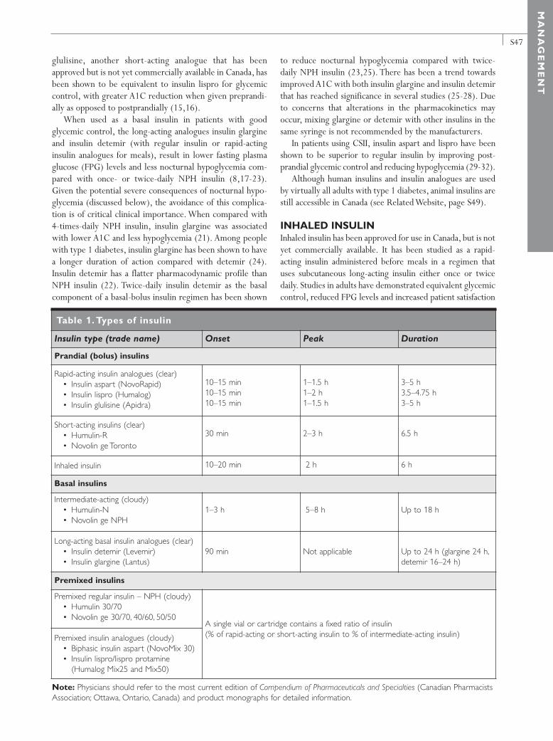
S47
glulisine, another short-acting analogue that has beenapproved but is not yet commercially available in Canada, hasbeen shown to be equivalent to insulin lispro for glycemiccontrol, with greater A1C reduction when given preprandi-ally as opposed to postprandially (15,16).
When used as a basal insulin in patients with goodglycemic control, the long-acting analogues insulin glargineand insulin detemir (with regular insulin or rapid-actinginsulin analogues for meals), result in lower fasting plasmaglucose (FPG) levels and less nocturnal hypoglycemia com-pared with once- or twice-daily NPH insulin (8,17-23).Given the potential severe consequences of nocturnal hypo-glycemia (discussed below), the avoidance of this complica-tion is of critical clinical importance. When compared with4-times-daily NPH insulin, insulin glargine was associatedwith lower A1C and less hypoglycemia (21). Among peoplewith type 1 diabetes, insulin glargine has been shown to havea longer duration of action compared with detemir (24).Insulin detemir has a flatter pharmacodynamic profile thanNPH insulin (22). Twice-daily insulin detemir as the basalcomponent of a basal-bolus insulin regimen has been shown
to reduce nocturnal hypoglycemia compared with twice-daily NPH insulin (23,25). There has been a trend towardsimproved A1C with both insulin glargine and insulin detemirthat has reached significance in several studies (25-28). Dueto concerns that alterations in the pharmacokinetics mayoccur, mixing glargine or detemir with other insulins in thesame syringe is not recommended by the manufacturers.
In patients using CSII, insulin aspart and lispro have beenshown to be superior to regular insulin by improving post-prandial glycemic control and reducing hypoglycemia (29-32).
Although human insulins and insulin analogues are usedby virtually all adults with type 1 diabetes, animal insulins arestill accessible in Canada (see Related Website, page S49).
INHALED INSULINInhaled insulin has been approved for use in Canada, but is notyet commercially available. It has been studied as a rapid-acting insulin administered before meals in a regimen thatuses subcutaneous long-acting insulin either once or twicedaily. Studies in adults have demonstrated equivalent glycemiccontrol, reduced FPG levels and increased patient satisfaction
MA
NA
GE
ME
NT
Note: Physicians should refer to the most current edition of Compendium of Pharmaceuticals and Specialties (Canadian PharmacistsAssociation; Ottawa, Ontario, Canada) and product monographs for detailed information.
Table 1. Types of insulin
Insulin type (trade name) Onset Peak Duration
Prandial (bolus) insulins
Rapid-acting insulin analogues (clear)• Insulin aspart (NovoRapid)• Insulin lispro (Humalog)• Insulin glulisine (Apidra)
Short-acting insulins (clear)• Humulin-R• Novolin ge Toronto
Inhaled insulin
10–15 min10–15 min10–15 min
30 min
10–20 min
1–1.5 h1–2 h1–1.5 h
2–3 h
2 h
3–5 h3.5–4.75 h3–5 h
6.5 h
6 h
Basal insulins
Intermediate-acting (cloudy)• Humulin-N• Novolin ge NPH
Long-acting basal insulin analogues (clear)• Insulin detemir (Levemir)• Insulin glargine (Lantus)
1–3 h
90 min
5–8 h
Not applicable
Up to 18 h
Up to 24 h (glargine 24 h,detemir 16–24 h)
Premixed insulins
Premixed regular insulin – NPH (cloudy)• Humulin 30/70• Novolin ge 30/70, 40/60, 50/50
Premixed insulin analogues (cloudy)• Biphasic insulin aspart (NovoMix 30)• Insulin lispro/lispro protamine
(Humalog Mix25 and Mix50)
A single vial or cartridge contains a fixed ratio of insulin (% of rapid-acting or short-acting insulin to % of intermediate-acting insulin)
S48
compared with subcutaneous short-acting or rapid-actinginsulins (33-36). The short-term safety data demonstrate noclinically significant pulmonary dysfunction (33,34,37). It isrecommended, however, that inhaled insulin not be used inthose with abnormal baseline spirometry (i.e. forced expira-tory volume in 1 second [FEV1] <70% predicted) (38).
HYPOGLYCEMIAInsulin-induced hypoglycemia is a major obstacle for indi-viduals trying to achieve glycemic targets. Hypoglycemiacan be severe and result in confusion, coma or seizure,requiring the assistance of other individuals. Significant riskof hypoglycemia often necessitates less stringent glycemicgoals. The negative social and emotional impact of hypo-glycemia may make patients reluctant to intensify therapy.The diabetes healthcare team should review the patient’sexperience with hypoglycemia at each visit. This shouldinclude an estimate of cause, frequency, symptoms, recogni-tion, severity and treatment.
Intensive vs. conventional insulin therapy Hypoglycemia is the most common adverse effect of inten-sive insulin therapy in patients with type 1 diabetes. In theDCCT, 35% of patients in the conventional treatment groupand 65% in the intensive group experienced at least 1episode of severe hypoglycemia (39,40). In a meta-analysis of14 trials, the median incidence of severe hypoglycemia was4.6 and 7.9 episodes per 100 patient-years in the conven-tionally treated and intensively treated patients, respectively(41). Studies have suggested that with adequate self-manage-ment education, appropriate glycemic targets, SMBG andprofessional support, intensive therapy may result in lesshypoglycemia than reported in the DCCT (42-45).
Rapid-acting insulin analogues vs. regular insulinAlthough there are no differences in the magnitude and tem-poral pattern of the physiologic, symptomatic and counter-regulatory hormonal responses to hypoglycemia induced byregular human insulin or rapid-acting analogues (46,47), thefrequency of hypoglycemic events has been shown to bereduced with rapid-acting insulin analogues compared withregular insulin (8-11).
Long-acting insulin analogues vs. intermediate-acting insulinsStudies have shown reduced incidence of nocturnal hypo-glycemia when a long-acting insulin analogue is used in lieuof an intermediate-acting insulin as the basal insulin (48-51).This is an important clinical consideration, as nocturnalhypoglycemia has potential for significant adverse effects.
Lifestyle factorsDeviations from recommended or appropriate self-manage-ment behaviours (such as eating less food, taking more insulin,
engaging in more activity) account for 85% of hypoglycemicepisodes (52,53). For patients managed with fixed-doseinsulin regimens, care should be taken to develop an individu-alized meal and activity plan that the person can and will fol-low (54). Adding bedtime snacks may be helpful to avoidnocturnal hypoglycemia among those taking NPH as the basalinsulin, or in those individuals at high risk of severe hypo-glycemia (regardless of insulin type), particularly when bed-time plasma glucose (PG) levels are <7.0 mmol/L (55,56).
Knowledge of the acute effects of exercise is mandatory.Low- to moderate-intensity exercise lowers blood glucose(BG) levels both during and after the activity, increasing therisk of a hypoglycemic episode. These effects on BG levelscan be modified by altering diet, insulin and the type andtiming of exercise. In contrast, high-intensity exercise raisesBG levels during and immediately after the event. SMBGbefore, during and, especially for many hours after exerciseis important for establishing response to exercise and guidingthe appropriate management of exercise. If ketosis is present(urine ketone level >8.0 mmol/L or blood ketone level>3.0 mmol/L), exercise should not be performed, as meta-bolic deterioration will occur (57).
Hypoglycemia unawareness and nocturnal hypoglycemia Asymptomatic hypoglycemia is the presence of a biochemi-cally documented low BG level without any symptoms.Hypoglycemia unawareness occurs when the threshold forthe development of autonomic warning symptoms is close toor lower than the threshold for the neuroglycopenic symp-toms, such that the first signs of hypoglycemia will often beconfusion or loss of consciousness. Severe hypoglycemic reac-tions are the primary barrier to achieving glycemic targets in people with type 1 diabetes (58). Severe hypoglycemicepisodes occur frequently during sleep or in the absence ofhypoglycemia awareness that alerts patients to take actions tocorrect their BG levels (59,60). The sympathoadrenalresponse to hypoglycemia is reduced during sleep (61).Asymptomatic nocturnal hypoglycemia is common and oftenlasts >4 hours (59,62-65). Severe hypoglycemia, resulting inseizures, is more likely to occur at night than during the day(66). To reduce the risk of asymptomatic nocturnal hypo-glycemia, individuals using intensive insulin therapy shouldperiodically monitor overnight BG levels at a time that corre-sponds with the peak action time of their overnight insulin.
In people with type 1 diabetes, hypoglycemia was report-ed to occur at a mean rate of approximately 2 episodes perweek. Increasing frequency of hypoglycemia can lead to adecrease in the normal responses to hypoglycemia (67),which, in turn, can lead to decreased awareness of hypo-glycemia and defective glucose counterregulation.
Hypoglycemia unawareness and defective glucose coun-terregulation are potentially reversible. Strict avoidance ofhypoglycemia for a period of 2 days to 3 months has been
2008 CLINICAL PRACTICE GUIDELINES
S49
associated with improvement in the recognition of severehypoglycemia, in the counterregulatory hormone responses,or both (42,67-73). Structured educational and psychobe-havioural programs (e.g. blood glucose awareness training)may help improve detection of hypoglycemia and reduce fre-quency of severe hypoglycemia (74,75).
OTHER RELEVANT GUIDELINESTargets for Glycemic Control, p. S29Monitoring Glycemic Control, p. S32Pharmacologic Management of Type 2 Diabetes, p. S53Hypoglycemia, p. S62In-hospital Management of Diabetes, p. S71Management of Acute Coronary Syndromes, p. S119Type 1 Diabetes in Children and Adolescents, p. S150
Type 2 Diabetes in Children and Adolescents, p. S162Diabetes and Pregnancy, p. S168Diabetes in the Elderly, p. S181
RELATED WEBSITEHealth Canada information about animal insulin:http://hc-sc.gc.ca/dhp-mps/brgtherap/activit/fs-fi/qa_qr_insulin_02_2006_e.html Accessed September 1, 2008.
REFERENCES1. Pickup J, Mattock M, Kerry S. Glycaemic control with con-
tinuous subcutaneous insulin infusion compared with intensiveinsulin injections in patients with type 1 diabetes: meta-analy-sis of randomised controlled trials. BMJ. 2002;324:705.
2. Tsui E, Barnie A, Ross S, et al. Intensive insulin therapy withinsulin lispro: a randomized trial of continuous subcutaneousinsulin infusion versus multiple daily insulin injection. DiabetesCare. 2001;24:1722-1727.
3. DeVries JH, Snoek FJ, Kostense PJ, et al; Dutch Insulin PumpStudy Group. A randomized trial of continuous subcutaneousinsulin infusion and intensive injection therapy in type 1 dia-betes for patients with long-standing poor glycemic control.Diabetes Care. 2002;25:2074-2080.
4. Hirsch IB, Bode BW, Garg S, et al; Insulin Aspart CSII/MDIComparison Study Group. Continuous subcutaneous insulininfusion (CSII) of insulin aspart versus multiple daily injectionof insulin aspart/insulin glargine in type 1 diabetic patientspreviously treated with CSII. Diabetes Care. 2005;28:533-538.
5. Retnakaran R, Hochman J, DeVries JH, et al. Continuous sub-cutaneous insulin infusion versus multiple daily injections: theimpact of baseline A1c. Diabetes Care. 2004;27:2590-2596.
6. The Diabetes Control and Complications Trial Research Group.The effect of intensive treatment of diabetes on the develop-ment and progression of long-term complications in insulin-dependent diabetes mellitus. N Engl J Med. 1993;329:977-986.
7. Nathan DM, Cleary PA, Backlund JY, et al; Diabetes Controland Complications Trial/Epidemiology of DiabetesInterventions and Complications (DCCT/EDIC) StudyResearch Group. Intensive diabetes treatment and cardiovas-cular disease in patients with type 1 diabetes. N Engl J Med.2005;353:2643-2653.
8. DeWitt DE, Hirsch IB. Outpatient insulin therapy in type 1and type 2 diabetes mellitus: scientific review. JAMA. 2003;289:2254-2264.
9. Siebenhofer A, Plank J, Berghold A, et al. Short acting insulinanalogues versus regular human insulin in patients with dia-betes mellitus. Cochrane Database Syst Rev. 2006;(2):CD003287.
10. Heller SR, Colagiuri S, Vaaler S, et al. Hypoglycaemia withinsulin aspart: a double-blind, randomised, crossover trial insubjects with type1 diabetes. Diabet Med. 2004;21:769-775.
11. Plank J, Siebenhofer A, Berghold A, et al. Systematic reviewand meta-analysis of short-acting insulin analogues in patientswith diabetes mellitus. Arch Intern Med. 2005;165:1337-1344.
12. Schernthaner G,Wein W, Shnawa N, et al. Preprandial vs. post-
MA
NA
GE
ME
NT
Insulin regimens for type 1 diabetes1.To achieve glycemic targets in adults with type 1 dia-
betes, multiple daily insulin injections (prandial [bolus]and basal insulin) or the use of CSII as part of an inten-sive diabetes management regimen is the treatment ofchoice [Grade A, Level 1A (6)].
2. Rapid-acting insulin analogues (aspart or lispro), in com-bination with adequate basal insulin, should be consid-ered over regular insulin to improve A1C whileminimizing the occurrence of hypoglycemia [Grade B,Level 2 (9,11)] and to achieve postprandial glucose tar-gets [Grade B, Level 2 (76)].
3. Insulin aspart or insulin lispro should be used when CSIIis used in adults with type 1 diabetes [Grade B, Level 2(29,30)].
4.A long-acting insulin analogue (detemir, glargine) may beconsidered as an alternative to NPH as the basal insulin[Grade B, Level 2 (17-20)] to reduce the risk of hypo-glycemia [Grade B, Level 2 (50), for detemir; Grade C, Level3 (51), for glargine], including nocturnal hypoglycemia[Grade B, Level 2 (50), for detemir; Grade D, Consensus,for glargine].
Hypoglycemia5.All individuals with type 1 diabetes should be counselled
about the risk and prevention of insulin-induced hypo-glycemia, and risk factors for severe hypoglycemiashould be identified and addressed [Grade D, Consensus].
6. In individuals with hypoglycemia unawareness, the fol-lowing strategies should be implemented to reduce therisk of hypoglycemia and to attempt to regain hypo-glycemia awareness:• Increased frequency of SMBG, including periodic
assessment during sleeping hours [Grade D, Consensus].• Less stringent glycemic targets with avoidance of
hypoglycemia [Grade C, Level 3 (72,73)].• Consideration of a psychobehavioural intervention
program (blood glucose awareness training), if available[Grade B, Level 2 (75)].
RECOMMENDATIONS
S50
prandial insulin lispro – a comparative crossover trial inpatients with type 1 diabetes. Diabet Med. 2004;21:279-284.
13. Jovanovic L, Giammattei J, Acquistapace M, et al. Efficacycomparison between preprandial and postprandial insulinaspart administration with dose adjustment for unpredictablemeal size. Clin Ther. 2004;26:1492-1497.
14. Bott U, Ebrahim S, Hirschberger S, et al. Effect of the rapid-acting insulin analogue insulin aspart on quality of life andtreatment satisfaction in patients with type 1 diabetes. DiabetMed. 2003;20:626-634.
15. Dreyer M, Prager R, Robinson A, et al. Efficacy and safety ofinsulin glulisine in patients with type 1 diabetes. Horm MetabRes. 2005;37:702-707.
16. Garg SK, Rosenstock J,Ways K. Optimized basal-bolus insulinregimens in type 1 diabetes: insulin glulisine versus regularhuman insulin in combination with basal insulin glargine.Endocr Pract. 2005;11:11-17.
17. Warren E, Weatherley-Jones E, Chilcott J, et al. Systematicreview and economic evaluation of a long-acting insulin ana-logue, insulin glargine. Health Technol Assess. 2004;8:1-57.
18. Wang F, Carabino JM,Vergara CM. Insulin glargine: a system-atic review of a long-acting insulin analogue. Clin Ther. 2003;25:1541-1577.
19. Dunn CJ, Plosker GL, Keating GM, et al. Insulin glargine: anupdated review of its use in the management of diabetes mel-litus. Drugs. 2003;63:1743-1778.
20. Chapman TM, Perry CM. Insulin detemir: a review of its usein the management of type 1 and 2 diabetes mellitus. Drugs.2004;64:2577-2595.
21. Rossetti P, Pampanelli S, Fanelli C, et al. Intensive replacementof basal insulin in patients with type 1 diabetes given rapid-act-ing insulin analog at mealtime: a 3-month comparison betweenadministration of NPH insulin four times daily and glargineinsulin at dinner or bedtime. Diabetes Care. 2003;26:1490-1496.
22. Plank J, Bodenlenz M, Sinner F, et al. A double-blind, ran-domized, dose-response study investigating the pharmaco-dynamic and pharmacokinetic properties of the long-actinginsulin analog detemir. Diabetes Care. 2005;28:1107-1112.
23. De Leeuw I, Vague P, Selam JL, et al. Insulin detemir used inbasal-bolus therapy in people with type 1 diabetes is associat-ed with a lower risk of nocturnal hypoglycaemia and lessweight gain over 12 months in comparison to NPH insulin.Diabetes Obes Metab. 2005;7:73-82.
24. Porcellati F, Rossetti P, Busciantella NR, et al. Comparison ofpharmacokinetics and dynamics of the long-acting insulinanalogs glargine and detemir at steady state in type 1 diabetes:a double-blind, randomized, crossover study. Diabetes Care.2007;30:2447-2452.
25. Home P, Bartley P, Russell-Jones D, et al; Study to Evaluate theAdministration of Detemir Insulin Efficacy, Safety andSuitability (STEADINESS) Study Group. Insulin detemir offersimproved glycemic control compared with NPH insulin inpeople with type 1 diabetes: a randomized clinical trial.Diabetes Care. 2004;27:1081-1087.
26. Hermansen K, Fontaine P, Kukolja KK, et al. Insulin analogues(insulin detemir and insulin aspart) versus traditional humaninsulins (NPH insulin and regular human insulin) in basal-bolus therapy for patients with type 1 diabetes. Diabetologia.2004;47:622-629.
27. Kudva YC, Basu A, Jenkins GD, et al. Randomized controlledclinical trial of glargine versus ultralente insulin in the treat-ment of type 1 diabetes. Diabetes Care. 2005;28:10-14.
28. Ashwell SG, Amiel SA, Bilous RW, et al. Improved glycaemiccontrol with insulin glargine plus insulin lispro: a multicentre,randomized, cross-over trial in people with type 1 diabetes.Diabet Med. 2006;23:285-292.
29. Zinman B, Tildesley H, Chiasson JL, et al. Insulin lispro inCSII: results of a double-blind crossover study. Diabetes. 1997;46:440-443.
30. Bode B, Weinstein R, Bell D, et al. Comparison of insulinaspart with buffered regular insulin and insulin lispro in con-tinuous subcutaneous insulin infusion: a randomized study intype 1 diabetes. Diabetes Care. 2002;25:439-444.
31. Radermecker RP, Scheen AJ. Continuous subcutaneous insulininfusion with short-acting insulin analogues or human regularinsulin: efficacy, safety, quality of life, and cost-effectiveness.Diabetes Metab Res Rev. 2004;20:178-188.
32. Siebenhofer A, Plank J, Berghold A, et al. Meta-analysis ofshort-acting insulin analogues in adult patients with type 1 dia-betes: continuous subcutaneous insulin infusion versus injec-tion therapy. Diabetologia. 2004;47:1895-1905.
33. Quattrin T, Bélanger A, Bohannon NJ, et al; Exubera Phase IIIStudy Group. Efficacy and safety of inhaled insulin (Exubera)compared with subcutaneous insulin therapy in patients withtype 1 diabetes: results of a 6-month, randomized, compara-tive trial. Diabetes Care. 2004;27:2622-2627.
34. Skyler JS,Weinstock RS, Raskin P, et al; Inhaled Insulin PhaseIII Type 1 Diabetes Study Group. Use of inhaled insulin in abasal/bolus insulin regimen in type 1 diabetic subjects: a 6-month, randomized, comparative trial. Diabetes Care. 2005;28:1630-1635.
35. Gerber RA, Cappelleri JC, Kourides IA, et al.Treatment satis-faction with inhaled insulin in patients with type 1 diabetes: arandomized controlled trial. Diabetes Care. 2001;24:1556-1559.
36. Skyler JS, Jovanovic L, Klioze S, et al; Inhaled Human InsulinType 1 Diabetes Study Group.Two-year safety and efficacy ofinhaled human insulin (Exubera) in adult patients with type 1diabetes. Diabetes Care. 2007;30:579-585.
37. Royle P,Waugh N, McAuley L, et al. Inhaled insulin in diabetesmellitus. Cochrane Database Syst Rev. 2004;(3):CD003890.
38. Exubera Product Monograph. Kirkland, QC: Pfizer CanadaInc.; 2007.
39. The Diabetes Control and Complications Trial ResearchGroup. Adverse events and their association with treatmentregimens in the Diabetes Control and Complications Trial.Diabetes Care. 1995;18:1415-1427.
40. The Diabetes Control and Complications Trial Research Group.Hypoglycemia in the Diabetes Control and Complications Trial.
2008 CLINICAL PRACTICE GUIDELINES
S51
Diabetes. 1997;46:271-286.41. Egger M, Davey Smith G, Stettler C, et al. Risk of adverse
effects of intensified treatment in insulin-dependent diabetesmellitus: a meta-analysis. Diabet Med. 1997;14:919-928.
42. Fanelli CG, Epifano L, Rambotti AM, et al. Meticulous pre-vention of hypoglycemia normalizes the glycemic thresholdsand magnitude of most of neuroendocrine responses to, symp-toms of, and cognitive function during hypoglycemia in inten-sively treated patients with short-term IDDM. Diabetes.1993;42:1683-1689.
43. Bott S, Bott U, Berger M, et al. Intensified insulin therapy and therisk of severe hypoglycaemia. Diabetologia. 1997;40:926-932.
44. Ahern J, Tamborlane WV. Steps to reduce the risks of severehypoglycemia. Diabetes Spectrum. 1997;10:39-41.
45. Bolli GB. How to ameliorate the problem of hypoglycemia inintensive as well as nonintensive treatment of type 1 diabetes.Diabetes Care. 1999;22(suppl 2):B43-B52.
46. Torlone E, Fanelli C, Rambotti AM, et al. Pharmacokinetics,pharmacodynamics and glucose counterregulation followingsubcutaneous injection of the monomeric insulin analogue[Lys(B28),Pro(B29)] in IDDM. Diabetologia. 1994;37:713-720.
47. McCrimmon RJ, Frier BM. Symptomatic and physiologicalresponses to hypoglycaemia induced by human soluble insulinand the analogue lispro human insulin. Diabet Med.1997;14:929-936.
48. Garg SK, Gottlieb PA, Hisatomi ME, et al. Improved glycemiccontrol without an increase in severe hypoglycemic episodes inintensively treated patients with type 1 diabetes receivingmorning, evening, or split dose insulin glargine. Diabetes ResClin Pract. 2004;66:49-56.
49. Garg SK, Paul JM, Karsten JI, et al. Reduced severe hypo-glycemia with insulin glargine in intensively treated adultswith type 1 diabetes. Diabetes Technol Ther. 2004;6:589-595.
50. Goldman-Levine JD, Lee KW. Insulin detemir – a new basalinsulin analog. Ann Pharmacother. 2005;39:502-507.
51. Mullins P, Sharplin P,Yki-Jarvinen H, et al. Negative binomialmeta-regression analysis of combined glycosylated hemoglobinand hypoglycemia outcomes across eleven phase III and IVstudies of insulin glargine compared with neutral protamineHagedorn insulin in type 1 and type 2 diabetes mellitus. ClinTher. 2007;29:1607-1619.
52. Clarke WL, Cox DJ, Gonder-Frederick LA, et al. The rela-tionship between nonroutine use of insulin, food, and exerciseand the occurrence of hypoglycemia in adults with IDDM andvarying degrees of hypoglycemic awareness and metaboliccontrol. Diabetes Educ. 1997;23:55-58.
53. Fritsche A, Stumvoll M, Renn W, et al. Diabetes teaching pro-gram improves glycemic control and preserves perception ofhypoglycemia. Diabetes Res Clin Pract. 1998;40:129-135.
54. Cryer PE, Fisher JN, Shamoon H. Hypoglycemia. DiabetesCare. 1994;17:734-755.
55. Kaufman FR, Halvorson M, Kaufman ND. A randomized,blinded trial of uncooked cornstarch to diminish nocturnalhypoglycemia at diabetes camp. Diabetes Res Clin Pract. 1995;
30:205-209.56. Kalergis M, Schiffrin A, Gougeon R, et al. Impact of bedtime
snack composition on prevention of nocturnal hypoglycemia inadults with type 1 diabetes undergoing intensive insulin man-agement using lispro insulin before meals: a randomized, place-bo-controlled, crossover trial. Diabetes Care. 2003;26:9-15.
57. Berger M, Berchtold P, Cüppers HJ, et al. Metabolic and hor-monal effects of muscular exercise in juvenile type diabetics.Diabetologia. 1977;13:355-365.
58. Cryer PE. Banting Lecture. Hypoglycemia: the limiting factorin the management of IDDM. Diabetes. 1994;43:1378-1389.
59. The DCCT Research Group. Epidemiology of severe hypo-glycemia in the Diabetes Control and Complications Trial. AmJ Med. 1991;90:450-459.
60. Daneman D, Frank M, Perlman K, et al. Severe hypoglycemiain children with insulin-dependent diabetes mellitus: frequen-cy and predisposing factors. J Pediatr. 1989;115:681-685.
61. Berlin I, Sachon CI, Grimaldi A. Identification of factors asso-ciated with impaired hypoglycaemia awareness in patients withtype 1 and type 2 diabetes mellitus. Diabetes Metab. 2005;31:246-251.
62. Porter PA, Byrne G, Stick S, et al. Nocturnal hypoglycaemiaand sleep disturbances in young teenagers with insulindependent diabetes mellitus. Arch Dis Child. 1996;75:120-123.
63. Gale EA, Tattersall RB. Unrecognised nocturnal hypogly-caemia in insulin-treated diabetics. Lancet. 1979;1:1049-1052.
64. Beregszàszi M,Tubiana-Rufi N, Benali K, et al. Nocturnal hypo-glycemia in children and adolescents with insulin-dependentdiabetes mellitus: prevalence and risk factors. J Pediatr. 1997;131:27-33.
65. Vervoort G, Goldschmidt HM, van Doorn LG. Nocturnalblood glucose profiles in patients with type 1 diabetes mellituson multiple (> or = 4) daily insulin injection regimens. DiabetMed. 1996;13:794-799.
66. Davis EA, Keating B, Byrne GC, et al. Hypoglycemia: inci-dence and clinical predictors in a large population-based sam-ple of children and adolescents with IDDM. Diabetes Care.1997;20:22-25.
67. Ovalle F, Fanelli CG, Paramore DS, et al. Brief twice-weeklyepisodes of hypoglycemia reduce detection of clinical hypo-glycemia in type 1 diabetes mellitus. Diabetes. 1998;47:1472-1479.
68. Dagogo-Jack S, Rattarasarn C, Cryer PE. Reversal of hypo-glycemia unawareness, but not defective glucose counterregu-lation, in IDDM. Diabetes. 1994;43:1426-1434.
69. Fanelli C, Pampanelli S, Epifano L, et al. Long-term recoveryfrom unawareness, deficient counterregulation and lack ofcognitive dysfunction during hypoglycaemia, following institu-tion of rational, intensive insulin therapy in IDDM.Diabetologia. 1994;37:1265-1276.
70. Dagogo-Jack S, Fanelli CG, Cryer PE. Durable reversal ofhypoglycemia unawareness in type 1 diabetes. Diabetes Care.1999;22:866-867.
71. Davis M, Mellman M, Friedman S, et al. Recovery of epineph-
MA
NA
GE
ME
NT

S52
rine response but not hypoglycemic symptom threshold afterintensive therapy in type 1 diabetes. Am J Med. 1994;97:535-542.
72. Liu D, McManus RM, Ryan EA. Improved counter-regulatoryhormonal and symptomatic responses to hypoglycemia inpatients with insulin-dependent diabetes mellitus after 3months of less strict glycemic control. Clin Invest Med.1996;19:71-82.
73. Lingenfelser T, Buettner U, Martin J, et al. Improvement ofimpaired counterregulatory hormone response and symptomperception by short-term avoidance of hypoglycemia inIDDM. Diabetes Care. 1995;18:321-325.
74. Kinsley BT,Weinger K, Bajaj M, et al. Blood glucose awarenesstraining and epinephrine responses to hypoglycemia duringintensive treatment in type 1 diabetes. Diabetes Care. 1999;22:1022-1028.
75. Schachinger H, Hegar K, Hermanns N, et al. Randomized con-trolled clinical trial of Blood Glucose Awareness Training(BGAT III) in Switzerland and Germany. J Behav Med. 2005;28:587-594.
76. DeVries JH, Lindholm A, Jacobsen JL, et al; Tri-ContinentalInsulin Aspart Study Group. A randomized trial of insulinaspart with intensified basal NPH insulin supplementation inpeople with type 1 diabetes. Diabet Med. 2003;20:312-318.
2008 CLINICAL PRACTICE GUIDELINES

S53
MA
NA
GE
ME
NT
INTRODUCTIONAs people with type 2 diabetes form a heterogeneous group,treatment regimens and therapeutic targets should be indi-vidualized. Blood glucose (BG) levels close to the normalrange should be the goal for individuals in whom it is deemedsafe. As type 2 diabetes is characterized by insulin resistanceand ongoing decline in beta cell function, glucose levels willlikely worsen over time (1) and treatment must be dynamic.The number of available antihyperglycemic agents is expand-ing, requiring the clinician to consider many of the followingfactors when choosing medications: degree of hyperglycemia,risk of hypoglycemia, medication side effects, concomitantmedical conditions, ability to adhere to regimen and patientpreferences. Lifestyle modification, including nutritionaltherapy and physical activity, should continue to be empha-sized while pharmacotherapy is being used.
TREATMENT REGIMENS The diagnosis of type 2 diabetes is often delayed, and 20 to50% of people with type 2 diabetes present with microvascu-lar and/or macrovascular complications at the time of diagno-sis (2,3).When lifestyle interventions fail to control BG levelsadequately, pharmacologic treatment becomes necessary.
In the face of more severe hyperglycemia (i.e. glycatedhemoglobin [A1C] ≥9.0%), combinations of agents are usuallyrequired.The lag period before adding other antihyperglycemicagent(s) should be kept to a minimum, taking into account thecharacteristics of the different medications.With timely adjust-ments to and/or additions of antihyperglycemic agents, the tar-get A1C level should be attainable within 6 to 12 months.
In general, A1C will decrease by about 0.5 to 1.5% withmonotherapy, depending on the agent used and the baselineA1C level (4). Generally, the higher the baseline A1C, thegreater the A1C reduction for each given agent. In general,as A1C levels decrease toward normal levels (<7.3%), post-prandial BG control assumes greater importance for furtherA1C reduction (5).
The initial use of combinations of submaximal doses ofantihyperglycemic agents produces more rapid and improvedglycemic control and fewer side effects compared tomonotherapy at maximal doses (6-9). Furthermore, manypatients on monotherapy with the late addition of anotherantihyperglycemic agent may not readily attain target BG lev-els (1). When combining antihyperglycemic agents with orwithout insulin, classes of agents that have different mecha-nisms of action should be used. Simultaneous use of agentsfrom different classes but with similar mechanisms of action(e.g. sulfonylureas and meglitinides; and DPP-4 inhibitors) iscurrently untested and may be less effective at improvingglycemia and is not recommended at this time.
There is debate over which antihyperglycemic agent(including insulin) should be used initially and which agentsshould be added subsequently.There is also debate over whichagents within a given class might be preferred in specific situ-ations. Symptomatic patients with high BG and A1C levelsrequire agents that lower BG levels quickly (e.g. insulin).However, the issue of how to reach glycemic targets may beless important than the need to achieve that target. ImprovedBG and A1C levels are associated with better outcomes, evenif recommended glycemic targets cannot be reached (3). Eachof the agents listed in Table 1 (10-51) and Figure 1 has advan-tages and disadvantages (e.g. degree of BG lowering, risk ofhypoglycemia and nonglycemic benefits/risks).
The recommendation to use metformin as the initial agentin most patients is based on its effectiveness in lowering BG,its relatively mild side effect profile and its demonstrated ben-efit in overweight patients (52).While monotherapy with aninsulin sensitizer (thiazolidinedione [TZD]) produces morelong-lasting glycemic control compared to metformin or sul-fonylurea therapy (45), the edema, weight gain, small risk ofcongestive heart failure (CHF), increased risk of fractures inwomen (44,46) and inconsistent data regarding cardiovascu-lar outcomes (53) offset the potential for this class to be rec-ommended as first-line therapy. Although meta-analyses of
Pharmacologic Management of Type 2 DiabetesCanadian Diabetes Association Clinical Practice Guidelines Expert CommitteeThe initial draft of this chapter was prepared by William Harper MD FRCPC, Amir Hanna MBBCh FRCPC,Vincent Woo MD FRCPC, Keith G. Dawson MD PhD FRCPC, Jean-François Yale MDCSPQ, Lori MacCallum BScPhm PharmD, Maureen Clement MD CCFP, Scot Simpson BSP PharmDMSc and Maryann Hopkins BSP CDE
• If glycemic targets are not achieved within 2 to 3 months of lifestyle management, antihyperglycemic pharmacotherapy should be initiated.
• Timely adjustments to and/or additions of antihyper-glycemic agents should be made to attain target A1Cwithin 6 to 12 months.
• In patients with marked hyperglycemia (A1C ≥9.0%),antihyperglycemic agents should be initiated concomi-tantly with lifestyle management, and considerationshould be given to either initiating combination therapywith 2 agents or initiating insulin.
KEY MESSAGES
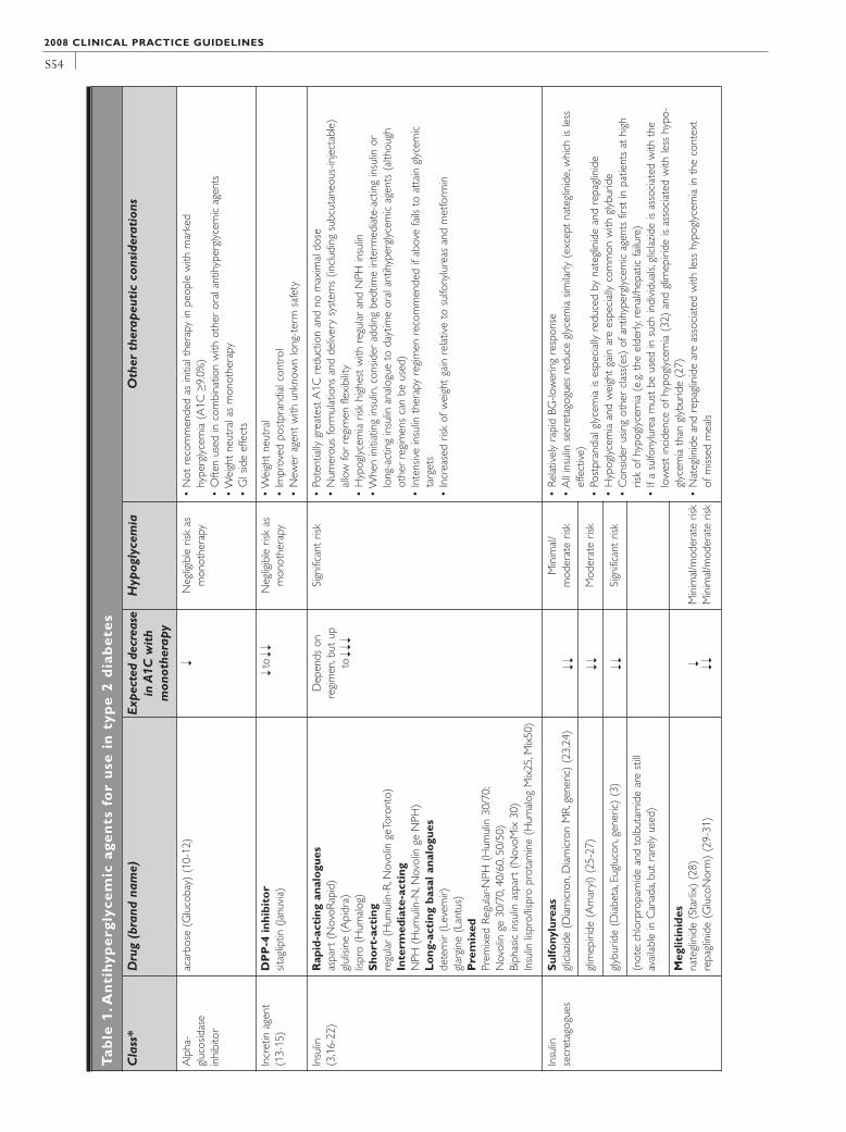
S54
2008 CLINICAL PRACTICE GUIDELINES
Tab
le1
.An
tih
ype
rgly
cem
icag
en
tsfo
ru
sein
typ
e2
dia
be
tes
Cla
ss*
Dru
g(b
rand
nam
e)Ex
pect
edde
crea
sein
A1C
wit
hm
onot
hera
py
Hyp
ogly
cem
iaO
ther
ther
apeu
tic
cons
ider
atio
ns
Alp
ha-
gluc
osid
ase
inhi
bito
r
acar
bose
(Glu
coba
y)(1
0-12
)N
eglig
ible
risk
asm
onot
hera
py•
Not
reco
mm
ende
das
initi
alth
erap
yin
peop
lew
ithm
arke
dhy
perg
lyce
mia
(A1C≥9
.0%
)•
Ofte
nus
edin
com
bina
tion
with
othe
ror
alan
tihyp
ergl
ycem
icag
ents
•W
eigh
tne
utra
las
mon
othe
rapy
•G
Isid
eef
fect
s
Incr
etin
agen
t(1
3-15
)D
PP
-4in
hibi
tor
sitag
liptin
(Janu
via)
toN
eglig
ible
risk
asm
onot
hera
py•
Wei
ght
neut
ral
•Im
prov
edpo
stpr
andi
alco
ntro
l•
New
erag
ent
with
unkn
own
long
-ter
msa
fety
Insu
lin(3
,16-
22)
Rap
id-a
ctin
gan
alog
ues
aspa
rt(N
ovoR
apid
)gl
ulisi
ne(A
pidr
a)lis
pro
(Hum
alog
)S
hort
-act
ing
regu
lar
(Hum
ulin
-R,N
ovol
inge
Toro
nto)
Inte
rmed
iate
-act
ing
NPH
(Hum
ulin
-N,N
ovol
inge
NPH
)L
ong
-act
ing
basa
lana
logu
esde
tem
ir(L
evem
ir)gl
argi
ne(L
antu
s)P
rem
ixed
Prem
ixed
Regu
lar-N
PH(H
umul
in30
/70;
Nov
olin
ge30
/70,
40/6
0,50
/50)
Biph
asic
insu
linas
part
(Nov
oMix
30)
Insu
linlis
pro/
lispr
opr
otam
ine
(Hum
alog
Mix
25,M
ix50
)
Dep
ends
onre
gim
en,b
utup
to
Sign
ifica
ntris
k•
Pote
ntia
llygr
eate
stA
1Cre
duct
ion
and
nom
axim
aldo
se•
Num
erou
sfo
rmul
atio
nsan
dde
liver
ysy
stem
s(in
clud
ing
subc
utan
eous
-inje
ctab
le)
allo
wfo
rre
gim
enfle
xibi
lity
•H
ypog
lyce
mia
risk
high
est
with
regu
lar
and
NPH
insu
lin•
Whe
nin
itiat
ing
insu
lin,c
onsid
erad
ding
bedt
ime
inte
rmed
iate
-act
ing
insu
linor
long
-act
ing
insu
linan
alog
ueto
dayt
ime
oral
antih
yper
glyc
emic
agen
ts(a
lthou
ghot
her
regi
men
sca
nbe
used
)•
Inte
nsiv
ein
sulin
ther
apy
regi
men
reco
mm
ende
dif
abov
efa
ilsto
atta
ingl
ycem
icta
rget
s•
Incr
ease
dris
kof
wei
ght
gain
rela
tive
tosu
lfony
lure
asan
dm
etfo
rmin
Insu
linse
cret
agog
ues
Sul
fony
lure
asgl
icla
zide
(Dia
mic
ron,
Dia
mic
ron
MR,
gene
ric)
(23,
24)
Min
imal
/m
oder
ate
risk
•Re
lativ
ely
rapi
dBG
-low
erin
gre
spon
se•
All
insu
linse
cret
agog
ues
redu
cegl
ycem
iasim
ilarly
(exc
ept
nate
glin
ide,
whi
chis
less
effe
ctiv
e)•
Post
pran
dial
glyc
emia
ises
peci
ally
redu
ced
byna
tegl
inid
ean
dre
pagl
inid
e•
Hyp
ogly
cem
iaan
dw
eigh
tga
inar
ees
peci
ally
com
mon
with
glyb
urid
e•
Con
sider
usin
got
her
clas
s(es
)of
antih
yper
glyc
emic
agen
tsfir
stin
patie
nts
athi
ghris
kof
hypo
glyc
emia
(e.g
.the
elde
rly,r
enal
/hep
atic
failu
re)
•If
asu
lfony
lure
am
ust
beus
edin
such
indi
vidu
als,
glic
lazi
deis
asso
ciat
edw
ithth
elo
wes
tin
cide
nce
ofhy
pogl
ycem
ia(3
2)an
dgl
imep
iride
isas
soci
ated
with
less
hypo
-gl
ycem
iath
angl
ybur
ide
(27)
•N
ateg
linid
ean
dre
pagl
inid
ear
eas
soci
ated
with
less
hypo
glyc
emia
inth
eco
ntex
tof
miss
edm
eals
glim
epiri
de(A
mar
yl)
(25-
27)
Mod
erat
eris
k
glyb
urid
e(D
iabe
ta,E
uglu
con,
gene
ric)
(3)
Sign
ifica
ntris
k
(not
e:ch
lorp
ropa
mid
ean
dto
lbut
amid
ear
est
illav
aila
ble
inC
anad
a,bu
tra
rely
used
)
Meg
litin
ides
nate
glin
ide
(Sta
rlix)
(28)
repa
glin
ide
(Glu
coN
orm
)(2
9-31
)M
inim
al/m
oder
ate
risk
Min
imal
/mod
erat
eris
k
Ý
ÝÝ
ÝÝÝ
ÝÝ
ÝÝ
ÝÝÝ
ÝÝÝ

S55
MA
NA
GE
ME
NT
Met
form
inG
luco
phag
e,G
lum
etza
,gen
eric
(33,
34)
Neg
ligib
leris
kas
mon
othe
rapy
•Im
prov
edca
rdio
vasc
ular
outc
omes
inov
erw
eigh
tsu
bjec
ts•
Con
trai
ndic
ated
ifC
rCl/e
GFR
<30
mL/
min
orhe
patic
failu
re•
Cau
tion
ifC
rCl/e
GFR
<60
mL/
min
•W
eigh
tne
utra
las
mon
othe
rapy
,pro
mot
esle
ssw
eigh
tga
inw
hen
com
bine
dw
ithot
her
antih
yper
glyc
emic
agen
ts,i
nclu
ding
insu
lin•
GIs
ide
effe
cts
TZD
s(3
5-45
)pi
oglit
azon
e(A
ctos
)ro
siglit
azon
e(A
vand
ia)
Neg
ligib
leris
kas
mon
othe
rapy
•Lo
nger
dura
tion
ofgl
ycem
icco
ntro
lwith
mon
othe
rapy
com
pare
dto
met
form
inor
glyb
urid
e•
Mild
BPlo
wer
ing
•Be
twee
n6
and
12w
eeks
requ
ired
toac
hiev
efu
llgl
ycem
icef
fect
•W
eigh
tga
in(w
aist
-to-
hip
ratio
not
incr
ease
d)•
May
indu
ceed
ema
and
orhe
art
failu
re•
Avo
idin
patie
nts
with
hear
tfa
ilure
•H
ighe
rra
tes
hear
tfa
ilure
whe
nco
mbi
ned
with
insu
lin†
•Ra
reoc
curr
ence
ofm
acul
ared
ema
•Ra
reoc
curr
ence
offr
actu
res
infe
mal
es(4
4,46
)•
Sugg
estio
nof
incr
ease
dris
kof
card
iova
scul
arev
ents
with
rosig
litaz
one
awai
tsfu
rthe
rst
udy
Ant
iobe
sity
agen
tsor
lista
t(X
enic
al)
(47-
49)
Non
e•
Prom
ote
wei
ght
loss
•G
lyce
mic
bene
fitm
aybe
limite
dto
thos
ew
hoac
tual
lylo
sew
eigh
t•
Orli
stat
can
caus
edi
arrh
eaan
dot
her
GIs
ide
effe
cts
•Si
butr
amin
eca
nin
crea
sehe
art
rate
and
BPsib
utra
min
e(M
erid
ia)
(50,
51)
Non
e
Com
bine
dfo
rmul
atio
nsA
vand
amet
(met
form
in+
rosig
litaz
one)
Neg
ligib
leris
kas
mon
othe
rapy
See
met
form
in,T
ZD
s,an
dsu
lfony
lure
as
Ava
ndar
yl(g
limep
iride
+ro
siglit
azon
e)M
oder
ate
risk
ÝÝ
ÝÝ Ý Ý
ÝÝÝ
ÝÝÝ
*List
edin
alph
abet
ical
orde
r† C
ombi
ning
insu
linw
itha
TZD
isno
tan
appr
oved
indi
catio
nin
Can
ada
:<1.
0%re
duct
ion
inA
1C:1
.0–2
.0%
redu
ctio
nin
A1C
:>2.
0%re
duct
ion
inA
1C
A1C
=gl
ycat
edhe
mog
lobi
nBG
=bl
ood
gluc
ose
BP=
bloo
dpr
essu
reC
rCl=
crea
tinin
ecl
eara
nce
eGFR
=es
timat
edgl
omer
ular
filtr
atio
nra
teG
I=ga
stro
inte
stin
alTZ
D=
thia
zolid
ined
ione
No
te:P
hysic
ians
shou
ldre
fer
toth
em
ost
rece
nted
ition
ofth
eCo
mpe
ndiu
mof
Phar
mac
eutic
als
and
Spec
ialti
es(C
anad
ian
Phar
mac
ists
Ass
ocia
tion,
Ott
awa,
Ont
ario
,Can
ada)
for
prod
uct
mon
ogra
phs
and
deta
iled
pres
crib
ing
info
rmat
ion.
ÝÝÝ
ÝÝÝ

S56
2008 CLINICAL PRACTICE GUIDELINES
If not at target
Figure 1. Management of hyperglycemia in type 2 diabetes
Timely adjustments to and/or addition of antihyperglycemic agentsshould be made to attain target A1C within 6 to 12 months
• Add another drug from a different class; or• Add bedtime basal insulin to other agent(s); or • Intensify insulin regimen
Note: Physicians should refer to the most recent edition of the Compendium of Pharmaceuticals and Specialties (CanadianPharmacists Association, Ottawa, Ontario, Canada) for product monographs and for detailed prescribing information
*Less hypoglycemia in the context of missed meals
A1C = glycated hemoglobinBP = blood pressureCHF = congestive heart failure
DPP-4 = dipeptidyl peptidase-4GI = gastrointestinalTZD = thiazolidinedione
= <1.0% decrease in A1C= 1.0–2.0% decrease in A1C= >2.0% decrease in A1C
Initiate pharmacotherapy immediately without waiting for effect from lifestyle interventions:
• Consider initiating metformin concurrently with another agent from a different class; or
• Initiate insulin
A1C <9.0%
Ý ÝÝÝÝ
Ý
A1C ≥9.0% Symptomatic hyperglycemia with metabolic decompensation
L
I
F
E
S
T
Y
L
E
If not at target
Add an agent best suited to the individual based on the advantages/disadvantages listed below and the information contained in Table 1 (agents listed in alphabetical order)
Class A1C Hypoglycemia Other advantages Other disadvantages
Alpha-glucosidaseinhibitor
Rare Improved postprandial controlWeight neutral
GI side effects
Incretin agent:DPP-4 inhibitor to Rare
Improved postprandial controlWeight neutral New agent (unknown long-term safety)
Insulin Yes No dose ceilingMany types, flexible regimens
Weight gain
Insulin secretagogue:
MeglitinideSulfonylurea
to Yes*Yes
Improved postprandial controlNewer sulfonylureas (gliclazide,glimepiride) are associated withless hypoglycemia than glyburide
Requires TID to QID dosingWeight gain
TZD Rare Durable monotherapy Requires 6–12 weeks for maximal effectWeight gainEdema, rare CHF, rare fractures in females
Weight loss agent None Weight loss GI side effects (orlistat)Increased heart rate/BP (sibutramine)
ÝÝ
Ý ÝÝÝÝ
ÝÝ
ÝÝÝ
ÝÝÝ
Initiate insulin± metformin
Initiate metformin
Lifestyle intervention (initiation of nutrition therapy and physical activity)
Clinical assessment
S57
MA
NA
GE
ME
NT
smaller underpowered studies suggested possible cardiovas-cular harm specific to TZD use (54,55) this has not beendemonstrated in larger randomized clinical trials (56-58).
In patients for whom hypoglycemia is a particular concern,agents associated with less hypoglycemia are preferred.Table 1and Figure 1 provide information to aid decision-making.
A combination of oral antihyperglycemic agents andinsulin often effectively controls glucose levels.When insulinis added to oral antihyperglycemic agent(s), a single injectionof intermediate-acting (NPH) (6,59), or an extended long-acting insulin analogue (insulin glargine or insulin detemir)(19) may be added. This approach may result in betterglycemic control with a smaller dose of insulin (60) and mayinduce less weight gain and less hypoglycemia than that seenwhen oral agents are stopped and insulin is used alone (33).The addition of bedtime insulin to metformin therapy leadsto less weight gain than insulin plus a sulfonylurea or twice-daily NPH insulin (16).While combining insulin with a TZDis not an approved indication in Canada, the addition of suchagents to insulin in carefully selected patients improvesglycemic control and reduces insulin requirements (61).Such combination can result in increased weight, fluid reten-tion and, in few patients, CHF. Inhaled insulin (approved, butnot yet available in Canada) can also be added to oral antihy-perglycemic therapy to help control BG levels, but can cause
cough and slight reductions in pulmonary function tests (62).The use of inhaled insulin should be restricted to non-smokers and those without respiratory disorders. Pulmonaryfunction tests should be done at baseline, 6 months and annu-ally during inhaled insulin therapy.
Insulin can be used at diagnosis in individuals with markedhyperglycemia and can be used temporarily during illness,pregnancy, stress, or for a medical procedure or surgery.Thereis no evidence that exogenous insulin accelerates the risk ofmacrovascular complications of diabetes, and its appropriateuse should be encouraged (63).When insulin is used in type 2diabetes, the insulin regimen should be tailored to achieve goodmetabolic control while trying to avoid severe hypoglycemia.With intensive glycemic control, there is an increased risk ofhypoglycemia, but this risk is lower in people with type 2 dia-betes than in those with type 1 diabetes.The number of insulininjections (1–4 per day) and the timing of injections may varydepending on each individual’s situation (64).The reduction inA1C achieved with insulin therapy depends on the dose andnumber of injections per day of insulin.
As type 2 diabetes progresses, insulin doses will likely needto be increased, additional doses of basal insulin (intermediate-acting or long-acting analogues) may need to be added, andprandial insulin (short-acting or rapid-acting analogues orinhaled insulin) may also be required.
1. In people with type 2 diabetes, if glycemic targets are notachieved using lifestyle management within 2 to 3 months,antihyperglycemic agents should be initiated [Grade A, Level1A (3)]. In the presence of marked hyperglycemia (A1C≥9.0%), antihyperglycemic agents should be initiated con-comitantly with lifestyle management, and considerationshould be given to initiating combination therapy with 2agents or initiating insulin treatment in symptomatic indi-viduals [Grade D, Consensus].
2. If glycemic targets are not attained when a single antihy-perglycemic agent is used initially, an antihyperglycemicagent or agents from different classes should be added.The lag period before adding other agent(s) should bekept to a minimum, taking into account the characteristicsof the different agents.Timely adjustments to and/or addi-tions of antihyperglycemic agents should be made in order to attain target A1C within 6 to 12 months[Grade D, Consensus].
3. Pharmacological treatment regimens should be individual-ized taking into consideration the degree of hyperglycemiaand the properties of the antihyperglycemic agents includ-ing: effectiveness in lowering BG, durability of glycemiccontrol, side effects, contraindications, risk of hypo-glycemia, presence of diabetes complications or comor-bidities, and patient preferences [Grade D, Consensus].The following factors and the information shown in Table 1 and Figure 1 should also be taken into account:• Metformin should be the initial drug used in both over-
weight patients [Grade A, Level 1A (52)] and nonover-
weight patients [Grade D, Consensus].• Other classes of antihyperglycemic agents, including
insulin, should be added to metformin, or used in com-bination with each other, if glycemic targets are not met, taking into account the information in Figure 1 and Table 1 [Grade D, Consensus].
4. When basal insulin is added to antihyperglycemic agents, long-acting analogues (insulin detemir or insulinglargine) may be considered instead of NPH to reducethe risk of nocturnal and symptomatic hypoglycemia[Grade A, Level 1A (71)].
5. The following antihyperglycemic agents (listed in alphabet-ical order), should be considered to lower postprandialBG levels:• Alpha-glucosidase inhibitor [Grade B, Level 2 (10)]• Premixed insulin analogues (i.e. biphasic insulin aspart
and insulin lispro/protamine) instead of regular/NPH premixtures [Grade B, Level 2 (72,73)]
• DPP-4 inhibitor [Grade A, Level 1 (13,14,74)].• Inhaled insulin [Grade B, Level 2 (20)].• Meglitinides (repaglinide, nateglinide) instead of
sulfonylureas [Grade B, Level 2 (75,76)]• Rapid-acting insulin analogues (aspart, glulisine, lispro)
instead of short-acting insulin (i.e. regular insulin) [GradeB, Level 2 (21,77,78)].
6. All individuals with type 2 diabetes currently using orstarting therapy with insulin or insulin secretagoguesshould be counselled about the recognition and preven-tion of drug-induced hypoglycemia [Grade D, Consensus].
RECOMMENDATIONS
S58
2008 CLINICAL PRACTICE GUIDELINES
HYPOGLYCEMIA Medication-induced hypoglycemia is the most commoncause of hypoglycemia. It is estimated that hypoglycemia ofany severity occurs annually in up to approximately 20% ofpatients taking insulin secretagogues (65). Although thesehypoglycemic episodes are rarely fatal, they can be associat-ed with serious clinical sequelae.Therefore, it is important toprevent, recognize and treat hypoglycemic episodes second-ary to the use of insulin secretagogues. Few large, random-ized clinical trials have compared the rates of hypoglycemiabetween these agents.
In the United Kingdom Prospective Diabetes Study(UKPDS), the proportion of adults with type 2 diabetes whoexperienced a severe hypoglycemic episode per year was sig-nificantly higher in the intensive group than in the conven-tional group (3), particularly for patients using insulintherapy.Although the risk of hypoglycemia was less than thatseen in the patients with type 1 diabetes in the DiabetesControl and Complications Trial, each year approximately3% of patients treated with insulin in the UKPDS experi-enced a severe hypoglycemic episode, and 40% had a hypo-glycemic episode of any severity (3).
Lower rates of hypoglycemia have been observed in somestudies of patients with type 2 diabetes treated with rapid-acting insulin analogues (insulin aspart, insulin lispro, insulinglulisine) compared to those treated with short-acting (reg-ular) insulin (66,67). Use of long-acting basal insulin ana-logues (insulin detemir, insulin glargine) reduces the risk ofnocturnal hypoglycemia compared to treatment with NPHinsulin (19,68-70).
OTHER RELEVANT GUIDELINESTargets for Glycemic Control, p. S29Insulin Therapy in Type 1 Diabetes, p. S46Hypoglycemia, p. S62Management of Obesity in Diabetes, p. S77Type 2 Diabetes in Children and Adolescents, p. S162Diabetes and Pregnancy, p. S168Diabetes in the Elderly, p. S181
RELEVANT APPENDIXAppendix 3: Examples of Insulin Initiation and TitrationRegimens in People With Type 2 Diabetes
REFERENCES1. Turner RC, Cull CA, Frighi V, et al. Glycemic control with
diet, sulfonylurea, metformin, or insulin in patients with type2 diabetes mellitus: progressive requirement for multiple ther-apies (UKPDS 49). JAMA. 1999;281:2005-2012.
2. Klein R, Klein BE, Moss SE, et al. The Wisconsin epidemio-logical study of diabetic retinopathy. III. Prevalence and risk ofdiabetic retinopathy when age at diagnosis is 30 or more years.Arch Ophthalmol. 1984;102:527-532.
3. UK Prospective Diabetes Study (UKPDS) Group. Intensive
blood-glucose control with sulphonylureas or insulin com-pared with conventional treatment and risk of complications inpatients with type 2 diabetes (UKPDS 33). Lancet. 1998;352:837-853.
4. Bloomgarden ZT, Dodis R, Viscoli CM, et al. Lower baselineglycemia reduces apparent oral agent glucose-lowering efficacy:a meta-regression analysis. Diabetes Care. 2006;29:2137-2139.
5. Monnier L, Lapinski H, Colette C. Contributions of fastingand postprandial plasma glucose increments to the overalldiurnal hyperglycemia of type 2 diabetic patients: variationswith increasing levels of HbA(1c). Diabetes Care. 2003;26:881-885.
6. Garber AJ, Larsen J, Schneider SH, et al. Simultaneous gly-buride/metformin therapy is superior to componentmonotherapy as an initial pharmacological treatment for type2 diabetes. Diabetes Obes Metab. 2002;4:201-208.
7. Rosenstock J, Goldstein BJ,Vinik AI, et al. Effect of early addi-tion of rosiglitazone to sulphonylurea therapy in older type 2diabetes patients (>60 years): the Rosiglitazone Early vs.Sulphonylurea Titration (RESULT) study. Diabetes Obes Metab.2006;8:49-57.
8. Rosenstock J, Rood J, Cobitz A, et al. Initial treatment withrosiglitazone/metformin fixed-dose combination therapycompared with monotherapy with either rosiglitazone or met-formin in patients with uncontrolled type 2 diabetes. DiabetesObes Metab. 2006;8:650-660.
9. Rosenstock J, Rood J, Cobitz A, et al. Improvement in gly-caemic control with rosiglitazone/metformin fixed-dose com-bination therapy in patients with type 2 diabetes with verypoor glycaemic control. Diabetes Obes Metab. 2006;8:643-649.
10. Chiasson J-L, Josse RG, Hunt JA, et al.The efficacy of acarbosein the treatment of patients with non-insulin-dependent dia-betes mellitus. A multicenter controlled clinical trial. AnnIntern Med. 1994;121:928-935.
11. Hoffmann J, Spengler M. Efficacy of 24-week monotherapywith acarbose, glibenclamide, or placebo in NIDDM patients.The Essen Study. Diabetes Care. 1994;17:561-566.
12. Holman RR, Cull CA,Turner RC. A randomized double-blindtrial of acarbose in type 2 diabetes shows improved glycemiccontrol over 3 years (U.K. Prospective Diabetes Study 44).Diabetes Care. 1999;22:960-964.
13. Aschner P, Kipnes MS, Lunceford JK, et al. Effect of the dipep-tidyl peptidase-4 inhibitor sitagliptin as monotherapy onglycemic control in patients with type 2 diabetes. Diabetes Care.2006;29:2632-2637.
14. Charbonnel B, Karasik A, Liu J, et al. Efficacy and safety of thedipeptidyl peptidase-4 inhibitor sitagliptin added to ongoingmetformin therapy in patients with type 2 diabetes inade-quately controlled with metformin alone. Diabetes Care. 2006;29:2638-2643.
15. Bosi E, Camisasca RP, Collober C, et al. Effects of vildagliptinon glucose control over 24 weeks in patients with type 2 dia-betes inadequately controlled with metformin. Diabetes Care.2007;30:890-895.

S59
MA
NA
GE
ME
NT
16. Yki-Järvinen H, Ryysy L, Nikkilä K, et al. Comparison of bed-time insulin regimens in patients with type 2 diabetes mellitus.A randomized, controlled trial. Ann Intern Med. 1999;130:389-396.
17. Wright A, Burden AC, Paisey RB, et al. Sulfonylurea inade-quacy: efficacy of addition of insulin over 6 years in patientswith type 2 diabetes in the U.K. Prospective Diabetes Study(UKPDS 57). Diabetes Care. 2002;25:330-336.
18. Ohkubo Y, Kishikawa H, Araki E, et al. Intensive insulin thera-py prevents the progression of diabetic microvascular compli-cations in Japanese patients with non-insulin-dependentdiabetes mellitus: a randomized prospective 6-year study.Diabetes Res Clin Pract. 1995;28:103-117.
19. Rosenstock J, Schwartz SL, Clark CM Jr, et al. Basal insulintherapy in type 2 diabetes: 28-week comparison of insulinglargine (HOE 901) and NPH insulin. Diabetes Care. 2001;24:631-636.
20. Weiss SR, Cheng SL, Kourides IA, et al, for the Inhaled InsulinPhase II Study Group. Inhaled insulin provides improvedglycemic control in patients with type 2 diabetes mellitus inad-equately controlled with oral agents: a randomized controlledtrial. Arch Intern Med. 2003;27:2277-2282.
21. Dailey G, Rosenstock J, Moses RG, et al. Insulin glulisine pro-vides improved glycemic control in patients with type 2 dia-betes. Diabetes Care. 2004;27:2363-2368.
22. Haak T,Tiengo A, Draeger E, et al. Lower within-subject vari-ability of fasting blood glucose and reduced weight gain withinsulin detemir compared to NPH insulin in patients with type2 diabetes. Diabetes Obes Metab. 2005;7:56-64.
23. Harrower A. Gliclazide modified release: from once-dailyadministration to 24-hour blood glucose control. Metabolism.2000;49(10 suppl 2):7-11.
24. Tessier D, Dawson K, Tétrault JP, et al. Glibenclamide vs gli-clazide in type 2 diabetes of the elderly. Diabet Med. 1994;11:974-980.
25. Schade DS, Jovanovic L, Schneider J. A placebo-controlled,randomized study of glimepiride in patients with type 2 dia-betes mellitus for whom diet therapy is unsuccessful. J ClinPharmacol. 1998;38:636-641.
26. Dills DG, Schneider J. Clinical evaluation of glimepiride ver-sus glyburide in NIDDM in a double-blind comparative study.Horm Metab Res. 1996;28:426-429.
27. Holstein A, Plaschke A, Egberts EH. Lower incidence of severehypoglycaemia in patients with type 2 diabetes treated withglimepiride versus glibenclamide. Diabetes Metab Res Rev. 2001;17:467-473.
28. Horton ES, Clinkingbeard C, Gatlin M, et al. Nateglinidealone and in combination with metformin improves glycemiccontrol by reducing mealtime glucose levels in type 2 diabetes.Diabetes Care. 2000;23:1660-1665.
29. Wolffenbuttel BHR, Landgraf R.A 1-year multicenter random-ized double-blind comparison of repaglinide and glyburide forthe treatment of type 2 diabetes. Diabetes Care. 1999;22:463-467.
30. Moses R, Slobodniuk R, Boyages S, et al. Effect of repaglinideaddition to metformin monotherapy on glycemic control inpatients with type 2 diabetes. Diabetes Care. 1999;22:119-124.
31. Damsbo P, Clauson P, Marbury TC, et al. A double-blind ran-domized comparison of meal-related glycemic control byrepaglinide and glyburide in well-controlled type 2 diabeticpatients. Diabetes Care. 1999;22:789-794.
32. Schernthaner G, Grimaldi A, Di Mario U, et al. GUIDE study:double-blind comparison of once-daily gliclazide MR andglimepiride in type 2 diabetic patients. Eur J Clin Invest. 2004;34:535-542.
33. United Kingdom Prospective Diabetes Study Group. UnitedKingdom Prospective Diabetes Study 24: a 6-year, random-ized, controlled trial comparing sulfonylurea, insulin, andmetformin therapy in patients with newly diagnosed type 2diabetes that could not be controlled with diet therapy. AnnIntern Med. 1998;128:165-175.
34. Garber AJ, Duncan TG, Goodman AM, et al. Efficacy of met-formin in type II diabetes: results of a double-blind, placebo-controlled, dose-response trial. Am J Med. 1997;103:491-497.
35. Aronoff S, Rosenblatt S, Braithwaite S, et al. Pioglitazonehydrochloride monotherapy improves glycemic control in thetreatment of patients with type 2 diabetes: a 6-month ran-domized placebo-controlled dose-response study. DiabetesCare. 2000;23:1605-1611.
36. Raskin P, Rappaport EB, Cole ST, et al. Rosiglitazone short-term monotherapy lowers fasting and post-prandial glucose inpatients with type II diabetes. Diabetologia. 2000;43:278-284.
37. Nolan JJ, Jones NP, Patwardhan R, et al. Rosiglitazone takenonce daily provides effective glycaemic control in patients withtype 2 diabetes mellitus. Diabet Med. 2000;17:287-294.
38. Lebovitz HE, Dole JF, Patwardhan R, et al. Rosiglitazonemonotherapy is effective in patients with type 2 diabetes. J ClinEndocrinol Metab. 2001;86:280-288.
39. Fonseca V, Rosenstock J, Patwardhan R, et al. Effect of met-formin and rosiglitazone combination therapy in patients withtype 2 diabetes mellitus: a randomized controlled trial. JAMA.2000;283:1695-1702.
40. Kipnes MS, Krosnick A, Rendell MS, et al. Pioglitazonehydrochloride in combination with sulfonylurea therapyimproves glycemic control in patients with type 2 diabetesmellitus: a randomized, placebo-controlled study. Am J Med.2001;111:10-17.
41. Einhorn D, Rendell M, Rosenzweig J, et al. Pioglitazonehydrochloride in combination with metformin in the treat-ment of type 2 diabetes mellitus: a randomized, placebo-con-trolled study. Clin Ther. 2000;22:1395-1409.
42. Yale J-F,Valiquett TR, Ghazzi MN, et al.The effect of a thiazo-lidinedione drug, troglitazone, on glycemia in patients withtype 2 diabetes mellitus poorly controlled with sulfonylureaand metformin. A multicenter, randomized, double-blind,placebo-controlled trial. Ann Intern Med. 2001;134:737-745.
43. Schwartz S, Raskin P, Fonseca V, et al. Effect of troglitazone ininsulin-treated patients with type II diabetes mellitus. N Engl J
S60
2008 CLINICAL PRACTICE GUIDELINES
Med. 1998;338:861-866.44. Meymeh RH, Wooltorton E. Diabetes drug pioglitazone
(Actos): risk of fracture. CMAJ. 2007;177:723-724.45. Kahn SE, Haffner SM, Heise MA, et al. Glycemic durability of
rosiglitazone, metformin, or glyburide monotherapy. N Engl JMed. 2006;355:2427-2443.
46. Kahn SE, Zinman B, Lachin JM, et al; A Diabetes OutcomeProgression Trial (ADOPT) Study Group. Rosiglitazone-asso-ciated fractures in type 2 diabetes: an analysis from A DiabetesOutcome Progression Trial. Diabetes Care. 2008;31:845-851.
47. Miles JM, Leiter L, Hollander P, et al. Effect of orlistat in over-weight and obese patients with type 2 diabetes treated withmetformin. Diabetes Care. 2002;25:1123-1128.
48. Kelley DE, Bray GA, Pi-Sunyer FX, et al. Clinical efficacy oforlistat therapy in overweight and obese patients with insulin-treated type 2 diabetes: A 1-year randomized controlled trial.Diabetes Care. 2002;25:1033-1041.
49. Hollander PA, Elbein SC, Hirsch IB, et al. Role of orlistat inthe treatment of obese patients with type 2 diabetes. A 1-yearrandomized double-blind study. Diabetes Care. 1998;21:1288-1294.
50. Vettor R, Serra R, Fabris R, Pagano C, Federspil G. Effect ofsibutramine on weight management and metabolic control intype 2 diabetes: a meta-analysis of clinical studies. DiabetesCare. 2005;28:942-999.
51. Finer N, Bloom SR, Frost GS, et al. Sibutramine is effective forweight loss and diabetic control in obesity with type 2 dia-betes: a randomised, double-blind, placebo-controlled study.Diabetes Obes Metab. 2000;2:105-112.
52. UK Prospective Diabetes Study (UKPDS) Group. Effect ofintensive blood-glucose control with metformin on complica-tions in overweight patients with type 2 diabetes (UKPDS 34).Lancet. 1998;352:854-865.
53. Avandia – Highlights of Prescribing Information. March 2008.Research Triangle Park, NC: GlaxoSmithKline. Available at:http://www.gsk.com/products/prescription_medicines/us/medicines-ae.htm. Accessed September 1, 2008.
54. Nissen SE, Wolski K. Effect of rosiglitazone on the risk ofmyocardial infarction and death from cardiovascular causes.N Engl J Med. 2007;356:2457-2471.
55. Singh S, Loke YK, Furberg CD. Long-term risk of cardiovascu-lar events with rosiglitazone: a meta-analysis. JAMA. 2007;298:1189-1195.
56. Dormandy JA, Charbonnel B, Eckland DJ, et al. Secondaryprevention of macrovascular events in patients with type 2 dia-betes in the PROactive Study (PROspective pioglitAzoneClinical Trial In macroVascular Events): a randomised con-trolled trial. Lancet. 2005;366:1279-1289.
57. Home PD, Pocock SJ, Beck-Nielsen H, et al. RosiglitazoneEvaluated for Cardiovascular Outcomes – An interim analysis.N Engl J Med. 2007;357:28-38.
58. The Action to Control Cardiovascular Risk in Diabetes StudyGroup. Effects of intensive glucose lowering in type 2 dia-betes. N Engl J Med. 2008;358:2545-2559.
59. Yki-Järvinen H, Kauppila M, Kujansuu E, et al. Comparison ofinsulin regimens in patients with non-insulin-dependent dia-betes mellitus. N Engl J Med. 1992;327:1426-1433.
60. Johnson JL, Wolf SL, Kabadi UM. Efficacy of insulin and sul-fonylurea combination therapy in type II diabetes. A meta-analysis of the randomized placebo-controlled trials. ArchIntern Med. 1996;156:259-264.
61. Yu JG, Krusynska MT, Mulford MI, et al. A comparison oftroglitazone and metformin on insulin requirements in eug-lycemic intensively insulin-treated type 2 diabetic patients.Diabetes. 1999;48:2414-2421.
62. Barnett AH, Dreyer M, Lange P, Serdarevic-Pehar M.An open,randomized, parallel-group study to compare the efficacy andsafety profile of inhaled human insulin (Exubera) with met-formin as adjunctive therapy in patients with type 2 diabetespoorly controlled on a sulfonylurea. Diabetes Care. 2006;29:1282-1287.
63. American Diabetes Association. Implications of the UnitedKingdom Prospective Diabetes Study. Diabetes Care. 1998;21:2180-2184.
64. Abraira C, Colwell JA, Nuttall FQ, et al. Veterans AffairsCooperative Study on glycemic control and complications intype II diabetes (VA CSDM). Results of the feasibility trial.Diabetes Care. 1995;18:1113-1123.
65. Jennings AM, Wilson RM, Ward JD. Symptomatic hypo-glycemia in NIDDM patients treated with oral hypoglycemicagents. Diabetes Care. 1989;12:203-208.
66. Anderson JH Jr, Brunelle RL, Keohane P, et al. Mealtime treat-ment with insulin analog improves postprandial hyperglycemiaand hypoglycemia in patients with non-insulin-dependent dia-betes mellitus. Arch Intern Med. 1997;157:1249-1255.
67. Anderson JH Jr, Brunelle RL, Koivisto VA, et al. Improvedmealtime treatment of diabetes mellitus using an insulin ana-logue. Clin Ther. 1997;19:62-72.
68. Yki-Järvinen H, Dressler A, Ziemen M. Less nocturnal hypo-glycemia and better post-dinner glucose control with bedtimeinsulin glargine compared with bedtime NPH insulin duringinsulin combination therapy in type 2 diabetes. Diabetes Care.2000;23:1130-1136.
69. Fritsche A, Schweitzer MA, Haring HU, et al. Glimepiridecombined with morning insulin glargine, bedtime neutral pro-tamine hagedorn insulin, or bedtime insulin glargine inpatients with type 2 diabetes. A randomized, controlled trial.Ann Int Med. 2003;138:952-959.
70. Janka HU, Plewe G, Riddle MC, et al. Comparison of basalinsulin added to oral agents versus twice-daily premixedinsulin as initial insulin therapy for type 2 diabetes. DiabetesCare. 2005;28:254-259.
71. Horvath K, Jeitler K, Berghold A, et al. A long-acting insulinanalogues versus NPH insulin (human isophane insulin) fortype 2 diabetes mellitus (Review). Cochrane Database Syst Rev.2007;(2):CD005613.
72. Roach P,Yue L, Arora V. Improved postprandial glycemic con-trol during treatment with Humalog Mix25, a novel prota-
S61
MA
NA
GE
ME
NT
mine-based insulin lispro formulation. Humalog Mix25 StudyGroup. Diabetes Care. 1999;22:1258-1261.
73. Boehm BO, Home PD, Behrend C, et al. Premixed insulinaspart 30 vs. premixed human insulin 30/70 twice daily: a ran-domized trial in type 1 and type 2 diabetic patients. Diabet Med.2002;19:393-399.
74. Ahren B, Gomis R, Standl E, et al. Twelve- and 52-week effi-cacy of the dipeptidyl peptidase IV inhibitor LAF237 in met-formin-treated patients with type 2 diabetes. Diabetes Care.2004;27:2874-2880.
75. Derosa G, Mugellini A, Ciccarelli L, et al. Comparisonbetween repaglinide and glimepiride in patients with type 2diabetes mellitus: a one-year, randomized, double-blind assess-ment of metabolic parameters and cardiovascular risk factors.Clin Ther. 2003;25:472-484.
76. Ristic S, Collober-Maugeais C, Pecher E, et al. Comparison ofnateglinide and gliclazide in combination with metformin, fortreatment of patients with Type 2 diabetes mellitus inade-quately controlled on maximum doses of metformin alone.Diabet Med. 2006;23:757-762.
77. Ross SA, Zinman B, Campos RV, et al. A comparative study ofinsulin lispro and human regular insulin in patients with type 2diabetes mellitus and secondary failure of oral hypoglycemicagents. Clin Invest Med. 2001;24:292-298.
78. Rosenfalck AM,Thorsby P, Kjems L, et al. Improved postpran-dial glycaemic control with insulin aspart in type 2 diabeticpatients treated with insulin. Acta Diabetol. 2000;37:41-46.

S62
INTRODUCTIONDrug-induced hypoglycemia is a major obstacle for individu-als (especially those with type 1 diabetes) trying to achieveglycemic targets. Hypoglycemia can be severe and result inconfusion, coma or seizure requiring the assistance of otherindividuals. Significant risk of hypoglycemia often necessi-tates less stringent glycemic goals. The negative social andemotional impact of hypoglycemia may make patients reluc-tant to intensify therapy. As such, it is important to prevent,recognize and treat hypoglycemic episodes secondary to theuse of insulin or insulin secretagogues. (See “Insulin Therapyin Type 1 Diabetes,” p. S46, and “Pharmacologic Managementof Type 2 Diabetes,” p. S53, for further discussion of drug-induced hypoglycemia.)
DEFINITION OF HYPOGLYCEMIAHypoglycemia is defined by: 1) the development of auto-nomic or neuroglycopenic symptoms (Table 1); 2) a lowplasma glucose (PG) level (<4.0 mmol/L for patients treat-ed with insulin or an insulin secretagogue); and 3) symp-toms responding to the administration of carbohydrate (1).The severity of hypoglycemia is defined by clinical manifes-tations (Table 2).
COMPLICATIONS OF SEVERE HYPOGLYCEMIA Short-term risks of hypoglycemia include the dangerous sit-uations that can arise while an individual is hypoglycemic,whether at home or work (e.g. driving, operating machin-ery). In addition, prolonged coma is sometimes associated
with transient neurological symptoms such as paresis, con-vulsions and encephalopathy. The potential long-term com-plications of severe hypoglycemia are mild intellectualimpairment and permanent neurologic sequelae such ashemiparesis and pontine dysfunction.The latter are rare andhave been reported only in case studies.
Retrospective studies have suggested a link between fre-quent severe hypoglycemia (≥5 episodes since diagnosis) anda decrease in intellectual performance. These changes weresmall but, depending on an individual’s occupation, could beclinically meaningful. In contrast, prospective studies havenot found an association between intensive insulin therapyand cognitive function (2,3). A meta-analysis concluded thatlowered cognitive performance in people with diabetesappeared to be associated with the presence of microvascularcomplications, but not with the occurrence of severe hypo-glycemic episodes or with poor metabolic control (4).
The major risk factors for severe hypoglycemia inpatients with type 1 diabetes include prior episode of severehypoglycemia (5-7), current low glycated hemoglobin(A1C) (<6.0%) (6,8-10), hypoglycemia unawareness (11),
HypoglycemiaCanadian Diabetes Association Clinical Practice Guidelines Expert CommitteeThe initial draft of this chapter was prepared by Jean-François Yale MD CSPQ
2008 CLINICAL PRACTICE GUIDELINES
• It is important to prevent, recognize and treat hypo-glycemic episodes secondary to the use of insulin orinsulin secretagogues.
• The goals of treatment for hypoglycemia are to detectand treat a low blood glucose (BG) level promptly byusing an intervention that provides the fastest rise in BG to a safe level, to eliminate the risk of injury and to relieve symptoms quickly.
• It is important to avoid overtreatment, since this canresult in rebound hyperglycemia and weight gain.
KEY MESSAGES
Table 1. Symptoms of hypoglycemia
Neurogenic (autonomic) Neuroglycopenic
Trembling Palpitations Sweating Anxiety Hunger NauseaTingling
Difficulty concentrating ConfusionWeakness DrowsinessVision changes Difficulty speakingHeadacheDizziness
Table 2. Severity of hypoglycemia
Mild: Autonomic symptoms are present.The individual is ableto self-treat.
Moderate: Autonomic and neuroglycopenic symptoms arepresent.The individual is able to self-treat.
Severe: Individual requires assistance of another person.Unconsciousness may occur. PG is typically <2.8 mmol/L.
PG = plasma glucose

S63
long duration of diabetes (9,12), autonomic neuropathy(13), adolescence (14) and preschool-age children unable todetect and/or treat mild hypoglycemia on their own.Patients at high risk for severe hypoglycemia should beinformed of their risk and counselled, along with their sig-nificant others, on preventing and treating hypoglycemia(including use of glucagon), preventing driving and indus-trial accidents through self-blood glucose monitoring andtaking appropriate precautions prior to the activity, and doc-umenting blood glucose (BG) readings taken during sleep-ing hours. Individuals may need to have their insulinregimen adjusted appropriately to lower their risk. Risk fac-tors for severe hypoglycemia are shown in Table 3.
TREATMENT OF HYPOGLYCEMIA The goals of treatment for hypoglycemia are to detect andtreat a low BG level promptly by using an intervention thatprovides the fastest rise in BG to a safe level, to eliminate therisk of injury and to relieve symptoms quickly. It is alsoimportant to avoid overtreatment, since this can result inrebound hyperglycemia and weight gain.
Evidence suggests that 15 g of glucose (monosaccharide)is required to produce an increase in BG of approximately2.1 mmol/L within 20 minutes, with adequate symptomrelief for most people (Table 4) (15-19). This has not beenwell studied in patients with gastropathy. A 20-g oral glu-cose dose will produce a BG increment of approximately3.6 mmol/L at 45 minutes (16,17). Other choices such asmilk and orange juice are slower to increase BG levels andprovide symptom relief (16,17). Glucose gel is quite slow(<1.0 mmol/L increase at 20 minutes) and must be swal-lowed to have a significant effect (15,20). Patients taking an alpha-glucosidase inhibitor (acarbose) must use glucose(dextrose) tablets (21) or, if unavailable, milk or honey to treat hypoglycemia. Glucagon 1 mg subcutaneously orintramuscularly produces a significant increase in BG (from3.0 mmol/L to 12.0 mmol/L) within 60 minutes (22).Theeffect is impaired in individuals whom have consumed morethan 2 standard alcoholic drinks in the previous few hours,or in those who have advanced liver disease (23).
OTHER RELEVANT GUIDELINESTargets for Glycemic Control, p. S29Monitoring Glycemic Control, p. S32Insulin Therapy in Type 1 Diabetes, p. S46Pharmacologic Management of Type 2 Diabetes, p. S53Type 1 Diabetes in Children and Adolescents, p. S150Diabetes and Pregnancy, p. S168Diabetes in the Elderly, p. S181
MA
NA
GE
ME
NT
Table 3. Risk factors for severe hypoglycemia in patients with type 1 diabetes
• Prior episode of severe hypoglycemia• Current low A1C (<6.0%)• Hypoglycemia unawareness• Long duration of diabetes• Autonomic neuropathy• Low economic status• Adolescence• Preschool-age children unable to detect and/or treat mild
hypoglycemia on their own
Table 4. Examples of 15 g of carbohydratefor the treatment of mild to moderate hypoglycemia
• 15 g of glucose in the form of glucose tablets• 15 mL (3 teaspoons) or 3 packets of table sugar
dissolved in water• 175 mL (3/4 cup) of juice or regular soft drink• 6 Life Savers (1=2.5 g of carbohydrate)• 15 mL (1 tablespoon) of honey
A1C = glycated hemoglobin
1. Mild to moderate hypoglycemia should be treated by theoral ingestion of 15 g of carbohydrate, preferably as glu-cose or sucrose tablets or solution.These are preferableto orange juice and glucose gels [Grade B, Level 2 (15)].Patients should be encouraged to wait 15 minutes, retestBG and retreat with another 15 g of carbohydrate if theBG level remains <4.0 mmol/L [Grade D, Consensus].
2. Severe hypoglycemia in a conscious person should betreated by the oral ingestion of 20 g of carbohydrate,preferably as glucose tablets or equivalent. Patientsshould be encouraged to wait 15 minutes, retest BG and retreat with another 15 g of glucose if the BG level remains <4.0 mmol/L [Grade D, Consensus].
3. Severe hypoglycemia in an unconscious individual >5 years of age, in the home situation, should be treatedwith 1 mg of glucagon subcutaneously or intramuscular-ly. Caregivers or support persons should call for emer-gency services and the episode should be discussed with the diabetes healthcare team as soon as possible[Grade D, Consensus].
4. For individuals at risk of severe hypoglycemia, supportpersons should be taught how to administer glucagon by injection [Grade D, Consensus].
5.To treat severe hypoglycemia with unconsciousness,when intravenous access is available, glucose 10 to 25 g(20 to 50 cc of D50W) should be given over 1 to 3minutes [Grade D, Consensus].
6.To prevent repeated hypoglycemia, once the hypo-glycemia has been reversed, the person should have theusual meal or snack that is due at that time of the day.If a meal is >1 hour away, a snack (including 15 g of car-bohydrate and a protein source) should be consumed[Grade D, Consensus].
RECOMMENDATIONS

S64
RELATED WEBSITEBegg IS, Yale J-F, Houlden RL, et al. Canadian DiabetesAssociation’s clinical practice guidelines for diabetes andprivate and commercial driving. Can J Diabetes. 2003;27:128-140. Available at: http://www.diabetes.ca/files/DrivingGuidelinesBeggJune03.pdf. Accessed September 1, 2008.
REFERENCES1. Hepburn DA. Symptoms of hypoglycaemia. In: Frier BM, Fisher
BM, eds. Hypoglycaemia and Diabetes: Clinical and PhysiologicalAspects. London, UK: Edward Arnold; 1993:93-103.
2. The Diabetes Control and Complications Trial ResearchGroup. Effects of intensive diabetes therapy on neuropsycho-logical function in adults in the Diabetes Control andComplications Trial. Ann Intern Med. 1996;124:379-388.
3. Reichard P, Pihl M. Mortality and treatment side-effects dur-ing long-term intensified conventional insulin treatment in the Stockholm Diabetes Intervention Study. Diabetes. 1994;43:313-317.
4. Brands AM, Biessels GJ, de Haan EH, et al.The effects of type1 diabetes on cognitive performance: a meta-analysis. DiabetesCare. 2005;28:726-735.
5. The Diabetes Control and Complications Trial ResearchGroup. Adverse events and their association with treatmentregimens in the Diabetes Control and Complications Trial.Diabetes Care. 1995;18:1415-1427.
6. The Diabetes Control and Complications Trial ResearchGroup. Hypoglycemia in the Diabetes Control andComplications Trial. Diabetes. 1997;46:271-286.
7. Mühlhauser I, Overmann H, Bender R, et al. Risk factors ofsevere hypoglycaemia in adult patients with type I diabetes—aprospective population based study. Diabetologia. 1998;41:1274-1282.
8. The DCCT Research Group. Epidemiology of severe hypo-glycemia in the Diabetes Control and Complications Trial. AmJ Med. 1991;90:450-459.
9. Davis EA, Keating B, Byrne GC, et al. Hypoglycemia: inci-dence and clinical predictors in a large population-based sam-ple of children and adolescents with IDDM. Diabetes Care.1997;20:22-25.
10. Egger M, Davey Smith G, Stettler C, et al. Risk of adverseeffects of intensified treatment in insulin-dependent diabetesmellitus: a meta-analysis. Diabet Med. 1997;14:919-928.
11. Gold AE, MacLeod KM, Frier BM. Frequency of severe hypo-glycemia in patients with type I diabetes with impaired aware-ness of hypoglycemia. Diabetes Care. 1994;17:697-703.
12. Mokan M, Mitrakou A, Veneman T, et al. Hypoglycemiaunawareness in IDDM. Diabetes Care. 1994;17:1397-1403.
13. Meyer C, Grossmann R, Mitrakou A, et al. Effects of auto-nomic neuropathy on counterregulation and awareness ofhypoglycemia in type 1 diabetic patients. Diabetes Care. 1998;21:1960-1966.
14. Diabetes Control and Complications Trial Research Group.Effect of intensive diabetes treatment on the development and
progression of long-term complications in adolescents withinsulin-dependent diabetes mellitus: Diabetes Control andComplications Trial. J Pediatr. 1994;125:177-188.
15. Slama G, Traynard PY, Desplanque N, et al. The search for anoptimized treatment of hypoglycemia. Carbohydrates intablets, solution, or gel for the correction of insulin reactions.Arch Intern Med. 1990;150:589-593.
16. Wiethop BV, Cryer PE.Alanine and terbutaline in treatment ofhypoglycemia in IDDM. Diabetes Care. 1993;16:1131-1136.
17. Brodows RG,Williams C, Amatruda JM. Treatment of insulinreactions in diabetics. JAMA. 1984;252:3378-3381.
18. Special problems. In: Skyler JS, ed. Medical Management of Type 1 Diabetes. 3rd ed. Alexandria, VA: American DiabetesAssociation; 1998:134-143.
19. Canadian Diabetes Association. The role of dietary sugars indiabetes mellitus. Beta Release. 1991;15:117-123.
20. Gunning RR, Garber AJ. Bioactivity of instant glucose. Failure ofabsorption through oral mucosa. JAMA. 1978;240:1611-1612.
21. Glucobay® (acarbose) [product monograph]. Toronto, ON:Bayer Inc.; 2007.
22. Cryer PE, Fisher JN, Shamoon H. Hypoglycemia. DiabetesCare. 1994;17:734-755.
23. Glucagon [product monograph]. Toronto, ON: Eli LillyCanada Inc.; 2007.
2008 CLINICAL PRACTICE GUIDELINES

S65
MA
NA
GE
ME
NT
Note to readers: Although the diagnosis and treatment of diabeticketoacidosis (DKA) in adults and in children share general princi-ples, there are significant differences in their application, largelyrelated to the increased risk of life-threatening cerebral edema withDKA in children and adolescents.The specific issues related to treat-ment of DKA in children and adolescents are addressed in “Type 1Diabetes in Children and Adolescents,” p. S150.
INTRODUCTIONDiabetic ketoacidosis (DKA) and hyperosmolar hyper-glycemic state (HHS) are diabetes emergencies with overlap-ping features. With insulin deficiency, hyperglycemia causesurinary losses of water and electrolytes (sodium, potassium,chloride) and the resultant extracellular fluid volume (ECFV)depletion. Potassium is shifted out of cells, and ketoacidosisoccurs as a result of elevated glucagon levels and absoluteinsulin deficiency (in the case of type 1 diabetes) or high cat-echolamine levels suppressing insulin release (in the case oftype 2 diabetes). In DKA, ketoacidosis is prominent, while inHHS the main features are ECFV depletion and hyperosmo-larity.
Risk factors for DKA include new diagnosis of diabetesmellitus, insulin omission, infection, myocardial infarction,abdominal crisis, trauma and possibly treatment with insulininfusion pumps.
HHS is much less common than DKA (1,2). In additionto the precipitating factors noted above for DKA, HHS hasalso been reported following cardiac surgery, and with theuse of certain drugs, including diuretics, glucocorticoids,lithium and atypical antipsychotics.
The clinical presentation of DKA includes symptoms ofhyperglycemia, Kussmaul respiration, acetone-odouredbreath, ECFV contraction, nausea, vomiting and abdominalpain. There may be a decreased level of consciousness. InHHS, there is often more profound ECFV contraction anddecreased level of consciousness (proportional to the eleva-tion in plasma osmolality). In addition, in HHS there can be avariety of neurological presentations, including seizures and astroke-like state that can resolve once osmolality returns tonormal (2-4). In both conditions, there may also be evidenceof a precipitating condition.
DIAGNOSISDKA or HHS should be suspected whenever patients havesignificant hyperglycemia, especially if they are ill or highlysymptomatic (see above). As outlined in Figure 1, to makethe diagnosis and determine the severity of DKA or HHS,the following should be assessed: plasma levels of electrolytes(and anion gap), glucose, creatinine, osmolality and beta-hydroxybutyric acid (beta-OHB) (if available), blood gases,serum and urine ketones, fluid balance, level of conscious-ness, precipitating factors and complications (5).
There are no definitive criteria for the diagnosis of DKA.Typically, the arterial pH is ≤7.3, serum bicarbonate is ≤15mmol/L, and the anion gap is >12 mmol/L with positiveserum and/or urine ketones (5-7). Plasma glucose is usually≥14.0 mmol/L, but can be lower (8). DKA is more chal-lenging to diagnose in the presence of the following condi-tions: 1) mixed acid-base disorders (such as associatedvomiting, which will raise the bicarbonate level); 2) if therehas been a shift in the redox potential favouring the presenceof beta-OHB (rendering serum ketone testing negative); or3) if the loss of ketoanions with sodium or potassium inosmotic diuresis has occurred, leading to a return of the plas-ma anion gap towards normal. It is therefore important tomeasure ketones in both the serum and urine. If there is anelevated anion gap, and serum ketones are negative, beta-OHB levels should be measured. Measurement of serum lac-tate should be considered in hypoxic states. In HHS, a moreprolonged duration of relative insulin insufficiency and inad-equate fluid intake (or high glucose intake) results in higherglucose levels (typically ≥34.0 mmol/L) and greater ECFVcontraction, but minimal acid-base disturbance (5,6).
Hyperglycemic Emergencies in AdultsCanadian Diabetes Association Clinical Practice Guidelines Expert CommitteeThe initial draft of this chapter was prepared by Jeannette Goguen MD MEd FRCPC and Danièle Pacaud MD FRCPC
• Diabetic ketoacidosis (DKA) and hyperosmolar hyper-glycemic state (HHS) should be suspected in ill patientswith diabetes. If either DKA or HHS is diagnosed,precipitating factors must be sought and treated.
• DKA and HHS are medical emergencies that requiretreatment and monitoring for multiple metabolic abnormalities and vigilance for complications.
• Ketoacidosis requires insulin administration (0.1U/kg/hour) for resolution; bicarbonate therapy should be considered only for extreme acidosis (pH ≤7.0).
KEY MESSAGES

S66
2008 CLINICAL PRACTICE GUIDELINES
Fig
ure
1.M
anag
em
en
to
fD
KA
inad
ult
s
Giv
e40
mm
ol/L
KCl
(max
40m
mol
/h)
+no
insu
linun
til[K
+]
≥3.3
mm
ol/L
Giv
e10
–40
mm
ol/L
KCl(
max
40m
mol
/h)
Less
aggr
essiv
eKC
Igi
ven
inre
nalf
ailu
re
Adj
ust
rate
ofin
sulin
infu
sion
base
don
anio
nga
pre
solu
tion
Avo
idhy
poka
lem
iaan
dhy
pogl
ycem
ia
NaH
CO
3
1am
poul
e/h
until
pH≥7
.0A
void
hypo
kale
mia
Add
D5W
orD
10W
toIV
fluid
sto
mai
ntai
npl
asm
agl
ucos
eof
12.0
–14.
0m
mol
/L
Dia
gno
seD
KA
gluc
ose,
anio
nga
p*+
seru
m/u
rine
keto
nes
and/
orse
rum
beta
-OH
B,pH
orbi
carb
onat
e
Mo
nito
rPl
asm
ael
ectr
olyt
es,a
nion
gap,
gluc
ose,
crea
tinin
e,pl
asm
aos
mol
ality
,flu
idba
lanc
e,le
velo
fcon
scio
usne
ssev
ery
2–4
hPr
ecip
itatin
gfa
ctor
s(T
able
1)C
ompl
icat
ions
(Tab
le1)
Man
age
IVflu
ids
Seru
m[K
+]
<3.
3m
mol
/L>
3.3
mm
ol/L
but<
5.0–
5.5
mm
ol/L
If[K
+]≥3
.3m
mol
/L,
adm
inist
erIV
shor
t-ac
ting
insu
lin0.
1U
/kg/
h
Aci
dosis
If[K
+]
<3.
3m
mol
/L,
corr
ect
hypo
kale
mia
befo
rest
artin
gin
sulin
pH<
7.0
Onc
epl
asm
agl
ucos
ere
ache
s14
.0m
mol
/L
Switc
hto
0.45
%N
aClt
ore
plac
eon
goin
glo
sses
Con
tinue
with
0.9%
NaC
lto
repl
ace
ongo
ing
loss
es
Cor
rect
edpl
asm
a[N
a+]†
isno
rmal
orhi
gh;a
ndRa
teof
fall
ofef
fect
ive
plas
ma
osm
olal
ity‡is
<3
mm
ol/k
g/h
Cor
rect
edpl
asm
a[N
a+]†
islo
w;o
rRa
teof
fall
ofef
fect
ive
plas
ma
osm
olal
ity‡is≥3
mm
ol/k
g/h
Onc
eeu
vole
mic
0.9%
NaC
l500
mL/
hx
4h,
then
250
mL/
hrx
4h
0.9%
NaC
l1–2
L/h
toco
rrec
thy
pote
nsio
n/sh
ock,
then
…
Seve
rede
ficit
(sho
ck)
Mild
tom
oder
ate
defic
it
ECFV
* Ani
onga
p=
plas
ma
[Na+
]–
plas
ma
[Cl- ]
–pl
asm
a[H
CO
3- ]† C
orre
cted
plas
ma
[Na+
]=
mea
sure
d[N
a+]
+3/
10x
([gl
ucos
e(m
mol
/L)]
–5)
‡ Effe
ctiv
epl
asm
aos
mol
ality
=[N
a+]
x2
+[g
luco
se(m
mol
/L)]
,rep
orte
das
mm
ol/k
g
Beta
-OH
B=
beta
-hyd
roxy
buty
ricac
idD
KA=
diab
etic
keto
acid
osis
ECFV
=ex
trac
ellu
arflu
idvo
lum
eIV
=in
trav
enou
s
Ý
Ý
Ý
Ý
Ý
Ý
Ý

S67
MA
NA
GE
ME
NT
MANAGEMENTObjectives of management include restoration of normalECFV and tissue perfusion; resolution of ketoacidosis; cor-rection of electrolyte imbalances and hyperglycemia; and thediagnosis and treatment of coexistent illness. The issues thatmust be addressed in the patient presenting with DKA orHHS are outlined in Table 1. A summary of fluid therapy isoutlined in Table 2, and a management algorithm and formu-las for calculating key measurements are provided in Figure 1.
Patients with DKA and HHS are best managed in anintensive care unit (ICU) or step-down setting (5-7) withspecialist care (9,10). Volume status (including fluid intakeand output), vital signs, neurologic status, plasma concentra-tions of electrolytes, anion gap, osmolality and glucose needto be monitored closely, initially as often as every 2 hours (5-7). Precipitating factors must be diagnosed and treated (5-7).
ECFV contractionThe sodium deficit is typically 7.0 to 10.0 mmol/kg in DKA(11) and 5 to 13 mmol/kg in HHS (12), which along withwater losses (100 mL/kg and 100–200 mL/kg, respectively)(11,12) results in decreased ECFV, usually with decreasedintracellular fluid volume. Restoring ECFV improves tissueperfusion and reduces plasma glucose levels by both dilutionand by increasing urinary glucose losses. ECFV re-expansionusing a rapid rate of initial fluid administration was associat-ed with an increased risk of cerebral edema (CE) in 1 study(13) but not in another (14). In adults, one should initiallyadminister intravenous (IV) normal saline 1 to 2 L/hour tocorrect shock, otherwise 500 mL/hour for 4 hours, then 250mL/hour of IV fluids (15,16).
Potassium deficitThe typical potassium deficit range is 2 to 5 mmol/kg inDKA and 4 to 6 mmol/kg in HHS (12,13).There have beenno randomized trials that have studied strategies for potassi-um replacement. Typical recommendations suggest thatpotassium supplementation should be started for plasmapotassium <5.0 to 5.5 mmol/L once diuresis has beenestablished, usually with the second litre of saline. If thepatient at presentation is normo- or hypokalemic, potassiumshould be given immediately, at concentrations in the IVfluid between 10 and 40 mmol/L, at a maximum rate of 40 mmol/hour. In the case of frank hypokalemia (potassium<3.3 mmol/L), insulin should be withheld until potassiumreplacement at 40 mmol/hour has restored plasma potassi-um to ≥3.3 mmol/L (5,6). It is reasonable to treat thepotassium deficit of HHS in the same way.
Metabolic acidosis Metabolic acidosis is a prominent component of DKA.Patients with HHS have minimal or no acidosis. Insulin isused to stop ketoacid production; IV fluid alone has no impact on parameters of ketoacidosis (17). Short-acting insulin(0.1 U/kg/h) is recommended (18-20). Although the use ofan initial bolus of IV insulin is recommended in some reviews(5), the effectiveness of this step has not been studied inadults. In children, using an initial bolus of IV insulin does notresult in faster resolution of ketoacidosis (21,22).The use ofsubcutaneous boluses of rapid-acting insulin analogues at 1- to2-hour intervals results in similar duration of ketoacidosiswith no more frequent occurrence of hypoglycemia com-pared to short-acting IV insulin 0.1 U/kg/hour (23-25).The
Table 1. Priorities* to be addressed in the management of patients presenting with hyperglycemicemergencies
Metabolic Precipitatingcause of
DKA/HHS
Othercomplications of DKA/HHS
•ECFV contraction•Potassium deficitand abnormal concentration
•Metabolic acidosis •Hyperosmolality(water deficit lead-ing to increasedcorrected sodiumconcentration plushyperglycemia)
•New diagnosisof diabetes
•Insulin omission
•Infection•Myocardialinfarction
•Drugs
•Hyper/hypokalemia•ECFV overexpan-sion
•Cerebral edema•Hypoglycemia•Pulmonary emboli•Aspiration•Hypocalcemia (if phosphate used)
•Stroke•Acute renal failure•Deep vein thrombosis
*Severity of issue will dictate priority of action
DKA = diabetic ketoacidosisECFV = extracellular fluid volumeHHS = hyperosmolar hyperglycemic state
DKA = diabetic ketoacidosisHHS = hyperosmolar hyperglycemic stateIV = intravenous
Table 2. Summary of fluid therapy forDKA and HHS in adults
1. Administer IV normal saline initially. If the patient is in shock,give 1 to 2 L/hour initially to correct shock; otherwise, give500 mL/hour for 4 hours, then 250 mL/hour for 4 hours.
2. Add potassium immediately if patient is normo- orhypokalemic. Otherwise, if initially hyperkalemic, only addpotassium once serum potassium falls to <5 to 5.5 mmol/Land patient is diuresing.
3. Once plasma glucose reaches 14.0 mmol/L, add glucose tomaintain plasma glucose at 12.0 to 14.0 mmol/L.
4. After hypotension has been corrected, switch normal salineto half-normal saline (with potassium chloride). However, ifplasma osmolality is falling more rapidly than 3 mmol/kg/hourand/or the corrected plasma sodium is reduced, maintain IV fluids at higher osmolality (i.e. may need to maintain onnormal saline).

S68
2008 CLINICAL PRACTICE GUIDELINES
dose of insulin should subsequently be adjusted based onongoing acidosis (26), using the plasma anion gap or beta-OHB measurements. Plasma glucose levels will fall due to multiple mechanisms, including ECFV re-expansion (27),glucose losses via osmotic diuresis (17) and insulin-mediatedreduced glucose production and increased cellular uptake ofglucose. Once plasma glucose reaches 14.0 mmol/L, IV glu-cose should be started to avoid hypoglycemia, targeting aplasma glucose of 12.0 to 14.0 mmol/L.
Similar doses of IV insulin can be used to treat HHS,although subjects are not acidemic and the fall in plasma glu-cose concentration is predominantly due to re-expansion ofECFV and osmotic diuresis (27). Insulin has been withheldsuccessfully in HHS (28), but generally its use is recom-mended to reduce plasma glucose levels (5,6).
Use of IV sodium bicarbonate to treat acidosis did notaffect outcome in randomized controlled trials (29-31).Sodium bicarbonate therapy can be considered in adultpatients in shock or with arterial pH ≤7.0. For example, onecan administer 1 ampoule (50 mmol) of sodium bicarbonateadded to 200 mL of D5W (or sterile water, if available) over1 hour, repeated every 1 to 2 hours until pH is ≥7.0 (5,6).Potential risks associated with the use of sodium bicarbonateinclude hypokalemia (32) and delayed occurrence of meta-bolic alkalosis.
Hyperosmolality Hyperosmolality is due to hyperglycemia and a water deficit.However, serum sodium concentration may be reduced dueto shift of water out of cells. The concentration of sodiumneeds to be corrected for the level of glycemia to determineif there is also a water deficit (see Figure 1). In patients withDKA, plasma osmolality is usually ≤320 mmol/kg. In HHS,plasma osmolality is typically >320 mmol/kg. Because of therisk of CE with rapid reductions in osmolality (33), it hasbeen recommended that the plasma osmolality be loweredno faster than 3 mmol/kg/hour (5,6). This can be achievedby monitoring plasma osmolality, by adding glucose to theinfusions when plasma glucose reaches 14.0 mmol/L tomaintain it at that level, and selecting the correct concen-tration of IV saline. Typically, after volume re-expansion,IV fluid is switched to half-normal saline because urinarylosses of electrolytes in the setting of osmotic diuresis areusually hypotonic. The potassium in the infusion will alsoadd to the osmolality. If osmolality falls too rapidly despitethe administration of glucose, consideration should be givento increasing the sodium concentration of the infusing solu-tion (5,6). Water imbalances can also be monitored usingthe corrected plasma sodium.
Phosphate deficiencyThere is currently no evidence to support the use of phos-phate therapy for DKA (34-36), and there is no evidencethat hypophosphatemia causes rhabdomyolysis in DKA (37).
However, because hypophosphatemia has been associatedwith rhabdomyolysis in other states, administration of potas-sium phosphate in cases of severe hypophosphatemia couldbe considered for the purpose of trying to prevent rhab-domyolysis.
COMPLICATIONSIn Ontario, in-hospital mortality in patients hospitalized foracute hyperglycemia ranged from <1% at ages 20 to 49years old to 16% in those over age 75 (38). Reported mor-tality in DKA ranges from 0.65 to 3.3% (2,9,39-41). InHHS, recent studies found mortality rates to be 12 to 17%,but included patients with mixed DKA and hyperosmolality(1,3,42). About 50% of deaths occur in the first 48 to 72hours. Mortality is usually due to the precipitating cause, toelectrolyte imbalances (especially hypo- and hyperkalemia)and to CE.
OTHER RELEVANT GUIDELINESType 1 Diabetes in Children and Adolescents, p. S150
REFERENCES1. Hamblin PS, Topliss DJ, Chosich N, et al. Deaths associated
with diabetic ketoacidosis and hyperosmolar coma, 1973-1988. Med J Aust. 1989;151:439-444.
2. Holman RC, Herron CA, Sinnock P. Epidemiologic character-istics of mortality from diabetes with acidosis or coma, UnitedStates, 1970-78. Am J Public Health. 1983;73:1169-1173.
3. Wachtel TJ, Tetu-Mouradjian LM, Goldman DL, et al.Hyperosmolarity and acidosis in diabetes mellitus: a three yearexperience in Rhode Island. J Gen Med. 1991;6:495-502.
4. Malone ML, Gennis B, Goodwin JS. Characteristics of diabetic
1. In patients with DKA, a protocol incorporating the prin-ciples illustrated in Figure 1 should be followed [GradeD, Consensus]. For HHS, a similar protocol can be used;however, in this case, the plasma glucose level is used to titrate the insulin dose [Grade D, Consensus].
2. In individuals with DKA, IV 0.9% sodium chloride shouldbe administered initially at 500 mL/hour for 4 hours,then 250 mL/hour for 4 hours [Grade B, Level 2 (15)]with consideration of a higher initial rate (1–2 L/hour) inthe presence of shock [Grade D, Consensus]. For personswith a HHS, IV fluid administration should be individual-ized based on the patient’s needs [Grade D, Consensus].
3. In patients with DKA, IV short-acting insulin should beadministered at an initial dose of 0.1 U/kg/hour [GradeB, Level 2 (19,20)]. The insulin infusion rate should bemaintained until the resolution of ketosis [Grade B, Level2 (24)] as measured by the normalization of the plasmaanion gap [Grade D, Consensus]. Once the plasma glucoseconcentration reaches 14.0 mmol/L, IV dextrose shouldbe started to avoid hypoglycemia [Grade D, Consensus].
RECOMMENDATIONS
S69
MA
NA
GE
ME
NT
ketoacidosis in older versus younger adults. J Am Geriatr Soc.1992;40:1100-1104.
5. Kitabchi AE, Umpierrez GE, Murphy MB, et al. Managementof hyperglycemic crises in patients with diabetes. Diabetes Care.2001;24:131-153.
6. Chiasson JL, Aris-Jilwan N, Belanger R, et al. Diagnosis andtreatment of diabetic ketoacidosis and the hyperglycemichyperosmolar state. CMAJ. 2003;168:859-866.
7. Lebovitz HE, Diabetic ketoacidosis. Lancet. 1995;345:767-772.8. Munro JF, Campbell IW, McCuish AC, et al. Euglycemic dia-
betic ketoacidosis. BMJ. 1973;2:578-580.9. May ME,Young C, King J. Resource utilization in treatment of
diabetic ketoacidosis in adults. Am J Med Sci. 1993;306:287-294.10. Levetan CS, Jablonski KA, Passaro MD, et al. Effect of physi-
cian specialty on outcomes in diabetic ketoacidosis. DiabetesCare. 1999;22:1790-1795.
11. Kreisberg RA. Diabetic ketoacidosis: new concepts and trendsin pathogenesis and treatment. Ann Intern Med. 1978;88:681-695.
12. Ennis ED, Stahl EJ, Kreisberg RA. The hyperosmolar hyper-glycemic syndrome. Diabetes Rev. 1994;2:115-126.
13. Mahoney CP, Vlcek BW, DelAguila M. Risk factors for devel-oping brain herniation during diabetic ketoacidosis. PediatrNeurol. 1999;2:721-727.
14. Rosenbloom AL. Intracerebral crises during treatment of dia-betic ketoacidosis. Diabetes Care. 1990;13:22-33.
15. Adrogue HJ, Barrero J, Eknoyan G. Salutary effects of modestfluid replacement in the treatment of adults with diabeticketoacidosis. JAMA. 1989;262:2108-2113.
16. Fein IA, Rackow EC, Sprung CL, et al. Relation of colloidosmotic pressure to arterial hypoxemia and cerebral edemaduring crystalloid volume loading of patients with diabeticketoacidosis. Ann Intern Med. 1982;96:570-575.
17. Owen OE, Licht JH, Sapir DG. Renal function and effects ofpartial rehydration during diabetic ketoacidosis. Diabetes. 1981;30:510-518.
18. Kitabchi AE, Ayyagari V, Guerra SM, et al.The efficacy of lowdose versus conventional therapy of insulin for treatment ofdiabetic ketoacidosis. Ann Intern Med. 1976;84:633-638.
19. Heber D, Molitch ME, Sperling MA. Low-dose continuousinsulin therapy for diabetic ketoacidosis. Prospective compari-son with “conventional” insulin therapy. Arch Intern Med.1977;137:1377-1380.
20. Butkiewicz EK, Leibson CL, O’Brien PC, et al. Insulin thera-py for diabetic ketoacidosis. Bolus insulin injection versus con-tinuous insulin infusion. Diabetes Care. 1995;18:1187-1190.
21. Fort P,Waters SM, Lifshitz F. Low-dose insulin infusion in thetreatment of diabetic ketoacidosis: bolus versus no bolus.J Pediatr. 1980;96:36-40.
22. Lindsay R, Bolte RG. The use of an insulin bolus in low-doseinsulin infusion for pediatric diabetic ketoacidosis. Pediatr EmergCare. 1989;5:77-79.
23. Della Manna T, Steinmetz L, Campos PR, et al. Subcutaneoususe of a fast-acting insulin analog: an alternative treatment for
pediatric patients with diabetic ketoacidosis. Diabetes Care.2005;28:1856-1861.
24. Umpierrez GE, Latif K, Stoever J, et al. Efficacy of subcuta-neous insulin lispro versus continuous intravenous regularinsulin for the treatment of patients with diabetic ketoacidosis.Am J Med. 2004;117:291-296.
25. Umpierrez GE, Cuervo R, Karabell A, et al.Treatment of dia-betic ketoacidosis with subcutaneous insulin aspart. DiabetesCare. 2004;27:1873-1888.
26. Wiggam MI, O’Kane MJ, Harper R, et al.Treatment of diabet-ic ketoacidosis using normalization of blood 3-hydroxybutyrateconcentration as the endpoint of emergency management.Diabetes Care. 1997;20:1347-1352.
27. Waldhäusl W, Kleinberger G, Korn A, et al. Severe hyper-glycemia: effects of rehydration on endocrine derangementsand blood glucose concentration. Diabetes. 1979;28:577-584.
28. Gerich JE, Martin MM, Recant LL. Clinical and metabolic char-acteristics of hyperosmolar nonketotic coma. Diabetes. 1971;20:228-238.
29. Morris LR, Murphy MB, Kitabchi AE. Bicarbonate therapy insevere diabetic ketoacidosis. Ann Intern Med. 1986;105:836-840.
30. Gamba G, Oseguera J, Castrejon M, et al. Bicarbonate thera-py in severe diabetic ketoacidosis: a double blind, randomizedplacebo controlled trial. Revista Investig Clinica. 1991;43:234-248.
31. Hale PJ, Crase J, Nattrass M. Metabolic effects of bicarbonatein the treatment of diabetic ketoacidosis. Br Med J Clin Res Ed.1984;289:1035-1038.
32. Soler NG, Bennet MA, Dixon K, et al. Potassium balance dur-ing treatment of diabetic ketoacidosis with special reference tothe use of bicarbonate. Lancet. 1972;2:665-667.
33. Carlotti AP, Bohn D, Mallie JP, et al.Tonicity balance, and notelectrolyte-free water calculations, more accurately guidestherapy for acute changes in natremia. Intensive Care Med.2001;27:921-924.
34. Keller U, Berger W. Prevention of hypophosphatemia by phos-phate infusion during treatment of diabetic ketoacidosis andhyperosmolar coma. Diabetes. 1980;29:87-95.
35. Wilson HK, Keuer SP, Lea AS, et al. Phosphate therapy in dia-betic ketoacidosis. Arch Intern Med. 1982;142:517-520.
36. Fisher JN, Kitabchi AE.A randomized study of phosphate ther-apy in the treatment of diabetic ketoacidosis. J Clin EndocrinolMetab. 1983:57:177-180.
37. Singhal PC, Abromovici M, Ayer S, et al. Determinants ofrhabdomyolysis in the diabetic state. Am J Nephrol. 1991;11:447-450.
38. Booth GL, Fang J.Acute complications of diabetes. In: Hux JE,Booth GL, Slaughter PM, et al, eds. Diabetes in Ontario:An ICESPractice Atlas. Toronto, ON: Institute for Clinical EvaluativeSciences; 2003:2.19-2.50. Available at http://www.ices.on.ca. Accessed September 1, 2008.
39. Bagg W, Sathu A, Streat S, et al. Diabetic ketoacidosis in adultsat Auckland Hospital, 1988-1996. Aust N Z J Med. 1998;28:604-608.

S70
2008 CLINICAL PRACTICE GUIDELINES
40. Umpierrez GE, Kelly JP, Navarrete JE, et al. Hyperglycemiccrises in urban blacks. Arch Intern Med. 1997;157;669-675.
41. Musey VC, Lee JK, Crawford R, et al. Diabetes in urbanAfrican-Americans. I. Cessation of insulin therapy is the majorprecipitating cause of diabetic ketoacidosis. Diabetes Care.1995;18:483-489.
42. Wachtel TJ, Silliman RA, Lamberton P. Predisposing factorsfor the diabetic hyperosmolar state. Arch Intern Med. 1987;147:499-501.
S71
MA
NA
GE
ME
NT
INTRODUCTIONDiabetes increases the risk for disorders that predispose indi-viduals to hospitalization, including cardiovascular disease(CVD), nephropathy, infection and lower-extremity amputa-tions. The majority of hospitalizations for patients with dia-betes are not directly related to the metabolic state, anddiabetes management is rarely the primary focus of care.Therefore, glycemic control and other diabetes care issuesare often not adequately addressed (1). A rapidly growingbody of literature supports targeted glycemic control in thehospital setting, with potential for improved mortality, mor-bidity and healthcare economic outcomes (2).
The precise prevalence of diabetes in hospitalized adultpatients is not known. One study reported a prevalence of26% of known diabetes in hospitalized patients in a commu-nity teaching hospital (3). An additional 12% of patients hadunrecognized diabetes or hospital-related hyperglycemia thatreverted to normoglycemia after discharge. Diabetes hasbeen reported to be the fourth most common comorbidcondition listed on all hospital discharges (4).
ROLE OF ORAL ANTIHYPERGLYCEMICAGENTSNo large studies have investigated the potential roles of var-ious oral antihyperglycemic agents (OHAs) on outcomes inhospitalized patients with diabetes. However, OHAs mayhave a role in stable patients who had good glycemic controlon OHAs prior to admission (unless newly developed condi-
tions, such as renal, hepatic or cardiac disturbances, repre-sent contraindications to their use).
ROLE OF SUBCUTANEOUS INSULINPatients with type 1 diabetes must be maintained on insulintherapy during hospitalization to prevent diabetic ketoaci-dosis. Stable patients who are able to eat should typicallyreceive the same dose of subcutaneous basal insulin (NPH,glargine, detemir) they were taking at home. Bolus (prandi-al) insulin (regular, lispro, aspart) may require adjustmentdepending on the patient’s intercurrent illness and ability toconsume meals. Correction-dose (supplemental) insulin isuseful to treat unanticipated hyperglycemia in hospitalizedpatients (2,5). This involves the adjustment of the patient’susual scheduled or programmed insulin to compensate forunanticipated hyperglycemia. If correction doses are fre-quently required, the scheduled insulin doses should beincreased. If patients are not able to eat their usual meals,prandial insulin doses might also need to be adjusted toavoid hypoglycemia.
Stable patients with type 2 diabetes using insulin at homeshould also continue their pre-admission insulin regimen,with adjustment as needed.
The use of “sliding scale” insulin therapy for inpatientmanagement of diabetes is a common practice. Sliding scaleinsulin therapy treats hyperglycemia after it has occurred.Studies have shown that this reactive approach is associatedwith higher rates of hyper- and hypoglycemia (6).
ROLE OF INTRAVENOUS INSULIN INFUSIONIntravenous (IV) insulin infusion therapy should be consid-ered during critical illness, or other illness requiring promptglycemic control, or prolonged fasting (NPO status) (7). IVinsulin infusion therapy should be administered only wherefrequent blood glucose (BG) monitoring and close nursingsupervision are possible. Staff education is a critical compo-nent of the implementation of an IV insulin infusion proto-col. IV insulin protocols should take into account the currentand previous BG levels (and, therefore, the rate of change),and the patient’s usual insulin dose. BG determinationsshould be performed every 1 to 2 hours until BG stability hasbeen demonstrated.
For NPO patients not receiving enteral or parenteral
In-hospital Management of DiabetesCanadian Diabetes Association Clinical Practice Guidelines Expert CommitteeThe initial draft of this chapter was prepared by Alun Edwards MB MRCP (UK) FRCPC,Alice Y.Y. Cheng MD FRCPC, Maureen Clement MD CCFP, Amir Hanna MB BCh FRCPC,Robyn Houlden MD FRCPC and Jacqueline James MD MEd FRCPC
• Diabetes increases the risk for disorders that predisposeindividuals to hospitalization, including cardiovascular diseases, nephropathy, infection and lower-extremityamputations.
• Use of “sliding scale” insulin therapy, although common,treats hyperglycemia after it has occurred.A proactiveapproach to management with the use of basal, bolus andcorrection insulin is preferred.
• Hypoglycemia remains a major impediment to achiev-ing optimal glycemic control in hospitalized patients.Healthcare institutions should have standardized treat-ment protocols that address mild, moderate and severehypoglycemia.
KEY MESSAGES

S72
2008 CLINICAL PRACTICE GUIDELINES
nutrition, dextrose infusions should be provided.To maintain effective blood levels of insulin, short- or
rapid-acting insulin should be administered 30 minutes to 2hours before discontinuation of IV insulin infusion.The ini-tial dose of subcutaneous insulin given after discontinuationof IV insulin infusion should be based on previously estab-lished dose requirements or the rate and pattern of in-hospital IV insulin infusion. Other parameters that affectsubcutaneous insulin dose determination include bodyweight, stress of illness and other comorbid conditions suchas renal insufficiency.
ORGANIZATION OF CARE Healthcare institutions should implement a program toimprove glycemic control in the inpatient setting.This shouldinclude the formation of a multidisciplinary steering com-mittee to provide educational programs, implement policiesto assess and monitor the quality of glycemic management,and produce standardized order sets, protocols and algo-rithms for diabetes care within the institution. The timelyconsultation of such teams has been demonstrated toimprove quality, reduce length of stay and lower costs (8,9).
Self-management in the hospital may be appropriate forcompetent adult patients who successfully conduct self-management of diabetes at home, have a stable level of con-sciousness, and have the physical skills needed to self-admin-ister insulin and perform self-monitoring of blood glucose(SMBG). A physician order for self-management should bewritten with respect to selection of food, SMBG, self-deter-mination and administration of insulin dose and type.
Bedside BG monitoringNo study has compared the effect of frequency of bedside BGtesting on the incidence of hyper- or hypoglycemia in thehospital. The frequency and timing of bedside BG monitor-ing should be individualized. Healthcare institutions mustimplement and maintain a quality-control program to ensurethe accuracy of bedside BG testing (10,11).
Safety – hypoglycemia Hypoglycemia remains a major impediment to achievingoptimal glycemic control in hospitalized patients. Healthcareinstitutions should have standardized treatment protocolsthat address mild, moderate and severe hypoglycemia.Healthcare workers should be educated about factors thatincrease the risk of hypoglycemia, such as sudden reductionin oral intake or discontinuation of enteral or parenteralnutrition, unexpected transfer from nursing unit after rapid-acting insulin administration, and reduction in corticosteroiddose (12).
Safety – insulin administration errorsInsulin is identified as 1 of the top 5 “high-risk medications”in the hospital setting. A systems approach may work to
reduce errors. This includes preprinted, approved, un-ambiguous standard orders for insulin administration, orcomputerized order entry (13).
THE CRITICALLY ILL PATIENTAcute hyperglycemia in the intensive care setting is notunusual and results from a number of factors, includingstress-induced counterregulatory hormone secretion, andpossibly the effect of medications administered in theintensive care unit (ICU) (14). Hyperglycemia in this set-ting has effects on multiple systems, including the CV, neu-rologic and immune systems (14). Van den Berghe andcolleagues (15) demonstrated impressive benefits of inten-sive glycemic control with IV insulin infusion among pre-dominantly surgical patients admitted to the ICU andrequiring mechanical ventilation. A subsequent analysis ofa heterogeneous ICU population with predominantly med-ical patients and utilizing historical controls demonstrateda reduction in mortality, length of stay, renal dysfunctionand requirement of transfusion among those receivingintensive glycemic control with an IV insulin infusion pro-tocol (16).
A meta-analysis of studies looking at the effects of insulintherapy for critically ill adult patients also demonstrated anoverall reduction in mortality, particularly among those withdiabetes and if glycemic control was a primary goal (17).However, this meta-analysis did not include any randomizedcontrolled trials (RCTs) of intensive insulin therapy in amedical ICU.To date, there has been only 1 RCT of intensiveinsulin therapy and glycemic control among medical ICUpatients (18). There was no difference in the primary out-come of in-hospital mortality between the groups. However,there was a significant reduction in the prespecified second-ary outcomes of renal dysfunction, length of stay and pro-longed mechanical ventilation. Mortality was increasedamong patients who stayed in the ICU for <3 days anddecreased in patients who stayed in the ICU for >3 days.
Perioperative glycemic control The management of individuals with diabetes at the time ofsurgery poses a number of challenges. Acute hyperglycemiais common secondary to the physiologic stress associatedwith surgery. Pre-existing diabetes-related complicationsand comorbidities may also influence clinical outcomes.Acute hyperglycemia has been shown to adversely affectimmune function (19) and wound healing (20) in animalmodels. Observational studies in humans have shown thathyperglycemia increases the risk of postoperative infections(21-23) and renal allograft rejection (24), and is associatedwith increased resource utilization (25).
In patients undergoing coronary artery bypass surgery, apre-existing diagnosis of diabetes has been identified as a riskfactor for postoperative sternal wound infections, delirium,renal dysfunction, respiratory insufficiency and prolonged

S73
MA
NA
GE
ME
NT
hospital stay (26-28). Intraoperative hyperglycemia duringcardiopulmonary bypass has been associated with increasedmorbidity and mortality rates in individuals with and with-out diabetes (29-31).
Studies investigating the role of diabetes as an independ-ent risk factor for short- and long-term mortality rates post-coronary artery bypass surgery yield mixed results(26,32,33). Patients with known diabetes, undiagnosed dia-betes and impaired fasting glucose identified by preoperativefasting plasma glucose (FPG) determination carry a higherrisk of postoperative mortality than those with normal pre-operative FPG levels (34). A diagnosis of diabetes may notinfluence early and midterm mortality in patients after off-pump coronary artery bypass (35).
In patients undergoing major noncardiac surgery, diabetesmay increase the risk of postoperative complications, includ-ing mortality (36,37).
Major surgeryIn patients undergoing coronary artery bypass surgery,improved intraoperative and postoperative glycemic controlwith a continuous IV insulin infusion or glucose insulinpotassium (GIK) infusion to achieve plasma glucose (PG)levels between 5.5 and 10.0 mmol/L has been shown todecrease the rate of deep sternal wound infections and mor-tality (38-40).The use of GIK to maintain PG levels between6.9 and 11.1 mmol/L was also associated with decreasedrates of recurrent ischemia, atrial fibrillation and length ofstay (40). However, among those without diabetes, tightintraoperative glycemic control initiated when PG levels roseabove 5.6 mmol/L during coronary artery bypass surgeryfailed to decrease neurologic complications associated withthe surgery (41). Among those with and without diabetesundergoing coronary artery bypass surgery, an RCT using acontinuous IV insulin infusion to maintain intraoperativeglycemic control between 4.4 and 5.6 mmol/L was com-pared with conventional intraoperative glycemic control(<11.1 mmol/L) (42). There was no additional benefit tomore aggressive control.
Minor and moderate surgeryThe appropriate perioperative glycemic targets for minor ormoderate surgeries are less clear.There are few interventionstudies assessing the impact of tight glycemic control onmorbidity or mortality in these settings; however, a numberof small studies that compared different methods of achiev-ing glycemic control during minor and moderate surgeriesdid not demonstrate any adverse effects of maintaining peri-operative glycemic levels between 5.0 and 11.0 mmol/L(43-45).
Rapid institution of perioperative control should be care-fully considered in patients with poorly controlled type 2diabetes undergoing monocular phacoemulsification cataractsurgery with moderate to severe nonproliferative diabetic
retinopathy, because of the possible increased risk of postop-erative progression of retinopathy and maculopathy (46).Theoutcome of vitrectomy does not appear to be influenced byperioperative control (47).
Given the data supporting tighter perioperative glycemiccontrol during major surgeries and the compelling datashowing the adverse effects of hyperglycemia, it is reasonableto target glycemic levels between 5.0 and 11.0 mmol/L forminor and moderate surgeries. However, the benefits ofimproved perioperative glycemic control must be weighedagainst the risk of perioperative hypoglycemia. Anestheticagents and postoperative analgesia may alter the patient’slevel of consciousness and awareness of hypoglycemia. Therisk of hypoglycemia can be reduced by frequent BG moni-toring and carefully designed management protocols.
Acute stroke Diabetes is well recognized as a major contributor toatherothrombotic cerebrovascular disease. About 21% ofpatients admitted with acute ischemic stroke have previouslydiagnosed diabetes; undiagnosed diabetes may increase theoverall prevalence to >50% (48,49). Observational studiessuggest that diabetes might increase the risk of mortality(50,51), infarct size or neurological impairment (49,50,52,53) and reduce the benefit from acute thrombolyticrevascularization (54). However, the results are inconsistent,and recent studies have failed to show an effect of diabetes onstroke morbidity or mortality (49,55).
Patients with diabetes who have higher BG values in thedays following a cerebral infarction are more likely to exhib-it infarct expansion, cerebral edema and worse short-termoutcome (52,53). In 1 small study of 25 patients, mean PGlevels >7.0 mmol/L were associated with increased infarctsize (52).These observations indicate the need for studies todetermine the effect of aggressive BG lowering in the earlystages of stroke management.
A randomized trial performed on 933 patients withincreased PG values (6.0 to 17.0 mmol/L) at the time ofadmission with acute stroke, compared the effect of GIKinfusion with saline infusion. No reduction in mortality orsignificant disability at 90 days was observed, even thoughBG and blood pressure (BP) values were significantly betterin the GIK group (56). This confirmed the findings of asmaller pilot study (57).
Patients with undefined neurological conditions admittedto an ICU and managed with IV insulin infusion to achieveintensive glycemic targets also showed no improvement inmortality compared to the control group (18).
At present, the apparent association between in-hospitalhyperglycemia and adverse outcomes for ischemic stroke hasnot been accompanied by evidence that therapy to correcthyperglycemia is beneficial. In view of this, no specific rec-ommendation regarding glycemic management during acutestroke can be made.
S74
2008 CLINICAL PRACTICE GUIDELINES
OTHER RELEVANT GUIDELINESPharmacologic Management of Type 2 Diabetes,
p. S53Management of Acute Coronary Syndromes, p. S119Treatment of Diabetes in Patients With Heart Failure,
p. S123
REFERENCES1. Roman SH, Chassin MR.Windows of opportunity to improve
diabetes care when patients with diabetes are hospitalized forother conditions. Diabetes Care. 2001;24:1371-1376.
2. Campbell KB, Braithwaite SS. Hospital management of hyper-glycemia. Clin Diabetes. 2004;22:81-88.
3. Umpierrez GE, Isaacs SD, Bazargan N, et al. Hyperglycemia:an independent marker of in-hospital mortality in patientswith undiagnosed diabetes. J Clin Endocrinol Metab. 2002;87:978-982.
4. Vasa F. Systematic strategies for improved outcomes for thehyperglycemic hospitalized patient with diabetes mellitus. AmJ Cardiol. 2005;96:41E-46E.
5. Magee MF, Clement S. Subcutaneous insulin therapy in thehospital setting: issues, concerns, and implementation. EndocrPract. 2004;10(suppl 2):81-88.
6. Queale WS, Seidler AJ, Brancati FL. Glycemic control and slid-ing scale insulin use in medical inpatients with diabetes melli-tus. Arch Intern Med. 1997;157:545-552.
7. Garber AJ, Moghissi ES, Bransome ED Jr, et al; AmericanCollege of Endocrinology Task Force on Inpatient DiabetesMetabolic Control. American College of Endocrinology posi-tion statement on inpatient diabetes and metabolic control.Endocr Pract. 2004;10:77-82.
8. Levetan CS, Salas JR,Wilets IF, et al. Impact of endocrine anddiabetes team consultation on hospital length of stay forpatients with diabetes. Am J Med. 1995;99:22-28.
9. Koproski J, Pretto Z, Poretsky L. Effects of an intervention bya diabetes team in hospitalized patients with diabetes. DiabetesCare. 1997;20:1553-1555.
10. Lewandrowski K, Cheek R, Nathan DM, et al. Implementationof capillary blood glucose monitoring in a teaching hospitaland determination of program requirements to maintain qual-ity testing. Am J Med. 1992;93:419-426.
11. Rumley AG. Improving the quality of near-patient blood glu-cose measurement. Ann Clin Biochem. 1997;34:281-286.
12. Clement S, Braithwaite SS, Magee MF, et al;American DiabetesAssociation Diabetes in Hospitals Writing Committee.Management of diabetes and hyperglycemia in hospitals.Diabetes Care. 2004;27:553-591.
13. Bates DW, Leape LL, Cullen DJ, et al. Effect of computerizedphysician order entry and a team intervention on prevention ofserious medication errors. JAMA. 1998;280:1311-1316.
14. Lewis KS, Lane-Gill SL, Bobek MG, et al. Intensive insulintherapy for critically ill patients. Ann Pharmacother. 2004;38:1243-1251.
15. Van den Berghe G,Wouters P,Weekers F, et al. Intensive insulintherapy in the critically ill patients. N Engl J Med. 2001;345:1359-1367.
16. Krinsley JS. Effect of an intensive glucose management proto-col on the mortality of critically ill adult patients. Mayo ClinProc. 2004;79:992-1000.
17. Pittas AG, Siegel RD, Lau J. Insulin therapy for critically ill hos-pitalized patients: a meta-analysis of randomized controlled
1. Provided that their medical conditions, dietary intakeand glycemic control are acceptable, patients with dia-betes should be maintained on their prehospitalizationoral antihyperglycemic agents or insulin regimens [GradeD, Consensus].
2. For hospitalized patients with diabetes treated withinsulin, a proactive approach that may include basal,prandial and correction-dose insulin, along with patternmanagement, is preferred over the “sliding scale” reac-tive approach using only short- or rapid-acting insulin[Grade D, Consensus].
3.To maintain intraoperative glycemic levels between 5.5and 10.0 mmol/L for patients with diabetes undergoingcoronary artery bypass surgery, a continuous IV insulininfusion alone [Grade C, Level 3 (38,39)] or with theaddition of glucose and potassium [Grade B, Level 2(40)], with an appropriate protocol and trained staff to ensure the safe and effective implementation of this therapy and to minimize the likelihood of hypo-glycemia, should be used.
4.A continuous IV insulin infusion should be used toachieve glycemic levels of 4.5 to 6.0 mmol/L in post-operative ICU patients with hyperglycemia (random PG >6.1 mmol/L) requiring mechanical ventilation toreduce morbidity and mortality [Grade A, Level 1A (15)],and in medical ICU patients with hyperglycemia (ran-dom PG >6.1 mmol/L) to reduce morbidity [Grade B,Level 2 (18)].
5. Perioperative glycemic levels should be maintainedbetween 5.0 and 11.0 mmol/L for most other surgicalsituations, with an appropriate protocol and trained staffto ensure the safe and effective implementation of thistherapy and minimize the likelihood of hypoglycemia[Grade D, Consensus].
6. In hospitalized patients, efforts must be made to ensurethat patients using insulin or insulin secretagogues haveready access to an appropriate form of glucose at alltimes, particularly when NPO or during diagnostic pro-cedures [Grade D, Consensus].
7. Measures to assess, monitor and improve glycemic con-trol within the inpatient setting should be implemented,and include hypoglycemia management protocols anddiabetes-specific discharge planning [Grade D, Consensus].Glucagon should be available for any patient at risk forsevere hypoglycemia when IV access is not readily avail-able [Grade D, Consensus].
RECOMMENDATIONS

S75
MA
NA
GE
ME
NT
trials. Arch Intern Med. 2004;164:2005-2011.18. Van den Berghe G, Wilmer A, Hermans G, et al. Intensive
insulin therapy in the medical ICU. N Engl J Med. 2006;354:449-461.
19. Kwoun MO, Ling PR, Lydon E, et al. Immunologic effects ofacute hyperglycemia in nondiabetic rats. J Parenter Enteral Nutr.1997;21:91-95.
20. Verhofstad MH, Hendriks T. Complete prevention of impairedanastomotic healing in diabetic rats requires preoperativeblood glucose control. Br J Surg. 1996;83:1717-1721.
21. Golden SH, Peart-Vigilance C, Kao WH, et al. Perioperativeglycemic control and the risk of infectious complications in acohort of adults with diabetes. Diabetes Care. 1999;22:1408-1414.
22. Pomposelli JJ, Baxter JK 3rd, Babineau TJ, et al. Early postop-erative glucose control predicts nosocomial infection rate indiabetic patients. J Parenter Enteral Nutr. 1998;22:77-81.
23. McAlister FA, Man J, Bistritz L, et al. Diabetes and coronaryartery bypass surgery: an examination of perioperativeglycemic control and outcomes. Diabetes Care. 2003;26:1518-1524.
24. Thomas MC, Mathew TH, Russ GR, et al. Early peri-operativeglycaemic control and allograft rejection in patients with dia-betes mellitus: a pilot study. Transplantation. 2001;72:1321-1324.
25. Estrada CA,Young JA, Nifong LW, et al. Outcomes and peri-operative hyperglycemia in patients with or without diabetesmellitus undergoing coronary artery bypass grafting. AnnThorac Surg. 2003;75:1392-1399.
26. Brandt M, Harder K, Walluscheck KP, et al. Coronary arterybypass surgery in diabetic patients. J Card Surg. 2004;19:36-40.
27. Bucerius J, Gummert JF,Walther T, et al. Diabetes in patientsundergoing coronary artery bypass grafting. Impact on periop-erative outcome. Z Kardiol. 2005;94:575-582.
28. Bucerius J, Gummert JF, Walther T, et al. Impact of diabetesmellitus on cardiac surgery outcome. Thorac Cardiovasc Surg.2003;51:11-16.
29. Doenst T, Wijeysundera D, Karkouti K, et al. Hyperglycemiaduring cardiopulmonary bypass is an independent risk factorfor mortality in patients undergoing cardiac surgery. J ThoracCardiovasc Surg. 2005;130:1144.
30. Gandhi GY, Nuttall GA, Abel MD, et al. Intraoperative hyper-glycemia and perioperative outcomes in cardiac surgerypatients. Mayo Clin Proc. 2005;80:862-866.
31. Ouattara A, Lecomte P, Le Manach Y, et al. Poor intraoperativeblood glucose control is associated with a worsened hospitaloutcome after cardiac surgery in diabetic patients.Anesthesiology. 2005;103:687-694.
32. Kubal C, Srinivasan AK, Grayson AD, et al. Effect of risk-adjusted diabetes on mortality and morbidity after coronaryartery bypass surgery. Ann Thorac Surg. 2005;79:1570-1576.
33. Calafiore AM, Di Mauro M, Di Giammarco G, et al. Effect ofdiabetes on early and late survival after isolated first coronary
bypass surgery in multivessel disease. J Thorac Cardiovasc Surg.2003;125:144-154.
34. Anderson RE, Klerdal K, Ivert T, et al.Are even impaired fast-ing blood glucose levels preoperatively associated withincreased mortality after CABG surgery? Eur Heart J. 2005;26:1513-1518.
35. Choi JS, Cho KR, Kim KB. Does diabetes affect the postoper-ative outcomes after total arterial off-pump coronary bypasssurgery in multivessel disease? Ann Thorac Surg. 2005;80:1353-1360.
36. Ganesh SP, Pietrobon R, Cecilio WA, et al.The impact of dia-betes on patient outcomes after ankle fracture. J Bone Joint SurgAm. 2005;87:1712-1718.
37. Juul AB,Wetterslev J, Kofoed-Enevoldsen A. Long-term post-operative mortality in diabetic patients undergoing major non-cardiac surgery. Eur J Anaesthesiol. 2004;21:523-529.
38. Furnary AP, Zerr KJ, Grunkemeier GL, et al. Continuousintravenous insulin infusion reduces the incidence of deep ster-nal wound infection in diabetic patients after cardiac surgicalprocedures. Ann Thorac Surg. 1999;67:352-360.
39. Furnary AP, Gao G, Grunkemeier GL, et al. Continuousinsulin infusion reduces mortality in patients with diabetesundergoing coronary artery bypass grafting. J Thorac CardiovascSurg. 2003;125:1007-1021.
40. Lazar HL, Chipkin SR, Fitzgerald CA, et al. Tight glycemiccontrol in diabetic coronary artery bypass graft patientsimproves perioperative outcomes and decreases recurrentischemic events. Circulation. 2004;109:1497-1502.
41. Butterworth J, Wagenknecht LE, Legault C, et al. Attemptedcontrol of hyperglycemia during cardiopulmonary bypass failsto improve neurologic or neurobehavioral outcomes inpatients without diabetes mellitus undergoing coronary arterybypass grafting. J Thorac Cardiovasc Surg. 2005;130:1319.
42. Gandhi GY, Nuttall GA, Abel MD, et al. Intensive intraopera-tive insulin therapy versus conventional glucose managementduring cardiac surgery: a randomized trial. Ann Intern Med.2007;146:233-243.
43. Raucoules-Aimé M, Lugrin D, Boussofara M, et al.Intraoperative glycaemic control in non-insulin-dependent andinsulin-dependent diabetes. Br J Anaesth. 1994;73:443-449.
44. Hemmerling TM, Schmid MC, Schmidt J, et al. Comparison ofa continuous glucose-insulin-potassium infusion versus inter-mittent bolus application of insulin on perioperative glucosecontrol and hormone status in insulin-treated type 2 diabetics.J Clin Anesth. 2001;13:293-300.
45. Christiansen CL, Schurizek BA, Malling B, et al. Insulin treat-ment of the insulin-dependent diabetic patient undergoingminor surgery. Continuous intravenous infusion compared withsubcutaneous administration. Anaesthesia. 1988;43:533-537.
46. Suto C, Hori S, Kato S, et al. Effect of perioperative glycemiccontrol in progression of diabetic retinopathy and maculopa-thy. Arch Ophthalmol. 2006;124:38-45.
47. Kamio S, Kawasaki R,Yamashita H. Influence of systemic con-ditions and glycemic control on complications of vitrectomy

S76
2008 CLINICAL PRACTICE GUIDELINES
for diabetic retinopathy [Japanese]. Folia OphthalmologicaJaponica. 2004;55:105-109.
48. Gray CS, Scott JF, French JM, et al. Prevalence and predictionof unrecognised diabetes mellitus and impaired glucose toler-ance following acute stroke. Age Ageing. 2004;33:71-77.
49. Megherbi SE, Milan C, Minier D, et al; European BIOMEDStudy of Stroke Care Group.Association between diabetes andstroke subtype on survival and functional outcome 3 monthsafter stroke: data from the European BIOMED Stroke Project.Stroke. 2003;34:688-694.
50. Capes SE, Hunt D, Malmberg K, et al. Stress hyperglycemiaand prognosis of stroke in nondiabetic and diabetic patients: asystematic overview. Stroke. 2001;32:2426-2432.
51. Hamidon BB, Raymond AA.The impact of diabetes mellitus onin-hospital stroke mortality. J Postgrad Med. 2003;49:307-309.
52. Baird TA, Parsons MW, Phanh T, et al. Persistent poststrokehyperglycemia is independently associated with infarct expan-sion and worse clinical outcome. Stroke. 2003;34:2208-2214.
53. Dora B, Mihci E, Eser A, et al. Prolonged hyperglycemia in theearly subacute period after cerebral infarction: effects on shortterm prognosis. Acta Neurol Belg. 2004;104:64-67.
54. Alvarez-Sabín J, Molina CA, Montaner J, et al. Effects ofadmission hyperglycemia on stroke outcome in reperfused tis-sue plasminogen activator-treated patients. Stroke. 2003;34:1235-1241.
55. Karapanayiotides T, Piechowski-Jozwiak B, van Melle G. Strokepatterns, etiology, and prognosis in patients with diabetes mel-litus. Neurology. 2004;62:1558-1562.
56. Gray CS, Hildreth AJ, Sandercock PA, et al; GIST TrialistsCollaboration. Glucose-potassium-insulin infusions in the man-agement of post-stroke hyperglycaemia: the UK Glucose Insulinin StrokeTrial (GIST-UK). Lancet Neurol. 2007;6:397-406.
57. Scott JF, Robinson GM, French JM, et al. Glucose potassiuminsulin infusions in the treatment of acute stroke patients withmild to moderate hyperglycemia: the Glucose Insulin in StrokeTrial (GIST). Stroke. 1999;30:793-799.
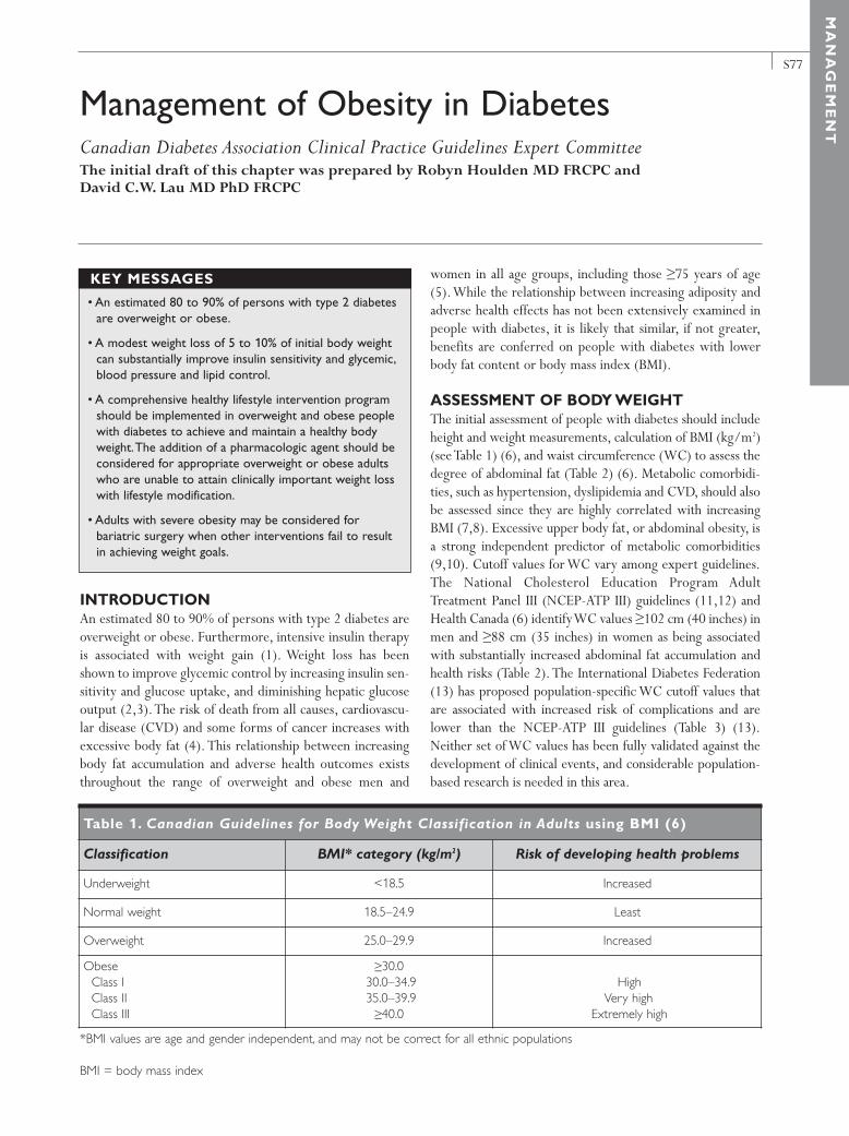
S77
MA
NA
GE
ME
NT
INTRODUCTION An estimated 80 to 90% of persons with type 2 diabetes areoverweight or obese. Furthermore, intensive insulin therapyis associated with weight gain (1). Weight loss has beenshown to improve glycemic control by increasing insulin sen-sitivity and glucose uptake, and diminishing hepatic glucoseoutput (2,3).The risk of death from all causes, cardiovascu-lar disease (CVD) and some forms of cancer increases withexcessive body fat (4). This relationship between increasingbody fat accumulation and adverse health outcomes existsthroughout the range of overweight and obese men and
women in all age groups, including those ≥75 years of age(5).While the relationship between increasing adiposity andadverse health effects has not been extensively examined inpeople with diabetes, it is likely that similar, if not greater,benefits are conferred on people with diabetes with lowerbody fat content or body mass index (BMI).
ASSESSMENT OF BODY WEIGHTThe initial assessment of people with diabetes should includeheight and weight measurements, calculation of BMI (kg/m2)(see Table 1) (6), and waist circumference (WC) to assess thedegree of abdominal fat (Table 2) (6). Metabolic comorbidi-ties, such as hypertension, dyslipidemia and CVD, should alsobe assessed since they are highly correlated with increasingBMI (7,8). Excessive upper body fat, or abdominal obesity, isa strong independent predictor of metabolic comorbidities(9,10). Cutoff values for WC vary among expert guidelines.The National Cholesterol Education Program AdultTreatment Panel III (NCEP-ATP III) guidelines (11,12) andHealth Canada (6) identify WC values ≥102 cm (40 inches) inmen and ≥88 cm (35 inches) in women as being associatedwith substantially increased abdominal fat accumulation andhealth risks (Table 2). The International Diabetes Federation(13) has proposed population-specific WC cutoff values thatare associated with increased risk of complications and arelower than the NCEP-ATP III guidelines (Table 3) (13).Neither set of WC values has been fully validated against thedevelopment of clinical events, and considerable population-based research is needed in this area.
Management of Obesity in DiabetesCanadian Diabetes Association Clinical Practice Guidelines Expert CommitteeThe initial draft of this chapter was prepared by Robyn Houlden MD FRCPC and David C.W. Lau MD PhD FRCPC
• An estimated 80 to 90% of persons with type 2 diabetesare overweight or obese.
• A modest weight loss of 5 to 10% of initial body weightcan substantially improve insulin sensitivity and glycemic,blood pressure and lipid control.
• A comprehensive healthy lifestyle intervention programshould be implemented in overweight and obese peoplewith diabetes to achieve and maintain a healthy bodyweight.The addition of a pharmacologic agent should beconsidered for appropriate overweight or obese adultswho are unable to attain clinically important weight losswith lifestyle modification.
• Adults with severe obesity may be considered forbariatric surgery when other interventions fail to resultin achieving weight goals.
KEY MESSAGES
Table 1. Canadian Guidelines for Body Weight Classification in Adults using BMI (6)
Classification BMI* category (kg/m2) Risk of developing health problems
Underweight <18.5 Increased
Normal weight 18.5–24.9 Least
Overweight 25.0–29.9 Increased
ObeseClass IClass IIClass III
≥30.0 30.0–34.9 35.0–39.9≥40.0
HighVery high
Extremely high
*BMI values are age and gender independent, and may not be correct for all ethnic populations
BMI = body mass index

S78
2008 CLINICAL PRACTICE GUIDELINES
TREATMENT OF OBESITYThe goals of therapy for overweight and obese people withdiabetes are to reduce body fat, attain and maintain ahealthy or lower body weight for the long term, and pre-vent weight regain. In general, obese people with diabeteshave greater difficulty with weight loss compared to simi-larly obese people without diabetes (14). A modest weightloss of 5 to 10% of initial body weight can substantiallyimprove insulin sensitivity, glycemic control, high bloodpressure (BP) and dyslipidemia (15-19).The optimal rate ofweight loss is 1 to 2 kg/month. A negative energy balanceof 500 kcal/day is typically required to achieve a weightloss of 0.45 kg/week (20).
Lifestyle interventions Lifestyle intervention is recommended for weight loss inorder to improve health status and quality of life (20,21). Inpeople with diabetes who are overweight or obese, achievinga healthy weight through an active lifestyle promotes a gen-eral sense of well-being and cardiovascular (CV) fitness,along with other benefits, such as reducing CVD, morbidity,
mortality and other complications attributable to obesity(22). Lifestyle interventions that combine dietary modifica-tion, increased and regular physical activity and behaviourtherapy are the most effective (23-25). Structured interdisci-plinary programs have demonstrated the best short- andlong-term results (24). Ongoing follow-up with the health-care team is important to plan individualized dietary andactivity changes to facilitate weight loss.Adjustments to anti-hyperglycemic agents may be required as the individual withdiabetes loses weight (26).
All weight-loss diets must be well balanced and nutrition-ally adequate to ensure optimal health. In general, a carbohy-drate intake of at least 100 g/day is required to spare proteinbreakdown and muscle wasting, and to avoid large shifts influid balance and ketosis. High-fibre foods that take longer toeat and digest are associated with greater satiety. Adequateprotein intake is required to maintain lean body mass andother essential physiological processes. Reduced intake of sat-urated fat and energy-dense foods should be emphasized toachieve the required daily energy deficit to promote weightloss. Very low-calorie diets with <900 kcal/day are not rec-ommended, except under medical supervision.
Because confusion over portion size of foods and bever-ages (27) may lead to overeating, people with diabetes shouldbe counselled by a dietitian on appropriate serving sizes andon how to select meals, preferably nutrient-rich meals (i.e.containing whole grains and legumes), which are associatedwith greater satiety and lower caloric intake (28).
Behavioural therapyTwo large-scale reviews of >100 individual studies evaluat-ing behaviour modification techniques support their effec-tiveness in promoting weight loss as adjuncts to lifestyleintervention (29,30).
Members of the healthcare team should consider using astructured approach to providing advice and feedback onphysical activity, healthy eating habits and weight loss (31-34).
Table 2. WC and risk of developing health problems (6)
WC cutoff points*† Risk of developinghealth problems
Men ≥102 cm (40 inches) Increased
Women ≥88 cm (35 inches) Increased
*WC cutoffs may be lower in some populations (e.g. older individuals, Asian population [See Table 3]), especially in the pres-ence of the metabolic syndrome (such as hypertriglyceridemia)†Increased WC can also be a marker for increased risk, even inpersons with normal weight
WC = waist circumference
*NCEP-ATP III guidelines (11,12) and Health Canada (6) define central obesity as WC values ≥102 cm (40 inches) in men and ≥88 cm (35 inches)
WC = waist circumference
Table 3. Ethnic-specific values for WC (13)
Country or ethnic group Central obesity as defined by WC
Men Women
Europid* ≥94 cm ≥80 cm
South Asian, Chinese, Japanese ≥90 cm ≥80 cm
South and Central American Use South Asian cutoff points until more specific data are available
Sub-Saharan African Use Europid cutoff points until more specific data are available
Eastern Mediterranean and Middle East (Arab) Use Europid cutoff points until more specific data are available

S79
MA
NA
GE
ME
NT
Pharmacotherapy Pharmacotherapy for overweight people with diabetes notonly improves glycemic control, but also results in a signifi-cant reduction in the doses of antihyperglycemic agents (26).Pharmacotherapy is an acceptable adjunct in the short- andlong-term management of obesity when lifestyle measuresfail to achieve the desired weight loss after an adequate trialof 3 to 6 months (20,35). Pharmacotherapy can be consid-ered for people with BMI ≥30.0 kg/m2 with no obesity-related comorbidities or risk factors, or BMI ≥27.0 kg/m2
with obesity-related comorbidities or risk factors (20).Antiobesity drug therapy may be considered as an adjunct tonutrition therapy, physical activity and behaviour modifica-tion to achieve a target weight loss of 5 to 10% of initial bodyweight and for weight maintenance (20,35).
Two medications, orlistat and sibutramine, have beenapproved in Canada for long-term management of obesity(Table 4). Drug therapy leads to even greater weight losswhen coupled with lifestyle intervention and behaviour mod-ification therapy. Both drugs have been shown to be effectivein obese people with type 2 diabetes, improving glycemic andmetabolic control, and resulting in favourable changes in lipidlevels, BP profile and fat distribution (26,36,37). In obesepeople with impaired glucose tolerance (IGT), orlistat alsoimproves glucose tolerance and reduces the progression totype 2 diabetes (38). Clinical trials with antiobesity agentshave confirmed a smaller degree of weight loss in people withdiabetes compared with obese people who do not have dia-betes (14,26).
When pharmacotherapy is being considered in the treat-ment of the obese or overweight person with type 2 dia-betes, the choice of drug should be based on the individual’sCV risk profile, dietary habits and concomitant disease(s).
People with irregular eating habits, such as those who “snack”frequently, may be better suited to sibutramine therapybecause of its long-acting satiety-enhancing properties.Combining orlistat and sibutramine therapy is not advocatedfor clinical use. Sibutramine should be avoided in patientswith ischemic heart disease, congestive heart failure or othermajor cardiac disease. Orlistat should be avoided in patientswith inflammatory or other chronic bowel disease.
Other available antiobesity drugs, such as diethylpropionand phentermine, are sympathomimetic noradrenergicappetite suppressants that are approved only for short-termuse of a few weeks. They are not recommended because ofmodest efficacy and frequent adverse side effects.
Currently, a number of new molecular entities that tar-get receptors and metabolic processes relevant to energymetabolism are being developed for the treatment of obesi-ty. Among these emerging strategies, cannabinoid type 1receptor antagonists currently appear to be the most prom-ising (39).
SurgeryIndividuals who are candidates for surgical proceduresshould be carefully selected after evaluation by an interdisci-plinary team with medical, surgical, psychiatric and nutri-tional expertise. Surgery is usually reserved for people withclass III obesity (BMI ≥40.0 kg/m2), or class II obesity(BMI=35.0–39.9 kg/m2) in the presence of comorbidities(40) and the inability to achieve weight-loss goals followingan adequate trial of lifestyle intervention. Long-term, if notlifelong, medical surveillance after surgical therapy is neces-sary for most people. Preferred surgical options for weightloss include laparoscopic vertical banded gastroplasty andlaparoscopic Roux-en-Y gastric bypass (41-43).
Table 4. Medications approved for the treatment of obesity in type 2 diabetes
Class Generic (trade)name
Recommended regimen
Action Adverse effects
Gastrointestinal lipaseinhibitor
orlistat (Xenical) 120 mg TID (during orup to 1 hour after eachmeal)
• Nonsystemic pancre-atic lipase inhibitorthat exerts its thera-peutic activity in thestomach and gastro-intestinal tract by reducing dietary fatdigestion and absorp-tion by about 30%
• Abdominal bloating,pain and cramping
• Steatorrhea• Fecal incontinence
Norepinephrine andserotonin reuptakeinhibitor
sibutramine (Meridia) 10–15 mg OD (in the morning)
• Reduces food intakeby enhancing satiety
• May increase thermo-genesis
• May prevent declinein energy expenditurewith weight loss
• Xerostomia• Increase heart rate
and blood pressure• Constipation • Dizziness

S80
2008 CLINICAL PRACTICE GUIDELINES
OTHER RELEVANT GUIDELINESPhysical Activity and Diabetes, p. S37Nutrition Therapy, p. S40
RELATED WEBSITESHealth Canada. Canadian Guidelines for Body WeightClassification in Adults. Quick Reference for Professionals.Available at: http://www.hc-sc.gc.ca/fn-an/alt_formats/hpfb-dgpsa/pdf/nutrition/cg_quick_ref-ldc_rapide_ref_e.pdf.Accessed September 1, 2008.
OBESITY CANADA GUIDELINES2006 Canadian Clinical Practice Guidelines on theManagement and Prevention of Obesity in Adults andChildren. Available at: http://www.cmaj.ca/cgi/content/full/176/8/S1/DC1. Accessed September 1, 2008.
REFERENCES1. UK Prospective Diabetes Study (UKPDS) Group. Intensive
blood-glucose control with sulphonylureas or insulin comparedwith conventional treatment and risk of complications inpatients with type 2 diabetes (UKPDS 33). Lancet. 1998;352:837-853.
2. Ruderman N, Chisholm D, Pi-Sunyer X, et al.The metabolical-ly obese, normal-weight individual revisited. Diabetes. 1998;47:699-713.
3. Markovic TP, Jenkins AB, Campbell LV, et al.The determinantsof glycemic responses to diet restriction and weight loss in obe-sity and NIDDM. Diabetes Care. 1998;21:687-694.
4. Calle EE, Rodriguez C, Walker-Thurmond K, et al.Overweight, obesity, and mortality from cancer in a prospec-tively studied cohort of U.S. adults. N Engl J Med. 2003;
348:1625-1638.5. Stevens J, Cai J, Pamuk ER, et al.The effect of age on the asso-
ciation between body-mass index and mortality. N Engl J Med.1998;338:1-7.
6. Health Canada. Canadian Guidelines for Body Weight Classification in Adults. Ottawa, ON: Health Canada; 2003. Publication H49-179/2003E.
7. Rabkin SW, Chen Y, Leiter L, et al. Risk factor correlates of bodymass index. Canadian Heart Health Surveys Research Group.CMAJ. 1997;157(suppl 1):S26-S31.
8. World Health Organization. Obesity: preventing and managingthe global epidemic. Report of a WHO consultation. WorldHealth Organ Tech Rep Ser. 2000;894:i-xii,1-253.
9. Reeder BA, Senthilselvan A, Després JP, et al.The association ofcardiovascular disease risk factors with abdominal obesity inCanada. Canadian Heart Health Surveys Research Group. CMAJ.1997;157(suppl 1):S39-S45.
10. Després JP, Lemieux I, Prud’homme D. Treatment of obesity:need to focus on high risk abdominally obese patients. BMJ.2001;322:716-720.
11. Expert Panel on Detection, Evaluation, and Treatment of HighBlood Cholesterol in Adults. Executive Summary of The ThirdReport of The National Cholesterol Education Program (NCEP)Expert Panel on Detection, Evaluation, and Treatment of HighBlood Cholesterol in Adults (Adult Treatment Panel III). JAMA.2001;285:2486-2497.
12. Grundy SM, Cleeman JI, Daniels SR, et al; American HeartAssociation; National Heart, Lung, and Blood Institute.Diagnosis and management of the metabolic syndrome: anAmerican Heart Association/National Heart, Lung, and BloodInstitute Scientific Statement. Circulation. 2005;112:2735-2752.
13. International Diabetes Federation. The IDF Consensus WorldwideDefinition of the Metabolic Syndrome. Brussels: IDFCommunications; 2006. Available at: http://www.idf.org/web data/docs/IDF_Meta_def_final.pdf. Accessed September1, 2008.
14. Wing RR, Marcus MD, Epstein LH, et al.Type II diabetic sub-jects lose less weight than their overweight nondiabetic spouses.Diabetes Care. 1987;10:563-566.
15. Dattilo AM, Kris-Etherton PM. Effects of weight reduction onblood lipids and lipoproteins: a meta-analysis. Am J Clin Nutr.1992;56:320-328.
16. Goldstein DJ. Beneficial health effects of modest weight loss. IntJ Obes Relat Metab Disord. 1992;16:397-415.
17. Elmer PJ, Grimm R Jr, Laing B, et al. Lifestyle intervention:results of the Treatment of Mild Hypertension Study (TOMHS).Prev Med. 1995;24:378-388.
18. Tuomilehto J, Lindström J, Eriksson JG, et al; Finnish DiabetesPrevention Study Group. Prevention of type 2 diabetes mellitusby changes in lifestyle among subjects with impaired glucose tol-erance. N Engl J Med. 2001;344:1343-1350.
19. Knowler WC, Barrett-Connor E, Fowler SE, et al; DiabetesPrevention Program Research Group. Reduction in the inci-dence of type 2 diabetes with lifestyle intervention or met-
1.A comprehensive healthy lifestyle intervention program(including a hypocaloric, nutritionally balanced diet, regu-lar physical activity or exercise, and behavioural modifi-cation techniques) for overweight and obese peoplewith, or at risk for diabetes, should be implemented toachieve and maintain a healthy body weight [Grade D,Consensus]. Members of the healthcare team should consider using a structured approach to providingadvice and feedback on physical activity, healthy eatinghabits and weight loss [Grade C, Level 3 (31-34)].
2. In overweight or obese adults with type 2 diabetes, apharmacologic agent such as orlistat [Grade A, Level 1A(26)] or sibutramine [Grade B, Level 2 (37)] should beconsidered as an adjunct to lifestyle modifications tofacilitate weight loss and improve glycemic control.
3.Adults with class III obesity (BMI ≥40.0 kg/m2) or class IIobesity (BMI 35.0 to 39.9 kg/m2) with other comorbidi-ties may be considered for bariatric surgery when otherlifestyle interventions are inadequate in achieving weightgoals [Grade C, Level 3 (43)].
RECOMMENDATIONS
S81
MA
NA
GE
ME
NT
formin. N Engl J Med. 2002;346:393-403.20. National Institutes of Health. Clinical Guidelines on the
Identification, Evaluation, and Treatment of Overweight andObesity in Adults – The Evidence Report. Obes Res. 1998;6(suppl 2):51S-209S.
21. Willett WC, Dietz WH, Colditz GA. Guidelines for healthyweight. N Engl J Med. 1999;341:427-434.
22. Williamson DF,Thompson TJ,Thun M, et al. Intentional weightloss and mortality among overweight individuals with diabetes.Diabetes Care. 2000;23:1499-1504.
23. Pavlou KN, Krey S, Steffee WP. Exercise as an adjunct to weightloss and maintenance in moderately obese subjects. Am J ClinNutr. 1989;49(5 suppl):1115-1123.
24. Wing RR, Hill JO. Successful weight loss maintenance. Annu RevNutr. 2001;21:323-341.
25. Wing RR, Goldstein MG, Acton KJ, et al. Behavioral scienceresearch in diabetes: lifestyle changes related to obesity, eatingbehavior, and physical activity. Diabetes Care. 2001;24:117-123.
26. Hollander PA, Elbein SC, Hirsch IB, et al. Role of orlistat in thetreatment of obese patients with type 2 diabetes. A 1-year ran-domized double-blind study. Diabetes Care. 1998;21:1288-1294.
27. Rolls BJ, Morris EL, Roe LS. Portion size of food affects energyintake in normal-weight and overweight men and women. Am JClin Nutr. 2002;76:1207-1213.
28. Rolls BJ, Roe LS, Meengs JS. Salad and satiety: energy densityand portion size of a first-course salad affect energy intake atlunch. J Am Diet Assoc. 2004;104:1570-1576.
29. Wing RR, Jeffery RW. Outpatient treatments of obesity: a com-parison of methodology and clinical results. Int J Obes. 1979;3:261-279.
30. Bennett GA. Behaviour therapy for obesity: a quantitativereview of the effects of selected treatment characteristics onoutcome. BehavTher. 1986;17:554-562.
31. Swinburn BA,Walter LG,Arroll B, et al.The green prescriptionstudy: a randomized controlled trial of written exercise adviceprovided by general practitioners. Am J Public Health. 1998;88:288-291.
32. Logsdon DN, Lazaro CM, Meier RV.The feasibility of behavioralrisk reduction in primary medical care. Am J Prev Med. 1989;5:249-256.
33. Campbell MK, DeVellis BM, Strecher VJ, et al. Improvingdietary behavior: the effectiveness of tailored messages in pri-mary care settings. Am J Public Health. 1994;84:783-787.
34. Lewis BS, Lynch WD.The effect of physician advice on exercisebehavior. Prev Med. 1993;22:110-121.
35. National Task Force on the Prevention and Treatment of Obesity.Long-term pharmacotherapy in the management of obesity.JAMA. 1996;276:1907-1915.
36. Scheen AJ, Lefèbvre PJ. Antiobesity pharmacotherapy in themanagement of type 2 diabetes. Diabetes Metab Res Rev.2000;16:114-124.
37. Finer N, Bloom SR, Frost GS, et al. Sibutramine is effective forweight loss and diabetic control in obesity with type 2 diabetes:a randomised, double-blind, placebo-controlled study. Diabetes
Obes Metab. 2000;2:105-112.38. Heymsfield SB, Segal KR, Hauptman J, et al. Effects of weight
loss with orlistat on glucose tolerance and progression to type 2diabetes in obese adults. Arch Intern Med. 2000;160:1321-1326.
39. Van Gaal LF, Rissanen AM, Scheen AJ, et al; RIO-Europe StudyGroup. Effects of the cannabinoid-1 receptor blocker rimona-bant on weight reduction and cardiovascular risk factors in over-weight patients: 1-year experience from the RIO-Europe study.Lancet. 2005;365:1389-1397.
40. Melissas J, Christodoulakis M, Spyridakis M, et al. Disordersassociated with clinically severe obesity: significant improve-ment after surgical weight reduction. South Med J. 1998;91:1143-1148.
41. Chapman AE, Kiroff G, Game P, et al. Laparoscopic adjustablegastric banding in the treatment of obesity: a systematic litera-ture review. Surgery. 2004;135:326-351.
42. Maggard MA, Shugarman LR, Suttorp M, et al. Meta-analysis:surgical treatment of obesity. Ann Intern Med. 2005;142:547-559.
43. Sjöström CD, Lissner L,Wedel H, et al. Reduction in incidenceof diabetes, hypertension and lipid disturbances after intention-al weight loss induced by bariatric surgery: the SOS InterventionStudy. Obes Res. 1999;7:477-484.
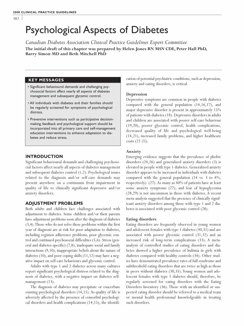
S82
INTRODUCTIONSignificant behavioural demands and challenging psychoso-cial factors affect nearly all aspects of diabetes managementand subsequent diabetes control (1,2). Psychological issuesrelated to the diagnosis and/or self-care demands maypresent anywhere on a continuum from impairment inquality of life to clinically significant depressive and/oranxiety disorders.
ADJUSTMENT PROBLEMS Both adults and children face challenges associated withadjustment to diabetes. Some children and/or their parentshave adjustment problems soon after the diagnosis of diabetes(3,4).Those who do not solve these problems within the firstyear of diagnosis are at risk for poor adaptation to diabetes,including regimen adherence problems, poor glycemic con-trol and continued psychosocial difficulties (5,6). Stress (gen-eral and diabetes-specific) (7,8), inadequate social and familyinteractions (9,10), inappropriate beliefs about the nature ofdiabetes (10), and poor coping skills (11,12) may have a neg-ative impact on self-care behaviours and glycemic control.
Adults with type 1 and 2 diabetes across many culturesreport significant psychological distress related to the diag-nosis of diabetes, with a negative impact on diabetes self-management (13).
The diagnosis of diabetes may precipitate or exacerbateexisting psychological disorders (14,15). As quality of life isadversely affected by the presence of comorbid psychologi-cal disorders and health complications (14,15), the identifi-
cation of potential psychiatric conditions, such as depression,anxiety and eating disorders, is critical.
DepressionDepressive symptoms are common in people with diabetescompared with the general population (14,16,17), andmajor depressive disorder is present in approximately 15%of patients with diabetes (18). Depressive disorders in adultsand children are associated with poorer self-care behaviour(19,20), poorer glycemic control, health complications,decreased quality of life and psychological well-being(14,21), increased family problems, and higher healthcarecosts (22-25).
AnxietyEmerging evidence suggests that the prevalence of phobicdisorders (24,26) and generalized anxiety disorders (3) iselevated in people with type 1 diabetes. Generalized anxietydisorder appears to be increased in individuals with diabetescompared with the general population (14 vs. 3 to 4%,respectively) (27). As many as 40% of patients have at leastsome anxiety symptoms (27), and fear of hypoglycemia(28,29) is not uncommon in those with diabetes. A recentmeta-analysis suggested that the presence of clinically signif-icant anxiety disorders among those with type 1 and 2 dia-betes is associated with poor glycemic control (28).
Eating disordersEating disorders are frequently observed in young womenand adolescent females with type 1 diabetes (30,31) and areassociated with poorer glycemic control (31,32) and anincreased risk of long-term complications (33). A meta-analysis of controlled studies of eating disorders and dia-betes showed a higher prevalence of bulimia in girls withdiabetes compared with healthy controls (34). Other stud-ies have demonstrated prevalence rates of full syndrome andsubthreshold eating disorders that are twice as high as thosein peers without diabetes (30,35). Young women and ado-lescent females with type 1 diabetes should, therefore, beregularly screened for eating disorders with the EatingDisorders Inventory (36). Those with an identified or sus-pected eating disorder should be referred to a medical teamor mental health professional knowledgeable in treatingsuch disorders.
Psychological Aspects of Diabetes Canadian Diabetes Association Clinical Practice Guidelines Expert CommitteeThe initial draft of this chapter was prepared by Helen Jones RN MSN CDE, Peter Hall PhD,Barry Simon MD and Beth Mitchell PhD
2008 CLINICAL PRACTICE GUIDELINES
• Significant behavioural demands and challenging psy-chosocial factors affect nearly all aspects of diabetesmanagement and subsequent glycemic control.
• All individuals with diabetes and their families should be regularly screened for symptoms of psychologicaldistress.
• Preventive interventions such as participative decision-making, feedback and psychological support should beincorporated into all primary care and self-managementeducation interventions to enhance adaptation to dia-betes and reduce stress.
KEY MESSAGES
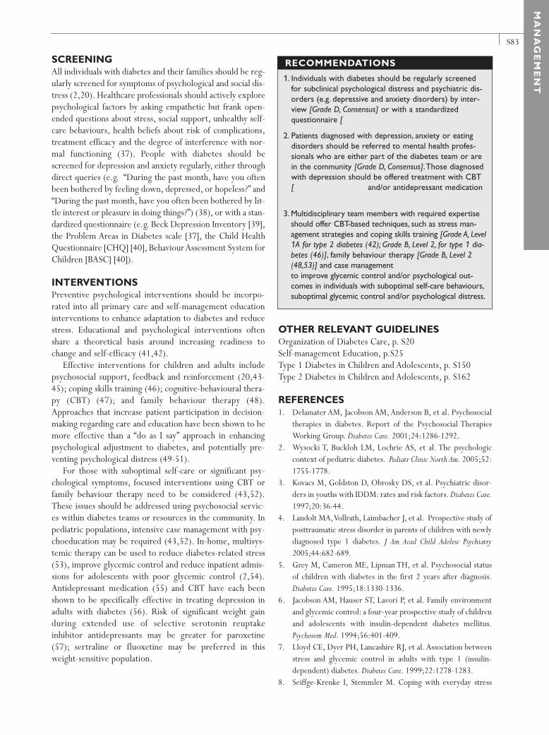
S83
SCREENING All individuals with diabetes and their families should be reg-ularly screened for symptoms of psychological and social dis-tress (2,20). Healthcare professionals should actively explorepsychological factors by asking empathetic but frank open-ended questions about stress, social support, unhealthy self-care behaviours, health beliefs about risk of complications,treatment efficacy and the degree of interference with nor-mal functioning (37). People with diabetes should bescreened for depression and anxiety regularly, either throughdirect queries (e.g. “During the past month, have you oftenbeen bothered by feeling down, depressed, or hopeless?” and“During the past month, have you often been bothered by lit-tle interest or pleasure in doing things?”) (38), or with a stan-dardized questionnaire (e.g. Beck Depression Inventory [39],the Problem Areas in Diabetes scale [37], the Child HealthQuestionnaire [CHQ] [40], Behaviour Assessment System forChildren [BASC] [40]).
INTERVENTIONSPreventive psychological interventions should be incorpo-rated into all primary care and self-management educationinterventions to enhance adaptation to diabetes and reducestress. Educational and psychological interventions oftenshare a theoretical basis around increasing readiness tochange and self-efficacy (41,42).
Effective interventions for children and adults includepsychosocial support, feedback and reinforcement (20,43-45); coping skills training (46); cognitive-behavioural thera-py (CBT) (47); and family behaviour therapy (48).Approaches that increase patient participation in decision-making regarding care and education have been shown to bemore effective than a “do as I say” approach in enhancingpsychological adjustment to diabetes, and potentially pre-venting psychological distress (49-51).
For those with suboptimal self-care or significant psy-chological symptoms, focused interventions using CBT orfamily behaviour therapy need to be considered (43,52).These issues should be addressed using psychosocial servic-es within diabetes teams or resources in the community. Inpediatric populations, intensive case management with psy-choeducation may be required (43,52). In-home, multisys-temic therapy can be used to reduce diabetes-related stress(53), improve glycemic control and reduce inpatient admis-sions for adolescents with poor glycemic control (2,54).Antidepressant medication (55) and CBT have each beenshown to be specifically effective in treating depression inadults with diabetes (56). Risk of significant weight gainduring extended use of selective serotonin reuptakeinhibitor antidepressants may be greater for paroxetine(57); sertraline or fluoxetine may be preferred in thisweight-sensitive population.
OTHER RELEVANT GUIDELINESOrganization of Diabetes Care, p. S20Self-management Education, p.S25Type 1 Diabetes in Children and Adolescents, p. S150Type 2 Diabetes in Children and Adolescents, p. S162
REFERENCES1. Delamater AM, Jacobson AM, Anderson B, et al. Psychosocial
therapies in diabetes. Report of the Psychosocial TherapiesWorking Group. Diabetes Care. 2001;24:1286-1292.
2. Wysocki T, Buckloh LM, Lochrie AS, et al. The psychologiccontext of pediatric diabetes. Pediatr Clinic North Am. 2005;52:1755-1778.
3. Kovacs M, Goldston D, Obrosky DS, et al. Psychiatric disor-ders in youths with IDDM: rates and risk factors. Diabetes Care.1997;20:36-44.
4. Landolt MA,Vollrath, Laimbacher J, et al. Prospective study ofposttraumatic stress disorder in parents of children with newlydiagnosed type 1 diabetes. J Am Acad Child Adolesc Psychiatry2005;44:682-689.
5. Grey M, Cameron ME, Lipman TH, et al. Psychosocial statusof children with diabetes in the first 2 years after diagnosis.Diabetes Care. 1995;18:1330-1336.
6. Jacobson AM, Hauser ST, Lavori P, et al. Family environmentand glycemic control: a four-year prospective study of childrenand adolescents with insulin-dependent diabetes mellitus.Psychosom Med. 1994;56:401-409.
7. Lloyd CE, Dyer PH, Lancashire RJ, et al. Association betweenstress and glycemic control in adults with type 1 (insulin-dependent) diabetes. Diabetes Care. 1999;22:1278-1283.
8. Seiffge-Krenke I, Stemmler M. Coping with everyday stress
MA
NA
GE
ME
NT
1. Individuals with diabetes should be regularly screenedfor subclinical psychological distress and psychiatric dis-orders (e.g. depressive and anxiety disorders) by inter-view [Grade D, Consensus] or with a standardizedquestionnaire [Grade B, Level 2 (39)].
2. Patients diagnosed with depression, anxiety or eatingdisorders should be referred to mental health profes-sionals who are either part of the diabetes team or arein the community [Grade D, Consensus].Those diagnosedwith depression should be offered treatment with CBT[Grade B, Level 2 (56)] and/or antidepressant medication[Grade A, Level 1A (55)].
3. Multidisciplinary team members with required expertiseshould offer CBT-based techniques, such as stress man-agement strategies and coping skills training [Grade A, Level1A for type 2 diabetes (42); Grade B, Level 2, for type 1 dia-betes (46)], family behaviour therapy [Grade B, Level 2(48,53)] and case management [Grade B, Level 2 (43,53)]to improve glycemic control and/or psychological out-comes in individuals with suboptimal self-care behaviours,suboptimal glycemic control and/or psychological distress.
RECOMMENDATIONS

S84
and links to medical and psychosocial adaptation in diabeticadolescents. J Adolesc Health. 2003;33:180-188.
9. Schafer LC, McCaul KD, Glasgow RE. Supportive and non-supportive family behaviors: relationships to adherence andmetabolic control in persons with type I diabetes. DiabetesCare. 1986;9:179-185.
10. Skinner TC, Hampson SE. Social support and personal modelsof diabetes in relation to self-care and well-being in adolescentswith type I diabetes mellitus. J Adolesc. 1998;21:703-715.
11. Peyrot MF, McMurry JF Jr. Stress buffering and glycemic con-trol.The role of coping styles. Diabetes Care. 1992;15:842-846.
12. Graue M,Wentzel-Larsen T, Bru E, et al. The coping styles ofadolescents with type 1 diabetes are associated with degree ofmetabolic control. Diabetes Care 2004; 27;1313-1317.
13. Peyrot, M, Rubin RR, Lauritzen T, et al. Psychosocial prob-lems and barriers to improved diabetes management: results ofthe Cross-National Diabetes Attitudes, Wishes and Needs(DAWN) Study. Diabet Med. 2005;22:1379-1385.
14. Goldney RD, Phillips PJ, Fisher LJ, et al. Diabetes, depression,and quality of life: a population study. Diabetes Care. 2004;27:1066-1070.
15. Northam EA, Matthews LK, Anderson PJ, et al. Psychiatricmorbidity and health outcome in type 1 diabetes – perspec-tives from a prospective longitudinal study. Diabet Med. 2005;22:152-157.
16. Anderson RJ, Freedland KE, Clouse RE, et al.The prevalenceof comorbid depression in adults with diabetes: a meta-analy-sis. Diabetes Care. 2001;24:1069-1078.
17. Dantzer C, Swendsen J, Maurice-Tison S, et al. Anxiety anddepression in juvenile diabetes: a critical review. Clin PsycholRev. 2003;23:787-800.
18. Garvard JA, Lustman PJ, Clouse RE. Prevalence of depressionin adults with diabetes. An epidemiological evaluation. DiabetesCare. 1993;16:1167-1178.
19. McKellar JD, Humphreys K, Piette JD. Depression increasesdiabetes symptoms by complicating patients’ self-care adher-ence. Diabetes Educ. 2004;30:485-492.
20. Wysocki T. Behavioural assessment and intervention in pedi-atric diabetes. Behav Modif. 2006;30:72-92.
21. Grey M,Whittemore R,Tamborlane W. Depression in type 1diabetes in children: natural history and correlates. J PsychosomRes. 2002;53:907-911.
22. Egede LE, Zheng D, Simpson K. Comorbid depression is asso-ciated with increased health care use and expenditures in indi-viduals with diabetes. Diabetes Care. 2002;25:464-470.
23. Garrison MM, Katon WJ, Richardson LP. The impact of psy-chiatric comorbidities on readmissions for diabetes in youth.Diabetes Care. 2005;28:2150-2154.
24. Popkin MK, Callies AL, Lentz RD, et al. Prevalence of majordepression, simple phobia, and other psychiatric disorders inpatients with long-standing type I diabetes mellitus. Arch GenPsychiatry. 1988;45:64-68.
25. Cote MP, Mullins LL, Hartman V, et al. Psychosocial correlatesof health care utilization for children and adolescents with type
1 diabetes mellitus. Children’s Health Care. 2003;32:1-16.26. Mollema ED, Snoek FJ,Adèr HJ, et al. Insulin-treated diabetes
patients with fear of self-injecting or fear of self-testing: psy-chological comorbidity and general well-being. J Psychosom Res.2001;51:665-672.
27. Grigsby AB, Anderson RJ, Freedland KE, et al. Prevalence of anxiety in adults with diabetes: a systematic review.J Psychosom Res. 2002;53:1053-1060.
28. Anderson RJ, DeGroot M, Grigsby AB, et al.Anxiety and poorglycemic control: a meta-analytic review of the literature. Int JPsychiatry Med. 2002:32:235-247.
29. Leiter LA, Yale J-F, Chiasson J-L, et al. Assessment of theimpact of fear of hypoglycemic episodes on glycemic andhypoglycemia management. Can J Diabetes. 2005;29:186-192.
30. Jones JM, Lawson ML, Daneman D, et al. Eating disorders inadolescent females with and without type 1 diabetes: cross sec-tional study. BMJ. 2000;320:1563-1566.
31. Daneman D, Olmsted M, Rydall A, et al. Eating disorders inyoung women with type 1 diabetes. Prevalence, problems andprevention. Horm Res. 1998;50(suppl 1):79-86.
32. Affenito SG, Backstrand JR, Welch GW, et al. Subclinical andclinical eating disorders in IDDM negatively affect metaboliccontrol. Diabetes Care. 1997;20:183-184.
33. Rydall AC, Rodin GM, Olmsted MP, et al. Disordered eatingbehavior and microvascular complications in young womenwith insulin-dependent diabetes mellitus. N Engl J Med. 1997;336:1849-1854.
34. Mannucci E, Rotella F, Ricca V, et al. Eating disorders inpatients with type 1 diabetes: a meta-analysis. J EndocrinolInvest. 2005;28:417-419.
35. Rodin G, Olmsead MP, Rydall AC, et al. Eating disorders inyoung women with type 1 diabetes mellitus. J Psychoso Res.2002;53: 943-949.
36. Garner DM, Olmstead MP. Eating Disorder Inventory (EDI)manual. Odessa, FL: Psychological Assessment Resources. 1984.
37. Welch GW, Jacobson AM, Polonsky WH.The Problem Areas inDiabetes scale.An evaluation of its clinical utility. Diabetes Care.1997;20:760-766.
38. Whooley MA, Avins AL, Miranda J, et al. Case-finding instru-ments for depression.Two questions are as good as many. J GenIntern Med. 1997;12:439-445.
39. Lustman PJ, Clouse RE, Griffith LS, et al. Screening fordepression in diabetes using the Beck Depression Inventory.Psychosom Med. 1997;59:24-31.
40. Cameron FJ, Smidts D, Hesketh K, et al. Early detection ofemotional and behavioural problems in children with diabetes:the validity of the Child Health Questionnaire as a screeninginstrument. Diabet Med. 2003;20:646-650.
41. Steed L, Cooke D, Newman S. A systematic review of psy-chosocial outcomes following education, self-management andpsychological interventions in diabetes mellitus. Patient EducCouns. 2003;51:5-15.
42. Ismail K, Winkley K, Rabe-Hesketh S. Systematic review andmeta-analysis of randomized controlled trials of psychological
2008 CLINICAL PRACTICE GUIDELINES

S85
interventions to improve glycaemic control in patients withtype 2 diabetes. Lancet. 2004;363:1589-1597.
43. Svoren BM, Butler D, Levine BS, et al. Reducing acute adverseoutcomes in youths with type 1 diabetes: a randomized, con-trolled trial. Pediatrics. 2003;112:914-922.
44. Piette JD,Weinberger M, McPhee SJ.The effect of automatedcalls with telephone nurse follow-up on patient-centered out-comes of diabetes care: a randomized, controlled trial. MedCare. 2000;38:218-230.
45. Jones H, Edwards L,Vallis TM, et al. Changes in diabetes self-care behaviors make a difference in glycemic control: theDiabetes Stages of Change (DiSC) study. Diabetes Care. 2003;26:732-737.
46. Grey M, Boland EA, Davidson M, et al. Short-term effects ofcoping skills training as adjunct to intensive therapy in adoles-cents. Diabetes Care. 1998;21:902-908.
47. Fosbury JA, Bosley CM, Ryle A, et al. A trial of cognitive ana-lytic therapy in poorly controlled type 1 patients. Diabetes Care.1997;20:959-964.
48. Wysocki T, Harris MA, Greco P, et al. Randomized, controlledtrial of behavior therapy for families of adolescents with insulin-dependent diabetes mellitus. J Pediatric Psychol.2000; 25:23-33.
49. Norris SL, Engelgau MM, Narayan KM. Effectiveness of self-management training in type 2 diabetes: a systematic review ofrandomized controlled trials. Diabetes Care. 2001;24:561-587.
50. Anderson BJ, Brackett J, Ho J, et al. An office-based interven-tion to maintain parent-adolescent teamwork in diabetes man-agement. Impact on parent involvement, family conflict, andsubsequent glycemic control. Diabetes Care. 1999;22:713-721.
51. Greenfield S, Kaplan SH,Ware JE Jr, et al. Patients’ participa-tion in medical care: effects on blood sugar control and quali-ty of life in diabetes. J Gen Intern Med. 1988;3:448-457.
52. Gage H, Hampson S, Skinner TC, et al. Educational and psy-chosocial programmes for adolescents with diabetes:approaches, outcomes and cost-effectiveness. Patient EducCouns. 2004;53:333-346.
53. Ellis DA, Frey MA, Naar-King S, et al.The effects of multisys-temic therapy on diabetes stress among adolescents withchronically poorly controlled type 1 diabetes: findings from arandomized, controlled trial. Pediatrics. 2005;16:826-832.
54. Ellis DA, Frey MA, Naar-King S, et al. Use of multisystemictherapy to improve regimen adherence among adolescentswith type 1 diabetes in chronic poor metabolic control: a ran-domized controlled trial. Diabetes Care. 2005;28:1604-1610.
55. Lustman PJ, Freedland KE, Griffith LS, et al. Fluoxetine fordepression in diabetes: a randomized double-blind placebo-controlled trial. Diabetes Care. 2000;23:618-623.
56. Lustman PJ, Griffith LS, Freedland KE, et al. Cognitive behav-ior therapy for depression in type 2 diabetes mellitus. A ran-domized, controlled trial. Ann Intern Med. 1998;129:613-621.
57. Fava M, Judge R, Hoog S, et al. Fluoxetine versus sertralineand paroxetine in major depressive disorders: changes inweight with long term treatment. J Clin Psychiatry. 2000;61:863-867.
MA
NA
GE
ME
NT

S86
INTRODUCTIONPeople with diabetes, especially those with renal and cardiaccomplications, are at high risk for morbidity and mortalityfrom influenza and pneumococcal disease (1). Studies inhigh-risk individuals, which included people with diabetes,have shown that influenza vaccination can reduce hospitaliza-tions by about 40% (2). However, there are few randomizedcontrolled trials that have specifically evaluated the use andbenefit of influenza or pneumococcal immunization in peo-ple with diabetes (1). Clinical practice recommendations forpeople with diabetes must therefore be extrapolated fromrecommendations for individuals at high risk of complica-tions associated with these infectious diseases (3-5).
INFLUENZA IMMUNIZATION IN ADULTSThe majority of studies on influenza immunization rely onobservational reports of increased death rates in people withdiabetes during influenza epidemics (6-9). One case-controlstudy of people with diabetes showed a 6-fold increased riskof hospitalization during influenza outbreaks compared tononepidemic years (9).
A retrospective case-control study demonstrated theeffectiveness of influenza vaccination in reducing rates ofhospitalization of people with diabetes for influenza, pneu-monia or diabetes-related events during 2 influenza epi-demics in Leicestershire, England, United Kingdom (10).The study detected a 79% reduction in hospitalization ratesduring the 2 epidemics in people with diabetes who had beenimmunized against influenza during the period immediatelypreceding the epidemic. Another nested case-control studyin the Netherlands demonstrated that vaccination was associ-
ated with a 56% reduction in any complication, a 54% reduc-tion in hospitalizations and a 58% reduction in deaths in people with type 2 diabetes (11).
PNEUMOCOCCAL IMMUNIZATION IN ADULTSNumerous studies have demonstrated the efficacy of immu-nization in reducing pneumococcal bacteremia in the generalpopulation (12-15).There is widespread acceptance that peo-ple with diabetes are at least as susceptible to pneumococcalinfection as other people with chronic diseases (1), and there-fore the use of the pneumococcal vaccine is encouraged in thispopulation. A one-time revaccination is recommended forindividuals >65 years of age if the original vaccine was admin-istered when they were <65 years of age and >5 years earlier.
RELATED WEBSITESNational Advisory Committee on Immunization. CanadianImmunization Guide. 7th ed. Ottawa, ON: Canadian MedicalAssociation; 2006. Available at: http://www.phac-aspc.gc.ca/publicat/cig-gci/index.html.Accessed September 1, 2008.
REFERENCES1. Smith SA, Poland GA. Use of influenza and pneumococcal vac-
cines in people with diabetes. Diabetes Care. 2000;23:95-108.2. Nichol KL, Nordin J, Mullooly J, et al. Influenza vaccination
and reduction in hospitalizations for cardiac disease and strokeamong the elderly. N Engl J Med. 2003;348:1322-1332.
3. Casey JI. Host defense abnormalities in diabetic patients. In:Rifkin H, Raskin P, eds. Diabetes Mellitus. Vol 5. Bowie, MD:Robert J. Brady Company; 1981:219-223.
4. Heymann AD, Shapiro Y, Chodick G, et al. Reduced hospital-izations and death associated with influenza vaccination amongpatients with and without diabetes. Diabetes Care. 2004;27:2581-2584.
Influenza and Pneumococcal Immunization Canadian Diabetes Association Clinical Practice Guidelines Expert CommitteeThe initial draft of this chapter was prepared by Vincent Woo MD FRCPC
2008 CLINICAL PRACTICE GUIDELINES
• Studies in high-risk individuals, which included peoplewith diabetes, have shown that influenza vaccination can reduce hospitalizations by approximately 40%.
• As people with diabetes are at least as susceptible topneumococcal infection as other people with chronic dis-eases, the use of the pneumococcal vaccine is encouraged.
• A one-time pheumococcal revaccination is recommen-ded for individuals >65 years of age if the original vaccinewas administered when they were <65 years of age and>5 years earlier.
KEY MESSAGES
1. People with diabetes should receive an annual influenzavaccine to reduce the risk of complications associatedwith influenza epidemics [Grade D, Consensus].
2. People with diabetes should be considered for vaccina-tion against pneumococcus [Grade D, Consensus].
RECOMMENDATIONS

S87
5. Smith SA, Poland GA, American Diabetes Association.Influenza and pneumococcal immunization in diabetes.Diabetes Care. 2004;27:S111-S113.
6. Eickhoff TC, Sherman IL, Serfing RE. Observations on excessmortality associated with epidemic influenza. JAMA. 1961;176:104-110.
7. Martin WJ. Recent changes in the death rate from influenza.Br Med J. 1950;1:267-268.
8. Stocks P, Camb MD. Influenza epidemics on the certified caus-es of death. Lancet. 1935:ii;386-395.
9. Bouter KP, Diepersloot RJA, van Romunde LKJ, et al. Effectof epidemic influenza on ketoacidosis, pneumonia and death indiabetes mellitus: a hospital register survey of 1976–1979 inThe Netherlands. Diabetes Res Clin Pract. 1991;12:61-68.
10. Colquhoun AJ, Nicholson KG, Botha JL, et al. Effectiveness ofinfluenza vaccine in reducing hospital admissions in peoplewith diabetes. Epidemiol Infect. 1997;119:335-341.
11. Looijmans-Van den Akker I, Verheij TJ, Buskens E, et al.Clinical effectiveness of first and repeat influenza vaccinationin adult and elderly diabetic patients. Diabetes Care. 2006;29:1771-1776.
12. Bolan G, Broome CV, Facklam RR, et al. Pneumococcal vac-cine efficacy in selected populations in the United States. AnnIntern Med. 1986;104:1-6.
13. Forrester HL, Jahnigen DW, LaForce FM. Inefficacy of pneu-mococcal vaccine in a high-risk population. Am J Med. 1987;83:425-430.
14. Schwartz JS. Pneumococcal vaccine: clinical efficacy and effec-tiveness. Ann Intern Med. 1982;96:208-220.
15. Shapiro ED, Clemens JD. A controlled evaluation of the pro-tective efficacy of pneumococcal vaccine for patients at highrisk of serious pneumococcal infections. Ann Intern Med. 1984;101:325-330.
MA
NA
GE
ME
NT

S88
INTRODUCTIONBeta cell replacement as a means of restoring endogenousinsulin secretion, either by whole organ pancreas transplantor islet transplant, has a number of potential advantages overstandard exogenous insulin therapy for the treatment of type1 diabetes, including improved glycemic control and thepotential for insulin independence. However, any advantagesmust be weighed against the risks and adverse effects of sur-gery and chronic immunosuppressive therapy that accompa-ny these treatments. Unfortunately, the absence of data fromrandomized controlled trials (RCTs) makes it difficult todraw firm conclusions regarding the efficacy of these thera-pies compared with intensive medical management of dia-betes. Nevertheless, some general recommendations can bemade regarding the role of pancreas and islet transplant inthe context of current clinical experience.
WHOLE PANCREAS TRANSPLANTATIONPancreas transplantation has progressed significantly in termsof surgical technique and immunosuppression since it wasfirst introduced in the 1960s (1). It is most commonly cate-gorized on the basis of the presence or absence of a kidneytransplant and the relative timing of the procedures: simul-taneous pancreas kidney (SPK) transplant; pancreas afterkidney (PAK) transplant; or pancreas transplant alone (PTA),in the absence of a kidney transplant. Worldwide, non-controlled pancreas graft and patient survival rates differslightly among these 3 categories (2). However, in theabsence of large RCTs, it is unclear whether these differ-ences are clinically significant.
Metabolic studies demonstrate a marked improvement inglycemic control and glycated hemoglobin (A1C) after suc-
cessful whole pancreas transplant, with most recipientsachieving insulin independence that can last for many years(3,4). A reduction in albuminuria has been shown at 1 yearposttransplant (5). Similarly, improvements in the histologicchanges of diabetic nephropathy have been reported after 5to 10 years posttransplant (6). Studies also show an improve-ment and/or stabilization of diabetic retinopathy (7) after aninitial risk of worsening due to a rapid reduction in glycemia(8). The benefits of pancreas transplant are less clear inpatients with advanced retinal disease (9). Peripheral senso-ry and motor neuropathies have been shown to improve afterpancreas transplant (10,11). Improvements in autonomicneuropathy are less consistent and may take longer to achieve(12,13). There is growing evidence that pancreas transplantimproves cardiovascular (CV) function (14) and may reducecardiac events (15). However, studies have generally beensmall and nonrandomized. It remains uncertain whether pan-creas transplant improves overall mortality rates (16).Finally, diabetes-related quality of life appears to improveafter pancreas transplant, although overall quality of life maynot change (17).
ISLET TRANSPLANTATIONIslet transplantation is a less invasive procedure than pancreastransplantation. It involves the infusion of islets isolated fromcadaveric pancreata via the portal vein into the liver (18).Unlike whole pancreas transplant recipients, most islet trans-plant recipients require at least 2 islet infusions to achieveinsulin independence, although there has been recent short-term success using single islet donors in some centres (19).The rate of posttransplant insulin independence at the mostexperienced centres is approximately 80% at 1 year, butdeclines to about 10% at 5 years (20-22). Rates of insulinindependence may be lower at less experienced centres(23,24). Most transplant recipients continue to have someendogenous insulin secretion even after insulin indepen-dence is lost. However, there are very few long-term dataregarding function of the transplanted islets after 5 years.Most published studies involve islet transplant in the absenceof a kidney transplant (islet transplant alone [ITA]).There issome evidence suggesting that islet transplant performed atthe same time as a kidney transplant (simultaneous islet kid-ney [SIK]) or after a kidney transplant (islet after kidney[IAK]) may have comparable results (25,26).
Pancreas and Islet TransplantationCanadian Diabetes Association Clinical Practice Guidelines Expert CommitteeThe initial draft of this chapter was prepared by Breay W. Paty MD FRCPC, Erin Keely MD FRCPCand Charlotte McDonald MD FRCPC
2008 CLINICAL PRACTICE GUIDELINES
• Pancreas transplant can result in prolonged insulin inde-pendence and a possible reduction in the progression of secondary complications of diabetes.
• Islet transplant can result in transient insulin indepen-dence and can reliably stabilize blood glucose concentra-tions in people with glycemic lability.
• The risks of chronic immunosuppression must be care-fully weighed against the potential benefits of pancreas or islet transplant for each individual.
KEY MESSAGES
S89
The principal benefit of islet transplant is stabilization ofblood glucose control in individuals with severe glycemiclability or hypoglycemia unawareness.This benefit is evidentand persists in most recipients, even in the absence of insulinindependence (27,28).The impact of islet transplantation ondiabetes complications remains uncertain. Renal functionappears to decline after ITA in patients with significant pre-existing renal dysfunction, although the degree of decline canvary (29,30). For this reason, particular caution may be war-ranted for patients with pre-existing renal dysfunction.Thereis some evidence that IAK transplant recipients showimproved endothelial and CV function compared to kidneytransplant recipients (31,32). Kidney graft survival rates alsoappear to improve with concomitant islet transplant (33).The impact of islet transplantation on diabetic retinopathyand neuropathy is still uncertain. Quality of life appears toimprove initially after islet transplantation, due primarily toa reduced fear of hypoglycemia, but declines with the loss ofinsulin independence (34,35).
RISKS OF PANCREAS AND ISLET TRANSPLANTATIONPancreas transplantation is associated with significant peri-operative risks, including graft pancreatitis, peripancreaticabscess, duodenal stump leak, venous or arterial thrombosis,and conversion from bladder to enteric drainage (36). Islettransplantation is associated with fewer procedural risks,which may include intraperitoneal hemorrhage, partial por-tal vein thrombosis, gallbladder puncture and a transient ele-vation of liver enzymes (37). Both pancreas and islettransplantation require chronic immunosuppression, whichis associated with a number of risks and side effects, includ-ing increased risk of infection and malignancy, nephrotoxicity,diarrhea, oral ulcers (in the case of islet transplant) and manyothers. These risks must be carefully weighed against thepotential benefits of transplant for each individual.
REFERENCES1. Larsen JL. Pancreas transplantation: indications and conse-
quences. Endocr Rev. 2004;25:919-946.2. Gruessner AC, Sutherland DE. Pancreas transplant outcomes
for United States (US) and non-US cases as reported to theUnited Network for Organ Sharing (UNOS) and theInternational Pancreas Transplant Registry (IPTR) as of June2004. Clin Transplant. 2005;19:433-455.
3. Robertson RP, Abid M, Sutherland DE, et al. Glucose home-ostasis and insulin secretion in human recipients of pancreastransplantation. Diabetes. 1989;38(suppl 1):97-98.
4. Robertson RP, Sutherland DE, Lanz KJ. Normoglycemia andpreserved insulin secretory reserve in diabetic patients 10-18years after pancreas transplantation. Diabetes. 1999;48:1737-1740.
5. Coppelli A, Giannarelli R,Vistoli F, et al.The beneficial effectsof pancreas transplant alone on diabetic nephropathy. DiabetesCare. 2005;28:1366-1370.
6. Fioretto P, Steffes MW, Sutherland DE, et al. Reversal oflesions of diabetic nephropathy after pancreas transplantation.N Engl J Med. 1998;339:69-75.
7. Giannarelli R, Coppelli A, Sartini M, et al. Effects of pancreas-kidney transplantation on diabetic retinopathy. Transpl Int.2005;18:619-622.
8. Landgraf R, Nusser J, Scheuer R, et al. Metabolic control andeffect on secondary complications of diabetes mellitus by pan-creatic transplantation. Baillieres Clin Gastroenterol. 1989;3:865-876.
9. Bandello F,Vigano C, Secchi A, et al. Effect of pancreas trans-plantation on diabetic retinopathy: a 20-case report.Diabetologia. 1991;34(suppl 1):S92-S94.
10. Kennedy WR, Navarro X, Goetz FC, et al. Effects of pancreat-ic transplantation on diabetic neuropathy. N Engl J Med. 1990;322:1031-1037.
11. Navarro X, Sutherland DE, Kennedy WR. Long-term effectsof pancreatic transplantation on diabetic neuropathy. AnnNeurol. 1997;42:727-736.
12. Solders G, Tydén G, Persson A, et al. Improvement of nerveconduction in diabetic neuropathy.A follow-up study 4 yr aftercombined pancreatic and renal transplantation. Diabetes.1992;41:946-951.
13. Tydén G, Bolinder J, Solders G, et al. Improved survival inpatients with insulin-dependent diabetes mellitus and end-stage diabetic nephropathy 10 years after combined pancreasand kidney transplantation. Transplantation. 1999;67:645-648.
14. Coppelli A, Giannarelli R, Mariotti R, et al. Pancreas trans-plant alone determines early improvement of cardiovascularrisk factors and cardiac function in type 1 diabetic patients.Transplantation. 2003;76:974-976.
15. La Rocca E, Fiorina P, di Carlo V, et al. Cardiovascular out-comes after kidney-pancreas and kidney-alone transplantation.Kidney Int. 2001;60:1964-1971.
16. Venstrom JM, McBride MA, Rother KI, et al. Survival afterpancreas transplantation in patients with diabetes and pre-served kidney function. JAMA. 2003;290:2817-2823.
17. Sureshkumar KK, Mubin T, Mikhael N, et al. Assessment ofquality of life after simultaneous pancreas-kidney transplanta-tion. Am J Kidney Dis. 2002;39:1300-1306.
MA
NA
GE
ME
NT
1. For individuals with type 1 diabetes and end-stage renaldisease who are undergoing or have undergone success-ful kidney transplant, pancreas transplant should be con-sidered [Grade D, Consensus].
2. For individuals with type 1 diabetes and preserved renalfunction, but with persistent metabolic instability character-ized by severe glycemic lability and/or severe hypoglycemiaunawareness despite best efforts to optimize glycemiccontrol, pancreas transplant [Grade D, Level 4 (4)] or islettransplant [Grade D, Level 4 (21)] may be considered.
RECOMMENDATIONS

S90
18. Robertson RP. Islet transplantation as a treatment for diabetes– a work in progress. N Engl J Med. 2004;350:694-705.
19. Hering BJ, Kandaswamy R,Ansite JD, et al. Single-donor, mar-ginal-dose islet transplantation in patients with type 1 dia-betes. JAMA. 2005;293:830-835.
20. Markmann JF, Deng S, Huang X, et al. Insulin independencefollowing isolated islet transplantation and single islet infu-sions. Ann Surg. 2003;237:741-749.
21. Ryan EA, Paty BW, Senior PA, et al. Five-year follow-up afterclinical islet transplantation. Diabetes. 2005;54:2060-2069.
22. Shapiro AM, Ricordi C, Hering BJ, et al. International trial ofthe Edmonton protocol for islet transplantation. N Engl J Med.2006;355:1318-1330.
23. Close NC, Hering BJ, Eggerman TL. Results from the inaugu-ral year of the Collaborative Islet Transplant Registry.Transplant Proc. 2005;37:1305-1308.
24. Hirshberg B, Rother KI, Digon BJ 3rd, et al. Benefits and risksof solitary islet transplantation for type 1 diabetes usingsteroid-sparing immunosuppression: the National Institutes ofHealth experience. Diabetes Care. 2003;26:3288-3295.
25. Lehmann R,Weber M, Berthold P, et al. Successful simultane-ous islet-kidney transplantation using a steroid-free immuno-suppression: two-year follow-up. Am J Transplant. 2004;4:1117-1123.
26. Toso C, Baertschiger R, Morel P, et al. Sequential kidney/islettransplantation: efficacy and safety assessment of a steroid-freeimmunosuppression protocol. Am J Transplant. 2006;6:1049-1058.
27. Ryan EA, Shandro T, Green K, et al. Assessment of the severi-ty of hypoglycemia and glycemic lability in type 1 diabetes sub-jects undergoing islet transplantation. Diabetes. 2004;53:955-962.
28. Paty BW, Senior PA, Lakey JR, et al. Assessment of glycemiccontrol after islet transplantation using the continuous glucosemonitor in insulin-independent versus insulin-requiring type 1diabetes subjects. Diabetes Technol Ther. 2006;8:165-173.
29. Rother KI, Harlan DM. Challenges facing islet transplantationfor the treatment of type 1 diabetes mellitus. J Clin Invest.2004;114:877-883.
30. Senior PA, Zeman M, Paty BW, et al. Changes in renal functionafter clinical islet transplantation: four-year observationalstudy. Am J Transplant. 2007;7:91-98.
31. Fiorina P, Folli F, Bertuzzi F, et al. Long-term beneficial effectof islet transplantation on diabetic macro-/microangiopathy intype 1 diabetic kidney-transplanted patients. Diabetes Care.2003;26:1129-1136.
32. Fiorina P, Gremizzi C, Maffi P, et al. Islet transplantation isassociated with an improvement of cardiovascular function intype 1 diabetic kidney transplant patients. Diabetes Care. 2005;28:1358-1365.
33. Fiorina P, Folli F, Zerbini G, et al. Islet transplantation is asso-ciated with improvement of renal function among uremicpatients with type I diabetes mellitus and kidney transplants.J Am Soc Nephrol. 2003;14:2150-2158.
34. Johnson JA, Kotovych M, Ryan EA, et al. Reduced fear ofhypoglycemia in successful islet transplantation. Diabetes Care.2004;27:624-625.
35. Poggioli R, Faradji RN, Ponte G, et al. Quality of life after islettransplantation. Am J Transplant. 2006;6:371-378.
36. Humar A, Kandaswamy R, Granger D, et al. Decreased surgi-cal risks of pancreas transplantation in the modern era. AnnSurg. 2000;231:269-275.
37. Ryan EA, Paty BW, Senior PA, et al. Risks and side effects ofislet transplantation. Curr Diab Rep. 2004;4:304-309.
2008 CLINICAL PRACTICE GUIDELINES

S91
MA
NA
GE
ME
NT
INTRODUCTIONComplementary and alternative medicine (CAM) has beendefined as “medicine that does not conform to the stan-dards of the medical community, is not widely taught in North American medical schools and is not available inNorth American hospitals” (1). It involves the use of herbalmedications as well as dietary supplements, including minerals,vitamins and other micronutrients.When used in a traditionalsystem (e.g. Chinese,Tibetan,Ayurvedic), an herb is often onlyone of a number of interventions, which could also includeacupuncture, yoga and multiple other herbs.
MANAGEMENTCAM in the management of diabetes has been included inthese guidelines, as it includes potential new therapeuticagents, and because studies have suggested that up to 30% ofpatients with diabetes use CAM for multiple indications (2),leading to potential side effects, drug interactions andincreased cost to the patient. In 1 Canadian study in predom-inantly Caucasian subjects, the most commonly used alterna-tive trace element for glycemic control was chromium (6%),followed by magnesium (2.2%) and vanadium (1%) (2).Herbs were rarely used.
There are several issues unique to CAM that have implica-tions in the assessment of the evidence for its use: trials aretypically of short duration, with small sample sizes and uniquepatient populations that may not be generalizable (3); publica-tions are often difficult to access, with only 10% referenced inMEDLINE (4); and there is a lack of standardization and puri-ty of available compounds, including their contamination withregular medications and toxic compounds (5).
The following herbs have been shown to improve glycemiccontrol in adults with type 2 diabetes: Aloe vera (6,7); Ipomoea
batatas (caiapo) (8); Coccinia indica (9); Ganoderma lucidum (10);Gymnema sylvestre (11); Ocimum tenuiflorum (holy basil or tulsi)(12); Salacia reticulata (13); pinitol (14); touchi (15); andPterocarpus marsupium (vijayasar) (16). However, as all of thestudies were small and of short duration, it is premature torecommend the use of these agents.
The following herbs have been shown to be ineffective forglycemic control in adults with type 2 diabetes: Syzgium cumi-ni (17); Tinospora crispa (18); French maritime pine bark (19);garlic (20); and soy phytoestrogens (21).The following dietarysupplements have been shown to be ineffective: coenzymeQ10 (22) and vitamin E (23-26). Glucosamine sulfate, used totreat osteoarthritis, does not affect glycemic control (27).
The following herbs have conflicting evidence withregards to glycemic control in adults with type 2 diabetes:Cinnamomum cassia (Chinese cinnamon) (28-31); Momordicacharantia (bitter melon or bitter gourd) (32,33); Trigonellafoenum-graecum (fenugreek) (34,35); and ginseng (36,37).Thefollowing dietary supplements have conflicting evidence:chromium (38-46); vanadium (47); magnesium (48-52);lipoic acid (53); vitamin C (52,54); and carnitine (55,56).
Studies have examined the combinations of herbs as usedby traditional practitioners. These studies included Tibetantraditional medicine (57), Chinese plants (58,59) andAyurvedic pancreas extract (60). Methodological concernsmake the results of these studies difficult to interpret.
COMPLICATIONSIt is important to consider potential harm from the use ofCAM. Most studies were of small sample size and short dura-tion, and thus may have missed harmful side effects.The useof Tinospora crispa was associated with markedly elevated liverenzymes in 2 patients and should be avoided (18).Alternativemedications should not be used in pregnancy – some areabortificants (e.g. Momordica charantia) (61).As well, there arecase reports of severe hypoglycemia with the use of bittermelon in children (61).
Impurities of substances are another concern.Contamination with regular medications and with heavymetals has been documented in several publications (5).Finally, certain CAM in common use for disorders other thandiabetes can result in side effects and drug interactions.Agents that have been associated with elevations in bloodpressure include the following: ginseng, licorice, yohimbine
Complementary and Alternative Medicine in the Management of DiabetesCanadian Diabetes Association Clinical Practice Guidelines Expert CommitteeThe initial draft of this chapter was prepared by Jeannette Goguen MD MEd FRCPC
• Up to 30% of patients with diabetes use complementaryand alternative medicine (CAM) for various indications.
• Most CAM studies have small sample sizes and are ofshort duration, and therefore may have missed harmfulside effects.
• Certain CAM in common use for disorders other thandiabetes can result in side effects and drug interactions.
KEY MESSAGES

S92
2008 CLINICAL PRACTICE GUIDELINES
and yerba mate. It is important to be aware of the followingdrug interactions: Hypericum perforata (St John’s wort) (usedin depression) induces CYP3A4 and can reduce levels ofstatins cleared by this mechanism; Gingko biloba (used forAlzheimer’s disease and intermittent claudication) reducesplatelet aggregation and can potentiate other medicationsthat affect bleeding; psyllium can retard the absorption ofsome drugs and minerals.
For a more detailed discussion of CAM and diabetes, seethe review by Yeh and colleagues (62).
REFERENCES1. Eisenberg DM, Davis RB, Ettner SL, et al. Trends in alterna-
tive medicine use in the United States, 1990-1997: results of afollow-up national survey. JAMA. 1998;280:1569-1575.
2. Ryan EA, Pick ME, Marceau C. Use of alternative medicinesin diabetes mellitus. Diabet Med. 2001;18:242-245.
3. Angell M, Kassirer JP. Alternative medicine – the risks ofuntested and unregulated remedies. N Engl J Med. 1998;339:839-841.
4. Ezzo J, Berman BM, Vickers AJ, et al. Complementary medi-cine and the Cochrane Collaboration. JAMA. 1998;280:1628-1630.
5. Saper RB, Kales SN, Paquin J, et al. Heavy metal content ofayurvedic herbal medicine products. JAMA. 2004;292:2868-2873.
6. Bunyapraphatsara N, Yongchaiyudha S, Rungpitarangsi V.Antidiabetic activity of Aloe vera L. juice II. Clinical trial in dia-betes mellitus patients in combination with glibenclamide.Phytomedicine. 1996;3:245-248.
7. Yongchaiyudha S, Rungpitarangsi V, Bunyapraphatsara N, et al.Antidiabetic activity of Aloe vera L. juice I. Clinical trial in newcases of diabetes mellitus. Phytomedicine. 1996;3:241-243.
8. Ludvik B, Neuffer B, Pacini G. Efficacy of Ipomoea batatas(Caiapo) on diabetes control in type 2 diabetic subjects treat-ed with diet. Diabetes Care. 2004;27:436-440.
9. Azad Khan AK,Akhtar S, Mahtab H. Coccinia indica in the treat-ment of patients with diabetes mellitus. Bangladesh Med ResCounc Bull. 1979;5:60-66.
10. Gao YH, Lan J, Dai XH, et al. A phase I/II study of Ling Zhi mushroom Ganoderma lucidum (W.Curt.:Fr.) Lloyd(Aphyllophoromycetidae) extract in patients with type 2 dia-betes. Int J Med Mushr. 2004;6:33-39.
11. Baskaran K, Kizar Ahamath B, Radha Shanmugasundaram K,et al.Antidiabetic effect of a leaf extract from Gymnema sylvestre
in non-insulin-dependent diabetes mellitus patients.J Ethnopharmacol. 1990;30:295-300.
12. Agrawal P, Rai V, Singh RB. Randomized placebo-controlled,single blind trial of holy basil leaves in patients with non-insulin-dependent diabetes mellitus. Int J Clin Pharmacol Ther.1996;34:406-409.
13. Jayawardena MH, de Alwis NM, Hettigoda V, et al. A doubleblind randomised placebo controlled cross over study of aherbal preparation containing Salacia reticulata in the treatmentof type 2 diabetes. J Ethnopharmacol. 2005;97:215-218.
14. Kim JI, Kim JC, Kang MJ, et al. Effects of pinitol isolated fromsoybeans on glycaemic control and cardiovascular risk factorsin Korean patients with type II diabetes mellitus: a randomizedcontrolled study. Eur J Clin Nutr. 2005;59:456-458.
15. Fujita H,Yamagami T, Ohshima K. Long-term ingestion of a fer-mented soybean-derived Touchi-extract with alpha-glucosidaseinhibitory activity is safe and effective in humans with border-line and mild type-2 diabetes. J Nutr. 2001;131:2105-2108.
16. Hariharan RS, Venkataraman S, Sunitha P, et al. Efficacy ofvijayasar (Pterocarpus marsupium) in the treatment of newlydiagnosed patients with type 2 diabetes mellitus: a flexibledose double-blind multicenter randomized controlled trial.Diabetologia Croatica. 2005;34:13-20.
17. Teixeira CC, Fuchs FD,Weinert LS, et al. The efficacy of folkmedicines in the management of type 2 diabetes mellitus:results of a randomized controlled trial of Syzygium cumini (L.)Skeels. J Clin Pharm Ther. 2006;31:1-5.
18. Sangsuwan C, Udompanthurak S, Vannasaeng S, et al.Randomized controlled trial of Tinospora crispa for additionaltherapy in patients with type 2 diabetes mellitus. J Med AssocThai. 2004;87:543-546.
19. Liu X, Wei J, Tan F, et al. Antidiabetic effect of PycnogenolFrench maritime pine bark extract in patients with diabetestype II. Life Sci. 2004;75:2505-2513.
20. Sitprija S, Plengvidhya C, Kangkaya V, et al. Garlic and diabetesmellitus phase II clinical trial. J Med Assoc Thai. 1987;70(suppl2):223-227.
21. Jayagopal V, Albertazzi P, Kilpatrick ES, et al. Beneficial effectsof soy phytoestrogen intake in postmenopausal women withtype 2 diabetes. Diabetes Care. 2002;25:1709-1714.
22. Eriksson JG, Forsén TJ, Mortensen SA, et al. The effect ofcoenzyme Q10 administration on metabolic control in patientswith type 2 diabetes mellitus. Biofactors. 1999;9:315-318.
23. Lonn E,Yusuf S, Hoogwerf B, et al. Effects of vitamin E on car-diovascular and microvascular outcomes in high-risk patientswith diabetes: results of the HOPE study and MICRO-HOPEsubstudy. Diabetes Care. 2002;25:1919-1927.
24. Boshtam M, Rafiei M, Golshadi ID, et al. Long term effects oforal vitamin E supplement in type II diabetic patients. Int JVitam Nutr Res. 2005;75:341-346.
25. Ble-Castillo JL, Carmona-Díaz E, Méndez JD, et al. Effect ofalpha-tocopherol on the metabolic control and oxidative stressin female type 2 diabetics. Biomed Pharmacother. 2005;59:290-295.
1. At this time, CAM is not recommended for glycemiccontrol for individuals with diabetes, as there is not sufficient evidence regarding safety and efficacy [Grade D, Consensus].
2. Individuals with diabetes should be routinely asked if they are using CAM [Grade D, Consensus].
RECOMMENDATIONS

S93
MA
NA
GE
ME
NT
26. Nazaimoon WW, Sakinah O, Gapor A, et al. Effects of palmolein tocopherol and tocotrienol on lipid peroxidation, lipidprofiles and glycemic control in non-insulin diabetes mellituspatients. Nutr Res. 1996;16:1901-1911.
27. Scroggie DA, Albright A, Harris MD. The effect of glu-cosamine-chondroitin supplementation on glycosylated hemo-globin levels in patients with type 2 diabetes mellitus: aplacebo-controlled, double-blinded, randomized clinical trial.Arch Intern Med. 2003;163:1587-1590.
28. Khan A, Safdar M, Ali Khan MM, et al. Cinnamon improvesglucose and lipids of people with type 2 diabetes. Diabetes Care.2003;26:3215-3218.
29. Vanschoonbeek K,Thomassen BJ, Senden JM, et al. Cinnamonsupplementation does not improve glycemic control in post-menopausal type 2 diabetes patients. J Nutr.2006;136:977-980.
30. Mang B, Wolters M, Schmitt B, et al. Effects of a cinnamonextract on plasma glucose, HbA, and serum lipids in diabetesmellitus type 2. Eur J Clin Invest. 2006;36:340-344.
31. Blevins SM, Leyva MJ, Brown J, et al. Effect of cinnamon onglucose and lipid levels in noninsulin-dependent type 2 dia-betes. Diabetes Care. 2007;30:2236-2237.
32. Rosales RF, Fernando RE. An inquiry to the hypoglycemicaction of Momodica charantia among type 2 diabetic patients.Phil J Intern Med. 2001;39:213-216.
33. John AJ, Cherian R, Subhash HS, et al. Evaluation of the effi-cacy of bitter gourd (momordica charantia) as an oral hypo-glycemic agent – a randomized controlled clinical trial. IndianJ Physiol Pharmacol. 2003;47:363-365.
34. Bordia A, Verma SK, Srivastava KC. Effect of ginger (Zingiberofficinale Rosc.) and fenugreek (Trigonella foenumgraecum L.) onblood lipids, blood sugar and platelet aggregation in patientswith coronary artery disease. Prostaglandins Leukot Essent FattyAcids. 1997;56:379-384.
35. Gupta A, Gupta R, Lal B. Effect of Trigonella foenum-graecum(fenugreek) seeds on glycaemic control and insulin resistancein type 2 diabetes mellitus: a double blind placebo controlledstudy. J Assoc Physicians India. 2001;49:1057-1061.
36. Sotaniemi EA, Haapakoski E, Rautio A. Ginseng therapy innon-insulin-dependent diabetic patients. Diabetes Care. 1995;18:1373-1375.
37. Vuksan V, Sung MK, Sievenpiper JL, et al. Korean red ginseng(Panax ginseng) improves glucose and insulin regulation in well-controlled, type 2 diabetes: results of a randomized, double-blind, placebo-controlled study of efficacy and safety. NutrMetab Cardiovasc Dis. 2008;18:46-56.
38. Althuis MD, Jordan NE, Ludington EA, et al. Glucose andinsulin responses to dietary chromium supplements: a meta-analysis. Am J Clin Nutr. 2002;76:148-155.
39. Martin J, Wang ZQ, Zhang XH, et al. Chromium picolinatesupplementation attenuates body weight gain and increasesinsulin sensitivity in subjects with type 2 diabetes. DiabetesCare. 2006;29:1826-1832.
40. Kleefstra N, Houweling ST, Jansman FG, et al. Chromiumtreatment has no effect in patients with poorly controlled,
insulin-treated type 2 diabetes in an obese Western population:a randomized, double-blind, placebo-controlled trial. DiabetesCare. 2006;29:521-525.
41. Ghosh D, Bhattacharya B, Mukherjee B, et al. Role of chromi-um supplementation in Indians with type 2 diabetes mellitus.J Nutr Biochem. 2002;13:690-697.
42. Anderson RA, Roussel AM, Zouari N, et al. Potential antioxi-dant effects of zinc and chromium supplementation in peoplewith type 2 diabetes mellitus. J Am Coll Nutr. 2001;20:212-218.
43. Anderson RA, Cheng N, Bryden NA, et al. Elevated intakes ofsupplemental chromium improve glucose and insulin variablesin individuals with type 2 diabetes. Diabetes. 1997;46:1786-1791.
44. Bahijiri SM, Mira SA, Mufti AM, et al.The effects of inorganicchromium and brewer’s yeast supplementation on glucose tol-erance, serum lipids and drug dosage in individuals with type2 diabetes. Saudi Med J. 2000;21:831-837.
45. Kleefstra N, Houweling ST, Bakker SJ, et al. Chromium treat-ment has no effect in patients with type 2 diabetes in a Westernpopulation: a randomized, double-blind, placebo-controlledtrial. Diabetes Care. 2007;30:1092-1096.
46. Balk EM,Tatsioni A, Lichtenstein AH, et al. Effect of chromi-um supplementation on glucose metabolism and lipids: a sys-tematic review of randomized controlled trials. Diabetes Care.2007;30:2154-2163.
47. Goldfine AB, Patti ME, Zuberi L, et al. Metabolic effects ofvanadyl sulfate in humans with non-insulin-dependent diabetesmellitus: in vivo and in vitro studies. Metabolism. 2000;49:400-410.
48. de Lordes Lima M, Cruz T, Pousada JC, et al. The effect ofmagnesium supplementation in increasing doses on the controlof type 2 diabetes. Diabetes Care. 1998;21:682-686.
49. Rodríguez-Morán M, Guerrero-Romero F. Oral magnesiumsupplementation improves insulin sensitivity and metaboliccontrol in type 2 diabetic subjects: a randomized double-blindcontrolled trial. Diabetes Care. 2003;26:1147-1152.
50. de Valk HW, Verkaaik R, van Rijn HJ, et al. Oral magnesiumsupplementation in insulin-requiring type 2 diabetic patients.Diabet Med. 1998;15:503-507.
51. Eibl NL, Kopp HP, Nowak HR, et al. Hypomagnesemia in typeII diabetes: effect of a 3-month replacement therapy. DiabetesCare. 1995;18:188-192.
52. Eriksson J, Kohvakka A. Magnesium and ascorbic acid supple-mentation in diabetes mellitus. Ann Nutr Metab. 1995;39:217-223.
53. Jacob S, Ruus P, Hermann R, et al. Oral administration ofRAC-alpha-lipoic acid modulates insulin sensitivity in patientswith type-2 diabetes mellitus: a placebo-controlled pilot trial.Free Radic Biol Med. 1999;27:309-314.
54. Chen H, Karne RJ, Hall G, et al. High-dose oral vitamin C par-tially replenishes vitamin C levels in patients with type 2 dia-betes and low vitamin C levels but does not improveendothelial dysfunction or insulin resistance. Am J Physiol Heart

S94
2008 CLINICAL PRACTICE GUIDELINES
Circ Physiol. 2006;290:H137-H145.55. Derosa G, Cicero AF, Gaddi A, et al.The effect of L-carnitine
on plasma lipoprotein(a) levels in hypercholesterolemicpatients with type 2 diabetes mellitus. Clin Ther. 2003;25:1429-1439.
56. Rahbar AR, Shakerhosseini R, Saadat N, et al. Effect of L-car-nitine on plasma glycemic and lipidemic profile in patientswith type II diabetes mellitus. Eur J Clin Nutr. 2005;59:592-596.
57. Namdul T, Sood A, Ramakrishnan L, et al. Efficacy of Tibetanmedicine as an adjunct in the treatment of type 2 diabetes.Diabetes Care. 2001;24:175-176.
58. Liu JP, Zhang M,Wang WY, et al. Chinese herbal medicines fortype 2 diabetes mellitus. Cochrane Database Syst Rev. 2004;(3):CD003642.
59. Vray M, Attali JR. Randomized study of glibenclamide versustraditional Chinese treatment in type 2 diabetic patients.Chinese-French Scientific Committee for the Study ofDiabetes. Diabete Metab. 1995;21:433-439.
60. Hsia SH, Bazargan M, Davidson MB. Effect of Pancreas Tonic(an ayurvedic herbal supplement) in type 2 diabetes mellitus.Metabolism. 2004;53:1166-1173.
61. Krawinkel MB, Keding GB. Bitter gourd (Momordica charantia):a dietary approach to hyperglycemia. Nutr Rev. 2006;64:331-337.
62. Yeh GY, Eisenberg DM, Kaptchuk TJ, et al. Systematic reviewof herbs and dietary supplements for glycemic control in dia-betes. Diabetes Care. 2003;26:1277-1294.

S95
CO
MP
LIC
AT
ION
SA
ND
CO
MO
RB
IDIT
IES
INTRODUCTIONDiabetes increases the prevalence of coronary artery disease(CAD) approximately 2- to 3-fold compared to individualswithout diabetes (1-3). Coronary and cerebrovascular eventsare responsible for >75% of the deaths in people with dia-betes, and are 40 times more likely to occur than the seriousconsequences of microvascular disease such as end-stagerenal failure (4). When a person with diabetes has an acutecoronary event, the short- and long-term outcomes are con-siderably worse than for the person without diabetes (5,6).
Individuals at high risk of cardiovascular (CV) morbidityand mortality should receive pharmacologic vascular protec-tive measures such as statin and angiotensin-convertingenzyme (ACE) inhibitor or angiotensin II antagonist (ARB)therapy and acetylsalicylic acid (ASA) therapy. The “high-risk” threshold for dyslipidemia treatment in the generalpopulation is defined as the level of risk for hard CAD eventsobserved in people with established CAD – a mean 20%10-year risk of cardiac death or nonfatal myocardial infarc-tion (MI) (7,8). Although a high proportion of people withdiabetes are at high risk for CAD (9) over a 10-year period,it is recognized that some do not have a risk equivalent to aperson with established CAD (10,11). The definitions of
“high risk” established in this section are those used in thepresent guidelines for dyslipidemia treatment and ACEinhibitor or ARB therapy and ASA therapy.
RISK FACTORSAge is the most powerful overall predictor of CAD risk. Inthe general population, the average male will reach a 20%10-year risk of a CAD event by age 60, the average female byage 65. Diabetes confers a risk that is equivalent to agingapproximately 15 years, with a transition from intermediaterisk to high risk in men at age 47.9 years, and in womenalmost 7 years later at age 54.3 years (2). It is therefore rec-ommended that people with diabetes be considered at highrisk if ≥45 years and male, or ≥50 years and female. For theyounger person (male <45 years or female <50 years) withdiabetes, the risk of developing CAD may be assessed from theevaluation of risk factors for CAD (both classical and diabetes-related).
Classical risk factors for CAD, such as smoking, hyper-tension and hyperlipidemia (elevated low-density lipoproteincholesterol [LDL-C] and low high-density lipoprotein cho-lesterol), add to the risk conferred by diabetes alone (12).Diabetes-related risk factors such as duration of diabetes>15 years (13) and hyperglycemia (as determined by glycat-ed hemoglobin [A1C] levels [14]), as well as the presence ofmicrovascular disease (micro- or macroalbuminuria [15],impaired renal function [16] or retinopathy [17]) and featuresof metabolic syndrome (18), add to the risk of prematureCAD events.
Type 1 diabetes is an independent risk factor for prema-ture CVD and mortality in young adults (20 to 39 years) (19).The presence of CAD in people with type 1 diabetes is relat-ed to age, duration of diabetes, presence of retinopathy, high-er A1C levels and higher albumin excretion rates, as well asto traditional CAD risk factors such as elevated total choles-terol and LDL-C cholesterol, smoking and excess bodyweight (20). A recent study (21) showed that for all agegroups, the majority of people with type 1 diabetes had atleast 1 CV risk factor. Even if an individual with type 1 dia-betes has a low short-term risk of a CV event (i.e. youngerand shorter duration of diabetes), his/her long-term risk isvery high. In the absence of firm data on risk, individuals areclassified as high-risk if >30 years old with a duration of dia-betes of >15 years.
Identification of Individuals at High Risk ofCoronary EventsCanadian Diabetes Association Clinical Practice Guidelines Expert CommitteeThe initial draft of this chapter was prepared by David Fitchett MD FRCPC,Lawrence A. Leiter MD FRCPC FACP and Guy Tremblay MD FRCPC
• Diabetes increases the prevalence of coronary artery disease (CAD) approximately 2- to 3-fold compared toindividuals without diabetes. People with diabetes devel-op CAD 10 to 12 years earlier than individuals withoutdiabetes.When a person with diabetes has an acutecoronary event, the short- and long-term outcomes are considerably worse than for the person without diabetes.
• People with diabetes should be considered to have a high10-year risk of CAD events if ≥45 years and male, or ≥50 years and female. For the younger person (male <45 years or female <50 years) with diabetes, the risk of developing CAD may be assessed from the evaluationof risk factors for CAD (both classical and diabetes-related).
• When assessing the need for pharmacologic measures to reduce risk in the younger person with diabetes, it is important to consider his or her high lifetime risk ofdeveloping CAD.
KEY MESSAGES

S96
2008 CLINICAL PRACTICE GUIDELINES
Subclinical vascular disease is common in people with dia-betes (22), and the detection of unrecognized disease willimmediately place a person at a high risk for CAD events. Ahistory of chest discomfort, unexplained dyspnea, exertion-al leg pain (23) or erectile dysfunction (24-26) may indicateCAD or peripheral arterial disease.The presence of a carotidor femoral bruit or a low ankle brachial index (27) suggestsvascular disease, and a duplex ultrasound study should beconsidered to establish the presence of atherosclerotic dis-ease. Measurement of the carotid intima thickness (28) anddetection of coronary calcification (29-31) and silentmyocardial ischemia (32) are additional tests that can be con-sidered in the person at risk. However, their role in the rou-tine screening of the younger person with diabetes for riskstratification is not yet established.
RISK TABLESRisk tables and equations such as the UKPDS allow the cal-culation of the absolute global risk of a coronary or CVevent for an individual with type 2 diabetes with no priorhistory of MI or stroke (33). In the future, the SCORE riskengine (34) may be valuable in helping clinicians establishabsolute vascular risk for a Canadian population. Otheravailable risk tables, such as PROCAM (35), the CV LifeExpectancy Model (36) and the Strong Heart Study (37),have limitations that may reduce their accuracy to predictoutcomes, especially in a younger population with diabetes.
RISK MANAGEMENT OF PATIENTS WITHDIABETES WITHOUT CVDStrategies to reduce CV events by initiating pharmacologicvascular protective measures could include the following: 1)a population health strategy of treating all patients with dia-betes; 2) a baseline risk strategy of treating only patients atmoderate to high risk; 3) an individual risk-factor strategy oftreating only patients with LDL-C above a certain threshold;and 4) an age cutoff strategy of treating patients above an agewhen the average risk crosses from intermediate to high risk(i.e. a combination of strategies 1 and 2).An analysis of these4 strategies (38) showed that the fourth strategy, based onthe age cutoff, was a good compromise between high effec-tiveness and high efficiency in reducing CV events. The agetransition from intermediate to high risk for CAD events of47.9 years for men and 54.3 years for women is based onCanadian observations (2) and provides the basis for the rec-ommendations for vascular protection.
OTHER RELEVANT GUIDELINESScreening for the Presence of Coronary Artery Disease, p. S99Vascular Protection in People With Diabetes, p. S102Dyslipidemia, p. S107Treatment of Hypertension, p. S115Management of Acute Coronary Syndromes, p. S119Treatment of Diabetes in People With Heart Failure, p. S123
REFERENCES1. Lee WL, Cheung AM, Cape D, et al. Impact of diabetes on
coronary artery disease in women and men: a meta-analysis ofprospective studies. Diabetes Care. 2000;23:962-968.
2. Booth GL, Kapral MK, Fung K, et al. Relation between ageand cardiovascular disease in men and women with diabetescompared with non-diabetic people: a population-based retro-spective cohort study. Lancet. 2006;368:29-36.
3. Lloyd-Jones DM, Leip EP, Larson MG, et al. Prediction of life-time risk for cardiovascular disease by risk factor burden at 50years of age. Circulation. 2006;113:791-798.
4. Gerstein HC. Reduction of cardiovascular events andmicrovascular complications in diabetes with ACE inhibitortreatment: HOPE and MICRO-HOPE. Diabetes Metab Res Rev.2002;18(suppl 3):S82-S85.
5. Malmberg K, Ryden L. Myocardial infarction in patients withdiabetes mellitus. Eur Heart J. 1988;9:259-264.
1.Assessment for CAD risk should be performed periodi-cally in people with diabetes and should include [GradeD, Consensus]:• CV history (dyspnea, chest discomfort) • Lifestyle (smoking, sedentary lifestyle, poor eating
habits)• Duration of diabetes • Sexual function history• Abdominal obesity• Lipid profile• Blood pressure• Reduced pulses or bruits• Glycemic control• Presence of retinopathy• Estimated glomerular filtration rate and random
albumin to creatinine ratio• Periodic electrocardiograms as indicated (see
“Screening for the Presence of Coronary ArteryDisease,” p. S99).
2.The following individuals with diabetes should be considered at high risk for CV events:• Men aged ≥45 years, women aged ≥50 years
[Grade B, Level 2 (2)].• Men <45 years and women <50 years with ≥1 of the following [Grade D, Consensus]:• Macrovascular disease (e.g. silent myocardial infarc-
tion or ischemia, evidence of peripheral arterial disease, carotid arterial disease or cerebrovasculardisease)
• Microvascular disease (especially nephropathy andretinopathy)
• Multiple additional risk factors, especially with afamily history of premature coronary or cere-brovascular disease in a first-degree relative
• Extreme level of a single risk factor (e.g. LDL-C >5.0 mmol/L, systolic BP >180 mm Hg)
• Duration of diabetes >15 years with age >30 years.
RECOMMENDATIONS

S97
CO
MP
LIC
AT
ION
SA
ND
CO
MO
RB
IDIT
IES
6. Malmberg K,Yusuf S, Gerstein HC, et al. Impact of diabetes on long-term prognosis in patients with unstable angina andnon-Q-wave myocardial infarction: results of the OASIS(Organization to Assess Strategies for Ischemic Syndromes)Registry. Circulation. 2000;102:1014-1019.
7. National Cholesterol Education Program (NCEP) Expert Panelon Detection, Evaluation, and Treatment of High BloodCholesterol in Adults (Adult Treatment Panel III).Third Reportof the National Cholesterol Education Program (NCEP) ExpertPanel on Detection, Evaluation, and Treatment of High BloodCholesterol in Adults (Adult Treatment Panel III) final report.Circulation. 2002;106:3143-3421.
8. McPherson R, Frohlich J, Fodor G, et al. CanadianCardiovascular Society position statement—recommendationsfor the diagnosis and treatment of dyslipidemia and preventionof cardiovascular disease. Can J Cardiol. 2006;22:913-927.
9. Haffner SM, Lehto S, Ronnemaa T, et al. Mortality for coro-nary heart disease in subjects with type 2 diabetes and in non-diabetic subjects with and without prior myocardial infarction.N Engl J Med. 1998;339:229-234.
10. Evans JMM,Wang J, Morris AD. Comparison of cardiovascularrisk between patients with type 2 diabetes and those who hadhad a myocardial infarction: cross sectional and cohort studies.BMJ. 2002;324:939-942.
11. Wannamethee SG, Shaper AG, Lennon L. Cardiovascular dis-ease incidence and mortality in older men with diabetes and inmen with coronary heart disease. Heart. 2004;90:1398-1403.
12. Turner RC, Millns H, Neil HA, et al. Risk factors for coronaryartery disease in non-insulin dependent diabetes mellitus:United Kingdom Prospective Diabetes Study (UKPDS: 23).BMJ. 1998;316:823-828.
13. Fox CS, Sullivan L, D’Agostino RB Sr, et al. The significanteffect of diabetes duration on coronary heart disease mortality:the Framingham Heart Study. Diabetes Care. 2004;27:704-708.
14. Selvin E, Marinopoulos S, Berkenblit G, et al. Meta-analysis:glycosylated hemoglobin and cardiovascular disease in diabetesmellitus. Ann Intern Med. 2004;141:421-431.
15. Dinneen SF, Gerstein HC. The association of microalbumin-uria and mortality in non-insulin-dependent diabetes mellitus.A systemic overview of the literature. Arch Intern Med. 1997;157:1413-1418.
16. Mann JF, Gerstein HC, Pogue J, et al. Cardiovascular risk inpatients with early renal insufficiency: implications for the useof ACE inhibitors. Am J Cardiovasc Drugs. 2002;2:157-162.
17. Faglia E, Favales F, Calia P, et al. Cardiac events in 735 type 2diabetic patients who underwent screening for unknownasymptomatic coronary heart disease: 5-year follow-up reportfrom the Milan Study on Atherosclerosis and Diabetes(MiSAD). Diabetes Care. 2002;25:2032-2036.
18. Alexander CM, Landsman PB, Teutsch SM, et al; ThirdNational Health and Nutrition Examination Survey (NHANESIII); National Cholesterol Education Program (NCEP). NCEP-defined metabolic syndrome, diabetes, and prevalence of coro-nary heart disease among NHANES III participants age 50
years and older. Diabetes. 2003;52:1210-1214.19. Laing SP, Swerdlow AJ, Slater SD, et al. The British Diabetic
Association Cohort Study, II: cause-specific mortality inpatients with insulin-treated diabetes mellitus. Diabet Med.1999;16:466-471.
20. Nathan DM, Cleary PA, Backlund JY, et al; Diabetes Controland Complications Trial/Epidemiology of DiabetesInterventions and Complications (DCCT/EDIC) StudyResearch Group. Intensive diabetes treatment and cardiovas-cular disease in patients with type 1 diabetes. N Engl J Med.2005;353:2643-2653.
21. Schwab KO, Doerfer J, Hecker W, et al; DPV Initiative of theGerman Working Group for Pediatric Diabetology. Spectrumand prevalence of atherogenic risk factors in 27,358 children,adolescents, and young adults with type 1 diabetes. Cross-sec-tional data from the German diabetes documentation and qual-ity management system (DPV). Diabetes Care.2006;29:218-225.
22. Barzilay JI, Spiekerman CF, Kuller LH, et al; CardiovascularHealth Study. Prevalence of clinical and isolated subclinical car-diovascular disease in older adults with glucose disorders: theCardiovascular Health Study. Diabetes Care. 2001;24:1233-1239.
23. Brand FN, Abbott RD, Kannel WB. Diabetes, intermittentclaudication, and risk of cardiovascular events. TheFramingham Study. Diabetes. 1989;38:504-509.
24. Gazzaruso C, Giordanetti S, De Amici E, et al. Relationshipbetween erectile dysfunction and silent myocardial ischemia inapparently uncomplicated type 2 diabetic patients. Circulation.2004;110:22-26.
25. Min JK,Williams KA, Okwuosa TM, et al. Prediction of coro-nary heart disease by erectile dysfunction in men referred fornuclear stress testing. Arch Intern Med. 2006;166:201-206.
26. Chiurlia E, D’Amico R, Ratti C, et al. Subclinical coronaryartery atherosclerosis in patients with erectile dysfunction.J Am Coll Cardiol. 2005;46:1503-1506.
27. Rutter MK, Wahid ST, McComb JM, et al. Significance ofsilent ischemia and microalbuminuria in predicting coronaryevents in asymptomatic patients with type 2 diabetes. J Am CollCardiol. 2002;40:56-61.
28. Chambless LE, Heiss G, Folsom AR, et al. Association of coro-nary heart disease incidence with carotid arterial wall thick-ness and major risk factors: the Atherosclerosis Risk inCommunities (ARIC) Study, 1987-1993. Am J Epidemiol. 1997;146:483-494.
29. Raggi P, Shaw LJ, Berman DS, et al. Prognostic value of coro-nary artery calcium screening in subjects with and without dia-betes. J Am Coll Cardiol. 2004;43:1663-1669.
30. Anand DV, Lim E, Hopkins D, et al. Risk stratification inuncomplicated type 2 diabetes: prospective evaluation of thecombined use of coronary artery calcium imaging and selec-tive myocardial perfusion scintigraphy. Eur Heart J. 2006;27:713-721.
31. Scholte AJ, Bax JJ, Wackers FJ. Screening of asymptomaticpatients with type 2 diabetes mellitus for silent coronaryartery disease: combined use of stress myocardial perfusion

S98
2008 CLINICAL PRACTICE GUIDELINES
imaging and coronary calcium scoring. J Nucl Cardiol. 2006;13:11-18.
32. Wackers FJ, Young LH, Inzucchi SE, et al; Detection ofIschemia in Asymptomatic Diabetics Investigators. Detectionof silent myocardial ischemia in asymptomatic diabetic sub-jects: the DIAD study. Diabetes Care. 2004;27:1954-1961.
33. Diabetes Trials Unit, Oxford Centre for Diabetes,Endocrinology & Metabolism. UKPDS Risk Engine. DiabetesTrials Unit, University of Oxford website.Available at: http://www.dtu.ox.ac.uk/riskengine/.Accessed September 1, 2008.
34. Conroy RM, Pyorale K, FitzGerald AP, et al; SCORE projectgroup. Estimation of ten-year risk of fatal cardiovascular diseasein Europe: the SCORE project. Eur Heart J. 2003;24:987-1003.
35. Assmann G, Cullen P, Schulte H. Simple scoring scheme forcalculating the risk of acute coronary events based on the 10-year follow-up of the prospective cardiovascular Munster(PROCAM) study. Circulation. 2002;105:310-315.
36. Grover SA, Paquet S, Levinton C, et al. Estimating the bene-fits of modifying risk factors of cardiovascular disease: a com-parison of primary vs. secondary prevention. Arch Intern Med.1998;158:655-662.
37. Lee ET, Howard BV,Wang W, et al Prediction of coronary heartdisease in a population with high prevalence of diabetes andalbuminuria: the Strong Heart Study. Circulation. 2006;113:2897-2905.
38. Siyambalapitiya S, Bulugahapitiya U, Sithole J, et al.Combining population health and baseline risk strategy bydetermining an age cut off for initiating statins in patients withdiabetes: a population-based study. Diabetes Care. 2007;30:2025-2059.

S99
CO
MP
LIC
AT
ION
SA
ND
CO
MO
RB
IDIT
IES
INTRODUCTIONThe majority (65 to 80%) of people with diabetes will diefrom heart disease (1,2). Compared to people without dia-betes, people with diabetes (especially women) are at higherrisk of developing heart disease, and at an earlier age. A highproportion of deaths occur in patients with no prior signs orsymptoms of cardiovascular disease (CVD). Furthermore,people with diabetes have a high prevalence of silent myocar-dial ischemia, and almost one-third of myocardial infarctions(MIs) occur without recognized or typical symptoms (silentMIs) (3).The goals of screening are to improve life expectan-cy and quality of life by preventing MI and heart failurethrough the early detection of coronary artery disease (CAD).
STRESS TESTINGExercise stress testing is useful in patients at high risk of CADfor the assessment of prognosis and the identification of indi-viduals who may benefit from coronary artery revasculariza-tion to improve long-term survival. The most predictiveclinical observation for CAD in the person with or withoutdiabetes is a history of chest pain or discomfort, but these fea-tures will be absent in a significant number (20 to 50%) ofpeople with diabetes (4-10). Clinical findings such as dyspneaon exertion, resting electrocardiogram (ECG) abnormalities
or multiple risk factors for atherosclerosis may also indicatethe presence of CAD. Recognition of such features is of clin-ical importance, as the outcome of CAD events is worse inpeople with diabetes when shortness of breath is the primarysymptom (4).
The presence of CAD risk factors and resting ECG abnor-malities identify patients with diabetes at increased risk ofimportant CAD and abnormal stress ECG or perfusion imag-ing results (11).A resting ECG at the time of diagnosis of dia-betes also provides a baseline to which future ECGs can becompared. In patients considered to be at high risk for CAD,a repeat resting ECG may detect changes that result fromsilent MI and lead to earlier detection of critical CAD.Thereis evidence that early screening and intervention in peoplewith diabetes and with silent ischemia is beneficial and mayimprove long-term survival (7,12). Screening with exerciseECG stress testing will find 3-vessel CAD in 13 to 15% ofthose with abnormal stress test findings (10,13) and lead toangiography with revascularization in 1 to 3% of asympto-matic individuals (10,13-15). The Definition of Ischemia inAsymptomatic Diabetes (DIAD) study (11) is prospectivelyinvestigating the value of routine adenosine stress myocardialperfusion scanning in asymptomatic patients with type 2 dia-betes ≥55 years for the prevention of coronary events. Thebaseline study showed either perfusion defects or stress-induced ECG abnormalities in 22% of patients and largedefects in 6%. In this study, multiple risk factors for CAD didnot help to predict the patients with positive screening testsfor CAD.Yet, a randomized pilot study on the impact of stresstesting to screen for CAD in asymptomatic subjects with dia-betes suggested a significant reduction in cardiac death and MI(16). Larger and adequately powered studies are necessary tosupport this provocative observation before clinical practice ischanged. However, it is important to keep in mind that thegoals of screening for CAD are to improve life expectancyand quality of life by preventing MI and heart failure throughearly detection.
The choice of initial stress test should be based on evalu-ation of the resting ECG, the individual’s ability to exercise,and local expertise and technology. ECG abnormalities thatlimit the diagnostic accuracy of a stress ECG include restingST depression (≥1 mm), left bundle branch block (LBBB) orright bundle branch block, an intraventricular conductiondefect with a QRS duration >120 ms, ventricular paced
Screening for the Presence of CoronaryArtery DiseaseCanadian Diabetes Association Clinical Practice Guidelines Expert CommitteeThe initial draft of this chapter was prepared by Paul Poirier MD PhD FRCPC FACC FAHA
• Compared to people without diabetes, people with dia-betes (especially women) are at higher risk of developingheart disease, and at an earlier age. Unfortunately, a largeproportion will have no symptoms before either a fatalor nonfatal myocardial infarction (MI). Hence, it is desir-able to identify patients at high risk for vascular events,especially patients with established severe coronaryartery disease (CAD).
• In individuals at high risk of CAD (based on age, gender,description of chest pain, history of prior MI and thepresence of several other risk factors), exercise stresstesting is useful for the assessment of prognosis.
• Exercise capacity is frequently impaired in people withdiabetes due to the high prevalence of obesity, sedentarylifestyle, peripheral neuropathy (both sensory and motor)and vascular disease. For those unable to perform anexercise test, pharmacologic or nuclear stress imagingmay be required.
KEY MESSAGES

S100
2008 CLINICAL PRACTICE GUIDELINES
rhythm or pre-excitation. Individuals with these resting ECGfindings should have a stress test with an imaging modalitysuch as scintigraphic myocardial perfusion imaging orechocardiography.
The strongest and most consistent prognostic markeridentified during exercise ECG stress testing is the person’smaximum exercise capacity (4). Although exercise capacityis decreased in individuals with diabetes (17,18), it is still ofprognostic importance (4). Silent ischemia is most likely tooccur in individuals with diabetes who are older (mean age65 years) and have elevated total cholesterol and proteinuria(14). An ECG with ST-T abnormalities at rest has beenshown to be most predictive for silent ischemia (OR 9.27,95% CI, 4.44-19.38) and the only significant predictor ofsilent ischemia in women (14).The relevance of ST-T abnor-malities as a predictive factor for silent ischemia emphasizesthe importance of recording a resting ECG in most individ-uals with type 2 diabetes.An abnormal ECG may indicate theneed for further investigations and result in the earlier detec-tion and treatment of CAD (14).An abnormal exercise ECGis associated with an annual CAD event rate of 2.1%, com-pared with 0.97% in subjects with normal exercise ECG(15). Myocardial ischemia (whether silent or symptomatic)detected during exercise stress testing in individuals withdiabetes is associated with poorer long-term survival comparedto individuals without diabetes (7). Silent MI is common(40%) in older asymptomatic people with type 2 diabetes,but is more frequent (65%) in those with diabetes who alsohave microalbuminuria (19). People with diabetes and silentischemia have an annual event rate for CAD of 6.2% (50% ofevents were new-onset angina and 50% cardiac death or MIs)(20). Thus, silent MI is a prelude not only to symptomaticischemia, but also to potentially fatal events.Also, it has beenshown in a randomized trial in patients with silent ischemia(the vast majority of whom did not have diabetes) that long-term anti-ischemic drug therapy (~11 years follow-up)reduces cardiac events (cardiac death, nonfatal MI, acutecoronary syndrome or revascularization) with preservationof ejection fraction (21).
Exercise capacity is frequently impaired in people withdiabetes due to the high prevalence of obesity, sedentarylifestyle, peripheral neuropathy (both sensory and motor)and vascular disease in this population. Individuals who can-not adequately exercise on a stress test have a poorer prog-nosis than those who can, regardless of the reason for thisincapacity. Perfusion imaging also provides important prog-nostic information. Myocardial perfusion imaging has similarpredictive value for cardiac death and nonfatal MI in individ-uals with diabetes as in those without diabetes (22). Forthose unable to perform an exercise ECG stress test, phar-macologic stress imaging using dipyridamole, adenosine ordobutamine testing is required. Stress echocardiography andstress nuclear imaging have similar values for cardiac eventsin the general population (23), but no comparative data are
available for the person with diabetes. In a meta-analysis ofperfusion imaging, an abnormal scan was predictive of futureCAD events in subjects with and without diabetes. However,the cardiac event rate in individuals with diabetes was signif-icantly greater than in those without diabetes (22). Thechoice of the optimal imaging modality to detect stress-induced MI is best determined by local availability andexpertise. The utility of newer CAD diagnostic modalitiessuch as computed tomography angiography, coronary arterycalcium scoring and cardiac magnetic resonance imaging iscurrently unknown in terms of guiding management deci-sions in patients with type 2 diabetes (24).
OTHER RELEVANT GUIDELINESIdentification of Individuals at High Risk of Coronary
Events, p. S95Vascular Protection in People With Diabetes, p. S102Dyslipidemia, p. S107Treatment of Hypertension, p. S115
1. In the following individuals, in addition to CAD riskassessment, a baseline resting ECG should be performed[Grade D, Consensus] in:
• All individuals >40 years of age• All individuals with duration of diabetes >15 years• All individuals (regardless of age) with hypertension,
proteinuria, reduced pulses or vascular bruitsA repeat resting ECG should be performed every 2 yearsin people considered at high risk for CV events [Grade D,Consensus].
2. Persons with diabetes should undergo investigation forCAD by exercise ECG stress testing as the initial test[Grade D, Consensus] in the presence of the following:
• Typical or atypical cardiac symptoms (e.g. unexplaineddyspnea, chest discomfort) [Grade C, Level 3 (4)]
• Resting abnormalities on ECG (e.g. Q waves) [GradeD, Consensus]
• Peripheral arterial disease (abnormal ankle-brachialratio) [Grade D, Level 4 (9)]
• Carotid bruits [Grade D, Consensus]• Transient ischemic attack [Grade D, Consensus]• Stroke [Grade D, Consensus]
3. Pharmacologic stress echocardiography or nuclear imag-ing should be used in individuals with diabetes in whomresting ECG abnormalities preclude the use of exerciseECG stress testing (e.g. LBBB or ST-T abnormalities)[Grade D, Consensus]. In addition, individuals who requirestress testing and are unable to exercise should undergopharmacologic stress echocardiography or nuclear imag-ing [Grade C, Level 3 (22)].
4. Individuals with diabetes who demonstrate ischemia atlow exercise capacity (<5 metabolic equivalents [METs])on stress testing should be referred to a cardiac special-ist [Grade D, Consensus].
RECOMMENDATIONS
S101
CO
MP
LIC
AT
ION
SA
ND
CO
MO
RB
IDIT
IES
Management of Acute Coronary Syndromes, p. S119Treatment of Diabetes in People With Heart Failure, p. S123
REFERENCES1. Lee WL, Cheung AM, Cape D, et al. Impact of diabetes on
coronary artery disease in women and men: a meta-analysis ofprospective studies. Diabetes Care. 2000;23:962-968.
2. Booth GL, Kapral MK, Fung K, et al. Relation between ageand cardiovascular disease in men and women with diabetescompared with non-diabetic people: a population-based retro-spective cohort study. Lancet. 2006;368:29-36.
3. Cohn PF, Fox KM, Daly C. Silent myocardial ischemia.Circulation. 2003;108:1263-1277.
4. Zellweger MJ, Hachamovitch R, Kang X, et al. Prognostic rele-vance of symptoms versus objective evidence of coronary arterydisease in diabetic patients. Eur Heart J. 2004;25:543-550.
5. Rajagopalan N, Miller TD, Hodge DO, et al. Identifying high-risk asymptomatic diabetic patients who are candidates forscreening stress single-photon emission computed tomogra-phy imaging. J Am Coll Cardiol. 2005;45:43-49.
6. Weiner DA, Ryan TJ, Parsons L, et al. Significance of silentmyocardial ischemia during exercise testing in patients withdiabetes mellitus: a report from the Coronary Artery SurgeryStudy (CASS) Registry. Am J Cardiol. 1991;68:729-734.
7. Inoguchi T,Yamashita T, Umeda F, et al. High incidence of silentmyocardial ischemia in elderly patients with non insulin-dependent diabetes mellitus. Diabetes Res Clin Pract. 2000;47:37-44.
8. Nesto RW, Phillips RT, Kett KG, et al. Angina and exertionalmyocardial ischemia in diabetic and nondiabetic patients:assessment by exercise thallium scintigraphy. Ann Intern Med.1988;108:170-175.
9. Bacci S,Villella M,Villella A, et al. Screening for silent myocar-dial ischaemia in type 2 diabetic patients with additionalatherogenic risk factors: applicability and accuracy of the exer-cise stress test. Eur J Endocrinol. 2002;147:649-654.
10. Wackers FJ, Young LH, Inzucchi SE, et al; Detection ofIschemia in Asymptomatic Diabetics Investigators. Detectionof silent myocardial ischemia in asymptomatic diabetic sub-jects: the DIAD study. Diabetes Care. 2004;27:1954-1961.
11. Myers J, Prakash M, Froelicher V, et al. Exercise capacity andmortality among men referred for exercise testing. N Engl JMed. 2002;346:793-801.
12. Sorajja P, Chareonthaitawee P, Rajagopalan N, et al. Improvedsurvival in asymptomatic diabetic patients with high-riskSPECT imaging treated with coronary artery bypass grafting.Circulation. 2005;112(9 suppl):I311-I316.
13. Paillole C, Ruiz J, Juliard JM, et al. Detection of coronary arterydisease in diabetic patients. Diabetologia. 1995;38:726-731.
14. Prevalence of unrecognized silent myocardial ischemia and itsassociation with atherosclerotic risk factors in noninsulin-dependent diabetes mellitus. Milan Study on Atherosclerosisand Diabetes (MiSAD) Group. Am J Cardiol. 1997;79:134-139.
15. Faglia E, Favales F, Calia P, et al; Milan Study on Atherosclerosis
and Diabetes (MiSAD). Cardiac events in 735 type 2 diabeticpatients who underwent screening for unknown asymptomaticcoronary heart disease: 5-year follow-up report from the MilanStudy on Atherosclerosis and Diabetes (MiSAD). Diabetes Care.2002;25:2032-2036.
16. Faglia E, Manuela M,Antonella Q, et al. Risk reduction of car-diac events by screening of unknown asymptomatic coronaryartery disease in subjects with type 2 diabetes mellitus at highcardiovascular risk: an open-label randomized pilot study. AmHeart J. 2005;149:e1-e6.
17. Poirier P, Garneau C, Bogaty P, et al. Impact of left ventriculardiastolic dysfunction on maximal treadmill performance innormotensive subjects with well-controlled type 2 diabetesmellitus. Am J Cardiol. 2000;85:473-477.
18. Poirier P, Bogaty P, Garneau C, et al. Diastolic dysfunction innormotensive men with well-controlled type 2 diabetes: impor-tance of maneuvers in echocardiographic screening for preclini-cal diabetic cardiomyopathy. Diabetes Care. 2001;24:5-10.
19. Rutter MK, McComb JM, Brady S, et al. Silent myocardialischemia and microalbuminuria in asymptomatic subjects withnon-insulin-dependent diabetes mellitus. Am J Cardiol. 1999;83:27-31.
20. Rutter MK, Wahid ST, McComb JM, et al. Significance ofsilent ischemia and microalbuminuria in predicting coronaryevents in asymptomatic patients with type 2 diabetes. J Am CollCardiol. 2002;40:56-61.
21. Erne P, Schoenenberger AW, Zuber M, et al, Effects of anti-ischaemic drug therapy in silent myocardial ischaemia type 1:the Swiss Interventional Study on Silent Ischaemia type 1(SWISSI I): a randomized, controlled pilot study. Eur Heart J2007;28:2110-2117.
22. Shaw LJ, Iskandrian AE. Prognostic value of gated myocardialperfusion SPECT. J Nucl Cardiol. 2004;11:171-185.
23. Schinkel AF, Bax JJ, Elhendy A, et al. Long-term prognosticvalue of dobutamine stress echocardiography compared withmyocardial perfusion scanning in patients unable to performexercise tests. Am J Med. 2004;117:1-9.
24. Bax JJ,Young LH, Frye RL, et al. Screening for coronary arterydisease in patients with diabetes. Diabetes Care. 2007;30:2729-2736.

S102
VASCULAR PROTECTIONIn order to reduce the excessive cardiovascular disease(CVD) risk associated with diabetes, all coronary risk factorsmust be addressed and treated aggressively. The Steno-2studies (1,2) demonstrated that a target-driven, comprehen-sive, multifaceted approach to risk factor managementapplied to high-risk patients with type 2 diabetes andmicroalbuminuria over a period of 7 years resulted in a>50% reduction of CVD (HR 0.47, 95% CI, 0.24–0.73)
and microvascular events (nephropathy HR 0.39, 95% CI,0.17–0.87; retinopathy HR 0.42, 95% CI, 0.21–0.86). It islikely that similar relative benefits would be achieved byapplying a comprehensive, multifaceted approach to risk fac-tor control in high-risk patients with diabetes who do nothave microalbuminuria.
Patients at the highest risk for CV events include thosewho have diabetes and atherosclerotic vascular disease thatincludes either clinically recognized disease (e.g. coronaryartery disease [CAD], peripheral arterial disease [PAD] andcerebrovascular disease) or clinically silent disease (e.g.silent myocardial ischemia or infarction, and PAD identifiedby the presence of bruits or abnormal ultrasound or ankle-brachial index). Other patients at high risk include thosewith microvascular disease and multiple risk factors orextreme levels of a single risk factor (Table 1) (see also“Identification of Individuals at High Risk of CoronaryEvents,” p. S95).
When deciding on appropriate treatment strategies, it isimportant to prioritize treatment goals. Since some of theavailable treatments, such as angiotensin converting enzyme(ACE) inhibitors and angiotensin II receptor antagonists(ARBs), have potential uses in controlling blood pressure(BP) as well as reducing the risks for CVD and nephropathy,it can be challenging to integrate the data to make recom-mendations for one application over another.Table 2 summa-rizes the priorities for vascular and renal protection, whileTable 3 summarizes recommended interventions for vascularprotection.
Vascular Protection in People With DiabetesCanadian Diabetes Association Clinical Practice Guidelines Expert CommitteeThe initial draft of this chapter was prepared by David Fitchett MD FRCPC and Maria Kraw MDFRCPC
2008 CLINICAL PRACTICE GUIDELINES
• The first priority in the prevention of macrovascularcomplications should be reduction of cardiovascular(CV) risk through a comprehensive, multifacetedapproach, integrating both lifestyle and pharmacologicmeasures.
• Treatment with angiotensin-converting enzyme (ACE)inhibitors has been shown to result in better outcomesfor people with atherosclerotic vascular disease, recentmyocardial infarction, left ventricular impairment andheart failure. In a similar population, angiotensin IIreceptor antagonists have been shown to be noninferiorto ACE inhibitors for vascular protection.
• Low-dose acetylsalicylic acid therapy may be consid-ered in people with stable CVD.The decision to pre-scribe antiplatelet therapy for primary prevention ofCV events, however, should be based on individual clinical judgment.
KEY MESSAGES
*See also “Identification of Individuals at High Risk of Coronary Events,” p. S95
BP = blood pressureCAD = coronary artery diseaseCV = cardiovascular
LDL-C = low-density lipoprotein cholesterolMI = myocardial infarctionPAD = peripheral arterial disease
Table 1. People with diabetes considered at high risk of a CV event*
• Men aged ≥45 years, women aged ≥50 years• Men <45 years and women <50 years with ≥1 of the following:
• Macrovascular disease (MI or ischemia, CAD, PAD, stroke, transient ischemic attack, cerebrovascular disease, evidence ofsilent MI or ischemia or PAD)
• Microvascular disease (especially nephropathy or retinopathy)• Multiple additional risk factors, especially with a family history of premature coronary or cerebrovascular disease in a first-
degree relative• Extreme level of a single risk factor (e.g. LDL-C >5.0 mmol/L, systolic BP >180 mm Hg)• Duration of diabetes >15 years with age >30 years

S103
RENIN-ANGIOTENSIN-ALDOSTERONESYSTEM INHIBITIONThe renin-angiotensin-aldosterone system (RAAS) plays acentral role in the pathophysiology of vascular and cardiacdisease, especially in people with diabetes. Interruption ofthe RAAS with ACE inhibitors has been shown to result inbetter outcomes for people with atherosclerotic vascular dis-ease, recent myocardial infarction (MI), left ventricular (LV)impairment and heart failure.
The Heart Outcomes Prevention Evaluation (HOPE) study(3) examined the hypothesis that ACE inhibitors would reducethe incidence of acute vascular events (CV mortality, nonfatalMI and stroke) in individuals at high risk. Subjects included inthe HOPE trial were >55 years of age and had proven coro-nary disease, cerebrovascular disease or PAD, or diabetes plus≥1 additional risk factor for vascular disease. In the overallpopulation, ramipril 10 mg daily reduced the primary end-point by 22% (p<0.001), with a significant reduction of eachof its components (CV death 26% [p<0.001], nonfatal MI20% [p<0.001] and stroke 32% [p<0.001]). The MICRO-HOPE analysis (4) of the 38% of subjects in the HOPE studywith diabetes (n=3577) showed an enhanced benefit fromramipril in this population. CV death was reduced by 37%(p<0.0001), MI by 22% (p<0.01) and stroke by 33%(p<0.0074).The subgroup of 2458 subjects with diabetes andCVD had a significant reduction of the primary outcome. Inthe subgroup of 1119 subjects with diabetes and no establishedCVD, the reduction of the primary endpoint did not achievesignificance. Other benefits observed in the MICRO-HOPEtrial included a reduced progression of nephropathy andreduced development of heart failure.
The European Trial on Reduction of Cardiac Events withPerindopril in Stable Coronary Artery Disease (EUROPA)study (5) included 12 218 subjects with CAD (prior MI, his-tory of revascularization, angiographically proven coronarydisease with >70% stenosis and chest pain with abnormalstress testing).Treatment with perindopril 8 mg daily result-ed in a significant 20% reduction of the primary compositeendpoint of CV mortality, resuscitated cardiac arrest and non-fatal MI (p<0.0003). In the 1502 subjects with diabetesenrolled in the EUROPA study (6), the benefits fromperindopril were similar to those observed in the overallgroup; however, the sample size was too small to show a sta-tistically significant benefit in this subgroup.
The Prevention of Events with Angiotensin ConvertingEnzyme inhibition (PEACE) study (7) randomized 8290 sub-jects with stable CAD and normal or mildly impaired LVfunction to receive either trandolapril 4 mg daily or placebo.A modified primary endpoint of CV death, nonfatal MI andcoronary revascularization was not significantly reduced (HR0.96, 95% CI, 0.88–1.06) during the median follow-up peri-od of 4.8 years.The majority of endpoints were due to coro-nary revascularization, which was not modified by ACEinhibition.
A combined analysis (8) of the 3 trials (HOPE, EUROPAand PEACE) showed that all-cause mortality, CV mortality,nonfatal MI, all stroke, congestive heart failure and revascu-larization by coronary bypass surgery, but not percutaneouscoronary intervention, were reduced by ACE inhibition treat-ment.The combined endpoint of CV death, nonfatal MI andstroke was reduced by 18% (OR 0.82, 95% CI, 0.76–0.88).A meta-analysis (9) of 7 studies of ACE inhibition in peoplewith CAD or diabetes plus 1 additional risk factor treated for
CO
MP
LIC
AT
ION
SA
ND
CO
MO
RB
IDIT
IES
Table 2. Priorities for vascular and renalprotection
Clinical strategy Target population
Step 1: Initiate vascular protection
All people with diabetes (see Table 3)
Step 2:Treat elevated BP All people with diabeteswhose BP remains ≥130/80mm Hg after applying vascularprotective measures (see“Treatment of Hypertension,”p. S115)
Step 3: Initiate renal protection
All people with diabetes whohave proteinuria after applyingvascular measures and afterachieving BP <130/80 mm Hg(See “Chronic Kidney Diseasein Diabetes,” p. S126)
BP = blood pressure
Table 3. Interventions for vascular protection
Population Interventions (in alphabetical order)
All people with diabetes
•Lifestyle modifications • Achievement and maintenance of a
healthy body weight (see “Managementof Obesity in Diabetes,” p. S77)
• Healthy diet (see “Nutrition Therapy,” p. S40) • Regular physical activity (see “Physical
Activity and Diabetes,” p. S37)• Smoking cessation
• Optimize BP control (see “Treatment ofHypertension,” p. S115)
• Optimize glycemic control (see “Targets for Glycemic Control,” p. S29)
People withdiabetes con-sidered athigh risk of aCV event(see Table 1)
• ACE inhibitor or ARB therapy (seeRecommendation #2)
• Antiplatelet therapy (see Recommendation#3)
• Lipid-lowering medication (primarily statins)(see “Dyslipidemia,” p. S107)
ACE = angiotensin converting enzymeARB = angiotension II receptor antagonistBP = blood pressureCV = cardiovascular

S104
at least 2 years showed decreased overall mortality (OR 0.86,95% CI, 0.79–0.93), CV mortality (OR 0.81, 95% CI,0.73–0.90), MI (OR 0.82, 95% CI, 0.75–0.89) and stroke(OR 0.77, 95% CI, 0.66–0.88).
Whether the benefits of ACE inhibition result from areduction of BP remains controversial. The benefits of ACEinhibition in both the HOPE and EUROPA trials wereobserved in individuals with or without a history of hyper-tension, and in those with higher and lower BP readings (4,5).Furthermore, recent analyses of BP trials have indicated abenefit of ACE inhibition beyond that of BP lowering (10).Also, trials of other agents such as calcium channel blockers,which lower BP in normotensive individuals with CAD, havefailed to reduce coronary events other than those related to areduction of angina (11).
The HOPE and EUROPA studies confirmed the benefit ofACE inhibition in people with diabetes and established vascu-lar disease (4,5). The HOPE study is the only study that hasincluded individuals with diabetes and no evident vascular dis-ease. In this relatively small subgroup, the point estimate ofbenefit was of similar magnitude to the overall group, yet didnot achieve significant benefit. Hence, the level of evidenceand the recommendation are less robust for this populationthan for people with both diabetes and CVD.
ARBs have been shown to be noninferior to ACE inhibi-tion for the prevention of events in patients with heart fail-ure and in those with reduced LV ejection fraction after MI.The Ongoing Telmisartan Alone and in Combination withRamipril Global Endpoint Trial (ONTARGET) study (12)compared the vascular protective properties of the ACEinhibitor ramipril with the ARB telmisartan and the combi-nation of the ACE and the ARB in patients at high risk for vas-cular events. Of the 25 620 patients entered in the study,9632 had diabetes. Of these patients, 7406 had diabetes andvascular disease, and 2226 had “high-risk diabetes.” TheONTARGET patients with “high-risk diabetes” (defined asdiabetes with non-macrovascular end-organ damage [i.e.with diabetic nephropathy or retinopathy]) were differentfrom the HOPE patients (diabetes with ≥1 CV risk factors).In the overall study, telmisartan 80 mg daily was noninferiorto ramipril 10 mg daily for the primary endpoint of death,MI, stroke and admission to hospital with heart failure, whenadministered for a median period of 56 months (RR 1.01,95% CI, 0.94–1.09). The telmisartan-treated patients hadless cough and angioedema, but more hypotensive episodes.The combination of ramipril and telmisartan was not superi-or to ramipril alone, and hypotensive adverse effects, includ-ing syncope were more frequent in the dual-treatment arm.For patients with diabetes, the primary end-point relativerisk point estimate was close to 1 and similar to the groupwithout diabetes, both for those receiving telmisartan com-pared to ramipril, as well as for those receiving the combina-tion of telmisartan and ramipril compared to ramipril alone.For patients with diabetes and nonvascular end-organ dam-
age, the point estimate was also close to 1, however due tothe small sample size the confidence intervals were wide. Insummary, for patients with diabetes with vascular disease,the ARB telmisartan was not inferior to ramipril to preventvascular events, however no confidence intervals or nonin-feriority margins for this population have yet been pub-lished. For patients with diabetes and nonvascular end-organdamage the sample size is too small to make any conclusiverecommendation.
ANTIPLATELET THERAPYIn addition to traditional risk factors for CVD such as smok-ing, hypertension, hyperglycemia and dyslipidemia, athero-sclerosis in people with diabetes can be accelerated by aprocoagulant state. Individuals with diabetes have a variety ofalterations in platelet function that can predispose them toincreased platelet activation and thrombosis, includingincreased turnover (13), enhanced aggregation (14) andincreased thromboxane synthesis (15). The efficacy ofantiplatelet agents in people with diabetes also appears to bereduced, particularly in those with poor metabolic control(16,17).
Acetylsalicylic acid (ASA) is the antiplatelet agent mostcommonly studied in the prevention of CV events in peoplewith diabetes. A number of primary, mixed primary/second-ary, and secondary CV event prevention trials have studiedthe effect of ASA in diabetes with varied results. The USPhysicians Health Study (18) was a primary prevention trialwith a subgroup of 533 male physicians with diabetes treatedwith 325 mg ASA every 2 days. ASA use reduced the risk ofMI by 60%, although the results were not significant due tothe small number of events (11/275 in the ASA group vs.26/258 in the placebo group, p=0.22). The PrimaryPrevention Project (PPP) trial (19) studied the effect of low-dose ASA (100 mg/day) in over 1000 people with diabetesand found a marginal decrease in major CV events (RR 0.90,95% CI, 0.50–1.62) with a nonsignificant 23% increase inCV deaths. This result is in contrast to the significant 41%reduction in major CV events seen in individuals without dia-betes.The Hypertension Optimal Treatment (HOT) (20) trialstudied the effect of 75 mg ASA daily in a subset of 1501 high-risk subjects with diabetes and hypertension. Fewer than 10%of subjects had clinical evidence of previous MI, stroke orother CAD. In the whole HOT population, ASA reduced therisk of pooled CV events by 15% and the risk of MI by 36%.Specific data on the diabetes subgroup were not included, butthe subjects with diabetes and CVD were reported to havehad similar outcomes to the overall HOT population.
The Antithrombotic Trialists (21) reported a meta-analysisof 195 randomized trials of antiplatelet therapy published upto 1997, including 9 trials with almost 5000 people with dia-betes. Compared to a 22% reduction in the risk of major CVevents among all 140 000 high-risk subjects on antiplatelettherapy, subjects with diabetes showed no significant benefit
2008 CLINICAL PRACTICE GUIDELINES

S105
(7±8% risk reduction). Within this meta-analysis, the EarlyTreatment Diabetic Retinopathy Study (ETDRS) (22) was theonly trial specifically designed to examine the effect of high-dose ASA in high-risk subjects with diabetes and retinopathy.The reduction in serious vascular events (vascular death, non-fatal MI, nonfatal stoke) was nonsignificant (RR 0.91, 99%CI, 0.75–1.11), although a larger reduction (although stillnonsignificant) was noted for fatal and nonfatal MI (RR 0.83,99% CI, 0.66–1.04).
Taken together, these studies suggest that ASA therapy mayconfer less benefit for CV event reduction in individuals withdiabetes than in those without diabetes. This may be due toincreased ASA resistance in people with diabetes, as well asASA-insensitive mechanisms of platelet activation and throm-bus formation. Given the known benefit of ASA in secondaryprevention of vascular events in the general population (21)and a trend toward MI reduction in people with diabetes andCAD (22), it is reasonable to consider prescribing ASA forpeople with diabetes and CAD.The decision to prescribe ASAfor primary prevention of CV events should be based on indi-vidual clinical judgment given the lack of evidence for bene-fit and the side effects of long-term use.
If an antiplatelet agent is to be used, ASA appears to be aseffective as other antiplatelet agents (20) and may be the bestchoice given that it is the most widely studied and the mosteconomical. Patients who cannot tolerate ASA should substi-tute an alternate antiplatelet agent, such as clopidogrel.Clopidogrel is an inhibitor of adenosine diphosphate-inducedplatelet aggregation that is effective for secondary preventionin people with diabetes. In the posthoc analysis of the diabet-ic subgroup (1914 patients) of the Clopidogrel Versus Aspirinin Patients with Risk of Ischemic Events (CAPRIE) trial, thecomposite vascular endpoint (ischemic stroke, MI or vasculardeath) occurred in 15.6% of those randomized to daily treat-ment with 75 mg clopidogrel vs. 17.7% of those on 325 mgof ASA (p=0.42) (23). The addition of clopidogrel to low-dose ASA was not shown to be of benefit in high-risk subjectswith diabetes in the Clopidogrel and Aspirin Versus AspirinAlone for the Prevention of Atherothrombotic Events(CHARISMA) trial (24).
The effective dose of ASA in people with diabetes remainscontroversial. It has been suggested that due to the increase inplatelet turnover and thromboxane synthesis in diabetes,higher doses or multiple daily dosing of ASA may be preferred(16). Clinical trials in subjects without diabetes suggest nodifferences in daily ASA dosages in terms of reducing CV risk.Similar results were seen in both the ETDRS and the PPP tri-als, despite the use of 650 mg per day in the former and 100mg per day in the latter.There have been no clinical trials onwhether multiple daily dosing would improve CV outcomes.Low-dose ASA (75–325 mg daily) is often recommended tolimit both gastrointestinal (GI) toxicity and the potentialadverse effects of prostaglandin inhibition on renal functionor BP control.
ASA therapy does not increase the risk of vitreous hemor-rhage in people with diabetic retinopathy (17), nor does itincrease stroke or fatal bleeds in those with adequately con-trolled hypertension (18). Antiplatelet agents should not beused in people with inherited or acquired bleeding disorders,recent GI bleeding or serious hepatic failure. ASA should notbe used in individuals <21 years of age because of the risk ofReye syndrome.
OTHER RELEVANT GUIDELINES Definition, Classification and Diagnosis of Diabetes
and Other Dysglycemic Categories, p. S10Screening for Type 1 and Type 2 Diabetes, p. S14Targets for Glycemic Control, p. S29Physical Activity and Diabetes, p. S37Nutrition Therapy, p. S40Management of Obesity in Diabetes, p. S77Identification of Individuals at High Risk of Coronary Events,
p. S95Screening for the Presence of Coronary Artery Disease, p. S99Dyslipidemia, p. S107Treatment of Hypertension, p. S115
CO
MP
LIC
AT
ION
SA
ND
CO
MO
RB
IDIT
IES
1.The first priority in the prevention of diabetes compli-cations should be the reduction of CV risk by vascularprotection through a comprehensive, multifacetedapproach [Grade D, Consensus, for all people with diabetes;Grade A, Level 1A (1), for people with type 2 diabetes age>40 years with microalbuminuria] as follows:• For all people with diabetes (in alphabetical order):
• Lifestyle modification• Achievement and maintenance
of a healthy body weight• Healthy diet• Regular physical activity• Smoking cessation
• Optimize BP control• Optimize glycemic control
• For all people with diabetes considered at high riskof a CV event (in alphabetical order):
• ACE inhibitor or ARB therapy • Antiplatelet therapy (as recommended)• Lipid-lowering medication (primarily statins)
2. Individuals with diabetes at high risk for CV events should receive an ACE inhibitor or ARB at doses that have demonstrated vascular protection [Grade A, Level 1A, for people with vascular disease (4,12); Grade B, Level 1A, for other high-risk groups (4,12)].
3. Low-dose ASA therapy (81–325 mg) may be consideredin people with stable CVD [Grade D, Consensus].Clopidogrel (75 mg) may be considered in peopleunable to tolerate ASA [Grade D, Consensus]. The deci-sion to prescribe antiplatelet therapy for primary pre-vention of CV events, however, should be based onindividual clinical judgment [Grade D, Consensus].
RECOMMENDATIONS

S106
Management of Acute Coronary Syndromes, p. S119Treatment of Diabetes in People With Heart Failure, p. S123
REFERENCES1. Gaede P, Vedel P, Larsen N, et al. Multifactorial intervention
and cardiovascular disease in patients with type 2 diabetes.N Engl J Med. 2003;348:383-393.
2. Gaede P, Lund-Andersen H, Parving H, et al. Effect of a mul-tifactorial intervention on mortality in type 2 diabetes. N EnglJ Med. 2008;358:580-591.
3. Yusuf S, Sleight P, Pogue J, et al. Effects of an angiotensin-con-verting-enzyme inhibitor, ramipril, on cardiovascular events inhigh-risk patients.The Heart Outcomes Prevention EvaluationStudy Investigators. N Engl J Med. 2000;342:145-153.
4. The Heart Outcomes Prevention Evaluation StudyInvestigators. Effects of ramipril on cardiovascular andmicrovascular outcomes in people with diabetes mellitus:results of the HOPE study and MICRO-HOPE substudy.Lancet. 2000;355:253-259.
5. Fox KM; EURopean trial On reduction of cardiac events withPerindopril in stable coronary Artery disease Investigators.Efficacy of perindopril in reduction of cardiovascular eventsamong patients with stable coronary artery disease: ran-domised, double-blind, placebo-controlled, multicentre trial(the EUROPA study). Lancet. 2003;362:782-788.
6. Daly CA, Fox KM, Remme WJ, et al; EUROPA Investigators.The effect of perindopril on cardiovascular morbidity andmortality in patients with diabetes in the EUROPA study:results from the PERSUADE substudy. Eur Heart J. 2005;26:1369-1378.
7. Braunwald E, Domanski W, Fowler SE, et al; PEACEInvestigators. Angiotensin-converting-enzyme inhibition in sta-ble coronary artery disease. N Engl J Med. 2004;351:2058-2068.
8. Dagenais GR, Pogue J, Fox K, et al. Angiotensin-converting-enzyme inhibitors in stable vascular disease without left ven-tricular systolic dysfunction or heart failure: a combinedanalysis of three trials. Lancet. 2006;368:581-588.
9. Danchin N, Cucherat M, Thuillez C, et al. Angiotensin-con-verting enzyme inhibitors in patients with coronary artery dis-ease and absence of heart failure or left ventricular systolicdysfunction: an overview of long-term randomized controlledtrials. Arch Intern Med. 2006;166:787-796.
10. Verdecchia P, Reboldi G, Angeli F, et al. Angiotensin-convert-ing enzyme inhibitors and calcium channel blockers for coro-nary heart disease and stroke prevention. Hypertension. 2005;46:386-392.
11. Poole-Wilson PA, Lubsen J, Kirwan BA, et al;A Coronary dis-ease Trial Investigating Outcome with Nifedipine gastrointesti-nal therapeutic system investigators. Effect of long-actingnifedipine on mortality and cardiovascular morbidity inpatients with stable angina requiring treatment (ACTIONtrial): randomised controlled trial. Lancet. 2004;364:849-857.
12. The ONTARGET Investigators.Telmisartan, ramipril, or bothin patients at high risk for vascular events. N Engl J Med. 2008;
385;15:1547-1559.13. DiMinno G, Silver MJ, Cerbone AM, et al.Trial of repeated low-
dose aspirin in diabetic angiopathy. Blood. 1986;68:886-891.14. Halushka PV, Rogers RC, Loadholt CB, et al. Increased platelet
thromboxane synthesis in diabetes mellitus. J Lab Clin Med.1981;97:87-96.
15. Davi G, Catalano I, Averna M, et al.Thromboxane biosynthe-sis and platelet function in type II diabetes mellitus. N Engl JMed. 1990;322:1769-1774.
16. Watala C, Golanski J, Pluta J, et al. Reduced sensitivity ofplatelets from type 2 diabetic patients to acetylsalicylic acid(aspirin) — its relation to metabolic control. Thromb Res.2004;113:101-113.
17. Friend M, Vucenik I, Miller M. Platelet responsiveness toaspirin in patients with hyperlipidaemia. BMJ. 2003;326:82-83.
18. Final report on the aspirin component of the ongoingPhysicians’ Health Study. Steering Committee of thePhysicians’ Health Study Research Group. N Engl J Med. 1989;321:129-135.
19. Sacco M, Pellegrini F, Roncaglioni MC, et al; PPPCollaborative Group. Primary prevention of cardiovascularevents with low-dose aspirin and vitamin E in type 2 diabeticpatients: results of the Primary Prevention Project (PPP) trial.Diabetes Care. 2003;26:3264-3272.
20. Hansson L, Zanchetti Al, Carruthers SG, et al. Effects of inten-sive blood-pressure lowering and low-dose aspirin in patientswith hypertension: principal results of the HypertensionOptimal Treatment (HOT) randomized trial. HOT StudyGroup. Lancet. 1998;351:1755-1762.
21. Antithrombotic Trialists’ Collaboration. Collaborative meta-analysis of randomised trials of antiplatelet therapy for preven-tion of death, myocardial infarction and stroke in high riskpatients. BMJ. 2002;324:71-86.
22. Aspirin effects on mortality and morbidity in patients with dia-betes mellitus. Early Treatment Diabetic Retinopathy Studyreport 14. ETDRS Investigators. JAMA. 1992;268:1292-1300.
23. Bhatt D, Marso S, Hirsch A, et al. Amplified benefit of clopi-dogrel versus aspirin in patients with diabetes mellitus. Am JCardiol. 2002;90:625-628.
24. CHARISMA Investigators. Clopidogrel and aspirin versusaspirin alone for the prevention of atherothrombotic events.N Engl J Med. 2006;354;16.
2008 CLINICAL PRACTICE GUIDELINES
S107
CO
MP
LIC
AT
ION
SA
ND
CO
MO
RB
IDIT
IES
DYSLIPIDEMIA IN DIABETESDiabetes is associated with a high risk of vascular disease (2- to 4-fold greater than that of individuals without dia-betes), with cardiovascular disease (CVD) being the primarycause of death among people with type 1 or type 2 diabetes(1-3). Aggressive management of all CV risk factors, includ-ing dyslipidemia, is therefore generally necessary (4). Themost common lipid pattern in people with type 2 diabetesconsists of hypertriglyceridemia (hyper-TG), low high-densi-ty lipoprotein cholesterol (HDL-C) and normal plasma con-centrations of low-density lipoprotein cholesterol (LDL-C).However, in the presence of even mild hyper-TG, LDL-Cparticles are typically small and dense and may be more sus-ceptible to oxidation. In addition, chronic hyperglycemia pro-motes the glycation of LDL-C, and both these processes arebelieved to increase the atherogenicity of LDL-C. In thosewith type 1 diabetes, plasma lipid and lipoprotein concentra-tions may be normal, but there may be oxidation and glyca-tion of the lipoproteins, which may impair their functionand/or enhance their atherogenicity.
RISK ASSESSMENT OF INDIVIDUALS WITH DIABETESPeople with diabetes should be assessed to determine theirshort- and long-term risks for CVD. Most individuals withestablished diabetes are at high risk for vascular events andshould be treated accordingly. Clinical assessment can iden-tify those with diabetes whose risk level might be considered
lower, but even in this group, it is important to consider thatthe average person with newly diagnosed type 2 diabetes mayhave had the disease for some time prior to diagnosis. Inaddition, all people with diabetes have an extremely high life-time risk of CVD; thus, even if the short-term risk is lower,early intervention to improve the lipid profile may be war-ranted. Physicians must also use their clinical judgement andcarefully weigh diabetes-specific as well as traditional CVDrisk factors in their decisions about when and how to imple-ment risk-reduction strategies in a given individual (see“Identification of Individuals at High Risk of CoronaryEvents,” p. S95).
SCREENINGThe burden of dyslipidemia is high in people with diabetes.Anational cross-sectional chart audit study of 2473 Canadianswith type 2 diabetes revealed that 55% of those with a diag-nosis of diabetes of ≤2 years had dyslipidemia. This propor-tion rose to 66% in those with diabetes for ≥15 years (5). Afasting lipid profile (total cholesterol [TC], HDL-C, TG andcalculated LDL-C) should therefore be conducted at the timeof diagnosis of diabetes, and then every 1 to 3 years, as clin-ically indicated. More frequent testing should be conductedif treatment for dyslipidemia is initiated. A fast of >8 hoursmay be inappropriate for individuals with diabetes, especial-ly if they are using a long-acting insulin. For screening in chil-dren and adolescents, please refer to the diabetes in childrensections, pages S150 and S162.
LIFESTYLE MODIFICATIONLifestyle interventions remain a key component of CVD pre-vention strategies and diabetes management in general.Individuals with type 2 diabetes are frequently overweightand sedentary. In those with a body mass index (BMI)≥25 kg/m2 and/or abdominal obesity (6), weight reduction
should be strongly recommended. Even a modest weight lossof 5 to 10% of initial body weight can be associated with animprovement in the lipid profile of individuals with dyslipi-demia and diabetes (7). As well, an energy-restricted, well-balanced diet that is low in dietary cholesterol, saturated fats,trans fatty acids and refined carbohydrates is essential. Inshort-term studies, a combination of various dietary inter-ventions (increased intake of viscous fibres, plant sterols,nuts and soy proteins) was shown to lower LDL-C by 30% in
Dyslipidemia Canadian Diabetes Association Clinical Practice Guidelines Expert CommitteeThe initial draft of this chapter was prepared by Lawrence A. Leiter MD FRCPC FACP,Jacques Genest MD FRCPC, Stewart B. Harris MD MPH FCFP FACPM, Gary Lewis MD FRCPC,Ruth McPherson MD PhD FRCPC, George Steiner MD FRCPC and Vincent Woo MD FRCPC
• The beneficial effects of lowering low-density lipoprotein(LDL-C) with statin therapy apply equally well to peoplewith diabetes as to those without.
• The primary target for most people with diabetes is anLDL-C of ≤2.0 mmol/L, which is generally achievablewith statin monotherapy.
• The secondary goal is a total cholesterol/high-densitylipoprotein cholesterol ratio of <4.0.This is often moredifficult to achieve than the primary LDL-C target, andmay require improved glycemic control, intensification oflifestyle changes (weight loss, physical activity, smokingcessation) and, if necessary, pharmacologic interventions.
KEY MESSAGES
S108
2008 CLINICAL PRACTICE GUIDELINES
highly motivated individuals with hypercholesterolemia butwithout diabetes (8). However, in a “real-world” setting, onlyone-third of individuals were able to adhere to this diet overa 1-year period of time (9). Regular aerobic exercise helpsindividuals lose weight and maintain this weight reductionover time (10), and may be associated with reductions in TGand elevations in HDL-C. Regular exercise can also improveglycemic control in people with type 2 diabetes (11) and isassociated with substantial reductions in CV morbidity andmortality in both type 1 (12) and type 2 diabetes (13-15).Indeed, a steep inverse relationship between fitness and mor-tality was observed in a cohort of men with diabetes, and thisassociation was independent of BMI (16). Smoking cessationshould be encouraged and supported.While lifestyle modifi-cation should be encouraged in all people with dyslipidemia,most will be unable to achieve recommended lipid targetswithout pharmacologic intervention. Accordingly, for mostpeople with diabetes, lifestyle interventions should be seenas an important adjunct to, but not a substitute for, pharma-cologic treatment.
LDL-CA number of studies have shown that the degree of LDL-Clowering with statins and the beneficial effects of loweringLDL-C apply equally well to people with and without dia-betes (17-24). Large, recently published trials have demon-strated the benefits of statin therapy in both the primary andsecondary prevention of vascular disease, and subgroupanalyses of these studies have shown similar benefits in sub-sets of participants with diabetes (17-19).While statin ther-apy across all subgroups has shown the same relative riskreduction in terms of outcomes, the absolute benefitdepends on absolute risk, which is increased in people withdiabetes. Subgroup analyses from statin trials have alsoshown similar benefits of LDL-C lowering, regardless ofbaseline LDL-C (20,22). Therefore, statin use should beconsidered for any person with diabetes at high risk of a vas-cular event. In the very small group of lower-risk individu-als with type 2 diabetes, the relative reduction in CVD riskwith statin therapy is likely to be similar to those at higherglobal risk for CVD, but the absolute benefit from statintherapy is predicted to be small. However, such individuals’global CVD risk will increase with age and in the presenceof additional risk factors for CVD.Therefore, repeated mon-itoring of the individual’s clinical condition and lipid screen-ing every 1 to 3 years, as outlined in the Screening sectionabove, are recommended.
The results of the Heart Protection Study (HPS) provideconsiderable insight into the importance of LDL-C lowering(21). In this large study involving >20 000 subjects, a similarbenefit in terms of risk ratio reduction was observed in sub-jects with baseline LDL-C >3.5 mmol/L, 3.0 to 3.5 mmol/Land <3.0 mmol/L.All randomized subjects were included inthis analysis. In the cohort with diabetes (n=5963, including
615 people with type 1 diabetes), treatment with 40 mg sim-vastatin daily resulted in a 27% reduction in CV events and a25% reduction in stroke relative to treatment with placebo.The risk reduction was similar in the cohorts with and with-out diabetes, and the treatment benefit was independent ofbaseline HDL-C and LDL-C levels (LDL-C <3.0 mmol/L or≥3.0 mmol/L), sex, vascular disease, type of diabetes (type1 vs. type 2) and glycated hemoglobin (A1C) (20). Theseresults confirmed that whatever the existing serum LDL-Clevel, lowering it further with the use of a statin is beneficial.However, the HPS did not demonstrate the effect of treatingLDL-C to any particular preset targets. In a post-hoc analy-sis of the entire study sample, the investigators found similarevent reductions in individuals with baseline LDL-C values<2.6 mmol/L, but this analysis was not performed in thesubset of people with diabetes who had baseline LDL-C val-ues <2.6 mmol/L because of insufficient power.
In the Collaborative Atorvastatin Diabetes Study (CARDS)(22), the first completed statin trial to be conducted exclu-sively in people with type 2 diabetes without known vasculardisease, mean baseline LDL-C was 3.1 mmol/L, and all sub-jects had at least 1 additional CV risk factor (i.e. in additionto known diabetes). CARDS demonstrated that treatmentwith atorvastatin 10 mg daily was safe and highly efficaciousin reducing the risk of first CVD events, including stroke.Treatment resulted in a mean LDL-C of 2.0 mmol/L andwas associated with a 37% reduced risk for CV events and a48% reduced risk for stroke. The study provided importantnew evidence to support the value of treating even so-called“normal” LDL-C levels in people with type 2 diabetes and noknown vascular disease. CARDS subjects all had at least 1 additional CV risk factor – a profile that would also applyto an estimated 70 (25) to 80% (22) of people with type 2diabetes; analysis of the United States (US) Third NationalHealth and Nutrition Examination Survey (NHANES III)data indicates that 82% of people with diabetes and no clini-cally evident coronary artery disease (CAD) have at least 1 ofthe CARDS entry criteria risk factors (22). The authors ofCARDS conclude that the data “challenge the use of a partic-ular threshold level of LDL-C as the sole arbiter of whichindividuals with type 2 diabetes should receive statin therapy... The absolute risk, determined by other risk factors inaddition to LDL-C, should drive the target levels.” Indeed,the authors question whether any individuals with type 2 dia-betes can be considered at sufficiently low risk for statintherapy to be withheld (22).A recently published subanalysisof the Anglo-Scandinavian Cardiac Outcomes Trial – LipidLowering Arm (ASCOT-LLA) revealed similar benefits ofatorvastatin 10 mg vs. placebo in people with type 2 dia-betes, hypertension and at least 3 additional risk factors (26).
The Atorvastatin Study for the Prevention of CoronaryHeart Disease Endpoints in Non-Insulin-Dependent DiabetesMellitus (ASPEN) (27) assessed the effect of 10 mg atorvas-tatin vs. placebo on CVD prevention in 2410 people with

S109
CO
MP
LIC
AT
ION
SA
ND
CO
MO
RB
IDIT
IES
type 2 diabetes. Although originally designed as a secondaryprevention trial, the protocol underwent several changes,including the addition of subjects without known CAD, andthe eventual switch of all patients with known CAD to open-label lipid-lowering medication. Mean LDL-C reduction over4 years in the atorvastatin group was 29% vs. placebo (p <0.0001). The composite primary endpoint was reducedby a nonsignificant 13.7%, which is generally believed to berelated to the methodological limitations of the study designand the protocol changes.
In the diabetic subset (n=1051) of the Treating to NewTargets (TNT) trial (24) conducted in individuals with sta-ble CAD, those subjects treated with atorvastatin 80 mgdaily who achieved a group mean LDL-C of 2.0 mmol/Lhad 25% fewer major CV events than those treated withatorvastatin 10 mg daily who achieved a mean LDL-C of2.5 mmol/L (p=0.026). Intensive therapy with atorvas-tatin 80 mg vs. therapy with 10 mg also reduced the rate ofall CV and cerebrovascular events. Notably, an increasedevent rate for all primary and secondary efficacy outcomeswas noted for the diabetes subgroup compared with theoverall study population, reinforcing the evidence that peo-ple with diabetes and CAD have an extremely high risk ofsubsequent CV events.
A recent meta-analysis of >90 000 statin-treated subjectsindicated that for every 1.0 mmol/L reduction in LDL-Cthere was an approximately 20% reduction in CVD events,regardless of baseline LDL-C. The proportional reductionswere very similar in all subgroups, including those with dia-betes without pre-existing vascular disease (28). Althoughthis linear relationship between the proportional CVD riskreduction and LDL-C lowering would suggest that there isno lower limit of LDL-C or specified LDL-C target (as theauthors suggest), the clinical trial evidence summarizedabove would suggest that a target LDL-C of ≤2.0 mmol/L iscurrently the most appropriate target for high-risk individu-als. This target is achievable in the vast majority of peoplewith either a statin alone or a statin in combination with asecond agent, such as a cholesterol absorption inhibitor. Forthose with an on-treatment LDL-C of 2.0 to 2.5 mmol/L,the physician should use clinical judgement as to whetheradditional LDL-C lowering is required.
Table 1 summarizes recommended treatment targets.Tables 2A and 2B summarize considerations that should guidethe choice of pharmacologic agent(s) to treat dyslipidemia.
People with impaired glucose tolerance (IGT) (particu-larly in the context of the metabolic syndrome) are at sig-nificant risk for the development of CVD. Indeed, somestudies suggest that their vascular risk is almost as high asindividuals with type 2 diabetes (29). No clinical trial oflipid-lowering agents has been conducted exclusively inpeople with IGT; however, given their increased CV risk,one can consider treating this population to the same targetsas people with diabetes (30). To reduce the CV morbidity
Table 1. Lipid targets for individuals withdiabetes at high risk for CVD
Index Target value
Primary target: LDL-C ≤2.0 mmol/L*
Secondary target:TC/HDL-C ratio <4.0
*Clinical judgement should be used to decide whether addi-tional LDL-C lowering is required for individuals with an on-treatment LDL-C of 2.0 to 2.5 mmol/L
CVD = cardiovascular diseaseHDL-C = high-density lipoprotein cholesterolLDL-C = low-density lipoprotein cholesterolTC = total cholesterol
*Prevention of statin-induced myopathy requires attention tofactors that increase risk, such as age >80 years (especiallywomen); small body frame and frailty; higher dose of statin;multisystem diseases (e.g. chronic renal insufficiency due todiabetes); multiple medications; hypothyroidism; perioperativeperiods; alcohol abuse; excessive grapefruit juice consumption;and specific concomitant medications such as fibrates (espe-cially gemfibrozil) (refer to specific statin package inserts forothers) (47)†Listed in alphabetical order
HDL-C = high-density lipoprotein cholesterolLDL-C = low-density lipoprotein cholesterolTG = triglyceride
Note: Physicians should refer to the most current edition of Compendium of Pharmaceuticals and Specialties (CanadianPharmacists Association, Ottawa, Ontario, Canada) for productmonographs and complete prescribing information.
Table 2A. First-line therapy to achieve primary lipid target of LDL-C≤2.0 mmol/L
Statins*
Generic name† Trade name Considerations
atorvastatin Lipitor
Drugs of choice to lower LDL-C.At higher doses,
modest TG-loweringeffects and
HDL-C–raisingeffects
fluvastatin Lescol
lovastatin Mevacor and generic
pravastatin Pravachol and generic
rosuvastatin Crestor
simvastatin Zocor and generic

S110
2008 CLINICAL PRACTICE GUIDELINES
and mortality associated with prediabetes and metabolicsyndrome, an aggressive approach aimed at associated CVrisk factors, including dyslipidemia, is warranted. Lifestyleinterventions aimed at reducing the risk of developing bothtype 2 diabetes and coronary disease are essential.
TC/HDL-C RATIO, HDL-C,TGThe TC/HDL-C ratio is a sensitive and specific index of CVrisk (31). This simple lipid ratio is recommended as a sec-ondary goal of therapy. Once the LDL-C goal of ≤2.0 mmol/Lhas been reached, one can consider lowering the TC/HDL-C ratio to the recommended goal of <4.0 (Table 1). This istypically more difficult to achieve than the primary LDL-Ctarget, requires ongoing reinforcement of lifestyle modifi-cation and frequently requires combination therapy. Evenwith such aggressive measures, this secondary target is fre-
quently not achieved.An elevated TC/HDL-C ratio in the face of an optimal
LDL-C of ≤2.0 mmol/L is usually associated with a lowHDL-C and/or elevated TG. This form of dyslipidemia ismore amenable to lifestyle modification (increase in physi-cal activity and weight reduction) and improvement inglycemic control than an isolated LDL-C elevation.Initially, treatment should consist of intensification oflifestyle modification and improvement of glycemic con-trol, using glucose-lowering therapies as needed. If theratio remains elevated after a 4- to 6-month trial of thesemeasures, and once glycemic control and LDL-C have beenoptimized, adjuvant lipid-modifying therapy may be used inconjunction with statin therapy.
If low HDL-C is the major cause of a persistently elevat-ed TC/HDL-C ratio (in those whose LDL-C is already opti-
Table 2B. Other lipid-modifying medications
Drug class* Generic name* (trade name)
Principal effects Other considerations
Bile acid sequestrants• cholestyramine resin (Questran,
Questran Light and generic)• colestipol HCl (Colestid)
• Lower LDL-C • GI intolerability, which worsens with increasing doses
• May elevate TG
Cholesterol absorption inhibitor• ezetimibe (Ezetrol)
• Lower LDL-C • Less effective than statins as monotherapy
• Effective when used in combinationwith a statin to further lower LDL-C
Fibrates• bezafibrate (Bezalip and generic)• fenofibrate (micronized/microcoated)
(Lipidil Micro/Lipidil Supra, Lipidil EZand generic)
• gemfibrozil (Lopid and generic)
• Lower TG• Variable effect on LDL-C• Highly variable effect on HDL-C
(more effective at raising HDL-C when baseline TG is high)
• May also increase creatinine and homocysteine levels
• Do not use gemfibrozil in combinationwith a statin due to increased risk ofmyopathy and rhabdomyolysis†
Nicotinic acid• Extended-release niacin (Niaspan)• Immediate-release niacin (generic,
non-prescription)• Long-acting (e.g. “no-flush”) niacin
(generic, nonprescription) Not recommended
• Raise HDL-C• Lower TG• Lower LDL-C
• Can cause dose-related deterioration of glycemic control
• Extended-release niacin has similar efficacy and better tolerability thanimmediate-release niacin
• Long-acting niacin should not be useddue to increased hepatotoxicity anddecreased efficacy (52)
*Listed in alphabetical order†See footnote to Table 2A regarding prevention of myopathy
HDL-C = high-density lipoprotein cholesterolLDL-C = low-density lipoprotein cholesterolTG = triglyceride
Note: Physicians should refer to the most current edition of Compendium of Pharmaceuticals and Specialties (CanadianPharmacists Association, Ottawa, Ontario, Canada) for product monographs and complete prescribing information.

S111
CO
MP
LIC
AT
ION
SA
ND
CO
MO
RB
IDIT
IES
mally controlled with a statin), niacin (immediate-releaseor extended-release formulation) is the adjuvant agent ofchoice. Combination lipid-lowering therapy with niacin isgenerally safe (32-35). Niacin can cause deterioration ofglycemic control (32) (although there is now evidence thatthe adverse effects of niacin on glycemia may have beenoveremphasized [33]).
In the placebo-controlled HDL Atherosclerosis TreatmentStudy (HATS) (34), combined low-dose simvastatin (10 to20 mg/day) and high-dose niacin (2 to 4 g/day) stabilizedcoronary atherosclerosis with an associated ≥13% absoluterisk reduction (up to 90% relative risk reduction) for CVoutcomes, although the number of subjects with diabeteswas small. In the Arterial Biology for the Investigation of theTreatment Effects of Reducing Cholesterol (ARBITER) 2trial, 1 g extended-release niacin added to existing statintherapy significantly improved HDL-C (21%),TG and non-HDL-C, and likely contributed to observed reduction ofcarotid intima-media thickness in subjects also treated witha statin (35).
Specific targets for TG are not provided in these guide-lines because there are very few clinical trial data to supportrecommendations based on specific TG target levels.Nonetheless, aTG level of <1.5 mmol/L is considered opti-mal, since below this level of hyper-TG there are fewer asso-ciated metabolic abnormalities such as low HDL-C, smalldense LDL particles and postprandial lipemia (36,37).Recognizing the atherogenicity of small, dense LDL particlesand remnant lipoproteins and the important antiatherogenicrole of HDL particles, it is important to improve these meta-bolic parameters by lifestyle modification, improvement inglycemic control and pharmacotherapy when indicated. Theatherogenic impact of LDL-C particle size will be minimizedand reductions in TC/HDL-C ratio will occur if very lowplasma concentrations of LDL-C are achieved.
To reduce the risk of pancreatitis, a fibrate is recom-mended for individuals with fasting TG levels >10.0 mmol/Lwho do not respond to other measures such as tight glycemiccontrol, weight loss and restriction of refined carbohydratesand alcohol. For those with moderate hyper-TG (4.5 to10.0 mmol/L), either a statin or a fibrate can be attemptedas first-line therapy, with the addition of a second lipid-low-ering agent of a different class if target lipid levels are notachieved after 4 to 6 months on monotherapy.While severalstudies have shown that CVD prevention is associated withfibrate treatment (38-42), there is much less evidence forCVD risk reduction with fibrates relative to statins in peoplewith diabetes. In some studies, no statistically significantreduction in the primary endpoint was demonstrated withfibrate therapy (43,44). Combination therapy with fenofi-brate (45,46) or bezafibrate plus a statin appears to be rela-tively safe if appropriate precautions are taken (Tables 2A and2B), but the efficacy of these approaches with regard to out-comes has yet to be established. Because of an increased risk
of myopathy and rhabdomyolysis, statins should not be usedin combination with gemfibrozil (47).
Although monotherapy with niacin or fibrates has beenshown to prevent CVD events, there is currently insufficientevidence for statin plus niacin and no evidence for fibrate plusniacin combinations to reduce CV risk in people with diabetes.However, adequately powered, event-reduction, prospective,randomized, controlled clinical trials are currently underwaywith various classes of agents to examine whether the additionof other therapies in individuals already treated with statinsfurther reduces CV events and/or prolongs survival (Actionto Control Cardiovascular Risk in Diabetes [ACCORD] forstatin plus fibrate; Atherothrombosis Intervention inMetabolic Syndrome with Low HDL-C/High Triglyceride andImpact on Global Health Outcomes [AIM HIGH] for statinplus extended-release niacin). Until the results of these clini-cal trials become available, for high-risk individuals who havea persistent elevation of TC/HDL-C despite achieving the pri-mary LDL-C target of ≤2.0 mmol/L, niacin or fibrates can beadded to statin therapy at the physician’s discretion.
ADDITIONAL LIPID MARKERS OF CVD RISKApo B, Apo B/Apo A1 ratioThere is 1 apolipoprotein B molecule (apo B) per LDL, verylow-density lipoprotein and intermediate-density lipopro-tein particle (all of which are atherogenic).Apo B has repeat-edly been shown to be a better risk marker for CVD eventsthan LDL-C; consequently, the measurement of apo B and itsmonitoring in response to lipid-lowering therapy has beenadvocated by some (48).The measurement of apo B is mostclinically useful in the individual with hyper-TG, since it pro-vides an indication of the total number of atherogeniclipoprotein particles in the circulation. In such cases, knowl-edge of the apo B level may guide the aggressiveness withwhich lipid-lowering therapy is pursued (i.e. more aggres-sive therapy in individuals in whom the apo B level is elevat-ed).An optimal level of apo B in high-risk individuals has notyet been precisely determined, but based on available evi-dence can be considered to be ~<0.9 g/L (49).
Apo A1 is a surrogate marker of the number of HDL par-ticles in the circulation (there may be 2 to 4 apo A1 moleculesper HDL particle). The apo B/apo A1 ratio was recentlyfound to be the best predictor of CVD risk, accounting for50% of population-attributable events in a population with-out diabetes (although its comparison to the TC/HDL-C ratioas a risk predictor was not reported in that study) (50).
There are, however, some limitations to the use of thesemeasures in guiding clinical decision-making. While bothapo B and the apo B/apo A1 ratio have been shown to pre-dict CVD events, there is no clinical trial evidence for spe-cific targets for these indices in individuals with or withoutdiabetes. In addition, although standardized, the measure-ment of apo B and apo A1 is currently not widely availablein Canada.
S112
2008 CLINICAL PRACTICE GUIDELINES
OTHER RELEVANT GUIDELINESDefinition, Classification and Diagnosis of Diabetes
and Other Dysglycemic Categories, p. S10Physical Activity and Diabetes, p. S37Nutrition Therapy, p. S40Management of Obesity in Diabetes, p. S77Identification of Individuals at High Risk of Coronary
Events, p. S95Screening for the Presence of Coronary Artery Disease,
p. S99Vascular Protection in People With Diabetes, p. S102Treatment of Hypertension, p. S115Management of Acute Coronary Syndromes, p. S119Treatment of Diabetes in People With Heart Failure, p. S123Type 1 Diabetes in Children and Adolescents, p. S150Type 2 Diabetes in Children and Adolescents, p. S162
REFERENCES1. Roglic G, Unwin N, Bennett PH, et al.The burden of mortal-
ity attributable to diabetes: realistic estimates for the year2000. Diabetes Care. 2005;28:2130-2135.
2. Morrish NJ,Wang SL, Stevens LK, et al. Mortality and causesof death in the WHO Multinational Study of Vascular Diseasein Diabetes. Diabetologia. 2001;44(suppl 2):S14-S21.
3. Booth GL, Rothwell D, Fung K, et al. Diabetes and cardiac dis-ease. In: Hux JE, Booth G, Laupacis A, eds. Diabetes in Ontario:An ICES Practice Atlas. 2002:5.95-5.127.
4. Gaede P, Vedel P, Larsen N, et al. Multifactorial interventionand cardiovascular disease in patients with type 2 diabetes.N Engl J Med. 2003;348:383-393.
5. Harris SB, Ekoé JM, Zdanowicz Y, et al. Glycemic control andmorbidity in the Canadian primary care setting (results of thediabetes in Canada evaluation study). Diabetes Res Clin Pract.2005;70:90-97.
6. International Diabetes Federation. The IDF consensus worldwidedefinition of the metabolic syndrome. International DiabetesFederation website.Available at: http://www.idf.org/webdata/docs/MetSyndrome_FINAL.pdf.Accessed September 1, 2008.
7. Dattilo AM, Kris-Etherton PM. Effects of weight reduction onblood lipids and lipoproteins: a meta-analysis. Am J Clin Nutr.1992;56:320-328.
8. Jenkins DJ, Kendall CW, Marchie A, et al. A dietary portfolio:maximal reduction of low-density lipoprotein cholesterol withdiet. Curr Atheroscler Rep. 2004;6:492-498.
9. Kendall CW, Jenkins DJ.Assessment of the longer-term effectsof a dietary portfolio of cholesterol-lowering foods in hyper-cholesterolemia. Am J Clin Nutr. 2006;83:582-591.
10. Wing RR.Weight loss in the management of type 2 diabetes.In: Gerstein HC, Haynes RB, eds. Evidence-based Diabetes Care.Hamilton, ON: BC Decker Inc.; 2001:252-276.
11. Boulé NG, Haddad E, Kenny GP, et al. Effects of exercise onglycemic control and body mass in type 2 diabetes mellitus: ameta-analysis of controlled clinical trials. JAMA. 2001;286:1218-1227.
1. People with type 1 or type 2 diabetes should beencouraged to adopt a healthy lifestyle to lower theirrisk of CVD.This entails adopting healthy eating habits,achieving and maintaining a healthy weight, engaging inregular physical activity and smoking cessation [Grade D,Consensus].
2. Fasting lipid levels (TC, HDL-C,TG and calculated LDL-C) should be measured at the time of diagnosis of dia-betes and then every 1 to 3 years as clinically indicated.More frequent testing should be performed if treatmentfor dyslipidemia is initiated [Grade D, Consensus].
3. Individuals at high risk of a vascular event should be treat-ed with a statin to achieve an LDL-C ≤2.0 mmol/L[Grade A, Level 1 (20,22), Level 2 (24)]. Clinical judgementshould be used as to whether additional LDL-C lower-ing is required for those with an on-treatment LDL-C of 2.0 to 2.5 mmol/L [Grade D, Consensus].
4.The primary target of therapy is LDL-C [Grade A,Level 1(20,22), Level 2 (24)]; the secondary target is TC/HDL-C ratio [Grade D, Consensus].
5. If the TC/HDL-C ratio is ≥4.0, consider strategies toachieve a TC/HDL-C ratio <4.0 [Grade D, Consensus],such as improved glycemic control, intensification oflifestyle modifications (weight loss, physical activity,smoking cessation) and, if necessary, pharmacologicinterventions [Grade D, Consensus].
6. If serum TG is >10.0 mmol/L despite best efforts atoptimal glycemic control and other lifestyle interven-tions (e.g. weight loss, restriction of refined carbohy-drates and alcohol), a fibrate should be prescribed toreduce the risk of pancreatitis [Grade D, Consensus].For those with moderate hyper-TG (4.5 to 10.0 mmol/L),either a statin or a fibrate can be attempted as first-line therapy, with the addition of a second lipid-lowering agent of a different class if target lipid levelsare not achieved after 4 to 6 months on monotherapy[Grade D, Consensus].
7. For individuals not at target(s) despite optimally dosedfirst-line therapy as described above, combination thera-py can be considered.Although there are as yet no com-pleted trials demonstrating clinical outcomes in subjectsreceiving combination therapy, pharmacologic treatmentoptions include (listed in alphabetical order):
• Statin plus ezetimibe [Grade B, Level 2 (51)].• Statin plus fibrate [Grade B, Level 2 (46), Level 3 (45)].• Statin plus niacin [Grade B, Level 2 (33)].
8. Plasma apo B can be measured, at the physician’s discre-tion, in addition to LDL-C and TC/HDL-C ratio, to mon-itor adequacy of lipid-lowering therapy in the high-riskindividual [Grade D, Consensus]. Target apo B should be<0.9 g/L [Grade D, Consensus].
RECOMMENDATIONS

S113
CO
MP
LIC
AT
ION
SA
ND
CO
MO
RB
IDIT
IES
12. Moy CS, Songer TJ, LaPorte RE, et al. Insulin-dependent dia-betes mellitus, physical activity, and death. Am J Epidemiol.1993;137:74-81.
13. Hu FB, Stampfer MJ, Solomon CG, et al. The impact of dia-betes mellitus on mortality from all causes and coronary heartdisease in women: 20 years of follow-up. Arch Intern Med.2001;161:1717-1723.
14. Wei M, Gibbons LW, Kampert JB, et al. Low cardiorespirato-ry fitness and physical inactivity as predictors of mortality inmen with type 2 diabetes. Ann Intern Med. 2000;132:605-611.
15. Warburton DE, Nichol CW, Bredin SS. Health benefits ofphysical activity: the evidence. CMAJ. 2006;174:801-809.
16. Church TS, Cheng YJ, Earnest CP, et al. Exercise capacity andbody composition as predictors of mortality among men withdiabetes. Diabetes Care. 2004;27:83-88.
17. Pyörälä K, Pedersen TR, Kjekshus J, et al. Cholesterol loweringwith simvastatin improves prognosis of diabetic patients withcoronary heart disease. A subgroup analysis of the ScandinavianSimvastatin Survival Study (4S). Diabetes Care. 1997;20:614-620.
18. Sacks FM, Pfeffer MA, Moye LA, et al.The effect of pravastatinon coronary events after myocardial infarction in patients withaverage cholesterol levels. Cholesterol and Recurrent EventsTrial investigators. N Engl J Med. 1996;335:1001-1009.
19. Prevention of cardiovascular events and death with pravastatinin patients with coronary heart disease and a broad range ofinitial cholesterol levels. The Long-term Intervention withPravastatin in Ischaemic Disease (LIPID) Study Group. N EnglJ Med. 1998;339:1349-1357.
20. Collins R, Armitage J, Parish S, et al; Heart Protection StudyCollaborative Group. MRC/BHF Heart Protection Study ofcholesterol-lowering with simvastatin in 5963 people with dia-betes: a randomised placebo-controlled trial. Lancet. 2003;361:2005-2016.
21. Heart Protection Study Collaborative Group. MRC/BHFHeart Protection Study of cholesterol lowering with simvas-tatin in 20,536 high-risk individuals: a randomised placebo-controlled trial. Lancet. 2002;360:7-22.
22. Colhoun HM, Betteridge DJ, Durrington PN, et al; CARDSInvestigators. Primary prevention of cardiovascular diseasewith atorvastatin in type 2 diabetes in the CollaborativeAtorvastatin Diabetes Study (CARDS): multicentre ran-domised placebo-controlled trial. Lancet. 2004;364:685-696.
23. LaRosa JC, Grundy SM, Waters DD, et al; Treating to NewTargets (TNT) Investigators. Intensive lipid lowering withatorvastatin in patients with stable coronary disease. N Engl JMed. 2005;352:1425-1435.
24. Shepherd J, Barter P, Carmena R, et al. Effect of lowering LDLcholesterol substantially below currently recommended levelsin patients with coronary heart disease and diabetes: theTreating to New Targets (TNT) study. Diabetes Care. 2006;29:1220-1226.
25. Evans JM, Wang J, Morris AD. Comparison of cardiovascularrisk between patients with type 2 diabetes and those who hadhad a myocardial infarction: cross sectional and cohort studies.
BMJ. 2002;324:939-942.26. Sever PS, Poulter NR, Dalhof B, et al. Reduction in cardiovascu-
lar events with atorvastatin in 2532 patients with type 2 diabetes.Anglo-Scandinavian Cardiac Outcomes Trial–lipid-lowering arm(ASCOT-LLA). Diabetes Care. 2005;28:1151-1157.
27. Knopp RH, D’emden M, Smilde SJ, et al. Efficacy and safetyof atorvastatin in the prevention of cardiovascular end points insubjects with type 2 diabetes: the Atorvastatin Study forPrevention of Coronary Heart Disease Endpoints in non-insulin-dependent diabetes mellitus (ASPEN). Diabetes Care.2006;29:1478-1485.
28. Baigent C, Keech A, Kearney PM, et al; Cholesterol TreatmentTrialists’ (CTT) Collaborators. Efficacy and safety of choles-terol-lowering treatment: prospective meta-analysis of datafrom 90,056 participants in 14 randomised trials of statins.Lancet. 2005;366:1267-1278.
29. Tominaga M, Eguchi H, Manaka H, et al. Impaired glucose tol-erance is a risk factor for cardiovascular disease, but notimpaired fasting glucose. Diabetes Care. 1999;22:920-924.
30. Girman CJ, Rhodes T, Mercuri M, et al; 4S Group and theAFCAPS/TexCAPS Research Group.The metabolic syndromeand risk of major coronary events in the ScandinavianSimvastatin Survival Study (4S) and the Air Force/TexasCoronary Atherosclerosis Prevention Study (AFCAPS/TexCAPS). Am J Cardiol. 2004;93:136-141.
31. Genest J, Frohlich J, Fodor G, et al; Working Group onHypercholesterolemia and Other Dyslipidemias. Recommen-dations for the management of dyslipidemia and the preven-tion of cardiovascular disease: summary of the 2003 update.CMAJ. 2003;169:921-924.
32. Elam MB, Hunninghake DB, Davis KB, et al. Effect of niacinon lipid and lipoprotein levels and glycemic control in patientswith diabetes and peripheral arterial disease: the ADMITstudy: a randomized trial. Arterial Disease MultipleIntervention Trial. JAMA. 2000;284:1263-1270.
33. Grundy SM, Vega GL, McGovern ME, et al; DiabetesMulticenter Research Group. Efficacy, safety, and tolerabilityof once-daily niacin for the treatment of dyslipidemia associat-ed with type 2 diabetes: results of the assessment of diabetescontrol and evaluation of the efficacy of niaspan trial. ArchIntern Med. 2002;162:1568-1576.
34. Brown BG, Zhao XQ, Chait A, et al. Simvastatin and niacin,antioxidant vitamins, or the combination for the prevention ofcoronary disease. N Engl J Med. 2001;345:1583-1592.
35. Taylor AJ, Sullenburger LE, Lee HJ, et al. Arterial Biology forthe Investigation of the Treatment Effects of ReducingCholesterol (ARBITER) 2: a double-blind, placebo-controlledstudy of extended-release niacin on atherosclerosis progres-sion in secondary prevention patients treated with statins.Circulation. 2004;110:3512-3517.
36. Griffin BA, Freeman DJ,Tait GW, et al. Role of plasma triglyc-eride in the regulation of plasma low density lipoprotein (LDL)subfractions: relative contribution of small, dense LDL to coro-nary heart disease risk. Atherosclerosis. 1994;106:241-253.
S114
2008 CLINICAL PRACTICE GUIDELINES
37. Packard CJ, Shepherd J. Lipoprotein heterogeneity andapolipoprotein B metabolism. Arterioscler Thromb Vasc Biol.1997;17:3542-3556.
38. Elkeles RS, Diamond JR, Poulter C, et al. Cardiovascular out-comes in type 2 diabetes. A double-blind placebo-controlledstudy of bezafibrate: the St. Mary’s, Ealing, Northwick ParkDiabetes Cardiovascular Disease Prevention (SENDCAP)Study. Diabetes Care. 1998;21:641-648.
39. Rubins HB, Robins SJ, Collins D, et al. Gemfibrozil for the sec-ondary prevention of coronary heart disease in men with lowlevels of high-density lipoprotein cholesterol. Veterans AffairsHigh-Density Lipoprotein Cholesterol Intervention TrialStudy Group. N Engl J Med. 1999;341:410-418.
40. Effect of fenofibrate on progression of coronary-artery diseasein type 2 diabetes: the Diabetes Atherosclerosis InterventionStudy, a randomised study. Lancet. 2001;357:905-910.
41. Frick MH, Elo O, Haapa K, et al. Helsinki Heart Study: pri-mary-prevention trial with gemfibrozil in middle-aged menwith dyslipidemia. Safety of treatment, changes in risk factors,and incidence of coronary heart disease. N Engl J Med. 1987;317:1237-1245.
42. Robins SJ, Rubins HB, Faas FH, et al; Veterans Affairs HDLIntervention Trial (VA-HIT). Insulin resistance and cardiovas-cular events with low HDL cholesterol: the Veterans AffairsHDL Intervention Trial (VA-HIT). Diabetes Care. 2003;26:1513-1517.
43. Keech A, Simes RJ, Barter P, et al; FIELD Study Investigators.Effects of long-term fenofibrate therapy on cardiovascular eventsin 9795 people with type 2 diabetes mellitus (the FIELD study):randomised controlled trial. Lancet. 2005;366:1849-1861.
44. Secondary prevention by raising HDL cholesterol and reducingtriglycerides in patients with coronary artery disease: theBezafibrate Infarction Prevention (BIP) Study. Circulation. 2000;102:21-27.
45. Durrington PN, Tuomilehto J, Hamann A, et al. Rosuvastatinand fenofibrate alone and in combination in type 2 diabetespatients with combined hyperlipidaemia. Diabetes Res ClinPract. 2004;64:137-151.
46. Athyros VG, Papageorgiou AA, Athyrou VV, et al. Atorvastatinand micronized fenofibrate alone and in combination in type 2diabetes with combined hyperlipidemia. Diabetes Care. 2002;25:1198-1202.
47. Pasternak RC, Smith SC Jr, Bairey-Merz C, et al; AmericanHeart Association; National Heart, Lung and Blood Institute.ACC/AHA/NHLBI clinical advisory on the use and safety ofstatins. J Am Coll Cardiol. 2002;40;567-572.
48. Barter PJ, Ballantyne CM, Carmena R, et al.Apo B versus cho-lesterol in estimating cardiovascular risk and in guiding thera-py: report of the thirty-person ten-country panel. J Intern Med.2006;259:247-258.
49. Walldius G, Jungner I, Holme I, et al. High apolipoprotein B,low apolipoprotein A-I, and improvement in the prediction offatal myocardial infarction (AMORIS study): a prospectivestudy. Lancet. 2001;358:2026-2033.
50. Yusuf S, Hawken S, Ounpuu S, et al; INTERHEART StudyInvestigators. Effect of potentially modifiable risk factors asso-ciated with myocardial infarction in 52 countries (the INTER-HEART Study): case-control study. Lancet. 2004;364:937-952.
51. Simons L, Tonkon M, Masana L, et al. Effects of ezetimibeadded to on-going statin therapy on the lipid profile of hyper-cholesterolemic patients with diabetes or metabolic syn-drome. Curr Med Res Opin. 2004;20:1437-1445.
52. McKenney J. New perspectives on the use of niacin in the treat-ment of lipid disorders. Arch Intern Med. 2004;164:697-705.

S115
CO
MP
LIC
AT
ION
SA
ND
CO
MO
RB
IDIT
IES
INTRODUCTIONMost people with diabetes will develop hypertension (1),which is a major determinant of both microvascular and car-diovascular (CV) complications. In the United KingdomProspective Diabetes Study (UKPDS), the risk of microvas-cular disease rose 13% for each 10 mm Hg rise in systolicblood pressure (BP) (2). CV risk is 2 to 7 times higher in peo-ple with diabetes (3-5), and up to 75% of this risk may beattributable to the presence of hypertension (6,7). In theUKPDS, the risk of both myocardial infarction (MI) and deathrose by 12% for every 10 mm Hg increase in systolic BP (2).
Hypertension is a treatable risk factor. Recent studiessuggest that a delay in the recognition and management ofhypertension, particularly in high-risk individuals, increasestheir risk of CV morbidity and mortality (8-10).Therefore,people with diabetes should be regularly screened (i.e. atevery diabetes-related clinic visit) for the presence of hyper-tension, and those with elevated BP should be aggressivelytreated to achieve target BP values in order to reduce therisk of both the micro- and macrovascular complications ofdiabetes.
In the prevention of diabetes-related complications, vas-cular protection (using a multifaceted, comprehensiveapproach to risk reduction) is the first priority, followed bycontrol of hypertension in those whose BP levels remainabove target, then nephroprotection for those with protein-uria despite the above measures (See “Vascular Protection inPeople With Diabetes,” p. S102).
BP TARGETSThe recommended BP targets are <130/80 mm Hg andapply regardless of whether nephropathy is present or not.The Hypertension Optimal Treatment (HOT) Trial (11) andUKPDS (12) provide level 2 evidence for a diastolic BP targetof <80 mm Hg. In both trials, subjects with diabetes wererandomized to treatments that yielded different mean dias-tolic BP values (HOT: 85, 83 and 81 mm Hg; UKPDS: 87 and82 mm Hg). Clinically important reductions in micro- andmacrovascular complications (11,12), CV death (11) and dia-betes-related death (12) were seen in the lowest BP groups.
The evidence for a systolic target of 130 mm Hg is weak-er and includes 2 prospective cohort studies (2,13) and thenormotensive Appropriate Blood Pressure Control inDiabetes (ABCD) randomized controlled trial (RCT) (14). Inthese studies, direct relationships were seen between highersystolic BP levels and death, coronary artery disease (CAD),nephropathy and proliferative retinopathy (2,13). ThePittsburgh Epidemiology of Diabetes Complications Study(13), a prospective cohort study of subjects with type 1 dia-betes, reported statistically significant associations betweenCV complications and mortality as systolic BP rose above 115mm Hg and as diastolic BP rose above 80 mm Hg. Althoughthis relationship extended to systolic BP values <130 mm Hg,the current evidence lacked the strength to identify and rec-ommend a systolic target lower than130 mm Hg.
Results from the normotensive ABCD trial, in which per-sons with diabetes were randomized to either a moderatetreatment strategy (achieved mean BP of 137/81 mm Hg) oran intensive treatment strategy (achieved mean BP of128/75 mm Hg) also support a systolic target of 130 mm Hg(14). Although no difference was seen between the 2 groupsin the prespecified primary outcome (creatinine clearance),statistically significant reductions in risks for other complica-tions, including selected measures of nephropathy, retinopa-thy and stroke, occurred in the intensively treated subjects.However, because a large number of secondary endpointswere tested for significance without adjustment for multiplecomparisons, and because estimates of treatment effects forsome secondary outcomes were based on small numbers(e.g. strokes) and were therefore unstable, the ABCD Trialfindings have not been accorded level 1 status. Stronger evidence for an optimal systolic BP awaits completion of the Action to Control Cardiovascular Risk in Diabetes
Treatment of Hypertension Canadian Diabetes Association Clinical Practice Guidelines Expert CommitteeThe initial draft of this chapter was prepared by Bruce Culleton MD FRCPC,Denis Drouin MD FRCPC, Pierre LaRochelle MD PhD FRCPC, Lawrence A. Leiter MD FRCPC FACP,Philip McFarlane MD FRCPC and Sheldon Tobe MD FRCPC
• In the prevention of diabetes-related complications, vas-cular protection (using a multifaceted, comprehensiveapproach to risk reduction) is the first priority, followedby control of hypertension in those whose blood pres-sure (BP) levels remain above target, then nephropro-tection for those with proteinuria despite the abovemeasures.
• People with diabetes and elevated BP should be aggres-sively treated to achieve a target BP of <130/80 mm Hgto reduce the risk of both micro- and macrovascularcomplications.
• Most people with diabetes will require multiple BP-lowering medications to achieve BP targets.
KEY MESSAGES

S116
2008 CLINICAL PRACTICE GUIDELINES
(ACCORD) trial, in which thousands of people with diabetesare being randomized to systolic BP targets of <120 or <140mm Hg.
Note that the recommended BP targets are based onoffice determinations. Although the concept of home BPmonitoring and 24-hour continuous ambulatory BP moni-toring to guide treatment in people with diabetes is attrac-tive, the role of such techniques remains unclear.
TREATMENT OF HYPERTENSIONConcurrent with lifestyle modification, an angiotensin-con-verting enzyme (ACE) inhibitor or angiotensin II receptorblocker (ARB) is recommended as initial therapy. This isbased, in part, on several randomized trials that have estab-lished the capacity of both drug classes to prevent majorrenal outcomes in subjects with diabetic nephropathy (15-18). The recommendations are also founded on the diabeticsubstudy of the Heart Outcomes Prevention Evaluation(MICRO-HOPE) (ramipril vs. placebo) (19) and theLosartan Intervention for Endpoint Reduction (LIFE) study(losartan vs. atenolol) (20). In these trials, people with dia-betes were clearly identified as a subgroup of a priori inter-est, and large reductions in major prespecified outcomes,including all-cause mortality (19,20), CV mortality (19,20)and nonfatal CV events (19,20), were seen in subjects givenan ACE inhibitor or an ARB.The use of atenolol as an activecomparator in LIFE does not weaken conclusions about thebenefits of ARBs, because atenolol had previously beenshown to reduce major CV outcomes in individuals with dia-betes and hypertension (12,21).
Recommendations for the use of dihydropyridine (DHP)calcium channel blockers (CCBs) and thiazide-like diureticsare based on the results of the clinical outcomes in theAntihypertensive and Lipid-Lowering Treatment to PreventHeart Attack Trial (ALLHAT) substudy (22) in people withdiabetes.This study in 13 101 subjects with hypertension anddiabetes was prespecified in the protocol and showed no sig-nificant differences in the incidence of the primary outcome(fatal coronary heart disease and nonfatal MI) for thoseassigned to a thiazide-like diuretic compared to an ACEinhibitor or a DHP CCB as first-line therapy.
The antihypertensive drugs recommended as second-linetherapy are supported by several generally less definitive stud-ies. For example, the apparent equivalence of captopril andatenolol in the UKPDS (which was insufficiently powered to detect a difference) warrants a grade B recommendationfor cardioselective beta blockers (21). The InternationalVerapamil-Trandolapril Study (INVEST), an RCT of 22 576patients with CAD and hypertension, compared atenolol- toverapamil-based treatment with the addition of an ACEinhibitor in 80 and 75% of cases, respectively. A prespecifiedanalysis of subjects with diabetes found no differences in thefirst occurrence of the primary outcomes of death and fataland nonfatal strokes (23).
1. Blood pressure should be measured at every diabetes clinicvisit for the assessment of hypertension [Grade D, Consensus].
2. Hypertension should be diagnosed in people with diabetes according to national hypertension guidelines(http://www.hypertension.ca/chep) [Grade D, Consensus].
3. Persons with diabetes and hypertension should be treated to attain systolic BP <130 mm Hg [Grade C,Level 3 (2,13,14)] and diastolic BP <80 mm Hg [Grade B,Level 2 (11,12)].These target BP levels are the same asthe BP treatment thresholds [Grade D, Consensus].
4. Lifestyle interventions to reduce BP should be consid-ered, including achieving and maintaining a healthy weight and limiting sodium and alcohol intake [Grade D,Consensus]. Lifestyle recommendations should be initiat-ed concurrently with pharmacological intervention toreduce BP [Grade D, Consensus].
5. For persons with diabetes and normal urinary albuminexcretion and without chronic kidney disease, with BP≥130/80 mm Hg, despite lifestyle interventions:
• Any of the following medications (listed in alphabet-ical order) is recommended, with special considera-tion to ACE inhibitors and ARBs given theiradditional renal benefits [Grade D, Consensus, for thespecial consideration to ACE inhibitors and ARBs]:
• ACE inhibitor [Grade A, Level 1A (19)] • ARB [Grade A, Level 1A (20); Grade B, Level 2,
for non-left ventricular hypertrophy (20)]• DHP CCB [Grade B, Level 2 (22)]• Thiazide-like diuretic [Grade A, Level 1A (22)]
• If the above drugs are contraindicated or cannot be tolerated, a cardioselective beta blocker [Grade B,Level 2 (21)] or non-DHP CCB [Grade B, Level 2(23)] can be substituted.
• Additional antihypertensive drugs should be used iftarget BP levels are not achieved with standard-dosemonotherapy [Grade C, Level 3 (12,22)].
• Add-on drugs should be chosen from the first-linechoices listed above [Grade D, Consensus].
6. For people with diabetes and albuminuria (persistentalbumin to creatinine ratio [ACR] ≥2.0 mg/mmol in menand ≥2.8 mg/mmol in women), an ACE inhibitor or an ARB is recommended as initial therapy [Grade A, Level1A (15-18)]. If BP remains ≥130/80 mm Hg despitelifestyle interventions and the use of an ACE inhibitoror ARB, additional antihypertensive drugs should beused to obtain target BP [Grade D, Consensus].
7. For persons with diabetes and a normal urinary albuminexcretion rate, with no chronic kidney disease and withisolated systolic hypertension, a long-acting DHP CCB[Grade C, Level 3 (26)] is an alternative initial choice to anACE inhibitor [Grade B, Level 2 (19)], an ARB [Grade B, Level2 (20)] or a thiazide-like diuretic [Grade B, Level 2 (22,25)].
8.Alpha-blockers are not recommended as first-lineagents for the treatment of hypertension in personswith diabetes [Grade A, Level 1A (27)].
RECOMMENDATIONS

S117
CO
MP
LIC
AT
ION
SA
ND
CO
MO
RB
IDIT
IES
Add-on therapy consists of combinations of first-line ther-apies.The results of a large RCT, the Action in Diabetes andVascular Disease: Preterax and Diamicron-MR ControlledEvaluation (ADVANCE) trial, which assessed the fixed com-bination of an ACE inhibitor (perindopril) plus a thiazide-likediuretic (indapamide) vs. placebo in 11 140 individuals withtype 2 diabetes, were recently published (24). Mean entry BPwas 145±22 / 81±11 mm Hg, and 75% of the patients werereceiving BP-lowering medication prior to the addition of the combination or placebo. A mean systolic BP reduction of5.6 mm Hg (95% CI, 5.2–6.0) and a mean diastolic BPreduction of 2.2 mm Hg (95% CI, 2.0–2.4) were associatedwith a reduction in total and CV mortality. No other trialshave specifically compared various second-line medications inhypertensive patients with diabetes.
The key objective in the management of hypertension isto obtain systolic and diastolic BP targets, and multiple drugswill often be needed to meet such targets. Specifically, directrelationships have been seen between the size of the incre-mental BP reduction and the subsequent reduction in hyper-tension-related complications (2,13,24). For example, in theUKPDS, 29% of subjects randomized to tight BP controlrequired ≥3 antihypertensive drugs by the trial’s end (12). InALLHAT (22), the mean number of medications was >2,and up to one-third of subjects required >3 medications.Thus, any BP reduction was associated with a lower risk ofcomplications, but larger BP reductions were associated withlarger reductions in risk and required multiple medications.
Two studies have looked post hoc at the effects of thiazide-like diuretics (Systolic Hypertension in the Elderly Program[SHEP]) (25) and long-acting DHP CCBs (SystolicHypertension in Europe [Syst-Eur] Trial) (26) in subjects withisolated systolic hypertension and diabetes. In both cases, therewere statistically significant reductions in CV events.
The recommendation to avoid alpha-blockers asmonotherapy or as add-on therapy ahead of other antihyper-tensive classes is based on ALLHAT, in which the alpha-blocker arm of the trial was stopped early because of asignificantly higher risk for stroke and combined CV eventscompared to subjects randomized to diuretic therapy (27).
OTHER RELEVANT GUIDELINESPhysical Activity and Diabetes, p. S37Nutrition Therapy, p. S40Identification of Individuals at High Risk of Coronary
Events, p. S95Screening for the Presence of Coronary Artery Disease, p. S99Vascular Protection in People With Diabetes, p. S102Chronic Kidney Disease in Diabetes, p. S126
RELATED WEBSITESCanadian Hypertension Education Program. Available at:http://www.hypertension.ca/chep. Accessed September 1,2008.
REFERENCES1. Geiss LS, Rolka DB, Engelgau MM. Elevated blood pressure
among U.S. adults with diabetes, 1988-1994. Am J Prev Med.2002;22:42-48.
2. Adler AI, Stratton IM, Neil HA, et al. Association of systolicblood pressure with macrovascular and microvascular compli-cations of type 2 diabetes (UKPDS 36): prospective observa-tional study. BMJ. 2000;321:412-419.
3. Booth GL, Rothwell DM, Fung K, et al. Diabetes and cardiacdisease. In: Hux J, Booth G, Slaughter P, et al. Diabetes inOntario: An ICES Practice Atlas. Toronto, ON: Institute forClinical Evaluative Sciences; 2003:5.95-5.112.
4. Hanefeld M, Schmechel H, Schwanebeck U, et al. Predictors ofcoronary heart disease and death in NIDDM: the DiabetesIntervention Study experience. Diabetologia. 1997;40(suppl 2):S123-S124.
5. Hanefeld M, Fischer S, Julius U, et al. Risk factors for myocar-dial infarction and death in newly detected NIDDM: theDiabetes Intervention Study, 11-year follow-up. Diabetologia.1996;39:1577-1583.
6. Sowers JR, Epstein M, Frohlich ED. Diabetes, hypertension,and cardiovascular disease: an update. Hypertension. 2001;37:1053-1059.
7. Sowers JR, Epstein M. Diabetes mellitus and associated hyper-tension, vascular disease, and nephropathy. An update.Hypertension. 1995;26(6 Pt 1):869-879.
8. Hansson L, Lindholm LH, Niskanen L, et al. Effect ofangiotensin-converting-enzyme inhibition compared withconventional therapy on cardiovascular morbidity and mortal-ity in hypertension: the Captopril Prevention Project(CAPPP) randomised trial. Lancet. 1999;353:611-616.
9. The ALLHAT Officers and Coordinators for the ALLHATCollaborative Research Group. Major outcomes in high-riskhypertensive patients randomized to angiotensin-convertingenzyme inhibitor or calcium channel blocker vs diuretic: TheAntihypertensive and Lipid-Lowering Treatment to PreventHeart Attack Trial (ALLHAT). JAMA. 2002;288:2981-2997.
10. Julius S, Kjeldsen SE, Weber M, et al; VALUE trial group.Outcomes in hypertensive patients at high cardiovascular risktreated with regimens based on valsartan or amlodipine: theVALUE randomised trial. Lancet. 2004;363:2022-2031.
11. Hansson L, Zanchetti A, Carruthers SG, et al. Effects of inten-sive blood-pressure lowering and low-dose aspirin in patientswith hypertension: principal results of the HypertensionOptimal Treatment (HOT) randomised trial. HOT StudyGroup. Lancet. 1998;351:1755-1762.
12. UK Prospective Diabetes Study Group. Tight blood pressurecontrol and risk of macrovascular and microvascular complica-tions in type 2 diabetes: UKPDS 38. BMJ. 1998;317:703-713.
13. Orchard TJ, Forrest KY, Kuller LH, et al; PittsburghEpidemiology of Diabetes Complications Study. Lipid andblood pressure treatment goals for type 1 diabetes: 10-yearincidence data from the Pittsburgh Epidemiology of DiabetesComplications Study. Diabetes Care. 2001;24:1053-1059.
S118
2008 CLINICAL PRACTICE GUIDELINES
14. Schrier RW, Estacio RO, Esler A, et al. Effects of aggressiveblood pressure control in normotensive type 2 diabeticpatients on albuminuria, retinopathy and strokes. Kidney Int.2002;61:1086-1097.
15. Lewis EJ, Hunsicker LG, Bain RP, et al.The effect of angiotensin-converting-enzyme inhibition on diabetic nephropathy. TheCollaborative Study Group. N Engl J Med. 1993;329:1456-1462.
16. Lewis EJ, Hunsicker LG, Clarke WR, et al; CollaborativeStudy Group. Renoprotective effect of the angiotensin-recep-tor antagonist irbesartan in patients with nephropathy due totype 2 diabetes. N Engl J Med. 2001;345:851-860.
17. Brenner BM, Cooper ME, de Zeeuw D, et al; RENAAL StudyInvestigators. Effects of losartan on renal and cardiovascularoutcomes in patients with type 2 diabetes and nephropathy.N Engl J Med. 2001;345:861-869.
18. The ACE Inhibitors in Diabetic Nephropathy Trialist Group.Should all patients with type 1 diabetes mellitus and microal-buminuria receive angiotensin-converting enzyme inhibitors?A meta-analysis of individual patient data. Ann Intern Med.2001;134:370-379.
19. Heart Outcomes Prevention Evaluation Study Investigators.Effects of ramipril on cardiovascular and microvascular out-comes in people with diabetes mellitus: results of the HOPEstudy and MICRO-HOPE substudy. Lancet. 2000;355:253-259.
20. Lindholm LH, Ibsen H, Dahlof B, et al; LIFE Study Group.Cardiovascular morbidity and mortality in patients with dia-betes in the Losartan Intervention For Endpoint reduction inhypertension study (LIFE): a randomised trial against atenolol.Lancet. 2002;359:1004-1010.
21. UK Prospective Diabetes Study Group. Efficacy of atenololand captopril in reducing risk of macrovascular and microvas-cular complications in type 2 diabetes: UKPDS 39. BMJ. 1998;317:713-720.
22. Whelton PK, Barzilay J, Cushman WC, et al; ALLHATCollaborative Research Group. Clinical outcomes in antihy-pertensive treatment of type 2 diabetes, impaired fasting glu-cose concentration, and normoglycemia:Antihypertensive andLipid-Lowering Treatment to Prevent Heart Attack Trial (ALL-HAT). Arch Intern Med. 2005;165:1401-1409.
23. Bakris GL, Gaxiola E, Messerli FH, et al; INVESTInvestigators. Clinical outcomes in the diabetes cohort of theINternational VErapamil SR-Trandolapril study. Hypertension.2004;44:637-642.
24. Patel A, ADVANCE Collaborative Group. Effects of a fixedcombination of perindopril and indapamide on macrovascularand microvascular outcomes in patients with type 2 diabetesmellitus (the ADVANCE trial): a randomized controlled trial.Lancet. 2007;370:829-840.
25. Curb JD, Pressel SL, Cutler JA, et al. Effect of diuretic-basedantihypertensive treatment on cardiovascular disease risk inolder diabetic patients with isolated systolic hypertension.Systolic Hypertension in the Elderly Program CooperativeResearch Group. JAMA. 1996;276:1886-1892.
26. Tuomilehto J, Rastenyte D, Birkenhager WH, et al. Effects of
calcium-channel blockade in older patients with diabetes andsystolic hypertension. Systolic Hypertension in Europe TrialInvestigators. N Engl J Med. 1999;340:677-684.
27. ALLHAT Collaborative Research Group. Major cardiovascularevents in hypertensive patients randomized to doxazosin vschlorthalidone: the antihypertensive and lipid-lowering treat-ment to prevent heart attack trial (ALLHAT). JAMA. 2000;283:1967-1975.

S119
CO
MP
LIC
AT
ION
SA
ND
CO
MO
RB
IDIT
IES
INTRODUCTIONAcute myocardial infarction (AMI) is responsible for about11% of deaths in Canada each year. This represents abouthalf of all deaths attributable to coronary artery disease (1).Approximately 30% of hospital admissions for AMI are inpatients with diabetes (2-6).The hospital admission rates forAMI, corrected for age and sex differences, are over 3-foldhigher in patients with diabetes (7). Diabetes is an inde-pendent predictor of increased short- and long-term mor-tality, recurrent MI and the development of heart failure inpatients with AMI (8-10). Predictors of 1-year mortality inthe person with diabetes and AMI include blood glucose(BG) level at hospital admission, age, blood pressure (BP),prior MI, duration of diabetes, insulin therapy and urinealbumin level (11,12).
THERAPEUTIC STRATEGIES IN ACUTECORONARY SYNDROMESGuidelines for the management of patients with acute coro-nary syndromes (ACS) have been developed by the AmericanCollege of Cardiology/American Heart Association (13-15)and the European Society of Cardiology (16). In most situa-tions, there are no clinical trials that specifically addressmanagement of the patient with diabetes and ACS. However,subgroup analyses in patients with diabetes and ACS showeither a similar or enhanced benefit from treatment com-pared to the overall group for a) reperfusion with fibrinoly-
sis (17) or primary angioplasty (18,19) for ST-segment ele-vation ACS; and b) high-risk non-ST-segment elevation ACSwith an early invasive strategy (20), the use of dualantiplatelet therapy with acetylsalicylic acid (ASA) and clopi-dogrel (21), and glycoprotein IIb/IIIa inhibitors in patientswith non-ST segment elevation ACS (22).
ISSUES IN THE MANAGEMENT OF THEPATIENT WITH DIABETES AND ACSThrombolysis and ocular hemorrhage There is concern that the risk of ocular hemorrhage isincreased in the person with diabetes. In the GlobalUtilization of Streptokinase and t-PA for Occluded CoronaryArteries (GUSTO 1) trial, there was no intra-ocular hemor-rhage in the more than 6000 patients with diabetes whoreceived thrombolytic therapy (23). Intra-ocular hemorrhageis an extremely rare complication of diabetes; consequently,diabetic retinopathy should not be considered a contraindica-tion to fibrinolysis in patients with ST-segment elevation MI(STEMI) and diabetes (23).
Glycemic controlHyperglycemia in the early hours after presentation is associ-ated with increased in-hospital and 6-month mortality, inde-pendent of the presence of diabetes (24-26), and admissionBG is an independent predictor of survival after AMI (25).The Diabetes Mellitus Insulin Glucose Infusion in AcuteMyocardial Infarction (DIGAMI 1) study (27-32) indicatedthat tight glycemic control with the use of intravenous insulinin the early hours after presentation, followed by multidosesubcutaneous insulin treatment over the subsequent months,resulted in a 30% reduction in 1-year mortality.The DIGAMI2 study (33) failed to achieve the study goals, both in the num-ber of patients recruited and in glycemic control, but despitethese limitations, it did demonstrate that outcomes wereclosely related to glycemic control, however achieved. Studieshave shown that glucose-insulin-potassium infusion in patientswith AMI do not improve outcomes. However, these proto-cols often resulted in increased BG levels, and therefore can-not be used as evidence for outcomes associated withglycemic control. In the Hyperglycemia: Intensive InsulinInfusion in Infarction (HI-5) study (34) of glucose and insulinin patients with AMI, patients with BG maintained <8.0mmol/L had lower mortality than subjects with higher levels.
Management of Acute Coronary SyndromesCanadian Diabetes Association Clinical Practice Guidelines Expert CommitteeThe initial draft of this chapter was prepared by David Fitchett MD FRCPC
• Diabetes is an independent predictor of increased short-and long-term mortality, recurrent myocardial infarction(MI) and the development of heart failure in patientswith acute MI (AMI).
• Patients with an AMI and hyperglycemia should receiveinsulin-glucose infusion therapy to maintain blood glucosebetween 7.0 and 10.0 mmol/L for at least 24 hours, followedby multidose subcutaneous insulin for at least 3 months.
• People with diabetes are less likely to receive recom-mended treatment such as revascularization, thromboly-sis, beta blockers or acetylsalicylic acid (ASA) thanpeople without diabetes. Efforts should be directed atpromoting adherence to existing proven therapies in thehigh-risk patient with MI and diabetes.
KEY MESSAGES

S120
2008 CLINICAL PRACTICE GUIDELINES
Long-term management The discharge prescription for a patient with ACS includesdual antiplatelet therapy with ASA and clopidogrel, a beta-adrenergic blocker, an angiotensin-converting enzyme(ACE) inhibitor or an angiotensin II antagonist (ARB) anda statin.
An ACE inhibitor should be given within the first 24hours to patients with anterior AMI, pulmonary conges-tion or left ventricular ejection fraction (LVEF) <40%.Benefits from ACE inhibition are observed in patients withdiabetes (35,36) who have a LVEF <40% or heart failureduring hospitalization. An ARB may be used for patientswho cannot tolerate an ACE inhibitor and have either clin-ical or radiologic signs of heart failure or LVEF <40%(37). Most patients with diabetes and ACS will benefitfrom ACE inhibitor or ARB therapy to prevent recurrentvascular events (see “Vascular Protection in People WithDiabetes,” p. S102).
Long-term beta blockade provides similar long-term ben-efit for the patient with or without diabetes (38,39).Mortality is reduced 23% (95% CI, 15–31%), and 42patients treated for 2 years will result in 1 life saved (37).Beta blockers are used less often in patients with diabetes fol-lowing ACS, despite a greater absolute benefit (40). Part ofthis care gap may result from concern that beta blockadecould both prolong an episode of hypoglycemia and/or maskhypoglycemic symptoms. However, the treatment benefitsoutweigh this relatively small risk. Use of a beta-1 selectivebeta blocker (e.g. metoprolol or bisoprolol) may reduce therisk of hypoglycemia.
Treatment gapDespite their significantly higher risk of death and recurrentvascular events, people with diabetes are less likely to be followed by a cardiologist (41) or to receive recommendedevidence-based treatment such as revascularization, throm-bolysis, beta blockers or ASA than people without diabetes(42-47). The treatment gap may be 1 reason for the pooreroutcomes seen in the patient with diabetes. Efforts shouldbe directed at promoting adherence to existing proven ther-apies in the high-risk patient with MI and diabetes.Strategies such as quality assurance assessment and struc-tured order sheets should be developed to promoteimproved application of evidence-based proven therapy inthe patient with MI.
OTHER RELEVANT GUIDELINESInsulin Therapy in Type 1 Diabetes, p. S46Pharmacologic Management of Type 2 Diabetes, p. S53In-hospital Management of Diabetes, p. S71Screening for the Presence of Coronary Artery
Disease, p. S99Vascular Protection in People With Diabetes, p. S102
REFERENCES1. Healthy Ontario.com.Available at: http://www.healthyontario.
com/ConditionDetails.aspx?disease_id=163. AccessedSeptember 1, 2008.
2. Hochman JS, McCabe CH, Stone PH, et al, for the TIMIInvestigators. Outcome and profile of women and men pre-senting with acute coronary syndromes: a report from TIMIIIIB. Thrombolysis in Myocardial Infarction. J Am Coll Cardiol.1997;30:141-148.
3. Malmberg K, Ryden L. Myocardial infarction in patients withdiabetes mellitus. Eur Heart J. 1988;9:259-264.
4. Bittl JA, Strony J, Brinker JA, et al, for the HirulogAngioplasty Study Investigators. Treatment with bivalirudin(Hirulog) as compared with heparin during coronary angio-plasty for unstable or postinfarction angina. N Engl J Med.1995;333:764-769.
5. Topol EJ, Fuster V, Harrington RA, et al. Recombinant hirudinfor unstable angina pectoris.A multicenter, randomized angio-graphic trial. Circulation. 1994;89:1557-1566.
6. Stone PH,Thompson B, Anderson HV, et al. Influence of race,sex, and age on management of unstable angina and non-Q-wave myocardial infarction: the TIMI III registry. JAMA. 1996;275:1104-1112.
7. Booth GL, Rothwell DM, Fung K, et al. Diabetes and cardiacdisease. In: Hux J, Booth G, Slaughter P, et al. Diabetes inOntario: An ICES Practice Atlas. Toronto, ON: Institute forClinical Evaluative Sciences; 2003:5.95-5.112.
8. Haffner SM, Lehto S, Ronnemaa T, et al. Mortality for coro-nary heart disease in subjects with type 2 diabetes and in non-diabetic subjects with and without prior myocardial infarction.N Engl J Med. 1998;339:229-234.
9. Sprafka JM, Burke GL, Folsom AR, et al.Trends in prevalence
1. In patients with diabetes and acute STEMI, the presenceof retinopathy should not be a contraindication to fibri-nolysis [Grade B, Level 2 (23)].
2.All patients with AMI, regardless of whether or not theyhave a prior diagnosis of diabetes, should have their BGlevel measured on admission [Grade D, Consensus].Thosewith BG >12.0 mmol/L should receive insulin-glucoseinfusion therapy to maintain BG between 7.0 and 10.0mmol/L for at least 24 hours, followed by multidose sub-cutaneous insulin for at least 3 months [Grade A, Level 1A(29,32)].An appropriate protocol should be developedand staff trained to ensure the safe and effective imple-mentation of this therapy and to minimize the likelihoodof hypoglycemia [Grade D, Consensus].
3.As beta blockers provide similar or enhanced survivalbenefit in patients with diabetes and MI compared topatients without diabetes, they should be prescribed andnot withheld because of concern about the risks associ-ated with hypoglycemia [Grade D, Consensus].
RECOMMENDATIONS
S121
CO
MP
LIC
AT
ION
SA
ND
CO
MO
RB
IDIT
IES
of diabetes mellitus in patients with myocardial infarction andeffect of diabetes on survival. The Minnesota Heart Survey.Diabetes Care. 1991;14:537-543.
10. Fuller JH, Stevens LK,Wang SL. Risk factors for cardiovascu-lar mortality and morbidity: the WHO Mutinational Study ofVascular Disease in Diabetes. Diabetologia. 2001;44(suppl 2):S54-S64.
11. Malmberg K, Ryden L, Hamsten A, et al. Mortality predictionin diabetic patients with myocardial infarction: experiencesfrom the DIGAMI study. Cardiovasc Res. 1997;34:248-253.
12. Stevens RJ, Coleman RL, Adler AI, et al. Risk factors formyocardial infarction case fatality and stroke case fatality intype 2 diabetes: UKPDS 66. Diabetes Care. 2004;27:201-207.
13. Antman EM,Anbe DT,Armstrong PW, et al.ACC/AHA guide-lines for the management of patients with ST elevation myocar-dial infarction — executive summary: a report of the AmericanCollege of Cardiology/American Heart Association Task Forceon Practice Guidelines (Writing Committee to Revise the 1999Guidelines for the Management of Patients with AcuteMyocardial Infarction). Circulation. 2004;110:588-636.
14. Braunwald E,Antman EM, Beasley JW, et al.ACC/AHA 2002guideline update for the management of patients with unstableangina and non-ST-segment elevation myocardial infarction—summary article: a report of the American College ofCardiology/American Heart Association task force on practiceguidelines (Committee on the Management of Patients withUnstable Angina). J Am Coll Cardiol. 2002;40:1366-1374.
15. Braunwald E, Antman EM, Beasley JW, et al. ACC/AHAguidelines for the management of patients with unstable angi-na and non-ST-segment elevation myocardial infarction. Areport of the American College of Cardiology/AmericanHeart Association Task Force on Practice Guidelines(Committee on the Management of Patients with UnstableAngina). J Am Coll Cardiol. 2000;36:970-1062.
16. Bertrand ME, Simmoons ML, Fox KA, et al. Management ofacute coronary syndromes: acute coronary syndromes withoutpersistent ST segment elevation. Recommendations of the TaskForce of the European Society of Cardiology. Eur Heart J. 2000;21:1406-1432.
17. Fibrinolytic Therapy Trialists (FTT) Collaborative Group.Indications for fibrinolytic therapy in suspected acute myocar-dial infarction: collaborative overview of early mortality andmajor morbidity results from all randomized trials of morethan 1000 patients. Lancet. 1994;343:311-322.
18. Grines C, Patel A, Zijlstra F, et al; PCAT Collaborators.Percutaneous transluminal coronary angioplasty. Primarycoronary angioplasty compared with intravenous thrombolyt-ic therapy for acute myocardial infarction: six-month follow upand analysis of individual patient data from randomized trials.Am Heart J. 2003;145:47-57.
19. Bonnefoy E, Steg PG, Chabaud S, et al. Is primary angioplastymore effective than prehospital fibrinolysis in diabetics withacute myocardial infarction? Data from the CAPTIM random-ized clinical trial. Eur Heart J. 2005;26:1712-1718.
20. Cannon CP, Weintraub WS, Demopoulos LA et al; TACTICS(Treat Angina with Aggrastat and Determine Cost of Therapywith an Invasive or Conservative Strategy) — Thrombolysis inMyocardial Infarction 18 Invest. Comparison of early invasiveand conservative strategies in patients with unstable coronarysyndromes treated with the glycoprotein IIb/IIIa inhibitortirofiban. N Engl J Med. 2001;344:1879-1887.
21. Yusuf S, Zhao F, Mehta SR, et al; Clopidogrel in UnstableAngina to Prevent recurrent Events Trial Investigators. Effectsof clopidogrel in addition to aspirin in patients with acutecoronary syndromes without ST-segment elevation. N Engl JMed. 2001;345:494-502.
22. Roffi M, Chew DP, Mukherjee D, et al. Platelet glycoproteinIIb/IIIa inhibitors reduce mortality in diabetic patients withnon-ST-segment-elevation acute coronary syndromes.Circulation. 2001;104:2767-2771.
23. Mahaffey KW, Granger CB,Toth CA, et al. Diabetic retinopa-thy should not be a contraindication to thrombolytic therapyfor acute myocardial infarction: a review of ocular hemorrhageincidence and location in the GUSTO-I trial. GlobalUtilization of Streptokinase and t-PA for Occluded CoronaryArteries. J Am Coll Cardiol. 1997;30:1606-1610.
24. Ishihara M, Kojima S, Sakamoto T, et al. Acute hyperglycemiais associated with adverse outcome after acute myocardialinfarction in the coronary intervention era. Am Heart J. 2005;150:814-820.
25. Hadjadj S, Coisne D, Mauco G, et al. Prognostic value ofadmission plasma glucose and HbA in acute myocardial infarc-tion. Diabet Med. 2004;21:305-310.
26. Ainla T, Baburin A, Teesalu R, et al. The association betweenhyperglycaemia on admission and 180-day mortality in acutemyocardial infarction patients with and without diabetes.Diabet Med. 2005;22:1321-1325.
27. Almbrand B, Johannesson M, Sjostrand B, et al. Cost-effec-tiveness of intense insulin treatment after acute myocardialinfarction in patients with diabetes mellitus; results from theDIGAMI study. Eur Heart J. 2000;21:733-739.
28. Davies MJ, Lawrence IG. DIGAMI (Diabetes Mellitus, InsulinGlucose Infusion in Acute Myocardial Infarction): theory andpractice. Diabetes Obes Metab. 2002;4:289-295.
29. Malmberg K. Prospective randomised study of intensive insulintreatment on long term survival after acute myocardial infarc-tion in patients with diabetes mellitus. DIGAMI (DiabetesMellitus, Insulin Glucose Infusion in Acute MyocardialInfarction) Study Group. BMJ. 1997;314:1512-1515.
30. Malmberg K, Ryden L, Hamsten A, et al. Effects of insulintreatment on cause-specific one-year mortality and morbidityin diabetic patients with acute myocardial infarction. DIGAMIStudy Group. Diabetes Insulin-Glucose in Acute MyocardialInfarction. Eur Heart J. 1996;17:1337-1344.
31. Malmberg KA, Efendic S, Ryden LE. Feasibility of insulin-glu-cose infusion in diabetic patients with acute myocardial infarc-tion. A report from the multicenter trial: DIGAMI. DiabetesCare. 1994;17:1007-1014.

S122
2008 CLINICAL PRACTICE GUIDELINES
32. Malmberg K, Ryden L, Efendic S, et al. Randomized trial ofinsulin-glucose infusion followed by subcutaneous insulin treat-ment in diabetic patients with acute myocardial infarction(DIGAMI study): effects on mortality at 1 year. J Am Coll Cardiol.1995;26:57-65.
33. Malmberg K, Ryden L,Wedel H, et al; DIGAMI Investigators.Intense metabolic control by means of insulin in patients withdiabetes mellitus and acute myocardial infarction (DIGAMI 2):effects on mortality and morbidity. Eur Heart J. 2005;26:650-661.
34. Cheung NW, Wong VW, McLean M. The Hyperglycemia:Intensive Insulin Infusion in Infarction (HI-5) study: a random-ized controlled trial of insulin infusion therapy for myocardialinfarction. Diabetes Care. 2006;29:765-770.
35. Gustafsson I, Torp-Pedersen C, Kober L, et al. Effect of theangiotensin-converting enzyme inhibitor trandolapril on mor-tality and morbidity in diabetic patients with left ventriculardysfunction after acute myocardial infarction. Trace StudyGroup. J Am Coll Cardiol. 1999;34:83-89.
36. Borghi C, Bacchelli S, Esposti DD, et al; SMILE Study. Effectsof the early ACE inhibition in diabetic nonthrombolyzedpatients with anterior acute myocardial infarction. DiabetesCare. 2003;26:1862-1868.
37. Freemantle N, Cleland J, Young P, et al. Beta blockade aftermyocardial infarction: systematic review and meta regressionanalysis. BMJ. 1999;318:1730-1737.
38. Kjekshus J, Gilpin E, Cali G, et al. Diabetic patients and beta-blockers after myocardial infarction. Eur Heart J. 1990;11:43-50.
39. Dargie HJ. Effect of carvedilol on outcome after myocardialinfarction in patients with left-ventricular dysfunction: theCAPRICORN randomised trial. Lancet. 2001;357:1385-1390.
40. Younis N, Burnham P, Patwala A, et al. Beta blocker prescrib-ing differences in patients with and without diabetes followinga first myocardial infarction. Diabet Med. 2001;18:159-161.
41. Alter DA, Khaykin Y, Austin PC, et al. Processes and outcomesof care for diabetic acute myocardial infarction patients inOntario: do physicians undertreat? Diabetes Care. 2003;26:1427-1434.
42. Franklin K, Goldberg RJ, Spencer F, et al. Implications of dia-betes in patients with acute coronary syndromes. The GlobalRegistry of Acute Coronary Events. Arch Intern Med. 2004;164:1457-1463.
43. Eagle KA, Goodman SG,Avezum A, et al. Practice variation andmissed opportunities for reperfusion in ST-segment-elevationmyocardial infarction: findings from the Global Registry ofAcute Coronary Events (GRACE). Lancet. 2002;359:373-377.
44. Ohman EM, Roe MT, Smith SC Jr., et al; CRUSADEInvestigators. Care of non-ST-segment elevation patients:insights from the CRUSADE national quality improvement ini-tiative. Am Heart J. 2004;148(5 suppl):S34-S39.
45. Norhammar A, Malmberg K, Ryden L, et al. Under utilisationof evidence-based treatment partially explains for theunfavourable prognosis in diabetic patients with acute myocar-dial infarction. Eur Heart J. 2003;24:838-844.
46. Yan RT, Yan AT, Tan M, et al; Canadian Acute CoronarySyndrome Registry Investigators. Underuse of evidence-basedtreatment partly explains the worse clinical outcomes in dia-betic patients with acute coronary syndromes. Am Heart J.2006;152:676-683.
47. Brogan GX Jr., Peterson ED, Mulgund J, et al.Treatment dis-parities in the care of patients with and without diabetes pre-senting with non-ST-segment elevation acute coronarysyndromes. Diabetes Care. 2006;29:9-14.

S123
CO
MP
LIC
AT
ION
SA
ND
CO
MO
RB
IDIT
IES
INTRODUCTIONType 2 diabetes often occurs in association with other car-diovascular risk factors such as hypertension, dyslipidemia,smoking and obesity, which together are strongly associatedwith atherosclerosis, ischemic heart disease and left ventric-ular (LV) dysfunction. LV dysfunction can be clinically silentor associated with the typical clinical signs and symptoms ofheart failure (e.g. peripheral edema, shortness of breath andfatigue), although the elderly may have atypical symptoms(1). These symptoms need to be differentiated from otherconditions that may have similar presentations, such aschronic obstructive pulmonary disease, pneumonia, anemia,varicose veins, depression etc.
HEART FAILURE IN PEOPLE WITH DIABETESThe diagnosis of heart failure is made by association of typicalclinical signs and symptoms with objective evidence such asthat obtained from a chest X-ray, an echocardiogram or plas-ma natriuretic peptide testing (brain natriuretic peptide[BNP] and prohormone of BNP [NT-pro-BNP]) (1).Documentation of systolic and diastolic myocardial functionis recommended at the time of diagnosis of heart failure orwith a significant change in clinical stability. Heart failure canoccur over the entire range of LV ejection fractions (LVEFs),from <10% to >60%.The measurement of plasma BNP andNT-pro-BNP, which are acutely released by ventricularmyocytes when the myocardium is stretched due to increased
filling pressures, may help make an accurate diagnosis whereclinical uncertainty exists (2). However, the practising physi-cian may still underrecognize and misdiagnose heart failure.This has significant clinical implications, as the prognosis ofuntreated or undertreated heart failure is poor, yet very effec-tive proven therapies are widely available to most physicians.
Diabetes is associated with increased prevalence of heartfailure, both systolic (commonly defined as an LVEF <40%)and diastolic (commonly defined as an LVEF >50%, but alsoreferred to as preserved systolic function or preserved ejec-tion fraction). However, the overlap between systolic anddiastolic heart failure is considerable, and many people have acombination of systolic and diastolic dysfunction, althoughone is often reported to be predominant. Current tests suchas echocardiography do usually fully characterize all aspectsof systolic and diastolic dysfunction in individuals.
It is recognized that diabetes can cause heart failure inde-pendently of ischemic heart disease by causing a diabetic car-diomyopathy (3). Epidemiological studies have shown that theincidence of heart failure is 2- to 4-fold higher in people withdiabetes compared to those without diabetes (4,5).While anincrease in glycated hemoglobin among individuals with dia-betes is a recognized risk factor for heart failure (6-10), nostudy to date has demonstrated that improved glycemic con-trol significantly reduces the incidence of heart failure (11).Microalbuminuria is also an independent risk factor for heartfailure, especially in people with diabetes. In individuals withand without diabetes, increasing urinary albumin to creati-nine ratio is associated with a stepwise increase (2- to 4-fold)in the risk of heart failure development (8,12). Angiotensin-converting enzyme (ACE) inhibitors significantly reduce uri-nary albumin excretion, and in large clinical trials of subjectswith cardiovascular disease or diabetes they have been shownto lower the risk of new-onset heart failure (13-15).
TREATMENT OF INDIVIDUALS WITHBOTH DIABETES AND HEART FAILUREIn most heart failure clinical trials, diabetes is present in overone-third of subjects. In the large landmark clinical trials ofheart failure, there is no evidence to suggest that treatmentchoices for heart failure should be different in subjects withdiabetes compared to those without diabetes. No large,prospective trials of heart failure have tested different heartfailure drugs or doses in subjects with diabetes vs. those with-
Treatment of Diabetes in People With Heart FailureCanadian Diabetes Association Clinical Practice Guidelines Expert CommitteeThe initial draft of this chapter was written by Malcolm Arnold MD FRCPC FRCP FACC
• Heart failure is still underrecognized and misdiagnosed.This has significant clinical implications, as the prognosisof untreated or undertreated heart failure is poor, yetvery effective proven therapies are widely available tomost physicians.
• Diabetes can cause heart failure independently ofischemic heart disease by causing a diabetic cardiomy-opathy. The incidence of heart failure is 2- to 4-fold high-er in people with diabetes compared to those without.
• Generally, heart failure in people with diabetes should betreated similarly to heart failure in those without diabetes,although comorbidities such as renal dysfunction may bemore prevalent in people with diabetes and may influenceheart failure drug doses and monitoring of therapy.
KEY MESSAGES

S124
2008 CLINICAL PRACTICE GUIDELINES
out diabetes. Generally, heart failure in people with diabetesshould be treated similarly to those without diabetes, althoughcomorbidities such as renal dysfunction may be more preva-lent in people with diabetes and may influence heart failuredrug doses and monitoring of therapy. Treatment choices fordiabetes (i.e. dietary and/or pharmacologic therapy) eachhave advantages and disadvantages in heart failure patients.
MetforminMetformin is an effective oral antihyperglycemic agent but,based on isolated case reports and a biochemical rationale fora risk of lactic acidosis (16-18), it is approved for use undera warning in the setting of several conditions, including heartfailure.Two large meta-analyses and a smaller case series haveevaluated the occurrence and outcomes of lactic acidosiswith the use of metformin or other antihyperglycemic agentsin over 40 000 subjects, including those with heart failure.Only subjects with a serum creatinine of up to 150 µmol/Lwere included in the meta-analyses, and up to 200 µmol/L inthe case series. Lactic acidosis was not increased, and cardio-vascular outcomes in heart failure patients taking metforminwere better than in those taking other antihyperglycemicagents.The current evidence suggests that patients with heartfailure fare at least as well, if not better, with metformin thanwith other antihyperglycemic agents if they have only mild tomoderate renal dysfunction (estimated glomerular filtrationrate [eGFR] >30 mL/min). As such, metformin should stillbe considered as first-line therapy in heart failure patientswith mild to moderate renal dysfunction (16-18).
ThiazolidinedionesThiazolidinediones (TZDs) are known to cause fluid retention,although this is generally mild. Recent studies suggest that thisis not a direct toxic effect on the myocardium.The ProspectivePioglitazone Clinical Trial In Macrovascular Events (PROAC-TIVE) study of pioglitazone in individuals at risk of cardiacischemic events showed that TZDs were associated with fewercardiac ischemic events, but at the cost of an increase in heartfailure hospitalizations (2% absolute excess over 2.8 years, or<1% per year) (19). The recently completed DiabetesReduction Assessment With Ramipril and RosiglitazoneMedication (DREAM) study tested whether development ofdiabetes could be prevented by rosiglitazone and/or ramipril(2x2 factorial design). In >5000 subjects, a significant reduc-tion of new glucose intolerance and cardiovascular events(0.8% absolute reduction) were seen with rosiglitazone, but asmall excess of new-onset heart failure was also observed(0.4% absolute excess) (20). A recently completed random-ized trial comparing the efficacy of rosiglitazone, metformin orglyburide monotherapy in people with type 2 diabetes report-ed a greater treatment failure rate of monotherapy with gly-buride or metformin compared to rosiglitazone, but anincrease in reported heart failure with rosiglitazone. Whenonly adjudicated events were considered, there was no signifi-
cant difference in cardiovascular-related or heart failure-relat-ed mortality in any arm (21). Recent reports suggest that thefluid retention can be safely managed with careful observation,taking care not to increase diuretic therapy in the absence ofeither symptoms or signs of central volume overload ratherthan just peripheral edema (17,18). In an addition to productmonographs in November 2007, Health Canada advised that,“Treatment with all rosiglitazone products is now contraindi-cated in patients with any stage of heart failure, (i.e. NYHAClass I, II, III or IV).” (22) A recent meta-analysis (23) has notconfirmed any difference in the risk of congestive heart failurebetween rosiglitazone and pioglitazone. Glitazones may beused cautiously in patients with stable mild heart failure if closespecialist monitoring is available, but should not be used inpatients with unstable or severe heart failure.
A detailed discussion of the rationale and evidence for thetreatment approach to heart failure patients is available in theCanadian Cardiovascular Society consensus recommenda-tions (http://www.hfcc.ca) (1,24).
1. Individuals with diabetes and heart failure should receive thesame heart failure therapies as those identified in the evi-dence-based Canadian Cardiovascular Society heart failurerecommendations (http://www.hfcc.ca) [Grade D, Consensus].
2. Unless contraindicated, metformin may be used in peo-ple with type 2 diabetes and heart failure [Grade C, Level3 (16,17)]. Metformin should be temporarily withheld ifrenal function acutely worsens, and should be discontin-ued if renal function significantly and chronically worsens[Grade D, Consensus].
3. Physicians should be aware that people taking TZDs areat increased risk of heart failure and may present withsymptoms such as increased dyspnea and peripheraledema [Grade B, Level 2 (19,20)].
4. In people with diabetes and heart failure and an eGFR<60 mL/min:• Starting doses of ACE inhibitors or angiotensin
receptor II antagonists (ARBs) should be halved [Grade D, Consensus].
• Serum electrolytes and creatinine, blood pressure and body weight, as well as heart failure symptoms and signs, should be monitored more frequently [Grade D, Consensus].
• Dose uptitration should be more gradual (with moni-toring of blood pressure, serum potassium and creati-nine) [Grade D, Consensus].
• The target drug doses should be those identified inthe evidence-based Canadian Cardiovascular Societyrecommendations on heart failure (http://www.hfcc.ca),if well tolerated [Grade D, Consensus].
5. Beta blockers should be prescribed when indicated forsystolic heart failure, as they provide similar benefits inpeople with diabetes compared with people without dia-betes [Grade B, Level 2 (25,26)].Where hypoglycemia is aparticular concern, a selective beta blocker such as bisopro-lol or metoprolol may be preferred [Grade D, Consensus].
RECOMMENDATIONS

S125
CO
MP
LIC
AT
ION
SA
ND
CO
MO
RB
IDIT
IES
OTHER RELEVANT GUIDELINESPhysical Activity and Diabetes, p. S37Nutrition Therapy, p. S40Pharmacologic Management of Type 2 Diabetes, p. S53Screening for the Presence of Coronary Artery Disease, p. S99Vascular Protection in People With Diabetes, p. S102Management of Acute Coronary Syndromes, p. S119
REFERENCES1. Arnold JM, Liu P, Demers C, et al; Canadian Cardiovascular
Society. Canadian Cardiovascular Society consensus recom-mendations on heart failure 2006: diagnosis and management.Can J Cardiol. 2006;22:23-45.
2. Maisel AS, Krishnaswamy P, Nowak RM, et al. Rapid measure-ment of B-type natriuretic in the emergency diagnosis of heartfailure; Breathing Not Properly Multinational StudyInvestigators. N Engl J Med. 2002;347:161-167.
3. Bell DS. Heart failure: the frequent, forgotten, and often fatalcomplication of diabetes. Diabetes Care. 2003;26:2433-2441.
4. Kannel WB, Hjortland M, Castelli WP. Role of diabetes in con-gestive heart failure: the Framingham Study. Am J Cardiol.1974;34:29-34.
5. He J, Ogden LG, Bazzano LA, et al. Risk factors for congestiveheart failure in US men and women: NHANES I epidemiolog-ic follow-up study. Arch Intern Med. 2001;161:996-1002.
6. Johansson S, Wallander MA, Ruigomez A, et al. Incidence ofnewly diagnosed heart failure in UK general practice. Eur JHeart Fail. 2001;3:225-231.
7. Nichols GA, Gullion CM, Koro CE, et al. The incidence ofcongestive heart failure in type 2 diabetes: an update. DiabetesCare. 2004;27:1879-1884.
8. Vaur L, Gueret P, Lievre M, et al; DIABHYCAR Study Group(type 2 DIABetes, HYpertension, CArdiovascular events andRamipril study). Development of congestive heart failure intype 2 diabetic patients with microalbuminuria or proteinuria:observations from the DIABHYCAR (type 2 DIABetes,HYpertension, CArdiovascular events and Ramipril) study.Diabetes Care. 2003;26:855-860.
9. Stratton IM, Adler AI, Neil HA, et al. Association of glycaemiawith macrovascular and microvascular complications of type 2diabetes (UKPDS 35): prospective observational study. BMJ.2000;321:405-412.
10. Iribarren C, Karter AJ, Go AS, et al. Glycemic control andheart failure among adult patients with diabetes. Circulation.2001;103:2668-2673.
11. Intensive blood-glucose control with sulphonylureas or insulincompared with conventional treatment and risk of complica-tions in patients with type 2 diabetes (UKPDS 33). UKProspective Diabetes Study (UKPDS) Group. Lancet. 1998;352:837-853.
12. Gerstein HC, Mann JF,Yi Q, et al; HOPE Study Investigators.Albuminuria and risk of cardiovascular events, death, andheart failure in diabetic and nondiabetic individuals. JAMA.2001;286:421-426.
13. Arnold JM,Yusuf S,Young J, et al; HOPE Investigators. Preventionof Heart Failure in Patients in the Heart Outcomes PreventionEvaluation (HOPE) Study. Circulation. 2003;107:1284-1290.
14. Fox KM; EUropean trial on Reduction Of cardiac events withPerindopril in stable coronary Artery disease (EUROPA)Investigators. Efficacy of perindopril in reduction of cardiovas-cular patients with stable coronary artery disease: a random-ized, double-blind, placebo-controlled, multicentre trial (theEUROPA study). Lancet. 2003;362:782-788.
15. Braunwald E, Domanski MJ, Fowler SE, et al; PEACE TrialInvestigators. Angiotensin-converting-enzyme inhibition in sta-ble coronary artery disease. N Engl J Med.2004;351:2058-2068.
16. Eurich DT, Majumdar SR, McAlister FA, et al. Improved clin-ical outcomes associated with metformin in patients with dia-betes and heart failure. Diabetes Care. 2005;28:2345-2351.
17. Masoudi FA, Inzucchi SE,Wang Y, et al.Thiazolidinediones, met-formin, and outcomes in older patients with diabetes and heartfailure: an observational study. Circulation. 2005;111:583-590.
18. Masoudi FA,Wang Y, Inzucchi SE, et al. Metformin and thiazo-lidinedione use in Medicare patients with heart failure. JAMA.2003;290:81-85.
19. Dormandy JA, Charbonnel B, Eckland DJ, et al; PROACTIVEInvestigators. Secondary prevention of macrovascular events inpatients with type 2 diabetes in the PROactive Study (PROspectivepioglitAzone Clinical Trial In macroVascular Events): a ran-domised controlled trial. Lancet. 2005;366:1279-1289.
20. DREAM (Diabetes REduction Assessment with ramipril androsiglitazone Medication) Trial Investigators. Effect of rosigli-tazone on the frequency of diabetes in patients with impairedglucose tolerance or impaired fasting glucose: a randomisedcontrolled trial. Lancet. 2006;368:1096-1105.
21. Kahn SE, Haffner SM, Heise MA, et al; ADOPT Study Group.Glycemic durability of rosiglitazone, metformin, or glyburidemonotherapy. N Engl J Med. 2006;355:2427-2443.
22. GlaxoSmithKline. Health Canada Endorsed Important SafetyInformation on Avandia®,Avandamet® and Avandaryl®. CardiacSafety of Avandia® (rosiglitazone maleate).November 6,2007.Available at: avandia_hpc-cps_5-eng.pdf. Accessed September 1, 2008.
23. Lago RM, Singh PP, Nesto RW. Congestive heart failure andcardiovascular death in patients with prediabetes and type 2diabetes given thiazolidinediones: a meta-analysis of random-ized clinical trials. Lancet. 2007;370:1129-1136.
24. Arnold JM, Howlett JG, Dorian P, et al. CanadianCardiovascular Society Consensus Conference recommenda-tions on heart failure update 2007: prevention, managementduring intercurrent illness or acute decompensation, and useof biomarkers. Can J Cardiol. 2007;23:21-45.
25. Bell DS, Lukas MA, Holdbrook FK, et al.The effect of carvedilolon mortality risk in heart failure patients with diabetes: resultsof a meta-analysis. Curr Med Res Opin. 2006;22:287-296.
26. Haas SJ,Vos T, Gilbert RE, et al.Are beta-blockers as efficaciousin patients with diabetes mellitus as in patients without diabetesmellitus who have chronic heart failure? A meta-analysis oflarge-scale clinical trials. Am Heart J. 2003;146:848-853.
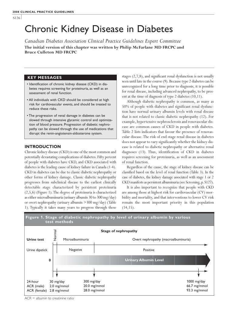
Figure 1. Stage of diabetic nephropathy by level of urinary albumin by various test methods
S126
INTRODUCTIONChronic kidney disease (CKD) is one of the most common andpotentially devastating complications of diabetes. Fifty percentof people with diabetes have CKD, and CKD associated withdiabetes is the leading cause of kidney failure in Canada (1-4).CKD in diabetes can be due to classic diabetic nephropathy orother forms of kidney damage. Classic diabetic nephropathyprogresses from subclinical disease to the earliest clinicallydetectable stage characterized by persistent proteinuria(2,5,6) (Figure 1).The degree of proteinuria is characterizedas either microalbuminuria (urinary albumin 30 to 300 mg/day)or overt nephropathy (urinary albumin >300 mg/day) (Table1). Typically it takes many years to progress through these
stages (2,7,8), and significant renal dysfunction is not usuallyseen until late in the course (9). Because type 2 diabetes can beunrecognized for a long time prior to diagnosis, it is possiblefor renal disease, including advanced nephropathy, to be pres-ent at the time of diagnosis of type 2 diabetes (10,11).
Although diabetic nephropathy is common, as many as50% of people with diabetes and significant renal dysfunc-tion have normal urinary albumin levels with renal diseasethat is not related to classic diabetic nephropathy (12). Forexample, hypertensive nephrosclerosis and renovascular dis-ease are common causes of CKD in people with diabetes.Table 2 lists indicators that favour the presence of renovas-cular disease.The risk of end-stage renal disease in diabetesdoes not appear to vary significantly whether the kidney dis-ease is related to diabetic nephropathy or alternative renaldiagnoses (13). Thus, identification of CKD in diabetesrequires screening for proteinuria, as well as an assessmentof renal function.
Regardless of the cause, the stage of kidney disease can beclassified based on the level of renal function (Table 3). In thecase of diabetes, the kidney damage associated with stage 1 or 2CKD manifests as persistent albuminuria (see Screening, p. S127).
It is also important to recognize that people with CKDare among those at highest risk for cardiovascular (CV) mor-bidity and mortality, and that interventions to lower CV riskremain the most important priority in this population(14,15).
Chronic Kidney Disease in DiabetesCanadian Diabetes Association Clinical Practice Guidelines Expert CommitteeThe initial version of this chapter was written by Philip McFarlane MD FRCPC and Bruce Culleton MD FRCPC
2008 CLINICAL PRACTICE GUIDELINES
• Identification of chronic kidney disease (CKD) in dia-betes requires screening for proteinuria, as well as anassessment of renal function.
• All individuals with CKD should be considered at highrisk for cardiovascular events, and should be treated toreduce these risks.
• The progression of renal damage in diabetes can beslowed through intensive glycemic control and optimiza-tion of blood pressure. Progression of diabetic nephro-pathy can be slowed through the use of medications thatdisrupt the renin-angiotensin-aldosterone system.
KEY MESSAGES
Urine test
Stage of nephropathy
Urine dipstick
Nor
mal
Microalbuminuria Overt nephropathy (macroalbuminuria)
Positive
Urinary Albumin Level
Negative
24-hour 30 mg/dayACR (male) 2.0 mg/mmolACR (female) 2.8 mg/mmol
300 mg/day20.0 mg/mmol28.0 mg/mmol
1000 mg/day66.7 mg/mmol93.3 mg/mmol
ACR = albumin to creatinine ratio
0

S127
SCREENINGIdentification of CKD in diabetes is usually a clinical diag-nosis, requiring a kidney biopsy only when clinical indica-tors leave doubt as to the diagnosis. A person with diabetesis considered to have CKD if he or she has classic diabeticnephropathy (as evidenced by persistent albuminuriaregardless of level of kidney function), or significantlyreduced kidney function (as evidenced by an estimatedglomerular filtration rate [eGFR] ≤60 mL/min). Table 4lists indicators that favour the diagnosis of either diabetic ornondiabetic nephropathy (16-19).As kidney damage is oftenasymptomatic until severe, screening must be performed to
identify renal damage in order to delay or prevent loss ofrenal function through early initiation of effective therapies,and to manage complications in those identified with renaldisease. In adults, screening is performed by measuring uri-nary albumin levels and estimating the level of kidney func-tion (Figure 2).
CO
MP
LIC
AT
ION
SA
ND
CO
MO
RB
IDIT
IES
CKD = chronic kidney diseaseeGFR = estimated glomerular filtration rateGFR = glomerular filtration rate
eGFR = estimated glomerular filtration rate
Table 3. Stages of CKD of all types
Stage Qualitative description
eGFR (mL/min)
1 Kidney damage,normal GFR
≥90
2 Kidney damage, mildlydecreased GFR
60–89
3 Moderately decreased GFR
30–59
4 Severely decreasedGFR
15–29
5 End-stage renal disease <15 (or dialysis)
Table 4. Clinical and laboratory factorsfavouring the diagnosis of clinicaldiabetic nephropathy or an alternate renal diagnosis
Favours diabeticnephropathy
Favours alternate renal diagnosis
• Persistent albuminuria
• Bland urine sediment
• Slow progression of disease
• Low eGFR associated withovert proteinuria
• Presence of other compli-cations of diabetes
• Known duration of diabetes >5 years
• Extreme proteinuria (>6 g/day)
• Persistent hematuria (micro-scopic or macroscopic) oractive urinary sediment
• Rapidly falling eGFR
• Low eGFR with little or noproteinuria
• Other complications of dia-betes not present or rela-tively not as severe
• Known duration of diabetes≤5 years
• Family history of nondiabeticrenal disease (e.g. polycystickidney disease)
• Signs or symptoms of systemic disease
Table 1. Stages of classic diabeticnephropathy according to urinary albumin level
Stage ofnephropathy
Urine dipstick
for protein
Urine ACR(mg/mmol)
24- urinecollection
for albumin*(mg/day)
Normal Negative <2.0 (men)<2.8 (women)
<30
Micro-albuminuria
Negative 2.0–20.0 (men)2.8–28.0 (women)
30–300
Overtnephropathy(macroalbu-minuria)
Positive >20.0 (men)>28.0 (women)
>66.7 (men)>93.3 (women)
>300
>1000
*Values are for urinary albumin, not total urinary protein, whichwill be higher than urinary albumin levels. ACR results may beelevated with conditions other than diabetic nephropathy (see text and Table 4)
ACR = albumin to creatinine ratio
ACE = angiotensin-converting enzymeACR = albumin to creatinine ratioARB = angiotensin II receptor antagonisteGFR = estimated glomerular filtration rate
Table 2. Factors favouring the presence of renovascular disease
• Severe or refractory hypertension• Low eGFR with normal or near-normal ACR• Low or low-normal serum potassium (especially if patient is
on an ACE inhibitor or an ARB)• Flank or abdominal bruits• >30% rise in serum creatinine following initiation of an ACE
inhibitor or an ARB• Presence of aortic or peripheral arterial disease• “Flash” pulmonary edema• Asymmetric renal size on ultrasound• Advanced hypertensive retinopathy

S128
Urine testingScreening for microalbuminuria should be performed usinga random urine test for albumin to creatinine ratio (ACR).As transient microalbuminuria unrelated to diabeticnephropathy can occur, persistent microalbuminuria (atleast 2 of 3 ACR tests positive taken at 1- to 8-week inter-vals) should be demonstrated before the diagnosis of
nephropathy is made. Overt nephropathy rarely normalizeswithout treatment, and repeat ACR testing is not requiredto make the diagnosis of nephropathy in those with ACR val-ues in the overt nephropathy range. A urine dipstick testshould also be performed, either in the laboratory or at thepoint of care, as a screen for renal disease other than diabet-ic nephropathy.
2008 CLINICAL PRACTICE GUIDELINES
Figure 2. Screening for CKD in adults
Screen annually when no transient causes of albuminuria or low eGFR are present, and when acute renal failureor nondiabetic kidney disease is not suspected
Type 1 diabetes: Annually in individuals with duration of diabetes >5 years Type 2 diabetes: At diagnosis of diabetes and annually thereafter
Order random urine ACR andserum creatinine for eGFR
Order serum creatinine for eGFR in 3 months, and 2 repeatrandom urine ACRs performed over the next 3 months
At 3 monthseGFR ≤60 mL/min or 2 or 3 out of 3 ACRs abnormal?
CKD diagnosed
Order urine routine and microscopic and urine dipstick
No evidence of CKDRescreen in 1 year
CKD in diabetesdiagnosed
See treatment guidelines
Nondiabetic renaldisease suspected
Work up or refer
ACR = albumin to creatinine ratio CKD = chronic kidney disease eGFR = estimated glomerular filtration rate
Yes
No
No
No Yes
eGFR ≤60 mL/min or ACR abnormal
Suspicion of nondiabetic renal disease (based on clinical findings or laboratory tests)? (See Table 4)

S129
Twenty-four-hour urine collections are frequently per-formed incorrectly, are unpopular with patients and areunnecessary in routine diabetes care (20-24). However, a 24-hour collection can be useful when there is doubt about theaccuracy of an eGFR, when screening for nonalbumin uri-nary proteins (e.g. multiple myeloma) or when estimatingdaily sodium intake in an individual with refractory edema orhypertension. Individuals should be counselled to discard thefirst morning urine on the day of collection, and then collectall subsequent urine for a 24-hour period, including the firstmorning urine of the next day.
Renal function testingDiabetic nephropathy and damage from other conditions suchas hypertension and renovascular disease can lead to a loss ofrenal function in people with diabetes.An estimate of the kid-ney’s ability to filter toxins from the blood should be made.Serum creatinine is the most commonly used measure ofrenal function; however, the creatinine may falsely indicatethat a person’s renal function is normal (25,26). Individualscan lose up to 50% of their renal function before serum cre-atinine levels rise into the abnormal range (27).The eGFR isa more sensitive method of identifying low kidney function inpeople with diabetes. In Canada, the eGFR is most often cal-culated using the abbreviated Modification of Diet in RenalDisease (MDRD) equation, which takes into account the per-son’s serum creatinine, age and sex. Clinicians can furtheradjust the eGFR for race. Calculation of the MDRD glomeru-lar filtration rate (GFR) is complicated and typically an elec-tronic aid (either a spreadsheet or an Internet-based tool) isused, or the GFR is calculated and reported by the laborato-ry automatically when a serum creatinine is ordered (28).
Delaying screening for CKDAs the ACR can be elevated with recent major exercise (29),fever (30), urinary tract infection, congestive heart failure(31), menstruation or acute severe elevations of blood pres-sure (BP) or blood glucose (BG) (32,33), screening for albu-minuria should be delayed in the presence of theseconditions. Intravascular volume contraction or any acute ill-ness can transiently lower kidney function, and GFR estima-tion for screening purposes should be delayed until suchconditions resolve.
TREATMENT AND FOLLOW-UPAll people with CKD should be considered to be at high riskfor CV events and should be treated to reduce these risks.The progression of renal damage in diabetes can be slowedthrough intensive glycemic control (34) and optimization ofBP (35). Progression of diabetic nephropathy can be slowedthrough the use of medications that disrupt the renin-angiotensin-aldosterone system (RAAS) (36). BP andglycemic targets are the same as for those individuals withdiabetes without nephropathy.
In addition to BP control, some antihypertensive havebeen shown to have additional renal-protective properties. Intype 1 diabetes, angiotensin-converting enzyme (ACE)inhibitors have been shown to decrease albuminuria and pre-vent worsening of nephropathy (37), and angiotensin IIreceptor antagonists (ARBs) have been shown to reduce pro-teinuria (38). In type 2 diabetes, ACE inhibitors and ARBshave been shown to decrease albuminuria and prevent wors-ening of nephropathy, and ARBs have been shown to delaythe time to dialysis in those with renal dysfunction at baseline(39-42). In type 2 diabetes, ACE inhibitors have been shownto reduce the chance of developing new nephropathy(39,43). ACE inhibitor plus ARB combination therapy hasbeen shown to lower BP and proteinuria in type 2 diabetesmore effectively than monotherapy with either agent (44-46).These renal-protective effects also appear to be presentin proteinuric individuals with diabetes and normal or near-normal BP. ACE inhibitors have been shown to reduce pro-gression of diabetic nephropathy in normotensive individualswith type 1 (47-50) or type 2 diabetes (51). In people withdiabetes, hypertension and proteinuria, nondihydropyridinecalcium channel blockers (non-DHP CCBs) (diltiazem andverapamil) have been shown to decrease albuminuria and areassociated with a slower loss of renal function (52-55).However, non-DHP CCBs do not prevent the developmentof nephropathy (43).
In CKD from causes other than diabetic nephropathy, ACEinhibition has been shown to reduce proteinuria, slow progres-sion of renal disease and delay the need for dialysis (56,57).Theissue of whether ARBs and ACE inhibitors are similarly effec-tive in CKD that is not caused by diabetic nephropathy remainscontroversial (58). Compared to monotherapy with eitheragent, ACE inhibitor plus ARB combination therapy has beenshown to reduce proteinuria (59,60).
In people with CKD and diabetes with or without hyper-tension, an ACE inhibitor or an ARB would be the preferredinitial agent for prevention of renal disease progression. Todate, there have been no large-scale hard-endpoint trials for second-line agents in nephropathy (see The Role ofProteinuria Reduction, p. S130).
Treating CKD in diabetes safelyIndividuals starting therapy with an ACE inhibitor or anARB should be monitored within 1 to 2 weeks of initiationor titration of treatment for significant worsening of renalfunction or the development of significant hyperkalemia.Periodic monitoring should continue in those whose serumcreatinine or potassium level increases above normal labo-ratory limits until these values have stabilized. Serum crea-tinine typically increases up to 30% above baseline afterinitiation of an ACE inhibitor or ARB, and usually stabilizesafter 2 to 4 weeks of treatment (61). ACE inhibitors andARBs can be used safely in people with renovascular dis-ease, unless the individual has only a single functioning kid-
CO
MP
LIC
AT
ION
SA
ND
CO
MO
RB
IDIT
IES
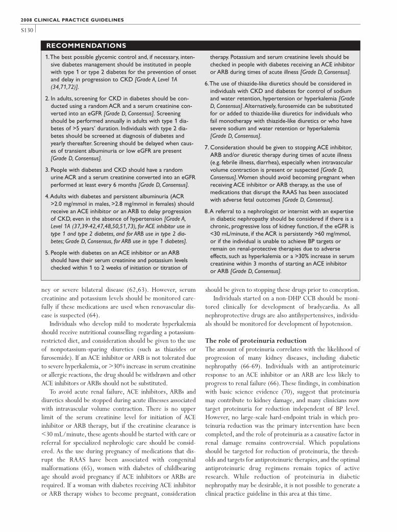
S130
ney or severe bilateral disease (62,63). However, serumcreatinine and potassium levels should be monitored care-fully if these medications are used when renovascular dis-ease is suspected (64).
Individuals who develop mild to moderate hyperkalemiashould receive nutritional counselling regarding a potassium-restricted diet, and consideration should be given to the useof nonpotassium-sparing diuretics (such as thiazides orfurosemide). If an ACE inhibitor or ARB is not tolerated dueto severe hyperkalemia, or >30% increase in serum creatinineor allergic reactions, the drug should be withdrawn and otherACE inhibitors or ARBs should not be substituted.
To avoid acute renal failure, ACE inhibitors, ARBs anddiuretics should be stopped during acute illnesses associatedwith intravascular volume contraction. There is no upperlimit of the serum creatinine level for initiation of ACEinhibitor or ARB therapy, but if the creatinine clearance is<30 mL/minute, these agents should be started with care orreferral for specialized nephrologic care should be consid-ered. As the use during pregnancy of medications that dis-rupt the RAAS have been associated with congenitalmalformations (65), women with diabetes of childbearingage should avoid pregnancy if ACE inhibitors or ARBs arerequired. If a woman with diabetes receiving ACE inhibitoror ARB therapy wishes to become pregnant, consideration
should be given to stopping these drugs prior to conception.Individuals started on a non-DHP CCB should be moni-
tored clinically for development of bradycardia. As allnephroprotective drugs are also antihypertensives, individu-als should be monitored for development of hypotension.
The role of proteinuria reductionThe amount of proteinuria correlates with the likelihood ofprogression of many kidney diseases, including diabeticnephropathy (66-69). Individuals with an antiproteinuricresponse to an ACE inhibitor or an ARB are less likely toprogress to renal failure (66).These findings, in combinationwith basic science evidence (70), suggest that proteinuriamay contribute to kidney damage, and many clinicians nowtarget proteinuria for reduction independent of BP level.However, no large-scale hard-endpoint trials in which pro-teinuria reduction was the primary intervention have beencompleted, and the role of proteinuria as a causative factor inrenal damage remains controversial. Which populationsshould be targeted for reduction of proteinuria, the thresh-olds and targets for antiproteinuric therapies, and the optimalantiproteinuric drug regimens remain topics of activeresearch. While reduction of proteinuria in diabeticnephropathy may be desirable, it is not possible to generate aclinical practice guideline in this area at this time.
2008 CLINICAL PRACTICE GUIDELINES
1.The best possible glycemic control and, if necessary, inten-sive diabetes management should be instituted in peoplewith type 1 or type 2 diabetes for the prevention of onsetand delay in progression to CKD [Grade A, Level 1A(34,71,72)].
2. In adults, screening for CKD in diabetes should be con-ducted using a random ACR and a serum creatinine con-verted into an eGFR [Grade D, Consensus]. Screeningshould be performed annually in adults with type 1 dia-betes of >5 years’ duration. Individuals with type 2 dia-betes should be screened at diagnosis of diabetes andyearly thereafter. Screening should be delayed when caus-es of transient albuminuria or low eGFR are present[Grade D, Consensus].
3. People with diabetes and CKD should have a randomurine ACR and a serum creatinine converted into an eGFRperformed at least every 6 months [Grade D, Consensus].
4.Adults with diabetes and persistent albuminuria (ACR>2.0 mg/mmol in males, >2.8 mg/mmol in females) shouldreceive an ACE inhibitor or an ARB to delay progressionof CKD, even in the absence of hypertension [Grade A,Level 1A (37,39-42,47,48,50,51,73), for ACE inhibitor use intype 1 and type 2 diabetes, and for ARB use in type 2 dia-betes; Grade D, Consensus, for ARB use in type 1 diabetes].
5. People with diabetes on an ACE inhibitor or an ARBshould have their serum creatinine and potassium levelschecked within 1 to 2 weeks of initiation or titration of
therapy. Potassium and serum creatinine levels should bechecked in people with diabetes receiving an ACE inhibitoror ARB during times of acute illness [Grade D, Consensus].
6.The use of thiazide-like diuretics should be considered inindividuals with CKD and diabetes for control of sodiumand water retention, hypertension or hyperkalemia [GradeD, Consensus].Alternatively, furosemide can be substitutedfor or added to thiazide-like diuretics for individuals whofail monotherapy with thiazide-like diuretics or who havesevere sodium and water retention or hyperkalemia[Grade D, Consensus].
7. Consideration should be given to stopping ACE inhibitor,ARB and/or diuretic therapy during times of acute illness(e.g. febrile illness, diarrhea), especially when intravascularvolume contraction is present or suspected [Grade D,Consensus].Women should avoid becoming pregnant whenreceiving ACE inhibitor or ARB therapy, as the use ofmedications that disrupt the RAAS has been associatedwith adverse fetal outcomes [Grade D, Consensus].
8.A referral to a nephrologist or internist with an expertisein diabetic nephropathy should be considered if there is achronic, progressive loss of kidney function, if the eGFR is<30 mL/minute, if the ACR is persistently >60 mg/mmol,or if the individual is unable to achieve BP targets orremain on renal-protective therapies due to adverseeffects, such as hyperkalemia or a >30% increase in serumcreatinine within 3 months of starting an ACE inhibitor or ARB [Grade D, Consensus].
RECOMMENDATIONS
S131
ReferralMost people with CKD and diabetes will not require referralto a specialist in renal disease. However, specialist care may benecessary when renal dysfunction is severe, when there aredifficulties implementing renal-protective strategies or whenthere are problems managing the sequelae of renal disease(see Recommendation #8).
OTHER RELEVANT GUIDELINESTargets for Glycemic Control, p. S29Identification of Individuals at High Risk of Coronary
Events, p. S95Vascular Protection in People With Diabetes, p. S102Treatment of Hypertension, p. S115Type 1 Diabetes in Children and Adolescents, p. S150Diabetes and Pregnancy, p. S168
RELATED WEBSITESCanadian Hypertension Society. Available at http://www.
hypertension.ca/chs. Accessed September 1, 2008.Canadian Society of Nephrology. Available at http://www.
csnscn.ca. Accessed September 1, 2008.Nephron Information Center. Available at: http://www.
nephron.com. Accessed September 1, 2008.
REFERENCES1. Canadian Institute for Health Information (CIHI). Canadian
Organ Replacement Registry (CORR): 2001 Annual Report. Ottawa,ON: CIHI; 2001.
2. Warram JH, Gearin G, Laffel L, et al. Effect of duration of typeI diabetes on the prevalence of stages of diabetic nephropathydefined by urinary albumin/creatinine ratio. J Am Soc Nephrol.1996;7:930-937.
3. Reenders K, de Nobel E, van den Hoogen HJ, et al. Diabetesand its long-term complications in general practice: a survey ina well-defined population. Fam Pract. 1993;10:169-172.
4. Weir MR. Albuminuria predicting outcome in diabetes: inci-dence of microalbuminuria in Asia-Pacific Rim. Kidney Int.2004;66(suppl 92):S38-S39.
5. Mathiesen ER, Ronn B, Storm B, et al. The natural course ofmicroalbuminuria in insulin-dependent diabetes: a 10-yearprospective study. Diabet Med. 1995;12:482-487.
6. Lemley KV,Abdullah I, Myers BD, et al. Evolution of incipientnephropathy in type 2 diabetes mellitus. Kidney Int. 2000;58:1228-1237.
7. Gall MA, Nielsen FS, Smidt UM, et al. The course of kidneyfunction in type 2 (non-insulin-dependent) diabetic patientswith diabetic nephropathy. Diabetologia. 1993;36:1071-1078.
8. Jacobsen P, Rossing K,Tarnow L, et al. Progression of diabet-ic nephropathy in normotensive type 1 diabetic patients.Kidney Int. 1999;71(suppl):S101-S105.
9. Hasslacher C, Ritz E,Wahl P, et al. Similar risks of nephropa-thy in patients with type I or type II diabetes mellitus. NephrolDial Transplant. 1989;4:859-863.
10. Ballard DJ, Humphrey LL, Melton LJ, et al. Epidemiology ofpersistent proteinuria in type II diabetes mellitus. Population-based study in Rochester, Minnesota. Diabetes. 1988;37:405-412.
11. Winaver J,Teredesai P, Feldman HA, et al. Diabetic nephropa-thy as the mode of presentation of diabetes mellitus.Metabolism. 1979;28:1023-1030.
12. Middleton RJ, Foley RN, Hegarty J, et al. The unrecognizedprevalence of chronic kidney disease in diabetes. Nephrol DialTransplant. 2006;21:88-92.
13. Ruggenenti P, Gambara V, Perna A, et al. The nephropathy ofnon-insulin-dependent diabetes: predictors of outcome rela-tive to diverse patterns of renal injury. J Am Soc Nephrol.1998;9:2336-2343.
14. Gerstein HC, Mann JF,Yi Q, et al.Albuminuria and risk of car-diovascular events, death, and heart failure in diabetic and non-diabetic individuals. JAMA. 2001;286:421-426.
15. Gaede P, Vedel P, Parving HH, et al. Intensified multifactorialintervention in patients with type 2 diabetes mellitus andmicroalbuminuria: the Steno type 2 randomised study. Lancet.1999;353:617-622.
16. VenkataRaman TV, Knickerbocker F, Sheldon CV. Unusualcauses of renal failure in diabetics: two case studies. J Okla StateMed Assoc. 1990;83:164-168.
17. Clinical path conference. Unusual renal complications in dia-betes mellitus. Minn Med. 1967;50:387-393.
18. Amoah E, Glickman JL, Malchoff CD, et al. Clinical identifi-cation of nondiabetic renal disease in diabetic patients withtype I and type II disease presenting with renal dysfunction. AmJ Nephrol. 1988;8:204-211.
19. El-Asrar AM,Al-Rubeaan KA,Al-Amro SA, et al. Retinopathyas a predictor of other diabetic complications. Int Ophthalmol.2001;24:1-11.
20. Ahn CW, Song YD, Kim JH, et al.The validity of random urinespecimen albumin measurement as a screening test for diabet-ic nephropathy. Yonsei Med J. 1999;40:40-45.
21. Kouri TT, Viikari JS, Mattila KS, et al. Microalbuminuria.Invalidity of simple concentration-based screening tests forearly nephropathy due to urinary volumes of diabetic patients.Diabetes Care. 1991;14:591-593.
22. Rodby RA, Rohde RD, Sharon Z, et al. The urine protein tocreatinine ratio as a predictor of 24-hour urine protein excre-tion in type 1 diabetic patients with nephropathy. TheCollaborative Study Group. Am J Kidney Dis. 1995;26:904-909.
23. Chaiken RL, Khawaja R, Bard M, et al. Utility of untimed uri-nary albumin measurements in assessing albuminuria in blackNIDDM subjects. Diabetes Care. 1997;20:709-713.
24. Bakker AJ. Detection of microalbuminuria. Receiver operatingcharacteristic curve analysis favors albumin-to-creatinine ratioover albumin concentration. Diabetes Care. 1999;22:307-313.
25. Gault MH, Longerich LL, Harnett JD, et al. Predictingglomerular function from adjusted serum creatinine. Nephron.1992;62:249-256.
26. Bending JJ, Keen H, Viberti GC. Creatinine is a poor marker
CO
MP
LIC
AT
ION
SA
ND
CO
MO
RB
IDIT
IES
S132
of renal failure. Diabet Med. 1985;2:65-66.27. Shemesh O, Golbetz H, Kriss JP, et al. Limitations of creatinine
as a filtration marker in glomerulopathic patients. Kidney Int.1985;28:830-838.
28. Levey AS, Bosch JP, Lewis JB, et al. A more accurate methodto estimate glomerular filtration rate from serum creatinine: anew prediction equation. Modification of Diet in RenalDisease Study Group. Ann Intern Med. 1999;130:461-470.
29. Huttunen NP, Kaar M, Puukka R, et al. Exercise-induced pro-teinuria in children and adolescents with type 1 (insulindependent) diabetes. Diabetologia. 1981;21:495-497.
30. Solling J, Solling K, Mogensen CE. Patterns of proteinuria andcirculating immune complexes in febrile patients. Acta MedScand. 1982;212:167-169.
31. Ritz E. Nephropathy in type 2 diabetes. J Intern Med. 1999;245:111-126.
32. Wiseman M,Viberti G, Mackintosh D, et al. Glycaemia, arte-rial pressure and micro-albuminuria in type 1 (insulin-depend-ent) diabetes mellitus. Diabetologia. 1984;26:401-405.
33. Ravid M, Savin H, Lang R, et al. Proteinuria, renal impair-ment, metabolic control, and blood pressure in type 2 diabetesmellitus. A 14-year follow-up report on 195 patients. ArchIntern Med. 1992;152:1225-1229.
34. Wang PH, Lau J, Chalmers TC. Meta-analysis of effects ofintensive blood-glucose control on late complications of type Idiabetes. Lancet. 1993;341:1306-1309.
35. Maki DD, Ma JZ, Louis TA, et al. Long-term effects of antihy-pertensive agents on proteinuria and renal function. Arch InternMed. 1995;155:1073-1080.
36. Kasiske BL, Kalil RS, Ma JZ, et al. Effect of antihypertensivetherapy on the kidney in patients with diabetes: a meta-regres-sion analysis. Ann Intern Med. 1993;118:129-138.
37. Lewis EJ, Hunsicker LG, Bain RP, et al. The effect ofangiotensin-converting-enzyme inhibition on diabeticnephropathy. The Collaborative Study Group. N Engl J Med.1993;329:1456-1462.
38. Andersen S,Tarnow L, Rossing P, et al. Renoprotective effectsof angiotensin II receptor blockade in type 1 diabetic patientswith diabetic nephropathy. Kidney Int. 2000;57:601-606.
39. Strippoli GF, Craig MC, Schena FP, et al. Role of blood pres-sure targets and specific antihypertensive agents used to pre-vent diabetic nephropathy and delay its progression. J Am SocNephrol. 2006;17(suppl 2):S153-S155.
40. Lewis EJ, Hunsicker LG, Clarke WR, et al. Renoprotectiveeffect of the angiotensin-receptor antagonist irbesartan inpatients with nephropathy due to type 2 diabetes. N Engl J Med.2001;345:851-860.
41. Brenner BM, Cooper ME, de Zeeuw D, et al. Effects of losar-tan on renal and cardiovascular outcomes in patients with type2 diabetes and nephropathy. N Engl J Med. 2001;345:861-869.
42. Parving HH, Lehnert H, Brochner-Mortensen J, et al. Theeffect of irbesartan on the development of diabetic nephropa-thy in patients with type 2 diabetes. N Engl J Med. 2001;345:870-878.
43. Ruggenenti P, Fassi A, Ilieva AP, et al. Preventing microalbu-minuria in type 2 diabetes. N Engl J Med. 2004;351:1941-1951.
44. Wade VL, Gleason BL. Dual blockade of the renin-angiotensinsystem in diabetic nephropathy. Ann Pharmacother. 2004;38:1278-1282.
45. Mogensen CE, Neldam S,Tikkanen I, et al. Randomised con-trolled trial of dual blockade of renin-angiotensin system inpatients with hypertension, microalbuminuria, and non-insulin dependent diabetes: the Candesartan And LisinoprilMicroalbuminuria (CALM) Study. BMJ. 2000;321:1440-1444.
46. Igarashi M, Hirata A, Kadomoto Y, et al. Dual blockade ofangiotensin II with enalapril and losartan reduces proteinuriain hypertensive patients with type 2 diabetes. Endocr J. 2006;53:493-501.
47. Laffel LM, McGill JB, Gans DJ. The beneficial effect ofangiotensin-converting enzyme inhibition with captopril ondiabetic nephropathy in normotensive IDDM patients withmicroalbuminuria. North American Microalbuminuria StudyGroup. Am J Med. 1995;99:497-504.
48. Mathiesen ER, Hommel E, Giese J, et al. Efficacy of captopril inpostponing nephropathy in normotensive insulin dependent dia-betic patients with microalbuminuria. BMJ. 1991;303:81-87.
49. Jerums G, Allen TJ, Campbell DJ, et al. Long-term compari-son between perindopril and nifedipine in normotensivepatients with type 1 diabetes and microalbuminuria. Am JKidney Dis. 2001;37:890-899.
50. ACE Inhibitors in Diabetic Nephropathy Trialist Group. Shouldall patients with type 1 diabetes mellitus and microalbumin-uria receive angiotensin-converting enzyme inhibitors? Ameta-analysis of individual patient data. Ann Intern Med.2001;134:370-379.
51. Ravid M, Savin H, Jutrin I, et al. Long-term stabilizing effectof angiotensin-converting enzyme inhibition on plasma creati-nine and on proteinuria in normotensive type II diabeticpatients. Ann Int Med. 1993;118:577-581.
52. Bakris GL, Copley JB, Vicknair N, et al. Calcium channelblockers versus other antihypertensive therapies on progres-sion of NIDDM associated nephropathy. Kidney Int. 1996;50:1641-1650.
53. Bakris GL, Weir MR, DeQuattro V, et al. Effects of an ACEinhibitor/calcium antagonist combination on proteinuria indiabetic nephropathy. Kidney Int. 1998;54:1283-1289.
54. Perez-Maraver M, Carrera MJ, Micalo T, et al. Renoprotectiveeffect of diltiazem in hypertensive type 2 diabetic patients withpersistent microalbuminuria despite ACE inhibitor treatment.Diabetes Res Clin Pract. 2005;70:13-19.
55. Bakris GL,Weir MR, Secic M, et al. Differential effects of cal-cium antagonist subclasses on markers of nephropathy pro-gression. Kidney Int. 2004;65:1991-2002.
56. Ruggenenti P, Perna A, Gherardi G, et al. Renal function andrequirement for dialysis in chronic nephropathy patients onlong-term ramipril: REIN follow-up trial. Gruppo Italiano diStudi Epidemiologici in Nefrologia (GISEN). Ramipril
2008 CLINICAL PRACTICE GUIDELINES

S133
Efficacy in Nephropathy. Lancet. 1998;352:1252-1256.57. Maschio G, Alberti D, Locatelli F, et al. Angiotensin-convert-
ing enzyme inhibitors and kidney protection: the AIPRI trial.The ACE Inhibition in Progressive Renal Insufficiency (AIPRI)Study Group. J Cardiovasc Pharmacol. 1999;33(suppl 1):S16-S20.
58. Shoda J, Kanno Y, Suzuki H. A five-year comparison of therenal protective effects of angiotensin-converting enzymeinhibitors and angiotensin receptor blockers in patients withnon-diabetic nephropathy. Intern Med. 2006;45:193-198.
59. Parving HH, Persson F, Lewis JB, et al. Aliskiren combinedwith losartan in type 2 diabetes and nephropathy. N Engl J Med.2008;358;23:2433-2446.
60. Minutolo R, Balletta MM, Catapano F, et al. Mesangial hyper-cellularity predicts antiproteinuric response to dual blockade ofRAS in primary glomerulonephritis. Kidney Int.2006; 70:1170-1176.
61. Bakris GL, Weir MR. Angiotensin-converting enzymeinhibitor-associated elevations in serum creatinine: is this acause for concern? Arch Intern Med. 2000;160:685-693.
62. Reams GP, Bauer JH, Gaddy P. Use of the converting enzymeinhibitor enalapril in renovascular hypertension. Effect onblood pressure, renal function, and the renin-angiotensin-aldosterone system. Hypertension. 1986;8:290-297.
63. Franklin SS, Smith RD. Comparison of effects of enalaprilplus hydrochlorothiazide versus standard triple therapy onrenal function in renovascular hypertension. Am J Med. 1985;79:14-23.
64. Miyamori I, Yasuhara S, Takeda Y, et al. Effects of convertingenzyme inhibition on split renal function in renovascularhypertension. Hypertension. 1986;8:415-421.
65. Cooper WO, Hernandez-Diaz S, Arbogast PG, et al. Majorcongenital malformations after first-trimester exposure toACE inhibitors. N Engl J Med. 2006;354:2443-2451.
66. de Zeeuw D, Remuzzi G, Parving HH, et al. Proteinuria, a tar-get for renoprotection in patients with type 2 diabeticnephropathy: lessons from RENAAL. Kidney Int. 2004;65:2309-2320.
67. de Zeeuw D, Remuzzi G, Parving HH, et al. Albuminuria, atherapeutic target for cardiovascular protection in type 2 dia-betic patients with nephropathy. Circulation. 2004;110:921-927.
68. Keane WF, Brenner BM, de Zeeuw D, et al.The risk of devel-oping end-stage renal disease in patients with type 2 diabetesand nephropathy: the RENAAL study. Kidney Int. 2003;63:1499-1507.
69. Forsblom CM, Groop PH, Ekstrand A, et al. Predictive valueof microalbuminuria in patients with insulin-dependent dia-betes of long duration. BMJ. 1992;305:1051-1053.
70. Zoja C, Benigni A, Remuzzi G. Cellular responses to proteinoverload: key event in renal disease progression. Curr OpinNephrol Hypertens. 2004;13:31-37.
71. The Diabetes Control and Complications (DCCT) ResearchGroup. Effect of intensive therapy on the development andprogression of diabetic nephropathy in the Diabetes Control
and Complications Trial. Kidney Int. 1995;47:1703-1720.72. UK Prospective Diabetes Study (UKPDS) Group. Intensive
blood-glucose control with sulphonylureas or insulin comparedwith conventional treatment and risk of complications in patientswith type 2 diabetes (UKPDS 33). Lancet. 1998;352:837-853.
73. Viberti G,Wheeldon NM; MicroAlbuminuria Reduction WithVALsartan (MARVAL) Study Investigators. Microalbuminuriareduction with valsartan in patients with type 2 diabetes mel-litus: a blood pressure-independent effect. Circulation. 2002;106:672-678.
CO
MP
LIC
AT
ION
SA
ND
CO
MO
RB
IDIT
IES

S134
INTRODUCTIONDiabetic retinopathy is the most common cause of new casesof legal blindness in people of working age (1). The EyeDiseases Prevalence Research Group determined the crudeprevalence rate of retinopathy in the adult diabetic populationof the United States to be 40.3%; sight-threatening retinopa-thy occurred at a rate of 8.2% (2). Previous data showed theprevalence rate of proliferative retinopathy to be 23% in peo-ple with type 1 diabetes, 14% in people with type 2 diabetesand on insulin therapy, and 3% in people receiving oral anti-hyperglycemic therapies (3). Macular edema occurs in 11, 15and 4% of these groups, respectively (4). First Nations popu-lations in Canada have high rates of diabetes and its complica-tions (5,6). It is estimated that approximately 2 millionindividuals in Canada (i.e. almost all people with diagnoseddiabetes) have some form of diabetic retinopathy (7).
Visual loss is associated with significant morbidity, includ-ing increased falls, hip fractures and a 4-fold increase in mortality (8). Among individuals with type 1 diabetes, limbamputation and visual loss due to diabetic retinopathy are the2 independent predictors of early death (9).
DEFINITION AND PATHOGENESISDiabetic retinopathy is clinically exclusively defined, diag-nosed and treated based on the extent of retinal vascular dis-ease. Three distinct forms of diabetic retinopathy aredescribed: 1) macular edema, which includes diffuse or focalvascular leakage at the macula; 2) progressive accumulation ofblood vessel change that includes microaneurysms, intrareti-nal hemorrhage, vascular tortuosity and vascular malforma-tion (together known as nonproliferative diabetic retinopathy)that ultimately leads to abnormal vessel growth (proliferativediabetic retinopathy); and 3) retinal capillary closure, a formof vascular change detected by fluorescein angiography, which
is also well recognized as a potentially blinding complicationof diabetes, but currently has no treatment options.
SCREENING AND DIAGNOSISSince laser therapy for sight-threatening diabetic retinopathyreduces the risk of blindness (10-13), ophthalmic screeningstrategies are intended to detect treatable disease. Sight-threatening diabetic retinopathy includes severe nonprolifera-tive diabetic retinopathy, proliferative diabetic retinopathy orclinically significant macular edema. Screening programs con-sider the differences in incidence and prevalence of retinopa-thy observed in type 1 and type 2 diabetes, and distinguishbetween children and adults (see Table 1) (14-19).
Diabetic retinopathy rarely develops in children with type 1 diabetes <10 years of age, regardless of the duration ofdiabetes (18). Among patients <15 years of age, irrespectiveof age of onset of diabetes, the prevalence of mild nonprolif-
RetinopathyCanadian Diabetes Association Clinical Practice Guidelines Expert CommitteeThe initial draft of this chapter was prepared by Shelley R. Boyd MD FRCSC DABO and Filiberto Altomare MD FRCSC
2008 CLINICAL PRACTICE GUIDELINES
• Screening is important for early detection of treatabledisease. Screening intervals for diabetic retinopathy varyaccording to the individual’s age and type of diabetes.
• Tight glycemic control reduces the onset and progres-sion of sight-threatening diabetic retinopathy.
• Laser therapy reduces the risk of significant visual loss.
KEY MESSAGES
Table 1. Screening for retinopathy
When to initiate screening• 5 years after diagnosis of type 1 diabetes in all individuals ≥15 years
• In all individuals at diagnosis of type 2 diabetes
Screening methods• 7-standard field, stereoscopic-colour fundus photography
with interpretation by a trained reader (gold standard) • Direct ophthalmoscopy or indirect slit-lamp fundoscopy
through dilated pupil• Digital fundus photography
If retinopathy is present• Diagnose retinopathy severity and establish appropriate
monitoring intervals (1 year or less) • Treat sight-threatening retinopathy with laser therapy • Review glycemic, BP and lipid control, and adjust therapy
to reach targets as per guidelines* • Screen for other diabetes complications
If retinopathy is not present• Type 1 diabetes: rescreen annually • Type 2 diabetes: rescreen every 1–2 years • Review glycemic, BP and lipid control, and adjust therapy
to reach targets as per guidelines* • Screen for other diabetes complications
*See Other Relevant Guidelines, p. S136BP = blood pressure
S135
erative retinopathy was 2%, and none had sight-threateningdiabetic retinopathy (11,18,20). However, the prevalence rateincreases sharply after 5 years’ duration of diabetes in post-pubertal individuals with type 1 diabetes (18). In theWisconsin Epidemiology Study of Diabetic Retinopathy 4-year incidence study, no person <17 years of age developedproliferative retinopathy or macular edema (16,21,22).Conversely, in people with type 2 diabetes, retinopathy may bepresent in 21 to 39% of patients soon after clinical diagnosis,but is sight-threatening in only about 3% (4,17,19,23). In theUnited Kingdom Prospective Diabetes Study (UKPDS), fewpatients without retinopathy at diagnosis of diabetes had dis-ease progression to the point of requiring photocoagulation inthe following 3 to 6 years (24). More recently, progressionrates of diabetic retinopathy were prospectively evaluated(14,15,25).The Liverpool Diabetic Eye Study reported the 1-year cumulative incidence of sight-threatening diabeticretinopathy in individuals with type 1 or type 2 diabetes whoat baseline had no diabetic retinopathy, had backgroundretinopathy, or had mild preproliferative retinopathy. In peoplewith type 1 diabetes, the incidence in these groups was 0.3,3.6 and 13.5%, respectively (14), and in individuals with type2 diabetes it was 0.3, 5.0 and 15.0%, respectively (15).Although the incidence of sight-threatening diabetic retinopa-thy in the group without baseline diabetic retinopathy is low(14,15,24,25), there have been no studies comparing variousscreening intervals in their effectiveness to reduce the risk ofvision loss (26).
The gold standard for diagnosing diabetic retinopathy isstereoscopic colour fundus photographs in 7 standard fields(27). However, practical common screening strategies for dia-betic retinopathy include clinical examination with ophthal-moscopy with or without diagnostic tools such as fundusphotography.The accuracy of direct ophthalmoscopy to assessseverity of retinopathy can vary widely (28) and is deemedinadequate through an undilated pupil (29-31). Combiningdirect ophthalmoscopy with slit-lamp fundus biomicroscopy,diagnostic accuracy was comparable to the gold standard of 7-field stereophotography (32). Optical coherence tomography(OCT) is also currently under investigation for its use in thediagnosis of diabetic macular edema (33,34). Telemedicineprograms are widely employed in Canada and internationallyfor the identification and triage of patients with diabeticretinopathy (35).
PREVENTION OF ONSET AND PROGRESSIONLonger duration of diabetes, elevated glycated hemoglobin(A1C), increased blood pressure (BP), dyslipidemia, lowhematocrit, pregnancy (with type 1 diabetes) and severeretinopathy itself are associated with disease progression(16-19,22,36-41).
Glycemic control In the Diabetes Control and Complications Trial, intensive
insulin therapy in people with type 1 diabetes reduced therisk of onset of retinopathy by 76%, and the rate of progres-sion by 54%, compared to conventional therapy (42,43). Intype 1 diabetes, rapid improvement of glycemic control maybe associated with transient early worsening of retinopathyduring the first 12 months, but this effect is offset by long-term gain (44).
The UKPDS demonstrated that in type 2 diabetes, hyper-glycemia is an independent risk factor for the incidence andprogression of retinopathy (45,46).Tight glycemic control istherefore recommended (45,47).
Anecdotal reports and retrospective analyses of individu-als receiving thiazolidinediones (TZDs) suggest a correlationwith increased diabetic macular edema (48,49). Individualswho experience changes in vision while on a TZD should bereferred to an ophthalmologist for assessment.
BP controlIn type 1 and type 2 diabetes, elevated diastolic BP is a sig-nificant risk factor for the development of macular edema(22,50), and elevated systolic BP is a risk factor for visionloss (51). In hypertensive individuals, development and pro-gression of retinopathy can be reduced by treatment withantihypertensive agents (52). Further lowering of BP in nor-motensive people with type 2 diabetes also reduces the pro-gression of retinopathy (53).
Lipid controlDyslipidemia is an independent risk factor for retinal hardexudates and clinically significant macular edema in type 1diabetes (54). Similarly, in the Early Treatment DiabeticRetinopathy Study (ETDRS) trial, in which most participantshad type 2 diabetes, elevated low-density lipoprotein choles-terol was associated with increased risk of developing hardexudates (38).
TREATMENTTreatment for diabetic retinopathy includes retinal photo-coagulation and vitreoretinal surgery.
Residual vision can often be improved by an accurate spec-tacle correction and/or magnifying aids, with instructions foruse. People with impaired vision should be informed of theservices in their community that will assist with retraining foremployment, encourage independence and improve theirquality of life (55,56).
Laser therapyAs determined in the Diabetic Retinopathy Study (DRS) andthe ETDRS, laser therapy by panretinal photocoagulation tothe retinal periphery reduces severe visual loss and reduceslegal blindness by 90% in people with severe nonprolifera-tive or proliferative retinopathy (11-13). As determined bythe ETDRS, focal and/or grid laser treatment to the maculafor clinically significant macular edema reduces the incidence
CO
MP
LIC
AT
ION
SA
ND
CO
MO
RB
IDIT
IES

S136
of moderate visual loss by 50% (10).The first study to eval-uate the long-term outcome of laser treatment confirmed itsbenefit (57).
Surgical interventionThe Diabetic Retinopathy Vitrectomy Study (DRVS) Groupevaluated the benefit of early vitrectomy (<6 months) in thetreatment of severe vitreous hemorrhage (58) and very severeproliferative diabetic retinopathy (59). People with type 1diabetes of <20 years’ duration and severe vitreous hemor-rhage were more likely to achieve good vision with early vit-rectomy compared to conventional management (58).Similarly, early vitrectomy was associated with a higher chanceof visual recovery in people with either type 1 or 2 diabeteswith very severe proliferative diabetic retinopathy (59).Surgical advances in vitrectomy since the DRVS trials havedemonstrated reduced side effects with more consistentfavourable visual outcomes, thus supporting vitrectomy in advanced proliferative diabetic retinopathy (60).Furthermore, these advances have expanded surgical indica-tions to include vitrectomy for diffuse macular edema, result-ing in structural and functional improvements (61). It is worthnoting that systemic treatment with acetylsalicylic acid (ASA)does not increase the risk or severity of vitreous hemorrhage(62-64).The risk of vitreous hemorrhage or foveola blot hem-orrhage associated with warfarin therapy is unknown.
Pharmacologic interventionStudies investigating local and systemic pharmacologic treat-ments for diabetic retinopathy are underway, but to date nophase III clinical trial has been successful in achieving its pri-mary endpoint. Nonetheless, earlier phase studies stronglysuggested that intra-ocular delivery of anti-vascular endothe-lial growth factor (anti-VEGF) agents or steroid could beeffective in reducing diabetic macular edema or retinal neo-vascularization. In particular, pegaptanib, an anti-VEGFaptamer approved for the treatment of “wet” age-related mac-ular degeneration, has been shown, in a phase II trial (65) anda pilot study (66), to reduce diabetic macular edema andimprove visuals outcomes compared to control interventions.A retrospective review of patients treated for edema demon-strated a reduction in neovascularization (67). Similarly, ameta-analysis by the Cochrane Collaboration (68) supportsthe intravitreal injection of the steroid triamcinolone acetate(69-71), or the use of implanted intra-ocular devices thatrelease fluocinalone acetonide (72,73) or dexamethasone(74). Finally, 3 phase III clinical trials that are evaluating theeffects of renin-angiotensin-aldosterone system blockade arenearing completion, and are of particular note.These are theDiabetic Retinopathy Candesartan Trial (DIRECT) (75), theretinal measurement substudy (AdRem) of the Action inDiabetes in Vascular Disease: Preterax and Diamicron MRControlled Evaluation (ADVANCE) trial (76) and the ReninAngiotensin System Study (RASS) (77). These studies are
based upon data from the EURODIAB Controlled Trial ofLisinopril in Insulin Dependent Diabetes Mellitus (EUCLID)(78) and the United Kingdom Prospective Diabetes Study(UKPDS) (79).The EUCLID study, designed to evaluate renaldisease, demonstrated that the odds ratio for risk of progres-sion of diabetic retinopathy was 0.5 in patients treated withangiotensin-converting enzyme (ACE) inhibitors comparedto those treated with placebo. However, this study was under-powered for ophthalmic outcomes. Similarly, the UKPDSsuggested a reduction in the need for laser therapy in patientswith type 2 diabetes who received an angiotensin II receptorblocker. Taken together, better understanding of the mecha-nisms of diabetic retinopathy and recent development ofpharmacologic therapies for other indications suggest thatnew therapies are on the horizon.
OTHER RELEVANT GUIDELINESTargets for Glycemic Control, p. S29Dyslipidemia, p. S107Treatment of Hypertension, p. S115Type 1 Diabetes in Children and Adolescents, p. S150Type 2 Diabetes in Children and Adolescents, p. S162Diabetes and Pregnancy, p. S168
2008 CLINICAL PRACTICE GUIDELINES
1. In individuals ≥15 years of age with type 1 diabetes,screening and evaluation for retinopathy by an expertprofessional should be performed annually starting 5years after the onset of diabetes [Grade A, Level 1 (16,18)].
2. In individuals with type 2 diabetes, screening and evalua-tion for diabetic retinopathy by an expert professionalshould be performed at the time of diagnosis of dia-betes [Grade A, Level 1 (17,21)].The interval for follow-up assessments should be tailored to the severity of theretinopathy. In those with no or minimal retinopathy, therecommended interval is 1 to 2 years [Grade A, Level 1(17,21)].
3. Screening for diabetic retinopathy should be performedby experienced professionals, either in person orthrough interpretation of retinal photographs takenthough dilated pupils [Grade A, Level 1 (31)].
4.To prevent the onset and delay the progression of dia-betic retinopathy, people with diabetes should be treat-ed to achieve optimal control of blood glucose [Grade A,Level 1A (42,45)] and BP [Grade A, Level 1A (52)]. Peoplewith abnormal lipids should be considered at high riskfor retinopathy [Grade A, Level 1 (54)].
5. Patients with sight-threatening diabetic retinopathy shouldbe assessed by a general ophthalmologist or retina spe-cialist [Grade D, Consensus]. Laser therapy and/or vitrecto-my [Grade A, Level 1A (10,12,58,59)] and/or pharmacologicintervention [Grade B, Level 2 (65)] should be considered.
6.Visually disabled people should be referred for low-vision evaluation and rehabilitation [Grade D, Consensus].
RECOMMENDATIONS

S137
REFERENCES1. Klein R, Klein BEK.Vision disorders in diabetes. In: Diabetes in
America. 2nd ed. Bethesda, MD: National Diabetes DataGroup, National Institute of Diabetes and Digestive andKidney Diseases, National Institutes of Health; 1995:293-338.
2. Kempen JH, O’Colmain BJ, Leske MC, et al. The prevalenceof diabetic retinopathy among adults in the United States. ArchOphthalmol. 2004;122:552-563.
3. Klein R, Klein BEK, Moss SE. Epidemiology of proliferativediabetic retinopathy. Diabetes Care. 1992;15:1875-1891.
4. Klein R, Klein BEK, Moss SE, et al.The Wisconsin epidemio-logic study of diabetic retinopathy. IV. Diabetic macularedema. Ophthalmology. 1984;91:1464-1474.
5. Maberley D,Walker H, Koushik A, et al. Screening for diabet-ic retinopathy in James Bay, Ontario: a cost-effectivenessanalysis. CMAJ 2003;168:160-164.
6. Kaur H, Maberley D, Chang A, et al.The current status of dia-betes care, diabetic retinopathy screening and eye-care in BritishColumbia’s First Nations Communities. Int J Circumpolar Health.2004;63:277-285.
7. A Clear Vision. Solutions to Canada’s Vision Loss Crisis. Toronto,ON: Canterbury Communications; 2004. Available at:http://www.costofblindness.org.Accessed September 1, 2008.
8. Vu HT, Keeffe JE, McCarty CA, et al. Impact of unilateral andbilateral vision loss on quality of life. Br J Ophthalmol. 2005;89:360-363.
9. Cusick M, Meleth AD, Agron E, et al. Associations of mortali-ty and diabetes complications in patients with type 1 and type2 diabetes: EarlyTreatment Diabetic Retinopathy Study reportno. 27. Diabetes Care. 2005;28:617-625.
10. Early Treatment Diabetic Retinopathy Study Research Group.Photocoagulation for diabetic macular edema. EarlyTreatmentDiabetic Retinopathy Study report number 1. Arch Ophthalmol.1985;103:1796-1806.
11. Ferris FL III. How effective are treatments for diabeticretinopathy? JAMA. 1993;269:1290-1291.
12. The Diabetic Retinopathy Study Research Group.Photocoagulation treatment of proliferative diabetic retinopa-thy: the second report of Diabetic Retinopathy Study findings.Ophthalmology. 1978;85:82-106.
13. Ferris F. Early photocoagulation in patients with either type Ior type II diabetes. Trans Am Ophthalmol Soc. 1996;94:505-537.
14. Younis N, Broadbent DM, Harding SP, et al. Incidence of sight-threatening retinopathy in type 1 diabetes in a systematicscreening programme. Diabet Med. 2003;20:758-765.
15. Younis N, Broadbent DM, Vora JP, et al. Incidence of sight-threatening retinopathy in patients with type 2 diabetes in theLiverpool Diabetic Eye Study: a cohort study. Lancet. 2003;361:195-200.
16. Klein R, Klein BEK, Moss SE, et al. The WisconsinEpidemiologic Study of Diabetic Retinopathy. IX. Four-year inci-dence and progression of diabetic retinopathy when age at diag-nosis is less than 30 years. Arch Ophthalmol. 1989;107:237-243.
17. Klein R, Klein BEK, Moss SE, et al. The Wisconsin
Epidemiologic Study of Diabetic Retinopathy. X. Four-year inci-dence and progression of diabetic retinopathy when age at diag-nosis is 30 years or more. Arch Ophthalmol. 1989;107:244-249.
18. Klein R, Klein BEK, Moss SE, et al. The WisconsinEpidemiologic Study of Diabetic Retinopathy. II. Prevalenceand risk of diabetic retinopathy when age at diagnosis is lessthan 30 years. Arch Ophthalmol. 1984;102:520-526.
19. Klein R, Klein BEK, Moss SE, et al. The WisconsinEpidemiologic Study of Diabetic Retinopathy. III. Prevalenceand risk of diabetic retinopathy when age at diagnosis is 30 ormore years. Arch Ophthalmol. 1984;102:527-532.
20. Klein R, Klein BEK, Moss SE, et al. Severe retinopathy ininsulin-taking children and young adults. Pediat Adolesc Endocr.1988;17:146-152.
21. Klein R, Klein BEK, Moss SE, et al. The WisconsinEpidemiologic Study of Diabetic Retinopathy. VII. Diabeticnonproliferative retinal lesions. Ophthalmology. 1987;94:1389-1400.
22. Klein R, Moss SE, Klein BEK, et al. The WisconsinEpidemiologic Study of Diabetic Retinopathy. XI. The inci-dence of macular edema. Ophthalmology. 1989;96:1501-1510.
23. Kohner EM, Aldington SJ, Stratton IM, et al. United KingdomProspective Diabetes Study, 30: diabetic retinopathy at diagno-sis of non-insulin-dependent diabetes mellitus and associatedrisk factors. Arch Ophthalmol. 1998;116:297-303.
24. Kohner EM, Stratton IM, Aldington SJ, et al. Relationshipbetween the severity of retinopathy and progression to photo-coagulation in patients with type 2 diabetes mellitus in theUKPDS (UKPDS 52). Diabet Med. 2001;18:178-184.
25. Maguire A, Chan A, Cusumano J, et al. The case for biennialretinopathy screening in children and adolescents. DiabetesCare. 2005;28:509-513.
26. Klein R. Screening interval for retinopathy in type 2 diabetes.Lancet. 2003;361:190-191.
27. Early Treatment Diabetic Retinopathy Study Research Group.Grading diabetic retinopathy from stereoscopic color fundusphotographs—an extension of the modified Airlie House clas-sification. ETDRS report number 10. Ophthalmology. 1991;98:786-806.
28. Hutchinson A, McIntosh A, Peters J, et al. Effectiveness ofscreening and monitoring tests for diabetic retinopathy—asystematic review. Diabet Med. 2000;17:495-506.
29. Nathan DM, Fogel HA, Godine JE, et al. Role of diabetologistin evaluating diabetic retinopathy. Diabetes Care. 1991;14:26-33.
30. Sussman EJ, Tsiaras WG, Soper KA. Diagnosis of diabetic eyedisease. JAMA. 1982;247:3231-3234.
31. Buxton MJ, Sculpher MJ, Ferguson BA, et al. Screening fortreatable diabetic retinopathy: a comparison of different meth-ods. Diabet Med. 1991;8:371-377.
32. Scanlon PH, Malhotra R, Greenwood RH, et al. Comparisonof two reference standards in validating two field mydriaticdigital photography as a method of screening for diabeticretinopathy. Br J Ophthalmol. 2003;87:1258-1263.
33. Lang GE. Optical coherence tomography findings in diabetic
CO
MP
LIC
AT
ION
SA
ND
CO
MO
RB
IDIT
IES
S138
retinopathy. Dev Ophthalmol. 2007;39:31-47.34. Massin P, Girach A, Erginay A, et al. Optical coherence tomog-
raphy: a key to the future management of patients with diabet-ic macular oedema. Acta Ophthalmol Scand. 2006;84:466-474.
35. Whited JD. Accuracy and reliability of teleophthalmology fordiagnosing diabetic retinopathy and macular edema: a reviewof the literature. Diabetes Technol Ther. 2006;8:102-122.
36. Davis MD, Fisher MR, Gangnon RE, et al. Risk factors forhigh-risk proliferative diabetic retinopathy and severe visualloss: Early Treatment Diabetic Retinopathy Study report #18.Invest Ophthalmol Vis Sci. 1998;39:233-252.
37. Klein BEK, Moss SE, Klein R. Effect of pregnancy on progres-sion of diabetic retinopathy. Diabetes Care. 1990;13:34-40.
38. Chew EY, Klein ML, Ferris FL III, et al. Association of elevat-ed serum lipid levels with retinal hard exudate in diabeticretinopathy. Early Treatment Diabetic Retinopathy Study(ETDRS) report 22. Arch Ophthalmol. 1996;114:1079-1084.
39. Qiao Q, Keinänen-Kiukaanniemi S, Läärä E. The relationshipbetween hemoglobin levels and diabetic retinopathy. J ClinEpidemiol. 1997;50:153-158.
40. The Diabetes Control and Complications Trial ResearchGroup. Effect of pregnancy on microvascular complications inthe Diabetes Control and Complications Trial. Diabetes Care.2000;23:1084-1091.
41. Chew EY, Mills JL, Metzger BE, et al. Metabolic control andprogression of retinopathy. The Diabetes in Early PregnancyStudy. Diabetes Care. 1995;18:631-637.
42. The Diabetes Control and Complications Trial ResearchGroup. The effect of intensive treatment of diabetes on thedevelopment and progression of long-term complications ininsulin-dependent diabetes mellitus. N Engl J Med. 1993;329:977-986.
43. The Diabetes Control and Complications Trial ResearchGroup. The effect of intensive diabetes treatment on the pro-gression of diabetic retinopathy in insulin-dependent diabetesmellitus. Arch Ophthalmol. 1995;113:36-51.
44. The Diabetes Control and Complications Trial ResearchGroup. Early worsening of diabetic retinopathy in the DiabetesControl and Complications Trial. Arch Ophthalmol. 1998;116:874-886.
45. UK Prospective Diabetes Study (UKPDS) Group. Intensiveblood-glucose control with sulphonylureas or insulin com-pared with conventional treatment and risk of complications inpatients with type 2 diabetes (UKPDS 33). Lancet. 1998;352:837-853.
46. Ohkubo Y, Kishikawa H, Araki E, et al. Intensive insulin thera-py prevents the progression of diabetic microvascular compli-cations in Japanese patients with non-insulin-dependentdiabetes mellitus: a randomized prospective 6-year study.Diabetes Res Clin Pract. 1995;28:103-117.
47. Klein R, Klein BEK, Moss SE, et al. Relationship of hyper-glycemia to the long-term incidence and progression of dia-betic retinopathy. Arch Intern Med. 1994;154:2169-2178.
48. Sivagnanam G. Rosiglitazone and macular edema. CMAJ.
2006;175:276.49. Kendall C,Wooltoroton E. Rosiglitazone (Avandia) and macu-
lar edema. CMAJ. 2006;174:623.50. Klein R, Klein BEK, Moss SE, et al. The Wisconsin
Epidemiologic Study of Diabetic Retinopathy. XV.The long-termincidence of macular edema. Ophthalmology. 1995;102:7-16.
51. Moss SE, Klein R, Klein BEK.Ten-year incidence of visual lossin a diabetic population. Ophthalmology. 1994;101:1061-1070.
52. UK Prospective Diabetes Study Group. Tight blood pressurecontrol and risk of macrovascular and microvascular complica-tions in type 2 diabetes: UKPDS 38. BMJ. 1998;317:703-713.
53. Schrier RW, Estacio RO, Esler A, et al. Effects of aggressiveblood pressure control in normotensive type 2 diabeticpatients on albuminuria, retinopathy and strokes. Kidney Int.2002;61:1086-1097.
54. Miljanovic B, Glynn RJ, Nathan DM, et al.A prospective studyof serum lipids and risk of diabetic macular edema in type 1diabetes. Diabetes. 2004;53:2883-2892.
55. Fonda GE. Optical treatment of residual vision in diabeticretinopathy. Ophthalmology. 1994;101:84-88.
56. Bernbaum M, Albert SG. Referring patients with diabetes andvision loss for rehabilitation: who is responsible? Diabetes Care.1996;19:175-177.
57. Chew EY, Ferris FL III, Csaky KG, et al.The long-term effectsof laser photocoagulation treatment in patients with diabeticretinopathy: the Early Treatment Diabetic Retinopathy Follow-up Study. Ophthalmology. 2003;110:1683-1689.
58. The Diabetic Retinopathy Vitrectomy Study Research Group.Early vitrectomy for severe vitreous hemorrhage in diabeticretinopathy. Four-year results of a randomized trial: DiabeticRetinopathy Study report 5. Arch Ophthalmol. 1990;108:958-964.
59. The Diabetic Retinopathy Vitrectomy Study Research Group.Early vitrectomy for severe proliferative diabetic retinopathy ineyes with useful vision. Results of a randomized trial—DiabeticRetinopathy Vitrectomy Study report 3. Ophthalmology.1988;95:1307-1320.
60. Smiddy WE, Flynn HW.Vitrectomy in the management of dia-betic retinopathy. Surv Ophthalmol. 1999;43:491-507.
61. Stolba U, Binder S, Gruber D, et al.Vitrectomy for persistentdiffuse macular edema. Am J Ophthalmol. 2005;140:295-301.
62. Chew EY, Klein ML, Murphy RP, et al. Effects of aspirin onvitreous/preretinal hemorrhage in patients with diabetes mel-litus. Early Treatment Diabetic Retinopathy Study report no.20. Arch Ophthalmol. 1995;113:52-55.
63. Early Treatment Diabetic Retinopathy Study Research Group.Effects of aspirin treatment on diabetic retinopathy. ETDRSreport number 8. Ophthalmology. 1991;98(5 suppl):757-765.
64. Chew EY, Benson WE, Remaley NA, et al. Results after lensextraction in patients with diabetic retinopathy: EarlyTreatment Diabetic Retinopathy Study report number 25. ArchOphthalmol. 1999;117:1600-1606.
65. Cunningham ET Jr, Adamis AP, Altaweel M, et al. A phase IIrandomized double-masked trial of pegaptanib, an anti-
2008 CLINICAL PRACTICE GUIDELINES
S139
vascular endothelial growth factor aptamer, for diabetic macu-lar edema. Ophthalmology. 2005;112:1747-1757.
66. Chun DW, Heier JS,Topping TM, et al. A pilot study of multi-ple intravitreal injections of ranibizumab in patients with cen-ter-involving clinically significant diabetic macular edema.Ophthalmology. 2006;113:1706-1712.
67. Adamis AP, Altaweel M, Bressler NM, et al. Changes in retinalneovascularization after pegaptanib (Macugen) therapy in dia-betic individuals. Ophthalmology. 2006;113:23-28.
68. Grover D, Li J, Chong CW. Intravitreal steroid for macular edemain diabetes. Cochrane Database Syst Rev. 2008;(1):CD005656.
69. Avitabile T, Longo A, Reibaldi A. Intravitreal triamcinolonecompared with macular laser grid photocoagulation for thetreatment of cystoid macular edema. Am J Ophthalmol. 2005;140;695-702.
70. Jonas JB, Kampperter BA, Harder B, et al. Intravitreal triam-cinolone acetonide for diabetic macular edema: a prospective,randomized study. J Ocul Pharmacol Ther. 2006;22:200-207.
71. Sutter FK, Simpson JM, Gilles MC. Intravitreal triamcinolonefor diabetic macular edema that persists after laser treatment:three-month efficacy and safety results of a prospective, ran-domized, double-masked, placebo-controlled clinical trial.Ophthalmology. 2004;111:2044-2049.
72. Pearson PA, Baker CW, Eliott D, et al. Fluocinolone acetonideintravitreal implant for diabetic macular edema: 2 year results.Association for Research in Vision and Ophthalmology AnnualMeeting; April 25-29, 2004; Fort Lauderdale, FL.
73. Pearson PA, Levy B, Comstock T. Fluocinolone acetonideImplant Study Group. Fluocinolone acetonide intravitrealimplant to treat diabetic macular edema: 3-year results of amulti-center clinical trial. Association for Research in Visionand Ophthalmology Annual Meeting;April 30 to May 4, 2006;Fort Lauderdale, FL.
74. Kuppermann BD, Blumenkranz MS, Haller JA, et al.Randomized controlled study of an intravitreous dexametha-sone drug delivery system in patients with persistent macularedema. Arch Ophthalmol. 2007;125:309-317.
75. Sjølie AK, Porta M, Parving HH, et al; The DIRECTProgramme Study Group. The DIabetic REtinopathyCandesartan Trials (DIRECT) Programme: baseline character-istics. J Renin Angiotensin Aldosterone Syst. 2005;6:25-32.
76. ADVANCE Management Committee. Study rational and designof ADVANCE: Action in Diabetes and Vascular Disease –preterax and diamicron MR controlled evaluation. Diabetologia.2001;44:1118-1120.
77. Klein R, Zinman B, Gardiner R, et al.The relationship of dia-betic retinopathy to preclinical diabetic glomerulopathylesions in type 1 diabetic patients: the Renin-AngiotensinSystem Study. Diabetes. 2005;54:527-533.
78. Chaturvedi N, Sjolie AK, Stephenson JM, et al. Effect of lisino-pril on progression of retinopathy in normotensive peoplewith type 1 diabetes.The EUCLID Study Group. EURODIABControlled Trial of Lisinopril in Insulin-Dependent DiabetesMellitus. Lancet. 1998;351:28-31.
79. UK Prospective Diabetes Study (UKPDS) Group. Effects ofintensive blood-glucose control with metformin on complica-tions in overweight patients with type 2 diabetes (UKPDS 34).Lancet. 1998;352:854-865.
CO
MP
LIC
AT
ION
SA
ND
CO
MO
RB
IDIT
IES

S140
INTRODUCTIONDetectable sensorimotor polyneuropathy will develop within10 years of the onset of diabetes in 40 to 50% of people withtype 1 or type 2 diabetes (1). Although <50% of thesepatients have motor or sensory symptoms, the neuropathicpain associated with symptomatic disease is frequently both-ersome (2,3).While neuropathy is uncommon in people withtype 1 diabetes within the first 5 years after onset of diabetes,people with type 2 diabetes may have neuropathy at the timeof diagnosis (4). Risk factors for neuropathy are exposure tohigher levels of glycemia, elevated triglycerides, high bodymass index, smoking and hypertension (5). Foot ulceration,which depends on the degree of foot insensitivity (6), andamputation are important and costly sequelae of diabetic neu-ropathy (7). Both somatic and autonomic neuropathy mayoccur, and may require referral to a specialist experienced inmanaging the affected body system. Mononeuropathy, partic-ularly carpal tunnel syndrome, is common in people with dia-betes and can be difficult to diagnose (8).
Underdiagnosis of neuropathy is a fundamental problemin the primary care of people with diabetes, and impedes thebenefits of early identification, the management necessary toachieve improved glycemic control and the prevention ofneuropathy-related sequelae (9).
SCREENING FOR PERIPHERAL NEUROPATHYScreening for neuropathy can be performed rapidly and reli-ably using the 10-g Semmes-Weinstein monofilament or128-Hz tuning fork (10-13). Methods for using the monofil-ament or tuning fork to detect diabetic neuropathy are
explained in Appendix 4. Other screening maneuvers caninclude assessment of pinprick sensation (10) and reflexes. Inindividuals with significant early progressive symptoms ofneuropathy or in whom a clinical suspicion of nondiabeticneuropathy exists, referral for additional neurologic evalua-tion is indicated.
MANAGEMENT OF NEUROPATHY Intensive glycemic control is effective for primary preven-tion of or secondary intervention for neuropathy in peoplewith type 1 diabetes (3,14,15). In those with type 2 diabetes,lower blood glucose levels are associated with reduced fre-quency of neuropathy (2,16). Multiple medications are avail-able for effective management of neuropathic pain.There areinsufficient comparative studies to justify a recommendationon which oral medication should be attempted first.Commonly available and commonly used tricyclic antide-pressants (17,18), anticonvulsants (19,20) and opioid anal-gesics (21) are shown in Table 1. Combination therapy withgabapentin and opioid has been shown to achieve better anal-gesia at lower doses of each drug (22). Other antidepressantsinclude desipramine (18) (a tricyclic antidepressant), ven-lafaxine (23), nortriptyline and fluphenazine (24), andduloxetine (25) (a dual reuptake inhibitor). Other opioidanalgesics include tramadol (26) and sustained-release oxy-codone (21); other anticonvulsants include carbamazepine(27), oxcarbazepine (28), lamotrigine (29) and topiramate(30).Alternate therapeutic options include topical isosorbidedinitrate (31) and the antiarrhythmic mexiletine (32). Theefficacy of topical capsaicin is less clear (33,34).
Although subclinical autonomic neuropathic manifesta-tions are common, symptomatic involvement is infrequent.The diagnosis of symptomatic autonomic neuropathy is basedon exclusion of specific cardiovascular, gastrointestinal orgenitourinary pathology, usually requiring assessment by aspecialist in the affected system. Treatment of autonomicneuropathy is based primarily on expert opinion, butresearch in this field remains active.
OTHER RELEVANT GUIDELINESTargets for Glycemic Control, p. S29Foot Care, p. S143Type 1 Diabetes in Children and Adolescents, p. S150Type 2 Diabetes in Children and Adolescents, p. S162
NeuropathyCanadian Diabetes Association Clinical Practice Guidelines Expert CommitteeThe initial draft of this chapter was prepared by Vera Bril MD FRCPC and Bruce Perkins MD MPH FRCPC
2008 CLINICAL PRACTICE GUIDELINES
• Exposure to higher levels of glycemia, elevated triglyc-erides, high body mass index, smoking and hypertensionare risk factors for neuropathy.
• Intensive glycemic control is effective for primary pre-vention or secondary intervention of neuropathy in peo-ple with type 1 diabetes.
• In people with type 2 diabetes, lower blood glucose levels are associated with reduced frequency of neu-ropathy.
KEY MESSAGES

S141
RELEVANT APPENDIXAppendix 4: Rapid Screening for Diabetic Neuropathy
REFERENCES1. Dyck PJ, Kratz KM, Karnes JL, et al.The prevalence by staged
severity of various types of diabetic neuropathy, retinopathy,and nephropathy in a population-based cohort: the RochesterDiabetic Neuropathy Study. Neurology. 1993;43:817-824.
2. Partanen J, Niskanen L, Lehtinen J, et al. Natural history ofperipheral neuropathy in patients with non-insulin-dependentdiabetes mellitus. N Engl J Med. 1995;333:89-94.
3. The Diabetes Control and Complications Trial ResearchGroup. The effect of intensive treatment of diabetes on thedevelopment and progression of long-term complications ininsulin-dependent diabetes mellitus. N Engl J Med. 1993;329:977-986.
4. Singleton JR, Smith AG, Bromberg MB. Increased prevalenceof impaired glucose tolerance in patients with painful sensoryneuropathy. Diabetes Care. 2001;24:1448-1453.
CO
MP
LIC
AT
ION
SA
ND
CO
MO
RB
IDIT
IES
Table 1. Oral medications for the management of neuropathic pain*
Medication Suggested starting dose† Suggested titration† Common or serious side effects
Tricyclic antidepressants
Amitriptyline (17,18)
10 mg QHS Increase weekly by 10 mg/day to amaximum of 150 mg/day
Dry mouthBlurred visionConstipationUrinary retentionDizzinessDrowsinessCardiac arrhythmias
(particularly in the elderly)
Anticonvulsants
Gabapentin (19)‡ 300 mg TID Increase weekly by 300 mg/day toa maximum of 3600 mg/day
Dizziness SomnolenceAtaxiaFatiguePeripheral edema
Pregabalin (20) 75 mg BID May double weekly to a maximumof 300 mg BID
Weight gainPeripheral edemaDizziness Somnolence
Opioid analgesics‡
Sustained-releaseoxycodone (21)
10 mg BID Increase every 3 days by 10 mg toa maximum of 60 mg BID
ConstipationNauseaSomnolence
*Clinically important outcomes in the clinical trial setting are generally defined by a 30 to 50% decrease in pain (as assessed byvisual analogue scores). Few patients achieve complete pain relief in these clinical trials.
† Dose ranges are for adults and are generalized from clinical trials – smaller starting doses and slower titration schedules may beindicated. Optimal doses are the lowest doses required for maximum efficacy without significant side effects. Although required for some agents, dose adjustments for renal and liver dysfunction are not shown here. Physicians should refer to the most currentedition of the Compendium of Pharmaceuticals and Specialties (Canadian Pharmacists Association, Ottawa, Ontario, Canada) forproduct monographs and complete prescribing information.
‡Combination therapy with gabapentin and an opioid has been shown to achieve better analgesia at lower doses of each drug (22).
1. In people with type 2 diabetes, screening for peripheralneuropathy should begin at diagnosis of diabetes andoccur annually thereafter. In people with type 1 diabetes,annual screening should commence after 5 years’ post-pubertal duration of diabetes [Grade D, Consensus].
2. Screening for peripheral neuropathy should be conduct-ed by assessing loss of sensitivity to the 10-g monofila-ment or loss of sensitivity to vibration at the dorsum ofthe great toe [Grade A, Level 1 (10)].
3. People with diabetes should be treated with intensifiedglycemic control to prevent the onset and progressionof neuropathy [Grade A, Level 1A, for type 1 diabetes(3,14); Grade B, Level 2 (16), for type 2 diabetes].
4.Antidepressants [Grade A, Level 1A (23,25)], anticonvul-sants [Grade A, Level 1A (19,20,22,28)], opioid analgesics[Grade A, Level 1A (22)] and topical isosorbide dinitrate[Grade B, Level 2 (31)] should be considered alone or incombination for relief of painful peripheral neuropathy.
RECOMMENDATIONS
S142
5. Tesfaye S, Chaturvedi N, Eaton SE, et al; EURODIABProspective Complications Study Group. Vascular risk factorsand diabetic neuropathy. N Engl J Med. 2005;352:341-350.
6. Young MJ, Breddy JL,Veves A, et al.The prediction of diabeticneuropathic foot ulceration using vibration perception thresh-olds. A prospective study. Diabetes Care. 1994;17:557-560.
7. Reiber GE, Boyko EJ, Smith DG. Lower extremity foot ulcersand amputations in diabetes. In: Diabetes in America. 2nd ed.Bethesda, MD: National Diabetes Data Group, NationalInstitutes of Health, National Institute of Diabetes andDigestive and Kidney Diseases; 1995:409-428.
8. Perkins BA, Olaleye D, Bril V. Carpal tunnel syndrome inpatients with diabetic polyneuropathy. Diabetes Care. 2002;25:565-569.
9. Herman WH, Kennedy L. Underdiagnosis of peripheral neu-ropathy in type 2 diabetes. Diabetes Care. 2005;28:1480-1481.
10. Perkins BA, Olaleye D, Zinman B, et al. Simple screening testsfor peripheral neuropathy in the diabetes clinic. Diabetes Care.2001;24:250-256.
11. Rith-Najarian SJ, Stolusky T, Gohdes DM. Identifying diabeticpatients at high risk for lower-extremity amputation in a pri-mary health care setting. A prospective evaluation of simplescreening criteria. Diabetes Care. 1992;15:1386-1389.
12. Kästenbauer T, Sauseng S, Brath H, et al. The value of theRydel-Seiffer tuning fork as a predictor of diabetic polyneu-ropathy compared with a neurothesiometer. Diabet Med. 2004;21:563-567.
13. Rahman M, Griffin SJ, Rathmann W, et al. How should periph-eral neuropathy be assessed in people with diabetes in primarycare? A population-based comparison of four measures. DiabetMed. 2003;20:368-374.
14. Reichard P, Berglund B, Britz A, et al. Intensified convention-al insulin treatment retards the microvascular complications ofinsulin-dependent diabetes mellitus (IDDM): the StockholmDiabetes Intervention Study (SDIS) after 5 years. J Intern Med.1991;230:101-108.
15. The Diabetes Control and Complications Trial ResearchGroup. The effect of intensive diabetes therapy on the devel-opment and progression of neuropathy. Ann Intern Med. 1995;122:561-568.
16. UK Prospective Diabetes Study (UKPDS) Group. Intensiveblood-glucose control with sulphonylureas or insulin com-pared with conventional treatment and risk of complications inpatients with type 2 diabetes (UKPDS 33). Lancet. 1998;352:837-853.
17. Max MB, Culnane M, Schafer SC, et al. Amitriptyline relievesdiabetic neuropathy pain in patients with normal or depressedmood. Neurology. 1987;37:589-596.
18. Max MB, Lynch SA, Muir J, et al. Effects of desipramine,amitriptyline, and fluoxetine on pain in diabetic neuropathy.N Engl J Med. 1992;326:1250-1256.
19. Backonja M, Beydoun A, Edwards KR, et al. Gabapentin forthe symptomatic treatment of painful neuropathy in patientswith diabetes mellitus: a randomized controlled trial.
JAMA.1998;280:1831-1836.20. Richter RW, Portenoy R, Sharma U, et al. Relief of painful dia-
betic peripheral neuropathy with pregabalin: a randomized,placebo-controlled trial. J Pain. 2005;6:253-260.
21. Gimbel JS, Richards P, Portenoy RK. Controlled-release oxy-codone for pain in diabetic neuropathy: a randomized con-trolled trial. Neurology. 2003;60:927-934.
22. Gilron I, Bailey JM,Tu D, et al. Morphine, gabapentin, or theircombination for neuropathic pain. N Engl J Med. 2005;352:1324-1334.
23. Rowbotham MC, Goli V, Kunz NR, et al.Venlafaxine extendedrelease in the treatment of painful diabetic neuropathy: a dou-ble-blind, placebo-controlled study. Pain. 2004;110:697-706.
24. Gomez-Perez FJ, Rull JA, Dies H, et al. Nortriptyline andfluphenazine in the symptomatic treatment of diabetic neuropa-thy. A double-blind cross-over study. Pain. 1985;23:395-400.
25. Raskin J, Smith TR,Wong K, et al. Duloxetine versus routinecare in the long-term management of diabetic peripheral neu-ropathic pain. J Palliat Med. 2006;9:29-40.
26. Harati Y, Gooch C, Swenson M, et al. Double-blind random-ized trial of tramadol for the treatment of the pain of diabeticneuropathy. Neurology. 1998;50:1842-1846.
27. McQuay H, Carroll D, Jadad AR, et al. Anticonvulsant drugsfor management of pain: a systematic review. BMJ. 1995;311:1047-1052.
28. Dogra S, Beydoun S, Mazzola J, et al. Oxcarbazepine in painfuldiabetic neuropathy: a randomized, placebo-controlled study.Eur J Pain. 2005;9:543-554.
29. Vinik AI,Tuchman M, Safirstein B, et al. Lamotrigine for treat-ment of pain associated with diabetic neuropathy: results oftwo randomized, double-blind, placebo-controlled studies.Pain. 2007;128:169-179.
30. Raskin P, Donofrio PD, Rosenthal NR, et al; CAPSS-141 StudyGroup. Topiramate vs placebo in painful diabetic neuropathy:analgesic and metabolic effects. Neurology. 2004;63:865-873.
31. Yuen KC, Baker NR, Rayman G.Treatment of chronic painfuldiabetic neuropathy with isosorbide dinitrate spray: a double-blind placebo-controlled cross-over study. Diabetes Care. 2002;25:1699-1703.
32. Stracke H, Meyer UE, Schumacher HE, et al. Mexiletine in thetreatment of diabetic neuropathy. Diabetes Care.1992;15:1550-1555.
33. Low PA, Opfer-Gehrking TL, Dyck PJ, et al. Double-blind,placebo-controlled study of the application of capsaicin cream inchronic distal painful polyneuropathy. Pain. 1995;62:163-168.
34. The Capsaicin Study Group.Treatment of painful diabetic neu-ropathy with topical capsaicin. A multicenter, double-blind,vehicle-controlled study. Arch Intern Med. 1991;151:2225-2229.
2008 CLINICAL PRACTICE GUIDELINES
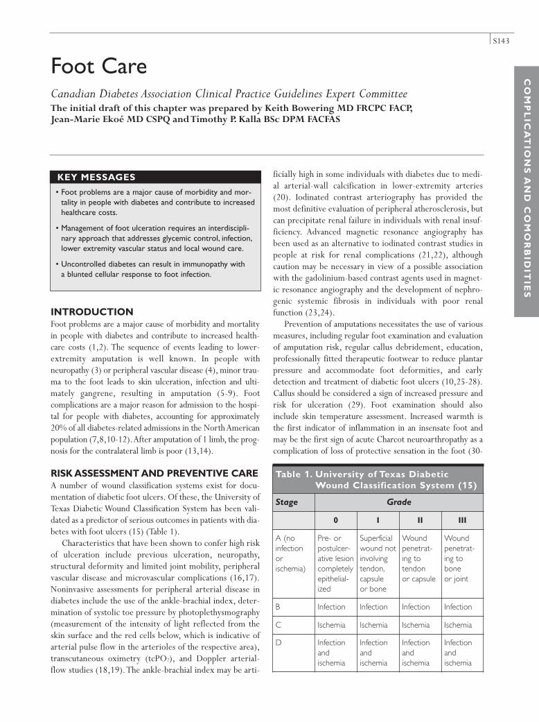
S143
CO
MP
LIC
AT
ION
SA
ND
CO
MO
RB
IDIT
IES
INTRODUCTIONFoot problems are a major cause of morbidity and mortalityin people with diabetes and contribute to increased health-care costs (1,2). The sequence of events leading to lower-extremity amputation is well known. In people withneuropathy (3) or peripheral vascular disease (4), minor trau-ma to the foot leads to skin ulceration, infection and ulti-mately gangrene, resulting in amputation (5-9). Footcomplications are a major reason for admission to the hospi-tal for people with diabetes, accounting for approximately20% of all diabetes-related admissions in the North Americanpopulation (7,8,10-12).After amputation of 1 limb, the prog-nosis for the contralateral limb is poor (13,14).
RISK ASSESSMENT AND PREVENTIVE CAREA number of wound classification systems exist for docu-mentation of diabetic foot ulcers. Of these, the University ofTexas Diabetic Wound Classification System has been vali-dated as a predictor of serious outcomes in patients with dia-betes with foot ulcers (15) (Table 1).
Characteristics that have been shown to confer high riskof ulceration include previous ulceration, neuropathy,structural deformity and limited joint mobility, peripheralvascular disease and microvascular complications (16,17).Noninvasive assessments for peripheral arterial disease indiabetes include the use of the ankle-brachial index, deter-mination of systolic toe pressure by photoplethysmography(measurement of the intensity of light reflected from theskin surface and the red cells below, which is indicative ofarterial pulse flow in the arterioles of the respective area),transcutaneous oximetry (tcPO2), and Doppler arterial-flow studies (18,19).The ankle-brachial index may be arti-
ficially high in some individuals with diabetes due to medi-al arterial-wall calcification in lower-extremity arteries(20). Iodinated contrast arteriography has provided themost definitive evaluation of peripheral atherosclerosis, butcan precipitate renal failure in individuals with renal insuf-ficiency. Advanced magnetic resonance angiography hasbeen used as an alternative to iodinated contrast studies inpeople at risk for renal complications (21,22), althoughcaution may be necessary in view of a possible associationwith the gadolinium-based contrast agents used in magnet-ic resonance angiography and the development of nephro-genic systemic fibrosis in individuals with poor renalfunction (23,24).
Prevention of amputations necessitates the use of variousmeasures, including regular foot examination and evaluationof amputation risk, regular callus debridement, education,professionally fitted therapeutic footwear to reduce plantarpressure and accommodate foot deformities, and earlydetection and treatment of diabetic foot ulcers (10,25-28).Callus should be considered a sign of increased pressure andrisk for ulceration (29). Foot examination should alsoinclude skin temperature assessment. Increased warmth isthe first indicator of inflammation in an insensate foot andmay be the first sign of acute Charcot neuroarthropathy as acomplication of loss of protective sensation in the foot (30-
Foot CareCanadian Diabetes Association Clinical Practice Guidelines Expert CommitteeThe initial draft of this chapter was prepared by Keith Bowering MD FRCPC FACP,Jean-Marie Ekoé MD CSPQ and Timothy P. Kalla BSc DPM FACFAS
• Foot problems are a major cause of morbidity and mor-tality in people with diabetes and contribute to increasedhealthcare costs.
• Management of foot ulceration requires an interdiscipli-nary approach that addresses glycemic control, infection,lower extremity vascular status and local wound care.
• Uncontrolled diabetes can result in immunopathy with a blunted cellular response to foot infection.
KEY MESSAGES
Table 1. University of Texas DiabeticWound Classification System (15)
Stage Grade
0 I II III
A (noinfectionorischemia)
Pre- orpostulcer-ative lesioncompletelyepithelial-ized
Superficialwound notinvolvingtendon,capsule or bone
Woundpenetrat-ing to tendon or capsule
Woundpenetrat-ing tobone or joint
B Infection Infection Infection Infection
C Ischemia Ischemia Ischemia Ischemia
D Infectionandischemia
Infectionandischemia
Infectionandischemia
Infectionandischemia

S144
2008 CLINICAL PRACTICE GUIDELINES
32). In addition, an acute Charcot foot may be associatedwith erythema and swelling, with overall clinical characteris-tics very similar to cellulitis (33,34).
MANAGEMENTAppropriate management can prevent or heal diabetic footulcers, thereby greatly reducing the amputation rate(6,9,10,25,26,35,36). All people with diabetes should beinstructed on proper foot care (including smoking cessationstrategies) (Appendix 5), should strive to reach recommend-ed glycemic targets, and should receive early referrals to ahealthcare professional trained in foot care management ifproblems occur (37).
Management of foot ulceration requires an interdiscipli-nary approach (38) that addresses glycemic control, infection,lower-extremity vascular status and local wound care (39).
Essentials of good wound care involve provision of an opti-mal wound environment, off-loading of the ulcer site, and, innonischemic wounds, regular debridement of nonviable tis-sue. In general, wound dressings that maintain a moist woundenvironment should be selected (40) (Appendix 6).Expeditious debridement may be performed with sharp instru-ments or biologically with medical-grade maggots (41,42).
Pressure offloading may be achieved with temporaryfootwear until the ulcer heals and the character of the footstabilizes. Removable and irremovable cast-walkers andtotal-contact casting have demonstrated proven efficacy aspressure-reducing devices in plantar-surface ulcers (43-45). Although very effective in healing noninfected, non-ischemic plantar-surface neuropathic ulcers, total-contactcasting requires careful individual selection and personneltrained specifically in its application due to its potential forcomplications (46).
Infections that complicate diabetic foot ulcers occur fre-quently and may be imminently limb threatening (47). Surfacecultures (as opposed to cultures of deeper tissues) of ulcers inpeople with diabetes have produced inconsistent results indetermining the bacterial pathogens involved (48-50). Initialantibiotic therapy is typically empiric and broad spectrum,with subsequent antibiotics tailored to results from appropri-ate cultures. Studies to date do not clearly identify a particularantibiotic agent that is more efficacious in reducing amputa-tion, accelerating ulcer healing or resolving infection (51).Uncontrolled diabetes can result in immunopathy with ablunted cellular response to infection. Up to 50% of patientswith diabetes who have a significant limb infection may nothave systemic signs of fever or leukocytosis at presentation(52). Deep infections require prompt surgical debridement inaddition to appropriate antibiotic therapy (53).
In medically suitable individuals with peripheral arterial dis-ease, distal limb revascularization has proven benefit in long-term limb salvage (54).Where bony foot deformities preventfitting of appropriate footwear and/or offloading of pressure-related ulcers, consultation from a surgeon skilled in foot sur-
gery may be considered to address the deformity (55-57).Hyperbaric oxygen therapy may be useful as an adjunct to
systemic antibiotics in individuals with deep, long-standing,nonhealing foot infections, provided there is an adequateperfused capillary bed in the wound area (i.e. by measuringtcPO2 response to 100% oxygen challenge). Few studies sup-port its use in treating uncomplicated neuropathic orischemic diabetic foot ulcers. There are no evidence-basedcriteria to select people for hyperbaric oxygen therapy andto predict their response (58).
OTHER RELEVANT GUIDELINESTargets for Glycemic Control, p. S29Neuropathy, p. S140
RELEVANT APPENDICESAppendix 5: Diabetes and Foot Care: A Patient’s ChecklistAppendix 6: Diabetic Foot Ulcers: Essentials of Management
REFERENCES1. American Diabetes Association: clinical practice recommenda-
tions 2004. Diabetes Care. 2004;27(suppl 1):S63-64.2. Reiber GE, Boyko EJ, Smith DG. Lower extremity foot ulcers and
amputations in diabetes. In: Diabetes in America. 2nd ed. Bethesda,MD: National Diabetes Data Group, National Institutes of Health,National Institute of Diabetes and Digestive and Kidney Diseases;1995:409-428.
3. Eastman RC. Neuropathy in diabetes. In: Diabetes in America.
1. In people with diabetes, foot examinations by both theindividual and healthcare providers should be an integralcomponent of diabetes management to decrease therisk of foot lesions and amputations [Grade B, Level 2(26,37)], and should be performed at least annually andat more frequent intervals in those at high risk [Grade D,Consensus].Assessment by healthcare providers shouldinclude structural abnormalities (e.g. range of motion of ankles and toe joints, callus pattern, bony deformities,skin temperatures), evaluation for neuropathy andperipheral arterial disease, ulcerations and evidence of infection [Grade D, Level 4 (9,50)].
2. People at high risk of foot ulceration and amputationshould receive foot care education (including counsellingto avoid foot trauma), professionally fitted footwear,smoking cessation strategies and early referrals to ahealthcare professional trained in foot care managementif problems occur [Grade B, Level 2 (37)].
3. Individuals who develop a foot ulcer should be managedby a multidisciplinary healthcare team with expertise in the management of foot ulcers to prevent recurrentfoot ulcers and amputation [Grade C, Level 3 (38)].
4.Any infection in a diabetic foot must be treated aggressively [Grade D, Level 4 (53)].
RECOMMENDATIONS

S145
CO
MP
LIC
AT
ION
SA
ND
CO
MO
RB
IDIT
IES
2nd ed. Bethesda, MD: National Diabetes Data Group,National Institutes of Health, National Institute of Diabetesand Digestive and Kidney Diseases; 1995:339-348.
4. Palumbo PJ, Melton LJ III. Peripheral vascular disease and dia-betes. In: Diabetes in America. 2nd ed. Bethesda, MD: NationalDiabetes Data Group, National Institutes of Health, NationalInstitute of Diabetes and Digestive and Kidney Diseases;1995:401-408.
5. Boyko EJ, Lipsky BA. Infection and diabetes. In: Diabetes inAmerica. 2nd ed. Bethesda, MD: National Diabetes DataGroup, National Institutes of Health, National Institute ofDiabetes and Digestive and Kidney Diseases; 1995:485-499.
6. Frykberg RG. Diabetic foot ulcerations. In: Frykberg RG, ed.The High Risk Foot in Diabetes Mellitus. New York, NY: ChurchillLivingstone; 1991:151-195.
7. Frykberg RG,Veves A. Diabetic foot infections. Diabetes MetabRev. 1996;12:255-270.
8. Gibbons GW, Eliopoulos GM, Kozak GP, et al. Infection of thediabetic foot. In: Kozak GP, Campbell DR, Frykberg RG, et al,eds. Management of Diabetic Foot Problems. Philadelphia, PA:WBSaunders; 1995:121.
9. Reiber GE, Pecoraro RE, Koepsell TD. Risk factors for ampu-tation in patients with diabetes mellitus. A case-control study.Ann Intern Med. 1992;117:97-105.
10. Bild DE, Selby JV, Sinnock P, et al. Lower-extremity amputa-tion in people with diabetes. Epidemiology and prevention.Diabetes Care. 1989;12:24-31.
11. Reiber GE. Epidemiology of the diabetic foot. In: Levin ME,O’Neal LW, Bowker JH, eds. The Diabetic Foot. 5th ed. St.Louis, MO: Mosby; 1993:1-15.
12. Crude and age-adjusted hospital discharge rates for a lowerextremity condition as the first-listed or any-listed diagnosisper 1,000 diabetic population, United States, 1980-2003.Atlanta, GA: Department of Health & Human Services,Centers for Disease Control and Prevention; 2007. Availableat: http://www.cdc.gov/diabetes/statistics/hosplea/diabetes_complications/fig6.htm. Accessed September 1, 2008.
13. Ebskov B, Josephsen P. Incidence of reamputation and deathafter gangrene of the lower extremity. Prosthet Orthot Int. 1980;4:77-80.
14. Most RS, Sinnock P. The epidemiology of lower extremityamputations in diabetic individuals. Diabetes Care. 1983;6:87-91.
15. Armstrong DG, Lavery LA, Harkless LB. Validation of a dia-betic wound classification system. The contribution of depth,infection, and ischemia to risk of amputation. Diabetes Care.1998;21:855-859.
16. Boyko EJ, Ahroni JH, Stensel V, et al. A prospective study ofrisk factors for diabetic foot ulcer. The Seattle Diabetic FootStudy. Diabetes Care. 1999;22:1036-1042.
17. Fernando DJ, Masson EA,Veves A, et al. Relationship of limit-ed joint mobility to abnormal foot pressures and diabetic footulceration. Diabetes Care. 1991;14:8-11.
18. Kalani M, Brismar K, Fagrell B, et al. Transcutaneous oxygentension and toe blood pressure as predictors for outcome of
diabetic foot ulcers. Diabetes Care. 1999;22:147-151.19. Faglia E, Caravaggi C, Marchetti R, et al; SCAR (SCreening for
ARteriopathy) Study Group. Screening for peripheral arterialdisease by means of the ankle-brachial index in newly diagnosedtype 2 diabetic patients. Diabet Med. 2005;22:1310-1314.
20. Young MJ, Adams JE, Anderson GF, et al. Medial arterial cal-cification in the feet of diabetic patients and matched non-dia-betic control subjects. Diabetologia. 1993;36:615-621.
21. Brillet PY,Vayssairat M,Tassart M, et al. Gadolinium-enhancedMR angiography as first-line preoperative imaging in high-riskpatients with lower limb ischemia. J Vasc Interv Radiol. 2003;14:1139-1145.
22. Lapeyre M, Kobeiter H, Desgranges P, et al. Assessment ofcritical limb ischemia in patients with diabetes: comparison ofMR angiography and digital subtraction angiography. AJR Am JRoentgenol. 2005;185:1641-1650.
23. Pedersen M. Safety update on the possible causal relationshipbetween gadolinium-containing MRI agents and nephrogenicsystemic fibrosis. J Magn Reson Imaging. 2007;25:881-883.
24. Centers for Disease Control and Prevention (CDC).Nephrogenic fibrosing dermopathy associated with exposure togadolinium-containing contrast agents – St. Louis, Missouri,2002-2006. MMWR Morb Mortal Wkly Rep. 2007;56:137-141.
25. Assal JP, Mühlhauser I, Pernet A, et al. Patient education as thebasis for diabetes care in clinical practice and research.Diabetologia. 1985;28:602-613.
26. Litzelman DK, Slemenda CW, Langefeld CD, et al. Reductionof lower extremity clinical abnormalities in patients with non-insulin-dependent diabetes mellitus.A randomized, controlledtrial. Ann Intern Med. 1993;119:36-41.
27. Malone JM, Snyder M,Anderson G, et al. Prevention of ampu-tation by diabetic education. Am J Surg. 1989;158:520-524.
28. Viswanathan V, Madhavan S, Gnanasundaram S, et al.Effectiveness of different types of footwear insoles for the dia-betic neuropathic foot: a follow-up study. Diabetes Care. 2004;27:474-477.
29. Sage RA,Webster JK, Fisher SG. Outpatient care and morbid-ity reduction in diabetic foot ulcers associated with chronicpressure callus. J Am Podiatr Med Assoc. 2001;91:275-279.
30. Lavery LA, Higgins KR, Lanctot DR, et al. Preventing diabet-ic foot ulcer recurrence in high-risk patients: use of tempera-ture monitoring as a self-assessment tool. Diabetes Care. 2007;30:14-20.
31. Armstrong DG, Lavery LA. Monitoring healing of acuteCharcot’s arthropathy with infrared dermal thermometry.J Rehabil Res Dev. 1997;34:317-321.
32. Yu GV, Hudson JR. Evaluation and treatment of stage 0Charcot’s neuroarthropathy of the foot and ankle. J Am PodiatrMed Assoc. 2002;92:210-220.
33. Frykberg RG, Zgonis T, Armstrong DG, et al; AmericanCollege of Foot and Ankle Surgeons. Diabetic foot disorders.A clinical practical guideline (2006 revision). J Foot Ankle Surg.2006;45(5 suppl):S1-S66.
34. Ledermann HP, Morrison WB. Differential diagnosis of pedal

S146
2008 CLINICAL PRACTICE GUIDELINES
osteomyelitis and diabetic neuroarthropathy: MR imaging.Semin Musculoskelet Radiol. 2005;9:272-283.
35. Apelqvist J, Larsson J.What is the most effective way to reduceincidence of amputation in the diabetic foot? Diabetes Metab ResRev. 2000;16(suppl 1):S75-S83.
36. Valk GD, Kriegsman DM,Assendelft WJ. Patient education forpreventing diabetic foot ulceration. Cochrane Database Syst Rev.2001;(4):CD001488.
37. McCabe CJ, Stevenson RC, Dolan AM. Evaluation of a diabet-ic foot screening and protection programme. Diabet Med.1998;15:80-84.
38. Dargis V, Pantelejeva O, Jonushaite A, et al. Benefits of a mul-tidisciplinary approach in the management of recurrent dia-betic foot ulceration in Lithuania: a prospective study. DiabetesCare. 1999;22:1428-1431.
39. Margolis DJ, Kantor J, Berlin JA. Healing of diabetic neuro-pathic foot ulcers receiving standard treatment. A meta-analy-sis. Diabetes Care. 1999;22:692-695.
40. Atiyeh BS, Ioannovich J, Al-Amm CA, et al. Management ofacute and chronic open wounds: the importance of moist envi-ronment in optimal wound healing. Curr Pharm Biotechnol.2002;3:179-195.
41. Steed DL, Donohoe D,Webster MW, et al. Effect of extensivedebridement and treatment on the healing of diabetic footulcers. Diabetic Ulcer Study Group. J Am Coll Surg. 1996;183:61-64.
42. Sherman RA. Maggot therapy for treating diabetic foot ulcersunresponsive to conventional therapy. Diabetes Care. 2003;26:446-451.
43. Armstrong DG, Lavery LA,Wu S, et al. Evaluation of remov-able and irremovable cast walkers in the healing of diabeticfoot wounds: a randomized controlled trial. Diabetes Care.2005;28:551-554.
44. Katz IA, Harlan A, Miranda-Palma B, et al. A randomized trialof two irremovable off-loading devices in the management ofplantar neuropathic diabetic foot ulcers. Diabetes Care.2005;28:555-559.
45. Armstrong DG, Nguyen HC, Lavery LA, et al. Off-loading thediabetic foot wound: a randomized clinical trial. Diabetes Care.2001;24:1019-1022.
46. Nabuurs-Franssen MH, Sleegers R, Huijberts MS, et al. Totalcontact casting of the diabetic foot in daily practice: a prospec-tive follow-up study. Diabetes Care. 2005;28:243-247.
47. Lavery LA, Armstrong DG,Wunderlich RP, et al. Risk factorsfor foot infections in individuals with diabetes. Diabetes Care.2006;29:1288-1293.
48. Perry CR, Pearson RL, Miller GA. Accuracy of cultures ofmaterial from swabbing of the superficial aspect of the woundand needle biopsy in the preoperative assessment ofosteomyelitis. J Bone Joint Surg Am. 1991;73:745-749.
49. Senneville E, Melliez H, Beltrand E, et al. Culture of percuta-neous bone biopsy specimens for diagnosis of diabetic footosteomyelitis: concordance with ulcer swab cultures. Clin InfectDis. 2006;42:57-62.
50. Slater RA, Lazarovitch T, Boldur I, et al. Swab cultures accu-rately identify bacterial pathogens in diabetic foot wounds notinvolving bone. Diabet Med. 2004;21:705-709.
51. Nelson EA, O’Meara S, Golder S, et al; DASIDU SteeringGroup. Systematic review of antimicrobial treatments for dia-betic foot ulcers. Diabet Med. 2006;23:348-359.
52. Eneroth M, Apelqvist J, Stenström A. Clinical characteristicsand outcome in 223 diabetic patients with deep foot infec-tions. Foot Ankle Int. 1997;18:716-722.
53. Tan JS, Friedman NM, Hazelton-Miller C, et al. Can aggres-sive treatment of diabetic foot infections reduce the need forabove-ankle amputation? Clin Infect Dis. 1996;23:286-291.
54. Pomposelli FB, Kansal N, Hamdan AD, et al.A decade of expe-rience with dorsalis pedis artery bypass: analysis of outcome inmore than 1000 cases. JVasc Surg. 2003;37:307-315.
55. Singh N, Armstrong DG, Lipsky BA. Preventing foot ulcers inpatients with diabetes. JAMA. 2005;293:217-228.
56. Blume PA, Paragas LK, Sumpio BE, et al. Single-stage surgicaltreatment of noninfected diabetic foot ulcers. Plast ReconstrSurg. 2002;109:601-609.
57. Sayner LR, Rosenblum BI, Giurini JM. Elective surgery of thediabetic foot. Clin Podiatr Med Surg. 2003;20:783-792.
58. Räkel A, Huot C, Ekoé JM. Canadian Diabetes AssociationTechnical Review: The diabetic foot and hyperbaric oxygentherapy. Can J Diabetes. 2006;30:411-421.

S147
CO
MP
LIC
AT
ION
SA
ND
CO
MO
RB
IDIT
IES
INTRODUCTIONErectile dysfunction (ED) affects approximately 34 to 45%of men with diabetes, and has been demonstrated to nega-tively impact quality of life among those affected across allage strata. Furthermore, studies indicate that 40% of diabet-ic men >60 years of age have complete ED (1-9).Among thediabetic population, risk factors include increasing age, dura-tion of diabetes, poor glycemic control, cigarette smoking,hypertension, dyslipidemia, androgen deficiency states (10)and cardiovascular disease (CVD) (5,7,11,12). ED as amarker of potential CV events has been reported by numer-ous investigators (13-20). Diabetic retinopathy has beenshown to correlate with the presence of ED (5,7,21).Organic causes of ED include microvascular and macrovas-cular disease, and neuropathy. In addition, psychological orsituational factors may cause or contribute to ED.
Compared with the general population, multiple studieshave reported men with diabetes having higher rates ofhypogonadism (10,22-24). Importantly, phosphodiesterasetype 5 (PDE5) inhibitors appear to be less effective in hypo-gonadal states (23,25,26), where treatment of nonrespon-ders to PDE5 with testosterone replacement is successful inroughly 50% of individuals. In addition, ED is a side effect ofmany drugs commonly prescribed to men with diabetes,such as some antihypertensives and antidepressants.
SCREENINGAll adult men with diabetes should be regularly screened forED with a sexual function history. Screening for ED in men
with type 2 diabetes should begin at diagnosis of diabetes.Validated questionnaires (e.g. International Index of ErectileFunction [27,28] or Sexual Health Inventory for Men [29])have been shown to be both sensitive and specific in deter-mining the presence of ED and providing a means of assessingresponse to therapy.
TREATMENTWhile no randomized clinical trials have demonstrated thatinterventions that improve glycemic control also reduce theincidence and progression of ED, the Diabetes Control andComplications Trial and United Kingdom ProspectiveDiabetes Study showed that intensive glycemic control waseffective for primary prevention of and secondary interven-tion for neuropathy, a condition that can impair sensory feed-back from the penis, leading to reduced erectile function(30-32). The current data still show that tight glycemic con-trol does not reverse ED (33-35).
The current mainstays of therapy are PDE5 inhibitors.They have been reported to have a major impact on erectilefunction, quality of life, and should be offered as first-linetherapy to men with diabetes wishing treatment for ED(36-41).
Evolving evidence supports the potential beneficial effectsof PDE5 inhibitors on endothelial function and lower urinarytract symptoms. Contraindications for the use of PDE5inhibitors include unstable angina or untreated cardiacischemia and concomitant use of nitrates (42,43).
Referral to a specialist in ED should be offered to menwho do not respond to PDE5 inhibitors or for whom the useof PDE5 inhibitors is contraindicated. Second-line therapies(e.g. vacuum constriction devices, intracorporal injectiontherapy with prostaglandin E1 [PGE1] alone or in combina-tion with papaverine and phentolamine [triple therapy],intraurethral therapy using PGE1) or third-line therapy(penile prosthesis) may be considered for these men (44).
EJACULATORY DISORDERSEjaculatory disorders are another common disorder of sexu-al function in men with diabetes, occurring in up to 32%(45).They range in scope from retrograde ejaculation, usual-ly secondary to autonomic neuropathy with incomplete clo-sure of the bladder neck during ejaculation, to premature orretarded ejaculation.
Erectile DysfunctionCanadian Diabetes Association Clinical Practice Guidelines Expert CommitteeThe initial draft of this chapter was prepared by Gerald Brock MD FRCPC,Richard Bebb MD ABIM FRCPC and William Harper MD FRCPC
• Erectile dysfunction (ED) affects approximately 34 to45% of men with diabetes, has been demonstrated tonegatively impact quality of life among those affectedacross all age strata, and may be the earliest sign of cardiovascular disease.
• All adult men with diabetes should be regularly screenedfor ED with a sexual function history.
• The current mainstays of therapy are phosphodiesterasetype 5 inhibitors.They have been reported to have amajor impact on erectile function and quality of life,and should be offered as first-line therapy to men with diabetes wishing treatment for ED.
KEY MESSAGES

S148
2008 CLINICAL PRACTICE GUIDELINES
OTHER RELEVANT GUIDELINESScreening for the Presence of Coronary Artery Disease,
p. S99Diabetes in the Elderly, p. S181
REFERENCES1. Chew KK, Earle CM, Stuckey BG, et al. Erectile dysfunction
in general medicine practice: prevalence and clinical corre-lates. Int J Impot Res. 2000;12:41-45.
2. Maatman TJ, Montague DK, Martin LM. Erectile dysfunctionin men with diabetes mellitus. Urology. 1987;29:589-592.
3. Rubin A, Babbott D. Impotence and diabetes mellitus. JAMA.1958;168:498-500.
4. Kolodny RC, Kahn CB, Goldstein HH, et al. Sexual dysfunc-tion in diabetic men. Diabetes. 1974;23:306-309.
5. McCulloch DK, Campbell IW, Wu FC, et al. The prevalence of diabetic impotence. Diabetologia. 1980;18:279-283.
6. Zemel P. Sexual dysfunction in the diabetic patient with hyper-tension. Am J Cardiol. 1988;61:27H-33H.
7. McCulloch DK,Young RJ, Prescott RJ, et al. The natural his-tory of impotence in diabetic men. Diabetologia. 1984;26:437-440.
8. Bacon CG, Hu FB, Giovannucci E, et al. Association of typeand duration of diabetes with erectile dysfunction in a largecohort of men. Diabetes Care. 2002;25:1458-1463.
9. De Berardis G, Pellegrini F, Franciosi M, et al. Identifyingpatients with type 2 diabetes with a higher likelihood of erec-tile dysfunction: the role of the interaction between clinicaland psychological factors. J Urol. 2003;169:1422-1428.
10. Alexopoulou O, Jamart J, Maiter D, et al. Erectile dysfunctionand lower androgenicity in type 1 diabetic patients. DiabetesMetab. 2001;27:329-336.
11. Naliboff BD, Rosenthal M. Effects of age on complications in
adult onset diabetes. J Am Geriatr Soc. 1989;37:838-842.12. Feldman HA, Goldstein I, Hatzichristou DG, et al. Impotence
and its medical and psychosocial correlates: results of theMassachusetts Male Aging Study. J Urol. 1994;151:54-61.
13. Grover SA, Lowensteyn I, Kaouache M, et al. The prevalenceof erectile dysfunction in the primary care setting: importanceof risk factors for diabetes and vascular disease. Arch Intern Med.2006;166:213-219.
14. Barrett-Connor E. Cardiovascular risk stratification and car-diovascular risk factors associated with erectile dysfunction:assessing cardiovascular risk in men with erectile dysfunction.Clin Cardiol. 2004;27(suppl 1):I8-I13.
15. Billups KL. Erectile dysfunction as an early sign of cardiovas-cular disease. Int J Impot Res. 2005;17(suppl 1):S19-S24.
16. Thompson IM, Tangen CM, Goodman PJ, et al. Erectile dys-function and subsequent cardiovascular disease. JAMA.2005;294:2996-3002.
17. Gazzaruso C. Erectile dysfunction and coronary atherothrom-bosis in diabetic patients: pathophysiology, clinical features andtreatment. Expert Rev Cardiovasc Ther. 2006;4:173-180.
18. Barrett-Connor E. Heart disease risk factors predict erectiledysfunction 25 years later (the Rancho Bernardo Study). Am JCardiol. 2005;96:3M-7M.
19. Min JK,Williams KA, Okwuosa TM, et al. Prediction of coro-nary heart disease by erectile dysfunction in men referred fornuclear stress testing. Arch Intern Med. 2006;166:201-206.
20. Chiurlia E, D’Amico R, Ratti C, et al. Subclinical coronaryartery atherosclerosis in patients with erectile dysfunction.J Am Coll Cardiol. 2005;46:1503-1506.
21. Klein R, Klein BEK, Lee KE, et al. Prevalence of self-report-ed erectile dysfunction in people with long-term IDDM.Diabetes Care. 1996;19:135-141.
22. Dhindsa S, Prabhakar S, Sethi M, et al. Frequent occurrence ofhypogonadotropic hypogonadism in type 2 diabetes. J ClinEndocrinol Metab. 2004;89:5462-5468.
23. Boyanov MA, Boneva Z, Christov VG. Testosterone supple-mentation in men with type 2 diabetes, visceral obesity andpartial androgen deficiency. Aging Male. 2003;6:1-7.
24. Shabsigh R, Rajfer J, Aversa A, et al. The evolving role oftestosterone in the treatment of erectile dysfunction. Int J ClinPract. 2006;60:1087-1092.
25. Shabsigh R, Kaufman JM, Steidle C, et al. Randomized studyof testosterone gel as adjunctive therapy to sildenafil in hypo-gonadal men with erectile dysfunction who do not respond tosildenafil alone. J Urol. 2004;172:658-663.
26. Kalinchenko SY, Kozlov GI, Gontcharov NP, et al. Oral testos-terone undecanoate reverses erectile dysfunction associatedwith diabetes mellitus in patients failing on sildenafil citratetherapy alone. Aging Male. 2003;6:94-99.
27. Rosen RC, Riley A,Wagner G, et al.The International Index ofErectile Function (IIEF): a multidimensional scale for assess-ment of erectile dysfunction. Urology. 1997;49:822-830.
28. Cappelleri JC, Rosen RC, Smith MD, et al. Diagnostic evalua-tion of the erectile function domain of the International Index
1.All adult men with diabetes should be regularlyscreened for ED with a sexual function history [Grade D, Consensus].
2.A PDE5 inhibitor should be offered as first-line therapyto men with diabetes with ED if there are no con-traindications to its use [Grade A, Level 1A (36-43)]
3. Referral to a specialist in ED should be considered foreugonadal men who do not respond to PDE5 inhibitors,or for whom the use of PDE5 inhibitors is contraindi-cated [Grade D, Consensus].
4. Men with diabetes and ED who do not respond toPDE5 therapy should be investigated for hypogonadism[Grade D, Level 4 (22,23,25,26)].
5. Men with diabetes and ejaculatory dysfunction who wishfertility should be referred to a healthcare professionalexperienced in the treatment of ejaculatory dysfunction[Grade D, Consensus].
RECOMMENDATIONS
S149
CO
MP
LIC
AT
ION
SA
ND
CO
MO
RB
IDIT
IES
of Erectile Function. Urology. 1999;54:346-351.29. Ramanathan R, Mulhall J, Rao S, et al. Prospective correlation
between the International Index of Erectile Function (IIEF)and Sexual Health Inventory for Men (SHIM): implications forcalculating a derived SHIM for clinical use. J Sex Med. 2007;4:1334-1344.
30. Valiquette L, Montorsi F, Auerbach S;Vardenafil Study Group.First-dose success with vardenafil in men with erectile dys-function and associated comorbidities: RELY-I. Int J Clin Pract.2006;60:1378-1385.
31. The Diabetes Control and Complications Trial ResearchGroup. The effect of intensive diabetes therapy on the devel-opment and progression of neuropathy. Ann Intern Med. 1995;122:561-568.
32. The Diabetes Control and Complications Trial ResearchGroup. The effect of intensive treatment of diabetes on thedevelopment and progression of long-term complications ininsulin-dependent diabetes mellitus. N Engl J Med. 1993;329:977-986.
33. UK Prospective Diabetes Study (UKPDS) Group. Intensiveblood-glucose control with sulphonylureas or insulin com-pared with conventional treatment and risk of complications inpatients with type 2 diabetes (UKPDS 33). Lancet. 1998;352:837-853.
34. Azad N, Emanuele NV,Abraira C, et al.The effects of intensiveglycemic control on neuropathy in the VA Cooperative Studyon Type II Diabetes Mellitus (VA CSDM). J DiabetesComplications. 1990;13:307-313.
35. El-Sakka AI, Hassoba HM, Sayed HM, et al. Pattern ofendocrinal changes in patients with sexual dysfunction. J SexMed. 2005;2:551-558.
36. Fonseca V, Seftel A, Denne J, et al. Impact of diabetes mellituson the severity of erectile dysfunction and response to treat-ment: analysis of data from tadalafil clinical trials. Diabetologia.2004;47:1914-1923.
37. Rendell MS, Rajfer J,Wicker PA, et al. Sildenafil for treatmentof erectile dysfunction in men with diabetes: a randomizedcontrolled trial. JAMA. 1999;281:421-426.
38. Boulton AJM, Selam J-L, Sweeney M, et al. Sildenafil citratefor the treatment of erectile dysfunction in men with type IIdiabetes mellitus. Diabetologia. 2001;44:1296-1301.
39. Goldstein I,Young JM, Fischer J, et al.Vardenafil, a new phos-phodiesterase type 5 inhibitor, in the treatment of erectile dys-function in men with diabetes: a multicenter double-blindplacebo-controlled fixed-dose study. Diabetes Care. 2003;26:777-783.
40. Sáenz de Tejada I,Anglin G, Knight JR, et al. Effects of tadalafilon erectile dysfunction in men with diabetes. Diabetes Care.2002;25:2159-2164.
41. Carson CC, Lue TF. Phosphodiesterase type 5 inhibitors forerectile dysfunction. BJU Int. 2005;96:257-280.
42. Briganti A, Salonia A, Gallina A, et al. Drug insight: oral phos-phodiesterase type 5 inhibitors for erectile dysfunction. NatClin Pract Urol. 2005;2:239-247.
43. DeBusk R, Drory Y, Goldstein I, et al. Management of sexualdysfunction in patients with cardiovascular disease: recom-mendations of the Princeton Consensus Panel. Am J Cardiol.2000;86:175-181.
44. Herschorn S. Cardiovascular safety of PDE5 inhibitors. Can JUrol. 2003;10(suppl 1):23-28.
45. Carson CC, Mulcahy JJ, Govier FE. Efficacy, safety and patientsatisfaction outcomes of the AMS 700CX inflatable penileprosthesis: results of a long-term multicenter study. J Urol.2000;164:376-380.

S150
Unless otherwise specified, the term “child” or “children” is used forindividuals 0 to 18 years of age, and the term “adolescent” for those13 to 18 years of age.
INTRODUCTIONDiabetes mellitus is the most common endocrine disease andone of the most common chronic conditions in children.Type2 diabetes and other types of diabetes, including genetic defectsof beta cell function such as maturity-onset diabetes of theyoung (MODY), are increasing in frequency and should beconsidered when clinical presentation is atypical for type 1diabetes.This section addresses those areas of type 1 diabetesmanagement that are specific to children.
EDUCATIONChildren with new-onset type 1 diabetes and their familiesrequire intensive diabetes education by an interdisciplinarypediatric diabetes healthcare (DHC) team to provide themwith the necessary skills and knowledge to manage this dis-ease. The complex physical, developmental and emotionalneeds of children and their families necessitate specialized careto ensure the best long-term outcomes (1). Education topicsmust include insulin action and administration, dosage adjust-ment, blood glucose (BG) and ketone testing, sick-day man-agement and prevention of diabetic ketoacidosis (DKA),nutrition therapy, exercise, and prevention, detection, andtreatment of hypoglycemia.Anticipatory guidance and lifestylecounselling should be part of routine care, especially duringcritical developmental transitions (e.g. upon school entry,beginning high school). Healthcare providers should regularlyinitiate discussions with children and their families aboutschool, diabetes camp, psychological issues, substance abuse,driver’s licence and career choices.
Children with new-onset diabetes who present with DKArequire a short period of hospitalization to stabilize the asso-ciated metabolic derangements and to initiate insulin thera-py. Outpatient education for well children with new-onsetdiabetes has been shown to be less expensive than inpatienteducation and associated with similar or slightly better out-comes when appropriate resources are available (2).
GLYCEMIC TARGETSAs improved metabolic control reduces both the onset andprogression of diabetes-related complications in adults andadolescents with type 1 diabetes (3,4) aggressive attemptsshould be made to reach the recommended glycemic targetsoutlined in Table 1. However, clinical judgement is required todetermine which children can reasonably and safely achievethese targets.Treatment goals and strategies must be tailoredto each child, with consideration given to individual risk fac-tors.Young age at diabetes onset (<7 years of age) has beenassociated with poorer cognitive function in many studies (5).Episodes of severe hypoglycemia have been associated withpoorer cognitive function in some follow-up studies, whileother studies have found chronic hyperglycemia in young chil-dren to be associated with poorer cognitive performance (6-8).
INSULIN THERAPYInsulin therapy is the mainstay of medical management oftype 1 diabetes. A variety of insulin regimens can beemployed, but few have been studied specifically in childrenwith new-onset diabetes. The choice of insulin regimendepends on many factors, including the child’s age, durationof diabetes, family lifestyle, socioeconomic factors, and fam-ily, patient and physician preferences. Regardless of theinsulin regimen used, all children should be treated to meetglycemic targets.
The honeymoon period, which can last up to 2 years post-diagnosis, is characterized by good glycemic control and lowinsulin requirements (<0.5 units/kg/day). At the end of thisperiod, more intensive management may be required to con-tinue meeting glycemic targets.Two methods of intensive dia-betes management have been used: multiple daily injection(MDI) regimens and continuous subcutaneous insulin infu-sion (CSII, insulin pump therapy). CSII is safe and effectiveand can be initiated at any age (9). Most (10-13), but not all(14), randomized controlled trials (RCTs) of CSII in children
Type 1 Diabetes in Children and AdolescentsCanadian Diabetes Association Clinical Practice Guidelines Expert CommitteeThe initial draft of this chapter was prepared by Margaret L. Lawson MD MSc FRCPC,Danièle Pacaud MD FRCPC and Diane Wherrett MD FRCPC
2008 CLINICAL PRACTICE GUIDELINES
• Suspicion of diabetes in a child should lead to immediateconfirmation of the diagnosis and initiation of treatmentto reduce the likelihood of diabetic ketoacidosis (DKA).
• Management of pediatric DKA differs from DKA inadults because of the increased risk for cerebral edema.Pediatric protocols should be used.
• Children should be referred for diabetes education andongoing care to a diabetes team with pediatric expertise.
KEY MESSAGES
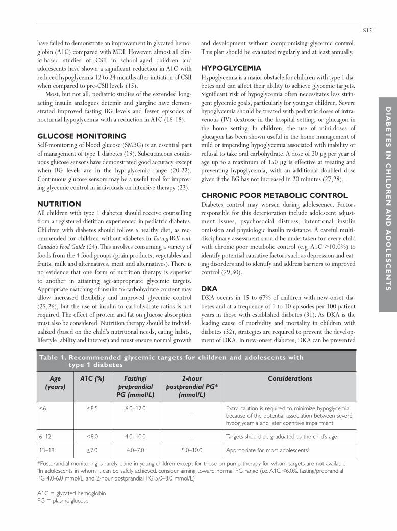
S151
have failed to demonstrate an improvement in glycated hemo-globin (A1C) compared with MDI. However, almost all clin-ic-based studies of CSII in school-aged children andadolescents have shown a significant reduction in A1C withreduced hypoglycemia 12 to 24 months after initiation of CSIIwhen compared to pre-CSII levels (15).
Most, but not all, pediatric studies of the extended long-acting insulin analogues detemir and glargine have demon-strated improved fasting BG levels and fewer episodes ofnocturnal hypoglycemia with a reduction in A1C (16-18).
GLUCOSE MONITORINGSelf-monitoring of blood glucose (SMBG) is an essential partof management of type 1 diabetes (19). Subcutaneous contin-uous glucose sensors have demonstrated good accuracy exceptwhen BG levels are in the hypoglycemic range (20-22).Continuous glucose sensors may be a useful tool for improv-ing glycemic control in individuals on intensive therapy (23).
NUTRITION All children with type 1 diabetes should receive counsellingfrom a registered dietitian experienced in pediatric diabetes.Children with diabetes should follow a healthy diet, as rec-ommended for children without diabetes in Eating Well withCanada’s Food Guide (24).This involves consuming a variety offoods from the 4 food groups (grain products, vegetables andfruits, milk and alternatives, meat and alternatives). There isno evidence that one form of nutrition therapy is superior to another in attaining age-appropriate glycemic targets.Appropriate matching of insulin to carbohydrate content mayallow increased flexibility and improved glycemic control(25,26), but the use of insulin to carbohydrate ratios is notrequired.The effect of protein and fat on glucose absorptionmust also be considered. Nutrition therapy should be individ-ualized (based on the child’s nutritional needs, eating habits,lifestyle, ability and interest) and must ensure normal growth
and development without compromising glycemic control.This plan should be evaluated regularly and at least annually.
HYPOGLYCEMIA Hypoglycemia is a major obstacle for children with type 1 dia-betes and can affect their ability to achieve glycemic targets.Significant risk of hypoglycemia often necessitates less strin-gent glycemic goals, particularly for younger children. Severehypoglycemia should be treated with pediatric doses of intra-venous (IV) dextrose in the hospital setting, or glucagon inthe home setting. In children, the use of mini-doses ofglucagon has been shown useful in the home management ofmild or impending hypoglycemia associated with inability orrefusal to take oral carbohydrate. A dose of 20 µg per year ofage up to a maximum of 150 µg is effective at treating andpreventing hypoglycemia, with an additional doubled dosegiven if the BG has not increased in 20 minutes (27,28).
CHRONIC POOR METABOLIC CONTROLDiabetes control may worsen during adolescence. Factorsresponsible for this deterioration include adolescent adjust-ment issues, psychosocial distress, intentional insulin omission and physiologic insulin resistance. A careful multi-disciplinary assessment should be undertaken for every childwith chronic poor metabolic control (e.g. A1C >10.0%) toidentify potential causative factors such as depression and eat-ing disorders and to identify and address barriers to improvedcontrol (29,30).
DKA DKA occurs in 15 to 67% of children with new-onset dia-betes and at a frequency of 1 to 10 episodes per 100 patientyears in those with established diabetes (31). As DKA is theleading cause of morbidity and mortality in children with diabetes (32), strategies are required to prevent the develop-ment of DKA. In new-onset diabetes, DKA can be prevented
DIA
BE
TE
SIN
CH
ILD
RE
NA
ND
AD
OL
ES
CE
NT
S
*Postprandial monitoring is rarely done in young children except for those on pump therapy for whom targets are not available†In adolescents in whom it can be safely achieved, consider aiming toward normal PG range (i.e. A1C ≤6.0%, fasting/preprandial PG 4.0-6.0 mmol/L, and 2-hour postprandial PG 5.0–8.0 mmol/L)
A1C = glycated hemoglobinPG = plasma glucose
Table 1. Recommended glycemic targets for children and adolescents with type 1 diabetes
Age(years)
A1C (%) Fasting/preprandialPG (mmol/L)
2-hour postprandial PG*
(mmol/L)
Considerations
<6 <8.5 6.0–12.0–
Extra caution is required to minimize hypoglycemiabecause of the potential association between severehypoglycemia and later cognitive impairment
6–12 <8.0 4.0–10.0 – Targets should be graduated to the child’s age
13–18 ≤7.0 4.0–7.0 5.0–10.0 Appropriate for most adolescents†

S152
through earlier recognition and initiation of insulin therapy.Public awareness campaigns about the early signs of diabeteshave significantly reduced the frequency of DKA in new-onsetdiabetes (33). In children with established diabetes, DKAresults from failing to take insulin or poor sick-day manage-ment. Risk is increased in children with poor metabolic control or previous episodes of DKA, peripubertal and ado-lescent girls, children with psychiatric disorders and thosewith difficult family circumstances (34). The frequency ofDKA in established diabetes can be decreased with educationand family support (35) as well as access to 24-hour telephoneservices for parents of children with diabetes (36,37).
Management of DKAWhile most cases of DKA are corrected without event, 0.7to 3.0% of pediatric cases are complicated by cerebral edema(CE) (38), which is associated with significant morbidity (21to 35%) and mortality (21 to 24%) (39). In contrast, CE hasrarely been reported in adults (34,39).Although the cause ofCE is still unknown, several factors are associated withincreased risk (Table 2) (40-43). A bolus of insulin prior toinfusion is not recommended (44) since it does not offer anyfaster resolution of acidosis (45,46) and may contribute toCE (47). Recent evidence suggests early insulin administra-tion (within the first hour of fluid replacement) may increasethe risk for CE (48). Special caution should be exercised inyoung children with DKA and new-onset diabetes or agreater degree of acidosis and extracellular fluid volume(ECFV) depletion because of the increased risk of CE. Use ofbedside criteria may allow earlier identification of patientswho require treatment for CE (49). DKA should be managedaccording to published protocols for management of pedi-atric DKA (50) (Figure 1).
IMMUNIZATION Historically, national guidelines have recommended influen-za and pneumococcal immunization for children with type 1diabetes (51-53). Currently, there is no evidence supportingincreased morbidity or mortality from influenza or pneumo-coccus in children with type 1 diabetes (54,55). However,
the management of type 1 diabetes can be complicated by ill-ness, thus requiring parental knowledge of sick-day manage-ment and increased attention during periods of illness. Forthis reason, parents may choose to immunize their children.
SMOKING PREVENTION AND CESSATIONSmoking is a significant risk factor for both macrovascularand microvascular complications of diabetes (56). Smokingprevention should be emphasized throughout childhood andadolescence.
CONTRACEPTION AND SEXUAL HEALTHCOUNSELLINGAdolescents with diabetes should receive regular counsellingabout sexual health and contraception. Unplanned pregnan-cies should be avoided, as pregnancy in females with type 1diabetes with suboptimal metabolic control results in higherrisks of maternal and fetal complications (57).
PSYCHOLOGICAL ISSUESSome children and their parents have adjustment problemssoon after the diagnosis of diabetes (58,59). Although mostresolve these problems within the first year after diagnosis,those who do not are at risk for poor adaptation to diabetes,including regimen adherence problems, poor glycemic con-trol and continued psychosocial difficulties (60,61). Stress(general and diabetes-specific) (62), inadequate social andfamily support (63,64), inappropriate beliefs about the natureof diabetes (63) and poor coping skills (65) may have a nega-tive impact on self-care behaviours and glycemic control.
The diagnosis of diabetes may precipitate or exacerbateexisting psychological disorders (66). As quality of life anddiabetes control may be adversely affected by the presence ofcomorbid psychological disorders and health complications(66), the identification of potential psychiatric conditions,such as depression, anxiety and eating disorders, is critical.All children with diabetes and their families should be regu-larly screened for symptoms of psychological distress(67,68) (See “Psychological Aspects of Diabetes,” p. S82).
Eating disorders Ten percent of adolescent females with type 1 diabetes meetthe Diagnostic and Statistical Manual of Mental Disorders (4thEdition) criteria for eating disorders compared to 4% of theirage-matched peers without diabetes (69). Furthermore, eat-ing disorders are associated with poor metabolic control andearlier onset and more rapid progression of microvascularcomplications (70). Eating disorders should be suspected inthose adolescent and young adult females who are unable toachieve and maintain metabolic targets especially wheninsulin omission is suspected. It is important to identify indi-viduals with eating disorders because different managementstrategies are required to optimize metabolic control andprevent microvascular complications (71).
2008 CLINICAL PRACTICE GUIDELINES
Table 2. Risks factors for CE
• Younger age (<5 years)• New-onset diabetes• High initial serum urea • Low initial par tial pressure of ar terial carbon
dioxide (pCO2)• Rapid administration of hypotonic fluids• IV bolus of insulin• Ear ly IV insulin infusion (within first hour of
administration of fluids)• Failure of serum sodium to rise during treatment• Use of bicarbonate
CE = cerebral edemaIV = intravenous

S153
DIA
BE
TE
SIN
CH
ILD
RE
NA
ND
AD
OL
ES
CE
NT
S
Figure 1. Immediate assessment and management of DKA in children
BG = blood glucoseDKA = diabetic ketoacidosis
ECG = electrocardiogramICU = intensive care unit
IV = intravenousPG = plasma glucose
SC = subcutaneous
Re-evaluation• IV fluid calculations • Insulin delivery system
and dose • Need for additional
resuscitation • Consider sepsis
Transition to SC insulinStart SC insulin then stop IVinsulin after an appropriate interval
Management• Give mannitol 0.5–1 g/kg• Restrict IV fluids by one-third• Call senior staff• Move to ICU• Consider cranial imaging only
after patient stabilized
Immediate assessment
Clinical signs• Dehydration (assess)• Deep sighing respiration
(Kussmaul)• Smell of ketones• Lethargy/drowsiness
± vomiting
Clinical history• Polyuria• Polydipsia• Weight loss (weigh)• Abdominal pain• Tiredness• Vomiting • Confusion
Biochemical features and investigations
• Ketones in urine• Elevated BG • Acidemia• Blood gases, urea, electrolytes • Other investigations as
indicated
Diagnosis of DKA confirmedContact senior staff
• Shock (reduced peripheralpulses)
• Reduced consciousness orcoma
• Dehydration >5%• Not in shock• Acidotic (hyperventilation)• Vomiting
• Minimal dehydration• Tolerating oral fluid
No improvement
Therapy• Start with SC insulin• Continue oral hydration
Resuscitation• Airway ± nasogastric tube• Breathing (100% O2)• Circulation (0.9% NaCl 10–20
mL/kg over 1–2 h), and repeatuntil circulation is restored),but do not exceed 30 mL/kg
IV therapy• Calculate fluid requirements• Correct over 48 hours• 0.9% NaCl• ECG for abnormal T-waves• Add 40 mmol/L KCl
Continuous IV insulin infusion 0.1 U/kg/h to be started 1–2 hours after IV rehydration initiated
• Hourly BG• Hourly fluid input and output• Neurologic status at least
hourly
• Electrolytes every 2 h afterstart of IV therapy
• Monitor ECG for T-wavechanges
Acidosis not improving
PG 14.0–17.0 mmol/L; orPG falling >5.0 mmol/L/h
Neurological deterioration (Warningsigns: headache, slowing heart rate, irri-tability, decreased level of consciousness,incontinence, specific neurological signs)
IV therapy• Change to 0.45% NaCl + 5% glucose • Adjust [Na+] infusion to promote an
increase in measured serum [Na+]
Exclude hypoglycemiaIs it cerebral edema?
Improvement• Clinically well• Tolerating oral fluids
Critical observations
Adapted with permission from Reference 50.
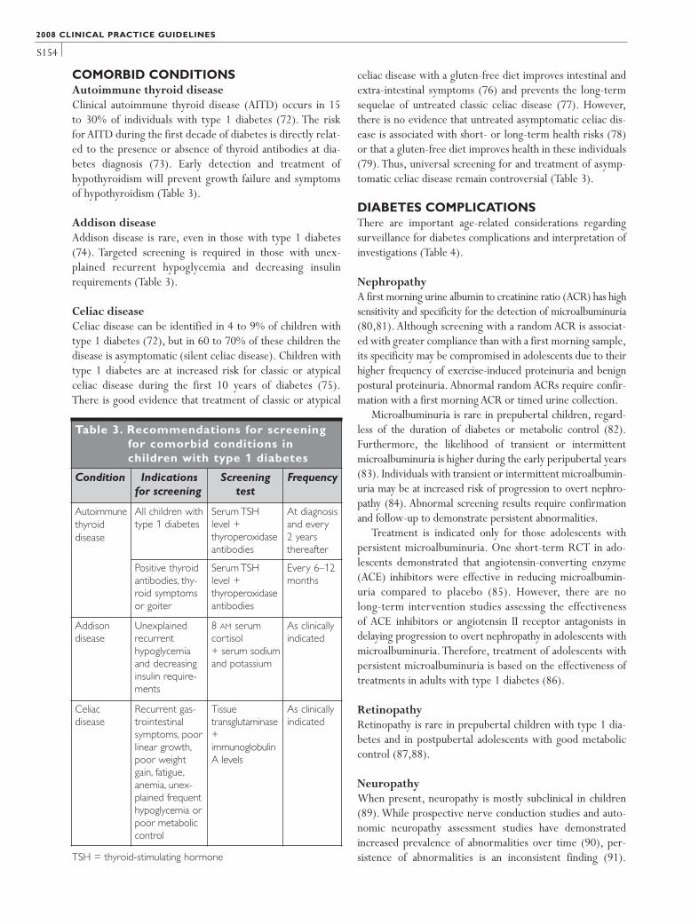
S154
COMORBID CONDITIONS Autoimmune thyroid diseaseClinical autoimmune thyroid disease (AITD) occurs in 15 to 30% of individuals with type 1 diabetes (72). The risk for AITD during the first decade of diabetes is directly relat-ed to the presence or absence of thyroid antibodies at dia-betes diagnosis (73). Early detection and treatment ofhypothyroidism will prevent growth failure and symptomsof hypothyroidism (Table 3).
Addison diseaseAddison disease is rare, even in those with type 1 diabetes(74). Targeted screening is required in those with unex-plained recurrent hypoglycemia and decreasing insulinrequirements (Table 3).
Celiac diseaseCeliac disease can be identified in 4 to 9% of children withtype 1 diabetes (72), but in 60 to 70% of these children thedisease is asymptomatic (silent celiac disease). Children withtype 1 diabetes are at increased risk for classic or atypicalceliac disease during the first 10 years of diabetes (75).There is good evidence that treatment of classic or atypical
celiac disease with a gluten-free diet improves intestinal andextra-intestinal symptoms (76) and prevents the long-termsequelae of untreated classic celiac disease (77). However,there is no evidence that untreated asymptomatic celiac dis-ease is associated with short- or long-term health risks (78)or that a gluten-free diet improves health in these individuals(79). Thus, universal screening for and treatment of asymp-tomatic celiac disease remain controversial (Table 3).
DIABETES COMPLICATIONS There are important age-related considerations regardingsurveillance for diabetes complications and interpretation ofinvestigations (Table 4).
NephropathyA first morning urine albumin to creatinine ratio (ACR) has highsensitivity and specificity for the detection of microalbuminuria(80,81). Although screening with a random ACR is associat-ed with greater compliance than with a first morning sample,its specificity may be compromised in adolescents due to theirhigher frequency of exercise-induced proteinuria and benignpostural proteinuria. Abnormal random ACRs require confir-mation with a first morning ACR or timed urine collection.
Microalbuminuria is rare in prepubertal children, regard-less of the duration of diabetes or metabolic control (82).Furthermore, the likelihood of transient or intermittentmicroalbuminuria is higher during the early peripubertal years(83). Individuals with transient or intermittent microalbumin-uria may be at increased risk of progression to overt nephro-pathy (84). Abnormal screening results require confirmationand follow-up to demonstrate persistent abnormalities.
Treatment is indicated only for those adolescents withpersistent microalbuminuria. One short-term RCT in ado-lescents demonstrated that angiotensin-converting enzyme(ACE) inhibitors were effective in reducing microalbumin-uria compared to placebo (85). However, there are nolong-term intervention studies assessing the effectivenessof ACE inhibitors or angiotensin II receptor antagonists indelaying progression to overt nephropathy in adolescents withmicroalbuminuria. Therefore, treatment of adolescents withpersistent microalbuminuria is based on the effectiveness oftreatments in adults with type 1 diabetes (86).
RetinopathyRetinopathy is rare in prepubertal children with type 1 dia-betes and in postpubertal adolescents with good metaboliccontrol (87,88).
Neuropathy When present, neuropathy is mostly subclinical in children(89). While prospective nerve conduction studies and auto-nomic neuropathy assessment studies have demonstratedincreased prevalence of abnormalities over time (90), per-sistence of abnormalities is an inconsistent finding (91).
2008 CLINICAL PRACTICE GUIDELINES
Table 3. Recommendations for screeningfor comorbid conditions in children with type 1 diabetes
Condition Indicationsfor screening
Screeningtest
Frequency
Autoimmunethyroid disease
All children withtype 1 diabetes
Serum TSH level + thyroperoxidaseantibodies
At diagnosisand every2 years thereafter
Positive thyroidantibodies, thy-roid symptomsor goiter
Serum TSH level + thyroperoxidaseantibodies
Every 6–12months
Addison disease
Unexplainedrecurrent hypoglycemiaand decreasinginsulin require-ments
8 AM serum cortisol + serum sodiumand potassium
As clinicallyindicated
Celiac disease
Recurrent gas-trointestinalsymptoms, poorlinear growth,poor weightgain, fatigue,anemia, unex-plained frequenthypoglycemia orpoor metaboliccontrol
Tissue transglutaminase +immunoglobulin A levels
As clinicallyindicated
TSH = thyroid-stimulating hormone

S155
Vibration and monofilament testing have suboptimal sensi-tivity and specificity in adolescents (92).With the exceptionof intensifying diabetes management to achieve and maintainglycemic targets, no other treatment modality has been stud-ied in children and adolescents.
Dyslipidemia Most children with type 1 diabetes should be considered atlow risk for vascular disease associated with dyslipidemia.The exceptions are those with longer duration of disease,microvascular complications or other cardiovascular disease(CVD) risk factors including smoking, hypertension, obesityand/or family history of premature CVD (93). Dyslipidemiascreening should be targeted at those >12 years of age andyounger children with specific risk factors for dyslipidemia.Statin therapy has not been studied specifically in childrenwith diabetes, and there is no evidence linking specific low-density lipoprotein cholesterol (LDL-C) cutoffs in childrenwith diabetes with long-term outcomes. In pubertal childrenwithout diabetes but with familial hypercholesterolemia,statin therapy is safe and effective at lowering LDL-C levels,and attenuating progression of carotid intima-media thick-ness, a surrogate marker for future vascular disease (94).
HypertensionUp to 16% of adolescents with type 1 diabetes have hyper-tension (95). Twenty-four-hour ambulatory blood pres-sure (BP) monitoring has been used to exclude white coathypertension and to identify loss of diurnal systolicrhythm (nondippers) with nocturnal hypertension insome normotensive adolescents with type 1 diabetes (96).These abnormalities may be predictive of future microal-buminuria (96). However, the role of ambulatory BP mon-itoring in routine care remains uncertain. Children withtype 1 diabetes and confirmed hypertension should betreated according to the guidelines for children withoutdiabetes (97).
TRANSITION TO ADULT CAREThe change of physician or DHC team can have a majorimpact on disease management and metabolic control in theperson with diabetes. Between 25 and 65% of young adultshave no medical follow-up during the transition from pedi-atric to adult diabetes care services (98,99). Those with nofollow-up are more likely to experience hospitalization forDKA during this period. Organized transition services maydecrease the rate of loss of follow-up (100).
DIA
BE
TE
SIN
CH
ILD
RE
NA
ND
AD
OL
ES
CE
NT
S
Table 4. Screening for diabetes complications, dyslipidemia and hyperglycemia in children with type 1 diabetes
Complication Indications and intervals for screening Screening method
Nephropathy • Yearly screening commencing at 12 years ofage in those with duration of type 1 diabetes>5 years
• First morning (preferred) or random ACR• Abnormal ACR requires confirmation at least 1
month later with a first morning ACR, and ifabnormal, followed by timed, overnight or 24-hoursplit urine collections for albumin excretion rate
• Repeated sampling should be done every 3–4months over a 12-month period to demonstratepersistence
Retinopathy • Yearly screening commencing at 15 years ofage with duration of type 1 diabetes >5 years
• Screening interval can increase to 2 years ifgood glycemic control, duration of diabetes<10 years, and no retinopathy at initial assessment
• 7-standard field, stereoscopic-colour fundus pho-tography with interpretation by a trained reader(gold standard); or
• Direct ophthalmoscopy or indirect slit-lamp fun-doscopy through dilated pupil; or
• Digital fundus photography
Neuropathy • Postpubertal adolescents with poor metaboliccontrol should be screened yearly after 5years’ duration of type 1 diabetes
• Question and examine for symptoms of numb-ness, pain, cramps and paresthesia, as well as skinsensation, vibration sense, light touch and anklereflexes
Dyslipidemia • Delay screening post-diabetes diagnosis untilmetabolic control has stabilized
• Screen at 12 and 17 years of age• <12 years of age: screen only those with BMI
>95th percentile, family history of hyperlipi-demia or premature CVD
• Fasting total cholesterol, high-density lipoproteincholesterol, triglycerides, calculated low-densitylipoprotein cholesterol
Hypertension • Screen all children with type 1 diabetes atleast twice a year
• Use appropriate cuff size
ACR = albumin to creatinine ratio BMI = body mass index CVD = cardiovascular disease

S156
2008 CLINICAL PRACTICE GUIDELINES
1.All children with diabetes should have access to an expe-rienced pediatric DHC team and specialized care startingat diagnosis [Grade D, Level 4 (1)].
2. Children with new-onset type 1 diabetes who are med-ically stable should receive their initial education and man-agement in an outpatient setting, providing appropriatepersonnel and daily telephone consultation service areavailable in the community [Grade B, Level 1A (2)].
3.To ensure ongoing and adequate metabolic control, pedi-atric and adult diabetes care services should collaborateto prepare adolescents and young adults for the transitionto adult diabetes care [Grade C, Level 3 (100)].
Glycemic targets4. Glycemic targets should be graduated with age (Table 1):
• Children <6 years of age should aim for an A1C of<8.5% [Grade D, Consensus]. Extra caution should be usedto minimize hypoglycemia because of the potential asso-ciation in this age group between severe hypoglycemiaand later cognitive impairment [Grade D, Level 4 (101)].
• Children 6 to 12 years of age should aim for an A1Ctarget of <8.0% [Grade D, Consensus].
• Adolescents should aim for the same glycemic targetsas adults [Grade A, Level 1A (4)].
5. Children with persistently poor diabetes control (e.g.A1C>10%) should be referred to a tertiary pediatric diabetesteam and/or mental health professional for a comprehen-sive interdisciplinary assessment [Grade D, Consensus].Intensive family and individualized psychological interven-tions aimed at improving glycemic control should be con-sidered to improve chronically poor metabolic control[Grade A, Level 1A (102,103)].
Insulin therapy6. Children with new-onset diabetes should be started on
at least 2 daily injections of short-acting insulin or rapid-acting insulin analogues combined with an intermediate-or long-acting insulin [Grade D, Consensus].
7. Insulin therapy should be assessed at each clinicalencounter to ensure it still enables the child to meetA1C targets, minimizes the risk of hypoglycemia andallows flexibility in carbohydrate intake, daily scheduleand activities [Grade D, Consensus]. This assessment should include consideration of:• Increased frequency of injections [Grade D, Consensus]• Change in the type of basal (long-acting analogue) and/or
prandial (rapid-acting analogue) insulin [Grade B, Level 2(17), for adolescents; Grade D, Consensus, for younger children].
• Change to CSII therapy [Grade C, Level 3 (104)].
Hypoglycemia8. In children, the use of mini-doses of glucagon (20 µg per
year of age to a maximum of 150 µg) should be consid-ered in the home management of mild or impendinghypoglycemia associated with inability or refusal to takeoral carbohydrate [Grade D, Level 4 (27)].
9. In the home situation, severe hypoglycemia in an uncon-scious child >5 years of age should be treated with 1 mgof glucagon subcutaneously or intramuscularly. In children≤5 years of age, a dose of 0.5 mg of glucagon should begiven.The episode should be discussed with the diabeteshealthcare team as soon as possible and considerationgiven to reducing insulin doses for the next 24 hours toavoid further severe hypoglycemia [Grade D, Consensus].
10. Dextrose 0.5 to 1 g/kg should be given over 1 to 3 min-utes to treat severe hypoglycemia with unconsciousnesswhen IV access is available [Grade D, Consensus].
Diabetic ketoacidosis11.To prevent DKA in children with diabetes:
• Targeted public awareness campaigns should be con-sidered to educate parents and other caregivers (e.g. teachers) about the early symptoms of diabetes [Grade C, Level 3 (33)].
• Comprehensive education and support services[Grade C, Level 3 (35)], as well as 24-hour telephoneservices [Grade C, Level 3 (36)], should be available for families of children with diabetes.
12. DKA in children should be treated according to pedi-atric-specific protocols [Grade D, Consensus]. If appropri-ate expertise/facilities are not available locally, thereshould be immediate consultation with a centre withexpertise in pediatric diabetes [Grade D, Consensus].
13. In children in DKA, rapid administration of hypotonic flu-ids should be avoided [Grade D, Level 4 (41)]. Circulatorycompromise should be treated with only enough isoton-ic fluids to correct circulatory inadequacy [Grade D,Consensus]. Restoration of ECFV should be extendedover a 48-hour period with regular reassessments offluid deficits [Grade D, Level 4 (41)].
14. In children in DKA, IV insulin bolus should not be given; anIV infusion of short-acting insulin should be used at an ini-tial dose of 0.1 units/kg/hour [Grade D, Level 4 (45)]. Theinsulin infusion should not be started until 1 hour afterstarting fluid replacement therapy [Grade D, Level 4 (48)].
15. In children in DKA, the insulin infusion rate should bemaintained until the plasma anion gap normalizes. OncePG reaches 14.0 to 17.0 mmol/L, IV glucose should bestarted to avoid hypoglycemia [Grade D, Consensus].
16. In children in DKA, administration of sodium bicarbonateshould be avoided except in extreme circulatory com-promise, as this may contribute to CE [Grade D, Level 4(40)].
Microvascular complications17. Prepubertal children and those in the first 5 years of
diabetes should be considered at very low risk formicroalbuminuria [Grade A, Level 1 (82,83)]. Screening for microalbuminuria should be performed annually commencing at 12 years of age in children with type 1diabetes >5 years’ duration [Grade D, Consensus].
RECOMMENDATIONS
Delivery of care

S157
OTHER RELEVANT GUIDELINESSelf-management Education, p. S25Targets for Glycemic Control, p. S29Monitoring Glycemic Control, p. S32Insulin Therapy in Type 1 Diabetes, p. S46Hypoglycemia, p. S62Psychological Aspects of Diabetes, p. S82Type 2 Diabetes in Children and Adolescents, p. S162
REFERENCES1. Glasgow AM, Weissberg-Benchell J, Tynan WD, et al.
Readmissions of children with diabetes mellitus to a children’shospital. Pediatrics. 1991;88:98-104.
2. Clar C,Waugh N,Thomas S. Routine hospital admission versusout-patient or home care in children at diagnosis of type 1 dia-betes mellitus. Cochrane Database Syst Rev. 2006;(2):CD004099.
3. Diabetes Control and Complications Trial Research Group.
DIA
BE
TE
SIN
CH
ILD
RE
NA
ND
AD
OL
ES
CE
NT
S
18.Adolescents with type 1 diabetes should be screened formicroalbuminuria with a first morning urine ACR (pre-ferred) [Grade B, Level 2 (81)] or a random ACR [GradeD, Consensus].Abnormal results should be confirmed[Grade B, Level 2 (105)] at least 1 month later with a first morning ACR, and if abnormal, followed by timed,overnight, or 24-hour split urine collections for albuminexcretion rate [Grade D, Consensus]. Microalbuminuriashould not be diagnosed in adolescents unless it is per-sistent as demonstrated by 3 consecutive timed collec-tions obtained at 3- to 4-month intervals over a12-month period [Grade D, Consensus].
19.Adolescents with persistent microalbuminuria should betreated as per adult guidelines [Grade D, Consensus].
20. Proliferative retinopathy should be considered rare in pre-pubertal children, and within the first 5 years of diagnosisof diabetes [Grade B, Level 2 (87,106)]. In children ≥15years of age with type 1 diabetes, screening and evaluationfor retinopathy by an expert professional should be per-formed annually starting 5 years after the onset of dia-betes [Grade D, Consensus].The screening interval can beincreased to every 2 years in children with type 1 diabeteswho have good glycemic control, duration of diabetes <10years, and no significant retinopathy (as determined by anexpert professional) [Grade D, Consensus].
21. Postpubertal children with type 1 diabetes of >5 years’duration and poor metabolic control should be ques-tioned about symptoms of numbness, pain, cramps andparesthesia, and examined for skin sensation, vibrationsense, light touch and ankle reflexes [Grade D, Consensus].
Comorbid conditions and other complications 22. Children with type 1 diabetes who are <12 years of age
should be screened for dyslipidemia if they have otherrisk factors such as obesity (BMI >95th percentile forage and gender), and/or a family history of dyslipidemiaor premature CVD. Routine screening for dyslipidemiashould begin at 12 years of age, with repeat screeningafter 5 years [Grade D, Consensus].
23. Children with type 1 diabetes and dyslipidemia should betreated as per lipid guidelines for adults with diabetes[Grade D, Consensus].
24.All children with type 1 diabetes should be screened forhypertension at least twice annually [Grade D, Consensus].
25. Children with type 1 diabetes and BP readings persist-ently above the 95th percentile for age should receivelifestyle counselling, including weight loss if overweight[Grade D, Level 4 (107)]. If BP remains elevated, treatmentshould be initiated based on recommendations for chil-dren without diabetes [Grade D, Consensus].
26. Influenza immunization should be offered to childrenwith diabetes as a way to avoid an intercurrent illnessthat could complicate diabetes management [Grade D,Consensus].
27. Formal smoking prevention and cessation counsellingshould be part of diabetes management for children withdiabetes [Grade D, Consensus].
28.Adolescent females with type 1 diabetes should receivecounselling on contraception and sexual health in orderto avoid unplanned pregnancy [Grade D, Consensus].
29.Adolescent females with type 1 diabetes have a 2-foldincreased risk for eating disorders [Grade B, Level 2 (69)]and should be regularly screened using nonjudgementalquestions about weight and shape concerns, dieting,binge eating and insulin omission for weight loss [GradeD, Consensus].
30. Children with type 1 diabetes who have thyroid antibod-ies should be considered high risk for autoimmune thy-roid disease [Grade C, Level 3 (73)]. Children with type 1diabetes should be screened at diabetes diagnosis withrepeat screening every 2 years using a serum TSH andthyroperoxidase antibodies [Grade D, Consensus]. Morefrequent screening is indicated in the presence of posi-tive thyroid antibodies, thyroid symptoms, or goiter[Grade D, Consensus].
31. Children with type 1 diabetes and symptoms of classicor atypical celiac disease (Table 3) should undergo celiacscreening [Grade D, Consensus], and if confirmed, be treat-ed with a gluten-free diet to improve symptoms [GradeD, Level 4 (76)] and prevent the long-term sequelae ofuntreated classic celiac disease [Grade D, Level 4 (77)].Parents should be informed that the need for screeningand treatment of asymptomatic (silent) celiac disease iscontroversial [Grade D, Consensus].
RECOMMENDATIONS
S158
Effect of intensive treatment of diabetes on the developmentand progression of long-term complications in insulin-depend-ent diabetes mellitus. N Engl J Med. 1993;329:977-986.
4. Diabetes Control and Complications Trial Research Group.Effect of intensive diabetes treatment on the development andprogression of long-term complications in adolescents withinsulin-dependent diabetes mellitus: Diabetes Control andComplications Trial. J Pediatr. 1994;125:177-188.
5. Ryan C, Gurtunca N, Becker D. Hypoglycemia: a complicationof diabetes therapy in children. Pediatr Clin North Am. 2005;52:1705-1733.
6. Schoenle EJ, Schoenle D, Molinari L, et al. Impaired intellec-tual development in children with Type I diabetes: associationwith HbA(1c), age at diagnosis and sex. Diabetologia. 2002;45:108-114.
7. Ferguson SC, Blane A,Wardlaw J, et al. Influence of an early-onset age of type 1 diabetes on cerebral structure and cogni-tive function. Diabetes Care. 2005;28:1431-1437.
8. Strudwick SK, Carne C, Gardiner J, et al. Cognitive function-ing in children with early onset type 1 diabetes and severehypoglycemia. J Pediatr. 2005;147:680-685.
9. Phillip M, Battelino T, Rodriguez H, et al. Use of insulin pumptherapy in the pediatric age-group. Consensus statement fromthe European Society for Paediatric Endocrinology, theLawson Wilkins Pediatric Endocrine Society, and theInternational Society for Pediatric and Adolescent Diabetes,endorsed by the American Diabetes Association and theEuropean Association for the Study of Diabetes. Diabetes Care.2007;30:1653-1662.
10. Fox LA, Buckloh LM, Smith SD, et al. A randomized con-trolled trial of insulin pump therapy in young children withtype 1 diabetes. Diabetes Care. 2005;28:1277-1281.
11. DiMeglio LA, Pottorff TM, Boyd SR, et al.A randomized, con-trolled study of insulin pump therapy in diabetic preschoolers.J Pediatr. 2004;145:380-384.
12. Wilson DM, Buckingham BA, Kunselman EL, et al. A two-center randomized controlled feasibility trial of insulin pumptherapy in young children with diabetes. Diabetes Care. 2005;28:15-19.
13. Weintrob N, Benzaquen H, Galatzer A, et al. Comparison ofcontinuous subcutaneous insulin infusion and multiple dailyinjection regimens in children with type 1 diabetes: a random-ized open crossover trial. Pediatrics. 2003;112:559-564.
14. Doyle EA, Weinzimer SA, Steffen AT, et al. A randomized,prospective trial comparing the efficacy of continuous subcu-taneous insulin infusion with multiple daily injections usinginsulin glargine. Diabetes Care. 2004;27:1554-1558.
15. Weinzimer SA, Sikes KA, Steffen AT, et al. Insulin pump treat-ment of childhood type 1 diabetes. Pediatr Clin North Am. 2005;52:1677-1688.
16. Alemzadeh R, Berhe T,Wyatt DT. Flexible insulin therapy withglargine insulin improved glycemic control and reduced severehypoglycemia among preschool-aged children with type 1 dia-betes mellitus. Pediatrics. 2005;115:1320-1324.
17. Murphy NP, Keane SM, Ong KK, et al. Randomized cross-overtrial of insulin glargine plus lispro or NPH insulin plus regularhuman insulin in adolescents with type 1 diabetes on intensiveinsulin regimens. Diabetes Care. 2003;26:799-804.
18. Robertson KJ, Schoenle E, Gucev Z, et al. Insulin detemircompared with NPH insulin in children and adolescents withType 1 diabetes. Diabet Med. 2007;24:27-34.
19. Nordly S, Mortensen HB, Andreasen AH, et al. Factors associ-ated with glycaemic outcome of childhood diabetes care inDenmark. Diabet Med. 2005;22:1566-1573.
20. Diabetes Research in Children Network (DIRECNET) StudyGroup.The accuracy of the CGMS in children with type 1 dia-betes: results of the diabetes research in children network(DirecNet) accuracy study. Diabetes Technol Ther. 2003;5:781-789.
21. Fiallo-Scharer R, Diabetes Research in Children NetworkStudy Group. Eight-point glucose testing versus the continu-ous glucose monitoring system in evaluation of glycemic con-trol in type 1 diabetes. J Clin Endocrinol Metab. 2005;90:3387-3391.
22. Tansey MJ, Beck RW, Buckingham BA, et al. Accuracy of themodified Continuous Glucose Monitoring System (CGMS)sensor in an outpatient setting: results from a DiabetesResearch in Children Network (DirecNet) Study. DiabetesTechnol Ther. 2005;7:109-114.
23. Deiss D, Bolinder J, Riveline JP, et al. Improved glycemic con-trol in poorly controlled patients with type 1 diabetes usingreal-time continuous glucose monitoring. Diabetes Care. 2006;29:2730-2732.
24. Health Canada. Eating Well with Canada’s Food Guide. Ottawa,ON: Health Canada, Health Products and Food Branch, Officeof Nutrition Policy and Promotion; 2007. Publication H39-166/1990E.
25. Kaufman FR, Halvorson M, Carpenter S. Use of a plasticinsulin dosage guide to correct blood glucose levels out of thetarget range and for carbohydrate counting in subjects withtype 1 diabetes. Diabetes Care. 1999;22:1252-1257.
26. Patton SR, Dolan LM, Powers SW. Dietary adherence andassociated glycemic control in families of young children withtype 1 diabetes. J Am Diet Assoc. 2007;107:46-52.
27. Hartley M, Thomsett MJ, Cotterill AM. Mini-dose glucagonrescue for mild hypoglycaemia in children with type 1 dia-betes: the Brisbane experience. J Paediatr Child Health. 2006;42:108-111.
28. Haymond MW, Schreiner B. Mini-dose glucagon rescue forhypoglycemia in children with type 1 diabetes. Diabetes Care.2001;24:643-645.
29. Northam EA, Todd S, Cameron FJ. Interventions to promoteoptimal health outcomes in children with Type 1 diabetes—arethey effective? Diabet Med. 2006;23:113-121.
30. Wysocki T, Buckloh LM, Lochrie AS, et al. The psychologiccontext of pediatric diabetes. Pediatr Clin North Am. 2005;52:1755-1778.
31. Levy-Marchal C, Patterson CC, Green A. Geographic variation
2008 CLINICAL PRACTICE GUIDELINES

S159
of presentation at diagnosis of type 1 diabetes in children: theEURODIAB study. Diabetologia. 2001;44(suppl 3):B75-80.
32. Dahlquist G, Kallen B. Mortality in childhood-onset type 1diabetes: a population-based study. Diabetes Care. 2005;28:2384-2387.
33. Vanelli M, Chiari G, Ghizzoni L, et al. Effectiveness of a pre-vention program for diabetic ketoacidosis in children: an 8-year study in schools and private practices. Diabetes Care. 1999;22:7-9.
34. Keenan HT, Foster CM, Bratton SL. Social factors associatedwith prolonged hospitalization among diabetic children.Pediatrics. 2002;109:40-44.
35. Drozda DJ, Dawson VA, Long DJ, et al. Assessment of theeffect of a comprehensive diabetes management program onhospital admission rates of children with diabetes mellitus.Diabetes Educ. 1990;16:389-393.
36. Hoffman WH, O’Neill P, Khoury C, et al. Service and educa-tion for the insulin-dependent child. Diabetes Care. 1978;1:285-288.
37. Chiari G, Ghidini B,Vanelli M. Effectiveness of a toll-free tele-phone hotline for children and adolescents with type 1 dia-betes. A 5-year study. Acta Biomed. 2003;74(suppl 1):45-48.
38. Edge JA, Hawkins MM, Winter DL, et al. The risk and out-come of cerebral oedema developing during diabetic ketoaci-dosis. Arch Dis Child. 2001;85:16-22.
39. Rosenbloom AL. Intracerebral crises during treatment of dia-betic ketoacidosis. Diabetes Care. 1990;13:22-33.
40. Glaser N, Barnett P, McCaslin I, et al. Risk factors for cerebraledema in children with diabetic ketoacidosis. N Engl J Med.2001;344:264-269.
41. Harris GD, Fiordalisi I, Harris WL, et al. Minimizing the risk ofbrain herniation during treatment of diabetic ketoacidemia: aretrospective and prospective study. J Pediatr. 1990;117:22-31.
42. Harris GD, Fiordalisi I. Physiologic management of diabeticketoacidemia. A 5-year prospective pediatric experience in231 episodes. Arch Pediatr Adolesc Med. 1994;148:1046-1052.
43. Hale PM, Rezvani I, Braunstein AW, et al. Factors predictingcerebral edema in young children with diabetic ketoacidosisand new onset type I diabetes. Acta Paediatr. 1997;86:626-631.
44. Carlotti AP, Bohn D, Mallie JP, et al.Tonicity balance, and notelectrolyte-free water calculations, more accurately guidestherapy for acute changes in natremia. Intensive Care Med.2001;27:921-924.
45. Fort P,Waters SM, Lifshitz F. Low-dose insulin infusion in thetreatment of diabetic ketoacidosis: bolus versus no bolus.J Pediatr. 1980;96:36-40.
46. Lindsay R, Bolte RG. The use of an insulin bolus in low-doseinsulin infusion for pediatric diabetic ketoacidosis. PediatrEmerg Care. 1989;5:77-79.
47. Hoorn EJ, Carlotti AP, Costa LA, et al. Preventing a drop ineffective plasma osmolality to minimize the likelihood of cere-bral edema during treatment of children with diabetic ketoaci-dosis. J Pediatr. 2007;150:467-473.
48. Edge JA, Jakes RW, Roy Y, et al.The UK case-control study of
cerebral oedema complicating diabetic ketoacidosis in chil-dren. Diabetologia. 2006;49:2002-2009.
49. Muir AB, Quisling RG, Yang MC, et al. Cerebral edema inchildhood diabetic ketoacidosis: natural history, radiographicfindings, and early identification. Diabetes Care. 2004;27:1541-1546.
50. Wolfsdorf J, Craig ME, Daneman D, et al; for the InternationalSociety for Pediatric and Adolescent Diabetes. Diabeticketoacidosis. Pediatr Diabetes. 2007;8:28-43.
51. National Advisory Committee on Immunization. CanadianImmunization Guide.7th ed. Ottawa, ON: Public Health Agencyof Canada; 2006.
52. Infectious Diseases and Immunization Committee, CanadianPaediatric Society. Pneumococcal vaccine for children. PaediatrChild Health. 2002;6:214-217.
53. Infectious Diseases and Immunization Committee, CanadianPaediatric Society. Recommendations for the use of influenzavaccine for children. Paediatr Child Health. 2004;9:283-284.
54. Davies P, Nwokoro C, Leigh M.Vaccinations against influenzaand pneumococcus in children with diabetes: telephone ques-tionnaire survey. BMJ. 2004;328:203.
55. Irwin DE, Weatherby LB, Huang WY, et al. Impact of patientcharacteristics on the risk of influenza/ILI-related complica-tions. BMC Health Serv Res. 2001;I:8.
56. Scott LJ, Warram JH, Hanna LS, et al. A nonlinear effect ofhyperglycemia and current cigarette smoking are major deter-minants of the onset of microalbuminuria in type 1 diabetes.Diabetes. 2001;50:2842-2849.
57. Ray JG, O’Brien TE, Chan WS. Preconception care and therisk of congenital anomalies in the offspring of women withdiabetes mellitus: a meta-analysis. QJM. 2001;94:435-444.
58. Kovacs M, Goldston D, Obrosky DS, et al. Psychiatric disor-ders in youths with IDDM: rates and risk factors. Diabetes Care.1997;20:36-44.
59. Landolt MA,Vollrath M, Laimbacher J, et al. Prospective studyof posttraumatic stress disorder in parents of children withnewly diagnosed type 1 diabetes. J Am Acad Child AdolescPsychiatry. 2005;44:682-689.
60. Grey M, Cameron ME, Lipman TH, et al. Psychosocial statusof children with diabetes in the first 2 years after diagnosis.Diabetes Care. 1995;18:1330-1336.
61. Jacobson AM, Hauser ST, Lavori P, et al. Family environmentand glycemic control: a four-year prospective study of childrenand adolescents with insulin-dependent diabetes mellitus.Psychosom Med. 1994;56:401-409.
62. Seiffge-Krenke I, Stemmler M. Coping with everyday stressand links to medical and psychosocial adaptation in diabeticadolescents. J Adolesc Health. 2003;33:180-188.
63. Skinner TC, Hampson SE. Social support and personal modelsof diabetes in relation to self-care and well-being in adoles-cents with type I diabetes mellitus. J Adolesc.1998;21:703-715.
64. Anderson BJ, Vangsness L, Connell A, et al. Family conflict,adherence, and glycaemic control in youth with short durationtype 1 diabetes. Diabet Med. 2002;19:635-642.
DIA
BE
TE
SIN
CH
ILD
RE
NA
ND
AD
OL
ES
CE
NT
S
S160
65. Graue M,Wentzel-Larsen T, Bru E, et al.The coping styles ofadolescents with type 1 diabetes are associated with degree ofmetabolic control. Diabetes Care. 2004;27:1313-1317.
66. Northam EA, Matthews LK, Anderson PJ, et al. Psychiatricmorbidity and health outcome in type 1 diabetes—perspec-tives from a prospective longitudinal study. Diabet Med. 2005;22:152-157.
67. Wysocki T, Buckloh LM, Lochrie AS, et al. The psychologiccontext of pediatric diabetes. Pediatr Clin North Am. 2005;52:1755-1778.
68. Wysocki T. Behavioural assessment and intervention in pedi-atric diabetes. Behav Modif. 2006;30:72-92.
69. Jones JM, Lawson ML, Daneman D, et al. Eating disorders inadolescent females with and without type 1 diabetes: cross sec-tional study. BMJ. 2000;320:1563-1566.
70. Rydall AC, Rodin GM, Olmsted MP, et al. Disordered eatingbehavior and microvascular complications in young womenwith insulin-dependent diabetes mellitus. N Engl J Med. 1997;336:1849-1854.
71. Crow SJ, Keel PK, Kendall D. Eating disorders and insulin-dependent diabetes mellitus. Psychosomatics. 1998;39:233-243.
72. Barker JM. Clinical review:Type 1 diabetes-associated autoim-munity: natural history, genetic associations, and screening.J Clin Endocrinol Metab. 2006;91:1210-1217.
73. Glastras SJ, Craig ME,Verge CF, et al.The role of autoimmu-nity at diagnosis of type 1 diabetes in the development of thy-roid and celiac disease and microvascular complications.Diabetes Care. 2005;28:2170-2175.
74. Marks SD, Girgis R, Couch RM. Screening for adrenal anti-bodies in children with type 1 diabetes and autoimmune thy-roid disease. Diabetes Care. 2003;26:3187-3188.
75. Cerutti F, Bruno G, Chiarelli F, et al.Younger age at onset andsex predict celiac disease in children and adolescents with type1 diabetes: an Italian multicenter study. Diabetes Care. 2004;27:1294-1298.
76. Mayer M, Greco L, Troncone R, et al. Compliance of adoles-cents with celiac disease with a gluten-free diet. Gut. 1991;32:881-885.
77. Holmes GK, Prior P, Lane MR, et al. Malignancy in coeliacdisease—effect of a gluten free diet. Gut. 1989;30:333-338.
78. Lang-Muritano M, Molinari L, Dommann-Scherrer C, et al.Incidence of enteropathy-associated T-cell lymphoma in celiacdisease: implications for children and adolescents with type 1diabetes. Pediatr Diabetes. 2002;3:42-45.
79. Rami B, Sumnik Z, Schober E, et al. Screening detected celiacdisease in children with type 1 diabetes mellitus: effect on theclinical course (a case control study). J Pediatr GastroenterolNutr. 2005;41:317-321.
80. Gatling W, Knight C, Hill RD. Screening for early diabeticnephropathy: which sample to detect microalbuminuria?Diabet Med. 1985;2:451-455.
81. Shield JP, Hunt LP, Baum JD, et al. Screening for diabeticmicroalbuminuria in routine clinical care: which method? ArchDis Child. 1995;72:524-525.
82. Donaghue KC, Craig ME, Chan AK. Prevalence of diabetescomplications 6 years after diagnosis in an incident cohort ofchildhood diabetes. Diabet Med. 2005;22:711-718.
83. Schultz CJ, Konopelska-Bahu T, Dalton RN, et al.Microalbuminuria prevalence varies with age, sex, and puber-ty in children with type 1 diabetes followed from diagnosis ina longitudinal study. Oxford Regional Prospective StudyGroup. Diabetes Care. 1999;22:495-502.
84. Stone ML, Craig ME, Chan AK, et al. Natural history and riskfactors for microalbuminuria in adolescents with type 1 dia-betes: a longitudinal study. Diabetes Care. 2006;29:2072-2077.
85. Cook J, Daneman D, Spino M, et al. Angiotensin convertingenzyme inhibitor therapy to decrease microalbuminuria innormotensive children with insulin-dependent diabetes melli-tus. J Pediatr. 1990;117:39-45.
86. ACE Inhibitors in Diabetic Nephropathy Trialist Group. Shouldall patients with type 1 diabetes mellitus and microalbumin-uria receive angiotensin-converting enzyme inhibitors? Ameta-analysis of individual patient data. Ann Intern Med. 2001;134:370-379.
87. Maguire A, Chan A, Cusumano J, et al. The case for biennialretinopathy screening in children and adolescents. DiabetesCare. 2005;28:509-513.
88. Huo B, Steffen AT, Swan K, et al. Clinical outcomes and cost-effectiveness of retinopathy screening in youth with type 1 dia-betes. Diabetes Care. 2007;30:362-363.
89. Karavanaki K, Baum JD. Coexistence of impaired indices ofautonomic neuropathy and diabetic nephropathy in a cohort ofchildren with type 1 diabetes mellitus. J Pediatr EndocrinolMetab. 2003;16:79-90.
90. Olsen BS, Sjølie A-K, Hougaard P, et al. A 6-year nationwidecohort study of glycaemic control in young people with type 1diabetes. Risk markers for the development of retinopathy,nephropathy, and neuropathy. J Diabetes Complications. 2000;14:295-300.
91. Donaghue KC, Fung ATW, Fairchild JM, et al. Prospectiveassessment of autonomic and peripheral nerve function in ado-lescents with diabetes. Diabet Med. 1996;13:65-71.
92. Nelson D, Mah JK, Adams C, et al. Comparison of conven-tional and non-invasive techniques for the early identificationof diabetic neuropathy in children and adolescents with type 1diabetes. Pediatr Diabetes. 2006;7:305-310.
93. Celermajer DS,Ayer JGJ. Childhood risk factors for adult car-diovascular disease and primary prevention in childhood.Heart. 2006;92:1701-1706.
94. Arambepola C, Farmer AJ, Perera R, et al. Statin treatment forchildren and adolescents with heterozygous familial hypercho-lesterolaemia: a systematic review and meta-analysis.Atherosclerosis. 2007;195:339-347.
95. Eppens MC, Craig ME, Cusumano J, et al. Prevalence of dia-betes complications in adolescents with type 2 compared withtype 1 diabetes. Diabetes Care. 2006;29:1300-1306.
96. Lurbe E, Redon J, Kesani A, et al. Increase in nocturnal bloodpressure and progression to microalbuminuria in type 1 dia-
2008 CLINICAL PRACTICE GUIDELINES
S161
betes. N Engl J Med. 2002;347:797-805.97. National Heart, Lung and Blood Institute. Blood Pressure Tables
for Children and Adolescents from the Fourth Report on the Diagnosis,Evaluation, and Treatment of High Blood Pressure in Children andAdolescents. 2004. Available at: http://www.nhlbi.nih.gov/guidelines/hypertension/child_tbl.htm. Accessed September1, 2008.
98. Frank M. Factors associated with non-compliance with a med-ical follow-up regimen after discharge from a pediatric dia-betes clinic. Can J Diabetes. 1996;20:13-20.
99. Pacaud D, Yale JF, Stephure D, et al. Problems in transitionfrom pediatric care to adult care for individuals with diabetes.Can J Diabetes. 2005;29:13-18.
100.Kipps S, Bahu T, Ong K, et al. Current methods of transfer ofyoung people with type 1 diabetes to adult services. DiabetMed. 2002;19:649-654.
101. Hershey T, Perantie DC,Warren SL, et al. Frequency and tim-ing of severe hypoglycemia affects spatial memory in childrenwith type 1 diabetes. Diabetes Care. 2005;28:2372-2377.
102. Winkley K, Ismail K, Landau S, et al. Psychological interven-tions to improve glycaemic control in patients with type 1 dia-betes: systematic review and meta-analysis of randomisedcontrolled trials. BMJ. 2006;333:65.
103. Armour TA, Norris SL, Jack L Jr, et al. The effectiveness offamily interventions in people with diabetes mellitus: a sys-tematic review. Diabet Med. 2005;22:1295-1305.
104. McMahon SK, Airey FL, Marangou DA, et al. Insulin pumptherapy in children and adolescents: improvements in keyparameters of diabetes management including quality of life.Diabet Med. 2005;22:92-96.
105. Houlihan CA, Tsalamandris C, Akdeniz A, et al. Albumin tocreatinine ratio: a screening test with limitations. Am J KidneyDis. 2002;39:1183-1189.
106. Klein R, Klein BEK, Moss SE, et al. The WisconsinEpidemiologic Study of Diabetic Retinopathy. IX. Four-year inci-dence and progression of diabetic retinopathy when age at diag-nosis is less than 30 years. Arch Ophthalmol. 1989;107:237-243.
107. Rocchini AP, Katch V,Anderson J, et al. Blood pressure in obeseadolescents: effect of weight loss. Pediatrics. 1988;82:16-23.
DIA
BE
TE
SIN
CH
ILD
RE
NA
ND
AD
OL
ES
CE
NT
S

S162
Unless otherwise specified, the term “child” is used for individuals 0 to 18years of age, and the term “adolescent” for those 13 to 18 years of age.
INTRODUCTIONType 2 diabetes in children has increased in frequency inNorth America over the past 2 decades (1). Most of these chil-dren are from ethnic groups at high risk for type 2 diabetes,namely of Aboriginal, African, Hispanic or Asian descent.Limited Canadian prevalence data are available.The prevalenceof type 2 diabetes in Canadian Aboriginal children 5 to 18years of age is as high as 1%, with the highest prevalence in thePlains Cree of Central Canada (2,3). Data from the UnitedStates suggest a 10- to 30-fold increase in the number of chil-dren with type 2 diabetes over the past 10 to 15 years (4).
PREVENTIONBreastfeeding has been shown to reduce the risk of youth-onset type 2 diabetes in some populations (5).
Obesity is a major modifiable risk factor for the develop-ment of type 2 diabetes. In 2004, 18% of Canadian childrenand adolescents were overweight and 8% were obese (6).Studies on prevention of obesity in children are limited andhave generally not been demonstrated to be successful (7). Inobese children, standard lifestyle interventions in the form ofdietary recommendations and regular clinic visits have beenshown to have little benefit (7). However, lifestyle interven-tion trials that included dietary and exercise interventions,intensive counselling and family involvement have demon-strated long-term (5 to 10 years) weight maintenance (7).
The role of pharmacotherapy in the treatment of childhood
obesity is controversial, as there are few controlled trials andno long-term safety or efficacy data (8). Several studies suggestthat lifestyle changes plus pharmacotherapy may act synergis-tically when lifestyle intervention is aggressively pursued (8).Orlistat may be considered to aid in weight reduction andweight maintenance when added to a regimen of lifestyleintervention in adolescents (9-11). Metformin, orlistat andsibutramine each have potential for short-term positive effectson weight, glycemia, insulin sensitivity and/or lipids, but nopediatric studies have been performed to assess prevention of diabetes or long-term complications. In addition, safetyconcerns exist for sibutramine and possibly orlistat. In obeseadolescents with evidence of severe insulin resistance, phar-macologic therapy with metformin or orlistat should only beconsidered after a comprehensive evaluation of the child’smetabolic status, family history, and review of lifestyle inter-vention. Due to a lack of data in prepubertal children, the useof antiobesity drugs should only be considered within the con-text of a supervised clinical trial. Bariatric surgery in adoles-cents should be limited to exceptional cases and be performedonly by experienced teams.
SCREENING AND DIAGNOSISAlthough not proven in children, it is generally assumed thatearlier diagnosis of diabetes will lead to interventions that willimprove glycemia and reduce the related short- and long-term complications (12). Children with type 2 diabetes fromhigh-risk ethnic groups (Hispanic, African and Asian) havebeen identified in school-based screening studies in theUnited States (1) and Japan (13), but most have been report-ed as part of case series (4).
Risk factors for the development of type 2 diabetes inchildren include history of type 2 diabetes in a first- or sec-ond-degree relative (14), being a member of a high-risk pop-ulation (e.g. people of Aboriginal, Hispanic, South Asian,Asian or African descent) (1), overweight (14-17), impairedglucose tolerance (IGT) (18), polycystic ovary syndrome(PCOS) (19), exposure to diabetes in utero (20,21), acan-thosis nigricans (1,22), hypertension and dyslipidemia (23),and nonalcoholic fatty liver disease (NAFLD) (24). Atypicalantipsychotic medications may cause significant weight gainand insulin resistance in children (25). Neuropsychiatric dis-orders and use of neuropsychiatric medications are morecommon in obese children at diagnosis of type 2 diabetes
Type 2 Diabetes in Children and AdolescentsCanadian Diabetes Association Clinical Practice Guidelines Expert CommitteeThe initial draft of this chapter was prepared by Elizabeth A.C. Sellers MD MSc FRCPC,Constadina Panagiotopoulos MD FRCPC and Margaret L. Lawson MD MSc FRCPC
2008 CLINICAL PRACTICE GUIDELINES
• Anticipatory guidance regarding healthy eating andactive lifestyle is recommended to prevent obesity.
• Regular targeted screening for type 2 diabetes is recommended in children at risk.
• Children with type 2 diabetes should receive care inconsultation with an interdisciplinary pediatric diabeteshealthcare team.
• Early screening, intervention and optimization of glycemiccontrol are essential, as onset of type 2 diabetes duringchildhood is associated with severe and early onset ofmicrovascular complications.
KEY MESSAGES

S163
compared to the general pediatric population (26).While a fasting plasma glucose (FPG) is the recommend-
ed routine screening test for children, the oral glucose toler-ance test (OGTT) may have a higher detection rate (15,27)in children who are very obese (body mass index [BMI] ≥99thpercentile for age and gender) and who have multiple riskfactors for type 2 diabetes. An OGTT may also be more sen-sitive in less obese children who have multiple risk factors.
The diagnostic criteria for diabetes in children are thesame as for adults.
CLASSIFICATION In most children, the presence of clinical risk factors, modeof presentation and early course of the disease indicatewhether the child has type 1 or type 2 diabetes. However,differentiation may be difficult in some. Children with type 2diabetes can present with diabetic ketoacidosis (DKA)(28,29). Testing for the absence of islet autoantibodies maybe useful (30-32). Fasting insulin levels are not helpful atdiagnosis, as levels may be low due to glucose toxicity (33).DNA diagnostic testing for genetic defects in beta cell func-tion should be considered in children who have a strong fam-ily history suggestive of autosomal-dominant inheritance andwho are lacking features of insulin resistance. This may behelpful when diabetes classification is unclear, and may leadto more appropriate management (34,35).
MANAGEMENTChildren with type 2 diabetes should receive care in conjunc-tion or consultation with an interdisciplinary pediatric dia-betes healthcare team.To be effective, treatment programs foradolescents with type 2 diabetes need to address the lifestyleand health habits of the entire family, emphasizing healthy eat-ing and physical activity (36). In addition, psychologicalissues, such as depression, self-destructive behaviour patternsand smoking cessation, need to be addressed and interven-tions offered as required. In Aboriginal children, lifestyleintervention has improved glycemic control to within thenormal range in <2 weeks (37). Insulin is required in thosewith severe metabolic decompensation at diagnosis (e.g.DKA, glycated hemoglobin [A1C] ≥9.0%, symptoms ofsevere hyperglycemia), but may be successfully weaned onceglycemic targets are achieved, particularly if lifestyle changesare effectively adopted (38).There are limited data about thesafety or efficacy of oral antihyperglycemic agents in the pedi-atric population. Metformin has been shown to be safe in ado-lescents for up to 16 weeks, reducing A1C by 1.0 to 2.0% andlowering FPG with similar side effects as seen in adults (39).
IMMUNIZATIONThe recommendations for influenza and pneumococcal immu-nization in Canada do not address the issue of type 2 diabetesin children, and there are no studies evaluating the usefulnessof the influenza or pneumococcal vaccine in this population.
There is no reason not to manage these children in a similarfashion to those with type 1 diabetes. Some children with type2 diabetes may, however, have other factors (e.g. Aboriginalheritage) that may place them at higher risk of increasedinfluenza- and pneumococcal-related morbidity (40,41).
COMPLICATIONSShort-term complications of type 2 diabetes in childreninclude DKA and hyperglycemic hyperosmolar state (HHS).High morbidity and mortality rates have been reported inyouth presenting with combined DKA and HHS at onset oftype 2 diabetes (42-44).
Evidence suggests that early-onset type 2 diabetes in ado-lescence is associated with severe and early-onset microvas-cular complications (including retinopathy, neuropathy,nephropathy) (12,45,46). Although neither retinopathy norneuropathy has been described in adolescents with type 2diabetes at diagnosis, 1 study found that 1 in 5 youth withtype 2 diabetes had peripheral nerve abnormalities and morethan half had autonomic neuropathy after a median durationof diabetes of 1.3 years (46).Therefore, it is prudent to con-sider screening for these complications at diagnosis and year-ly thereafter until the natural history is better understood(Table 1). As well, Aboriginal youth in Canada are atincreased risk of renal diseases not associated with diabetes(47). Given that the documentation of persistent albumin-uria may indicate 1 of several possible diagnoses, includingunderlying primary renal disease, diabetic nephropathy orfocal sclerosing glomerulosclerosis (a comorbid conditionassociated with obesity), referral to a pediatric nephrologistfor assessment of etiology and treatment is recommended.
COMORBID CONDITIONSChildren with type 2 diabetes have an increased prevalenceof dyslipidemia (46,48). Screening for dyslipidemia at diag-nosis and every 1 to 3 years as clinically indicated thereafteris recommended. In children with familial dyslipidemia and apositive family history of early cardiovascular events, a statinshould be started if the low-density lipoprotein cholesterollevel remains >4.2 mmol/L after a 3- to 6-month trial ofdietary intervention (49). A similar approach seems reason-able in the absence of evidence to recommend a specificintervention in children with type 2 diabetes.
Similarly, as up to 36% of adolescents with type 2 dia-betes have hypertension (46), screening should begin at diag-nosis of diabetes and continue at every diabetes-relatedclinical encounter thereafter (50). (See “Type 1 Diabetes inChildren and Adolescents,” p. S150, for additional discussionon treatment of dyslipidemia and hypertension.)
Since most adolescents with type 2 diabetes show clinicalevidence of obesity and insulin resistance, surveillanceshould occur for comorbid complications associated withinsulin resistance, including PCOS (51) and NAFLD (52)(Table 1).
DIA
BE
TE
SIN
CH
ILD
RE
NA
ND
AD
OL
ES
CE
NT
S

S164
2008 CLINICAL PRACTICE GUIDELINES
Table 1. Screening for diabetes complications and comorbidities in children with type 2 diabetes
Complication/Comorbid condition
Indications and intervals for screening
Screening test
Dyslipidemia Screening should commence at diagnosis of diabetes and every 1–3 years thereafter as clinically indicated
Fasting TC, HDL-C,TG, calculated LDL-C
Hypertension At diagnosis of diabetes and at everydiabetes-related clinical encounter thereafter (at least twice annually)
BP measurement using appropriate size cuff
NAFLD Yearly screening commencing at diagnosis of diabetes
ALT
Nephropathy Yearly screening commencing at diagnosis of diabetes
• First morning (preferred) or random ACR• Abnormal ACR requires confirmation at least 1 month
later with a first morning ACR and if abnormal, follow-upwith timed, overnight or 24-hour split urine collectionsfor albumin excretion rate
• Repeated sampling should be done every 3–4 monthsover a 6- to 12-month period to demonstrate persistence
Neuropathy Yearly screening commencing at diagnosis of diabetes
Questioned and examined for :• symptoms of numbness, pain, cramps, and paresthesia• skin sensation• vibration sense• light touch, and• ankle reflexes
PCOS Yearly screening commencing at puberty in females with oligo/amenorrhea, acne and/or hirsutism
Androgen levels, including DHEAS and free testosterone
Retinopathy Yearly screening commencing at diagnosis of diabetes
• 7-standard field, stereoscopic-colour fundus photography with interpretation by a trained reader (gold standard); or
• Direct ophthalmoscopy or indirect slit-lamp fundoscopythrough dilated pupil; or
• Digital fundus photography
ACR = albumin to creatinine ratioALT = alanine aminotransferaseBP = blood pressureDHEAS = dehydroepiandrosterone sulfateHDL-C = high-density lipoprotein cholesterolLDL-C = low-density lipoprotein cholesterol NAFLD = nonalcoholic fatty liver diseasePCOS = polycystic ovary syndromeTC = total cholesterolTG = triglycerides
S165
OTHER RELEVANT GUIDELINESDefinition, Classification and Diagnosis of Diabetes and
Other Dysglycemic Categories, p. S10Screening for Type 1 and Type 2 Diabetes, p. S14Prevention of Diabetes, p. S17Hyperglycemic Emergencies in Adults, p. S65Dyslipidemia, p. S107Treatment of Hypertension, p. S115 Retinopathy, p. S134Type 1 Diabetes in Children and Adolescents, p. S150Type 2 Diabetes in Aboriginal Peoples, p. S187
REFERENCES1. Fagot-Campagna A, Pettitt DJ, Engelgau MM, et al.Type 2 dia-
betes among North American children and adolescents: an epi-demiologic review and a public health perspective. J Pediatr.2000;136:664-672.
2. Dean HJ, Young TK, Flett B, et al. Screening for type-2 dia-betes in aboriginal children in northern Canada [letter]. Lancet.1998;352:1523-1524.
3. Harris SB, Perkins BA, Whalen-Brough E. Non-insulindependent diabetes mellitus among First Nations children.New entity among First Nations people of north western
Ontario. Can Fam Physician. 1996;42:869-876.4. Fagot-Campagna A. Emergence of type 2 diabetes mellitus in
children: epidemiological evidence. J Pediatr Endocrinol Metab.2000;13(suppl 6):1395-1402.
5. Taylor JS, Kacmar JE, Nothnagle M, et al.A systematic reviewof the literature associating breastfeeding with type 2 diabetesand gestational diabetes. J Am Coll Nutr. 2005;24:320-326.
6. Shields M. Measured Obesity: Overweight Canadian Children andAdolescents. Nutrition: Findings from the Canadian CommunityHealth Survey. Ottawa, ON: Statistics Canada; 2005(1).Catalogue no. 82-620-MWE2005001.
7. Summerbell CD, Waters E, Edmunds LD, et al. Interventionsfor preventing obesity in children. Cochrane Database Syst Rev.2005;(3):CD001871.
8. Freemark M. Pharmacotherapy of childhood obesity: an evi-dence-based conceptual approach. Diabetes Care. 2007;30:395-402.
9. McDuffie JR, Calis KA, Uwaifo GI, et al. Efficacy of orlistat asan adjunct to behavioral treatment in overweight AfricanAmerican and Caucasian adolescents with obesity-related co-morbid conditions. J Pediatr Endocrinol Metab. 2004;17:307-319.
10. Ozkan B, Bereket A,Turan S, et al. Addition of orlistat to con-ventional treatment in adolescents with severe obesity. Eur J
DIA
BE
TE
SIN
CH
ILD
RE
NA
ND
AD
OL
ES
CE
NT
S
1. Anticipatory guidance promoting healthy eating, the main-tenance of a healthy weight and regular physical activity isrecommended as part of routine pediatric care [Grade D,Consensus].
2. Intensive lifestyle intervention, including dietary and exer-cise interventions, family counselling and family-orientedbehaviour therapy, should be undertaken for obese chil-dren in order to achieve and maintain a healthy bodyweight [Grade D, Consensus].
3. Children 10 years of age, or younger if puberty is estab-lished, should be screened for type 2 diabetes every 2years using an FPG test if they have ≥2 of the followingrisk factors [Grade D, Consensus]:
• Obesity (BMI ≥95th percentile for age and gender) • Member of high-risk ethnic group and/or family history
of type 2 diabetes and/or exposure to diabetes in utero • Signs or symptoms of insulin resistance (including acan-
thosis nigricans, hypertension, dyslipidemia, NAFLD) • IGT• Use of antipsychotic medications/atypical neuroleptics
4.Very obese children (BMI ≥99th percentile for age and gen-der) who meet the criteria in recommendation 3 shouldhave an OGTT performed annually [Grade D, Consensus].
5. Commencing at the time of diagnosis of type 2 diabetes,all children should receive intensive counselling, includinglifestyle modification, from an interdisciplinary pediatrichealthcare team [Grade D, Consensus].
6.The target A1C for most children with type 2 diabetesshould be ≤7.0% [Grade D, Consensus].
7. In children with type 2 diabetes and an AIC ≥9.0%, and inthose with severe metabolic decompensation (e.g. DKA),insulin therapy should be initiated, but may be successfullyweaned once glycemic targets are achieved, particularly if lifestyle changes are effectively adopted [Grade D, Level 4 (38)].
8. In children with type 2 diabetes, if glycemic targets are notachieved within 3 to 6 months using lifestyle modificationsalone, 1 of the following should be initiated: metformin[Grade B, Level 2 (39)] or insulin [Grade D, Consensus].Metformin may be used at diagnosis in those children presenting with an A1C >7.0% [Grade B, Level 2 (39)].
9. Children with type 2 diabetes should be screened annuallyfor microvascular complications (nephropathy, neuropathy,retinopathy) beginning at diagnosis of diabetes [Grade D,Level 4 (46)].
10.All children with type 2 diabetes and persistent albumin-uria (2 abnormal of 3 samples over a 6- to 12-month period) should be referred to a pediatric nephrologist for assessment of etiology and treatment [Grade D,Consensus].
11. Children with type 2 diabetes should have a fasting lipidprofile measured at diagnosis of diabetes and every 1 to 3 years thereafter as clinically indicated [Grade D,Consensus].
12. Children with type 2 diabetes should be screened forhypertension beginning at diagnosis of diabetes and atevery diabetes-related clinical encounter thereafter (atleast biannually) [Grade D, Consensus].
RECOMMENDATIONS

S166
Pediatr. 2004;163:738-741.11. Chanoine JP, Hampl S, Jensen C, et al. Effect of orlistat on
weight and body composition in obese adolescents: a random-ized controlled trial. JAMA. 2005;293:2873-2883.
12. Krakoff J, Lindsay RS, Looker HC, et al. Incidence of retinopa-thy and nephropathy in youth-onset compared with adult-onset type 2 diabetes. Diabetes Care. 2003;26:76-81.
13. Owada M, Hanaoka Y,Tanimoto Y, et al. Descriptive epidemi-ology of non-insulin dependent diabetes mellitus detected byurine glucose screening in school children in Japan. ActaPaediatr Jpn. 1990;32:716-724.
14. Pinhas-Hamiel O, Dolan LM, Daniels SR, et al. Increased inci-dence of non-insulin-dependent diabetes mellitus among ado-lescents. J Pediatr. 1996;128:608-615.
15. Sinha R, Fisch G, Teague B, et al. Prevalence of impaired glu-cose tolerance among children and adolescents with markedobesity. N Engl J Med. 2002;346:802-810.
16. McCance DR, Pettitt DJ, Hanson RL, et al. Glucose, insulinconcentrations and obesity in childhood and adolescence aspredictors of NIDDM. Diabetologia. 1994;37:617-623.
17. Hanley AJ, Harris SB, Gittelsohn J, et al. Overweight amongchildren and adolescents in a Native Canadian community:prevalence and associated factors. Am J Clin Nutr. 2000;71:693-700.
18. Weiss R, Taksali SE, Tamborlane WV, et al. Predictors ofchanges in glucose tolerance status in obese youth. DiabetesCare. 2005;28:902-909.
19. Palmert MR, Gordon CM, Kartashov AI, et al. Screening forabnormal glucose tolerance in adolescents with polycysticovary syndrome. J Clin Endocrinol Metab. 2002;87:1017-1023.
20. Dabelea D, Hanson RL, Lindsay RS, et al. Intrauterine expo-sure to diabetes conveys risks for type 2 diabetes and obesity:a study of discordant sibships. Diabetes. 2000;49:2208-2211.
21. Young TK, Martens PJ,Taback SP, et al.Type 2 diabetes melli-tus in children: prenatal and early infancy risk factors amongNative Canadians. Arch Pediatr Adolesc Med. 2002;156:651-655.
22. Stoddart ML, Blevins KS, Lee ET, et al. Association of acan-thosis nigricans with hyperinsulinemia compared with otherselected risk factors for type 2 diabetes in Cherokee Indians:the Cherokee Diabetes Study. Diabetes Care. 2002;25:1009-1014.
23. Weiss R, Dziura J, Burgert TS, et al. Obesity and the metabol-ic syndrome in children and adolescents. N Engl J Med. 2004;350:2362-2374.
24. Perseghin G, Bonfanti R, Magni S, et al. Insulin resistance andwhole body energy homeostasis in obese adolescents with fattyliver disease. Am J Physiol Endocrinol Metab. 2006;291:E697-703.
25. Stigler KA, Potenza MN, Posey DJ, et al.Weight gain associat-ed with atypical antipsychotic use in children and adolescents:prevalence, clinical relevance, and management. Pediatr Drugs.2004;6:33-44.
26. Levitt Katz LE, Swami S, Abraham M, et al. Neuropsychiatricdisorders at the presentation of type 2 diabetes mellitus in chil-
dren. Pediatr Diabetes. 2005;6:84-89.27. Reinehr T, Andler W, Kapellen T, et al. Clinical characteristics
of type 2 diabetes mellitus in overweight European Caucasianadolescents. Exp Clin Endocrinol Diabetes. 2005;113:167-170.
28. Pinhas-Hamiel O, Dolan LM, Zeitler PS. Diabetic ketoacidosisamong obese African-American adolescents with NIDDM.Diabetes Care. 1997;20:484-486.
29. Sellers EA, Dean HJ. Diabetic ketoacidosis: a complication oftype 2 diabetes in Canadian aboriginal youth [letter]. DiabetesCare. 2000;23:1202-1204.
30. Dabelea D, Palmer JP, Bennett PH, et al. Absence of glutamicacid decarboxylase antibodies in Pima Indian children with dia-betes mellitus [letter]. Diabetologia. 1999;42:1265-1266.
31. Sellers E, Eisenbarth G, Young TK, et al. Diabetes-associatedautoantibodies in aboriginal children [letter]. Lancet. 2000;355:1156.
32. Hathout EH,Thomas W, El-Shahawy M, et al. Diabetic autoim-mune markers in children and adolescents with type 2 diabetes. Pediatrics. 2001;107:E102.Available at: http://www.pediatrics.org/cgi/content/full/107/6/e102. AccessedSeptember 1, 2008.
33. Ferrannini E. Insulin resistance versus insulin deficiency innon-insulin dependent diabetes mellitus: problems andprospects. Endocr Rev. 1998;19:477-490.
34. Sellers EA,Triggs-Raine B, Rockman-Greenberg C, et al.Theprevalence of the HNF-1alpha G319S mutation in Canadianaboriginal youth with type 2 diabetes. Diabetes Care. 2002;25:2202-2206.
35. Hattersley AT. Molecular genetics goes to the diabetes clinic.Clin Med. 2005;5:476-481.
36. Pinhas-Hamiel O, Standiford D, Hamiel D, et al. The type 2family: a setting for development and treatment of adolescenttype 2 diabetes mellitus. Arch Pediatr Adolesc Med. 1999;153:1063-1067.
37. Anderson KA, Dean HJ. The effect of diet and exercise on anative youth with poorly controlled non-insulin dependentdiabetes mellitus. Beta Release. 1990;14:105-106.
38. Sellers EA, Dean HJ. Short-term insulin therapy in adolescentswith type 2 diabetes mellitus. J Pediatr Endocrinol Metab. 2004;17:1561-1564.
39. Jones KL, Arslanian S, Peterokova VA, et al. Effect of met-formin in pediatric patients with type 2 diabetes: a randomizedcontrolled trial. Diabetes Care. 2002;25:89-94.
40. Advisory Committee on Immunization Practices. Preventingpneumococcal disease among infants and children. Recom-mendations of the Advisory Committee on ImmunizationPractices (ACIP). MMWR Recomm Rep. 2000; 49:1-35.
41. Crighton EJ, Elliott SJ, Moineddin R, et al.A spatial analysis ofthe determinants of pneumonia and influenza hospitalizationsin Ontario (1992-2001). Soc Sci Med. 2007;64:1636-1650.
42. Morales AE, Rosenbloom AL. Death caused by hyperglycemichyperosmolar state at the onset of type 2 diabetes. J Pediatr.2004;144:270-273.
43. Bhowmick SK, Levens KL, Rettig KR. Hyperosmolar hyper-
2008 CLINICAL PRACTICE GUIDELINES

S167
glycemic crisis: an acute life-threatening event in children andadolescents with type 2 diabetes mellitus. Endocr Pract. 2005;11:23-29.
44. Carchman RM, Dechert-Zeger M, Calikoglu AS, et al. A newchallenge in pediatric obesity: pediatric hyperglycemic hyper-osmolar syndrome. Pediatr Crit Care Med. 2005;6:20-24.
45. Yokoyama H, Okudaira M, Otani T, et al. Existence of early-onset NIDDM Japanese demonstrating severe diabetic compli-cations. Diabetes Care. 1997;20:844-847.
46. Eppens MC, Craig ME, Cusumano J, et al. Prevalence of dia-betes complications in adolescents with type 2 compared withtype 1 diabetes. Diabetes Care. 2006;29:1300-1306.
47. Bulloch B, Postl BD, Ogborn MR. Excess prevalence of nondiabetic renal disease in native American children in Manitoba.Pediatr Nephrol. 1996;10:702-704.
48. Kershnar AK, Daniels SR, Imperatore G, et al. Lipid abnor-malities are prevalent in youth with type 1 and type 2 diabetes:the SEARCH for Diabetes in Youth Study. J Pediatr. 2006;149:314-319.
49. National Cholesterol Education Program (NCEP): highlightsof the report of the Expert Panel on Blood Cholesterol Levelsin Children and Adolescents. Pediatrics. 1992;89:495-501.
50. Ettinger LM, Freeman K, DiMartino-Nardi JR, et al.Microalbuminuria and abnormal ambulatory blood pressure inadolescents with type 2 diabetes mellitus. J Pediatr. 2005;147:67-73.
51. Norris AW, Svoren BM. Complications and comorbidities oftype 2 diabetes. Pediatr Ann. 2005;34:710-718.
52. Nadeau KJ, Klingensmith G, Zeitler P.Type 2 diabetes in chil-dren is frequently associated with elevated alanine aminotrans-ferase. J Pediatr Gastroenterol Nutr. 2005;41:94-98.
DIA
BE
TE
SIN
CH
ILD
RE
NA
ND
AD
OL
ES
CE
NT
S

S168
INTRODUCTIONThis chapter covers both pregnancy in pre-existing diabetes(pregestational diabetes) and gestational diabetes; as outlinedin the text that follows, some of the management principlesare common to all types of diabetes in pregnancy, includingmonitoring and lifestyle factors.
Blood glucose targets during pregnancyNormal maternal blood glucose (1) and glycated hemoglobin(A1C) (2) levels during pregnancy are considerably lowerthan in nonpregnant adults: fasting and preprandial(mean±SD) 4.3±0.7 mmol/L; 1h postprandial 6.1±0.9mmol/L; 2h postprandial 5.4±0.6 mmol/L; and 24-h mean5.3 mmol/L.Values are higher in obese women (1).
While there is uncertainty about the precise levels ofmaternal plasma glucose (PG) required to prevent complica-tions, there appears to be a glycemic threshold that identifiesthe majority of fetuses at risk. A mean PG <6.0 mmol/L isassociated with a lower incidence of macrosomia, while ratesof other complications increase at higher PG levels (3). Evenin women without diabetes, fetal abdominal circumferencecorrelates with postprandial PG levels (4). Current treat-ment of diabetes in pregnancy often results in higher mean
blood glucose levels than those in nondiabetic pregnancy (5).Recommended glycemic targets for preconception and dur-ing pregnancy are shown in Table 1.
PREGESTATIONAL DIABETESRecent large studies of women with pregestational diabetescontinue to show higher rates of complications compared tothe general population, including perinatal mortality, con-genital malformations, hypertension, preterm delivery,large-for-gestational-age infants, cesarean delivery andneonatal morbidities (6-20). Adverse outcomes in pregnan-cies in women with type 2 diabetes, including congenitalanomalies (8) and perinatal mortality (6), may be worse thanin those with type 1 diabetes and may have actually increasedover the past decade (9,21).
Preconception carePreconception care for women with pregestational diabetesis associated with better outcomes, but <50% of womenreceive such care, and it is less common in women with type2 diabetes (8,14). Higher A1C levels are associated withpoorer outcomes (14), but even women who achieve tightglycemic control (A1C <7.0%) have an increased rate ofcomplications (16). By discussing pregnancy prior to con-ception, healthcare providers may be able to improve out-
Diabetes and PregnancyCanadian Diabetes Association Clinical Practice Guidelines Expert CommitteeThe initial draft of this chapter was prepared by David Thompson MD FRCPC, Sarah Capes MDMSc FRCPC, Denice S. Feig MD MSc FRCPC,Tina Kader MD FRCPC CDE, Erin Keely MD FRCPC,Sharon Kozak BSN and Edmond A. Ryan MD FRCPC
2008 CLINICAL PRACTICE GUIDELINES
Pregestational diabetes• All women with pre-existing type 1 or type 2 diabetes
should receive preconception care to optimize glycemiccontrol, assess complications, review medications andbegin folate supplementation.
• Care by an interdisciplinary diabetes healthcare teamcomposed of diabetes nurse educators, dietitians, obste-tricians and endocrinologists, both prior to conceptionand during pregnancy, has been shown to minimizematernal and fetal risks in women with pre-existing type 1 or type 2 diabetes.
Gestational diabetes mellitus (GDM)• The suggested screening test for GDM is the
Gestational Diabetes Screen – a 50-g glucose load followed by a plasma glucose test measured 1 h later.
• Untreated GDM leads to increased maternal and peri-natal morbidity, while intensive treatment is associatedwith outcomes similar to control populations.
KEY MESSAGES
Table 1. Recommended glycemic targetsfor preconception and duringpregnancy*
Pre-pregnancy A1C≤ ≤7.0%*
During pregnancy
Fasting and preprandial PG 3.8–5.2 mmol/L
1h postprandial PG 5.5–7.7 mmol/L
2h postprandial PG 5.0–6.6 mmol/L
A1C ≤ ≤6.0% (normal)
*A1C ≤6.0% if this can be safely achieved. In some women,particularly those with type 1 diabetes, higher targets may benecessary to avoid excessive hypoglycemia
A1C = glycated hemoglobinPG = plasma glucose

S169
comes by educating women about the importance of strictglycemic control and encouraging them to participate in pre-pregnancy care.
Assessment and management of complicationsRetinopathyWomen with type 1 (22,23) and type 2 diabetes (24) shouldhave ophthalmologic assessments before conception, duringthe first trimester, as needed during pregnancy and withinthe first year postpartum (25). The risk of progression ofretinopathy is increased with poor glycemic control duringpregnancy, and such progression may occur up to 1 yearpostpartum (24,25). Additional risk factors for retinopathyprogression include chronic and pregnancy-induced hyper-tension, pre-eclampsia and more severe pre-existingretinopathy (22,26-28). Pregnancy does not affect the long-term outcome of mild to moderate retinopathy (25).
HypertensionThe incidence of hypertension complicating pregnancy is 40 to45% in women with type 1 and type 2 diabetes (28). Type 1diabetes is more often associated with pre-eclampsia; type 2diabetes with chronic hypertension. Of the risk factors forhypertension, poor glycemic control in early pregnancy ispotentially modifiable. Some (29,30) but not all (31) studieshave found that increased urinary protein excretion in earlypregnancy raises the risk of developing hypertension.
Any type of hypertension is strongly associated withadverse outcomes. A number of antihypertensive medica-tions are known to be safe and effective in pregnancy, includ-ing calcium channel blockers, beta-blockers, labetalol,hydralazine and methyldopa (32).
Chronic kidney diseasePrior to conception, women should be screened for chronickidney disease (CKD) according to the guidelines (see“Chronic Kidney Disease in Diabetes,” p. S126). In the pres-ence of early CKD, monitoring of renal function using a ran-dom albumin-to-creatinine ratio (ACR) and estimatedglomerular filtration rate (eGFR) from serum creatinineshould occur each trimester. Microalbuminuria and overtnephropathy are associated with increased risk of maternaland fetal complications (33-37). Proteinuria increases duringpregnancy, but in women with a normal GFR, pregnancy hasno adverse effects on long-term renal function as long asblood pressure and blood glucose are well controlled (33-36,38). In women with elevated serum creatinine, however,pregnancy can lead to a permanent deterioration in renalfunction (39,40).
Angiotensin-converting enzyme (ACE) inhibitors andangiotensin II receptor blockers (ARBs) are associated withan increased risk of congenital malformations and fetopathy,and their use should be avoided throughout pregnancy(41,42).
Cardiovascular disease Although rare, cardiovascular disease (CVD) can occur inwomen of reproductive age with diabetes. Myocardial infarc-tion in pregnancy is associated with poor maternal and fetaloutcomes (43,44).Women with known CVD should be eval-uated and counselled about the significant risks associatedwith pregnancy.
ManagementCare by an interdisciplinary diabetes healthcare (DHC) teamcomposed of diabetes nurse educators, dietitians, obstetri-cians and endocrinologists, both prior to conception andduring pregnancy, has been shown to minimize maternal andfetal risks in women with diabetes (20,45-47). An earlyworking relationship should be established between thewoman and DHC team to optimize care, facilitate planningof pregnancy, ensure adequate self-care practices and discussthe need for social support during pregnancy.
Women should begin supplementing their diet with mul-tivitamins containing 5 mg folic acid at least 3 months pre-conception and continue until 12 weeks postconception.From this time and continuing through the pregnancy, thefirst 6 weeks postpartum and as long as breastfeeding con-tinues, supplementation should consist of a multivitaminwith 0.4 to 1.0 mg folic acid (48).
Glycemic control Hyperglycemia has adverse effects on the fetus throughoutpregnancy: at conception and during the first trimester, itincreases the risk of fetal malformations; later in pregnancy,it increases the risk of macrosomia and metabolic complica-tions at birth (49). As a result, meticulous glycemic controlis required for optimal maternal and fetal outcomes.Glycemic targets recommended for pregnancy are outlinedin Table 1 (1,3,45,50-52).
During pregnancy there is a blunting of the normal count-er- regulatory hormone responses to hypoglycemia (53,54).This and the risk of recurrent hypoglycemic episodes as aresult of striving to reach glycemic targets may lead to hypo-glycemia unawareness. Women with type 1 diabetes may,therefore, be at high risk of severe hypoglycemia, especiallyduring the first trimester before relative insulin resistancefrom the placental hormones develops, and care should betaken to counsel these patients about the risks.There do notappear to be significant adverse effects on the neonate frommaternal hypoglycemia (55); however, in the presence ofhypoglycemia unawareness, there may be an increased risk ofmacrosomia related to erratic glycemic control, as well as anincreased risk of maternal seizures (56,57).
Monitoring Self-monitoring of blood glucose (SMBG) is essential duringpregnancy (6). Both preprandial and postprandial testing arerecommended to guide therapy in order to achieve glycemic
DIA
BE
TE
SIN
SP
EC
IAL
PO
PU
LA
TIO
NS

S170
targets (50). Due to the increased risk of nocturnal hypo-glycemia during pregnancy, testing during the night is oftennecessary in patients receiving insulin (56). Because starva-tion ketosis is common in pregnancy and may have detri-mental effects on the fetus, urine and/or blood monitoringof ketones is warranted to confirm that the diet is adequate(58,59).
Lifestyle During pregnancy, women should be evaluated and fol-lowed by a registered dietitian to ensure that nutrition ther-apy promotes euglycemia, appropriate weight gain and
adequate nutritional intake (60,61). Meal planning shouldemphasize moderate carbohydrate restriction and distribu-tion over 3 meals and 3 snacks, 1 of which should be at bed-time. Hypocaloric diets are not recommended, as theyresult in weight loss and significant ketosis and are likelyinadequate in required nutrients such as protein and calci-um (62). Pre-pregnancy body mass is a potent predictor ofbirth weight and should be considered when making rec-ommendations about energy intake and rate of weight gain(62). Physical activity should be encouraged, unless obstet-rical contraindications exist or glycemic control is wors-ened by the activity (63).
2008 CLINICAL PRACTICE GUIDELINES
1. Women with type 1 or type 2 diabetes of reproductiveage should:a. Use reliable birth control if sexually active and if
glycemic control is not optimal [Grade D, Consensus].b. Be counselled about the necessity of pregnancy plan-
ning, including the importance of good glycemic controland the need to stop potentially embryopathic drugsprior to pregnancy [Grade D, Consensus].
2. Before attempting to become pregnant, women with type1 or type 2 diabetes should:a. Receive preconception counselling regarding optimal
diabetes management and nutrition, preferably in con-sultation with an interdisciplinary pregnancy team, tooptimize maternal and neonatal outcomes [Grade C,Level 3 (47,88,89)].
b. Strive to attain a preconception A1C ≤7.0% (<6.0% ifsafely achievable) to decrease the risk of:• Spontaneous abortions [Grade C, Level 3 (90), for type 1
diabetes; Grade D, Consensus, for type 2 diabetes]• Congenital malformations [Grade C, Level 3 (47,91,92)] • Pre-eclampsia [Grade C, Level 3 (93,94)] • Progression of retinopathy in pregnancy [Grade A, Level
1A (24), for type 1 diabetes; Grade D, Consensus, for type2 diabetes].
c. Supplement their diet with multivitamins containing 5mg folic acid at least 3 months preconception and con-tinuing until at least 12 weeks postconception [Grade D,Consensus]. From 12 weeks postconception andthroughout the pregnancy, the first 6 weeks postpartumand as long as breastfeeding continues, supplementationshould consist of a multivitamin with 0.4 to 1.0 mg folicacid [Grade D, Consensus].
d. Discontinue medications considered to be potentiallyembryopathic, including any from the following classes:• ACE inhibitors and ARBs [Grade C, Level 3 (42)]. In the
setting of hypertension, these may be replaced withantihypertensives that are known to be safe in preg-nancy (calcium channel blockers, beta-blockers,labetalol, hydralazine and methyldopa) [Grade D,Consensus].
• Statins [Grade D, Level 4 (95)].e. Undergo an ophthalmologic evaluation by an eye care
specialist. Repeat assessments should be performed dur-ing the first trimester, as needed during the rest of preg-nancy and within the first year postpartum [Grade A,
Level 1, for type 1 diabetes (24,96); Grade D, Consensus,for type 2 diabetes].
f. Be screened for nephropathy [Grade D, Consensus].If microalbuminuria or overt nephropathy is found,glycemic and blood pressure control should be opti-mized to minimize maternal and fetal complications andprogression of nephropathy [Grade C, Level 3 (33,37)].
3. Women with type 2 diabetes who are planning a pregnan-cy or become pregnant should:a. Switch from oral antihyperglycemic agents to insulin
[Grade D, Consensus]. This should preferably be done prepregnancy, except in the setting of PCOS, wheremetformin can be safely used for ovulation induction[Grade D, Consensus].The safety of metformin beyondovulation induction in women with type 2 diabetesremains unknown [Grade D, Consensus].
b. Receive an individualized insulin regimen to achieveglycemic targets, with consideration given to intensiveinsulin therapy [Grade A, Level 1 (65)].
4. Pregnant women with type 1 or type 2 diabetes should:a. Strive to achieve target glucose values:
• Fasting/preprandial PG: 3.8 to 5.2 mmol/L• 1h postprandial PG: 5.5 to 7.7 mmol/L• 2h postprandial PG l: 5.0 to 6.6 mmol/L
b. Perform SMBG, both pre- and postprandially (≥4times/day if needed) to achieve glycemic targets andimprove pregnancy outcomes [Grade C, Level 3 (47)].
c. Receive nutrition counselling from a registered dietitianwho is part of the DHC team during pregnancy [GradeC, Level 3 (89)] and postpartum [Grade D, Consensus].Recommendations for weight gain during pregnancyshould be based on pregravid body mass index (BMI)[Grade D, Consensus].
d.Avoid ketosis during pregnancy [Grade C, Level 3 (97)].
5. Women with type 1 diabetes in pregnancy should receiveintensive insulin therapy with multiple daily injections or aninsulin pump to attain glycemic targets during pregnancy[Grade A, Level 1A (20,65)].
Postpartum6. Women with type 1 diabetes in pregnancy should be
screened for postpartum thyroiditis with a thyroid-stimulating hormone test at 6 weeks postpartum [Grade D, Consensus].
RECOMMENDATIONS

S171
Pharmacologic interventionsInsulinInsulin therapy must be individualized and regularly adaptedto the changing needs of pregnancy (20,46,50,64,65).Intensive insulin therapy with multiple daily injections orcontinuous subcutaneous insulin infusion (CSII or the insulinpump) is recommended to achieve glycemic targets prior topregnancy.Women using CSII should be educated about theincreased risk of diabetic ketoacidosis (DKA) in the event ofinsulin pump failure, because DKA is a potentially fatal com-plication for the fetus (66). Short-acting analogues aspart andlispro can be safely used in pregnancy (67-70). However,although aspart and lispro can help women achieve postpran-dial targets without severe hypoglycemia (71,72), no signifi-cant improvements in A1C or in fetal or maternal outcomeshave been demonstrated compared with regular insulin inpregnant women with pregestational diabetes (73). There isinsufficient evidence on the use of detemir or glargine inpregnancy, but in women who cannot tolerate NPH becauseof nocturnal hypoglycemia, consideration may be given tothe use of detemir following a discussion of the risks andbenefits.While there are case series of patients using glarginein pregnancy with no adverse effects (74), theoretical con-siderations would suggest that patients should avoid glargineuse in pregnancy (75).
Oral antihyperglycemic agents and type 2 diabetesA meta-analysis of first-trimester use of either glyburide ormetformin did not show an increased incidence of congeni-tal anomalies (76). However, studies have found increasedperinatal mortality and pre-eclampsia in women treated withmetformin and/or glyburide compared to those treated withinsulin, despite similar glycemic control (77,78).As a result,oral agents are not recommended for glycemic control inwomen with type 2 diabetes during pregnancy.
Metformin and polycystic ovary syndromeWomen with polycystic ovary syndrome (PCOS), some ofwhom also had type 2 diabetes, have been treated with met-formin to increase fertility and decrease miscarriage rates.Treatment of PCOS with metformin reduces testosteronelevels and improves insulin levels and insulin resistance bothbefore and during pregnancy (79,80). Although metformincrosses the placenta, 1 small study found no increase in therate of congenital malformations, neonatal hypoglycemia orabnormal growth and motor development at 18 months (81).
PostpartumBreastfeedingAll women should be encouraged to breastfeed, since thismay reduce offspring obesity, especially in the setting ofmaternal obesity (82).
Few studies have examined breastfeeding and use of oralagents. Three case series (83-85) found metformin in the
milk and plasma of breastfeeding women who were takingmetformin 500 mg BID or TID, but infant exposure was wellbelow the 10% “level of concern” (0.18 to 0.65%). A studylooking at weight, height and motor-social development upto 6 months of age in children of mothers taking metforminwhile breastfeeding showed normal development and no dif-ference from formula-fed infants (81). One case series thatlooked at women taking glyburide or glipizide while breast-feeding found neither drug in the breast milk, and the maxi-mum theoretical infant dose again was well below 10%(<1.5%), with no hypoglycemia found in the 3 infants test-ed (86).There are no studies to date looking at thiazolidine-dione (TZD) use while breastfeeding. As a result, metforminand glyburide can be considered for use during breastfeed-ing, although further long-term studies are needed to betterclarify the safety of these drugs.
Postpartum thyroiditisWomen with type 1 diabetes have a high risk of autoimmunethyroid disease (87) and should be screened for postpartumthyroiditis with a thyroid-stimulating hormone test at 6 weekspostpartum.
GESTATIONAL DIABETES MELLITUSDefinition and prevalenceGestational diabetes mellitus (GDM) is defined as hyper-glycemia with onset or first recognition during pregnancy(98). The prevalence of GDM is population-specific andreflects the underlying incidence of diabetes in that popula-tion (99). In Canada, the prevalence of GDM is higher thanpreviously thought, varying from 3.7% in the non-Aboriginal (but probably multiethnic) population to 8–18%in Aboriginal populations (99-101).
Screening and diagnosis Given conclusive evidence demonstrating that treatment ofGDM is worthwhile (102), it is important to make the diag-nosis of this generally asymptomatic condition. TheHyperglycemia and Adverse Pregnancy Outcomes (HAPO)study was designed to determine whether hyperglycemiaduring pregnancy was associated with increased risk ofmaternal or fetal complications compared to overt diabetes.This large study (N=23 316) confirms that an increase in theglucose level during the oral glucose tolerance test (OGTT)of 1 standard deviation in pregnancy is associated with fetalhyperinsulinemia, increased birth weight, higher rates ofcesarian deliveries and more neonatal hypoglycemia (103).However, the international approach to the diagnosis ofGDM remains fragmented (104).
The suggested screening test is the Gestational DiabetesScreen (GDS) – a 50-g glucose load, with a PG measured 1 hlater. The 2003 Canadian Diabetes Association guidelines (aswell as these current guidelines) recommend diagnosingGDM if the glucose level 1 h after the GDS is ≥10.3 mmol/L.
DIA
BE
TE
SIN
SP
EC
IAL
PO
PU
LA
TIO
NS
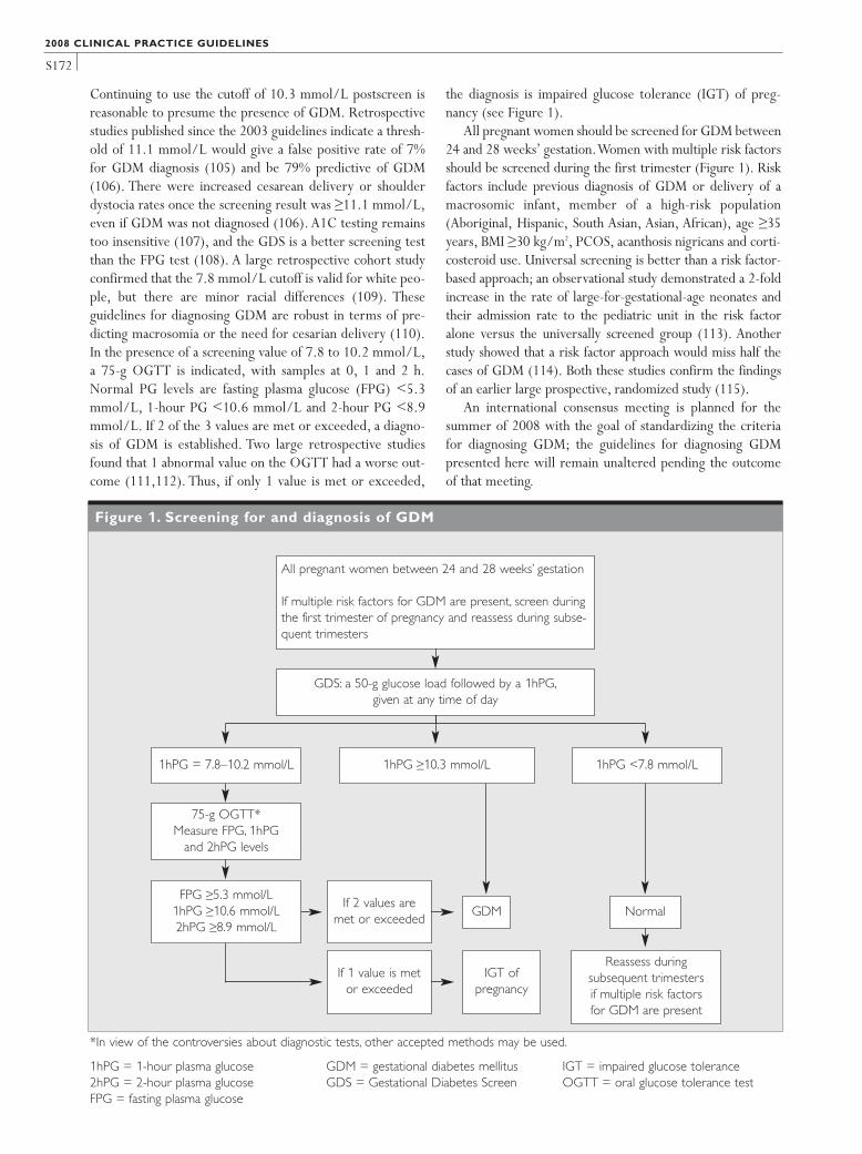
S172
Continuing to use the cutoff of 10.3 mmol/L postscreen isreasonable to presume the presence of GDM. Retrospectivestudies published since the 2003 guidelines indicate a thresh-old of 11.1 mmol/L would give a false positive rate of 7%for GDM diagnosis (105) and be 79% predictive of GDM(106). There were increased cesarean delivery or shoulderdystocia rates once the screening result was ≥11.1 mmol/L,even if GDM was not diagnosed (106). A1C testing remainstoo insensitive (107), and the GDS is a better screening testthan the FPG test (108). A large retrospective cohort studyconfirmed that the 7.8 mmol/L cutoff is valid for white peo-ple, but there are minor racial differences (109). Theseguidelines for diagnosing GDM are robust in terms of pre-dicting macrosomia or the need for cesarian delivery (110).In the presence of a screening value of 7.8 to 10.2 mmol/L,a 75-g OGTT is indicated, with samples at 0, 1 and 2 h.Normal PG levels are fasting plasma glucose (FPG) <5.3mmol/L, 1-hour PG <10.6 mmol/L and 2-hour PG <8.9mmol/L. If 2 of the 3 values are met or exceeded, a diagno-sis of GDM is established. Two large retrospective studiesfound that 1 abnormal value on the OGTT had a worse out-come (111,112). Thus, if only 1 value is met or exceeded,
the diagnosis is impaired glucose tolerance (IGT) of preg-nancy (see Figure 1).
All pregnant women should be screened for GDM between24 and 28 weeks’ gestation.Women with multiple risk factorsshould be screened during the first trimester (Figure 1). Riskfactors include previous diagnosis of GDM or delivery of amacrosomic infant, member of a high-risk population(Aboriginal, Hispanic, South Asian, Asian, African), age ≥35years, BMI ≥30 kg/m2, PCOS, acanthosis nigricans and corti-costeroid use. Universal screening is better than a risk factor-based approach; an observational study demonstrated a 2-foldincrease in the rate of large-for-gestational-age neonates andtheir admission rate to the pediatric unit in the risk factoralone versus the universally screened group (113). Anotherstudy showed that a risk factor approach would miss half thecases of GDM (114). Both these studies confirm the findingsof an earlier large prospective, randomized study (115).
An international consensus meeting is planned for thesummer of 2008 with the goal of standardizing the criteriafor diagnosing GDM; the guidelines for diagnosing GDMpresented here will remain unaltered pending the outcomeof that meeting.
2008 CLINICAL PRACTICE GUIDELINES
Figure 1. Screening for and diagnosis of GDM
All pregnant women between 24 and 28 weeks’ gestation
If multiple risk factors for GDM are present, screen duringthe first trimester of pregnancy and reassess during subse-quent trimesters
GDS: a 50-g glucose load followed by a 1hPG,given at any time of day
1hPG = 7.8–10.2 mmol/L
75-g OGTT*Measure FPG, 1hPG
and 2hPG levels
FPG ≥5.3 mmol/L1hPG ≥10.6 mmol/L2hPG ≥8.9 mmol/L
If 2 values aremet or exceeded
GDM Normal
If 1 value is metor exceeded
Reassess during subsequent trimestersif multiple risk factors for GDM are present
IGT of pregnancy
1hPG ≥10.3 mmol/L 1hPG <7.8 mmol/L
*In view of the controversies about diagnostic tests, other accepted methods may be used.
1hPG = 1-hour plasma glucose2hPG = 2-hour plasma glucoseFPG = fasting plasma glucose
GDM = gestational diabetes mellitusGDS = Gestational Diabetes Screen
IGT = impaired glucose toleranceOGTT = oral glucose tolerance test

S173
ManagementUntreated GDM (116) or IGT (102) leads to increasedmaternal and perinatal morbidity, while intensive treatmentis associated with outcomes similar to control populations(52,117,118). Some women with GDM actually have undi-agnosed type 2 diabetes, and this group has an increased riskof offspring having congenital malformations (101,119,120).Women at high risk of type 2 diabetes (advanced maternalage, strong family history, previous GDM, ethnic predisposi-tion, marked obesity) should be assessed for diabetes at thefirst prenatal visit if this has not been done in the 6 monthsbefore pregnancy.The recommended glycemic targets (Table1), use of SMBG and lifestyle interventions are similar for alltypes of diabetes in pregnancy. Since many women of differ-ent high-risk ethnic backgrounds have GDM, it is importantto have culturally relevant educational materials available.Use of 1-hour or 2-hour postprandial glucose levels appearsto be equally effective in therapy (121).
Monitoring SMBG is essential during pregnancy (6). Both preprandialand postprandial testing are recommended to guide therapyin order to achieve glycemic targets (50,116). Due to theincreased risk of nocturnal hypoglycemia during pregnancy,testing during the night is often necessary in patients receiv-ing insulin (56). Because starvation ketosis is common inpregnancy and may have detrimental effects on the fetus,urine and/or blood monitoring of ketones is warranted toconfirm that the diet is adequate (58,59).
Lifestyle During pregnancy, women should be evaluated and followedby a registered dietitian to ensure that nutrition therapy pro-motes euglycemia, appropriate weight gain and adequatenutritional intake (60,61,122,123). Meal planning shouldemphasize moderate carbohydrate restriction and distribu-tion over 3 meals and 3 snacks, 1 of which should be at bed-time. Hypocaloric diets are not recommended, as they resultin weight loss and significant ketosis and are likely inadequatein required nutrients, such as protein and calcium. Pre-pregnancy body mass is a potent predictor of birth weightand should be considered when making recommendationsabout energy intake and rate of weight gain (62). Detailedrecommendations for nutritional management of GDM areavailable (123). Physical activity should be encouraged unlessobstetrical contraindications exist or glycemic control isworsened by the activity (63,124).
Pharmacologic interventionsInsulinIf women with GDM or IGT do not achieve glycemic targetswithin 2 weeks from nutrition therapy alone, insulin therapyshould be initiated (125,126). In some cases, assessment offetal growth by early third-trimester ultrasound can be used
to guide therapy (127,128). The use of insulin to achieveglycemic targets has been shown to reduce fetal and mater-nal morbidities (52,126).A variety of protocols can be used,with multiple injections being the most effective (65). Insulinusually needs to be continuously adjusted to achieve targetglucose values. Although short-acting analogues aspart andlispro can help achieve postprandial glucose targets withoutsevere hypoglycemia (71,72), improvements in fetal out-comes or in fetal growth parameters have not been demon-strated with the use of lispro compared to regular insulin inclinical trials in women with GDM (73).
GlyburideGlyburide is safe and effective at controlling glucose levels inover 80% of patients with GDM (129-131) and does notcross the placenta (132). Women with higher fasting andpostprandial glucose values on their OGTT (133), or whileon diet therapy (132), are less likely to respond to glyburide.Despite good glucose levels, however, some studies reportmore adverse perinatal outcomes in women treated with gly-buride than with insulin (134,135). Glyburide can be con-sidered for women in whom insulin cannot be used.
MetforminIn a recent study (136), 751 women with GDM were ran-domly assigned to open treatment with metformin (withsupplemental insulin if required) or insulin. Of the womenassigned to metformin, 46.3% received supplementalinsulin. Metformin (alone or with supplemental insulin) wasnot associated with increased perinatal complications com-pared with insulin. There was less severe hypoglycemia inneonates receiving metformin, but more spontaneouspreterm delivery (i.e. <37 weeks’ gestation). While met-formin appears to be a safe alternative to insulin therapy, itdoes cross the placenta. Results of the offspring follow-up ofthe Metformin in Gestational Diabetes trial (MiG TOFU),expected in several years, will provide more data on its long-term safety.
As the use of metformin or glyburide during pregnancy iscurrently not an approved indication in Canada, such usewould be considered off-label and would therefore requirethe appropriate discussion with patients.
PostpartumBreastfeedingAll women should be encouraged to breastfeed, since thismay reduce offspring obesity, especially in the setting ofmaternal obesity, and prevent the development of type 2 dia-betes (82,137,138).
Long-term maternal risks With the diagnosis of GDM, there is evidence of both impair-ment of insulin secretion and action (139,140).These defectspersist postpartum and increase the risk of impaired fasting
DIA
BE
TE
SIN
SP
EC
IAL
PO
PU
LA
TIO
NS

S174
glucose, IGT and type 2 diabetes (141,142). The cumulativeincidence increases markedly in the first 5 years postpartumand more slowly after 10 years (143,144). At 3 to 6 monthspostpartum, risks of dysglycemia are in the 16 to 20% range,and the cumulative risks increase to a 30 to 60% range, depend-ing on time since the index pregnancy and the population stud-ied.The strongest predictor of early postpartum developmentof diabetes is elevated FPG during pregnancy (145,146).
Some women with GDM, especially lean women <30years of age who require insulin during pregnancy, progressto type 1 diabetes (147,148).Women with positive autoanti-bodies (anti-GAD, IA-2) are more likely to have diabetes by6 months postpartum (149).
Postpartum testing is essential to identify women who con-tinue to have diabetes, those who develop diabetes after tem-porary normalization and those at risk, including those withIGT. However, many women do not receive adequate postpar-tum follow-up (150,151), and it is essential that the impor-tance of follow-up be explicitly communicated with the woman
and her caregivers who are responsible for postpartum testing.Women should be screened postpartum to determine
their glucose status. Postnatal FPG has been the most consis-tently found variable in determining women at high risk forearly postpartum diabetes (152).A FPG alone, however, willmiss many women with some degree of abnormal glucosetolerance (153), and therefore, a 75-g OGTT should be donebetween 6 weeks and 6 months postpartum.
Metabolic syndrome has been shown to be more preva-lent in women with GDM, especially those who are obeseand non-Caucasian in some (154-156) but not all studies(157). Given the increased risk of CVD with metabolic syn-drome, consideration should be given to screening for allcomponents of metabolic syndrome in the postpartum careof women with GDM. Education on lifestyle modification toprevent diabetes and CVD should begin in pregnancy andcontinue postpartum. Emphasis on targeted strategies thatincorporate women’s exercise beliefs may increase participa-tion rates (158) (see “Prevention of Diabetes,” p. S17).
2008 CLINICAL PRACTICE GUIDELINES
7. All pregnant women should be screened for GDM [Grade C, Level 3 (113,115)]. For most women, screeningshould be performed between 24 and 28 weeks’ gesta-tion [Grade D, Consensus]. Women with multiple risk fac-tors should be screened during the first trimester and,if negative, should be reassessed during subsequenttrimesters [Grade D, Consensus].
8. Screening for GDM should be conducted using the GDS– a 50-g glucose load followed by a PG test measured 1 hlater [Grade D, Level 4 (108)]. If GDM is strongly suspect-ed, an OGTT can be performed without an initial GDS[Grade D, Consensus].
9. Women who have a positive screening test (a 1hPG of 7.8 to 10.2 mmol/L on the GDS) should undergo an OGTT in order to diagnose GDM.A value of ≥10.3mmol/L is considered diagnostic of GDM, in which casean OGTT does not need to be performed [Grade D,Consensus].
10. GDM is diagnosed when at least 2 of the following val-ues on the OGTT are met or exceeded. If 1 value ismet or exceeded, a diagnosis of IGT of pregnancy ismade [Grade D, Consensus]:• FPG: ≥5.3 mmol/L• 1hPG: ≥10.6 mmol/L• 2hPG: ≥8.9 mmol/L
11. Women with GDM should:a. Strive to achieve target glucose values:
• Fasting/preprandial PG: 3.8 to 5.2 mmol/L• 1h postprandial PG: 5.5 to 7.7 mmol/L• 2h postprandial PG: 5.0 to 6.6 mmol/L
b. Perform SMBG both pre- and postprandially (≥4 timesper day, if needed) to achieve glycemic targets andimprove pregnancy outcomes [Grade C, Level 3 (47)].
c. Receive nutrition counselling from a registered dietitian
during pregnancy [Grade C, Level 3 (89)] and postpartum[Grade D, Consensus]. Recommendations for weight gainduring pregnancy should be based on pregravid BMI[Grade D, Consensus].
d.Avoid ketosis during pregnancy [Grade C, Level 3 (97)].
12. If women with GDM do not achieve glycemic targetswithin 2 weeks using nutrition therapy alone, insulintherapy should be initiated [Grade D, Consensus],with up to 4 injections/day considered [Grade A,Level 1A (65)].
13. Glyburide [Grade B, Level 2 (130,134,135)] or metformin[Grade B, Level 2 (136)] may be considered as second-line agents in women with GDM who are nonadherentto or who refuse insulin. Glyburide may be preferred, asmetformin use is more likely to need supplementalinsulin for glycemic control and metformin crosses theplacenta with unknown long-term effects. Use of oralagents in pregnancy is off-label and should be discussedwith the patient [Grade D, Consensus].
Postpartum14. As women who have had GDM are defined as high risk
of developing subsequent type 2 diabetes, they shouldbe re-evaluated postpartum [Grade D, Consensus]. A 75-gOGTT should be performed between 6 weeks and 6months postpartum to establish their glucose status.Women who are suspected of having had pre-existingdiabetes should be monitored more closely postpartum.All women with GDM should be counselled on ahealthy lifestyle.
15. Women with previous GDM should follow the screen-ing and prevention guidelines for other high-risk groupsscreened for type 2 diabetes [Grade D, Consensus] andshould be screened for type 2 diabetes when planninganother pregnancy [Grade D, Consensus].
RECOMMENDATIONS
S175
Long-term risks in offspringThere is compelling evidence that offspring exposed to GDMare at increased risk of obesity and IGT, especially if large forgestational age and born to obese mothers (58,159-162). Ina Canadian cohort of children exposed to GDM, 7% had IGTat age 7 to 11 years (162). In the Pima Indian population, asmany as 70% of offspring exposed to diabetes in utero hadtype 2 diabetes at age 25 to 35 years (163). Breastfeedingmay lower the risk (82,138,164). The importance of tightglycemic control during pregnancy to prevent these out-comes is not clearly established.The need for increased sur-veillance of these children requires further study.
Planning subsequent pregnanciesWomen with previous GDM should plan future pregnanciesin consultation with their healthcare providers (165,166).Glucose tolerance should be assessed prior to conception toassure normoglycemia at the time of conception, and any glu-cose abnormality should be treated. In an effort to reducethe risk of congenital anomalies and optimize pregnancy out-comes, all women should take a folic acid supplement of 0.4to 1.0 mg (48).
OTHER RELEVANT GUIDELINESScreening for Type 1 and Type 2 Diabetes, p. S14Targets for Glycemic Control, p. S29Chronic Kidney Disease in Diabetes, p. S126Type 1 Diabetes in Children and Adolescents, p. S150
REFERENCES1. Yogev Y, Ben-Haroush A, Chen R, et al. Diurnal glycemic pro-
file in obese and normal weight nondiabetic pregnant women.Am J Obstet Gynecol. 2004;191:949-953.
2. Nielsen LR, Ekbom P, Damm P, et al. HbA1c levels are signif-icantly lower in early and late pregnancy. Diabetes Care. 2004;27:1200-1201.
3. Langer O, Conway DL. Level of glycemia and perinatal out-come in pregestational diabetes. J Matern Fetal Med.2000;9:35-41.
4. Parretti E, Mecacci F, Papini M, et al. Third-trimester mater-nal glucose levels from diurnal profiles in nondiabetic preg-nancies: correlation with sonographic parameters of fetalgrowth. Diabetes Care. 2001;24:1319-1323.
5. Ben-Haroush A,Yogev Y, Chen R, et al. The postprandial glu-cose profile in the diabetic pregnancy. Am J Obstet Gynecol.2004;191:576-581.
6. Cundy T, Gamble G, Townend K, et al. Perinatal mortality intype 2 diabetes mellitus. Diabet Med. 2000;17:33-39.
7. Wylie BR, Kong J, Kozak SE, et al. Normal perinatal mortali-ty in type 1 diabetes mellitus in a series of 300 consecutivepregnancy outcomes. Am J Perinatol. 2002;19:169-176.
8. Roland JM, Murphy HR, Ball V, et al. The pregnancies ofwomen with type 2 diabetes: poor outcomes but opportunitiesfor improvement. Diabet Med. 2005;22:1774-1777.
9. Clausen TD, Mathiesen E, Ekbom P, et al. Poor pregnancy out-come in women with type 2 diabetes. Diabetes Care. 2005;28:323-328.
10. Gunton JE, Morris J, Boyce S, et al. Outcome of pregnancycomplicated by pre-gestational diabetes – improvement inoutcomes. Aust N Z J Obstet Gynaecol. 2002;42:478-481.
11. Hadden DR, Cull CA, Croft DJ, et al. Poor pregnancy out-come for women with type 2 diabetes. Diabetic Med. 2003;20:506-507.
12. Dunne F, Brydon P, Smith K, et al. Pregnancy in women withtype 2 diabetes: 12 years outcome data 1990-2002. Diabet Med.2003;20:734-738.
13. Farrell T, Neale L, Cundy T. Congenital anomalies in the off-spring of women with type 1, type 2 and gestational diabetes.Diabetic Med. 2002;19:322-326.
14. Boulot P, Chabbert-Buffet N, d’Ercole C, et al; Diabetes andPregnancy Group, France. French multicentric survey of out-come of pregnancy in women with pregestational diabetes.Diabetes Care. 2003;26:2990-2993.
15. dos Santos Silva I, Higgins C, Swerdlow AJ, et al. Birthweightand other pregnancy outcomes in a cohort of women with pre-gestational insulin-treated diabetes mellitus, Scotland, 1979-95. Diabet Med. 2005;22:440-447.
16. Evers IM, de Valk HW, Visser GH. Risk of complications ofpregnancy in women with type 1 diabetes: nationwideprospective study in the Netherlands. BMJ. 2004;328:915.
17. Jensen DM, Damm P, Moelsted-Pedersen L, et al. Outcomesin type 1 diabetic pregnancies: a nationwide, population-basedstudy. Diabetes Care. 2004;27:2819-2823.
18. Penney GC, Mair G, Pearson DW; Scottish Diabetes inPregnancy Group. Outcomes of pregnancies in women withtype 1 diabetes in Scotland: a national population-based study.BJOG. 2003;110:315-318.
19. Platt MJ, Stanisstreet M, Casson IF, et al. St Vincent’sDeclaration 10 years on: outcomes of diabetic pregnancies.Diabet Med. 2002;19:216-220.
20. The Diabetes Control and Complications Trial ResearchGroup. Pregnancy outcomes in the Diabetes Control andComplications Trial. Am J Obstet Gynecol. 1996;174:1343-1353.
21. McIntosh M, ed. Pregnancy in Women with Type 1 and Type 2Diabetes: 2002–2003. England, Wales and Northern Ireland.London, UK: Confidential Enquiry into Maternal and ChildHealth; 2005.
22. Klein BE, Moss SE, Klein R. Effect of pregnancy on progres-sion of diabetic retinopathy. Diabetes Care. 1990;13:34-40.
23. Omori Y, Minei S,Testuo T, et al. Current status of pregnancyin diabetic women. A comparison of pregnancy in IDDM andNIDDM mothers. Diabetes Res Clin Pract. 1994;24(suppl):S273-S278.
24. The Diabetes Control and Complications Trial ResearchGroup. Effect of pregnancy on microvascular complications inthe Diabetes Control and Complications Trial. Diabetes Care.2000;23:1084-1091.
25. Chew EY, Mills JL, Metzger BE, et al. Metabolic control and
DIA
BE
TE
SIN
SP
EC
IAL
PO
PU
LA
TIO
NS

S176
progression of retinopathy. The Diabetes in Early PregnancyStudy. National Institute of Child Health and HumanDevelopment Diabetes in Early Pregnancy Study. Diabetes Care.1995;18:631-637.
26. Lövestam-Adrian M, Agardh CD, Aberg A, et al. Pre-eclamp-sia is a potent risk factor for deterioration of retinopathy dur-ing pregnancy in type 1 diabetic patients. Diabetic Med. 1997;14:1059-1065.
27. Rosenn B, Miodovnik M, Kranias G, et al. Progression of dia-betic retinopathy in pregnancy: association with hypertensionin pregnancy. Am J Obstet Gynecol. 1992;166:1214-1218.
28. Cundy T, Slee F, Gamble G, et al. Hypertensive disorders ofpregnancy in women with type 1 and type 2 diabetes. DiabetMed. 2002;19:482-489.
29. Sibai BM, Caritis S, Hauth J, et al. Risks of preeclampsia andadverse neonatal outcomes among women with pregestationaldiabetes mellitus. National Institute of Child Health andHuman Development Network of Maternal-Fetal MedicineUnits. Am J Obstet Gynecol. 2000;182:364-369.
30. Schröder W, Heyl W, Hill-Grasshoff B, et al. Clinical value ofdetecting microalbuminuria as a risk factor for pregnancy-induced hypertension in insulin-treated diabetic pregnancies.Eur J Obstet Gynecol Reprod Biol. 2000;91:155-158.
31. How HY, Sibai B, Lindheimer M, et al. Is early-pregnancy pro-teinuria associated with an increased rate of preeclampsia inwomen with pregestational diabetes mellitus? Am J ObstetGynecol. 2004;190:775-778.
32. Chobanian AV, Bakris GL, Black HR, et al; Joint NationalCommittee on Prevention, Detection, Evaluation, andTreatment of High Blood Pressure. National Heart, Lung, andBlood Institute; National High Blood Pressure EducationProgram Coordinating Committee. Seventh report of the JointNational Committee on Prevention, Detection, Evaluation,and Treatment of High Blood Pressure. Hypertension. 2003;42:1206-1252.
33. Ekbom P, Damm P, Feldt-Rasmussen B, et al. Pregnancy out-come in type 1 diabetic women with microalbuminuria.Diabetes Care. 2001;24:1739-1744.
34. Dunne FP, Chowdhury TA, Hartland A, et al. Pregnancy out-come in women with insulin-dependent diabetes mellituscomplicated by nephropathy. QJM. 1999;92:451-454.
35. Bagg W, Neale L, Henley P, et al. Long-term maternal outcomeafter pregnancy in women with diabetic nephropathy. N Z MedJ. 2003;116:U566.
36. Rossing K, Jacobsen P, Hommel E, et al. Pregnancy and pro-gression of diabetic nephropathy. Diabetologia. 2002;45:36-41.
37. Reece EA, Leguizamon G, Homko C. Stringent controls indiabetic nephropathy associated with optimization of pregnan-cy outcomes. J Matern Fetal Med. 1998;7:213-216.
38. Leguizamon G, Reece EA. Effect of medical therapy on pro-gressive nephropathy: influence of pregnancy, diabetes andhypertension. J Matern Fetal Med. 2000;9:70-78.
39. Biesenbach G, Grafinger P, Stöger H, et al. How pregnancyinfluences renal function in nephropathic type 1 diabetic
women depends on their pre-conceptional creatinine clear-ance. J Nephrol. 1999;12:41-46.
40. Gordon M, Landon MB, Samuels P, et al. Perinatal outcomeand long-term follow-up associated with modern managementof diabetic nephropathy. Obstet Gynecol. 1996;87:401-409.
41. Quan A. Fetopathy associated with exposure to angiotensinconverting enzyme inhibitors and angiotensin receptor antag-onists. Early Hum Dev. 2006;82:23-28.
42. Cooper WO, Hernandez-Diaz S, Arbogast PG, et al. Majorcongenital malformations after first-trimester exposure toACE inhibitors. N Engl J Med. 2006;354:2443-2451.
43. Silfen SL,Wapner RJ, Gabbe SG. Maternal outcome in class Hdiabetes mellitus. Obstet Gynecol. 1980;55:749-751.
44. Bagg W, Henley PG, Macpherson P, et al. Pregnancy in womenwith diabetes and ischemic heart disease. Aust N Z J ObstetGynaecol. 1999;39:99-102.
45. Howorka K, Pumprla J, Gabriel M, et al. Normalization ofpregnancy outcome in pregestational diabetes through func-tional insulin treatment and modular out-patient educationadapted for pregnancy. Diabet Med. 2001;18:965-972.
46. Quevedo SF, Coustan DR. Diabetes and pregnancy. Use of anintegrated “team” approach provides the necessary compre-hensive care. R I Med J. 1989;72:129-132.
47. Ray JG, O’Brien TE, Chan WS. Preconception care and therisk of congenital anomalies in the offspring of women withdiabetes mellitus: a meta-analysis. QJM. 2001;94:435-444.
48. Wilson RD. Pre-conception vitamin/folic acid supplementa-tion 2007: the use of folic acid in combination with a multivi-tamin supplement for the prevention of neural tube defectsand other congenital anomalies. J Obstet Gynaecol Can. 2007;29:1003-1026.
49. Langer O. A spectrum of glucose thresholds may effectivelyprevent complications in the pregnant diabetic patient. SeminPerinatol. 2002;26:196-205.
50. Combs CA, Gunderson E, Kitzmiller JL, et al. Relationship offetal macrosomia to maternal postprandial glucose controlduring pregnancy. Diabetes Care. 1992;15:1251-1257.
51. de Veciana M, Major CA, Morgan MA, et al. Postprandial ver-sus preprandial blood glucose monitoring in women with ges-tational diabetes mellitus requiring insulin therapy. N Engl JMed. 1995;333:1237-1241.
52. Langer O, Rodriguez DA, Xenakis EM, et al. Intensified versusconventional management of gestational diabetes. Am J ObstetGynecol. 1994;170:1036-1046.
53. Rosenn BM, Miodovnik M, Khoury JC, et al. Counterregulatoryhormonal responses to hypoglycemia during pregnancy. ObstetGynecol. 1996;87:568-574.
54. Diamond MP, Reece EA, Caprio S, et al. Impairment of coun-terregulatory hormone responses to hypoglycemia in pregnantwomen with insulin-dependent diabetes mellitus. Am J ObstetGynecol. 1992;166:70-77.
55. Reece EA, Hagay Z, Roberts AB, et al. Fetal Doppler andbehavioral responses during hypoglycemia induced with theinsulin clamp technique in pregnant diabetic women. Am J
2008 CLINICAL PRACTICE GUIDELINES

S177
Obstet Gynecol. 1995;172:151-155.56. Egger M, Davey Smith G, Stettler C, et al. Risk of adverse
effects of intensified treatment in insulin-dependent diabetesmellitus: a meta-analysis. Diabet Med. 1997;14:919-928.
57. Rosenn BM, Miodovnik M, Khoury JC, et al. Deficient coun-terregulation: a possible risk factor for excessive fetal growthin IDDM pregnancies. Diabetes Care. 1997;20:872-874.
58. Silverman BL, Rizzo TA, Cho NH, et al. Long-term effects ofthe intrauterine environment. The Northwestern UniversityDiabetes in Pregnancy Center. Diabetes Care. 1998;21(suppl 2):B142-B149.
59. Ornoy A. Growth and neurodevelopmental outcome of chil-dren born to mothers with pregestational and gestational dia-betes. Pediatr Endocrinol Rev. 2005;3:104-113.
60. Franz MJ, Bantle JP, Beebe CA, et al. Evidence-based nutritionprinciples and recommendations for the treatment and pre-vention of diabetes and related complications. Diabetes Care.2002;25:148-198.
61. Jovanovic L. Medical nutritional therapy in pregnant womenwith pregestational diabetes mellitus. J Matern Fetal Med.2000;9:21-28.
62. Knopp RH, Magee MS, Raisys V, et al. Metabolic effects ofhypocaloric diets in management of gestational diabetes.Diabetes. 1991;40(suppl 2):165-171.
63. Sternfeld B, Quesenberry CP Jr, Eskenazi B, et al. Exerciseduring pregnancy and pregnancy outcome. Med Sci Sports Exerc.1995;27:634-640.
64. Jovanovic L, Druzin M, Peterson CM. Effect of euglycemia onthe outcome of pregnancy in insulin-dependent diabeticwomen as compared with normal control subjects. Am J Med.1981;71:921-927.
65. Nachum Z, Ben-Shlomo I,Weiner E, et al. Twice daily versusfour times daily insulin dose regimens for diabetes in pregnan-cy: randomised controlled trial. BMJ. 1999;319:1223-1227.
66. Chauhan SP, Perry KG Jr, McLaughlin BN, et al. Diabeticketoacidosis complicating pregnancy. J Perinatol. 1996;16:173-175.
67. Boskovic R, Feig DS, Derewlany L, et al. Transfer of insulinlispro across the human placenta: in vitro perfusion studies.Diabetes Care. 2003;26:1390-1394.
68. Persson B, Swahn ML, Hjertberg R, et al. Insulin lispro thera-py in pregnancies complicated by type 1 diabetes mellitus.Diabetes Res Clin Pract. 2002;58:115-121.
69. Wyatt JW, Frias JL, Hoyme HE, et al; IONS study group.Congenital anomaly rate in offspring of mothers with diabetestreated with insulin lispro during pregnancy. Diabet Med.2005;22:803-807.
70. Mathiesen ER, Kinsley B, Amiel SA, et al; Insulin AspartPregnancy Study Group. Maternal glycemic control and hypo-glycemia in type 1 diabetic pregnancy: a randomized trial ofinsulin aspart versus human insulin in 322 pregnant women.Diabetes Care. 2007;30:771-776.
71. Mecacci F, Carignani L, Cioni R, et al. Maternal metaboliccontrol and perinatal outcome in women with gestational dia-
betes treated with regular or lispro insulin: comparison withnon-diabetic pregnant women. Eur J Obstet Gynecol Reprod Biol.2003;111:19-24.
72. Pettitt DJ, Ospina P, Kolaczynski JW, et al. Comparison of aninsulin analog, insulin aspart, and regular human insulin withno insulin in gestational diabetes mellitus. Diabetes Care. 2003;26:183-186.
73. Plank J, Siebenhofer A, Berghold A, et al. Systematic reviewand meta-analysis of short-acting insulin analogues in patientswith diabetes mellitus. Arch Intern Med. 2005;165:1337-1344.
74. Gallen IW, Jaap A, Roland JM, et al. Survey of glargine use in115 pregnant women with type 1 diabetes. Diabet Med. 2008;25:165-169.
75. Kurtzhals P, Schaffer L, Sorensen A, et al. Correlations ofreceptor binding and metabolic and mitogenic potencies ofinsulin analogs designed for clinical use. Diabetes. 2000;49:999-1005.
76. Gutzin SJ, Kozer E, Magee LA, et al.The safety of oral hypo-glycemic agents in the first trimester of pregnancy: a meta-analysis. Can J Clin Pharmacol. 2003;10:179-183.
77. Hellmuth E, Damm P, Molsted-Pedersen L. Oral hypogly-caemic agents in 118 diabetic pregnancies. Diabet Med. 2000;17:507-511.
78. Ekpebegh CO, Coetzee EJ, van der Merwe L, et al. A 10-yearretrospective analysis of pregnancy outcome in pregestationaltype 2 diabetes: comparison of insulin and oral glucose-lower-ing agents. Diabet Med. 2007;24:253-258.
79. Glueck CJ, Wang P, Kobayashi S, et al. Metformin therapythroughout pregnancy reduces the development of gestationaldiabetes in women with polycystic ovary syndrome. FertilSteril. 2002;77:520-525.
80. Glueck CJ, Goldenberg N, Pranikoff J, et al. Height, weight,and motor-social development during the first 18 months oflife in 126 infants born to 109 mothers with polycystic ovarysyndrome who conceived on and continued metforminthrough pregnancy. Hum Reprod. 2004;19:1323-1330.
81. Glueck CJ, Salehi M, Sieve L, et al. Growth, motor, and socialdevelopment in breast- and formula-fed infants of metformin-treated women with polycystic ovary syndrome. J Pediatr.2006;148:628-632.
82. Mayer-Davis EJ, Rifas-Shiman SL, Zhou L, et al. Breast-feed-ing and risk for childhood obesity: does maternal diabetes orobesity status matter? Diabetes Care. 2006;29:2231-2237.
83. Gardiner SJ, Kirkpatrick CM, Begg EJ, et al.Transfer of met-formin into human milk. Clin Pharmacol Ther. 2003;73:71-77.
84. Briggs GG,Ambrose PJ, Nageotte MP, et al. Excretion of met-formin into breast milk and the effect on nursing infants. ObstetGynecol. 2005;105:1437-1441.
85. Hale TW, Kristensen JH, Hackett LP, et al. Transfer of met-formin into human milk. Diabetologia. 2002;45:1509-1514.
86. Feig DS, Briggs GG, Kraemer JM, et al.Transfer of glyburideand glipizide into breast milk. Diabetes Care. 2005;28:1851-1855.
87. Umpierrez GE, Latif KA, Murphy MB, et al.Thyroid dysfunc-
DIA
BE
TE
SIN
SP
EC
IAL
PO
PU
LA
TIO
NS
S178
tion in patients with type 1 diabetes: a longitudinal study.Diabetes Care. 2003;26:1181-1185.
88. Kitzmiller JL, Gavin LA, Gin GD, et al. Preconception care ofdiabetes. Glycemic control prevents congenital anomalies.JAMA. 1991;265:731-736.
89. McElvy SS, Miodovnik M, Rosenn B, et al. A focused precon-ceptional and early pregnancy program in women with type 1diabetes reduces perinatal mortality and malformation rates togeneral population levels. J Matern Fetal Med. 2000;9:14-20.
90. Jovanovic L, Knopp RH, Kim H, et al. Elevated pregnancylosses at high and low extremes of maternal glucose in earlynormal and diabetic pregnancy: evidence for a protectiveadaptation in diabetes. Diabetes Care. 2005;28:1113-1117.
91. Suhonen L, Hiilesmaa V, Teramo K. Glycaemic control duringearly pregnancy and fetal malformations in women with type Idiabetes mellitus. Diabetologia. 2000;43:79-82.
92. Guerin A, Nisenbaum R, Ray JG. Use of maternal GHb con-centration to estimate the risk of congenital anomalies in theoffspring of women with pre-pregnancy diabetes. DiabetesCare. 2007;30:1920-1925.
93. Hiilesmaa V, Suhonen L,Teramo K. Glycaemic control is asso-ciated with pre-eclampsia but not with pregnancy-inducedhypertension in women with type I diabetes mellitus.Diabetologia. 2000;43:1534-1539.
94. Hsu CD, Tan HY, Hong SF, et al. Strategies for reducing thefrequency of preeclampsia in pregnancies with insulin-depend-ent diabetes mellitus. Am J Perinatol. 1996;13:265-268.
95. Edison RJ, Muenke M. Central nervous system and limbanomalies in case reports of first-trimester statin exposure.N Engl J Med. 2004;350:1579-1582.
96. The Diabetes Control and Complications Trial ResearchGroup. Early worsening of diabetic retinopathy in the DiabetesControl and Complications Trial. Arch Ophthalmol.1998;116:874-886.
97. Rizzo T, Metzger BE, Burns WJ, et al. Correlations betweenantepartum maternal metabolism and child intelligence. N EnglJ Med. 1991;325:911-916.
98. Metzger BE, Coustan DR. Summary and recommendations ofthe Fourth International Workshop – Conference onGestational Diabetes Mellitus. Diabetes Care. 1998;21(suppl 2):B161-B167.
99. Dyck R, Klomp H, Tan LK, et al. A comparison of rates, riskfactors, and outcomes of gestational diabetes between aborig-inal and non-aboriginal women in the Saskatoon HealthDistrict. Diabetes Care. 2002;25:487-493.
100. Godwin M, Muirhead M, Huynh J, et al. Prevalence of gesta-tional diabetes mellitus among Swampy Cree women in MooseFactory, James Bay. CMAJ. 1999;160:1299-1302.
101. Harris SB, Caulfield LE, Sugamori ME, et al.The epidemiolo-gy of diabetes in pregnant Native Canadians. A risk profile.Diabetes Care. 1997;20:1422-1425.
102. Crowther CA, Hiller JE, Moss JR, et al; AustralianCarbohydrate Intolerance Study in Pregnant Women(ACHOIS) Trial Group. Effect of treatment of gestational dia-
betes mellitus on pregnancy outcomes. N Engl J Med. 2005;352:2477-2486.
103. HAPO Study Cooperative Research Group; Metzger BE,Lowe LP, Dyer AR, et al. Hyperglycemia and adverse pregnan-cy outcomes. N Engl J Med. 2008;358:1991-2002.
104. Cutchie WA, Cheung NW, Simmons D. Comparison of inter-national and New Zealand guidelines for the care of pregnantwomen with diabetes. Diabet Med. 2006;23:460-468.
105. Lanni S, Barrett D.The predictive value of the 1-h 50-g glucosescreen for diagnosing gestational diabetes mellitus in a high-risk population. J Matern Fetal Neonatal Med. 2004;15:375-379.
106. Cheng YW, Esakoff TF, Block-Kurbisch I, et al. Screening ordiagnostic: markedly elevated glucose loading test and perina-tal outcomes. J Matern Fetal Neonatal Med. 2006;19:729-734.
107. Agarwal MM, Dhatt GS, Punnose J, et al. Gestational diabetes:a reappraisal of HBA1c as a screening test. Acta Obstet GynecolScand. 2005;84:1159-1163.
108. Rey E, Hudon L, Michon N, et al. Fasting plasma glucose ver-sus glucose challenge test: screening for gestational diabetesand cost effectiveness. Clin Biochem. 2004;37:780-784.
109. Esakoff TF, Cheng YW, Caughey AB. Screening for gestationaldiabetes: different cut-offs for different ethnicities? Am J ObstetGynecol. 2005;193:1040-1044.
110. Agarwal MM, Dhatt GS, Punnose J, et al. Gestational diabetes:dilemma caused by multiple international diagnostic criteria.Diabet Med. 2005;22:1731-1736.
111. Chico A, Lopez-Rodo V, Rodriguez-Vaca D, et al. Features andoutcome of pregnancies complicated by impaired glucose tol-erance and gestational diabetes diagnosed using different crite-ria in a Spanish population. Diabetes Res Clin Pract. 2005;68:141-146.
112. McLaughlin GB, Cheng YW, Caughey AB. Women with oneelevated 3-hour glucose tolerance test value: are they at riskfor adverse perinatal outcomes? Am J Obstet Gynecol. 2006;194:e16-e19.
113. Cosson E, Benchimol M, Carbillon L, et al. Universal ratherthan selective screening for gestational diabetes mellitus mayimprove fetal outcomes. Diabetes Metab. 2006;32:140-146.
114. Pöyhönen-Alho MK,Teramo KA, Kaaja RJ, et al. 50gram oralglucose challenge test combined with risk factor-basedscreening for gestational diabetes. Eur J Obstet Gynecol ReprodBiol. 2005;121:34-37.
115. Griffin ME, Coffey M, Johnson H, et al. Universal vs. risk fac-tor-based screening for gestational diabetes mellitus: detectionrates, gestation at diagnosis and outcome. Diabet Med. 2000;17:26-32.
116. Langer O, Yogev Y, Most O, et al. Gestational diabetes: theconsequences of not treating. Am J Obstet Gynecol. 2005;192:989-997.
117. Thompson DM, Dansereau J, Creed M, et al. Tight glucosecontrol results in normal perinatal outcomes in 150 patientswith gestational diabetes. Obstet Gynecol. 1994;83:362-366.
118. Salim R, Hasanein J, Nachum Z, et al. Anthropometricparameters in infants of gestational diabetic women with
2008 CLINICAL PRACTICE GUIDELINES
S179
strict glycemic control. Obstet Gynecol. 2004;104:1021-1024.119. Martínez-Frías ML, Frías JP, Bermejo E, et al. Pre-gestational
maternal body mass index predicts an increased risk of con-genital malformations in infants of mothers with gestationaldiabetes. Diabet Med. 2005;22:775-781.
120. García-Patterson A, Erdozain L, Ginovart G, et al. In humangestational diabetes mellitus congenital malformations arerelated to pre-pregnancy body mass index and to severity ofdiabetes. Diabetologia. 2004;47:509-514.
121. Weisz B, Shrim A, Homko CJ, et al. One hour versus twohours postprandial glucose measurement in gestational dia-betes: a prospective study. J Perinatol. 2005;25:241-244.
122. Dornhorst A, Frost G.The principles of dietary managementof gestational diabetes: reflection on current evidence. J HumNutr Diet. 2002;15:145-156.
123. Anderson K, Barbeau M-C, Blagrave P, et al. Recommendationsfor nutrition best practice in the management of gestationaldiabetes mellitus. Can J Diet Pract Res. 2006;67:206-208.
124. Bung P, Bung C, Artal R, et al. Therapeutic exercise forinsulin-requiring gestational diabetics: effects on the fetus –results of a randomized prospective longitudinal study.J Perinat Med. 1993;21:125-137.
125. McFarland MB, Langer O, Conway DL, et al. Dietary therapyfor gestational diabetes: how long is long enough? ObstetGynecol. 1999;93:978-982.
126. Hadden DR.When and how to start insulin treatment in ges-tational diabetes: a UK perspective. Diabet Med. 2001;18:960-964.
127. Rossi G, Somigliana E, Moschetta M, et al. Adequate timingof fetal ultrasound to guide metabolic therapy in mild gesta-tional diabetes mellitus. Results from a randomized study.Acta Obstet Gynecol Scand. 2000;79:649-654.
128. Kjos SL, Schaefer-Graf U, Sardesi S, et al. A randomized con-trolled trial using glycemic plus fetal ultrasound parametersversus glycemic parameters to determine insulin therapy ingestational diabetes with fasting hyperglycemia. Diabetes Care.2001;24:1904-1910.
129. Kremer CJ, Duff P. Glyburide for the treatment of gestation-al diabetes. Am J Obstet Gynecol. 2004;190:1438-1439.
130. Langer O, Conway DL, Berkus MD, et al. A comparison ofglyburide and insulin in women with gestational diabetes mel-litus. N Engl J Med. 2000;343:1134-1138.
131. Chmait R, Dinise T, Moore T. Prospective observational studyto establish predictors of glyburide success in women withgestational diabetes mellitus. J Perinatol. 2004;24:617-622.
132. Elliott BD, Schenker S, Langer O, et al. Comparative placen-tal transport of oral hypoglycemic agents in humans: a modelof human placental drug transfer. Am J Obstet Gynecol. 1994;171:653-660.
133. Conway DL, Gonzales O, Skiver D. Use of glyburide for thetreatment of gestational diabetes: the San Antonio experi-ence. J Matern Fetal Neonatal Med. 2004;15:51-55.
134. Bertini AM, Silva JC,Taborda W, et al. Perinatal outcomes andthe use of oral hypoglycemic agents. J Perinat Med. 2005;
33:519-523.135. Jacobson GF, Ramos GA, Ching JY, et al. Comparison of gly-
buride and insulin for the management of gestational diabetesin a large managed care organization. Am J Obstet Gynecol.2005;193:118-124.
136. Rowan JA, Hague WM, Gao W, et al; MiG Trial Investigators.Metformin versus insulin for the treatment of gestational dia-betes. N Engl J Med. 2008;358:2003-2015.
137. Schaefer-Graf UM, Hartmann R, Pawliczak J, et al.Association of breast-feeding and early childhood overweightin children from mothers with gestational diabetes mellitus.Diabetes Care. 2006;29:1105-1107.
138. Stuebe AM, Rich-Edwards JW,Willett WC, et al. Duration oflactation and incidence of type 2 diabetes. JAMA. 2005;294:2601-2610.
139. Catalano PM, Drago NM, Amini SB. Longitudinal changes inpancreatic beta-cell function and metabolic clearance rate ofinsulin in pregnant women with normal and abnormal glu-cose tolerance. Diabetes Care. 1998;21:403-408.
140. Ergin T, Lembet A, Duran H, et al. Does insulin secretion inpatients with one abnormal glucose tolerance test valuemimic gestational diabetes mellitus? Am J Obstet Gynecol.2002;186:204-209.
141. Kjos SL, Peters RK, Xiang A, et al. Predicting future diabetesin Latino women with gestational diabetes. Utility of earlypostpartum glucose tolerance testing. Diabetes. 1995;44:586-591.
142. Pallardo F, Herranz L, Garcia-Ingelmo T, et al. Early postpar-tum metabolic assessment in women with prior gestationaldiabetes. Diabetes Care. 1999;22:1053-1058.
143. O’Sullivan JB. Diabetes mellitus after GDM. Diabetes.1991;40(suppl 2):131-135.
144. Kim C, Newton KM, Knopp RH. Gestational diabetes and theincidence of type 2 diabetes: a systematic review. DiabetesCare. 2002;25:1862-1868.
145. Kaufmann RC, Schleyhahn FT, Huffman DG, et al.Gestational diabetes diagnostic criteria: long-term maternalfollow-up. Am J Obstet Gynecol. 1995;172:621-625.
146. Schaefer-Graf UM, Buchanan TA, Xiang AH, et al. Clinicalpredictors for a high risk for the development of diabetes mel-litus in the early puerperium in women with recent gestation-al diabetes mellitus. Am J Obstet Gynecol. 2002;186:751-756.
147. Järvelä IY, Juutinen J, Koskela P, et al. Gestational diabetesidentifies women at risk for permanent type 1 and type 2 dia-betes in fertile age: predictive role of autoantibodies. DiabetesCare. 2006;29:607-612.
148. Dozio N, Beretta A, Belloni C, et al. Low prevalence of isletautoantibodies in patients with gestational diabetes mellitus.Diabetes Care. 1997;20:81-83.
149. Löbner K, Knopff A, Baumgarten A, et al. Predictors of post-partum diabetes in women with gestational diabetes mellitus.Diabetes. 2006;55:792-797.
150. Smirnakis KV, Chasan-Taber L,Wolf M, et al. Postpartum dia-betes screening in women with a history of gestational dia-
DIA
BE
TE
SIN
SP
EC
IAL
PO
PU
LA
TIO
NS

S180
betes. Obstet Gynecol. 2005;106:1297-1303.151. Clark HD, van Walraven C, Code C, et al. Did publication of
a clinical practice guideline recommendation to screen fortype 2 diabetes in women with gestational diabetes changepractice? Diabetes Care. 2003;26:265-268.
152. Holt RI, Goddard JR, Clarke P, et al. A postnatal fasting plas-ma glucose is useful in determining which women with gesta-tional diabetes should undergo a postnatal oral glucosetolerance test. Diabet Med. 2003;20:594-598.
153. Reinblatt SL, Morin L, Meltzer SJ.The importance of a post-partum 75 g oral glucose tolerance test in women with gesta-tional diabetes. J Obstet Gynaecol Can. 2006;28:690-694.
154. Kousta E, Efstathiadou Z, Lawrence NJ, et al. The impact ofethnicity on glucose regulation and the metabolic syndromefollowing gestational diabetes. Diabetologia. 2006;49:36-40.
155. Bo S, Monge L, Macchetta C, et al. Prior gestational hyper-glycemia: a long-term predictor of the metabolic syndrome.J Endocrinol Invest. 2004;27:629-635.
156. Lauenborg J, Mathiesen E, Hansen T, et al.The prevalence ofthe metabolic syndrome in a Danish population of womenwith previous gestational diabetes mellitus is three-fold high-er than in the general population. J Clin Endocrinol Metab.2005;90:4004-4010.
157. Albareda M, Caballero A, Badell G, et al. Metabolic syndromeat follow-up in women with and without gestational diabetesmellitus in index pregnancy. Metabolism. 2005;54:1115-1121.
158. Symons Downs D, Ulbrecht JS. Understanding exercisebeliefs and behaviors in women with gestational diabetes mel-litus. Diabetes Care. 2006;29:236-240.
159. Boney CM,Verma A,Tucker R, et al. Metabolic syndrome inchildhood: association with birth weight, maternal obesity,and gestational diabetes mellitus. Pediatrics. 2005;115:e290-e296.
160. Krishnaveni GV, Hill JC, Leary SD, et al.Anthropometry, glu-cose tolerance, and insulin concentrations in Indian children:relationships to maternal glucose and insulin concentrationsduring pregnancy. Diabetes Care. 2005;28:2919-2925.
161. Gillman MW, Rifas-Shiman S, Berkey CS, et al. Maternal ges-tational diabetes, birth weight, and adolescent obesity.Pediatrics. 2003;111:e221-e226.
162. Malcolm JC, Lawson ML, Gaboury I, et al. Glucose toleranceof offspring of mother with gestational diabetes mellitus in alow-risk population. Diabet Med. 2006;23:565-570.
163. Dabelea D, Knowler WC, Pettitt DJ. Effect of diabetes inpregnancy on offspring: follow-up research in the PimaIndians. J Matern Fetal Med. 2000;9:83-88.
164. Plagemann A, Harder T, Franke K, et al. Long-term impact ofneonatal breast-feeding on body weight and glucose tolerancein children of diabetic mothers. Diabetes Care. 2002;25:16-22.
165. MacNeill S, Dodds L, Hamilton DC, et al. Rates and risk fac-tors for recurrence of gestational diabetes. Diabetes Care.2001;24:659-662.
166. Gaudier FL, Hauth JC, Poist M, et al. Recurrence of gesta-tional diabetes mellitus. Obstet Gynecol. 1992;80:755-758.
2008 CLINICAL PRACTICE GUIDELINES
S181
DIA
BE
TE
SIN
SP
EC
IAL
PO
PU
LA
TIO
NS
INTRODUCTIONThe definition of “elderly” varies, with some studies definingthe elderly population as ≥60 years of age. Administrativeguidelines frequently classify people >65 years of age as eld-erly. Although there is no uniformly agreed-upon definitionof elderly, it is generally accepted that this is a concept thatreflects an age continuum starting sometime after age 60 andis characterized by a slow, progressive frailty that continuesuntil the end of life (1).
PREVENTION OF DIABETESLifestyle interventions are effective in the prevention of dia-betes in elderly people at high risk for the development ofthe disease (2,3). Acarbose (4) and rosiglitazone (5) are alsoeffective in the prevention of diabetes in elderly people athigh risk, but metformin is not (3).
MANAGEMENTGlycemic control As interdisciplinary interventions have been shown toimprove glycemic control in elderly individuals with diabetes,these people should be referred to a diabetes healthcare team(6,7). The same glycemic targets apply to otherwise healthyelderly as to younger people with diabetes (8-18). In peoplewith multiple comorbidities, a high level of functionaldependency and limited life expectancy, the goal should beless stringent, and clinicians should try to avoid symptoms ofhyperglycemia and prevent hypoglycemia.
Nutrition and physical activityNutrition education programs can improve metabolic controlin ambulatory older people with diabetes (19). Physical train-ing programs can be successfully implemented in older peo-
ple with diabetes, although comorbid conditions may preventaerobic physical training in many patients, and increasedactivity levels may be difficult to sustain.While the effects ofaerobic exercise programs on glucose and lipid metabolismare inconsistent (20-23), resistance training has been shownto result in modest improvements in glycemic control, as wellas improvements in strength, body composition and mobility(24-28). However, it appears difficult to maintain thesechanges outside of a supervised setting (29).
Oral antihyperglycemic agentsIn lean elderly people with type 2 diabetes, the principalmetabolic defect is impairment in glucose-induced insulinsecretion (30).Therefore, initial therapy for these individualsshould involve agents that stimulate insulin secretion. In obeseelderly people with type 2 diabetes, the principal metabolicdefect is resistance to insulin-mediated glucose disposal, withinsulin secretion being relatively preserved (31-33). Initialtherapy for obese older people with diabetes should involveagents that improve insulin resistance.
Alpha-glucosidase inhibitors are modestly effective inolder people with diabetes, but a substantial percentage ofindividuals cannot tolerate them because of gastrointestinalside effects (34-38).
Thiazolidinediones are effective agents, but are associatedwith an increased incidence of edema and congestive heartfailure (CHF) in older people and should be used with cau-tion in individuals with cardiovascular disease (CVD) (39-43). When used as monotherapy, they are less likely to failthan metformin or glyburide (43).
Sulfonylureas should be used with caution because therisk of hypoglycemia increases exponentially with age (44)and appears to be higher with glyburide (45,46). Gliclazideand glimepiride are preferred over glyburide in the elderlybecause they are associated with a lower frequency of hypo-glycemic and CV events (47-49). A long-acting formulationof gliclazide resulted in equivalent glycemic control and thesame frequency of hypoglycemic events as regular gliclazidein the elderly (50), and appears to result in a lower frequen-cy of hypoglycemic events than glimepiride (51).
Meglitinides (repaglinide and nateglinide) are associatedwith a lower frequency of hypoglycemia in the elderly com-pared to glyburide (52,53), and would be preferred in indi-viduals with irregular eating habits.
Diabetes in the Elderly Canadian Diabetes Association Clinical Practice Guidelines Expert CommitteeThe initial draft of this chapter was prepared by Graydon Meneilly MD FRCPC and Daniel Tessier MD MSc
• Diabetes in the elderly is metabolically distinct from andthe approach to therapy should be different than in peo-ple <60 years of age.
• Sulfonylureas should be used with caution because therisk of hypoglycemia increases exponentially with age.
• In elderly people, the use of premixed insulins as analternative to mixing insulins minimizes dose errors.
KEY MESSAGES

S182
2008 CLINICAL PRACTICE GUIDELINES
Insulin therapyInsulin regimens in the elderly should be individualized andselected to promote patient safety. In elderly people, the use ofpremixed insulins as an alternative to mixing insulins (54) andprefilled insulin pens as an alternative to conventional syringes(55,56) minimizes dose errors and may improve glycemiccontrol. Rapid-acting insulin analogue mixtures can be usedand be administered after meals (57-59), although recent datasuggest that the kinetics of regular and rapid-acting insulin aresimilar in the elderly (60). Multiple daily injections (MDI) maybe associated with greater improvements in glycemic control,health status and mood than twice-daily injections of long-acting insulin (61). In older people with poorly controlledtype 2 diabetes requiring insulin, both continuous subcuta-neous insulin infusion (CSII) and MDI can result in excellentglycemic control, with good safety and patient satisfaction(62). One study demonstrated equivalent glycemic controlin older people treated with either twice-daily insulin injec-tions or a combination of a single injection of NPH insulinwith an oral antihyperglycemic agent (63). Another studydemonstrated that once-daily glargine with continuation ofan oral antihyperglycemic agent resulted in better glycemiccontrol and a reduced rate of hypoglycemia when comparedto twice-daily injections of 30/70 insulin (64).
Prevention and treatment of complicationsHypertensionTreatment of isolated systolic hypertension or combined sys-tolic and diastolic hypertension in elderly people with dia-betes is associated with a significant reduction in CVmorbidity and mortality (65-68). Treatment of isolated sys-tolic hypertension may also preserve renal function in elderlypeople with diabetes (69).
Several different classes of antihypertensive agents havebeen shown to be effective in reducing the risk of CV eventsand end-stage renal disease, including thiazide-like diuretics,long-acting calcium channel blockers, angiotensin-convertingenzyme (ACE) inhibitors and angiotensin II receptor antago-nists (65-75). Any of these agents is a reasonable first choice(70-72), although the calcium channel blocker amlodipinemay be associated with an increased risk of CHF (72).Cardioselective beta blockers and alpha-adrenergic blockersare less likely to reduce CV risk than the above agents (70-74). ACE inhibitors may be particularly valuable for peoplewith diabetes and ≥1 other CV risk factor (76,77).
Recent data suggest there has been a significant improve-ment in the last decade in the number of older people treat-ed for hypertension, and therapies being used are moreconsistent with current clinical practice guidelines (78).
DyslipidemiaThe treatment of hypercholesterolemia with statins for bothprimary and secondary prevention of CV events has beenshown in most, although not all, studies to significantly
reduce CV morbidity and mortality in older people with dia-betes (79-88). The data on the use of fibrates in this patientpopulation are equivocal (89,90).
Erectile dysfunctionType 5 phosphodiesterase inhibitors appear to be effectivefor the treatment of erectile dysfunction in carefully selectedelderly people with diabetes (91-93).
DIABETES IN NURSING HOMESDiabetes is often undiagnosed in nursing home patients (94-96), and individuals frequently have established macro- andmicrovascular complications (97,98). In observational stud-ies, the degree of glycemic control varies between differentcentres (94,98). Undernutrition is a major problem in peo-ple with diabetes living in nursing homes (98).
There are very few intervention studies on diabetes innursing homes.The short-term substitution of a regular diet
1. In elderly individuals with impaired glucose tolerance, astructured program of lifestyle modification that includesmoderate weight loss and regular physical activity shouldbe considered to reduce the risk of type 2 diabetes[Grade A, Level 1A (2)].
2. Otherwise healthy elderly people with diabetes should betreated to achieve the same glycemic, blood pressure andlipid targets as younger people with diabetes [Grade D,Consensus]. In people with multiple comorbidities, a highlevel of functional dependency or limited life expectancy,the goals should be less stringent [Grade D, Consensus].
3. Elderly people with diabetes living in the communityshould be referred for interdisciplinary interventionsinvolving education and support [Grade C, Level 3(6,7,19)].
4.Aerobic exercise and/or resistance training may benefitelderly people with type 2 diabetes and should be rec-ommended for those individuals in whom it is not con-traindicated [Grade B, Level 2 (20,23-25)].
5. In elderly people with type 2 diabetes, sulfonylureasshould be used with caution because the risk of hypo-glycemia increases exponentially with age [Grade D, Level4 (44)]. In general, initial doses of sulfonylureas in theelderly should be half those used for younger people,and doses should be increased more slowly [Grade D,Consensus]. Gliclazide and gliclazide MR [Grade B, Level 2(48,51)] and glimepiride [Grade C, Level 3 (49)] are thepreferred sulfonylureas, as they are associated with areduced frequency of hypoglycemic events. Meglitinides(repaglinide and nateglinide) should be considered inpatients with irregular eating habits [Grade D, Consensus].
6. In elderly people, the use of premixed insulins and pre-filled insulin pens as alternatives to mixing insulins shouldbe considered to reduce dose errors, and to potentiallyimprove glycemic control [Grade B, Level 2 (54-56)].
RECOMMENDATIONS
S183
DIA
BE
TE
SIN
SP
EC
IAL
PO
PU
LA
TIO
NS
or a standard nutritional formula for a “diabetic diet” or a dia-betic nutritional formula did not modify the level ofglycemic control (99-101). For selected nursing home resi-dents with type 2 diabetes, substitution of regular insulin bymultiple injections with lispro insulin may improve glycemiccontrol and glycated hemoglobin (A1C) levels with areduced number of hypoglycemic episodes (102).
Screening for diabetes may be warranted in selected indi-viduals. In the absence of positive intervention studies onmorbidity or mortality in this population, the decision aboutscreening for diabetes should be made on an individual basis.
OTHER RELEVANT GUIDELINES Screening for Type 1 and Type 2 Diabetes, p. S14 Prevention of Diabetes, p. S17Organization of Diabetes Care, p. S20Self-management Education, p. S25Targets for Glycemic Control, p. S29Insulin Therapy in Type 1 Diabetes, p. S46Pharmacologic Management of Type 2 Diabetes, p. S53Hypoglycemia, p. S62Screening for the Presence of Coronary Artery Disease, p. S99 Dyslipidemia, p. S107Treatment of Hypertension, p. S115Erectile Dysfunction, p. S147
REFERENCES1. Tessier D, Meneilly GS. Diabetes management in the elderly.
In: Gerstein HC, Haynes RB, eds. Evidence-based Diabetes Care.Hamilton, ON: BC Decker Inc.; 2001:370-379.
2. Knowler WC, Barrett-Connor E, Fowler SE, et al. Reductionin the incidence of type 2 diabetes with lifestyle interventionor metformin. N Engl J Med. 2002;346:393-403.
3. Diabetes Prevention Program Research Group; Crandall J,Schade D, Ma Y, et al. The influence of age on the effects oflifestyle modification and metformin in prevention of diabetes.J Gerontol A Biol Med Sci. 2006;61:1075-1081.
4. Chiasson JL, Josse RG, Gomis R, et al; STOP-NIDDM TrialResearch Group. Acarbose for prevention of type 2 diabetesmellitus: the STOP-NIDDM randomised trial. Lancet. 2002;359:2072-2077.
5. DREAM (Diabetes REduction Assessment with ramipril androsiglitazone Medication) Trial Investigators; Gerstein HC,Yusuf S, Bosch J, et al. Effect of rosiglitazone on the frequencyof diabetes in patients with impaired glucose tolerance orimpaired fasting glucose: a randomised controlled trial. Lancet.2006;368:1096-1105.
6. Kronsbein P, Jörgens V, Mühlhauser I, et al. Evaluation of astructured treatment and teaching programme on non-insulin-dependent diabetes. Lancet. 1988;2:1407-1411.
7. Wilson W, Pratt C.The impact of diabetes education and peersupport upon weight and glycemic control of elderly personswith noninsulin dependent diabetes mellitus (NIDDM). Am JPublic Health. 1987;77:634-635.
8. Kuusisto J, Mykkänen L, Pyörälä K, et al. Non-insulin-depend-ent diabetes and its metabolic control are important predictorsof stroke in elderly subjects. Stroke. 1994;25:1157-1164.
9. Kuusisto J, Mykkänen L, Pyörälä K, et al. NIDDM and itsmetabolic control predict coronary heart disease in elderlysubjects. Diabetes. 1994;43:960-967.
10. Nathan DM, Singer DE, Godine JE, et al. Retinopathy in oldertype II diabetics. Association with glucose control. Diabetes.1986;35:797-801.
11. Morisaki N, Watanabe S, Kobayashi J, et al. Diabetic controland progression of retinopathy in elderly patients: five-yearfollow-up study. J Am Geriatr Soc. 1994;42:142-145.
12. Tanaka Y,Atsumi Y, Matsuoka K, et al. Role of glycemic controland blood pressure in the development and progression ofnephropathy in elderly Japanese NIDDM patients. DiabetesCare. 1998;21:116-120.
13. Stolk RP, Vingerling JR, de Jong PT, et al. Retinopathy, glu-cose, and insulin in an elderly population. The RotterdamStudy. Diabetes. 1995;44:11-15.
14. Beks PJ, Mackaay AJ, de Neeling JN, et al. Peripheral arterialdisease in relation to glycaemic level in an elderly Caucasianpopulation: the Hoorn study. Diabetologia. 1995;38:86-96.
15. The DECODE study group, on behalf of the EuropeanDiabetes Epidemiology Group. Glucose tolerance and mortal-ity: comparison of WHO and American Diabetes Associationdiagnostic criteria. Lancet. 1999;354:617-621.
16. The DECODE study group, on behalf of the EuropeanDiabetes Epidemiology Group. Glucose tolerance and cardio-vascular mortality: comparison of fasting and 2-hour diagnos-tic criteria. Arch Intern Med. 2001;161:397-405.
17. Kanaya AM, Grady D, Barrett-Connor E. Explaining the sexdifference in coronary heart disease mortality among patientswith type 2 diabetes mellitus: a meta-analysis. Arch Intern Med.2002;162:1737-1745.
18. Barzilay JI, Spiekerman CF,Wahl PW, et al. Cardiovascular dis-ease in older adults with glucose disorders: comparison ofAmerican Diabetes Association criteria for diabetes mellituswith WHO criteria. Lancet. 1999;354:622-625.
19. Miller CK, Edwards L, Kissling G, et al. Nutrition educationimproves metabolic outcomes among older adults with dia-betes mellitus: results from a randomized controlled trial. PrevMed. 2002;34:252-259.
20. Tessier D, Ménard J, Fülöp T, et al. Effects of aerobic physicalexercise in the elderly with type 2 diabetes mellitus. ArchGerontol Geriatr. 2000;31:121-132.
21. Skarfors ET,Wegener TA, Lithell H, et al. Physical training astreatment for type 2 (non-insulin-dependent) diabetes in eld-erly men. A feasibility study over 2 years. Diabetologia. 1987;30:930-933.
22. Ligtenberg PC, Godaert GLR, Hillenaar EF, et al. Influence ofa physical training program on psychological well-being in eld-erly type 2 diabetes patients. Psychological well-being, physi-cal training, and type 2 diabetes [letter]. Diabetes Care. 1998;21:2196-2197.
S184
2008 CLINICAL PRACTICE GUIDELINES
23. Ligtenberg PC, Hoekstra JBL, Bol E, et al. Effects of physicaltraining on metabolic control in elderly type 2 diabetes melli-tus patients. Clin Sci (Lond). 1997;93:127-135.
24. Dunstan DW, Daly RM, Owen N, et al. High-intensity resist-ance training improves glycemic control in older patients withtype 2 diabetes. Diabetes Care. 2002;25:1729-1736.
25. Castaneda C, Layne JE, Munoz-Orians L, et al. A randomizedcontrolled trial of resistance exercise training to improveglycemic control in older adults with type 2 diabetes. DiabetesCare. 2002;25:2335-2341.
26. Brandon LJ, Gaasch DA, Boyette LW, et al. Effects of long-term resistive training on mobility and strength in older adultswith diabetes. J Gerontol A Biol Sci Med Sci. 2003;58:740-745.
27. Cuff DJ, Meneilly GS, Martin A, et al. Effective exercisemodality to reduce insulin resistance in women with type 2diabetes. Diabetes Care. 2003;26:2977-2982.
28. Ibanez J, Izquierdo M, Argüelles I, et al. Twice-weekly pro-gressive resistance training decreases abdominal fat andimproves insulin sensitivity in older men with type 2 diabetes.Diabetes Care. 2005;28:662-667.
29. Dunstan DW, Daly RM, Owen N, et al. Home-based resistancetraining is not sufficient to maintain improved glycemic con-trol following supervised training in older individuals withtype 2 diabetes. Diabetes Care. 2005;28:3-9.
30. Meneilly GS, Elahi D. Metabolic alterations in middle-agedand elderly lean patients with type 2 diabetes. Diabetes Care.2005; 28:1498-1499.
31. Meneilly GS, Elliott T. Metabolic alterations in middle-agedand elderly obese patients with type 2 diabetes. Diabetes Care.1999;22:112-118.
32. Meneilly GS, Elliott T,Tessier D, et al. NIDDM in the elderly.Diabetes Care. 1996;19:1320-1325.
33. Arner P, Pollare T, Lithell H. Different aetiologies of type 2(non-insulin-dependent) diabetes mellitus in obese and non-obese subjects. Diabetologia. 1991;34:483-487.
34. Spengler M, Cagatay M. Evaluation of efficacy and tolerabilityof acarbose by postmarketing surveillance. Diabetes undStoffwechsel. 1992;1:218-222.
35. Johnston PS, Lebovitz HE, Coniff RF, et al. Advantages ofalpha-glucosidase inhibition as monotherapy in elderly type 2diabetic patients. J Clin Endocrinol Metab. 1998;83:1515-1522.
36. Orimo H, Akiguchi I, Shiraki M. Usefulness of acarbose in themanagement of non-insulin-dependent diabetes in the aged.In: Creutzfeldt W, ed. Acarbose. New York, NY: ExcerptaMedica; 1982:348-352.
37. Johansen K. Acarbose treatment of sulfonylurea-treated non-insulin dependent diabetics. A double-blind cross-over com-parison of an alpha-glucosidase inhibitor with metformin.Diabete Metab. 1984;10:219-223.
38. Josse RG, Chiasson JL, Ryan EA, et al. Acarbose in the treat-ment of elderly patients with type 2 diabetes. Diabetes Res ClinPract. 2003;59:37-42.
39. Delea TE, Edelsberg JS, Hagiwara M, et al. Use of thiazo-lidinediones and risk of heart failure in people with type 2 dia-
betes: a retrospective cohort study. Diabetes Care. 2003;26:2983-2989.
40. Chilcott J, Tappenden P, Jones ML, et al. A systematic reviewof the clinical effectiveness of pioglitazone in the treatment oftype 2 diabetes mellitus. Clin Ther. 2001;23:1792-1823.
41. Rajagopalan R, Perez A, Ye Z, et al. Pioglitazone is effectivetherapy for elderly patients with type 2 diabetes mellitus.Drugs Aging. 2004;21:259-271.
42. Kreider M, Heise M. Rosiglitazone in the management ofolder patients with type 2 diabetes mellitus. Int J Clin Pract.2002;56:538-541.
43. Kahn SE, Haffner SM, Heise MA, et al; ADOPT Study Group.Glycemic durability of rosiglitazone, metformin, or glyburidemonotherapy. N Engl J Med. 2006;355:2427-2443.
44. Asplund K, Wiholm BE, Lithner F. Glibenclamide-associatedhypoglycaemia: a report on 57 cases. Diabetologia. 1983;24:412-417.
45. Shorr RI, Ray WA, Daugherty JR, et al. Individual sulfonylureasand serious hypoglycemia in older people. J Am Geriatr Soc.1996;44:751-755.
46. Shorr RI, Ray WA, Daugherty JR, et al. Incidence and risk fac-tors for serious hypoglycemia in older persons using insulin orsulfonylureas. Arch Intern Med. 1997;157:1681-1686.
47. Johnsen SP, Monster TB, Olsen ML, et al. Risk and short-termprognosis of myocardial infarction among users of antidiabeticdrugs. Am J Ther. 2006;13:134-140.
48. Tessier D, Dawson K, Tétrault JP, et al. Glibenclamide vs gli-clazide in type 2 diabetes of the elderly. Diabet Med. 1994;11:974-980.
49. Holstein A, Plaschke A, Egberts EH. Lower incidence of severehypoglycaemia in patients with type 2 diabetes treated withglimepiride versus glibenclamide. Diabetes Metab Res Rev. 2001;17:467-473.
50. DiamicronMR Study Group, Drouin P. DiamicronMR oncedaily is effective and well tolerated in type 2 diabetes: a dou-ble-blind, randomized, multinational study. J DiabetesComplications. 2000;14:185-191.
51. Schernthaner G, Grimaldi A, Di Mario U, et al. GUIDE study:double-blind comparison of once-daily gliclazide MR andglimepiride in type 2 diabetic patients. Eur J Clin Invest. 2004;34:535-542.
52. Del Prato S, Heine RJ, Keilson L, et al. Treatment of patientsover 64 years of age with type 2 diabetes: experience fromnateglinide pooled database retrospective analysis. DiabetesCare. 2003;26:2075-2080.
53. Papa G, Fedele V, Rizzo MR, et al. Safety of type 2 diabetestreatment with repaglinide compared with glibenclamide inelderly people: a randomized, open-label, two-period, cross-over trial. Diabetes Care. 2006;29:1918-1920.
54. Coscelli C, Calabrese G, Fedele D, et al. Use of premixedinsulin among the elderly. Reduction of errors in patientpreparation of mixtures. Diabetes Care. 1992;15:1628-1630.
55. Corsi A, Torre E, Coronel GA, et al. Pre-filled insulin pen innewly insulin-treated diabetic patients over 60 years old. Diab
S185
DIA
BE
TE
SIN
SP
EC
IAL
PO
PU
LA
TIO
NS
Nutr Metab. 1997;10:78-81.56. Coscelli C, Lostia S, Lunetta M, et al. Safety, efficacy, accept-
ability of a pre-filled insulin pen in diabetic patients over 60years old. Diabetes Res Clin Pract. 1995;28:173-177.
57. Herz M, Sun B, Milicevic Z, et al. Comparative efficacy ofpreprandial or postprandial Humalog Mix75/25 versus gly-buride in patients 60 to 80 years of age with type 2 diabetesmellitus. Clin Ther. 2002;24:73-86.
58. Warren ML, Conway MJ, Klaff LJ, et al. Postprandial versuspreprandial dosing of biphasic insulin aspart in elderly type 2diabetes patients. Diabetes Res Clin Pract. 2004;66:23-29.
59. Galic E,Vrtovec M, Bozikov V, et al.The impact of the timingof Humalog Mix25 injections on blood glucose fluctuations inthe postprandial period in elderly patients with type 2 dia-betes. Med Sci Monit. 2005;11:P187-P192.
60. Meneilly GS. A comparison of insulin aspart and regularinsulin in elderly patients with type 2 diabetes. Diabetes ObesMetab. 2007;9:754-755.
61. Hendra TJ, Taylor CD. A randomised trial of insulin on well-being and carer strain in elderly type 2 diabetic subjects.J Diabetes Complications. 2004;18:148-154.
62. Herman WH, Ilag LL, Johnson SL, et al.A clinical trial of con-tinuous subcutaneous insulin infusion versus multiple dailyinjections in older adults with type 2 diabetes. Diabetes Care.2005;28:1568-1573.
63. Wolffenbuttel BH, Sels JP, Rondas-Colbers GJ, et al.Comparison of different insulin regimens in elderly patientswith NIDDM. Diabetes Care. 1996;19:1326-1332.
64. Janka HU, Plewe G, Busch K. Combination of oral antidiabet-ic agents with basal insulin versus premixed insulin alone inrandomized elderly patients with type 2 diabetes mellitus. J AmGeriatr Soc. 2007;55:182-188.
65. Curb JD, Pressel SL, Cutler JA, et al. Effect of diuretic-basedantihypertensive treatment on cardiovascular disease risk inolder diabetic patients with isolated systolic hypertension.Systolic Hypertension in the Elderly Program CooperativeResearch Group. JAMA. 1996;276:1886-1892.
66. Wang JG, Staessen JA, Gong L, et al. Chinese trial on isolatedsystolic hypertension in the elderly. Systolic Hypertension inChina (Syst-China) Collaborative Group. Arch Intern Med.2000;160:211-220.
67. Tuomilehto J, Rastenyte D, Birkenhäger WH, et al. Effects ofcalcium-channel blockade in older patients with diabetes andsystolic hypertension. Systolic Hypertension in Europe TrialInvestigators. N Engl J Med. 1999;340:677-684.
68. Lindholm LH, Hansson L, Ekbom T, et al. Comparison of anti-hypertensive treatments in preventing cardiovascular events inelderly diabetic patients: results from the Swedish Trial in OldPatients with Hypertension-2. STOP Hypertension-2 StudyGroup. J Hypertens. 2000;18:1671-1675.
69. Voyaki SM, Staessen JA,Thijs L, et al; Systolic Hypertension inEurope (Syst-Eur) Trial Investigators. Follow-up of renal func-tion in treated and untreated older patients with isolated sys-tolic hypertension. J Hypertens. 2001;19:511-519.
70. Barzilay JI, Davis BR, Bettencourt J, et al; ALLHATCollaborative Research Group. Cardiovascular outcomes usingdoxazosin vs. chlorthalidone for the treatment of hypertensionin older adults with and without glucose disorders: a reportfrom the ALLHAT study. J Clin Hypertens (Greenwich). 2004;6:116-125.
71. Rahman M, Pressel S, Davis BR, et al. Renal outcomes in high-risk hypertensive patients treated with an angiotensin-convert-ing enzyme inhibitor or a calcium channel blocker vs adiuretic: a report from the Antihypertensive and Lipid-Lowering Treatment to Prevent Heart Attack Trial (ALLHAT).Arch Intern Med. 2005;165:936-946.
72. Whelton PK, Barzilay J, Cushman WC, et al; ALLHATCollaborative Research Group. Clinical outcomes in antihy-pertensive treatment of type 2 diabetes, impaired fasting glu-cose concentration, and normoglycemia:Antihypertensive andLipid-Lowering Treatment to Prevent Heart Attack Trial (ALL-HAT). Arch Intern Med. 2005;165:1401-1409.
73. Kjeldsen SE, Dahlöf B, Devereux RB, et al; LIFE (LosartanIntervention for Endpoint Reduction) Study Group. Effects oflosartan on cardiovascular morbidity and mortality in patientswith isolated systolic hypertension and left ventricular hyper-trophy: a Losartan Intervention for Endpoint Reduction (LIFE)substudy. JAMA. 2002;288:1491-1498.
74. Dahlöf B, Sever PS, Poulter NR, et al; ASCOT Investigators.Prevention of cardiovascular events with an antihypertensiveregimen of amlodipine adding perindopril as required versusatenolol adding bendroflumethiazide as required, in the Anglo-Scandinavian Cardiac Outcomes Trial-Blood Pressure LoweringArm (ASCOT-BPLA): a multicentre randomised controlledtrial. Lancet. 2005;366:895-906.
75. Ferrari R; Perindopril and Remodeling in Elderly with AcuteMyocardial Infarction Investigators. Effects of angiotensin-con-verting enzyme inhibition with perindopril on left ventricularremodeling and clinical outcome: results of the randomizedPerindopril and Remodeling in Elderly with Acute MyocardialInfarction (PREAMI) Study. Arch Intern Med. 2006;166:659-666.
76. Heart Outcomes Prevention Evaluation Study Investigators.Effects of ramipril on cardiovascular and microvascular out-comes in people with diabetes mellitus: results of the HOPEstudy and MICRO-HOPE substudy. Lancet. 2000;355:253-259.
77. Fox KM; EURopean trial On reduction of cardiac events withPerindopril in stable coronary Artery disease Investigators.Efficacy of perindopril in reduction of cardiovascular eventsamong patients with stable coronary artery disease: ran-domised, double-blind, placebo-controlled, multicentre trial(the EUROPA study). Lancet. 2003;362:782-788.
78. McAlister FA, Campbell NR, Duong-Hua M, et al.Antihypertensive medication prescribing in 27,822 elderlyCanadians with diabetes over the past decade. Diabetes Care.2006;29:836-841.
79. Baigent C, Keech A, Kearney PM, et al; Cholesterol TreatmentTrialists’ (CTT) Collaborators. Efficacy and safety of choles-terol-lowering treatment: prospective meta-analysis of data
S186
2008 CLINICAL PRACTICE GUIDELINES
from 90,056 participants in 14 randomised trials of statins.Lancet. 2005;366:1267-1278.
80. Collins R, Armitage J, Parish S, et al; Heart Protection StudyCollaborative Group. Effects of cholesterol-lowering withsimvastatin on stroke and other major vascular events in 20536people with cerebrovascular disease or other high-risk condi-tions. Lancet. 2004;363:757-767.
81. Collins R, Armitage J, Parish S, et al; Heart Protection StudyCollaborative Group. MRC/BHF Heart Protection Study ofcholesterol-lowering with simvastatin in 5963 people with dia-betes: a randomised placebo-controlled trial. Lancet. 2003;361:2005-2016.
82. Heart Protection Study Collaborative Group. The effects ofcholesterol lowering with simvastatin on cause-specific mor-tality and on cancer incidence in 20,536 high-risk people: arandomised placebo-controlled trial (ISRCTN48489393).BMC Med. 2005;3:6.
83. LaRosa JC, He J,Vupputuri S. Effect of statins on risk of coro-nary disease: a meta-analysis of randomized controlled trials.JAMA. 1999;282:2340-2346.
84. Heart Protection Study Collaborative Group. MRC/BHFHeart Protection Study of cholesterol lowering with simvas-tatin in 20,536 high-risk individuals: a randomised placebo-controlled trial. Lancet. 2002;360:7-22.
85. Keech A, Colquhoun D, Best J, et al; LIPID Study Group.Secondary prevention of cardiovascular events with long-termpravastatin in patients with diabetes or impaired fasting glu-cose: results from the LIPID trial. Diabetes Care. 2003;26:2713-2721.
86. Neil HA, DeMicco DA, Luo D, et al; CARDS StudyInvestigators. Analysis of efficacy and safety in patients aged65-75 years at randomization: Collaborative AtorvastatinDiabetes Study (CARDS). Diabetes Care. 2006;29:2378-2384.
87. ALLHAT Officers and Coordinators for the ALLHATCollaborative Research Group. Major outcomes in moderate-ly hypercholesterolemic, hypertensive patients randomized topravastatin vs usual care: The Antihypertensive and Lipid-Lowering Treatment to Prevent Heart Attack Trial (ALLHAT-LLT). JAMA. 2002;288:2998-3007.
88. Sever PS, Poulter NR, Dahlöf B, et al. Reduction in cardiovas-cular events with atorvastatin in 2,532 patients with type 2 diabetes: Anglo-Scandinavian Cardiac Outcomes Trial – lipid-lowering arm (ASCOT-LLA). Diabetes Care. 2005;28:1151-1157.
89. Rubins HB, Robins SJ, Collins D, et al. Diabetes, plasmainsulin, and cardiovascular disease: subgroup analysis from theDepartment of Veterans Affairs high-density lipoprotein inter-vention trial (VA-HIT). Arch Intern Med. 2002;162:2597-2604.
90. Keech A, Simes RJ, Barter P, et al; FIELD study investigators.Effects of long-term fenofibrate therapy on cardiovascularevents in 9795 people with type 2 diabetes mellitus (theFIELD study): randomised controlled trial. Lancet. 2005;366:1849-1861.
91. Wagner G, Montorsi F, Auerbach S, et al. Sildenafil citrate
(VIAGRA) improves erectile function in elderly patients witherectile dysfunction: a subgroup analysis. J Gerontol A Biol SciMed Sci. 2001;56A:M113-M119.
92. Sáenz de Tejada I,Anglin G, Knight JR, et al. Effects of tadalafilon erectile dysfunction in men with diabetes. Diabetes Care.2002;25:2159-2164.
93. Goldstein I, Young JM, Fischer J, et al; Vardenafil DiabetesStudy Group. Vardenafil, a new phosphodiesterase type 5inhibitor, in the treatment of erectile dysfunction in men withdiabetes: a multicenter double-blind placebo-controlled fixed-dose study. Diabetes Care. 2003;26:777-783.
94. Hauner H, Kurnaz AA, Haastert B, et al. Undiagnosed diabetesmellitus and metabolic control assessed by HbA(1c) amongresidents of nursing homes. Exp Clin Endocrinol Diabetes.2001;109:326-329.
95. Sinclair AJ, Gadsby R, Penfold S, et al. Prevalence of diabetesin care home residents. Diabetes Care. 2001;24:1066-1068.
96. Aspray TJ, Nesbit K, Cassidy TP, et al. Diabetes in British nurs-ing and residential homes: a pragmatic screening study. DiabetesCare. 2006;29:707-708.
97. Wolffenbuttel BH, van Vliet S, Knols A, et al. Clinical charac-teristics and management of diabetic patients residing in anursing home. Diabetes Res Clin Pract. 1991;13:199-206.
98. Mooradian AD, Osterweil D, Petrasek D, et al. Diabetes melli-tus in elderly nursing home patients.A survey of clinical charac-teristics and management. J Am Geriatr Soc. 1988;36:391-396.
99. Coulston AM, Mandelbaum D, Reaven GM. Dietary manage-ment of nursing home residents with non-insulin-dependentdiabetes mellitus. Am J Clin Nutr. 1990;51:67-71.
100.Tariq SH, Karcic E, Thomas DR, et al. The use of a no-con-centrated-sweets diet in the management of type 2 diabetes innursing homes. J Am Diet Assoc. 2001;101:1463-1466.
101. Levinson Y, Epstein A, Adler B, et al. Successful use of asucrose-containing enteral formula in diabetic nursing homeelderly. Diabetes Care. 2006;29:698-700.
102.Velussi M. Lispro insulin treatment in comparison with regu-lar human insulin in type 2 diabetic patients living in nursinghomes. Diabetes Nutr Metab. 2002;15:96-100.
S187
DIA
BE
TE
SIN
SP
EC
IAL
PO
PU
LA
TIO
NS
INTRODUCTIONCanadian Aboriginal peoples are a heterogeneous populationcomprised of individuals of First Nations, Inuit and Métisheritage living in a range of environments from large cities tosmall, isolated communities. As in other countries, type 2diabetes has reached epidemic proportions amongAboriginal peoples in Canada, with the national age-adjustedprevalence 3 to 5 times higher than that of the general pop-ulation (1) and as high as 26% in individual communities (2).Aboriginal peoples are diagnosed with type 2 diabetes at amuch younger age (1), with high rates of diabetes in childrenand adolescents (3). As well, Aboriginal women are at morethan twice the risk of gestational diabetes mellitus (GDM)compared to non-Aboriginal women (4) and have high ratesof pre-existing type 2 diabetes in pregnancy (5). Prediabetesand metabolic syndrome are also more common in thesepopulations (6).
The high rate of type 2 diabetes is associated withincreased rates of cardiovascular disease (CVD), peripheralarterial disease, neuropathy and renal disease in this popula-
tion (7). In Manitoba, it is estimated that between the years1996 and 2016, there will be a 10-fold increase in CVD, a10-fold increase in lower-extremity amputations, a 10-foldincrease in dialysis starts and a 5-fold increase in blindnessamong Aboriginal peoples (8).
High rates of diabetes are likely the result of the inter-action of genetic susceptibility and local genetic mutationswith numerous social stressors and lifestyle factors (9-11).Decreased rates of physical activity and the replacement oftraditional foods with highly refined foods have resulted inhigh rates of obesity and diabetes risk factors in children (12)and adults (13).
Indicators of insulin resistance and hyperinsulinemia (e.g.elevated body mass index [BMI] and waist circumference[WC], and metabolic syndrome) are strong predictors of riskfor developing type 2 diabetes in Aboriginal peoples (14-16).Other predisposing factors include positive family historyand maternal pregnancy complicated by frank diabetes orGDM (which lead to increased incidence of diabetes in theoffspring) (17,18). As well, pregravid maternal obesity inAboriginal populations is associated with increased risk ofGDM and infant macrosomia (5). Rates of macrosomia con-tinue to rise in northern communities (19), and infantmacrosomia has been associated with increased rates of child-hood obesity (20), and hence adolescent and adult obesity.
SCREENING Due to the high prevalence of risk factors for diabetes in spe-cific Aboriginal groups (6,12,13,20), routine medical care inAboriginal peoples of all ages (starting in early childhood)should include identification of these modifiable risk factors(e.g. obesity, elevated WC or BMI, lack of physical activity,unhealthy eating habits), impaired fasting glucose (IFG) andimpaired glucose tolerance (IGT), to identify higher-riskindividuals who would benefit from diabetes preventionstrategies and individualized counselling.While WC is a morereliable predictor than BMI for development of diabetes(14), the standard cut-offs for BMI and WC should be usedin Aboriginal populations.
As part of routine medical care provided to Aboriginalpeople, screening for diabetes with a fasting plasma glucosetest should be considered every 1 to 2 years in individualswith ≥1 additional risk factor(s). Screening every 2 yearsshould also be considered from age 10 or established puberty
Type 2 Diabetes in Aboriginal Peoples Canadian Diabetes Association Clinical Practice Guidelines Expert CommitteeThe initial draft of this chapter was prepared by David Dannenbaum MD CCFP,Keith G. Dawson MD PhD FRCPC, Stewart B. Harris MD MPH FCPC FACPM and Jay Wortman MD
• Efforts to prevent diabetes should focus on all diabetesrisk factors, including pregravid obesity, to reduce gesta-tional diabetes mellitus, macrosomia and diabetes risk inoffspring; promotion of breast-feeding; and prevention ofchildhood, adolescent and adult obesity.
• Routine medical care in Aboriginal peoples should includeidentification of modifiable risk factors (e.g. lack of physi-cal activity, unhealthy eating habits, obesity resulting in elevated waist circumference and/or body mass index) inorder to identify higher-risk individuals who would benefitfrom diabetes prevention strategies and counselling.
• Screening for diabetes in adults should be consideredevery 1 to 2 years in Aboriginal individuals with ≥1 addi-tional risk factor(s). Screening every 2 years should alsobe considered from age 10 or established puberty inAboriginal children with ≥1 additional risk factor(s).
• Treatment of diabetes in Aboriginal peoples should fol-low current clinical practice guidelines using Aboriginal-specific community diabetes management programsdeveloped and delivered in partnership with the target communities.
KEY MESSAGES

S188
2008 CLINICAL PRACTICE GUIDELINES
(21) in Aboriginal children with ≥1 additional risk factor(s)(e.g. obesity, family history of type 2 diabetes, exposure todiabetes in utero, acanthosis nigricans, polycystic ovary syn-drome [PCOS], hypertension, dyslipidemia and use ofantipsychotic medications/atypical neuroleptics).An oral glu-cose tolerance test (OGTT) should be done annually in chil-dren who are very obese (BMI ≥99.5 percentile) (see “Type 2Diabetes in Children and Adolescents,” p. S162).
Those individuals with normal results but with diabetesrisk factors (in addition to Aboriginal heritage) should receivepost-test counselling on promotion of healthy lifestyles fordiabetes prevention.Annual OGTT testing in individuals withprediabetes (IFG and/or IGT) or PCOS should be encour-aged, as 20 to 50% of high-risk individuals with IFG may havea 2-hour plasma glucose ≥11.1 mmol/L (22).
PRIMARY PREVENTION Efforts to prevent diabetes should focus on all diabetes riskfactors, including pregravid obesity to reduce GDM, macro-somia and diabetes risk in offspring; promotion of breast-feeding; and prevention of childhood, adolescent and adultobesity.
Primary prevention of diabetes requires collaborationbetween community leaders, healthcare professionals andfunding agencies. Several initiatives with community-researcher partnerships are ongoing, and include programs tomobilize entire communities, promote environmentalchanges and prevent GDM (23-27).These partnerships sharethe common values of incorporating traditions and local cul-ture to promote empowerment, increased physical activity,balanced eating and healthy body weights. Such programsreinforce to individuals and families that they have some con-trol over their risk for diabetes.This can impact positively onthe approach to self-care and to the philosophy around familyor community activities, all of which affect family memberswho already have the disease or are at risk for diabetes.However, there remains a lack of published evidence thatthese interventions result in a reduction in the incidence ofdiabetes in the target communities (27).
Prevention of childhood obesity through moderate inter-ventions starting in infancy has been shown to be effective(28). In Zuni First Nations children, an educational compo-nent targeting decreased consumption of sugared beverages,knowledge of diabetes risk factors and a youth-oriented fit-ness centre significantly decreased insulin resistance (29).These types of interventions aimed at decreasing childhoodobesity, as well as efforts to promote breast-feeding in thefirst year of life (30), may help to reduce the risk for dia-betes.As well, strategies aimed at the prevention of maternalobesity prior to first conception or subsequent pregnancymay be important tools to decrease the incidence of GDMand type 2 diabetes in pregnancy, thereby potentiallydecreasing the incidence of diabetes in subsequent genera-tions of Aboriginal Canadians (5,18,26).
MANAGEMENTTreatment of diabetes in Aboriginal peoples should followcurrent clinical practice guidelines, with Aboriginal-specificcommunity diabetes management programs developed anddelivered in partnership with the target communities,reflecting a population health approach. Ideally, multidisci-plinary teams should include community members with localknowledge and expertise. Diabetes education programsshould consider various learning styles, incorporate local tra-ditions and culture, promote traditional activities and foods(provided they are safe, acceptable and accessible) and, ideal-ly, be taught in the language of the individual.
In Aboriginal communities, much of the responsibility fordiabetes care falls to community health representatives (locallay healthcare providers), who are often already overbur-dened.These individuals are able to provide better care whenthey have appropriate additional training and can focus ondiabetes. A number of communities have provided compre-hensive diabetes training to local lay people, who can thencombine their knowledge of diabetes with sensitivity to theculture and issues in their community.
Weight loss associated with a temporary return to a tra-ditional hunter-gatherer lifestyle was shown to significantlyimprove glycemic control among adult male volunteers in anAustralian Aboriginal community (31). A number of recentAmerican studies have demonstrated that carbohydrate-restricted diets, which resemble traditional Aboriginal diets,have a salutary effect on diabetes and metabolic syndrome(32-37). A focus on dietary change to a more carbohydrate-restricted diet may be warranted in both the prevention andtreatment of diabetes in Aboriginal populations.
Comprehensive management of diabetes in small remotecommunities remains difficult, due to discontinuities instaffing, lack of work-practice support and individuals’acceptance of services (38). In some communities, mobileteams of nurses, technicians and in some cases physiciansassess and treat community members with diagnosed dia-betes (39). Use of a nurse-directed hypertension treatmentprotocol has been shown to be effective in Aboriginal peoplesin Northern Canada (40). Use of a nurse case manager inlarge urban centres (caring for a mean of 365 Aboriginalpatients) was shown to be somewhat more effective thanusual care when assessing diabetes care on multiple parame-ters, and may be an effective strategy for remote and poorlyserviced communities (41). Retinal photography has beenshown to be another effective strategy for screening for dia-betic retinopathy in remote communities (42). Due to theheterogeneous settings of different high-risk groups, eachcommunity or region must determine the most cost-effective strategy to provide comprehensive diabetes care tobest suit their reality.
In the United States, federally funded on-reserve pro-grams include diabetes registries, use of flow charts, annualchart audits with continuous quality assurance, full-time

S189
DIA
BE
TE
SIN
SP
EC
IAL
PO
PU
LA
TIO
NS
dedicated diabetes clinical staff (e.g. nurses) and funding forcommunity initiatives. These programs have conclusivelydemonstrated improvements in conventional diabetes meas-ures (e.g. decreased glycated hemoglobin, improved lipidprofiles, reductions in blood pressure) (43).A similar nation-al program should be established in Canada for on- and off-reserve Aboriginal communities.
OTHER RELEVANT GUIDELINESScreening for Type 1 and Type 2 Diabetes, p. S14Prevention of Diabetes, p. S17Management of Obesity in Diabetes, p. S77Type 2 Diabetes in Children and Adolescents, p. S162
RELATED WEBSITESAboriginal Diabetes Initiative, First Nations and Inuit HealthBranch.Available at: http://www.hc-sc.gc.ca/fnihb/cp/adi/index.htm. Accessed September 1, 2008.National Aboriginal Diabetes Association. Available at:http://www.nada.ca. Accessed September 1, 2008.
REFERENCES1. Green C, Blanchard J, Young TK, et al. The epidemiology of
diabetes in the Manitoba-registered First Nation population:current patterns and comparative trends. Diabetes Care. 2003;26:1993-1998.
2. Harris SB, Gittelsohn J, Hanley A, et al. The prevalence ofNIDDM and associated risk factors in Native Canadians.Diabetes Care. 1997;20:185-187.
3. Fagot-Campagna A, Pettitt DJ, Engelgau MM, et al.Type 2 dia-betes among North American children and adolescents: an epi-demiologic review and a public health perspective. J Pediatr.2000;136:664-672.
4. Dyck R, Klomp H, Tan LK, et al. A comparison of rates, riskfactors, and outcomes of gestational diabetes between aborig-inal and non-aboriginal women in the Saskatoon health dis-trict. Diabetes Care. 2002;25:487-493.
5. Brennand EA, Dannenbaum D, Willows ND. Pregnancy out-comes of First Nation women in relation to pregravid weightand pregnancy weight gain. J Obstet Gynaecol Can. 2005;27:936-944.
6. Pollex RL, Hanley JG, Zinman B, et al. Synergism betweenmutant HNF1A and the metabolic syndrome in Oji Cree Type2 diabetes. Diabet Med. 2005;22;1510-1515.
7. Hanley AJ, Harris SB, Mamakeesick M, et al. Complications oftype 2 diabetes among Aboriginal Canadians: prevalence andassociated risk factors. Diabetes Care. 2005;28:2054-2057.
8. Diabetes in Canada. 2nd ed. Ottawa, ON: Centre for ChronicDisease Prevention and Control, Population and Public HealthBranch, Health Canada; 2002.
9. Dowse G, Zimmet P. The thrifty genotype in non-insulindependent diabetes. BMJ. 1993;306:532-533.
10. Hegele RA, Cao H, Harris SB, et al. The hepatocyte nuclearfactor-1alpha G319S. A private mutation in Oji-Cree associat-ed with type 2 diabetes. Diabetes Care. 1999;22:524.
11. Valencia ME,Weil EJ, Nelson RG, et al. Impact of lifestyle onprevalence of kidney disease in Pima Indians in Mexico and theUnited States. Kidney Int Suppl. 2005:97:S141-S144.
12. Hanley AJ, Harris SB, Gittelsohn J, et al. Overweight amongchildren and adolescents in a Native Canadian community:prevalence and associated factors. Am J Clin Nutr. 2000;71:693-700.
13. Young TK. Obesity among Aboriginal peoples in NorthAmerica: epidemiological patterns, risk factors and metabolicconsequences. In:Angel A,Anderson H, Bouchard C, et al, eds.Progress in Obesity Research. London, UK: John Libby; 1996:337-342.
14. Wang Z, Hoy WE. Body size measurements as predictors oftype 2 diabetes in Aboriginal people. Int J Obes Relat MetabDisord. 2004;28:1580-1584.
15. Hanson RL, Imperatore G, Bennett PH, et al. Components ofthe “metabolic syndrome” and incidence of type 2 diabetes.Diabetes. 2002;51:3120-3127.
16. Resnick HE, Jones K, Ruotolo G, et al. Insulin resistance, themetabolic syndrome, and risk of incident cardiovascular dis-ease in nondiabetic American Indians: the Strong Heart Study.Diabetes Care. 2003;26:861-867.
1. Starting in early childhood,Aboriginal people should beroutinely assessed for modifiable risk factors of diabetes(e.g. obesity, elevated WC, lack of physical activity,unhealthy eating habits), IFG or IGT in order to identifyhigher-risk individuals who would benefit from diabetesprevention strategies [Grade D, Consensus].
2. Screening for diabetes in Aboriginal children and adultsshould follow guidelines for high-risk populations (i.e.earlier and at more frequent intervals depending onpresence of additional risk factors) [Grade D, Consensus].
3. Culturally appropriate primary prevention programs for children and adults should be initiated in and byAboriginal communities to increase awareness of dia-betes, increase physical activity, improve eating habits andachieve healthy body weights, and to promote an environ-ment supportive of a healthy lifestyle [Grade D, Consensus].
4. Management of prediabetes and diabetes in Aboriginalpeople should follow the same clinical practice guidelinesas those for the general population with recognition of,respect for and sensitivity to the unique language, culturaland geographic issues as they relate to diabetes care andeducation in Aboriginal communities across Canada[Grade D, Consensus].
5.Aboriginal peoples in Canada should have access intheir communities to a diabetes management programthat would include the hiring of diabetes healthcareprofessionals, the establishment of diabetes registries,and ongoing quality assurance programs [Grade D,Consensus].
RECOMMENDATIONS

S190
2008 CLINICAL PRACTICE GUIDELINES
17. Franks PW, Looker HC, Kobes S, et al. Gestational glucose tol-erance and risk of type 2 diabetes in young Pima Indian off-spring. Diabetes. 2006;55:460-465.
18. Dean H, Flett B. Natural history of type 2 diabetes diagnosedin childhood: long-term follow-up in young adult years[abstract]. Diabetes. 2002;51(suppl 2):A24. Abstract 99-OR.
19. Dyck RF,Tan L. Differences in high birthweight rates betweenNorthern and Southern Saskatchewan: implications forAboriginal peoples. Chronic Dis Canada. 1995. Available at:http://www.phac-aspc.gc.ca/publicat/cdic-mcc/16-3/a_e.html. Accessed September 1, 2008.
20. Whitaker RC,Wright JA, Pepe MS, et al. Predicting obesity inyoung adulthood from children and parental obesity. N Engl JMed. 1997;337:899-873.
21. Gahagan S, Silverstein J; American Academy of PediatricsCommittee on Native American Child Health. Prevention andtreatment of type 2 diabetes mellitus in children, with specialemphasis on American Indian and Alaska Native children.Pediatrics. 2003;112:328-347.
22. Perry RC, Shankar RR, Fineberg N, et al. HbA1c measure-ment improved the detection of type 2 diabetes in high-riskindividuals with nondiagnostic levels of fasting plasma glucose.Diabetes Care. 2001;24:465-471.
23. Macaulay AC, Paradis G, Potvin L, et al. The KahnawakeSchools Diabetes Prevention Project: intervention, evaluation,and baseline results of a diabetes primary prevention programwith a native community in Canada. Prev Med. 1997;26:779-790.
24. Macaulay AC, Harris SB, Lévesque L, et al. Primary preventionof type 2 diabetes: a tale of 2 Aboriginal communities inCanada. Can J Diabetes. 2003;27:464-475.
25. Daniel M, Green LW, Marion SA, et al. Effectiveness of com-munity-directed diabetes prevention and control in a ruralAboriginal population in British Columbia, Canada. Soc SciMed. 1999;48:815-832.
26. Dyck RF, Sheppard MS, Cassidy H, et al. Preventing NIDDMamong Aboriginal people: is exercise the answer? Descriptionof a pilot project using exercise to prevent gestational diabetes.Int J Circumpolar Health. 1998;57(suppl 1):375-378.
27. Paradis G, Levesque L, Macaulay AC, et al. Impact of a diabetesprevention program on body size, physical activity, and dietamong Kanien'kehá:ka (Mohawk) children 6 to 11 years old:8-year results from the Kahnawake Schools DiabetesPrevention Project. Pediatrics. 2005;115:333-339.
28. Hakanen M, Langstrom H, Kaitosaari T, et al. Development ofoverweight in an atherosclerosis prevention trial started inearly childhood. The STRIP study. Int J Obes (Lond). 2006;30:618-626.
29. Ritenbaugh C, Teufel-Shone NI, Aickin MG, et al. A lifestyleintervention improves plasma insulin levels among NativeAmerican high school youth. Prev Med. 2003;36:309-319.
30. Young TK, Martens PJ,Taback SP, et al.Type 2 diabetes melli-tus in children: prenatal and early infancy risk factors amongnative Canadians. Arch Pediatr Adolesc Med. 2002;156:651-655.
31. O’Dea K. Marked improvement in carbohydrate and lipidmetabolism in diabetic Australian Aborigines after temporaryreversion to traditional lifestyle. Diabetes. 1984;33:596-603.
32. Boden G, Sargrad K, Homko C, et al. Effect of a low-carbohy-drate diet on appetite, blood glucose levels, and insulin resist-ance in obese patients with type 2 diabetes. Ann Intern Med.2005;142:403-411.
33. Gannon MC, Nuttall FQ. Effect of a high-protein, low-carbo-hydrate diet on blood glucose control in people with type 2diabetes. Diabetes. 2004;53:2375-2382.
34. Volek JS, Feinman RD. Carbohydrate restriction improves thefeatures of Metabolic Syndrome. Metabolic Syndrome may bedefined by the response to carbohydrate restriction. Nutr Metab(Lond). 2005;2:31. Available at: http://www.nutritionandmetabolism.com/content/pdf/1743-7075-2-31.pdf. AccessedSeptember 1, 2008.
35. Arora SK, McFarlane SI.The case for low carbohydrate diets indiabetes management. Nutr Metab (Lond). 2005,2:16. Availableat: http://www.nutritionandmetabolism.com/content/pdf/1743-7075-2-16.pdf. Accessed September 1, 2008.
36. Sharman MJ, Kraemer WJ, Love DM, et al. A ketogenic dietfavorably affects serum biomarkers for cardiovascular diseasein normal-weight men. J Nutr. 2002;132:1879-1885.
37. Allick G, Bisschop PH, Ackermans MT, et al. A low-carbohy-drate/high-fat diet improves glucoregulation in type 2 diabetesmellitus by reducing postabsorptive glycogenolysis. J ClinEndocrinol Metab. 2004,89:6193-6197.
38. Bailie RS, Si D, Robinson GW, et al. A multifaceted health-service intervention in remote Aboriginal communities: 3-year follow-up of the impact on diabetes care. Med J Aust.2004;181:195-200.
39. Jin AJ, Martin D, Maberley D, et al. Evaluation of a mobile dia-betes care telemedicine clinic serving Aboriginal communitiesin Northern British Columbia, Canada. Int J CircumpolarHealth. 2004;63(suppl 2):124-128.
40. Tobe SW, Pylypchuk G, Wentworth J, et al. Effect of nurse-directed hypertension treatment among First Nations peoplewith existing hypertension and diabetes mellitus: the DiabetesRisk Evaluation and Microalbuminuria (DREAM 3) random-ized controlled trial. CMAJ. 2006;174:1267-1271.
41. Wilson C, Curtis J, Lipke S, et al. Nurse case manager effec-tiveness and case load in a large clinical practice: implicationsfor workforce development. Diabet Med. 2005;22:1116-1120.
42. Maberley D,Walker H, Koushik A, et al. Screening for diabet-ic retinopathy in James Bay, Ontario: a cost-effectivenessanalysis. CMAJ. 2003;168:160-164.
43. Indian Health Service National Diabetes Program. SpecialDiabetes Program for Indians. Interim Report to Congress.2004. Available at: http://www.ihs.gov/MedicalPrograms/diabetes/resources/r_rtc2004index.asp.Accessed September 1,2008.

S191
DIA
BE
TE
SIN
SP
EC
IAL
PO
PU
LA
TIO
NS
INTRODUCTIONThe increase in immigration to Canada over the last 50 yearshas created a very ethnically diverse population. The 2006census enumerated over 6 million foreign-born people inCanada, accounting for 19.8% of the total population, the high-est proportion in 75 years (1). Among the foreign-born popu-lation who reported a mother tongue other than French orEnglish, most reported Chinese languages (18.6%), followedby Italian (6.6%), Punjabi (5.9%), Spanish (5.8%), German(5.4%),Tagalog (4.8%) and Arabic (4.7%). Recent immigrantsborn in Asia (including the Middle East) comprise the largestproportion (58.3%) of newcomers to Canada, compared to12.1% in 1971 (1).Toronto, Montreal and Vancouver are hometo 68.9% of recent immigrants. In contrast, only 27.1% ofCanada’s total population lives in these 3 cities (1).
Ethnic disparities in diabetes prevalence have been welldocumented in the United Kingdom and the United Stateswhere, compared with the general population, individuals ofSouth Asian, Chinese, African and Latin ancestry have higherrates of metabolic syndrome, impaired glucose tolerance(IGT), abdominal (central) obesity, insulin resistance (2-7),type 2 diabetes in childhood (8-10), gestational diabetes mel-litus (11), and diagnosed and undiagnosed type 2 diabetes withonset at a younger age (12).Those with type 2 diabetes havepoorer metabolic control (13-15) and experience higherrates of microvascular and macrovascular complications,which occur at younger ages than the general population(1,16-19). Individuals of South Asian descent representCanada’s fastest-growing immigrant population. Of all expa-
triate ethnic groups, they have the highest rates of morbidityand mortality from diabetes-related cardiovascular disease(CVD), with 40% higher age-standardized mortality fromcoronary artery disease than Caucasians (6,19-21).
Factors responsible for ethnic disparities in diabetesprevalence are multifactorial and include genetic susceptibil-ity, insulin resistance, inadequate socioeconomic resources,self-care capacity challenges, degree of acculturation, healthliteracy, psychosocial stressors, differences in treatmentresponse, and barriers to accessing healthcare. Traditionaldiabetes care systems designed for mainstream populationsare often of limited relevance to culturally diverse popula-tions, as these systems emphasize the reduction of behav-ioural risk factors and benefits of self-care behaviours, butignore the social, cultural, economic and physical environ-ments in which lifestyle practices are shaped and often con-strained.There is growing evidence that diabetes preventionand management strategies that target the social determi-nants of health, offer group support, provide services of amultidisciplinary team that includes community memberswith local knowledge and expertise are designed with anaffinity to the cultural traditions and socioeconomic reali-ties of the target ethnic group, and are delivered in the lan-guage of the individual, are associated with improved clinicaloutcomes and reduced ethnic disparities (22-30).
SCREENINGAs the relationship between body fat, waist circumference(WC) and disease varies between ethnic groups, there issome evidence to support the use of ethnic-specific bodymass index (BMI) (31) and WC (32) cutoffs to improve riskstratification and targeted risk management in different eth-nic groups. Asian-specific cutoffs for risk are BMI=22 to 25kg/m2 (“at risk”); and BMI ≥26 kg/m2 (“at higher risk”) (31),and WC ≥80 cm for women or ≥90 cm for men (32).
Opportunistic screening by family physicians is ideal butnot always accessible to high-risk new immigrant groups.Targeted, ethnic-specific, stepped screening approachesoffered in the community, and developed and delivered inpartnership with the target communities may refine riskstratification and identification of those who would benefitmost from a visit to a family physician (5).
In patients in whom a suspicion of prediabetes is high, a2-hour 75-g oral glucose tolerance test may be considered.
Type 2 Diabetes in High-risk Ethnic Populations Canadian Diabetes Association Clinical Practice Guidelines Expert CommitteeThe initial draft of this chapter was prepared by Elisabeth Harvey RNEC MScN,Stewart B. Harris MD MPH FCFP FACPM and Parmjit Sohal MD PhD CCFP
• There is some evidence to support the use of ethnic-specific body mass index and waist circumference cutoffs toimprove risk stratification and targeted risk management.
• The complex interplay between cultural context andlifestyle supports the use of ethnic-specific community-based diabetes prevention programs that focus onlifestyle modification.
• High-risk ethnic patients develop diabetes complications,particularly cardiovascular disease and renal failure, muchearlier than other populations, warranting aggressivemanagement of relevant risk factors, including hyperten-sion and dyslipidemia.
KEY MESSAGES

S192
2008 CLINICAL PRACTICE GUIDELINES
PRIMARY PREVENTIONSeveral large primary prevention clinical trials published inthe past 5 years have shown that progression of IGT can beprevented or delayed with lifestyle or pharmacological inter-ventions. In the Da Qing study (with 577 Chinese subjectswith IGT) and a Japanese study (with 458 Japanese subjectswith IGT), lifestyle interventions were associated with 46and 67% reductions, respectively, in the incidence of type 2diabetes (33,34). The Diabetes Prevention Program, a largeprospective randomized clinical trial in 3234 Americanadults with impaired fasting glucose (IFG) or IGT, demon-strated that lifestyle modifications reduced the incidence oftype 2 diabetes in a variety of high-risk racial/ethnic groups(35). The recently published Indian Diabetes PreventionProgram demonstrated a relative risk reduction of 28.5%with lifestyle intervention in native Asian Indians with IGTwho were younger, leaner and more insulin resistant than theabove populations (36). Progression of IGT to diabetes was18.3% per year. In a 3-year follow-up, 55% of the nonobeseyet highly insulin-resistant Indian population with IGT devel-oped diabetes (23).
The complex interplay between cultural context andlifestyle supports the use of ethnic-specific, community dia-betes prevention programs that focus on lifestyle modifica-tion.They should be developed and delivered in partnershipwith the target communities (5).
MANAGEMENTThe cultural dynamics influencing chronic illness manage-ment are complex and deeply rooted in the cultural traditionsand fabric of ethnic communities.There is a growing body ofevidence supporting the use of ethnic-specific communitydiabetes management programs that reflect the unique socio-cultural dynamics of and are delivered in partnership with thetarget communities (5,24-26). Individuals from high-risk eth-nic populations develop diabetes complications, particularlyCVD and renal failure, much earlier than other populations.Given the high CV mortality in South Asians, aggressive man-agement of risk factors, including hypertension and dyslipi-demia, is warranted to reduce morbidity and mortality (6).
OTHER RELEVANT GUIDELINESScreening for Type 1 and Type 2 Diabetes, p. S14Prevention of Diabetes, p. S17Organization of Diabetes Care, p. S20Self-management Education, p. S25Identification of Individuals at High Risk of Coronary
Events p. S95Type 2 Diabetes in Children and Adolescents, p. S162
REFERENCES1. Statistics Canada. The Daily. Ottawa, ON: Statistics Canada;
December 4, 2007. Catalogue 11-001-XIE. Available at:http://www.statcan.ca/Daily/English/071204/d071204.pdf.Accessed September 1, 2008.
2. Oldroyd J, Banerjee M, Heald A, et al. Diabetes and ethnicminorities. Postgrad Med J. 2005;81:486-490.
3. Egede LE, Dagogo-Jack S. Epidemiology of type 2 diabetes:focus on ethnic minorities. Med Clin North Am. 2005;89:949-975.
4. Bajaj M, Banerji MA.Type 2 diabetes in South Asians: a patho-physiologic focus on the Asian-Indian epidemic. Curr DiabetesRep. 2004;4:213-218.
5. Davachi S, Flynn MA, Edwards AL. A health region/commu-nity partnership for type 2 diabetes risk factor screening inIndo-Asian communities. Can J Diabetes. 2005;29:87-94.
6. Anand SS,Yusuf S, Vuksan V, et al. Differences in risk factors,arthrosclerosis, and cardiovascular disease between ethnicgroups in Canada: the Study of Heath Assessments and Risk inEthnic groups (SHARE). Lancet. 2000;356:279-284.
7. Lorenzo C,Williams K, Hunt KJ, et al.Trend in the prevalenceof the metabolic syndrome and its impact on cardiovasculardisease incidence: the San Antonio Heart Study. Diabetes Care.2006;29:625-630.
8. Khunti K, Davies M. Primary prevention of type 2 diabetes inpeople of South Asian origin: potential roles of schools. Br JDiabetes Vasc Dis. 2003;3:432-433.
9. Ehtisham S, Crabtree N, Clark P, et al. Ethnic differences ininsulin resistance and body composition in United Kingdomadolescents. J Clin Endocrinol Metab. 2005;90:3963-3969.
10. Zdravkovic V, Daneman D, Hamilton J. Presentation andcourse of type 2 diabetes in youth in a large multi-ethnic city.Diabet Med. 2004;21:1144-1148.
11. Ferrara A, Kahn HS, Quesenberry CP, et al. An increase in theincidence of gestational diabetes mellitus: NorthernCalifornia, 1991–2000. Obstet Gynecol. 2004;103:526-533.
12. Razak F,Anand S,Vuksan V, et al. Ethnic differences in the rela-tionships between obesity and glucose-metabolic abnormali-ties: a cross-sectional population-based study. Int J Obes (Lond).2005;29:656-667.
13. Hertz RP, Unger AN, Ferrario CM. Diabetes, hypertension,and dyslipidemia in Mexican Americans and non-Hispanicwhites. Am J Prev Med. 2006;30:103-110.
14. Davis TM, Cull CA, Holman RR; UK Prospective DiabetesStudy (UKPDS) Group. Relationship between ethnicity and
1. High-risk ethnic peoples should be screened for dia-betes according to clinical practice guidelines [Grade D,Consensus]. Ethnic-specific BMI and WC cutoff pointsshould be used for risk stratification [Grade D,Consensus].Where access to screening by a family physi-cian is not available, targeted community screening pro-grams should be provided for those at high risk ofdiabetes [Grade D, Consensus].
2. Community-based prevention and management programsaimed at high-risk ethnic peoples should be developedand delivered in partnership with target communities,and should reflect the local ethnocultural representation[Grade D, Consensus].
RECOMMENDATIONS

S193
DIA
BE
TE
SIN
SP
EC
IAL
PO
PU
LA
TIO
NS
glycemic control, lipid profiles, and blood pressure during thefirst 9 years of type 2 diabetes: UK Prospective Diabetes Study(UKPDS 55). Diabetes Care. 2001;24:1167-1174.
15. Mukhopadhyay B, Forouhi NG, Fisher BM, et al.A comparisonof glycaemic and metabolic control over time among SouthAsian and European patients with type 2 diabetes: results fromfollow-up in a routine diabetes clinic. Diabet Med. 2006;23:94-98.
16. McElduff P, Edwards R, Burns JA, et al. Comparison ofprocesses and intermediate outcomes between South Asianand European patients with diabetes in Blackburn, north-westEngland. Diabet Med. 2005;22:1226-1233.
17. Karter AJ, Ferrara A, Liu JY, et al. Ethnic disparities in diabet-ic complications in an insured population. JAMA. 2002;287:2519-2527.
18. Baskar V, Kamalakannan D, Holland MR, et al. Does ethnic ori-gin have an independent impact on hypertension and diabeticcomplications? Diabetes Obes Metab. 2006;8:214-219.
19. LaRosa JC, Brown CD. Cardiovascular risk factors in minori-ties. Am J Med. 2005;118:1314-1322.
20. Patel JV,Vyas A, Cruickshank JK, et al. Impact of migration oncoronary heart disease risk factors: comparison of Gujaratis inBritain and their contemporaries in villages of origin in India.Atherosclerosis. 2006;185:297-306.
21. Gupta M, Brister S. Is South Asian ethnicity an independentcardiovascular risk factor? Can J Cardiol. 2006;22:193-197.
22. Bonds DE, Zaccaro DJ, Karter AJ, et al. Ethnic and racial dif-ferences in diabetes care: The Insulin ResistanceAtherosclerosis Study. Diabetes Care. 2003;26:1040-1046.
23. O’Hare JP, Raymond NT, Mughal S, et al; UKADS StudyGroup. Evaluation of delivery of enhanced diabetes care topatients of South Asian ethnicity: the United Kingdom AsianDiabetes Study (UKADS). Diabet Med. 2004;21:1357-1365.
24. Baradaran H, Knill-Jones R.Assessing the knowledge, attitudesand understanding of type 2 diabetes amongst ethnic groups inGlasgow, Scotland. Pract Diabetes Int. 2004;21:143-148.
25. McDonald JT, Kennedy S. Is migration to Canada associatedwith unhealthy weight gain? Overweight and obesity amongCanada’s immigrants. Soc Sci Med. 2005;61:2469-2481.
26. Peyrot M, Rubin RR, Lauritzen T, et al. Psychosocial problemsand barriers to improved diabetes management: results of theCross-National Diabetes Attitudes, Wishes and Needs(DAWN) study. Diabet Med. 2005;22:1379-1385.
27. Raphael D, Anstice S, Raine K, et al. The social determinantsof the incidence and management of type 2 diabetes mellitus:are we prepared to rethink our questions and redirect ourresearch activities? Leadership Health Serv. 2003;16:10-20.
28. Jack L Jr, Liburd L, Spencer T, et al. Understanding the envi-ronmental issues in diabetes self-management educationresearch: a reexamination of 8 studies in community-basedsettings. Ann Intern Med. 2004;140:964-971.
29. Bray P,Thompson D,Wynn JD, et al. Confronting disparities indiabetes care: the clinical effectiveness of redesigning caremanagement for minority patients in rural primary care prac-
tices. J Rural Health. 2005;21:317-321.30. Two Feathers J, Kieffer EC, Palmisano G, et al. Racial and
Ethnic Approaches to Community Health (REACH) Detroitpartnership: improving diabetes-related outcomes amongAfrican American and Latino adults. Am J Public Health. 2005;95:1552-1560.
31. WHO Expert Consultation. Appropriate body-mass index forAsian populations and its implications for policy and interven-tion strategies. Lancet. 2004;363:157-163.
32. International Diabetes Federation (IDF). The IDF ConsensusWorldwide Definition of the Metabolic Syndrome. Brussels,Belgium: IDF; 2006. Available at: http://www.idf.org.Accessed September 1, 2008.
33. Pan XR, Li GW, Hu YH, et al. Effect of diet and exercise inpreventing NIDDM in people with impaired glucose toler-ance. The Da Qing IGT and Diabetes study. Diabetes Care.1997;20:537-544.
34. Kosaka K, Noda M, Kuzuya T. Prevention of type 2 diabetes bylifestyle interventions: a Japanese trial in IGT males. DiabetesRes Clin Pract. 2005;67:152-162.
35. Knowler WC, Barrett-Connor E, Fowler SE, et al. Reductionin the incidence of type 2 diabetes with lifestyle interventionor metformin. N Engl J Med. 2002;346:393-403.
36. Ramachandran A, Snehalatha C, Mary S, et al; Indian DiabetesPrevention Programme. The Indian Diabetes PreventionProgramme shows that lifestyle modification and metforminprevent type 2 diabetes in Asian Indian subjects with impairedglucose tolerance (IDPP-1). Diabetologia. 2006;49:289-297.

S194
2008 CLINICAL PRACTICE GUIDELINES
Appendix 1
Etiologic Classification of Diabetes Mellitus*
Type 1 diabetes mellitusBeta cell destruction, usually leading to absolute insulin deficiency
• Immune-mediated• Idiopathic
Type 2 diabetes mellitusMay range from predominant insulin resistance with relative insulin deficiency
to predominant secretory defect with insulin resistance
Gestational diabetes mellitusOnset or first recognition of glucose intolerance during pregnancy
Other specific types
Genetic defects of beta cell function• Chromosome 20, HNF-4alpha (formerly MODY1) • Chromosome 7, glucokinase (formerly MODY2) • Chromosome 12, HNF-1alpha (formerly MODY3)• Chromosome 13, IPF-1 (formerly MODY4)• Chromosome 17, HNF-1beta (MODY5)• Chromosome 2, NeuroD1 (MODY6)• Mitochondrial DNA• Neonatal diabetes (e.g. due to Kir6.2 mutation) • Others
Genetic defects in insulin action• Leprechaunism • Lipoatrophic diabetes • Rabson-Mendenhall syndrome • Type A insulin resistance • Others
Diseases of the pancreas • Cystic fibrosis • Fibrocalculous pancreatopathy • Hemochromatosis • Neoplasia • Pancreatitis • Trauma/pancreatectomy • Others
Endocrinopathies • Acromegaly • Aldosteronoma • Cushing syndrome • Glucagonoma • Hyperthyroidism • Pheochromocytoma • Somatostatinoma • Others
Infections• Congenital rubella • Cytomegalovirus • Others
Uncommon forms of immune-mediated diabetes• Anti-insulin receptor antibodies • “Stiff-man” syndrome • Others
Drug- or chemical-induced • Atypical antipsychotics • Beta-adrenergic agonists • Cyclosporine• Diazoxide • Glucocorticoids • Interferon alfa • Nicotinic acid • Pentamidine • Phenytoin • Protease inhibitors • Thiazide diuretics • Thyroid hormone• Others
Other genetic syndromes sometimes associated with diabetes • Down syndrome • Friedreich ataxia • Huntington chorea • Klinefelter syndrome • Laurence-Moon-Bardet-Biedl syndrome • Myotonic dystrophy• Porphyria • Prader-Willi syndrome • Turner syndrome• Wolfram syndrome • Others
*Patients with any form of diabetes may require insulin treatment at some stage of their illness. Such use of insulin does not, ofitself, classify the patient
HNF = hepatocyte nuclear factor IPF = insulin promoter factor MODY = maturity-onset diabetes of the young
Adapted with permission from: American Diabetes Association. Diagnosis and classification of diabetes mellitus. Diabetes Care.2007;30(suppl 1):S42-S47.

S195
AP
PE
ND
ICE
S
2008 CLINICAL PRACTICE GUIDELINES
SEE REVERSE FOR CARE OBJECTIVES AND TARGETS
Appendix 2. Sample Diabetes Patient Care Flow Sheet for Adults
Name Type 1 q Type 2 q Age at diagnosisDate of birth
Care objectives (risk factors, comorbidities) Self-management (discuss with patient)
q Hypertension (target <130/80 mm Hg) q Dyslipidemia q CAD q Smoking ___________ (date stopped)q PAD q Alcohol ___________ (assess/discussed)q CKD q Mental health diagnosis q PCOS q Foot diseaseq ED q Retinopathy
q Refer to diabetes teaching team _____________ (date)q Weight management:
Wt: ______ Ht: ______ BMI: ______ (normal: 18.5–24.9 kg/m2)Target wt: ______ WC: ______ (M <102 cm; F <88 cm)
q Physical activity (≥150 min/week): _____________________q Glucose meter/lab comparisonq Patient care plan (including pregnancy planning)
Nephropathy Neuropathyq Check feet for lesions and sensation
(10-g monofilament, 128 Hz tuning fork)q Check for pain, ED, GI symptoms
Date: ___________ Findings: _________________
Date: ___________ Findings: _________________
Date: ___________ Findings: _________________
Retinopathyq Annual eye exam
Date: ___________
Date: ___________
Ophthalmologist/optometrist:__________________
Date ACR target:M <2.0, F <2.8
eGFR/CrCI target: >60
Visits (3 to 6 months)Date BP Wt A1C
(Target ≤7%)
Notes (goals, clinical status) Diabetes medication baseline: Allergies,side effects, contraindications. Consider ACEI,ARB, statin ASA as indicated
Review SMBG records. Target: preprandial 4–7 mmol/L; 2-hour postprandial 5–10 mmol/L (5–8 mmol/L if not achieving A1C target)
Screen for diabetes complications annually, or as indicated
CAD assessmentq Not high risk q High risk
Definition M ≥45 y, F ≥50 y or has ≥≥1 of the following:macrovascular disease; microvascular disease;multiple risk factors (esp. family history);1 extreme risk factor ; duration of diabetes >15 y and age >30 y
Resting ECG: _____________________
Exercise stress test: ______________
Other: ___________________________
Lipids Targets for those at high risk for CADPrimary target: LDL-C ≤2.0 mmol/LSecondary target:TC/HDL-C <4.0
Vaccinations
q Annual influenza
Date: ___________
Date: ___________
q Pneumococcus
Date: ___________
Date TC LDL-C TC/HDL-C TG Medications

S196
2008 CLINICAL PRACTICE GUIDELINES
Care Objective Target
Self-monitoring of blood glucose
• Reinforce patient’s responsibility for regular monitoring as appropriate
• Ensure patient can use glucose meter, interpret SMBG results and modifytreatment as needed
• Develop an SMBG schedule with patient and review records
Preprandial (mmol/L)4.0–7.0 for most patients2-hour postprandial (mmol/L)5.0–10.0 for most patients5.0–8.0 if not achieving A1C target
Blood glucose control
• Measure A1C every 3 months for most adults• Consider testing at least every 6 months in adults during periods of
treatment and lifestyle stability, and when glycemic targets are being consistently achieved
A1C ≤7.0% for most patientsSee “Targets,” p. S29
Blood glucosemeter accuracy
• Compare meter results with laboratory measurements at least annually,and when indicators of glycemic control do not match meter
Simultaneous fasting glucose/meter labcomparison within 20%
Hypertension • Measure BP at diagnosis of diabetes and at every diabetes clinic visit <130/80 mm Hg
Waist circumference
• Measure as an indicator of abdominal fat Target WC: M <102 cm, F <88 cm (seeethnic-specific values in “Management ofObesity in Diabetes,” p. S77)
Body mass index • Calculate BMI: mass in kg/(height in m)2 Target BMI: 18.5–24.9 kg/m2
Nutrition • Encourage nutrition therapy (by a Registered Dietitian) as an integralpart of treatment and self-management (can reduce A1C by 1–2%)
Meet nutritional needs by following EatingWell with Canada’s Food Guide
Physical activity• Discuss and encourage aerobic and resistance exercise• Consider exercise ECG stress test for previously sedentary individuals at
high risk for CAD planning exercise more vigorous than brisk walking
Aerobic: ≥≥150 minutes/week Resistance: 3 sessions/week
Smoking • Encourage patient to stop at each visit; provide support as needed Smoking cessation
Retinopathy • Type 1 diabetes: Screen 5 years after diagnosis, then rescreen annually• Type 2 diabetes: Screen at diagnosis, then every 1–2 years if no
retinopathy present • Screening should be conducted by an experienced eye care professional
Early detection and treatment
Chronic kidney disease
• Identification of CKD requires screening for proteinuria using randomurine ACR and assessment of renal function using a serum creatinineconverted to eGFR
• Type 1 diabetes: In adults, screen after 5 years duration of diabetes, thenannually if no CKD
• Type 2 diabetes: Screen at diagnosis, then annually if no CKD• If CKD present, perform ACR and eGFR at least every 6 months
ACR (mg/mmol)Normal: M <2.0; F <2.8 Microalbuminuria: M 2.0–20.0, F 2.8–28.0Macroalbuminuria: M >20.0, F >28.0
CKD if eGFR ≤≤60 mL/min
Neuropathy/foot examination
• Type 1 diabetes: Screen 5 years after diagnosis, then rescreen annually• Type 2 diabetes: Screen at diagnosis, then annually• Screen for neuropathy with 10-g monofilament or 128-Hz tuning fork
at dorsum of great toe. In foot exam, look for structural abnormalities,neuropathy, arterial disease, ulceration, infection
Early detection and treatmentIf neuropathy present: foot care education,specialized footwear, smoking cessationIf ulcer present: manage by multidisciplinaryteam with expertise
CAD assessment • Conduct CAD risk assessment periodically: CV history, lifestyle, dura-tion of diabetes, sexual function, abdominal obesity, lipid profile, BP,reduced pulses, bruits, glycemic control, retinopathy, eGFR, ACR
• Measure baseline resting ECG, then every 2 years if: age >40 years,duration of diabetes >15 years, symptoms, hypertension, proteinuria,bruits or reduced pulses
• High-risk categories include:• Men ≥45 years, women ≥50 years or • Men <45 years, women <50 years with ≥1 of: macrovascular disease,
microvascular disease (especially retinopathy, nephropathy), multipleadditional risk factors (especially family history of premature coronaryor cerebrovascular disease in 1st-degree relative), extreme single risk(e.g. LDL-C >5.0 mmol/L, systolic BP >180 mm Hg) or duration ofdiabetes >15 years and age >30 years
Vascular protection: first priority in preven-tion of diabetes complications is reductionof CV risk by vascular protection througha comprehensive multifaceted approach:• All people with diabetes: optimize BP,
glycemic control and lifestyle (weight,exercise, smoking)
• People with diabetes and at high risk ofCV event, additional interventions: ACEinhibitor/ARB antiplatelet therapy (asindicated) and lipid-lowering medication(primarily statins)
Dyslipidemia • Measure fasting lipid levels (TC, HDL-C,TG and calculated LDL-C) at diagnosis of diabetes, then every 1–3 years as clinically indicated.Test more frequently if treatment initiated
Lipid targets for those at high risk for CAD:• Primary target: LDL-C ≤2.0 mmol/L• Secondary target:TC/HDL-C <4.0
Care objectives: People with diabetes will have better outcomes if primary healthcare providers: 1) identify patients with diabetes in theirpractice; 2) assist them by incorporating the suggested care objectives; 3) schedule diabetes-focused visits; and 4) use diabetes patient care flowsheets and systematic recall for visits.

S197
2008 CLINICAL PRACTICE GUIDELINES
Appendix 3
Examples of Insulin Initiation and Titration Regimens in PeopleWith Type 2 Diabetes
All people starting insulin should be counselled about the recognition, prevention and treatment of hypo-glycemia. Consider a change in type or timing of insulin administration if glycemic targets are not being reached.
Example A: Basal insulin (Humulin-N, Lantus, Levemir, Novolin ge NPH) added to oral antihyperglycemic agents
• Insulin should be titrated to achieve target fasting BG levels of 4.0 to 7.0 mmol/L.
• Individuals can be taught self-titration, or titration may be done in conjunction with a healthcare provider.
• Suggested starting dose is 10 units once daily at bedtime.
• Suggested titration is 1 unit per day until target is reached.
• A lower starting dose, slower titration and higher targets may be considered for elderly or normal-weight subjects.
• In order to safely titrate insulin, patients must perform SMBG at least once a day fasting.
• Insulin dose should not be increased if the individual experiences 2 episodes of hypoglycemia (BG <4.0 mmol/L) in 1 week or any episode of nocturnal hypoglycemia.
• For BG levels consistently <5.5 mmol/L, a reduction of 1 to 2 units of insulin may be considered to avoid nocturnal hypoglycemia.
• Oral antihyperglycemic agents (especially secretagogues) may need to be reduced if daytime hypoglycemia occurs.
Example B: Premixed insulin (Novolin 30/70, Humulin 30/70, NovoMix 30, Mix 25 or Mix 50)added to oral antihyperglycemic agents
• Suggested starting dose is 5 to 10 units once or twice daily (prebreakfast and/or presupper).
• Suggested titration is 1 to 2 units added to prebreakfast dose and/or presupper dose daily until targetBG values are reached based on prebreakfast and presupper BG readings.
• Prebreakfast premixed insulin achieves presupper target BG value (4.0 to 7.0 mmol/L).
• Presupper premixed insulin achieves target fasting BG value (4.0 to 7.0 mmol/L).
• 30/70 premixed insulin should be given 30 to 45 minutes before meals.
• NovoMix 30 and Mix 25 premixed insulin should be given immediately before eating.
• Stop increasing insulin when both target BG levels are reached.
• If both BG targets are not reached, continue to increase the relevant dose until both targets achieved.
• The individual needs to self-monitor BG at least twice daily to safely titrate insulin.
• Insulin dose should not be increased if the individual experiences 2 or more episodes of hypoglycemia(BG <4.0 mmol/L) in 1 week or any episode of nocturnal hypoglycemia.
• Oral antihyperglycemic agents (especially secretagogues) may need to be reduced if daytime hypoglycemia occurs.
Example C: Intensive insulin therapy with basal/bolus insulin
• Calculate total daily dose of 0.3 to 0.5 units/kg then distribute as follows:
a. 40% of total insulin dose as basal insulin (Humulin-N, Lantus, Levemir, Novolin ge NPH).
b. 20% of total insulin as bolus (prandial) insulin (Apidra, Humalog, Humulin R, Novolin ge Toronto,NovoRapid) 3 times per day rapid-acting insulin analogue or short-acting insulin).
AP
PE
ND
ICE
S
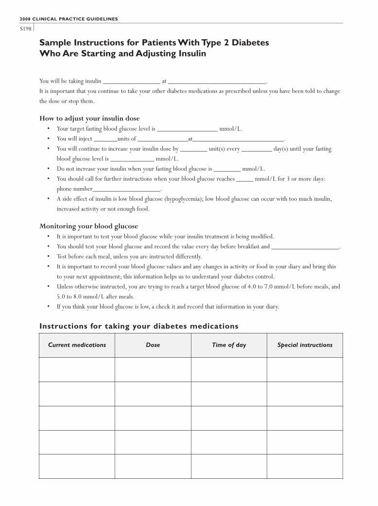
S198
Sample Instructions for Patients With Type 2 Diabetes Who Are Starting and Adjusting Insulin
You will be taking insulin _________________ at _____________________________.
It is important that you continue to take your other diabetes medications as prescribed unless you have been told to change
the dose or stop them.
How to adjust your insulin dose• Your target fasting blood glucose level is __________________ mmol/L.
• You will inject _______units of _______________at___________________________.
• You will continue to increase your insulin dose by ________ unit(s) every _________ day(s) until your fasting
blood glucose level is _____________ mmol/L.
• Do not increase your insulin when your fasting blood glucose is ________ mmol/L.
• You should call for further instructions when your blood glucose reaches _____ mmol/L for 3 or more days:
phone number____________________.
• A side effect of insulin is low blood glucose (hypoglycemia); low blood glucose can occur with too much insulin,
increased activity or not enough food.
Monitoring your blood glucose• It is important to test your blood glucose while your insulin treatment is being modified.
• You should test your blood glucose and record the value every day before breakfast and ____________________.
• Test before each meal, unless you are instructed differently.
• It is important to record your blood glucose values and any changes in activity or food in your diary and bring this
to your next appointment; this information helps us to understand your diabetes control.
• Unless otherwise instructed, you are trying to reach a target blood glucose of 4.0 to 7.0 mmol/L before meals, and
5.0 to 8.0 mmol/L after meals.
• If you think your blood glucose is low, a check it and record that information in your diary.
2008 CLINICAL PRACTICE GUIDELINES
Instructions for taking your diabetes medications
Current medications Dose Time of day Special instructions

S199
2008 CLINICAL PRACTICE GUIDELINES
Appendix 4
Rapid Screening for Diabetic Neuropathy
Multiple screening methods are published.These methods (1) are designed to screen for the presence or absence of diabetic neuropathy, as opposed to screening for specific sites on the feet that are at risk of ulceration (multisite testing). If neuropathy is identified by either of these methods, other sites may be tested to identify high-risk areas for ulceration.
1. Show the 10-g Semmes-Weinstein monofilament to thepatient.
2. Touch it first to the patient’s forehead or sternum so thatthe sensation is understood.
3. Instruct the patient to say “yes” every time the monofila-ment stimulus is perceived.
4. With the patient’s eyes closed, apply the monofilament tothe dorsum of the great toe proximal to the nail bed asshown in the illustration below. Use a smooth motion –touch the skin, bend the filament for a full second, then lift from the skin.
5. Perform this stimulus 4 times per foot in an arrhythmicmanner so the patient does not anticipate when the stimulus is to be applied.
6. Add up all correct stimuli for a score out of 8. A score of7 or 8 correct responses likely rules out the presence ofneuropathy.
1. Perkins BA, Olaleye D, Zinman B, et al. Simple screening tests for peripheral neuropathy in the diabetes clinic. Diabetes Care.2001;24:250-256.
Rapid Screening for Diabetic Neuropathy Using the 128-Hz Vibration Tuning Fork (The “On-Off” Method)
1. Strike the tuning fork against the palm of your hand hardenough that it will vibrate for approximately 40 seconds.
2. Apply the base of the tuning fork to the patient’s fore-head or sternum and ensure that the vibration sensation(not just the touch sensation) is understood.
3. With the patient’s eyes closed, apply the tuning fork tothe bony prominence situated at the dorsum of the firsttoe just proximal to the nail bed. Ask if the vibration sen-sation is perceived.
4. Ask the patient to tell you when the vibration stimulus is stopped, and then dampen the tuning fork with yourother hand.
5. One point is assigned for each vibration sensation per-ceived (vibration “on”). Another point is assigned if thecorrect timing of dampening of the vibration is perceived(vibration “off ”).
6. Repeat this procedure again on the same foot, then twiceon the other foot in an arrhythmic manner so the patientdoes not anticipate when the stimulus is to be applied.
7. Add up all correct stimuli for a score out of 8. A score of7 or 8 correct responses likely rules out the presence ofneuropathy.
Rapid Screening for Diabetic Neuropathy Using the 10-g Semmes-Weinstein Monofilament
AP
PE
ND
ICE
S

S200
2008 CLINICAL PRACTICE GUIDELINES
Appendix 5
Diabetes and Foot Care:A Patient’s Checklist
Many people with diabetes have problems with their feet. Ask your doctor to explain your risk factors for foot problems.You canprevent serious foot problems by following these basic guidelines.
DO… DON’T……check your feet every day for cuts, cracks, bruises, blisters,sores, infections or unusual markings.
…use a mirror to see the bottom of your feet if you can’t liftthem up.
…check the colour of your legs and feet. If there is swelling,warmth or redness or if you have pain, see your doctor or footspecialist right away.
… clean a cut or scratch with a mild soap and water and cover with a dry dressing for sensitive skin.
…trim your nails straight across.
…wash and dry your feet every day, especially between thetoes.
…apply a good skin lotion every day on your heels and soles.Wipe off any excess lotion.
…change your socks every day.
…always wear a good supportive shoe.
…always wear professionally fitted shoes from a reputablestore. Professionally fitted orthotics may help.
…choose shoes with low heels (under 5 cm high).
…buy shoes in the late afternoon (since your feet swell slightlyby then).
…avoid extreme cold and heat (including the sun).
…exercise regularly.
…see a foot care specialist if you need advice or treatment.
…cut your own corns or calluses.
…treat your own in-growing toenails or slivers with a razor orscissors. See your doctor or foot care specialist.
…use over-the-counter medications to treat corns and warts.They are dangerous for people with diabetes.
…apply heat to your feet with a hot water bottle or electricblanket.You could burn your feet without realizing it.
…soak your feet.
…take very hot baths.
…use lotion between your toes.
…walk barefoot inside or outside.
…wear tight socks, garters or elastics, or knee highs.
….wear over-the-counter insoles – they can cause blisters ifthey are not right for your feet.
…sit for long periods of time.
…smoke.
Adapted with permission from: Casella A. Feeling well…diabetes and foot care, a patient’s checklist. Knowing Diabetes.© Diabetes Hamilton, 2002

S201
2008 CLINICAL PRACTICE GUIDELINES
Appendix 6
Diabetic Foot Ulcers: Essentials of Management
REFERENCES1. Lavery LA, Baranoski S,Ayello EA. Options for off-loading the
diabetic foot. Adv Skin Wound Care. 2004;17:181-186.2. Lipsky BA, Berendt AR, Deery HG, et al; for the Infectious
Diseases Society of America. Diagnosis and treatment of dia-betic foot infections. Clin Infect Dis. 2004;39:885-910.
3. Frykberg RG, Zgonis T,Armstrong DG, et al; for the American
College of Foot and Ankle Surgeons. Diabetic foot disorders.Aclinical practice guideline (2006 revision). J Foot Ankle Surg.2006;45(5 suppl):S1-S66.
4. Schultz GS, Barillo DJ, Mozingo DW, et al; for the Wound BedAdvisory Members.Wound bed preparation and a brief historyof TIME. Int Wound J. 2004;1:19-32.
1. Assess underlying cause(s): neuropathy and/or ischemia.
2. Ulcers should be probed with a blunt-tipped instrument to detect sinus tracks or palpable bone suggestive of deep infections.
3. Plantar-surface ulcers require pressure relief. Individuals with plantar-surface foot ulcers should be non-weight-bearing as muchas possible and utilize off-loading footwear or appliances (1).
4. Clinically noninfected ulcers do not routinely require cultures or antibiotics (2).
5. More serious infections in chronic foot ulcers tend to be polymicrobial and typically require empiric use of broad spectrumsystemic antibiotics as soon as possible. Antibiotics can be subsequently tailored according to culture and sensitivity results.Cultures obtained by curettage or biopsy tend to be more reliable than surface swabs (3).
6.Wound bed preparation involves debridement of necrotic tissue (neuropathic wounds and noncritical ischemic wounds only)and maintenance of adequate moist wound environment with appropriate wound dressings. Hydrogels are used to increasewound bed moisture in dry or minimally draining neuropathic ulcers. Dressings that provide therapeutic levels of ionic silver or iodine may reduce critical degrees of wound bacterial colonization (4).
7. Comorbidities need to be managed (e.g. hyperglycemia).
8. Refer to a specialized wound clinic where available.
AP
PE
ND
ICE
S