cancer research symposium - ontario veterinary college
TRANSCRIPT
12th Annual ICCI
Cancer Research Symposium
Tuesday May 28, 2019
OVC LLC 9:00-5:00
2
Introductory Remarks
Welcome to the 12th annual Guelph ICCI Cancer Symposium! This meeting is an opportunity to
bring together cancer researchers from across campus and regional collaborators. Topics range from
basic science through to clinical application. We are very grateful to the amazing group of speakers
and poster presenters who will be sharing their findings with us today. Dr. David Vail is the 2019
Arthur Willis Distinguished speaker and will be giving the keynote address at 3:00.
In the past 12 years we have seen relationships and collaborations develop that were made possible
by these interactions and we hope that this year’s meeting will spark new collaborations and ideas.
This symposium is made possible by funding from the Arthur Willis Visiting Professorship in Canine
Oncology and support from the OVC Dean’s office.
Drs Geoff Wood and Michelle Oblak
Pathobiology and Clinical Studies, University of Guelph
ICCI Assistant Co-Directors
Administrative Support and Research Funding:
Thanks to Dr. Kaya Skowronski from the ICCI tumor bank for huge help organizing this
symposium along with support from Barb Gaudette and Daphne Summers from the OVC Office of
the Dean, Hospitality Services for help with set up and refreshments, and Marina Kashevska-
Gozdek and Melanie Knapp for assistance with the program and organization throughout the day.
The research projects presented here and the trainees performing these studies were
collectively supported by grants, scholarships and contracts from: CIHR; NSERC; Terry Fox
Research Institute; OGS; Cancer Research Society; OVC Pet Trust Fund; Smiling Blue Skies
Cancer Fund; OVC Graduate Scholarship; Art Rouse Cancer Biology Graduate Stipend;
BioCanRx; Vanier Canada Graduate Scholarship; Brock Doctoral Scholarship; QEII-GSST,
Government of Ontario, Stem Cell Network, and the CanaQuest Medical Corporation Cancer
Research Program.

3
ICCI 12th
Annual Cancer Research Symposium, Tuesday May 28th, 2019
Morning Session: Room 1714, OVC LLC
9:00-9:05 Welcome and Introductory Remarks
9:05-9:35 Guest Speaker
Utilizing vascular normalization to increase the uptake, efficacy, and impact of cancer therapies Dr. Jim Petrik; Department of Biomedical Sciences, University of Guelph
9:35- 10:20 Short talks from abstracts
1. Viral sensitizer-mediated enhancement of oncolytic NDV leads to rapid clearance of primary
tumours in a mouse model of melanoma
Thomas McAusland; Department of Pathobiology, University of Guelph
2. Combining Decitabine with Oncolytic Virotherapy Preferentially Kills Acute Myeloid
Leukemia Cells Via Lethal Oxidative Stress
Elaine Klafuric; Department of Pathobiology, University of Guelph
3. Investigating the potential to target colorectal cancer-linked Fusobacterium nucleatum with
Bdellovibrio and like organisms
Avery Robinson; Department of Molecular and Cellular Biology, University of Guelph
10:20-10:45 Coffee Break and Poster Viewing Room 1707 B & C, OVC LLC
10:45-11:30 Short talks from abstracts 1. Analysis of SNARE Regulation During Tumor Cell Invasion
Megan Brasher; Department of Molecular and Cellular Biology, University of Guelph
2. Characterization of extracellular vesicles obtained from cultured explants of canine
osteosarcoma and normal bone
Dr. Alicia Viloria-Petit; Department of Biomedical Sciences, University of Guelph
3. Lymphoma and Symmetric Dimethylarginine Concentraion in Dogs
Dr. Anthony Abrams-Ogg, Department of Clinical Studies, University of Guelph
11:30-12:20 Regional Keynote Speaker
Regulation of the Hippo pathway by AMPK family kinases in cancer
Dr. Liliana Attisano; Department of Biochemistry, University of Toronto
12:20- 1:30 Poster Session and Lunch Room 1707 B & C, OVC LLC
4
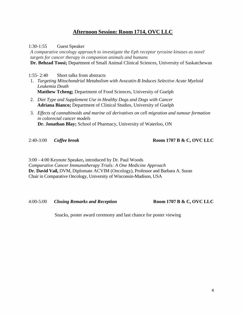
Afternoon Session: Room 1714, OVC LLC
1:30-1:55 Guest Speaker
A comparative oncology approach to investigate the Eph receptor tyrosine kinases as novel
targets for cancer therapy in companion animals and humans
Dr. Behzad Toosi; Department of Small Animal Clinical Sciences, University of Saskatchewan
1:55- 2:40 Short talks from abstracts
1. Targeting Mitochondrial Metabolism with Avocatin-B Induces Selective Acute Myeloid
Leukemia Death
Matthew Tcheng; Department of Food Sciences, University of Guelph
2. Diet Type and Supplement Use in Healthy Dogs and Dogs with Cancer
Adriana Bianco; Department of Clinical Studies, University of Guelph
3. Effects of cannabinoids and marine oil derivatives on cell migration and tumour formation
in colorectal cancer models
Dr. Jonathan Blay; School of Pharmacy, University of Waterloo, ON
2:40-3:00 Coffee break Room 1707 B & C, OVC LLC
3:00 - 4:00 Keynote Speaker, introduced by Dr. Paul Woods
Comparative Cancer Immunotherapy Trials: A One Medicine Approach
Dr. David Vail, DVM, Diplomate ACVIM (Oncology), Professor and Barbara A. Suran
Chair in Comparative Oncology, University of Wisconsin-Madison, USA
4:00-5:00 Closing Remarks and Reception Room 1707 B & C, OVC LLC
Snacks, poster award ceremony and last chance for poster viewing

5
KEYNOTE PRESENTATION
3:00 OVC LLC Room 1714
Dr. David M. Vail, DVM, Diplomate ACVIM (Oncology) Professor and Barbara A. Suran Chair in Comparative Oncology University of Wisconsin-Madison
Comparative Cancer Immunotherapy Trials: A One
Medicine Approach
Although immunotherapy is becoming one of the cornerstones of modern cancer therapy, the
majority of patients still fail to experience durable responses. This is further compounded by our
inability to predict or assess response owing to unusual response patterns unique to
immunotherapy. Recently, the scientific community has begun to explore the possibility that the
inclusion of companion species (companion dogs) in clinical trials of novel immunotherapeutic
agents, combinations of agents, and the assessment of response to immunotherapy may hold
promise. This stems from the fact that companion dogs have intact immune systems and
heterogenous tumour/tumour microenvironments which may better recapitulate the human
condition. Several examples of current cancer immunotherapy trials in pet dogs will be presented
to illustrate potential advantages and pitfalls of the comparative approach.
Dr. Vail received his DVM from the University of Saskatchewan in 1984 and subsequently
completed an internship in small animal medicine and surgery at Colorado State University prior
to practicing in his native Edmonton for two years. He followed up with a residency in Medical
Oncology at the Animal Cancer Center at Colorado State University, completed in 1990. He is
currently Professor and Barbara A. Suran Chair in Comparative Oncology at the University of
Wisconsin-Madison and a member of the UW Carbone Comprehensive Cancer Center. Dr. Vail
has published over 150 peer-reviewed scientific manuscripts and 50 book chapters in the field of
veterinary and comparative oncology. David is co-editor of the textbook Small Animal Clinical
Oncology. He has served in the past as President of the Veterinary Cancer Society, the Canine
Comparative Oncology and Genomics Consortium (CCOGC), Chairman of the Scientific
Advisory Boards for both the Morris Animal Foundation and the American College of Veterinary
Internal Medicine Foundation, and North American journal editor for Veterinary and Comparative
Oncology. He is a founding member of the Comparative Oncology Trials consortium. Dr. Vail
has been honored as the recipient of both the Mark L. Morris Sr. Distinguished Research Award
and the Pfizer Award for Veterinary Research Excellence.

6
Past ICCI Symposium Arthur Willis Distinguished Speakers
2018 Daniel Gustafson 2013 David Argyle
2017 William Eward 2012 Timothy Fan
2016 Jaime Modiano 2011 Cheryl London
2015 Nicola Mason 2010 Matthew Breen
2014 Deborah Knapp 2009 Barbara Kitchell

7
GUEST SPEAKER:
9:05-9:35
Utilizing vascular normalization to increase the uptake, efficacy, and impact of cancer
therapies
Dr. Jim Petrik; Department of Biomedical Sciences, University of Guelph
Tumors often initiate an aggressive program of angiogenesis in order to accommodate the oxygen and
metabolic needs of rapidly-growing tissue. As a result of the strong pro-angiogenic stimulus, blood vessels
form very rapidly and often are disorganized, immature, and dysfunctional. This dysfunctional vasculature
reduces perfusion and leads to areas of hypoxia and elevated interstitial fluid pressure. All of these
characteristics impede drug delivery to the tumor, significantly reducing the efficacy of anti-cancer therapy
and necessitating the administration of high levels of compound in order to have some uptake in to the
tumor. We have developed an approach to target this immature, dysfunctional tumor vasculature through
the use of the type I repeat region (3TSR) of thrombospondin-1 (TSP-1). TSP-1 is a large matricellular
glycoprotein with potent endogenous anti-angiogenic activity. We have shown previously that 3TSR
specifically targets the immature tumor vasculature in primary and metastatic ovarian tumors. 3TSR also
causes apoptotic tumor cell death. As a result of this bimodal function, 3TSR stimulates regression of
advanced stage ovarian cancer and induces tumor shrinkage, enhances vascular perfusion, and reduces
tumor hypoxia. Our previous data with 3TSR has shown that vascular normalization can enhance the uptake
and efficacy of a number of different therapeutic compounds and immune cells. However, the small size
of 3TSR results in rapid clearance from circulation and a serum half-life of approximately 14hrs. Here, we
discuss the development of a Fc fusion protein (Fc3TSR), which links two 3TSR molecules with a human
IgG protein. The half-life of Fc3TSR is approximately 8 days, which makes the compound more attractive
clinically. In addition to increased time in circulation, Fc3TSR also has better in vitro efficacy, potentially
due to enhancing clustering of its receptor CD36. This presentation will focus on the use of vascular
normalization to improve anti-cancer therapy and the development of Fc3TSR which we hope to take to
the clinic, initially for the treatment of advanced stage ovarian cancer.
REGIONAL KEYNOTE SPEAKER:
11:30-12:20
Inhibition of the Hippo pathway by AMPK-family kinases
Dr. Liliana Attisano; Donnelly Centre and Department of Biochemistry, 160 College Street, University
of Toronto
The Hippo signalling pathway is a key regulator of tissue growth and organogenesis. The pathway
is comprised of a core MST/LATS kinase cassette that phosphorylates and promotes cytoplasmic
localization of the transcriptional regulators, TAZ and YAP. Inactivation of the Hippo pathway is
a common feature in numerous cancers, yet mutations in pathway components are relatively rare.
To uncover novel Hippo pathway regulators, we conducted multidimensional high throughput
screens. These efforts uncovered two AMPK family kinases, MARK4 and NUAK2 as negative
regulators of the Hippo pathway. MARK kinases, including MARK3 and MARK4, phosphorylate
8
both SAV1 and MST1/2 and inhibit MST1/2-dependent activation of LATS. Moreover, we
showed that DLG5, acts as a scaffold to promote MARK-mediated phosphorylation of MST. In
contrast to MARKs, the AMPK family member, NUAK2 interacts with and phosphorylates LATS.
Interestingly, NUAK2 is induced by YAP/TAZ in cooperation with AP-1 and this is required for
robust YAP/TAZ signalling. Inhibition or loss of NUAK2 reduces the growth of cultured cancer
cells and mammary tumors in mice. In human patient samples, NUAK2 expression is elevated in
aggressive, high grade bladder cancer and strongly correlates with a YAP/TAZ gene signature.
Thus, we identified a positive feed forward loop in the Hippo pathway that establishes a key role
for NUAK2 in enforcing the tumour promoting activities of YAP/TAZ. Developing inhibitors for
therapeutic applications and understanding how these AMPK family members act to regulate the
Hippo pathway in distinct physiological contexts and their impact in human disease are
outstanding questions.
GUEST SPEAKER:
1:30-1:55
A comparative oncology approach to investigate the Eph receptor tyrosine kinases as novel
targets for cancer therapy in companion animals and humans
Dr. Behzad Toosi; Department of Small Animal Clinical Sciences, University of Saskatchewan
Naturally occurring tumors that develop in companion animals are unfortunate for both the animal
and the pet owner but represent an overlooked opportunity to facilitate the search and discovery
of new diagnostics and therapies for human malignancies. Testing of new therapies in animal
models that better represent human disease, such as naturally occurring tumors in pet dogs, has the
capacity to reduce the time for clinical development of new pharmaceutical agents for human
cancer therapy. At the same time, there exists an opportunity to bring the novel and advanced
diagnostics and therapeutics from human drug discovery and pharmaceutical research to veterinary
medicine. Our team is focused on identification of new cancer biomarkers of common canine and
human neoplasms and on the validation of spontaneous canine models of human malignancies
based on the role of receptor tyrosine kinases.
Protein kinases have been commonly identified as oncogenes because various cancers show a high
frequency of activating mutations in the genes encoding these proteins. Accordingly, receptor
tyrosine kinases represent promising targets for the development of new cancer therapies that
include monoclonal antibodies and small molecule inhibitors. The Eph family of receptor tyrosine
kinases with 14 members are often overexpressed in several human malignancies and their
function has been associated with tumor growth, invasiveness and metastasis. Our laboratory
investigates multiple canine spontaneous models of human cancers in terms of Eph receptor
expression, signaling mechanisms and function, and the regulation of response to treatment to
identify new diagnostics and therapies that will benefit both companion animals and humans.
9
SHORT TALKS FROM SUBMITTED ABSTRACTS
9:35-10:20 Morning Session
Viral sensitizer-mediated enhancement of oncolytic NDV leads to rapid clearance of
primary tumours in a mouse model of melanoma
Thomas M. McAusland, Jacob P. van Vloten, Lisa A. Santry, Joelle C. Ingrao, Matthew M.
Guilleman, Amira D. Rghei, Leo Susta, Khalil Karimi, and Byram W. Bridle and Sarah K.
Wootton
Department of Pathobiology, University of Guelph, Guelph, Ontario
The avian paramyxovirus, Newcastle disease virus (NDV), is a potent oncolytic virus that has been
shown to be safe and effective in a variety of preclinical cancer models as well as in human clinical
trials. NDV preferentially replicates in and lyses tumour cells while sparing normal cells. In
addition, NDV possesses strong immunostimulatory properties that can overcome cancer-induced
immunosuppression and generate effective anti-tumour immune responses. The oncolytic efficacy
of NDV is negatively impacted by tumours that retain intact antiviral signalling and sensing
capabilities. The objective of this research was to evaluate the use of viral sensitizer-mediated
combination therapy to enhance the anti-neoplastic efficacy of NDV. Here, we demonstrate that
treatment with a combination of NDV and viral sensitizer causes rapid regression and clearance of
B16-F10 tumours following intratumoral injection. In addition, we determined that the anti-tumour
efficacy of this combination therapy is reliant upon the actions of natural killer (NK) cells as
antibody depletion of NK cells abrogated therapeutic efficacy in this model. This viral sensitizer
does not act to increase virus replication, but rather acts as an immune-potentiator to activate cells
of the innate immune system to rapidly clear primary tumours. Although this treatment strategy is
reliant on the activity of NK cells, it is suspected that type I IFN also plays an important significant
role and we are currently investigating this mechanism. Taken together, these results suggest that
combining NDV with a viral sensitizer alerts the innate immune system to the presence of primary
tumours, and this in turn facilitates rapid tumor clearance.
Funding: OVC Scholarship
10
Combining Decitabine with Oncolytic Virotherapy Preferentially Kills Acute Myeloid
Leukemia Cells Via Lethal Oxidative Stress
Elaine M. Klafuric1,*, Megan R. Strachan-Whaley1,*, Lisa Santry1, Amanda W.K. AuYeung1,
Jacob P. van Vloten1, Robert C. Mould1, Thomas McAusland1, Khalil Karimi1, Anthony J.
Mutsaers2,3, Sarah K. Wootton1 and Byram W. Bridle1 1Departments of Pathobiology, Ontario Veterinary College, University of Guelph 2Department of Biomedical Sciences, Ontario Veterinary College, University of Guelph 3Department of Clinical Studies, Ontario Veterinary College, University of Guelph *contributed equally
Acute myeloid leukemias (AML) are aggressive hematological cancers for which the standard of
care has limited efficacy, with high rates of relapse. The DNA methyltransferase inhibitor
decitabine is an epigenetic modifier in clinical trials to treat leukemias, albeit with limited efficacy.
Oncolytic viruses (OVs) preferentially replicate in and kill cancer cells but perform poorly against
leukemias that are spread throughout normal tissues that can quench viral infections. However, we
discovered that combining decitabine with OVs induced durable remissions and resistance to
relapse in mouse models of acute T- and B-lymphocytic leukemias. Therefore, we hypothesized
that treatment with decitabine would sensitize AML cells to killing by oncolytic Newcastle disease
virus (NDV). In vitro resazurin dye-based assays supported the hypothesis. Further, most mice
challenged with C1498 AML cells and treated with decitabine seven and eight days later, followed
by NDV eleven days post-challenge, achieved durable remissions and resisted a homologous re-
challenge. Co-administration of the pan-reactive oxygen species (ROS) scavenger N-acetyl-L-
cysteine abrogated efficacy. This implicated induction of lethal oxidative stress as a mechanism of
action. Flow cytometric detection of ROS suggested that decitabine and NDV caused oxidative
stress in leukemia cells, with the combination therapy having an additive effect. More specific
reagents, such as dihydroethidium and mitoSOX, will be used to quantify cytoplasmic versus
mitochondrial ROS, like superoxide. Targeted ROS inhibitors will be employed to confirm which
subtypes of ROS are involved. In conclusion, treatment with clinically-approved decitabine
followed by NDV appears to be effective at preferentially killing AML cells via oxidative stress.
Funding: Operating funds were jointly provided by the Canadian Cancer Society Research
Institute and Canadian Institutes of Health Research-Institute of Cancer Research via an
Innovation Grant to BWB. Stipend funding: Ontario Veterinary College (OVC) Graduate
Scholarship (EMK); Canadian Institutes of Health Research Graduate Scholarship (Masters
Award) and OVC Pet Trust Scholarship (AWKAY); Ontario Graduate Scholarship and OVC
Graduate Scholarship (JPvV); OVC Pet Trust Scholarship (RCM); OVC Graduate Scholarship
(TM).
11
Investigating the potential to target colorectal cancer-linked Fusobacterium nucleatum with
Bdellovibrio and like organisms
A. Robinson1, W. Mun2, R. J. Mitchell2, E. Allen-Vercoe*1 1Department of Molecular and Cellular Biology, College of Biological Science, University of
Guelph 2Department of Biological Sciences, School of Life Sciences, Ulsan National Institute of Science
and Technology
The etiology of colorectal cancer (CRC) is highly complex: most cases of CRC are sporadic,
diagnosed in patients with no family history or genetic predisposition. One contributing factor to
sporadic CRC development is the human microbiome. Fusobacterium nucleatum is a Gram-
negative bacterium typically found as an oral commensal species. Yet, F. nucleatum has been
isolated at high abundance from cancerous colon mucosae and thus linked to CRC. Bdellovibrio
and like organisms (BALOs) are relatively small bacteria that hunt and prey upon other Gram-
negative bacterial cells. BALOs present an attractive potential application in human disease as [1]
BALOs do not damage human cells and [2] BALOs are proficient at invading and disintegrating
bacterial biofilms, penetrating through sessile bacterial cells far more efficiently than antibiotics
or bacterial viruses—bacteriophages. As BALOs are predatory bacteria that feed upon other Gram-
negative bacteria, BALOs pose an attractive potential application in eradicating virulent strains of
F. nucleatum. The current research serves to shed more light on the effect of different
environmental conditions during BALO-F. nucleatum co-incubation, and the range of F.
nucleatum which BALOs may predate. F. nucleatum exhibits a high degree of intraspecies
variation; not all strains of F. nucleatum are virulent. As such, investigating the predatory-prey
relationship between BALOs and F. nucleatum must include the range of potential Fusobacterium
prey strains. Additionally, the anaerobic environment of F. nucleatum is not propitious for aerobic
BALOs. As such, predation co-bacterial incubation assays must also be optimized. This
exploratory investigation will further elucidate the potential for BALO-based targeting of virulent
human-associated F. nucleatum strains.
Funding: Canadian Cancer Society CCSRI, Cancer Research UK, CIHR Canada Graduate
Scholarship – Masters Award
12
10:45-11:30 Second Morning Session
Analysis of SNARE Regulation During Tumor Cell Invasion
M. Brasher1, D. Martynowicz1, O. Grafinger1, M. Marchment1, R. Shannon1, A. Hucik1, E.
Shanks-Skinner1 and M. Coppolino1. 1Department of Molecular and Cellular Biology, Biological Sciences, University of Guelph
Tumor cell invasion involves targeted localization of proteins required for interactions with the
extracellular matrix and for proteolysis. The localization of many proteins during these cell-
extracellular matrix interactions relies on membrane trafficking mediated in part by SNAREs. The
SNARE protein syntaxin4 (Stx4) is involved in the formation of invasive structures called
invadopodia; however, it is unclear how Stx4 function is regulated during tumor cell invasion.
Munc18c is a known regulator of Stx4 activity, and here we show that Munc18c is required for
stx4-mediated invadopodium formation and invasion. Biochemical and microscopic analyses
revealed a physical association between Munc18c and Stx4, which was enhanced during
invadopodium formation. It was also found that an N-terminal Stx4-derived peptide associates
with Munc18c and inhibits endogenous interactions of Stx4 with SNAP23 and VAMP2.
Furthermore, expression of the Stx4 N-terminal peptide, which consists of residues 1-29 of Stx4,
decreased invadopodium formation and cell invasion in vitro. A smaller peptide that consisted of
residues 1-15 of Stx4 was also found to decrease invadopodium formation, cell migration and cell
invasion. Of note, cells expressing the Stx4 N-terminal peptide 1-29 residues exhibited impaired
trafficking of membrane type 1 matrix metalloproteinase (MT1-MMP) and EGF receptor (EGFR)
to the cell surface during invadopodium formation. Further work was done to determine VAMP2’s
role during invadopodium formation. In both VAMP2 knockdown experiments and inhibition
experiments, invadopodium formation and local cell invasion was decreased. This work further
advances our understanding of the role of SNARE function in the localization of proteins that drive
tumor cell invasion.
Funding: CRS and NSERC
Characterization of extracellular vesicles obtained from cultured explants of canine
osteosarcoma and normal bone
Alicia M. Viloria-Petit1*, Mackenzie Wong1, Anita K. Luu1, Michelle Oblak2, Brigitte Brisson2,
Geoffrey Wood3 1Department of Biomedical Sciences, Ontario Veterinary College, University of Guelph 2Department of Clinical Studies, Ontario Veterinary College, University of Guelph 3Department of Pathobiology, Ontario Veterinary College, University of Guelph
Osteosarcoma (OSA) is the most common bone tumour in dogs and humans. OSA often
metastasizes to the lungs in both species, and this is responsible for patient mortality. There are no
reliable biomarkers to predict metastatic relapse in OSA. Extracellular vesicles (EVs) are release
by tumour cells into blood circulation and contain cargo that reflects their cell of origin. The goal
of this research was to develop an explant culture protocol to isolate and characterize EVs and
their protein cargo in canine OSA tumour tissue, for subsequent identification of
prognostic/predictive signatures that could be assessed non-invasively in plasma of OSA patients.

13
Tumour and normal bone (NB) samples were obtained from canine OSA patients. Tissue was
processed and incubated in culture media containing 5% EV-free FBS and antibiotics under
standard conditions. After 24 hours, media was recovered, centrifuged, and stored. EVs were
isolated from media via size exclusion chromatography, and next characterized via
immunoblotting, transmission electron microscopy (TEM), and particle tracking analysis (PTA).
Protein cargo was assessed by ultra high performance liquid chromatography tandem mass
spectrometry (uHPLC MS/MS). EVs were shown to express flotillin and/or CD63 and PTA
showed different size distribution in OSA versus NB, suggesting different EV populations. TEM
revealed a predominance of cup-shaped EVs of 50-200 nm diameter in OSA samples. MS analysis
identified 354 distinct proteins in OSA as compared to NB EVs. OSA EVs were enriched in
proteins involved in protein translation, a number of which were reported to drive OSA
progression. Our results indicate that tissue explant cultures are useful tools for the identification
of prognostic/predictive EV signatures in canine OSA.
Funding: OVC Pet Trust, OVC Summer Assistantship, University of Guelph Graduate Tuition
Scholarship, OGS.
Lymphoma and Symmetric Dimethylarginine (SDMA) Concentration in Dogs
A. Abrams-Ogg1, Bronwyn Rutland2, Phillippe Levis2, Vicky Sabine3, Kaya Skowronski3,
Allison Majeed3, Dorothee Bienzle4, Alex Zur Linden3, Danielle Richardson5, Anthony
Mutsaers6, Paul Woods3
1Department of Clinical Studies, Ontario Veterinary College, University of Guelph 2VCA Canada 404 Veterinary Emergency and Referral Hospital, Newmarket, ON, Canada 3Department of Clinical Studies, Ontario Veterinary College, University of Guelph 4Department of Pathobiology, University of Guelph 5Animal Cancer Centre, Ontario Veterinary College, Guelph, ON 6Department of Biomedical Sciences, Ontario Veterinary College, Guelph
SDMA and creatinine were measured in 52 dogs with untreated LSA, LSA in CR or PR following
CHOP chemotherapy, and LSA in relapse. Mean, median and age range were 8, 7 and 3-13 years,
respectively. Fifty dogs had WHO Stage III-V multicentric nodal LSA, one dog had mediastinal
LSA, and one dog had bilateral renal LSA. No dog had clinical, clinicopathologic or sonographic
signs of pre-existing kidney disease when diagnosed with LSA. Results are:
Status N SDMA Creatinine SDMA:Creatinine
mean, median mean, median mean, median
(range) (range) (range)
[% > RI 0-14 ug/dL ] [% > RI 44-133 umol/L] ______
Untreated 36 18, 16 97, 81 0.21, 0.19
(5 – 90) (47 – 313) (0.06 – 0.53)
[72%] [12%]
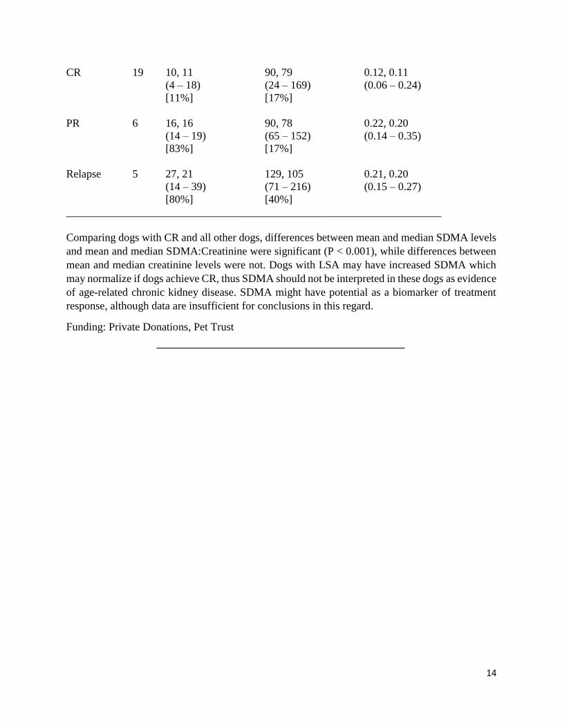
14
CR 19 10, 11 90, 79 0.12, 0.11
(4 – 18) (24 – 169) (0.06 – 0.24)
[11%] [17%]
PR 6 16, 16 90, 78 0.22, 0.20
(14 – 19) (65 – 152) (0.14 – 0.35)
[83%] [17%]
Relapse 5 27, 21 129, 105 0.21, 0.20
(14 – 39) (71 – 216) (0.15 – 0.27)
[80%] [40%]
____________________________________________________________________
Comparing dogs with CR and all other dogs, differences between mean and median SDMA levels
and mean and median SDMA:Creatinine were significant (P < 0.001), while differences between
mean and median creatinine levels were not. Dogs with LSA may have increased SDMA which
may normalize if dogs achieve CR, thus SDMA should not be interpreted in these dogs as evidence
of age-related chronic kidney disease. SDMA might have potential as a biomarker of treatment
response, although data are insufficient for conclusions in this regard.
Funding: Private Donations, Pet Trust
15
1:55-2:40 Afternoon Session
Targeting Mitochondrial Metabolism with Avocatin-B Induces Selective Acute Myeloid
Leukemia Death
M. Tcheng1, K. Rea 2, T. Akhtar2, V. Cunha3, AW Mohsen4, J. Vockley4, PA Spagnuolo1* 1Department of Food Sciences, Ontario Agricultural College, University of Guelph 2Department of Molecular and Cellular Biology, College of Biological Sciences, University of
Guelph 3Department of Chemistry, Faculty of Science, University of Alberta 4Department of Medical Genetics, Center for Rare Disease Therapy, University of Pittsburgh
Medical Center
Acute myeloid leukemia (AML) is an aggressive hematological malignancy characterized by high
relapse and low survival rates, driven by cancer cell resistance to induction therapy, the primary
anti-AML regimen. Unlike normal hematopoietic stem cells, the leukemic population exhibits an
altered mitochondrial phenotype characterized by increased dependence on fatty acid oxidation
(FAO), marking the leukemic mitochondria as an attractive pharmacological target. Previously,
avocatin-B, a mixture of two avocado-derived fatty alcohols avocadyne and avocadene, was shown
to targeted mitochondrial respiration to induce selective leukemic apoptosis. In the current project,
avocadyne (AYNE), a long chain, odd numbered, acetylenic fatty alcohol, was the most potent
FAO inhibitor towards AML cell lines and patient samples, while sparing healthy donor samples.
AYNE alone showed potent anti-AML activity in vivo, significantly reducing the amount of
leukemic cells in xenograft studies. The odd numbered carbon chain, terminal triple bond, and
stereochemistry of the hydroxyl groups were critical AYNE’s ability to directly inhibit FAO,
hinder mitochondrial respiration, and induce selective leukemic death. The next objective will
elucidate AYNE’s molecular target; AYNE resistant cell lines will be characterized by proteomics
and immunoblotting to determine the altered expression of FAO enzymes. Lentiviral knockdown
of intramitochondrial FAO enzymes with altered expression, co-immunoprecipitation of
intramitochondrial FAO enzymes from treated leukemic cells, and kinetic assays will determine
AYNE’s molecular target. Completion of these objectives is critical to our long term goal of
developing AYNE into a novel clinical therapeutic that exploits the altered mitochondrial
metabolism specific to AML.
Funding Source: Queen Elizabeth II Graduate Scholarship in Science and Technology (QEII-
GSST)
Diet Type and Supplement Use in Healthy Dogs and Dogs with Cancer
A.V. Bianco1, S. Abood1, A. Mutsears1,2, J.P. Woods1,3, J. Coe4, A. Verbrugghe*1
1Department of Clinical Studies, Ontario Veterinary College, University of Guelph 2Department of Biomedical Sciences, Ontario Veterinary College, University of Guelph 3Mona Campbell Centre for Animal Cancer, University of Guelph, Canada 4Department of Population Medicine, Ontario Veterinary College, University of Guelph
16
Nutritional supplements are commonly fed to dogs with cancer, many report doing so because of
a cancer diagnosis, however no study has compared nutritional supplement use between dogs with
cancer and healthy dogs. The aim of this study is to determine if differences exist in the
diet/supplement types fed to dogs with cancer and healthy dogs. An online survey containing 51
questions was administered using the Qualtrics Research Suite (Qualtrics, Provo, Utah, USA).
It was distributed among clients at the Ontario Veterinary College and through social media. Data
was analysed in SPSS (IBM SPSS Statistics for Macintosh, Version 25). Chi-squares and odds
ratios were used to compare categorical data. 353 surveys were analyzed. Commercial dry food
was the most frequently reported diet fed to both groups but owners of healthy dogs (n=221) were
four times more likely to feed commercial dry food compared to owners of dogs with cancer
(n=132) (p=2.37x10⁻⁹). Owners of dogs with cancer were two times more likely to feed a
homemade raw diet (p=0.0121) and four times more likely to feed a homemade cooked diet
(p=3.35x10⁻⁸). A variety of supplements were reported, such as multivitamin/minerals, cannabidiol
(CBD) oil, turmeric/curcumin, and mushroom supplements. Owners of dogs with cancer were five
times more likely to feed CBD oil (p=1.04x10⁻⁴), seven times more likely to feed
turmeric/curcumin (p=1.18x10⁻⁴) and 19 times more likely to feed mushroom supplements
(p=2.17x10⁻⁷). These results suggest owners of dogs with cancer make different dietary decisions
for their dogs compared to owners of healthy dogs.
Funding: OVC Scholarship, OGS Scholarship
Effects of cannabinoids and marine oil derivatives on cell migration and tumour formation
in colorectal cancer models
J. Fux1, J.Blay1,2 1School of Pharmacy, Faculty of Science, University of Waterloo 2Department of Pathology, School of Medicine, Dalhousie University
The cannabis plant is a rich source of bioactives that has been inadequately studied for medical
research purposes due to its past status as the source of a controlled substance. That changed in
2018 with the legalization of cannabis in Canada for both medical and recreational use and
increased attention in Canadian industry for the potential of formulations that might be used for
purposes of health support. In particular, the non-psychoactive cannabis constituent cannabidiol
(CBD) is of great interest for possible use in circumstances that involve altered activities in host
defence by disease changes in the immune and inflammatory systems. Since 2017 we have been
engaged in research to examine the potential for cannabinoids to alter the behaviour of cancer cells
in a way that will benefit patients at risk of or during the progression of cancer. There have been
indications that certain cannabinoids may have anticancer activities, but although cannabinoid-
based synthetics seem to have potential for direct killing, natural cannabinoids have been shown
not to have a significant ability for cytotoxicity. Given that cancer cells acquire or ‘hijack’ many
17
of the pathways used by cells of the immune system, metastatic change is a logical direction to
explore for thee natural cannabinoids. We have examined the potential for CBD and other
cannabinoids to alter cell migration and homing pathways, which in cancer form the basis of its
spread or metastasis. We describe here some of our recent findings and approaches using
preclinical models including transwell-based migration and invasion assays and a model of
extravasation and metastasis based upon the chick embryo vasculature. We report our results
following exposure of colorectal carcinoma cells either to CBD alone or in combination with
products derived from marine oils, particularly those from algae, which may have unique abilities
in this context. This research will build on our scientific understanding of the potential benefits of
oils extracted from cannabis and algae, particularly in the context of neoplastic disease, and may
help add to the economic impact of cannabis deregulation in Canada by enabling progress through
industry.
Funding: CanaQuest Medical Corporation Cancer Research Program.
18
LUNCH & POSTER SESSION 12:20-1:30
OVC LLC Room 1707 B & C
Posters will be displayed all day; authors please attend your posters from at
least 12:45-1:15. Judges for the Poster Competition will be evaluating posters
during this time.
POSTER ABSTRACTS
1) Investigating the effects of Hyperthermia and Heat Shock Protein 70 on microRNA
biogenesis
L. Abou Zeid1, R. Mosser1
1Department of Molecular and Cellular Biology, College of Biological Sciences, University of
Guelph
Heat shock, a form proteotoxic stress, is a major trigger of apoptosis. Molecular chaperones, such
as Heat Shock proteins 70 allow for the refolding of misfolded proteins, maintaining cellular
proteostasis. Additionally, HSP70 allows for cell survival following hyperthermia through
preventing the activation of caspases, which are proteases responsible for initiating apoptosis.
Along with disrupting proteostasis, hyperthermia can also influence gene expression through
altering microRNA (miRNA) levels and expression patterns. These small non-coding RNA can
silence the expression of target genes through interacting with their target mRNAs, leading to their
translational repression. MiRNAs are generated through a series of processing steps. The primary
miRNA transcript (pri-miRNA) is processed into a shorter precursor miRNA transcript (pre-
miRNA) by the ribonuclease Drosha and its partner DGCR8. The pre-miRNA is then converted to
the mature miRNA species by the ribonuclease Dicer and its binding protein, TRBP. The goal of
my research is to examine the effect of hyperthermia on miRNA processing and the potential role
of HSP70 in protecting the miRNA processing machinery in stressed cells. We were able to
demonstrate a disruptive effect of heat stress on miRNA processing as it resulted in the caspase-
mediated cleavage and degradation of Drosha, DGCR8, Dicer and TRBP. We also observed a
protective effect of HSP70 on miRNA processing under proteotoxic stress, as it was able to prevent
the cleavage of the miRNA processing proteins. We are currently investigating the effect of
hyperthermia on the function of the core miRNA processing proteins and the resulting alterations
in miRNA processing.
Funding: Natural Sciences and Engineering Research Council of Canada Discovery Grant
19
2) Low oxygen influences the composition of human ribosomes and alternative splicing of
ribosomal protein genes
Andrea Brumwell1, Lindsay Obress1, Jim Uniacke*1
1Department of Molecular and Cellular Biology, College of Biological Science, University of
Guelph
Ribosomes are often considered tightly regulated and static in composition due to their essential
role of catalyzing protein synthesis. This view is changing, as mutations in certain ribosomal
proteins are tolerated by cells, albeit with disease phenotypes known as “ribosomopathies”.
Additionally, specialized ribosomes have been observed in stressed bacteria and yeast cells which
possess transcript specificity during translation. Here, we show that the ribosomal protein
complement of human ribosomes is influenced by low oxygen (hypoxia), a key feature of the
tumor microenvironment. We quantified ribosomal protein levels in actively translating ribosomes
by Tandem Mass Tags mass spectrometry. Our data suggest that human ribosomes are more likely
to incorporate three proteins (RPL8, RPL27A, and RPL7L1) and over two-fold less likely to
incorporate RPS12 in hypoxia compared to normoxia. Furthermore, hypoxia affected the
expression of 20% of ribosomal protein genes and induced five alternative splicing events within
a subset of these genes. We propose that an alternative splicing event within RPS24 could act as a
hypoxic tumor biomarker based on splicing trends observed in spheroids, in vitro models of tumor
hypoxia, and human prostate tumor samples. This alternative splicing event within RPS24
produces protein isoforms with different C-termini, so current studies include elucidating the
mechanism of induction of this splicing event in spheroids and the functional implications of the
alternative protein isoforms in specialized translation and the cancer phenotype. This study
highlights the adaptability of a fundamental biological process in human cells under low oxygen
and other cellular stressors present in tumors.
Funding Sources: NSERC, Government of Ontario
3) Canine osteosarcoma plasma microRNA profile pre- and post-amputation
M. Edson1, D. Wood1, A. Viloria-Petit2, A. Mutsaers2, G. Wood*1
1Department of Pathobiology, Ontario Veterinary College, University of Guelph 2Department of Biomedical Sciences, Ontario Veterinary College, University of Guelph
Osteosarcoma is the most common primary bone tumor in both humans and dogs. The standard of
care for canine appendicular osteosarcoma involves amputation of the limb and adjuvant
chemotherapy. Although the median survival time is less than a year, patient’s treatment outcome
is hard to predict. There is currently no decisive method to determine which dogs will benefit the
most from this aggressive treatment. Therefore, it is necessary to discover and validate biomarkers
that can predict clinical outcome. MicroRNAs (miRNAs) are small non-coding RNAs that are
involved in numerous cell processes and have potential as biomarkers. miRNAs are present in
plasma, providing easy collection by blood sampling. This study aims to profile plasma miRNA
expression in healthy dogs versus dogs with osteosarcoma, both before and after amputation. By
examining matched pre- and post-amputation samples from the same individuals we hope to
determine which circulating miRNAs are potentially associated with the primary tumor. Plasma
samples of five dogs for each group were collected and pooled, miRNA was extracted, and reverse
transcribed to cDNA. Quantitative real-time PCR was conducted to determine miRNA expression
20
using a miRNA Array featuring 277 canine miRNAs. The miRNAs of interest from these findings
were then selected for a custom miRNA array (47 miRNAs + controls) and are being used to
examine correlations to clinical outcome in over 15 control and 30 osteosarcoma cases. Because
miRNAs are highly conserved across species, we anticipate that miRNAs with clinical utility in
our study may also benefit human osteosarcoma patients.
Funding: Pet Trust, OVC MSc Scholarship, Graduate Excellence Entrance Scholarship
4) Investigating the role of Nck cytoskeletal adaptors in mammary development and breast
cancer
A. Golding1, C. Martin1, L. New1, M. Tilak1, J. Ursini-Siegel2, R. Moorehead3, and N. Jones1 1 Department of Molecular and Cellular Biology, University of Guelph, Guelph, Ontario, Canada 2 Lady Davis Institute for Medical Research, McGill University, Montreal, Quebec, Canada 3 Department of Biomedical Science, University of Guelph, Guelph, Ontario, Canada
The adaptor proteins Nck1 and Nck2 are well established signaling nodes in cellular actin
cytoskeleton remodeling. Although they were first identified as oncogenes over 25 years ago, there
is scarce in vivo evidence supporting their ability to induce tumour development or metastasis. Our
lab has recently shown that Nck promotes endothelial cell migration, angiogenic remodeling, and
epithelial-to-mesenchymal transition (EMT), and others have reported a requirement for Nck in
invadopodia formation. These processes are all correlated with invasion and metastasis of breast
cancer cells. Accordingly, we have now determined that Nck1 and Nck2 are novel regulators of
breast cancer progression, as well as mammary gland morphogenesis. Systemic loss of Nck1 or
Nck2 produces defects, at different developmental stages, in mammary gland duct outgrowth,
branching area, and terminal end buds. Furthermore, we have found that Nck1 and Nck2 are both
upregulated in aggressive human breast cancers, including HER2+ and triple negative subtypes.
Using the MMTV-NIC transgenic mouse model of breast cancer, which allows simultaneous
expression of activated HER2/ErbB2 and Cre recombinase in mammary epithelial cells, we have
shown that deletion of both Nck1 and Nck2 results in a small but significant delay in tumour onset
and a profound reduction in metastasis. Protein analysis of tumours lacking Nck1 and Nck2 shows
alterations in focal adhesion signaling dynamics. These findings provide new physiological
insights verifying the role of Nck as an oncogene, and they reveal its potential as a target to inhibit
breast cancer.
5) β1 Integrin Mediates the Phosphorylation of MT1-MMP on Cytoplasmic Threonine-567
to Induce Tumour Cell Invasion
Olivia Grafinger1*, Genya Gorshtein1, Marc Coppolino1
1Department of Molecular & Cellular Biology, University of Guelph
The majority of all cancer-related deaths occur as a result of metastasis – the dissemination of
primary tumour cells through the body, resulting in the establishment of secondary tumours. In
order for primary cancer cells to migrate they must invade the dense protein-rich extracellular
matrix (ECM) which surrounds them. Many invasive cancer cells produce membrane protrusions,
21
known as invadopodia, which extend into the ECM and facilitate its degradation through their
enrichment in proteolytic enzymes. It has been found that digestion of the ECM is accomplished
primarily by the cell surface enzyme membrane type-1 matrix metalloproteinase (MT1-MMP),
allowing tunnels to be formed through which cells can navigate. Recently, it was determined that
MT1-MMP must be internalized from the plasma membrane and recycled to the migration front
for a cell to maintain its invasive phenotype. Endocytosis of the enzyme is dependent on a
phosphorylation event on its cytoplasmic domain, and we have previously found that β1 integrin
activation using a specific antibody results in MT1-MMP phosphorylation. Through the use of
nonphosphorylable mutant constructs, we show that MT1-MMP is phosphorylated on cytoplasmic
Threonine-567 downstream of β1 integrin activation. Further analyses suggest that invadopodia
formation and local cellular invasion downstream of β1-integrin-activating antibody treatment is
dependent on phosphorylation of MT1-MMP on Threonine-567.
Funding: OGS, NSERC
6) Inhibition of the copper chaperone Atox1 sensitizes human and canine osteosarcoma
cells to carboplatin chemotherapy
Jordon M. Inkol1, Andrew C. Poon1, Anthony J. Mutsaers1,2 1Department of Biomedical Sciences, 2Department of Clinical Studies, Ontario Veterinary
College, University of Guelph, Guelph, Ontario
Osteosarcoma (OSA) is the most common primary bone tumour in children and dogs. Treatment
of OSA is similar regardless of species: surgical resection and (neo)adjuvant chemotherapy.
Standard chemotherapy for OSA involves a platinum agent e.g. carboplatin or cisplatin. However,
OSA patient survival has not increased in several decades, and survival is limited by recurrent
disease and/or drug-resistant metastasis. Therefore, development of new or improved therapeutic
modalities are required. Recent studies suggest the copper chaperone Atox1 plays a role in
acquired platinum drug resistance by forming aggregates that prevent DNA adduct formation. This
in vitro study investigated targeting the platinum efflux pathway using a small molecule Atox1
inhibitor, DC_AC50 (DC), in OSA cells.
Two canine (Abrams and D17) and two human (MG63 and HOS) OSA cell lines were evaluated.
Clinically relevant doses of both carboplatin and DC decreased cancer cell viability in all cell lines.
Furthermore, combination treatment resulted in a synergistic relationship (CI < 1), also at clinically
relevant doses. Colony formation was also decreased at relevant doses. A significant dose-
dependent shift towards early apoptosis was detected with combination treatment, compared to
carboplatin or DC alone. DC treatment alone demonstrated a cytostatic effect, arresting OSA cells
in S phase. DC treatment also significantly attenuated migration capacity with no effect on
proliferation.
Targeting Atox1 may sensitize OSA cells to platinum chemotherapy, which may ultimately
improve outcomes for chemotherapy-resistant OSA patients, as well as increase our understanding
of the biological role of copper chaperones and transporters in cancer cells.
22
7) The Effects of Nidogen-1 on Proliferation and Migration in Claudin-low Mammary
Tumor Cells R. Jagroop1, R. A. Moorehead*1 1Department of Biomedical Sciences, Ontario Veterinary College, University of Guelph
Breast cancer is the most common type of cancer among women, with one subset of the triple-
negative subtype, claudin-low, known to be aggressive and metastatic. For invasion and metastasis
to occur, cancer cells must cross basement membranes (BMs), which contain structural proteins
such as laminin and collagen IV and linking proteins such as perlecan and nidogen, and colonize
on distant BMs. Nidogen is a glycoprotein that makes up 2-3% of basement membranes and has
two types: nidogen-1 (NID1) and nidogen-2 (NID2). There are limited studies on NID1 and cancer,
with results demonstrating decreased invasiveness and metastatic capabilities in Nid1 silenced
cells of various cancer types. Through previous work, a murine cell line representative of the
claudin-low subtype, known as RJ423, was developed; it demonstrated a 5000-fold increase in
Nid1 expression compared to the luminal subtypes. To test whether high Nid1 expression
contributes to the metastatic nature of claudin-low tumors, Nid1 levels were knocked down in
RJ423 cells and proliferation and migratory capabilities were assessed. Immunofluorescence using
a phospho-histone-H3 antibody demonstrated that suppressing NID1 reduced RJ423 cell
proliferation significantly. Additionally, apoptosis was assessed through flow cytometry to detect
annexin V levels; however, no significant differences. Furthermore, invasion assays demonstrated
a reduction in migration of collagen IV coated wells in NID1 suppressed cells. Currently,
conditioned media experiments to assess migration are being conducted along with qPCR to asses
EMT gene expression levels. Thus, this may provide a new area of NID1 targeted therapies to
lessen the metastatic nature of claudin-low breast cancer.
Funding: OVC Scholarship, CIHR
8) Self-Emulsifying Delivery Systems for Bioactive Avocado Polyhydroxylated Fatty
Alcohols
B. Kermanshahi1, N. Ahmed1, S. M. Ghazani1, K. Tait1, M. Tcheng1, A. Roma1, R. W. Smith2,
W. Tam3, M. A. Rogers1, A. G. Marangoni1, P. A. Spagnuolo1
1 Department of Food Science, University of Guelph, Guelph, Ontario, N1G 2WI, Canada 2University of Waterloo Mass Spectrometry Facility, Department of Chemistry, 200 University
Avenue West, Waterloo, ON, N2L 3G1, Canada 3Guelph-Waterloo Centre for Graduate Work in Chemistry and Biochemistry, Department of
Chemistry, University of Guelph
Avocatin B is a mixture of avocadene and avocadyne that possess novel anticancer activity by
accumulating in mitochondria and selectively inducing apoptosis of leukemia and leukemia stem
cells. Avocadene and avocadyne are avocado seed (Persea americana Mill.; Lauraceae) derived
seventeen carbon polyhydroxylated fatty alcohols (PFAs) that are currently being used in topical
cosmetic formulations for skin care products, and as food additives due to their insecticidal,
antimicrobial, and spore-inhibiting properties. Formulations of avocatin B suitable for in vivo
delivery and human oral consumption have not previously been described. We exploited the
natural surface active properties of avocadene and avocadyne to design self-emulsifying drug
23
delivery systems (SEDDS) that employ molecular self-assembly to form fine oil-in-water (O/W)
microemulsion droplet structures at the nanometer scale. Unlike typical emulsion based delivery
systems, the described compositions in this work were prepared without mechanical
homogenization using only one surfactant which provides significant advantages of i) a low
weight ratio of emulsifying component to oil component, and ii) fewer chemical toxicity concerns.
In vitro cytotoxicity testing of avocatin B SEDDS in acute myeloid leukemia (AML) cell lines,
TEX and AML-2, indicate significant increases in potency and bioactivity compared to
conventional dimethyl sulfoxide (DMSO) based delivery. A pilot pharmacokinetic evaluation of
avocatin B SEDDS in C57BL/6J mice revealed appreciable accumulation in whole blood and
biodistribution in key target tissues. We anticipate that data obtained from this study describe ideal
delivery systems to adequately evaluate the anti-leukemic activity of avocado PFAs in future pre-
clinical and clinical studies.
Funding: NSERC
9) Investigating molecular markers using tissue microarrays for the prognosis of canine
mast cell tumours
B. Knight1, G. Wood1, R. Foster1, B. L. Coomber2 1Department of Pathobiology, Ontario Veterinary College, University of Guelph 2Department of Biomedical Sciences, Ontario Veterinary College, University of Guelph
Mast cell tumours (MCTs) are the most common skin tumour of the dog, representing
approximately 21% of all skin tumours. Accurately predicting behaviour is critical in directing
patient therapy in canine MCTs, as they range from benign to a fatal systemic disease. Grading is
useful for prognosis, but it cannot predict the behaviour of each MCT. We hypothesized that
biomarker staining in tumour tissues will correlate with patient outcome. A clinically annotated
tissue microarray of skin canine MCTs (with and without adjunctive treatment) was created and
high-throughput immunohistochemical staining profiling of 244 tumours from 189 dogs was
performed. Mast cell tryptase levels were found to be prognostic in low-grade MCTs, with low
tryptase-expressing tumours having a decreased time to recurrence and/or metastasis compared to
high-tryptase expressing tumours. Two other proteins involved in protein degradation pathways
were also investigated: c-CBL, an E3 ubiquitin ligase, and beclin-1, an autophagy protein. High c-
CBL expressing tumours had a decreased MCT-related survival time in primary, adjunctive
therapy treated, subcutaneous MCTs. Beclin-1 staining level was a strong predictive biomarker for
MCTs. High beclin-1 expressing tumours showed poor response to adjunctive treatment compared
to low beclin-1 expressing tumours, especially for high-grade or high mitotic count tumours. These
findings will hopefully improve our ability to prognosticate MCTs and help decide whether to
pursue adjunctive treatment. Importantly, this is also the first evidence that autophagy inhibitors
may be useful in improving response to treatment for dogs with high-grade MCTs.
Funding: OVC Pet Trust
24
10) 3TSR improves oncolytic virus therapy and metronomic chemotherapy in advanced
stage epithelial ovarian cancer K. Matuszewska1, L. Santry1, J. van Vloten1, B. Bridle1, S. Wootton1, J. Lawler2, J. Petrik1; 1University of Guelph, Guelph, ON, CANADA, 2Beth Israel Deaconess Medical Center, Boston, MA.
Epithelial ovarian cancer (EOC) is the leading cause of death among all gynecological
malignancies. EOC lacks presentation of early symptoms and effective screening techniques,
forcing diagnosis at advanced stages when treatment strategies are largely ineffective. This
demonstrates the need for innovative approaches to combat advanced EOC. Oncolytic viruses
(OVs) selectively lyse malignant cells and activate anti-tumor immune cells while leaving normal
body cells unharmed. One difficulty with systemic delivery of OVs is that they cause vascular
collapse in the irregular vessels formed by tumors, impairing subsequent viral doses and tumor
perfusion of immune cells. Our lab has characterized the three type-1 repeat domains (3TSR) of
Thrombospondin-1, which harness the majority of its anti-angiogenic properties. 3TSR induces
vascular normalization and ovarian tumor cell apoptosis in an orthotopic, syngeneic mouse model
of advanced EOC, resulting in enhanced chemotherapy uptake, regression of metastatic disease
and prolonged survival. Recently, we have characterized the ability of 3TSR to combat vascular
collapse resulting from intravenous delivery of an oncolytic virus. Combining 3TSR with oncolytic
Newcastle Disease Virus (NDVF3aa) led to enhanced trafficking of immunological cells into both
the primary tumor core as well as to metastatic lesions. Given that women with advanced EOC are
treated with platinum and taxane-based chemotherapies at diagnosis, we seek to investigate the
immunological effect of adding chemotherapy to our 3TSR+NDV(F3aa) combination therapy.
Our data provides pre-clinical rationale to explore combining vascular normalization and oncolytic
virus therapy as a treatment strategy for malignancies that typically overcome single-agent therapy.
Funding: OVC Scholarship, OGS Scholarship
11) The Functional Utility Of A Unique Subset Of Bone Marrow-Derived Dendritic Cells
For Cancer Vaccines Robert Mould1, Jacob P. Van Vloten1, Corby Fink4 Ashley Ross1, Mankerat Singh1, Anthony
Mutsaers2,3, James J. Petrik2, Leonardo Susta1, Geoffrey Wood1, Sarah K. Wootton1, Gregory
Dekaban4, Byram W. Bridle1* and Khalil Karimi1* 1Department of Pathobiology 2Department of Biomedical Sciences 3Department of Clinical Studies, Ontario Veterinary College, University of Guelph, Guelph, 4Robarts Research Institute, University of Western, London, ON *contributed equally
The potency of dendritic cell (DC)-based vaccines as cancer biotherapies needs to be improved.
DC culturing protocols expand heterogeneous populations of cells that include subsets of
macrophages and DCs. When we compared the functionality of DCs differentiated from murine
bone marrow in the presence of GM-CSF, we identified a subset of DCs that produce IL-12 but
lack production of other inflammatory cytokines such as TNF-α. Interestingly, this population
could be expanded when IL-4 was added during a particular window to the DC culture. Using flow
25
cytometer cell sorting, we isolated subsets within the heterogenous cultures and vaccinated mice
with these cells after they were stimulated with lipopolysaccharide and pulsed with SIINFEKL
(OVA257-264) peptide. The IL-12 single producing subset of DCs outperformed all other subsets by
inducing the highest-magnitude SIINFEKL-specific CD8+ T cell responses. LPS-stimulated DCs
were phenotypically characterized 12-hours post-stimulation using flow cytometry. We confirmed
that not only the introduction, but also the timing of adding IL-4 into a DC culture was critical for
the expansion of the unique DC population. Furthermore, we were able to demonstrate the
existence of this subset in a very different culture protocol utilized by another lab, suggesting this
uniquely potent subset likely exists within many commonly used culturing methods. Notably,
isolation of our unique DC subset facilitated induction of high magnitude T cell responses at lower
doses than conventional mixed cell vaccines, which may help alleviate manufacturing burdens.
Future studies will determine why this unique subset is superior to other subsets.
Research and stipend funding provided by: Terry Fox Research Institute and Art Rouse Cancer
Biology Graduate Stipend
12) Inhibition of the Mevalonate Pathway with Simvastatin in Transformed Fallopian Tube
Epithelial Cells as a Novel Therapy in Epithelial Ovarian Cancer
Pereira M1, Shepherd T2, DiMattia G2, and Petrik J1
1Department of Biomedical Sciences, Ontario Veterinary College, University of Guelph 2Department of Ob/Gyn, Oncology, Anatomy and Cell Biology, & Biochemistry, Schulich
School of Medicine & Dentistry, Western University.
The 5-year survival rate for high-grade epithelial ovarian cancer (EOC) at <30% clearly indicates
an immediate need for new and innovative therapies. We have discovered that metastatic tumour
cells derived from the abdominal ascites (28-2 cells) in a murine EOC model acquired a gain-of-
function p53 mutation and displayed significant upregulation of the mevalonate pathway.
Enhanced mevalonate signaling provided a survival advantage to 28-2 cells such that they are
uniquely sensitive to inhibition of the rate-limiting enzyme of this pathway, HMG CoA reductase,
by simvastatin. As EOC originates from the distal fallopian tube epithelium (FTE), and acquisition
of p53 mutation is thought to be an initiating step, we hypothesize that transformed FTE cells will
be addicted to mevalonate signaling and treatment with simvastatin will inhibit this pathway to
induce disease regression. We have developed an orthotopic syngeneic model of early-stage EOC,
with introduction of immortalized murine FTE cells into the distal fallopian tube. We will evaluate
the role of p53 mutation and mevalonate signaling in reprogramming FTE cells within the
oviductal microenvironment and their role in initiating high-grade disease. Preliminary in vitro
studies demonstrated that treatment with simvastatin significantly reduced cell viability and
increased apoptosis in murine FTE cell lines harbouring a p53 mutation. Understanding the
mechanistic relationship between the acquisition of a p53 mutation and mevalonate pathway
upregulation may provide novel therapeutic avenues including the use of simvastatin to target
tumour initiating cells. Mevalonate pathway inhibition targets cancer cell metabolism and could
also reduce the burden of metastatic abdominal disease.
Funding Source: OVC Scholarship, Cancer Research Society
26
13) Cadherin-22 signaling as a modulator of cellular adhesion and migration in hypoxic
cancer cells S. Pascetta, J. Uniacke*
Department of Molecular and Cellular Biology, University of Guelph
Cell-cell adhesion is facilitated by adherens junctions in epithelial and endothelial tissues. These
contacts enable cells to collectively proliferate and migrate in processes such as embryonic
development, wound healing, and cancer progression. Cell migration and invasion are driven by
hypoxia, which arises in tumors through oncogene-driven proliferation of cancer cells in the
absence of efficient vasculature. Metastatic tumor cells have been shown to collectively migrate
and invade into nearby and distal tissues. How this is accomplished in hypoxic cancer cells is as
of yet largely unexplored. Cadherins are a superfamily of transmembrane proteins that mediate
cell-cell adhesion. Our lab has identified cadherin-22 as a hypoxia-induced cell-cell adhesion
protein that promotes glioblastoma and breast cancer cell-cell adhesion and invasion. Importantly,
cadherin-22 colocalizes with tumor hypoxia and correlates with low patient survival in glioma and
invasive ductal breast carcinoma patient tumor specimens. Here, I investigate the signaling
mechanisms utilized by cadherin-22 to develop its potential as a therapeutic target against breast
cancer metastasis. I will begin by investigating the relationship between cadherin-22 and of β -
catenin, a molecule known to be involved in the canonical Wnt signaling pathway. I hypothesize
that hypoxic induction of cadherin-22 sequesters active β-catenin to manipulate cell adhesion.
Thus far, I have demonstrated evidence for cadherin-22- β-catenin interaction via co-
immunoprecipitation as well as preliminary evidence for differential β-catenin activity in response
cadherin-22 expression levels as demonstrated by qRT-PCR data. Moving forward, I plan to
perform in vitro cell adhesion and migration assays, and measure β-catenin nuclear activity via
immunofluorescence.
14) Effect of Rapamycin on Canine Mast Cell Cancer Cell DNA Damage Responses
Following Radiation
M. Phan1, C. Ayoub1, V. Poirier2, B. Coomber*1
1Department of Biomedical Sciences, Ontario Veterinary College, University of Guelph 2Department of Clinical Studies, Ontario Veterinary College, University of Guelph
The mTOR pathway, highly activated in cancer cells, regulates several essential cell functions.
Inhibition of mTOR can be achieved using Rapamycin, a cytostatic compound demonstrating
radiosensitizing effects. We examined this in two canine mast cell cancer cell lines (MCT-1, MCT-
2) derived from naturally occurring tumours in pet dogs. Experiments were performed to
understand how altered mTOR signalling affects DNA strand break damage response. Preliminary
work revealed that Rapamycin pre-treatment led to sustained DNA damage foci after radiation
compared to untreated cells. Here we examine the kinetics of this response. MCT-1 cells were pre-
treated with Rapamycin for 24 or 48 hours before receiving radiation. MCT-1 and MCT-2 cells
were treated with Rapamycin for 4 or 7 days post-radiation. Rapamycin doses were 50%, 100%
and 150% of the plasma steady-state levels reported in canines: 5.5 nM, 11 nM and 16.5 nM
respectively. Each Rapamycin dose was combined with 3 radiation doses: 3 Gy, 6 Gy and 10 Gy.
27
Clonogenic survival of MCT-1 decreased after 4 and 7 days of Rapamycin treatment following 10
Gy. Western blots demonstrated a dose-dependent activation of S6K in MCT-1, while MCT-2
demonstrated a higher level of sensitivity. Radiation activates mTOR in MCT-1 in a dose-
dependent manner, possibly by RNF168 upregulation. Preliminary results from the comet assay
suggests that mTOR inhibition sustains DNA strand breaks following radiation. Rapamycin
potentially radiosensitizes canine mast cell cancer. Furthering our understanding of altering cell
signalling to enhance DNA damage in cancer provides insight into clinical applications and
therapeutic avenues.
Funding Source: OVC Pet Trust
15) Validating a semi-quantitative assessment method for degree of methylene blue staining
in canine sentinel lymph nodes
A. Ram1, M. Oblak1
1 Department of Clinical Studies, Ontario Veterinary College, University of Guelph
Sentinel lymph node (SLN) mapping provides important prognostic information for metastasis.
Methylene blue (MB) is the most common contrast agent used to identify SLNs, typically in
combination with another agent. One of the challenges with the use of MB is that it can be difficult
to discern whether a lymph node is stained blue, or if the discolouration is due to natural lymph
node coloration, as brown can often appear blue. In addition, the literature does not report an
objective means to score the degree of SLN staining, therefore making it very difficult to compare
data between studies. While ideally, a digital algorithm would be used in every case, this is not
practical in many situations. Therefore, a more objective, simple method for MB stain
quantification is necessary to improve reporting. The purpose of this prospective pilot study was
to develop a digital algorithm and validate a semi-quantitative scoring method for surface MB
staining in whole lymph nodes. LN were assessed ex vivo, photographed and scored based on
surface staining (0 – no blue stain, 1 – 1-50% stained, 2 – 51-100% stained). The lymph node
images were analyzed for signal-to-background ratio in Image J (using a threshold of 0-125).
Twelve lymph nodes were included. Scoring and analysis of lymph nodes depicted strong
agreement between the semi-quantitative scoring and image analysis (K = 0.875). Further analysis
will be performed to include interobserver variability and agreement between ex vivo and
photographic scores. Based on this preliminary work, the use of a semi-quantitative scoring system
shows promise for an objective assessment for MB staining in clinics and future research.
Funding: OVC Pet Trust
28
16) Branched Chain Amino Acid Transaminase 1 in Claudin-low Breast Cancer
L. Reynen, R. Jones, R. Moorehead
Department of Biomedical Science, Ontario Veterinary College, University of Guelph
Breast cancer, the most commonly diagnosed cancer in women, can be classified into five distinct
subtypes. One subtype, claudin-low breast cancer, accounts for approximately 7% of the breast
cancer cases and these tumors are notoriously aggressive. RNA sequencing of human claudin-low
breast cancers by other groups and RNA sequencing of a murine claudin-low mammary tumor cell
line by our group has revealed that Bcat1 is significantly up-regulated in this breast cancer subtype.
Bcat1 regulates the metabolism of branched chain amino acids and has been linked to numerous
pathologies. Based on this data we hypothesized that the expression of Bcat1 in claudin-low
mammary tumors is driving the aggressive nature of this cancer subtype and disrupting Bcat1 will
deter these features. Elevated expression of Bcat1 in the murine claudin-low cell line RJ423,
compared to the murine luminal mammary tumor cell line RJ345, has been confirmed at the mRNA
and protein level. Bcat1 has been transiently down-regulated ~70% in RJ423 cells using siRNA
and this suppression of Bcat1, contrary to the anticipated result, showed no effect on proliferation
based on phospho-histone H3 immunofluorescence or cell survival based on Annexin V staining.
Cell cycle analysis using Bromodeoxyuridine and 7-AAD by flow cytometry was completed,
however, no significant difference was observed. Further study analyzing the metabolic functions
of Bcat1 in claudin-low breast cancer is currently underway. This study will determine whether
further investigation into the effects of Bcat1 on claudin-low human breast cancer is prudent and
if Bcat1 may be used as a therapeutic target.
Funding: CIHR, OVC Scholarship
17) Targeting of Mitochondrial Bioenergetics by Shikonin as a Treatment for Acute
Myeloid Leukemia A. Roma1, P.N. Rao2, M.D. Minden3, D.A. Hess4, P.A. Spagnuolo1 1University of Guelph, Department of Food Science, Guelph, ON, Canada 2 University of Waterloo, School of Pharmacy, Waterloo, ON, Canada 3 Princess Margaret Cancer Center, Ontario Cancer Institute, Toronto, ON, Canada 4 University of Western Ontario, Robarts Research Institute, London, ON, Canada
Acute myeloid leukemia (AML) is a hematopoietic malignancy that results from the accumulation
of undifferentiated or poorly differentiated myeloid cells in the peripheral blood and bone marrow.
Current AML therapeutics are sub-optimal and contribute to a 5-year survival rate of only 24%.
AML has been characterized as having an altered metabolism, that contributes to its maintenance
and growth. These alterations distinguish AML cells from their unique normal hematopoietic
counterparts and present as possible targets for selective therapies. Considering this, we sought to
identify novel anti-AML compounds with potential for metabolic targeting. Through a high-
throughput screen of a nutraceutical library, we found that shikonin, a naphthoquinone, was a
potent inhibitor of leukemic proliferation. Shikonin induced cytotoxicity in a panel of leukemic
cell lines and preferentially targeted the clonogenic growth of primary leukemia cells while sparing
normal hematopoietic progenitor cells. Cytotoxicity of shikonin can likely be attributed to an
inhibition of mitochondrial respiration leading to a decrease in ATP production and thereafter,
29
energy depletion of the cell. This reduction in mitochondrial respiration was further assessed by
analysis of mitochondrial electron transport chain activities where it was found that shikonin
inhibited activity of complex II. Additionally, cells lacking complex II activity were less sensitive
to shikonin treatment. In an in vivo engraftment model of AML, shikonin significantly reduced the
engraftment of primary AML cells in the bone marrow and was well-tolerated. Together, these
results highlight shikonin’s ability to selectively target AML and warrant the further investigation
of shikonin as an electron transport chain-targeting agent.
Funding: Stem Cell Network
18) ICCI comparative oncology program: Utilizing spontaneous companion animal cancers
in clinical research studies as models for human cancers
V. Sabine1, K. Skowronski1, M. Oblak1, G. Wood2, B. Coomber3*, P. Woods1*
1Department of Clinical Studies, Ontario Veterinary College, University of Guelph 2Department of Pathobiology, Ontario Veterinary College, University of Guelph 3Department of Biomedical Sciences, Ontario Veterinary College, University of Guelph
Similar to people, cancer is common in companion animals (CA) with ~1:3 dogs and 1:7 cats
developing cancer and ~50% of pets >10 years old dying of the disease. Many CA cancers share
similar characteristics to human cancer types and studies in CA cancer patients enable valuable
clinical data to be obtained for translational research relevant to human cancer as well as benefiting
veterinary patients e.g. novel techniques and treatment options. Oncology-related clinical research
trials at OVC HSC are performed with the Institute for Comparative Cancer Investigation (ICCI).
The ICCI can assist with the administrative aspects of OVC HSC-based CA clinical research
projects, including paperwork requirements, recruiting patients, obtaining consent, liaising with
referring veterinarians, publicity & facilitating sample collection. Furthermore, the ICCI was the
first Canadian member in the National Institute of Health-National Cancer Institute (NIH-NCI)
Comparative Oncology Trials Consortium (COTC). Since 2014, over 1100 patients have been
recruited into 35 oncology-related studies (many OVC Pet Trust funded) at the OVC HSC.
Currently there are 11 studies recruiting oncology patients: 9 canine, 1 feline and 1 both species
(http://ovc.uoguelph.ca/icci/trials) and another 5 are closed for recruitment but are still actively
monitoring and collecting samples from recruited patients. Three studies (2 closed & 1 open) are
collaborations with COTC, all investigating osteosarcoma in dogs, results of which are of
particular importance for pediatric osteosarcoma. The ICCI comparative oncology program has
the potential to not only facilitate the improvement of healthcare and the lives of CA, but also
those for humans.
Funding Source: OVC Pet Trust and The Smiling Blue Skies Cancer Fund.
30
19) The ICCI Companion Animal Tumour Sample Bank: facilitating translational cancer
research
Kaya Skowronski1, Vicky Sabine1, Courtney Schott2, Karen Carlton2, Paul Woods1, Michelle
Oblak1, Geoffrey Wood2, Brenda Coomber3 1Clinical Studies, Ontario Veterinary College, University of Guelph 2Pathobiology, Ontario Veterinary College, University of Guelph 3Biomedical Sciences, Ontario Veterinary College, University of Guelph
The Companion Animal Tumour Sample Bank (CATSB) continues to successfully facilitate basic
and translational veterinary oncology research. Currently, CATSB has over 1300 cases banked
and has contributed samples to 20 intramural and extramural research projects. Located in the
OVC HSC Mona Campbell Centre for Animal Cancer, the CATSB is the only veterinary oncology
tissue bank in Canada and is registered with the Canadian Tissue Repository Network. Sample
types collected and stored at ultracold temperature are: serum, plasma, buffy coat, urine, and tissue.
Tissue samples (tumour and matched normal), are collected immediately following surgical
excision and are available as flash frozen, in RNAlater, and in CryoMatrix. Tumour tissue is also
formalin fixed, paraffin embedded, sectioned, and H&E stained for quality control analysis by a
pathologist. Prospective sampling can also be tailored to suit the needs of researchers. The three
most prevalent canine tumour types are soft tissue sarcoma, lymphoma, and osteosarcoma, but a
variety of other neoplasms have also been banked. There are also currently 12 primary cell lines
from canine and feline tumours available, with more in development. In addition to samples,
researchers can receive patient signalment, histopathology, and follow-up data. Researchers access
samples by filling out a short application form. A cost-recovery fee (which is subsidized for
University of Guelph researchers) is applied to enable the CATSB to continue its mission: to
facilitate veterinary research to improve the lives of companion animals with cancer, with the
potential to contribute to comparative human cancer research.
Funding: OVC Pet Trust and The Smiling Blue Skies Cancer Fund
20) Off-Target Infection of Stimulated T Cells by Vesicular Stomatitis Virus Has
Implications for Single- Versus Multi-Dosing Oncolytic Virotherapy Protocols
A.A. Stegelmeier1, A.W.K. AuYeung1, R. Mould1, T. McAusland1, L. Santry1, J. van Vloten1,
M. Whaley1, J.J. Petrik2, S.K. Wootton1*, B.W. Bridle1*
1Department of Pathobiology, Ontario Veterinary College, University of Guelph 2Department of Biomedical Sciences, Ontario Veterinary College, University of Guelph
The field of oncolytic virotherapy was founded on the premise that the dominant mechanism of
action was direct virus-mediated killing of cancer cells. This resulted in a paradigm of rapid,
repeated delivery of oncolytic viruses (OVs) to maximize infection of tumours before the immune
system cleared the virus. However, recent research argues that induction of tumour-specific
immune responses is an equally important mechanism. If OVs are to be used as in situ or
conventional transgene-encoding vaccines, the ideal dosing regimen may need to be reconsidered.
From an immunological perspective, rapid, multi-dosing protocols for vaccines are usually
avoided. Further, studies have shown that activation of leukocytes can promote their infection with
viruses. Therefore, we hypothesized that activated leukocytes might become susceptible to killing

31
by OVs, making single-dosing regimens superior in some contexts. Indeed, in vitro flow cytometry
studies demonstrated that activated T cells were susceptible to infection with vesicular stomatitis
virus (VSV), resulting in their death. Intravenous administration of VSV to mice followed two
days later by a second dose showed that multi-dosing potentiated infection of T cells in vivo.
Moreover, multi-dosing with a VSV-vectored booster vaccine at a two-day interval abrogated
survival of mice with intracranial melanomas, as compared to a single-dose protocol. Further,
infection of activated human blood-derived T cells was pronounced, suggesting the findings have
clinical relevance. These results suggest that multi-dosing protocols might be contraindicated for
some applications of OVs. Optimal dosing frequencies should be carefully evaluated before
oncolytic virotherapies enter clinical trials.
Operating funds were from the National Centre of Excellence in Biotherapeutics for Cancer
Treatment (BioCanRx) and the Terry Fox Research Institute (to BWB). Stipend funding was
from: Vanier Canada Graduate Scholarship, Brock Doctoral Scholarship, and the Ethel Rose
Charney Scholarship in the Human/Animal Bond (AAS); Canadian Institutes of Health Research
Graduate Scholarship (Masters Award) and Ontario Veterinary College (OVC) Pet Trust
Scholarship (AWKAY); Art Rouse Cancer Biology Graduate Stipend (RM); OVC Graduate
Scholarship (TM); Ontario Graduate Scholarship (JvV).
21) Targeted DNA sequencing of matched normal, primary, and metastatic canine
appendicular osteosarcoma
D. Szlachtycz1, A. Viloria-Petit2, L. Lukens3, G. Wood1
1Department of Pathobiology, University of Guelph 2 Department of Biomedical Sciences, University of Guelph 3 Department of Plant Agriculture, University of Guelph
Osteosarcoma (OSA) is the most common primary bone tumor in both humans and dogs. OSA of
long bones is a very aggressive cancer with the main cause of mortality being metastasis, which
occurs mostly in the lungs in both species. Canine appendicular OSA is similar to human
conventional OSA, but is more frequent and aggressive, making dogs a good model for the human
disease. Despite the clinical importance of metastases, we know relatively little about the
mutations found in these lesions, or how similar the genomes of primary tumours are to their
corresponding metastases. The purpose of this study was to conduct targeted sequencing of
selected cancer-related genes and compare matched normal, primary and metastases from the same
patient in a cohort of 8 dogs. Each sample had 4 high-depth sequencing runs, comprised of targeted
sequencing of 586 specific genes using Illumina Hi-Seq. Their reads were aligned to the dog
reference genome canfam3 using BWA. Variant calling was performed, using Mutect2 and
VarScan2. Variants were compared between the different groups. Varscan2 and arrayCGH were
used to call copy number changes in the dogs. These calls were processed by the R library
DNAcopy to predict large scale copy number aberrations. Overall, matched primary and metastatic
samples (within the same patient) had more in common than samples from the same location
(primary or metastasis) across different patients. The only gene which was mutated in multiple
matched primary-metastatic samples was TP53. There were several genes with predicted
32
consequential mutations that were only found in metastases and these may be important for the
phenotype of metastatic cells.
Funding: OVC Scholarship
22) Investigating the Synergistic Interaction between Adaptor Protein ShcD and Tie2
Receptor in Glioma Cell Invasion
M. Tilak1, S. Wismer1, M. Brasher1, and N. Jones*1
1Department of Molecular and Cellular Biology, University of Guelph, Guelph, Ontario, Canada
Malignant gliomas are often characterized by two key features: Misregulation of growth factor
receptor-mediated signaling cascades and induction of a malignant phenotype upon activation of
invasion and metastatic events. Receptor tyrosine kinases, phosphatases, and adaptor proteins with
modular interaction domains comprise a specialized signaling circuitry that translates extracellular
signals into responses through altered states of tyrosine phosphorylation. Prototypical adaptor
proteins such as the Shc (Src homology and collagen) family act as cytosolic sensors and bridge
different components of signaling pathways upon upstream receptor engagement. We previously
showed that ShcD, the most recently isolated member, holds oncological importance owing to its
overexpression in malignant gliomas compared to their benign counterparts and also demonstrated
that ShcD promotes constitutive phosphorylation of EGFR in the absence of its ligand EGF. Here,
we show that ShcD interacts with, and is phosphorylated by, Tie2/TEK, an angiogenic receptor
tyrosine kinase, which modulates cross-talk between glioma and endothelial cells in the tumor
microenvironment. Co-immunoprecipitation experiments confirmed that this interaction is
primarily mediated through the ShcD SH2 domain and Y1100 on the cytoplasmic tail of Tie2. We
generated U87 (more invasive) and T98G (less invasive) glioma-derived cell lines, stably
expressing ShcD and/or Tie2, to demonstrate increased invasion potential of cultured cells
resulting from a synergistic interaction between the two proteins shown by functional assays such
as transwell invasion and invadopodia-formation simultaneous with three-dimensional spheroid
migration, and invasion assays. This study will thus offer insights into the uncharted role of ShcD
as an active node in the cancer metastatic network.
Funding: NSERC
23) Quantifying T-Cell and Antibody Responses Induced by Antigen-Agnostic
Immunotherapies
Jacob P. van Vloten1, Lisa A. Santry1, Elaine M. Klafuric1, Thomas M. McAusland1, Khalil
Karimi1, Grant McFadden2, James J. Petrik3, Sarah K. Wootton1,† and Byram W. Bridle1,†
1Department of Pathobiology, Ontario Veterinary College, University of Guelph 2The Biodesign Institute, Arizona State University, Tempe, Arizona, USA, 85287 3Department of Biomedical Sciences, Ontario Veterinary College, University of Guelph †co-senior authors

33
Immunotherapies for cancers encompass any treatment that re-targets a patient’s own immune
system against their malignancy. Recent years has seen successful translation of immunotherapies
to the clinic, which is highlighted by immune checkpoint blockade and oncolytic virotherapy.
Research into the development and optimization of new immunotherapeutic strategies is gaining
momentum. To support evaluating these strategies, researchers require methods to detect and
quantify induced immune responses, specifically those governed by adaptive cytotoxic T cells and
antibody-producing B cells. Until recently, most methods to evaluate these immune responses
relied on prior knowledge of target antigens. However, genetic studies have shown that tumors can
harbor many antigens capable of eliciting immune responses, and these can differ substantially
across tumor types, between patients and even within the same tumour. This realization has led to
the testing of “antigen-agnostic” immunotherapies that allow tumor-derived cells to drive the
immune responses instead of pre-selecting target antigens. We have developed two independent
methods without the prerequisite of defining target antigens; one to quantify tumor-specific
cytotoxic and helper T cells, and one to quantify tumor-directed antibody responses. These
methods allow researchers to expand their preclinical research models to those that do not have
defined tumour antigens, and can be modified to monitor immune responses in veterinary and
human patients receiving immunotherapies.
Funding: Operating funds were provided by the National Centre of Excellence in Biotherapeutics
for Cancer Treatment and the Terry Fox Research Institute. Stipend funding was provided by
Ontario Veterinary College Graduate Scholarship for JPvV, EMK and TMM.
24) Preliminary experience using indocyanine green for intraoperative sentinel lymph node
mapping in dogs with solitary tumours
J. Wan1, ML. Oblak*1 1Department of Clinical Studies, Ontario Veterinary College, University of Guelph
The presence of lymphatic metastasis can significantly impact prognosis and treatment
recommendations. The first lymph node that drains a primary tumour is termed ‘sentinel’ (SLN)
but this lymph node does not follow an expected anatomic drainage pattern. Determining the SLN
allows for accurate determination of metastatic status leading to more accurate tumour staging and
decreased need for extensive lymph node dissection. Techniques are varied for SLN mapping but
often require a radioactive tracer. Recently, near infrared fluorescence imaging (NIRF) with
indocyanine green (ICG) for intraoperative SLN mapping has become standard of care in the
human literature. This diagnostic procedure has gained interest in veterinary oncology; however,
only a few studies are available. This report describes our preliminary experience with this
technology. Sixteen dogs with various solitary tumours were included. Diluted ICG was injected
in 4-quadrants around the tumour site. Using NIRF (Karl Storz VITOM ICGII camera), lymph
nodes were assessed for fluorescence and histopathology performed. Primary mass sites included
Oral (n=6), Lung (n=2), Mammary (n=2), Cutaneous (n=5), and subcutaneous (n=2). One dog had
two cutaneous masses at separate sites. The SLN identification rate was 87.5% (14/16) with a
mean of 2 SLN per patient. Thirty one percent (5/16) of cases had a metastatic SLN; however, a
false negative rate of 40% was identified. Overall, the intraoperative use of NIRF and ICG results
34
in a high rate of SLN detection; however, based on this data, an additional diagnostic method is
necessary to ensure all SLNs are identified.
Funding: Pet Trust
25) Investigating the Role of Nck during Invadopodia Formation and Invasion in
Metastatic Breast Cancer
Sarah Wismer, Megan Brasher, Manali Tilak, Claire Martin, Adam Golding, Marc Coppolino
and Nina Jones
Department of Molecular and Cellular Biology, College of Biological Science, University of
Guelph
Despite advances in breast cancer treatment, metastatic breast cancer remains incurable, and the
molecular signals that promote the invasion of cancer cells from primary tumours are poorly
understood. During invasion, metastatic cancer cells form actin-based structures, called
invadopodia, that protrude into and degrade the surrounding extracellular matrix (ECM). Here, we
identify the Nck family of cytoskeletal adaptor proteins as novel determinants of breast cancer cell
invasion. Specifically, we show that both Nck1 and Nck2 are prominently upregulated in
aggressive breast cancer subtypes associated with metastasis, including HER2+ and triple negative
subtypes. To test the role of Nck1/2 in invasive processes, stable lines of triple negative MDA-
MB-231 cells overexpressing Nck1 or Nck2 were created and their ability to form invadopodia,
degrade gelatin (an ECM analogue) and invade through an ECM-like matrix was quantified. 80%
of the Nck1 overexpressers and 82% of the Nck2 overexpressers formed invadopodia compared
to 58% of the control cells. The amount of gelatin degradation was 43.9% higher for Nck1
overexpressers and 33.2% for Nck2 overexpressers, compared to the control. In transwell invasion
assays, cells overexpressing Nck1 had a 1.78-fold increase in invasion, while cells overexpressing
Nck2 had a 1.72-fold increase, compared to the control. Ongoing experiments will further
characterize these cell models to determine how overexpression of Nck increases invadopodia
formation and invasion. A small molecule inhibitor of Nck is also being tested for its ability to
reduce invasion and invadopodia formation. Overall, Nck offers a potential target for the
characterization and treatment of metastatic breast cancer.
Funding: Cancer Research Society