canine natural killer cell-derived exosomes exhibit
TRANSCRIPT

Research ArticleCanine Natural Killer Cell-Derived Exosomes Exhibit AntitumorActivity in a Mouse Model of Canine Mammary Tumor
Jienny Lee ,1,2 Se-A Lee ,1 Na-Yeon Gu ,1 So Yeon Jeong ,1 Jeong Su Byeon ,1
Da-Un Jeong ,1 In-Ohk Ouh ,1 Yoon-Hee Lee ,1 and Bang-Hun Hyun 1
1Viral Disease Research Division, Animal and Plant Quarantine Agency, 177 Hyeoksin 8-ro, Gimcheon,Gyeongsangbuk-do 39660, Republic of Korea2Division of Regenerative Medicine Safety Control, Department of Chronic Disease Convergence Research, Korea National Instituteof Health, Korea Disease Control and Prevention Agency, 202 Osongsaengmyeong 2-ro, Cheongju,Chungcheongbuk-do 28159, Republic of Korea
Correspondence should be addressed to Jienny Lee; [email protected]
Received 3 January 2021; Accepted 14 August 2021; Published 6 September 2021
Academic Editor: Fernando José Dias
Copyright © 2021 Jienny Lee et al. This is an open access article distributed under the Creative Commons Attribution License,which permits unrestricted use, distribution, and reproduction in any medium, provided the original work is properly cited.
Natural killer (NK) cells are key immune cells engaged in fighting infection and malignant transformation. In this study, we foundthat canine NK cell-derived exosomes (NK-exosomes) separated from activated cytotoxic NK cell supernatants express specificmarkers including CD63, CD81, Alix, HSP70, TSG101, Perforin 1, and Granzyme B. We examined the antitumor effects of NK-exosomes in an experimental murine mammary tumor model using REM134 canine mammary carcinoma cell line. Weobserved changes in tumor size, tumor initiation, progression, and recurrence-related markers in the control, tumor group, andNK-exosome-treated tumor group. We found that the tumor size in the NK-exosome-treated tumor group decreased comparedwith that of the tumor group in the REM134-driven tumorigenic mouse model. We observed significant changes including theexpression of tumorigenesis-related markers, such as B cell-specific Moloney murine leukemia virus insertion site 1 (Bmi-1),vascular endothelial growth factor (VEGF), matrix metallopeptidase-3 (MMP-3), interleukin-1β (IL-1β), IL-6, tumor necrosisfactor-α (TNF-α), multidrug resistance protein (MDR), tumor suppressor protein p53 (p53), proliferating cell nuclear antigen(PCNA), and the apoptotic markers, B cell lymphoma-2 associated X (Bax) and B cell lymphoma-extra large (Bcl-xL) belongingto the Bcl-2 family, in the tumor group compared with those in the control group. The expression of CD133, a potent cancerstem cell marker, was significantly higher than that of the control. By contrast, the NK-exosome-treated tumor group exhibiteda significant reduction in Bmi-1, MMP-3, IL-1β, IL-6, TNF-α, Bax, Bcl-xL, and PCNA expression compared with that in thetumor group. Furthermore, the expression of CD133, which mediates tumorigenesis, was significantly decreased in the NK-exosome-treated tumor group compared with that in the tumor group. These findings indicate that canine NK-exosomesrepresent a promising therapeutic tool against canine solid tumors, including mammary carcinoma.
1. Introduction
Natural killer (NK) cells play an important role in theimmune response against various pathogens. NK cells areinnate lymphocytes that provide a rapid defense againsttumors and viral infections resulting in pathogen eliminationor limited viral spread [1]. The rapid response is primarilyattributed to the expression of multiple germline-encodedactivating receptors, among which natural cytotoxic recep-
tors and NKG2D are the most important for the recognitionand killing of infected cells [2]. NK cells have been character-ized in various mammals; however, canine NK cells have yetto be fully characterized.
In 2013, Randy Schekman, James Rothman, and ThomasC. Südhof were awarded the Nobel Prize in Physiology orMed-icine for their discovery of the regulatory mechanisms underly-ing the main intracellular vesicle transport system. Furtherstudies have suggested that exosomes play an important role
HindawiBioMed Research InternationalVolume 2021, Article ID 6690704, 14 pageshttps://doi.org/10.1155/2021/6690704

in several physiological and pathological processes [3], andrecently, exosomes have emerged as a novel therapeutic plat-form [4]. Exosomes are membrane vesicles with an approxi-mate diameter range of 50-200nm and are formed by cellularendocytosis during intercellular communication [5]. Variousresearch groups have reported that mesenchymal stem cell-(MSC-) derived exosomes play a role in renal disease, myocar-dial infarction, stroke, brain injury, and other pathologies [6–9]. Fan et al. reported that human fetal liver MSC-derived exo-somes also impair the NK cell function [10].
Recently, immune cell-derived exosomes have emergedas novel therapeutics [11]. They regulate cancer progressionand metastasis and play a mediating role between innate andadaptive immunity. Chimeric antigen receptor- (CAR-)based T cell adoptive immunotherapy is a distinct andpromising cancer therapy. Tang et al. demonstrated thepotential therapeutic efficacy of CAR-T cell-derived exo-somes as a cell-free modality for cancer therapy [12]. Asthe sentinel antigen-presenting cells of the immune system,dendritic cells (DCs) play a central role in initiatingantigen-specific immunity and tolerance. Pitt et al. reportedthe potential functional differences between DC- and DC-derived exosome-based cancer therapies [13]. Tian and Lireported the promise and challenges of DC-derived exo-somes for cancer immunotherapy [14]. Fais reported thathuman NK cell-derived exosomes (NK-exosomes) releaseNK cell-related markers and cytotoxic granules [15]. Recentstudies have confirmed that NK-exosomes exert effects onhepatocellular carcinoma, melanoma, lung cancer, multiplemyeloma, and neuroblastoma [16].
Canine mammary tumors are one of the most com-monly diagnosed in female dogs, and approximately 50%of these are malignant [17]. The primary aim of this studywas to investigate whether canine NK-exosomes respondto tumor cell-driven solid tumorigenesis. REM134, a caninemammary carcinoma cell line, is an excellent in vitro modelfor the analysis of cancer-associated genes and the study ofcancer biology and progression [18].
We sought to investigate various immune cell-basedtreatments using exosomes in the field of veterinary medi-cine on the basis of research advances in human medicine.Also, we investigated whether canine NK-exosomes exhibitantitumor effects against REM134-driven canine mammarytumorigenesis. We isolated exosomes from activated canineNK cells, which express cytotoxic NK cell receptors, andevaluated their effects in a REM134-driven canine mammarytumor model. To our knowledge, this study is the first toshow the antitumor effects of canine NK-exosomes in acanine mammary tumor model.
2. Materials and Methods
2.1. Isolation and Activation of Canine NK Cells. Canine NKcells were isolated using previously described methods [19].Peripheral blood mononuclear cells (PBMCs) were isolatedfrom a beagle dog (Genia Inc., Korea). Subsequently, CD5 neg-ative (CD5lo) cells were isolated by immunomagnetic separa-tion and cultured in a cell culture flask at 37°C in a 5% CO2incubator supplemented with 500U/mL human IL-2, 10ng/mL
human IL-15, and 5ng/mL canine IL-21 (all from R&D System,Minneapolis, USA). After 21 days, cell surface markers wereanalyzed via FACS flow cytometry of activated CD5lo cells.The activated canine NK cell markers were identified by label-ing, CD5lo cells (1 × 10⁵) with antibodies against surfacemarkers including mouse anti-dog CD3-FITC (cloneCA17.2A12, Bio-Rad, Hercules, USA), rat anti-dog CD4-FITC(clone YKIX302.9, Bio-Rad), rat anti-dog CD5-PE (cloneYKIX322.3, Bio-Rad), mouse anti-dog CD21-APC (cloneCA2.1D6, Bio-Rad), rat anti-dog CD45-APC (cloneYKIX716.13, Bio-Rad), and rat anti-dog major histocompatibil-ity complex (MHC)-II-FITC (clone YKIX334.2, eBioscience,San Diego, USA) for 1hr. The labeled cells were washed twicewith phosphate buffered saline and analyzed using a FACSCali-bur™ flow cytometer (Becton Dickinson, USA) with Cell QuestPro software (Becton Dickinson) for data analysis.
2.2. Cytotoxicity of Canine NK Cells. Cytotoxicity of canineNK cells was determined using a CytoTox 96 nonradioactivecytotoxicity assay (Promega, Madison, USA) according tothe manufacturers’ instructions. Activated CD5lo cells repre-senting effector cells (E) and REM134 serving as target cells(T) were cocultured at various E:T ratios (25 : 1, 12.5 : 1,6.25 : 1, 3.13 : 1, and 1.56 : 1). After incubating for 4 hr, theculture supernatants were collected and analyzed to measurethe levels of lactate dehydrogenase (LDH), a stable cytosolicenzyme, which is released during cell lysis.
2.3. Enzyme-Linked Immunosorbent Assay (ELISA) ofInterferon-Gamma (IFN-γ). The level of IFN-γ was deter-mined using a canine IFN-gamma DuoSet ELISA kit (R&DSystem) following the manufacturers’ instructions. CD5lo
cells (2 × 10⁶) were cocultured with target cells (REM134, 2× 10⁵) at a 10 : 1 E:T ratio in a 6-well plate. After 24hr ofcoculture, the cell-free culture supernatants were harvestedand analyzed for IFN-γ production. REM134 and CD5lo
cells cultured alone for 24 hr were used as the control. Theoptical density (OD) of each standard, sample, and controlwas determined at 450nm on basis of the average OD ofduplicates. The OD value was subtracted from the meanblank control to construct a standard curve. The sampleconcentration was determined using the correspondingmean absorbance from the standard curve.
2.4. Isolation and Characterization of NK-Exosomes. CanineCD5lo cells were activated with IL-2, IL-15, and IL-21 for 21days. The cell culture medium with exosome-depleted fetalbovine serum was replaced, and the culture supernatantwas harvested. The supernatant was centrifuged to removecells and debris and then filtered through a 0.22μm filter.The filtrate was centrifuged at 100,000 g for 90min at 4°C viaultracentrifugation. Western blot analysis was performed tomeasure CD63 (clone H5C6, Novus, Littleton, USA), CD81(catalog number TA343281, Origene, Rockville, USA), Alix(clone 3A9, Cell signaling technologies, Danvers, USA),HSP70 (clone C92F3A-5, Thermo Fisher, Rockford, USA),TSG101 (catalog number orb11527, Biorbyt, Cambridge,UK), Perforin 1 (clone A-2, Santa Cruz, Dallas, USA), andGranzyme B (clone 496B, Thermo Fisher) in the samples.
2 BioMed Research International
Exosomes typically measure 40-200nm in diameter andwere characterized via nanoparticle tracking analysis (NTA,Malvern Instruments, Worcestershire, UK).
2.5. In Vitro Sphere Formation and Drug Resistance Assay.REM134 cells (2 × 10⁵ cells/well in 1.5mL of medium) werelayered onto ultralow attachment 6-well plates. Spheres weregrown in serum-free DMEM/F12 medium (Gibco, Waltham,USA), 10ng/mL human recombinant basic fibroblast growthfactor (Invitrogen, Carlsbad, USA), 10ng/mLmouse recombi-nant EGF (Invitrogen), 20μg/mL insulin (Sigma, St. Luis,USA), 20nM progesterone (Sigma), 100μM putrescinechloride (Sigma), 30nM sodium selenite (Sigma), 25μg/mLtransferrin (Sigma), 0.5% methylcellulose (R&D System),and 1× B-27 supplement (Invitrogen) and allowed to growfor 7-10 days. Spheres were counted and then harvested forprotein extraction or split into a second generation of spheres,followed by lysis for protein extraction [20].
2.6. Animals. BALB/c nude mice used in these studies werepurchased from the Orient Company (Seongnam, Korea).The mice were housed at 23 ± 2°C (humidity 50 ± 5%) undera 12/12 hr light/dark cycle. The mice had free access to foodand tap water. Animal experiments were approved by theAnimal and Plant Quarantine Agency Guide following Insti-tutional Animal Care and Use Committee (IACUC) guide-lines (Approval number 2018-410). All of the procedures inthis study were performed according to the Animal and PlantQuarantine Agency Guide and IACUC, which are importantguidelines for animal research in the United States.
2.7. In Vivo Tumor Xenografts. Canine mammary tumorswere induced in mice, which were divided into three groups(control group, tumor group, and NK-exosome-treatedtumor group). REM134 cells (1 × 10⁴) were xenografted intothe mammary fat pad of BALB/c nude mice. The tumorvolume was monitored each week after cancer detectioncompared with that of the normal group. After 2 weeks oftumor induction, NK-exosomes (100μg per 1 time) wereadministered into the tumor site and tail vein twice weeklyfor 6 weeks. Tumor size and body weight of the mice wererecorded in the tumor group and the NK-exosome-treatedtumor group in the REM134-driven tumorigenic mousemodel [21]. At the end of the experiment, we dissected thetumor tissues from the mammary fat pad and weighed them.Tumor volume was calculated by the following formula:
Tumor volume mm3� �= 1/2 length × width2
� � ð1Þ
2.8. Quantitative Real-Time RT-PCR (qRT-PCR). Total RNAwas isolated from the cells and tissues and reverse transcribedinto cDNA (5μL), which was used for PCR analysis. TheqRT-PCR analysis was performed in 96-well plates with a Light-Cycler 480 (Roche Applied Science, Germany) using a SYBRGreen I Master kit (Roche Diagnostics, Germany) accordingto the manufacturer’s instructions. The primers used forPCR included CD133, B cell-specific Moloney murineleukemia virus insertion site 1 (Bmi-1), vascular endothelialgrowth factor (VEGF), matrix metallopeptidase-3 (MMP-3),interleukin-1β (IL-1β), IL-6, tumor necrosis factor-α (TNF-
α), multidrug resistance protein (MDR), B cell lymphoma-2associated X (Bax), B cell lymphoma-extra large (Bcl-xL),tumor suppressor protein p53 (p53), proliferating cellnuclear antigen (PCNA), and β-actin. The thermocyclingprogram used for amplification was as follows: predenatura-tion (95°C, 10min), followed by 45 cycles of denaturation(95°C, 10 sec), annealing (60°C, 10 sec), and elongation(72°C, 10 sec). Melting curve analysis was performed from60°C to 95°C to evaluate the homogeneity of the qRT-PCR products. The qRT-PCR results were calculated usingCt values. Relative quantification was conducted as previouslydescribed using β-actin as a reference gene. The 2−ΔCTmethoddescribed by Livak and Schmittgen was applied to normalizethe gene expression values [22]. Table 1 lists the primersequences and their respective annealing temperatures.
2.9. Statistical Analysis. The relative expression of specificmarker genes was analyzed using a one-way analysis ofvariance, and the differences between the two methods werecompared using Student’s t-test (SigmaPlot 12.0; Systat Soft-ware Inc., Erkrath, Germany).
3. Results
3.1. Characterization of Canine NK Cells. NK cells are asubset of large granular lymphocytes characterized as non-T and non-B cells. Canine NK cells were characterized byisolating CD5lo cells from canine whole blood-derivedPBMCs via immunomagnetic separation (Figure 1(a)). Thecells were activated under specific culture conditions supple-mented with IL-2, IL-15, and IL-21. After 21 days, cellsurface markers were analyzed via FACS flow cytometry ofactivated CD5lo cells. The flow cytometric analysis of acti-vated CD5lo cells indicated that they were positive forCD45 and MHC-II but negative for CD3, CD4, CD5, andCD21 (Figure 1(b)).
It is well known that canine NK cells express fewer cellsurface CD5 molecules [23]. Although activated CD5lo cellsare not non-T and non-B cells, it is necessary to determinewhether CD5lo cells can develop into cytotoxic NK cellsagainst target cells. Thus, we also found that activated canineCD5lo cells express high levels of NK cell-related receptors(NKp30, NKp44, NKp46, NKG2D, and CD244) as well asPerforin 1 and Granzyme B, which are found in NK cellgranules via qRT-PCR (Figure 1(c)).
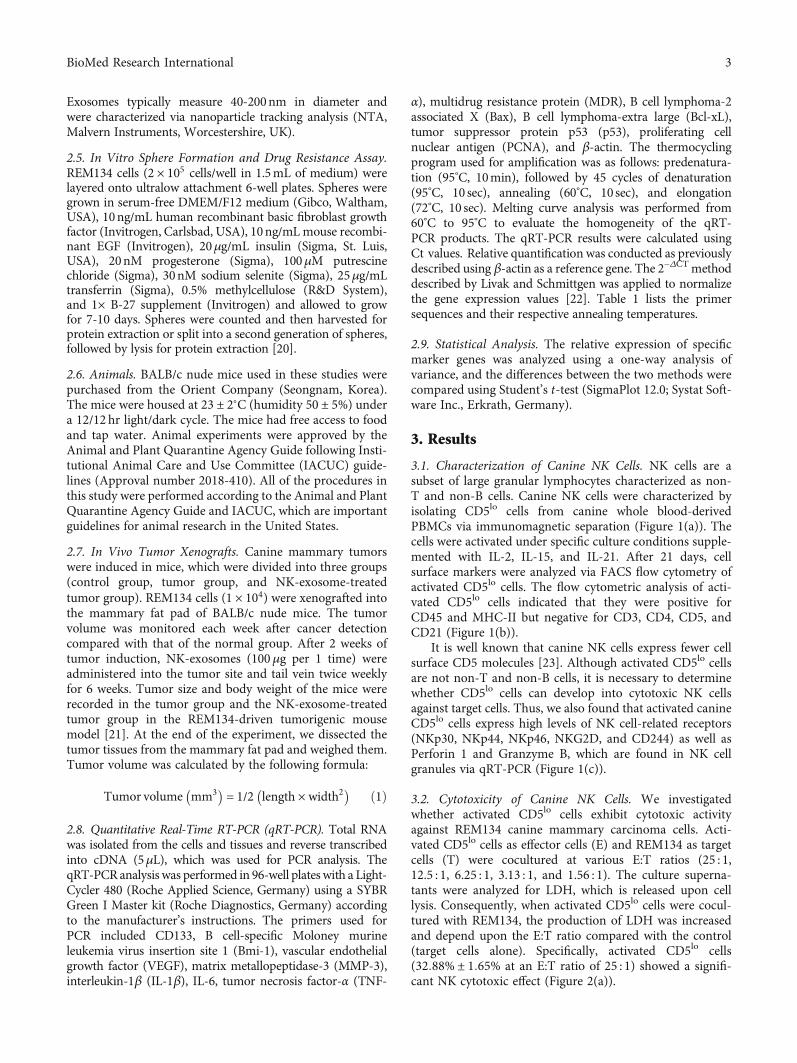
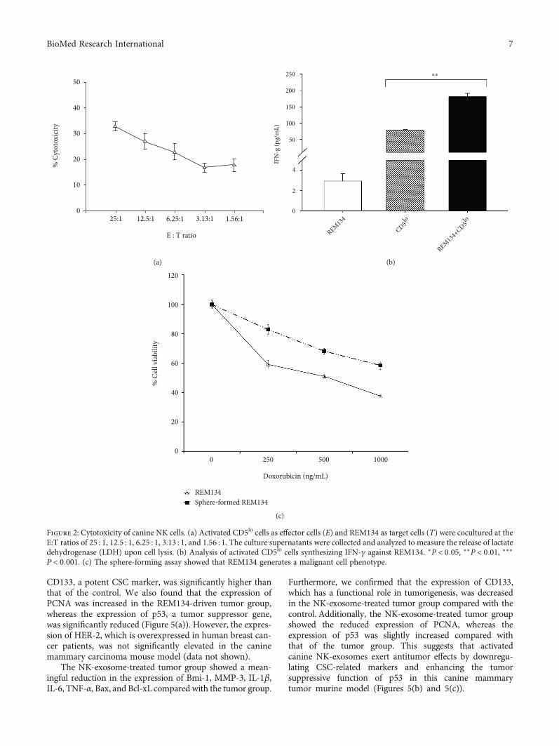
3.2. Cytotoxicity of Canine NK Cells. We investigatedwhether activated CD5lo cells exhibit cytotoxic activityagainst REM134 canine mammary carcinoma cells. Acti-vated CD5lo cells as effector cells (E) and REM134 as targetcells (T) were cocultured at various E:T ratios (25 : 1,12.5 : 1, 6.25 : 1, 3.13 : 1, and 1.56 : 1). The culture superna-tants were analyzed for LDH, which is released upon celllysis. Consequently, when activated CD5lo cells were cocul-tured with REM134, the production of LDH was increasedand depend upon the E:T ratio compared with the control(target cells alone). Specifically, activated CD5lo cells(32:88% ± 1:65% at an E:T ratio of 25 : 1) showed a signifi-cant NK cytotoxic effect (Figure 2(a)).
3BioMed Research International

The cytotoxic activity of NK cells was validated usingIFN-γ production and effector-mediated target cell assays.Significant IFN-γ production typically indicates NK cell
activation [24]. We also investigated whether activatedCD5lo cells secrete IFN-γ against REM134. Consequently,significant IFN-γ release was detected in activated CD5lo
Table 1: List of primers used for the reverse transcription-polymerase chain reaction.
Gene Primer sequence (5′-3′) Annealing temperature (°C) Accession number
CD133F-AGC CCT GTT GAA CGT GAA CA
60 KJ654317R-GTT GTA GCC ACT GGA GGG AC
Bmi-1F-CAC TGT GAA TAA TGA CTT CTT GCA T
60 NM_001287063R-AAG TTT ACT TTC CTT TGA TCG GTT T
VEGFF-GTA ATG ATG AGG GCC TAG AGT G
60 NM_001003175R-TAT GTG CTG GCC TTG ATG AG
MMP-3F-GTT GGA GGT GAC AGG GAA GG
60 AY183143R-CCA GGG AAG GTG GTG AAG TC
IL-1βF-GCC AAG ACC TGA ACC ACA GT
60 NM_001037971R-TGA CAC GAA ATG CCT CAG AC
IL-6F-CCT GGT GAT GGC TAC TGC TT
60 U12234R-TTG TTT GCA GAG GTG AGT GG
TNF-αF-GAG CCG ACG TGC CAA TG
60 Z70046R-CAA CCC ATC TGA CGG CAC TA
MDRF-GAG GAC TTG AAT GAG AAT GTT CCT
60 [60]R-CGG GTA AAG ATC CCT ATA ATC CTT
BaxF-CCG TGA GGT CTT CTT CCG AG
60 AB080230.1R-TAG AAG AGG GCA ACA ACC CG
Bcl-xLF-AAG GCG TTT CAG AGA AAA GGG
60 AB073983.1R-TTT TGA ATC ACC ACA CCG GC
p53F-TTC CTC CCC GAT GGC TCT TA
60 CFU62133R-AGA TGC CAA ACC AGA CCT CG
PCNAF-TCC TGC GCA AAA GAT GGA GT
60 XM_534355.4R-GAG AGA GCG GAG TGG CTT TT
NKp30F-TTG GCT CTG TCA CGT GGT AC
60 DQ003484R-CAG TGT CCC ATT CCC TGT CC
NKp44F-ATC GAG TGG CAG GGC AGA CA
60 XM_0846203R-TTC CTC CTT CAG ACC AAT CAT GGT
NKp46F-CCA GCA GAG CCC AAA ACA AC
60 NM_001284448R-CGG GAT GAA CGG AGA GAG TG
NKG2DF-ACG AAG GCA AAA GAG AAA GCC
60 XM_849013R-TGA TGA TTA TGG CAC CGC AT
CD244F-GGA GGA GGC TGG GGT TTT AC
60 XM_022415323R-GCG CCA TCT ACT CTC ACT CC
Perforin 1F-GAC AGA GCC CGT TGG AAG AA
60 FJ973622R-TTG GTG ATC CCC GAG TTG TG
Granzyme BF-GGA ACA GGA GAA GAC CCA GC
60 XM_547752R-TTG GCC TTC CTC TCA AGC AG
β-ActinF-GCT ACG TCG CCC TGG ACT TC
60 NM_001003349R-GCC CGT CGG GTA GTT CGT AG
4 BioMed Research International

00
100100
101
102
FL1-
H10
310
4
100
101
102
FL1-
H
103
104
100
101
102
FL1-
H
103
104
100
101
102
FL1-
H
103
104
101 102
FL2-H103 104
100 101 102
FL2-H
CD5-PE
CD3-
FITC
103 104 100 101 102
FL2-H103 104 100 101 102
FL2-H103 104
200 400 600
FSC-H
Isotype
0.55%
99.45% 0.00% 1.46% 94.51% 0.80% 1.28% 4.96%44.60%
0.00% 1.05% 52.89% 4.01% 0.67% 0.26% 93.50%
PBMC CD5lo cells CD5hi cells
800 1000 0 200 400 600
FSC-H
800 1000 0 200 400 600
FSC-H
800 1000 0 200 400 600
FSC-H
800 1000
200
400SSC-
H 600
800
1000
0
200
400SSC-
H 600
800
1000
0
200
400SSC-
H 600
800
1000
0
200
400SSC-
H 600
800
1000
(a)
00
100100
101
102
103
104
100
101
102
103
104
100
101
102
103
104
100
101
102
103
104
101 102 103 104 100 101 102 103 104 100 101 102 103 104 100 101 102 103 104
100 101 102 103 104100 101 102 103 104100 101 102 103 104100 101 102 103 104100
101
102
103
104
100
101
102
103
104
100
101
102
103
104
100
101
102
103
104
200 400FSC-H
FL2-H FL2-H FL2-H FL2-H
FL2-HFL2-HFL2-HFL2-HCD5-PE CD5-PE
CD5-PECD5-PE
Isotype
1.57% 0.53% 0.33%0.91%
96.20% 2.55% 0.01%99.38%
0.48% 0.13% 1.35%
1.35%98.40%0.00%
0.14%96.49%
3.37%
0.83%
97.80%
0.74%97.17%
IsotypeCD5lo cells CD5lo cells
FL4-
H
CD21
-APC
CD4-
FITC
MH
C2-F
ITC
CD45
-APC
FL4-
H
FL4-
H
FL4-
HFL
1-H
FL1-
H
FL1-
H
FL1-
H
FSC-H FSC-H FSC-H600 800 1000 0 200 400 600 800 1000 0 200 400 600 800 1000 0 200 400 600 800 1000
200
400SSC-
H 600
800
1000
0
200
400SSC-
H 600
800
1000
0
200
400SSC-
H 600
800
1000
0
200
400SSC-
H 600
800
1000
(b)
Figure 1: Continued.
5BioMed Research International

cells cocultured with REM134 (Figure 2(b)), suggesting thatactivated canine CD5lo cells exhibit NK cell characteristics.
One of the most important properties of cancer stemcells (CSCs) is tumorigenesis. It is known that single-cell-derived sphere formation facilitates CSC identification andtumor studies. We found that REM134 induces a malignanttype of cell and confirmed the impact of REM134 on thetumorigenic cell phenotype in a sphere-forming assay. Thesphere-formed REM134 exhibited the significantly increasedexpression of CD133 when compared with the control. Next,we analyzed the drug resistance phenotype of sphere-formedREM134 using doxorubicin, which is currently the mosteffective chemotherapeutic drug used to treat breast cancer[25]. The results indicated that single cell-derived sphere-formed REM134 is significantly resistant to doxorubicin incomparison with the adherent type of REM134. REM134may exhibit CSC properties upon tumor formation assphere-formed cells in the three-dimensional environmentof the body (Figure 2(c)).
3.3. Characterization of Canine NK-Exosomes. Next, we iso-lated and characterized NK-exosomes from cultured super-natants via ultracentrifugation. The isolated NK-exosomeswere approximately 136:6 ± 9:4 nm in diameter based onNTA (Figure 3(a)). We also confirmed the expression ofspecific exosome markers (CD63, CD81, Alix, HSP70, andTSG101) and NK cell markers (Perforin 1 and GranzymeB) in the activated NK-exosomes via western blot analysis(Figure 3(b)). These results suggest that activated canineNK-exosomes represent the properties of NK cells.
3.4. Effects of Canine NK-Exosomes on Tumor Progression.We established a REM134-driven tumor initiation modeland observed tumor formation and progression in BALB/cnude mice. Two weeks after tumor initiation, xenograft micewith REM134 tumors were injected intratumorally once andintravenously twice with NK-exosomes twice each week.Body weight and tumor size were monitored each week until8 weeks. Significant differences were detected within 4 weeksin only the REM134-driven tumor group in comparisonwith the tumor group treated with NK-exosomes. Inhibitionof tumor growth was observed in the tumor group treatedwith NK-exosomes but not in the untreated tumor group(Figures 4(a) and 4(b)). At the end of the experiment, wedissected tumor tissues from the mammary fat pad forweight analysis. Tumor weights were reduced in the tumorgroup treated with NK-exosomes compared with untreatedtumor group (Figure 4(c)). These results suggest that acti-vated canine NK-exosomes exhibit antitumor effects.
3.5. Effects of Canine NK-Exosomes on the Expression of CSCMarkers. Mounting evidence suggests that CSCs promotetumor progression, metastasis, and drug resistance [26].Thus, we investigated whether activated NK-exosomesregulate the expression of CSC-related markers on NK-exosomes during antitumor therapy. The results revealedsignificant changes including the increased expression oftumorigenesis-related markers, such as CD133, Bmi-1,VEGF, MMP-3, IL-1β, IL-6, TNF-α, and MDR, as well asthe apoptotic markers, Bax and Bcl-xL, in the REM134-driven tumor group. Among them, the expression of
700
600
Relat
ive e
xpre
ssio
n (fo
ld)
500
400
300
200
100
2
⁎⁎ ⁎⁎⁎⁎
⁎⁎
⁎⁎
⁎⁎
⁎⁎
1
0
NKp30NKp44
NKp46
NKG2DCD24
4
Perforin
1
Granzym
e B
CD5lo (w/o IL-2,IL-15,IL-21)CD5lo (with IL-2,IL-15,IL-21)
(c)
Figure 1: Isolation and characterization of canine NK cells. (a) CD5 negative (CD5lo) cells were isolated from canine PBMCs andactivated in specific culture supplemented with human IL-2 (500U/mL), IL-15 (10 ng/mL), and canine IL-21 (5 ng/mL) for 21 days.(b) Cell surface markers were analyzed by FACS flow cytometric analysis at activated CD5lo cells. (c) To determine whether CD5lo
cells develop into cytotoxic NK cells, the expression of activated NK cell markers (NKp30, NKp44, NKp46, NKG2D, CD244,Perforin 1, and Granzyme B) in activated canine CD5lo cells was analyzed by quantitative RT-PCR. β-Actin was used as a referencegene. ∗P < 0:05, ∗∗P < 0:01, ∗∗∗P < 0:001.
6 BioMed Research International
CD133, a potent CSC marker, was significantly higher thanthat of the control. We also found that the expression ofPCNA was increased in the REM134-driven tumor group,whereas the expression of p53, a tumor suppressor gene,was significantly reduced (Figure 5(a)). However, the expres-sion of HER-2, which is overexpressed in human breast can-cer patients, was not significantly elevated in the caninemammary carcinoma mouse model (data not shown).
The NK-exosome-treated tumor group showed a mean-ingful reduction in the expression of Bmi-1, MMP-3, IL-1β,IL-6, TNF-α, Bax, and Bcl-xL compared with the tumor group.
Furthermore, we confirmed that the expression of CD133,which has a functional role in tumorigenesis, was decreasedin the NK-exosome-treated tumor group compared with thecontrol. Additionally, the NK-exosome-treated tumor groupshowed the reduced expression of PCNA, whereas theexpression of p53 was slightly increased compared withthat of the tumor group. This suggests that activatedcanine NK-exosomes exert antitumor effects by downregu-lating CSC-related markers and enhancing the tumorsuppressive function of p53 in this canine mammarytumor murine model (Figures 5(b) and 5(c)).
25:10
10
20
30
40
50
12.5:1 6.25:1
E : T ratio
% C
ytot
oxic
ity
3.13:1 1.56:1
(a)
250 ⁎⁎
200
150
100
50
4
2
0
IFN
-g (p
g/m
L)
REM134
REM134+
CD5lo
CD5lo
(b)
120
100
80
60
40
20
00 250
Doxorubicin (ng/mL)
% C
ell v
iabi
lity
500 1000
REM134Sphere-formed REM134
(c)
Figure 2: Cytotoxicity of canine NK cells. (a) Activated CD5lo cells as effector cells (E) and REM134 as target cells (T) were cocultured at theE:T ratios of 25 : 1, 12.5 : 1, 6.25 : 1, 3.13 : 1, and 1.56 : 1. The culture supernatants were collected and analyzed to measure the release of lactatedehydrogenase (LDH) upon cell lysis. (b) Analysis of activated CD5lo cells synthesizing IFN-γ against REM134. ∗P < 0:05, ∗∗P < 0:01, ∗∗∗P < 0:001. (c) The sphere-forming assay showed that REM134 generates a malignant cell phenotype.
7BioMed Research International

4. Discussion
Canine tumors are invaluable models for the study of humancancers [27, 28]. Among the canine tumors, canine mam-mary tumors are the most frequent neoplasm in female dogs,and more than 50% are malignant [29–32]. In recent years,
several studies have advanced the understanding of thegenetic basis for canine mammary tumors [33–36]. Kimet al. reported cross-species oncogenic signatures for breastcancer in canine mammary tumors. They found molecularand histological discrepancies between canine mammarytumors and human breast cancers, despite their similarities
1.6
127
221
154
70 319351 450
1.4
1.2
1.0C
once
ntra
tion
(par
ticle
s/m
L)
0.8
0.6
0.4
0.2
00 100 200 300 400 500
Size (nm)
600 700 800 900 1000
(a)
Total NK-exosomes
CD63 (63 kDa)
CD81 (26 kDa)
Alix (95 kDa)
HSP70 (70 kDa)
TSG101 (44 kDa)
Perforin 1 (75 kDa)
Granzyme B (32 kDa)
(b)
Figure 3: Characterization of canine NK-exosomes. (a) Using nanoparticle tracking analysis, the size of NK cell-derived exosomes (NK-exosomes) was analyzed. (b) Specific markers (CD63, CD81, Alix, HSP70, TSG101, Perforin 1, and Granzyme B) were analyzed in NK-exosomes using western blot analysis.
8 BioMed Research International

[37]. For example, the amplification of HER2, a member ofthe human epidermal growth factor receptor family, hasbeen shown to play an important role in the developmentand progression of certain aggressive types of human breastcancer. The HER2 amplification occurs in 20-25% of humanbreast cancers and is associated with a high rate of relapseand poor prognosis [38]. Trastuzumab (Herceptin®) is amonoclonal antibody that inhibits downstream signal trans-duction by targeting the extracellular domain of the HER2receptor [39]. However, the efficiency of Herceptin® isapproximately 26%, even in HER2-overexpressing human
breast cancer patients [40]. Thus, the clinical benefit andthe specific strategy applied against a target gene may notbe straightforward in canine mammary tumors.
Recent studies have reported subpopulations of cellsknown as CSCs within tumors that are responsible for tumorinitiation, expansion, metastasis and recurrence after ther-apy. Tumors contain a large number of cancer cells alongwith a small population of CSCs [41]. CSCs are resistant tomultiple cancer therapies, including chemotherapy and radi-ation therapy [42]. A single cell-derived sphere formationassay can be used to identify CSCs and their drug resistance
25.0
24.0
23.0
22.0
21.0
20.0
19.0
18.0
17.0
16.0
15.02 3 4 5
Weeks
Body
wei
ght (
g)
6 7 8
ControlREM134REM134/NK-exosomes
(a)
1,5001,3501,2001,050
900750600
Tum
or v
olum
e (m
m3 )
450300150
02 3 4 5
Weeks
6 7 8
REM134REM134/NK-exosomes
(b)
1.0
0.9
0.8
0.7
0.6
Tum
or w
eigh
t (g)
0.5
0.4
0.3
0.2
0.1
0.0REM134 REM134/NK-exosomes
(c)
Figure 4: Effects of canine NK-exosomes on tumor progression in a mammary tumor model (a, b) REM134 cells (1 × 10⁴) induced tumorinitiation and obvious tumor formation and progression in BALB/c nude mice. After 2 weeks, xenograft mice carrying REM134 were onceintratumorally and twice intravenously injected with NK-exosomes each week. Body weight and tumor size of mice were monitored eachweek until 8 weeks. (c) Tumor tissues from the mammary fat pad were dissected and weighed at 8 weeks.
9BioMed Research International

800
700
600
500
400
300
Relat
ive e
xpre
ssio
n (fo
ld)
200
100
6
4
2
0CD133 Bmi-1 VEGF MMP-3 IL-1β TNFα MDR BAX Bcl-xL p53 PCNA
ControlREM134
IL-6
⁎⁎⁎
⁎⁎⁎
⁎⁎⁎
⁎⁎⁎
⁎⁎⁎
⁎⁎⁎
⁎⁎⁎
⁎⁎⁎
⁎⁎⁎
⁎⁎⁎
⁎
⁎
(a)
4
3
2
Relat
ive e
xpre
ssio
n (fo
ld)
1
0CD133 Bmi-1 VEGF MMP-3 IL-1β IL-6 TNFα MDR BAX Bcl-xL p53
REM134REM134/NK-exosomes
PCNA
⁎⁎⁎
⁎⁎⁎
⁎⁎⁎
⁎⁎⁎ ⁎⁎⁎
⁎⁎⁎
⁎⁎⁎
⁎
⁎⁎⁎⁎⁎⁎
⁎
(b)
REM134
REM134/NK-exosomes
(c)
Figure 5: Effects of canine NK-exosomes on the expression of cancer stem cell markers. (a, b) To examine whether activated NK-exosomesinfluence the expression of CSC-related markers (CD133, Bmi-1, VEGF, MMP-3, IL-1β, IL-6, TNF-α, MDR, Bax, Bcl-xL, p53, and PCNA),the expression level of tumorigenesis-related marker was examined in control, untreated tumor, and NK-exosome-treated tumor groups.∗P < 0:05, ∗∗P < 0:01, ∗∗∗P < 0:001. (c) We monitored the tumor volume of the mice in the tumor group (REM134, top) and NK-exosome-treated tumor group (REM134/NK-exosomes, bottom) in the REM134-driven tumorigenic mouse model. Representative imagesare shown (2, 4, 6, and 8 weeks after tumor induction).
10 BioMed Research International

mechanisms [43–45]. Michishita et al. reported that sphere-formed cells derived from canine mammary adenocarci-noma cell lines exhibit characteristics of CSCs [43]. In thisstudy, we found that REM134 generates cellular spheresand confirmed their impact on the tumorigenic cell pheno-type of REM134. We confirmed that sphere-formedREM134 cells exhibit significant doxorubicin resistancecompared with adherent-type REM134 cells. Hence, it isexpected that REM134 may exhibit properties of CSCs upontumor formation as sphere-formed cells in the three-dimensional environment of the body.
We focused on reducing the functional potential of CSCsand CSC-related markers to drive tumor initiation, expan-sion, metastasis, and recurrence. We induced REM134-driven tumor initiation and monitored tumor formationand progression in BALB/c nude mice. Expectedly, tumorformation and growth rate were accelerated by the entry ofa small number of cells into the body within 2 weeks, whichrepresents a very serious phase in humans. Surprisingly, thetumor growth was inhibited in mice treated with NK-exosomes compared with untreated mice. These results sug-gest that activated canine NK-exosomes exert antitumoreffects. CSC-targeted therapy is an interesting area of cancerresearch. Evidence increasingly suggests the presence ofCSCs in various solid tumors, with a potential role in tumorinitiation, progression, and recurrence [43]. Thus, the sup-pression of CSCs is an important strategy for the develop-ment of novel therapeutic agents and for the elucidation ofthe molecular and cellular mechanisms underlying tumori-genesis in veterinary research.
The immune system exploits highly destructive mecha-nisms. NK cells are a type of immune cell that appliesactivating receptors, such as NKG2D, to recognize and elim-inate infected and transformed cells that upregulate theligands for these receptors. Six to eight different NKG2Dligands are poorly expressed by normal cells but are upregu-lated in cancer cells [46]. Canine NK cells exhibit lowerlevels of CD5 molecules [23]. We confirmed that activatedcanine CD5lo cells express high levels of NKG2D and NKcell-related activating receptors. We also found that acti-vated canine CD5lo cells exhibit cytotoxic NK cell propertiesagainst target cells through LDH and IFN-γ production.This study is the first of its kind to examine the antitumoreffect of NK-exosomes in a canine mammary carcinomamurine model. A query of the Clinical Trials website(https://clinicaltrials.gov) using the search term “exosomes”returned more than 100 clinical studies. However, clinicalstudies investigating human breast cancer using exosomeswere not identified until June 2021. We focused on NK-exosomes that decrease the functional potential of CSCsand CSC-related markers that drive tumor initiation, expan-sion, metastasis, and recurrence. We observed significantchanges in tumorigenesis-related markers includingCD133, Bmi-1, VEGF, MMP-3, IL-1β, IL-6, TNF-α, andMDR in the tumor group compared with the control group.
Several CSC markers were also identified. CSCs are alimited subpopulation of cells with stem cell-like properties.CD133 is a transmembrane protein expressed on the sur-face of hematopoietic stem cells and is widely recognized
as a CSC marker. CD133 is also strongly associated withcancer-related signaling pathways and promotes tumorinvasion, progression, and migration [47]. Several reportssuggest that CD133 is a valuable prognostic marker inmelanoma, prostate cancer, and glioma [48–50]. Kim et al.suggested that CD133-expressing breast cancer cells exhibittumorigenesis, tumor self-renewal, high tumor cell prolifera-tion, and drug resistance properties [51].
Liu et al. reported that the overexpression of Bmi-1,polycomb complex protein, in normal mammary epithelialcells increased sphere formation [52]. Bmi-1 has been shownto regulate the self-renewal of both normal and malignantmammary stem cells [53]. Thus, the suppression of Bmi-1expression is related to the functional inhibition of CSCs,which form malignant tumor cells. Vascularization is animportant characteristic of breast cancer development.Accordingly, the regulation of angiogenic factors has beensuggested to play a therapeutic role [31, 54]. Pakravanet al. reported that MSC-derived exosomes regulate themTOR/HIF-1α signaling axis in human breast cancer cells,resulting in a significant reduction of the VEGF expression[55]. Lee et al. also reported that MSC-derived exosomessuppress angiogenesis by downregulating VEGF in murinebreast cancer cells [56]. MMPs are zinc-dependent endopep-tidases that constitute a family of 24 members in mammals[57]. The overexpression of MMP-3 had been shown todrive the formation of mammary tumors in mice [58].
IL-1β, IL-6, and TNF-α are major proinflammatorycytokines. Cytokines represent key mediators of inflamma-tion and play an important role in the interaction betweeninflammation and cancer. Proinflammatory states are associ-ated with carcinogenesis [59]. Cancer is a highly heteroge-neous disease, both within a single patient as well asamong different patients. Various single and combinationtherapies have been developed and refined for effective treat-ment. Recently, the evolution of MDR in cancer represents amajor challenge to successful treatment. MDR is a complexprocess involving multiple noncellular and cellular mecha-nisms [60]. In this study, we found that compared with thetumor group alone, the NK-exosome-treated tumor groupexhibited a significant decrease in the expression ofCD133, Bmi-1, MMP-3, IL-1β, IL-6, and TNF-α.
The Bcl-2 family of proteins is prominently involved inthe promotion or inhibition of apoptosis [61]. The proapop-totic multidomain molecules, Bax and Bak, mediate mito-chondrial apoptosis, whereas Bcl-xL, Mcl-1, Bcl2a1, andBcl-w are common prosurvival and antiapoptotic proteins[62]. The balance of proapoptotic and antiapoptotic proteinsregulates the fate of cells [63]. The p53 tumor suppressorgene prevents tumor development through the inhibitionand elimination of abnormally proliferating cells. It isknown that p53 mutations are associated with poor progno-sis and tumor aggressiveness [64]. He et al. reported thatMSCs inhibit tumor progression and enhance the radiosen-sitivity of human breast cancer cells. MSC-conditionedmedium suppresses the growth of breast cancer cells by reg-ulating stemness genes (Sox2, Oct4, Nanog, and c-Myc) andStat3 signaling pathway-related genes (cyclin D1, Bcl-xL,and p53) [65]. We also found that in the REM134-driven
11BioMed Research International

tumor group, the expression of Bax, BcL-xL, and PCNA wasincreased, whereas the expression of p53 was decreased. Bycontrast, the NK-exosome-treated REM134-driven tumorgroup showed significantly reduced expression of Bax,Bcl-xL, and PCNA, whereas the reduced expression of p53was similar to that of the tumor group. Therefore, NK-exosomes exert antitumor effects by downregulating CSCsand tumorigenesis-related markers and modulating the apo-ptotic protein expression. This study provides evidence sup-porting the role of CSCs in tumorigenesis in a model ofcanine mammary carcinoma. Further studies are requiredto elucidate the antitumor mechanism of NK-exosomes inmore detail.
Data Availability
The datasets used and/or analyzed during the current studyare available from the corresponding author on reasonablerequest.
Conflicts of Interest
The authors declare no conflicts of interest.
Authors’ Contributions
Jienny Lee, Se-A Lee, and Na-Yeon Gu contributed equallyto this work.
Acknowledgments
This work was funded by the Animal and Plant QuarantineAgency (B-1543083-2021-22-01), the Republic of Korea.
References
[1] E. Vivier, E. Tomasello, M. Baratin, T. Walzer, and S. Ugolini,“Functions of natural killer cells,” Nature Immunology, vol. 9,no. 5, pp. 503–510, 2008.
[2] F. M. Wensveen, V. Jelenčić, and B. Polić, “NKG2D: a masterregulator of immune cell responsiveness,” Frontiers in Immu-nology, vol. 9, p. 441, 2018.
[3] J. Wang, X. Sun, J. Zhao et al., “Exosomes: a novel strategy fortreatment and prevention of diseases,” Frontiers in Pharmacol-ogy, vol. 8, p. 300, 2017.
[4] I. L. Colao, R. Corteling, D. Bracewell, and I. Wall,“Manufacturing exosomes: a promising therapeutic platform,”Trends in Molecular Medicine, vol. 24, no. 3, pp. 242–256,2018.
[5] D. G. Phinney and M. F. Pittenger, “Concise review: MSC-derived exosomes for cell-free therapy,” Stem Cells, vol. 35,no. 4, pp. 851–858, 2017.
[6] L. Timmers, S. K. Lim, F. Arslan et al., “Reduction of myocar-dial infarct size by human mesenchymal stem cell conditionedmedium,” Stem Cell Research, vol. 1, no. 2, pp. 129–137, 2008.
[7] F. Arslan, R. C. Lai, M. B. Smeets et al., “Mesenchymal stemcell-derived exosomes increase ATP levels, decrease oxidativestress and activate PI3K/Akt pathway to enhance myocardialviability and prevent adverse remodeling after myocardialischemia/reperfusion injury,” Stem Cell Research, vol. 10,no. 3, pp. 301–312, 2013.
[8] H. Xin, Y. Li, Y. Cui, J. J. Yang, Z. G. Zhang, and M. Chopp,“Systemic administration of exosomes released frommesenchy-mal stromal cells promote functional recovery and neurovascu-lar plasticity after stroke in rats,” Journal of Cerebral Blood Flowand Metabolism, vol. 33, no. 11, pp. 1711–1715, 2013.
[9] D. R. Ophelders, T. G. Wolfs, R. K. Jellema et al., “Mesenchy-mal stromal cell-derived extracellular vesicles protect the fetalbrain after hypoxia-ischemia,” Stem Cells Translational Medi-cine, vol. 5, no. 6, pp. 754–763, 2016.
[10] Y. Fan, F. Herr, A. Vernochet, B. Mennesson, E. Oberlin, andA. Durrbach, “Human fetal liver mesenchymal stem cell-derived exosomes impair natural killer cell function,” StemCells and Development, vol. 28, no. 1, pp. 44–55, 2019.
[11] W. Yan and S. Jiang, “Immune cell-derived exosomes in thecancer-immunity cycle,” Trends Cancer, vol. 6, no. 6,pp. 506–517, 2020.
[12] X. J. Tang, X. Y. Sun, K. M. Huang et al., “Therapeutic poten-tial of CAR-T cell-derived exosomes: a cell-free modality fortargeted cancer therapy,” Oncotarget, vol. 6, no. 42,pp. 44179–44190, 2015.
[13] J. M. Pitt, F. André, S. Amigorena et al., “Dendritic cell–derived exosomes for cancer therapy,” The Journal of clinicalinvestigation, vol. 126, no. 4, pp. 1224–1232, 2016.
[14] H. Tian and W. Li, “Dendritic cell-derived exosomes for can-cer immunotherapy: hope and challenges,” Annals of Transla-tional Medicine, vol. 5, no. 10, p. 221, 2017.
[15] S. Fais, “NK cell-released exosomes: natural nanobulletsagainst tumors,” Oncoimmunology, vol. 2, no. 1, articlee22337, 2013.
[16] S. Oh, J. H. Lee, K. Kwack, and S. W. Choi, “Natural killer celltherapy: a new treatment paradigm for solid tumors,” Cancers,vol. 11, no. 10, p. 1534, 2019.
[17] I. Kaszak, A. Ruszczak, S. Kanafa, K. Kacprzak, M. Król, andP. Jurka, “Current biomarkers of canine mammary tumors,”Acta Veterinaria Scandinavica, vol. 60, no. 1, 2018.
[18] M. Norval, J. Maingay, and R. W. Else, “Studies of three caninemammary carcinoma cell lines–I. _In vitro_ properties,” Euro-pean Journal of Cancer and Clinical Oncology, vol. 20, no. 12,pp. 1489–1500, 1984.
[19] Y. C. Huang, S. W. Hung, T. R. Jan et al., “CD5-low expressionlymphocytes in canine peripheral blood show characteristics ofnatural killer cells,” Journal of Leukocyte Biology, vol. 84, no. 6,pp. 1501–1510, 2008.
[20] L. Y. Pang, A. Cervantes-Arias, R. W. Else, and D. J. Argyle,“Canine mammary cancer stem cells are Radio- and Chemo-Resistant and exhibit an epithelial-mesenchymal transitionphenotype,” Cancers, vol. 3, no. 2, pp. 1744–1762, 2011.
[21] M. M. Tomayko and C. P. Reynolds, “Determination of subcu-taneous tumor size in athymic (nude) mice,” Cancer Chemo-therapy and Pharmacology, vol. 24, no. 3, pp. 148–154, 1989.
[22] K. J. Livak and T. D. Schmittgen, “Analysis of Relative GeneExpression Data Using Real-Time Quantitative PCR and the2−ΔΔCT Method,” vol. 25, no. 4, pp. 402–408, 2001.
[23] C. S. Lin, C. P. Chang, H. C. Chiang, T. F. Chuang, C. H. Hsu,and C. C. Liu, “Activating natural killer (NK) cytotoxicity ofcanine CD5-CD21- cells requires low surface CD5 densityNK cells,” Iranian Journal of Veterinary Research, vol. 19,no. 2, pp. 87–95, 2018.
[24] A. Y. Mah and M. A. Cooper, “Metabolic regulation of naturalkiller cell IFN-γ production,” Critical Reviews in Immunology,vol. 36, no. 2, pp. 131–147, 2016.
12 BioMed Research International

[25] C. Christowitz, T. Davis, A. Isaacs, G. van Niekerk,S. Hattingh, and A. M. Engelbrecht, “Mechanisms ofdoxorubicin-induced drug resistance and drug resistanttumour growth in a murine breast tumour model,” BMC Can-cer, vol. 19, no. 1, p. 757, 2019.
[26] L. Pang, J. Ding, Y. Ge, J. Fan, and S. K. Fan, “Single-cell-derived tumor-sphere formation and drug-resistance assayusing an integrated microfluidics,” Analytical Chemistry,vol. 91, no. 13, pp. 8318–8325, 2019.
[27] J. L. Rowell, D. O. McCarthy, and C. E. Alvarez, “Dog modelsof naturally occurring cancer,” Trends in Molecular Medicine,vol. 17, no. 7, pp. 380–388, 2011.
[28] C. E. Alvarez, “Naturally occurring cancers in dogs: insightsfor translational genetics and medicine,” ILAR Journal,vol. 55, no. 1, pp. 16–45, 2014.
[29] S. A. Benjamin, A. C. Lee, and W. J. Saunders, “Classificationand behavior of canine mammary epithelial neoplasms basedon life-span observations in beagles,” Veterinary Pathology,vol. 36, no. 5, pp. 423–436, 1999.
[30] I. J. Fidler and R. S. Brodey, “The biological behavior of caninemammary neoplasms,” Journal of the American VeterinaryMedical Association, vol. 151, no. 10, pp. 1311–1318, 1967.
[31] S. B. Fox, D. G. Generali, and A. L. Harris, “Breast tumourangiogenesis,” Breast Cancer Research, vol. 9, no. 6, p. 216,2007.
[32] W. A. Priester and N. Mantel, “Occurrence of tumors indomestic animals. Data from 12 United States and Canadiancolleges of veterinary medicine,” Journal of the National Can-cer Institute, vol. 47, pp. 1333–1345, 1971.
[33] J. Beck, S. Hennecke, K. Bornemann-Kolatzki et al., “Genomeaberrations in canine mammary carcinomas and their detec-tion in cell-free plasma DNA,” Plo S one., vol. 8, no. 9, articlee75485, 2013.
[34] R. Klopfleisch, D. Lenze, M. Hummel, and A. D. Gruber, “Met-astatic canine mammary carcinomas can be identified by agene expression profile that partly overlaps with human breastcancer profiles,” BMC Cancer, vol. 10, no. 1, 2010.
[35] K. M. Pawłowski, H. Maciejewski, I. Dolka, J. A. Mol, T. Motyl,and M. Król, “Gene expression profiles in canine mammarycarcinomas of various grades of malignancy,” BMC VeterinaryResearch, vol. 9, no. 1, p. 78, 2013.
[36] D. Liu, H. Xiong, A. E. Ellis et al., “Molecular homology anddifference between spontaneous canine mammary cancer andhuman breast cancer,” Cancer Research, vol. 74, no. 18,pp. 5045–5056, 2014.
[37] T. M. Kim, I. S. Yang, B. J. Seung et al., “Cross-species onco-genic signatures of breast cancer in canine mammary tumors,”Nature Communications, vol. 11, no. 1, 2020.
[38] T. Cooke, J. Reeves, A. Lanigan, and P. Stanton, “HER2 as aprognostic and predictive marker for breast cancer,” Annalsof Oncology, vol. 12, pp. S23–S28, 2001.
[39] H. Maadi, B. Nami, J. Tong, G. Li, and Z. Wang, “The effectsof trastuzumab on HER2-mediated cell signaling in CHOcells expressing human HER2,” BMC Cancer, vol. 18, no. 1,2018.
[40] H. Tang, C. Song, F. Ye et al., “miR-200c suppresses stemnessand increases cellular sensitivity to trastuzumab in HER2+breast cancer,” Journal of Cellular and Molecular Medicine,vol. 23, no. 12, pp. 8114–8127, 2019.
[41] M. Luo, M. Brooks, and S. Wicha, “Epithelial-mesenchymalplasticity of breast cancer stem cells: implications for metasta-
sis and therapeutic resistance,” Current PharmaceuticalDesign, vol. 21, no. 10, pp. 1301–1310, 2015.
[42] Y. Kim, K. M. Joo, J. Jin, and D. H. Nam, “Cancer stemcells and their mechanism of chemo-radiation resistance,”International Journal of Stem Cells, vol. 2, no. 2, pp. 109–114, 2009.
[43] M. Michishita, R. Akiyoshi, H. Yoshimura et al., “Characteri-zation of spheres derived from canine mammary gland adeno-carcinoma cell lines,” Research in Veterinary Science, vol. 91,no. 2, pp. 254–260, 2011.
[44] H. F. Bahmad, K. Cheaito, R. M. Chalhoub et al., “Sphere-for-mation assay: three-dimensional in vitro culturing of prostatecancer stem/progenitor sphere-forming cells,” Frontiers inOncology, vol. 8, p. 347, 2018.
[45] X. L. Ma, Y. F. Sun, B. L. Wang et al., “Sphere-forming cultureenriches liver cancer stem cells and reveals Stearoyl-CoA desa-turase 1 as a potential therapeutic target,” BMCCancer, vol. 19,no. 1, p. 760, 2019.
[46] W. Deng, B. G. Gowen, L. Zhang et al., “A shed NKG2D ligandthat promotes natural killer cell activation and tumor rejec-tion,” Science, vol. 348, no. 6230, pp. 136–139, 2015.
[47] K. Tabu, T. Kimura, K. Sasai et al., “Analysis of an alternativehuman CD133 promoter reveals the implication of Ras/ERKpathway in tumor stem-like hallmarks,” Molecular Cancer,vol. 9, no. (1), p. 39, 2010.
[48] E. Monzani, F. Facchetti, E. Galmozzi et al., “Melanoma con-tains CD133 and ABCG2 positive cells with enhancedtumourigenic potential,” European Journal of Cancer, vol. 43,no. 5, pp. 935–946, 2007.
[49] G. D. Richardson, C. N. Robson, S. H. Lang, D. E. Neal, N. J.Maitland, and A. T. Collins, “CD133, a novel marker forhuman prostatic epithelial stem cells,” Journal of Cell Science,vol. 117, no. 16, pp. 3539–3545, 2004.
[50] F. Zeppernick, R. Ahmadi, B. Campos et al., “Stem cell markerCD133 affects clinical outcome in glioma patients,” ClinicalCancer Research, vol. 14, no. 1, pp. 123–129, 2008.
[51] S. J. Kim, Y. S. Kim, E. D. Jang, K. J. Seo, and J. S. Kim, “Prog-nostic impact and clinicopathological correlation of CD133and ALDH1 expression in invasive breast cancer,” Journal ofBreast Cancer, vol. 18, no. 4, pp. 347–355, 2015.
[52] S. Liu, G. Dontu, I. D. Mantle et al., “Hedgehog signaling andBmi-1 regulate self-renewal of normal and malignant humanmammary stem cells,” Cancer Research, vol. 66, no. 12,pp. 6063–6071, 2006.
[53] S. M. Chen, B. Y. Wang, C. H. Lee et al., “Hinokitiol up-regulates miR-494-3p to suppress BMI1 expression andinhibits self-renewal of breast cancer stem/progenitor cells,”Oncotarget, vol. 8, no. 44, pp. 76057–76068, 2017.
[54] R. Sharma, R. Sharma, T. P. Khaket, C. Dutta, B. Chakraborty,and T. K. Mukherjee, “Breast cancer metastasis: putative ther-apeutic role of vascular cell adhesion molecule-1,” CellularOncology, vol. 40, no. 3, pp. 199–208, 2017.
[55] K. Pakravan, S. Babashah, M. Sadeghizadeh et al., “Micro-RNA-100 shuttled by mesenchymal stem cell-derived exo-somes suppresses in vitro angiogenesis through modulatingthe mTOR/HIF-1α/VEGF signaling axis in breast cancercells,” Cellular Oncology, vol. 40, no. 5, pp. 457–470, 2017.
[56] J. K. Lee, S. R. Park, B. K. Jung et al., “Exosomes derived frommesenchymal stem cells suppress angiogenesis by down-regulating VEGF expression in breast cancer cells,” PLoSOne, vol. 8, no. 12, article e84256, 2013.
13BioMed Research International

[57] R. Roy, J. Yang, and M. A. Moses, “Matrix metalloproteinasesas novel biomarkers and potential therapeutic targets inhuman cancer,” Journal of Clinical Oncology, vol. 27, no. 31,pp. 5287–5297, 2009.
[58] M. D. Sternlicht, A. Lochter, C. J. Sympson et al., “The stromalproteinase MMP3/stromelysin-1 promotes mammary carci-nogenesis,” Cell, vol. 98, no. 2, pp. 137–146, 1999.
[59] A. M. DeMarzo, E. A. Platz, S. Sutcliffe et al., “Inflammation inprostate carcinogenesis,” Nature Reviews. Cancer, vol. 7, no. 4,pp. 256–269, 2007.
[60] J. Ahmad, S. Akhter, M. Ahmed Khan et al., “Engineerednanoparticles against MDR in cancer: the state of the art andits prospective,” Current Pharmaceutical Design, vol. 22,no. 28, pp. 4360–4373, 2016.
[61] J. M. Adams and S. Cory, “The Bcl-2 protein family: arbiters ofcell survival,” Science, vol. 281, no. 5381, pp. 1322–1326, 1998.
[62] M. C. Wei, W. X. Zong, E. H. Y. Cheng et al., “ProapoptoticBAX and BAK: a requisite gateway to mitochondrial dysfunc-tion and death,” Science, vol. 292, no. 5517, pp. 727–730, 2001.
[63] L. S. Loo, A. A. Soetedjo, H. H. Lau et al., “BCL-xL/BCL2L1 is acritical anti-apoptotic protein that promotes the survival ofdifferentiating pancreatic cells from human pluripotent stemcells,” Cell Death & Disease, vol. 11, no. 5, p. 378, 2020.
[64] M. Gasco, S. Shami, and T. Crook, “The p53 pathway in breastcancer,” Breast Cancer Research, vol. 4, no. 2, pp. 70–76, 2002.
[65] N. He, Y. Kong, X. Lei et al., “MSCs inhibit tumor progressionand enhance radiosensitivity of breast cancer cells by down-regulating Stat3 signaling pathway,” Cell Death & Disease,vol. 9, pp. 1–4, 2018.
14 BioMed Research International