capping of cdse–zns quantum dots with dhla and subsequent conjugation with proteins
TRANSCRIPT

Capping of CdSe–ZnS quantum dots with DHLA andsubsequent conjugation with proteinsAaron R. Clapp1, Ellen R. Goldman2 & Hedi Mattoussi1
1US Naval Research Laboratory, Optical Sciences Division, Code 5611, 2Center for Bio/Molecular Science and Engineering, Code 6900, 4555 Overlook Avenue SW,Washington, DC 20375, USA. Correspondence should be addressed to H.M. ([email protected])
Published online 28 September; corrected online 30 November 2006 (details online); doi:10.1038/nprot.2006.184
We provide a detailed protocol for designing water-soluble CdSe–ZnS quantum dots (QDs) based on cap exchange of the native
hydrophobic shell with dihydrolipoic acid (DHLA) ligands, and the preparation of functional QD bioconjugates for use in
immunoassays. Our conjugation strategy is based on non-covalent self-assembly between DHLA-capped QDs and protein appended
with either an electrostatic attachment domain (namely, the basic leucine zipper) or a polyhistidine tag. These bioconjugates
combine the properties of the QD and attached biomolecule to create structures with desirable luminescent and biologically specific
properties. This method also allows the preparation of mixed surface conjugates, which results in the conjugates gaining multiple
biological activities. Conjugation of DHLA-capped QDs to maltose binding protein (MBP), the immunoglobulin-G-binding b2 domain
of streptococcal protein G (PG) and avidin will be described. MBP and PG were modified by genetic fusion with either a charged
leucine zipper or a polyhistidine interaction domain.
INTRODUCTIONLuminescent quantum dots (QDs) are versatile inorganic probesthat have unique spectroscopic properties, including narrowand size-tunable photoemission profiles coupled with broadabsorption spectra. Their broad absorption spectra allow theflexibility to efficiently excite QD samples at wavelengths thatare far removed from their emission spectra, which translatesinto large experimental Stokes shifts. This is particularly beneficialfor the implementation of multiplexed assays in which multipledistinct QD samples can be simultaneously excited with a singleline that is far from their emission spectra, which simplifies theexperimental conditions and signal deconvolution. Considerablylarger than molecular dyes, the nanometer size QDs have muchlarger absorption cross-sections than commonly used organic dyes,and offer accessible surface area for the subsequent attachmentof molecules1–3.
Recently, there has been a growing emphasis on assemblingcustomized composite metal-organic nanostructures that mergedisparate yet desirable properties into a stable hybrid nanoparticle.In general, colloidal QDs are not intrinsically compatible withbiological environments due to the presence of hydrophobiccapping groups on their surfaces. However, this limitation can beovercome by using a variety of post-synthesis strategies that includeencapsulating the native hydrophilic QDs with a layer of amphi-philic molecules that interdigitate with the existing hydrophobicgroups, or completely replacing the native surface ligands withhydrophilic moieties (referred to as a ‘cap exchange’)2–7. Oncestabilized in an aqueous medium, QDs can be further processedto attach biomolecules to their surfaces.
In this protocol, we describe several practical approaches that wehave developed for constructing QD–protein bioconjugates basedon non-covalent self-assembly and their subsequent use to designand implement immunoassays specific for the detection of targetedmolecules8,9. Other conjugation approaches using carbodiimide-based covalent reactions or attachment of biotinylated proteins tostreptavidin-coated QDs have been reported (see summary inTable 1). Each of these approaches has inherent advantages and
disadvantages, which will guide the choice of conjugation methodto use, depending on the specific application and desired propertiesof the nanoparticle conjugate. The present conjugation method hasthe advantage that it does not rely on a particular linking chemistryand the constraints that this may bring. For example, the use of theconventional covalent cross-linking approach—based on 1-ethyl-3-(3-dimethylaminopropyl) carbodiimide hydrochloride (EDC) con-densation applied to QDs capped with COOH-terminated ligands(including dihydrolipoic acid (DHLA))—results in aggregatebuild-up, which can be attributed to a reduction in QD wateraffinity once the carboxy groups are reacted3,6. It does, however,require the use of proteins that have been engineered to express aleucine zipper domain or a polyhistidine tag. An example applica-tion is provided for a multiplexed sandwich immunoassay that iscapable of detecting four soluble toxins simultaneously, by whichthe specificity of toxin detection using surface-bound antibodiesand the ability to resolve multiple QD emission signals using asingle excitation source have been demonstrated.
EXPERIMENTAL DESIGNThis protocol uses high-quality CdSe nanoparticles with crystallinecores and narrow size distributions. These nanoparticles can beprepared by reacting organometallic precursors at high tempera-tures in a coordinating solvent mixture, following the approachesdescribed previously10,11. Subsequent overcoating of the CdSe coreswith a relatively thin layer (3–7 monolayers) of ZnS or CdSproduces highly luminescent CdSe–ZnS or CdSe–CdS core-shellQDs12–14. It is important to note that the more red-emitting QDstend to be anisotropic in shape compared with their smaller greenand yellow-emitting counterparts. This is due to the uniaxialsymmetry of the wurtzite crystal structure of the parent bulkCdSe (maintained within QDs), which increasingly affects theoverall shape of the nanoparticles when larger sizes (red-emitting)are grown. Aspect ratios of B1.2–2.0 have been reported for core-shell QDs that emit at wavelengths beyond 600 nm. For additionaldetails on the choice of precursors, growth/coordinating solvent
p
uor
G g
n ih si l
bu
P eru ta
N 600 2©
nat
ure
pro
toco
ls/
moc.er
ut an.
ww
w//:ptt
h
1258 | VOL.1 NO.3 | 2006 | NATURE PROTOCOLS
PROTOCOL

mixtures and general reaction conditions, we refer the reader toprevious publications10–14. In this report, we provide a briefdescription of water-soluble CdSe–ZnS QDs that we havedeveloped and refined, and their further use for applications infunctional assays. The procedure described here represents one ofseveral variations that has been developed and that is used byvarious research groups.
The first scheme that we describe is based on electrostatic self-assembly in which biomolecules having either a natural positivesurface charge (such as avidin) or engineered positively chargeddomains interact with negatively charged QDs capped with aDHLA solubilizing layer6. We have engineered maltose bindingprotein (MBP) and protein G (PG) appended with a positivelycharged leucine zipper attachment domain to form MBP–zb andPG–zb, respectively6,8. Adding these proteins to the QDs incombination enables the formation of mixed-surface bioconjugatesin which each protein provides a specific functionality. MBP–zballows purification of the QD bioconjugates over amylose resin,whereas a second protein (e.g., PG–zb or avidin) serves as abridging molecule for attaching antibodies to the QDs8,9. Nearlyall immunoassays developed in our laboratory have used mixed-surface QD bioconjugates using MBP.
A general approach for attaching biomolecules to QDs usesavidin as a bridging molecule. In principle, any biotinylatedmolecule (e.g., biotin-labeled antibody) can be readily attached toan avidin-coated QD; this obviates the need for an engineeredbridging protein such as PG–zb. However, there is a limit to thenumber of avidin molecules that can be self-assembled onto a QD,due to charge-induced agglomeration. The present protocolprovides a general description of how to prepare QD–antibodyconjugates via an avidin bridge. Figure 1 shows a schematic of atypical MBP–zb/avidin/antibody mixed-surface conjugate havingonly a few proteins attached to the surface (usual bioconjugates willhave a surface that is saturated with proteins). Alternatively, PG–zbspecifically interacts with the Fc domain of immunoglobulin G(IgG) and provides a structured orientation of antibodies on theQD with the binding sites directed outwards into the surroundingsolution and away from the nanocrystal surface. This arrangementcan potentially decrease heterogeneity and improve conjugateavidity. In addition, it does not require biotinylation of antibodies8.
The conjugation scheme using electrostatic self-assembly wasfurther expanded to include metal-affinity interactions betweenpolyhistidine-appended protein and DHLA-capped CdSe–ZnSQDs to form functional bioconjugates15–18. Many proteins areengineered to express an amino- or carboxy-terminal sequence ofrepeating His residues (typically five or more) for purificationon a Ni–nitrilotriacetic acid (Ni–NTA) column. This approachmay prove to be a facile and general route for the creationof QD–protein bioconjugates, because the metal-affinity-driven
self-assembly significantly simplifies the bioconjugation procedureand reduces the overall hydrodynamic size of the resulting nano-particle assembly by eliminating the need for a bridging protein.Self-assembly schemes using His-terminated proteins may bepreferable to methods that use an intermediate bridging protein(e.g., avidin or PG–zb), particularly for applications that requiremore compact bioconjugates. These considerations may be parti-cularly critical for intracellular delivery and imaging or fluorescenceresonance energy transfer (FRET) applications2–5,15,16. FRETinvolves the non-radiative transfer of excitation energy from anexcited donor (e.g., QD) to a ground-state proximal acceptor (e.g.,dye) via dipole–dipole interactions. Its efficiency, E, varies with thedonor–acceptor separation distance to the sixth power19. Compactassemblies made of QD–bioreceptor-dye conjugates are thereforecrucial for designing FRET-based assays15,16.
Currently, most commercially available water-soluble QDs arelarge in size (due to long-chain amphiphilic polymers used to makethe nanocrystals hydrophilic) and cannot allow easy implementa-tion of FRET-based assays and sensor design; useful FRET efficien-cies require the use of high acceptor-to-QD ratios to compensatefor the large separation distances3. DHLA-capped QDs directlycoupled with His-tagged proteins can significantly reduce theoverall size of bioconjugates, especially when compared with thestreptavidin–biotin functionalities that are offered in manycommercial conjugation kits. Bioconjugate size can be furtherreduced by using only relevant fragments of antibodies or shortpeptides appended with His-tags that adequately mimic fullprotein behavior17,18.
When preparing QD bioconjugates, it is important to considerthe protein-to-QD ratio. This can be determined from the molarratio of the solution precursors containing QDs and proteins that
p
uor
G g
n ih si l
bu
P eru ta
N 600 2©
nat
ure
pro
toco
ls/
moc.er
ut an.
ww
w//:ptt
h
TABLE 1 | A list of reported conjugation and functionalization methods for colloidal quantum dots. These methods require a passivating ligandbound to the quantum dot surface.
Covalent coupling Hydrogen bonding Hydrophobic bonding Electrostatic non-covalent self-assembly
Carbodiimides (-COOH) Avidin–biotin Phospholipids Charged proteinsNHS esters (-NH2) Oligonucleotides Block co-polymers Charged peptidesMaleimides (-SH) DNAIsocyanates (-OH) PolyelectrolytesMetal affinity (His–M2+)
Figure 1 | Schematic
representation of
a typical QD
bioconjugate. In this
example, a CdSe–ZnS
core-shell quantum dot
(QD) is capped with a
layer of dihydrolipoic
acid (DHLA) and
conjugated to maltose
binding protein (MBP)–
zb and avidin via
electrostatic self-
assembly. The
immunoglobulin G
(IgG) is biotinylated
and bound to one of four binding sites on the avidin molecule. Typical QD
bioconjugates have multiple copies of these surface-bound proteins
surrounding the central QD. (Figure is approximately to scale.)
Biotin–lgG
MBP
5 nmDHLA
ZnS
CdSe
Avidin
NATURE PROTOCOLS | VOL.1 NO.3 | 2006 | 1259
PROTOCOL

are used in the mixture to prepare self-assembled conjugates. Formixed-surface QD conjugates (with protein A and protein B), thedesired protein-to-QD ratios are realized by mixing the QDsolution with aliquots containing protein A and protein B at thedesired molar concentrations. In the following protocol that wedeveloped in our laboratory, conjugates usually have total protein-to-QD ratios of 10–20 (near saturation).
For conditions in which the total number of proteins initiallymixed in solution is lower than the number of available bindingsites on all QDs (i.e., binding is not sterically limited), the actualprotein-to-QD ratio of individual bioconjugates will be a distribu-tion around the molar average ratio of proteins to QD in themixture. If the binding is sufficiently random (i.e., well-mixed), thenumber of proteins per QD will naturally follow a Poissondistribution. For example, if proteins and QDs are mixed in a 1:1molar ratio, there will be an equal number of conjugates having oneprotein per QD as there are QDs with no protein at all. The rest ofthe conjugates would have two or more proteins per QD. Theseconsiderations become more important for applications that useFRET as a signal-transduction mechanism for quantifying eitherthe distance between fluorophores or the number of proximallylocated fluorophores. However, this effect can be largely mitigatedby using a larger average ratio of bound protein or peptide perQD (above 4:1).
Tests for proper conjugation should, particularly, be run for anynew bioconjugate system (e.g., a new QD sample or new proteins)and periodically checked once in common use. The binding of abiomolecule to a QD sample can be tested in a number of ways.One of the most practical methods is to measure the photolumi-nescence (PL) signal before and after conjugation. Due to a betterpassivation of surface charges with attached proteins when self-assembly is used, the bound bioconjugate sample should have alarger PL signal than the unmodified water-soluble QDs. Theamount of this change will vary from sample to sample (B20–200%). Enhancement in the QD bioconjugate PL (i.e., quantumyield) following self-assembly is reflective of binding interactions.We have consistently observed PL enhancement with our self-assembled QD–protein conjugates, regardless of the protein and theparticular DHLA-capped QD sample used. Using DHLA-cappedQDs, the final quantum yield of a bioconjugate will most often be15–40% depending on the particular sample, biomolecules usedand extent of surface coverage.
For QDs that have bound MBP (either via the leucine zipper orHis-tag), it is relatively simple to verify binding using an amylosecolumn. MBP–QDs that bind the column strongly and then releasefollowing addition of maltose are considered to be ideal. Contrarybehavior at either stage (i.e., no initial binding, or lack of releaseafter adding maltose) indicates poor conjugate formation.
MATERIALSREAGENTS.Selenium (99.99%).Cadmium acetylacetonate (Cd(acac)2).1,2-Hexadecanediol (HDDO).Trioctyl phosphine (TOP; 90–95%).Trioctyl phosphine oxide (TOPO).Hexadecylamine (HDA; 90%).Inert gas (nitrogen or argon).Solvents (hexane, toluene, butanol, ethanol, methanol, dimethylformamide).Diethylzinc (ZnEt2).Hexamethydisilathiane (TMS2S).Thioctic acid (DHLA precursor).Sodium borohydride.DHLA.Deionized water.Potassium tert-butoxide (K[t-BuO]).Millipore hydrophilic and organic filters (single use).Scintillation vials for purification of organic and aqueous QD solutions.Ultra-free centrifugal filters (Millipore).Sodium tetraborate buffer (10 mM, pH 9.5, Sigma).Amylose affinity resin (New England Biolabs).Maltose (Sigma).Small columns (e.g., Bio-Spin columns or Micro-Bio-Spin columns;
Bio-Rad).Eppendorf tubes, 1.5 ml volume (Eppendorf International).Phosphate-buffered saline (PBS, pH 7.4; Sigma).MBP engineered with a terminal basic leucine zipper attachment domain
(MBP–zb).MBP engineered with a terminal ployhistidine domain (MBP–His).Protein G engineered with a terminal basic leucine zipper attachment
(PG–zb).Avidin (Sigma), appropriate antibodies (e.g., IgG) and antigens.Single-chain antibody fragment engineered with a terminal polyhistidine
domain (ScFv–His)EQUIPMENT.Dual inert/vacuum line.Inert atmosphere glove box for handling air-sensitive materials.Rotovap for DHLA reduction and purification
.Centrifuge for solution purification
.UV-vis absorption spectrophotometer
.Fluorescence spectrophotometer
.96-well white microtiter plates (FluoroNunc Plates MaxiSorp surface; NalgeNunc International)
.Fluorescence microtiter plate reader
.Hand-held UV lamp (preferably UVA wavelength range)REAGENT SETUPMBP and protein G basic zipper fusion proteins Construction andpurification of the MBP and protein G appended with a basic leucine zipperdomain (MBP–zb and PG–zb) have been detailed previously6,8,20. Briefly, PCRwas used to introduce a unique Cys upstream of the sequence and to amplifythe DNA fragment coding for the basic zipper21. The amplified DNA segmentwas then ligated into the XmnI/XbaI sites within the polylinker that existsdownstream of the MalE gene in the commercially available pMal-c2 vector(New England Biolabs). Similarly, the PG–zb fusion protein was constructedby cloning the coding sequence for the b2 IgG binding domain ofstreptococcal protein G, and the tail including the poly-Asn linker,dimer-promoting cysteine, basic leucine zipper and C-terminal hexa–His taginto the expression vector pBad/HisB (Invitrogen).
For expression, both constructs were transformed into Escherichia coli Top10 (Invitrogen), grown at 37 1C to an optical density at 600 nm of B0.5 andthen induced with a final concentration of 1 mM isopropyl-b-D-thiogalacto-pyranoside (IPTG; Promega) or 0.002% (w/v) L-(+)arabinose, for MBP–zband PG–zb, respectively, and grown for an additional 2 h at 37 1C. A denatur-ing protein purification was necessary to avoid contamination with nucleicacids and proteases. Cells were suspended and stirred for 1 h in buffer con-taining 6 M guanidine HCl, spun to pellet debris and the supernatant loadedonto a Ni–NTA agarose (Qiagen) column. The loaded sample was thenwashed extensively with the 6 M guanidine HCl-containing buffer, buffer con-taining 8 M urea, and finally with PBS to facilitate re-folding of the proteinsbefore elution with 250 mM imidazole. Proteins were dialyzed extensivelyagainst PBS, concentrated to B1–2 mg ml–1 and stored over the short termat 4 1C or flash-frozen for storage at –80 1C.His-tagged MBP and single-chain antibody fragment Construction andpurification of the His-tagged MBP and single-chain antibody fragment havebeen detailed previously15,17,22. MBP–His was expressed in the Top 10 strainof E. coli, whereas the scFv–His was expressed in the Tuner strain of E. coli
p
uor
G g
n ih si l
bu
P eru ta
N 600 2©
nat
ure
pro
toco
ls/
moc.er
ut an.
ww
w//:ptt
h
1260 | VOL.1 NO.3 | 2006 | NATURE PROTOCOLS
PROTOCOL

(Novagen). Both proteins were extracted from the periplasmic space of E. coli,using an osmotic-shock-based protocol followed by purification on a Ni–NTAcolumn. Briefly, the induced cell pellet was resuspended in sucrose solution,followed by lysozyme digestion. EDTA was then added into the mixture,followed by the addition of MgCl2 to saturate EDTA. The spheroplasts wereseparated from the osmotic shockate by centrifugation and the shockate waspurified on Ni–NTA resin. His-tagged protein was eluted by the addition of PBScontaining 250 mM imidazole. Purified protein was dialyzed extensively againstPBS, concentrated to 1–2 mg ml–1 and stored at 4 1C.
Quantum dots Luminescent CdSe–ZnS core-shell QDs capped with hydro-phobic ligands (e.g., TOP/TOPO) are prepared step-wise using reaction of
organometallic precursors (for cadmium, zinc and sulfur) at hightemperature and in coordinating solvent mixtures, followed by sizeselection to purify the materials and to select samples with a narrow sizedistribution. It is commonly accepted that QDs that have a relatively thickovercoating layer of ZnS (exceeding three monolayers) should be used,as they provide aqueous dispersions that are more stable and highlyluminescent. Removal of the organic solvents (e.g., hexane or toluene)during transfer to buffers is crucial for optimized performance in biologicalassays. Commercially available QDs may be used; however, this procedureis optimized for laboratory-synthesized materials using the procedureoutlined below6.
PROCEDUREReagent preparation and purification: synthesis and purification of CdSe–ZnS core-shell QDs1| Prepare a 1 M stock solution of trioctylphosphine selenide (TOP:Se) by dissolving 7.9 g of Se (99.99%) into 100 ml of TOP(90–95%). A 2 M solution of TOP:Se could be used, but this higher concentration must be taken into account when adding Seprecursors to the reaction mixture.
2| Fill a 100 ml three-neck round-bottomed flask, fitted with a thermocouple temperature sensor, condenser and a nitrogen/vacuum inlet adapter, with TOPO (20 g), HDA (10 g) and TOP (5 ml) and heat to 120–140 1C under vacuum for 1–2 h.
3| Switch to nitrogen atmosphere and raise the temperature to 340–350 1C.
4| In a separate vial, mix Cd(acac)2 (620 mg), HDDO (1.2 g) and TOP (10 mL) and heat under vacuum to 100 1C; the solutionshould become homogeneous. This protocol is optimized for the above conditions. However, other comparable precursors,including CdO and cadmium acetate, can be used as Cd precursors.
5| Let the mixture cool to approximately 80 1C and add 10 ml of a 1 M TOP:Se (or 5 ml of a 2 M TOP:Se) and mix thoroughly.
6| Rapidly inject the solution mixture of cadmium and selenium precursors into the hot flask containing the coordinatingsolvent, and then quickly cool to a temperature of B100–200 1C (to prevent further growth of the nanoparticles).
7| The resulting nanoparticles can either be collected for subsequent processing and use or further grown to larger sizes withadditional heating to 250–280 1C for several minutes.’ PAUSE POINT Nanoparticles collected immediately following injection and without additional growth are usually small insize and have a first absorption peak at around 470–490 nm. Submitting the solution to additional heating allows furthergrowth of the nanocrystals (to a larger size) and shifts the location of the first absorption peak and emission maximum to thegreen, yellow, orange and red regions of the optical spectrum. Collecting absorption spectra from aliquots retrieved from thesolution provides information on when to stop the growth once the desired size (emission color) is reached.
8| The solution is then cooled to 60–80 1C, mixed with toluene (or hexane) and butanol, and centrifuged to remove anyunreacted metal salts and other impurities.
9| Purification is carried out using a solution mixture with excess methanol or ethanol, followed by centrifugation andremoval of the supernatant. Steps 10–18 below describe ZnS overcoating of the resulting core CdSe QDs after purification of theabove materials.
10| Mount a round-bottomed flask (100 ml or larger) with a pressure-equalizing addition funnel.
11| Load 20–30 g of TOPO into the round-bottomed flask and dry/de-gas at 120–140 1C for 2–3 h under vacuum. This permitsremoval of adsorbed water and reduces the presence of oxygen, as performed above in Step 2.
12| Add purified CdSe QD solution (dispersed in hexane or toluene) at 70–80 1C to a final Cd concentration of B0.1–1 mM,and remove the solvent by evaporation under vacuum. Complete removal of the solvent is desirable for effective growth of theovercoating ZnS layer.
13| Increase the temperature of the QD/TOPO solution to between 140 1C and 180 1C, depending on the initial core radius.m CRITICAL STEP The range of temperatures used for ZnS overcoating should be lower than those used for growing the core(see Step 7 above). The above temperature window applies, for overcoating, the full range of QD sizes throughout the spectrum.Lower temperatures are used for smaller core sizes, e.g., 140 1C is appropriate for overcoating a CdSe QD sample that hasa first absorption peak at B490–500 nm. If the temperature used is high, broadening in the size distribution quickly takes place,reducing the sample quality.
p
uor
G g
n ih si l
bu
P eru ta
N 600 2©
nat
ure
pro
toco
ls/
moc.er
ut an.
ww
w//:ptt
h
NATURE PROTOCOLS | VOL.1 NO.3 | 2006 | 1261
PROTOCOL

14| Separately, to a vial containing 4–5 ml of TOP, add equimolar amounts of diethylzinc (ZnEt2) and hexamethyldisilathiane(TMS2S) precursor that correspond to the desired overcoating layer for the appropriate CdSe nanocrystal radius. Use inertatmosphere (e.g., glove box) to carry out this step, as the ZnEt2 is pyrophoric.
15| Load the Zn and S precursor solution from Step 14 into a syringe (inside the glove box) and transfer the contents to theaddition funnel.
16| Slowly add the Zn and S precursor solution to the QD/TOPO solution at a rate of about 0.3–0.5 ml min–1.
17| Once the addition is complete, lower the solution temperature to 80 1C and leave the mixture stirring for several hours.’ PAUSE POINT This extra annealing step can result in a substantial improvement in the quality (in terms of stability andphotoluminescence quantum yield) of the core-shell QDs.
18| Add about 10 ml of butanol and hexanes and precipitate the ZnS-overcoated QDs with methanol to recover the QD productby centrifugation.
Reagent preparation and purification: water solubilization of QDs19| Preparation of DHLA. Prepare an aqueous solution of 0.25 M sodium bicarbonate.
20| Add 6 g of thiotic acid to 117 ml of the sodium bicarbonate solution (0.25 M) mounted in a cold bath (B0–5 1C).The steps described here are for the preparation of 5–6 g of reduced thiotic acid (DHLA)6,23.
21| Slowly add a total of 1.2 g of sodium borohydride (NaBH4) (in aliquots of 10–20 mg).
22| Let the mixture stir for B30 min. The resulting solution should be clear.’ PAUSE POINT Letting the reaction mixture stir for a longer time (1–2 h) can further improve the quality of the final product.
23| Add 100 ml of toluene; a two-phase solution will result.
24| Acidify the mixture to B pH 1.
25| The reduced thiotic acid will transfer fully into the organic phase, resulting in a whitish milky appearance.
26| Separate the two phases using a separatory funnel and collect the organic phase containing the product (reduced thiotic acid).
27| Add magnesium sulfate drying agent to remove excess water. The solution should become clear.
28| Filter the solution.
29| Remove the solvent under vacuum to produce pure DHLA.
30| An additional distillation step may be used to further purify the prepared DHLA. This could be carried out under modestvacuum (B0.3–0.5 atm) and at 140–150 1C.m CRITICAL STEP Pure DHLA is a transparent, colorless liquid that is made when thioctic acid precursor (yellow powder)is fully reduced6. High purity (nearly colorless liquid) should be used for cap-exchange and transfer into buffer solutions.
Transfer of TOP/TOPO-capped CdSe–ZnS QDs to water31| Disperse 100–500 mg of purified native hydrophobic QDs (e.g., TOP/TOPO-capped) in 0.3–1.0 ml of freshly prepared DHLA.
32| Heat the mixture to 60–80 1C for B30–120 min while stirring. This accelerates cap exchange and homogenizationof the dispersion.m CRITICAL STEP Longer incubation (for even several hours) of TOP/TOPO-capped QDs with DHLA can be beneficial as this mayimprove solubility and the efficiency of the cap-exchange process.
33| Dilute the QD solution in 3–5 ml dimethylformamide (DMF) or methanol.
34| Slowly add excess potassium-tert-butoxide (K[t-BuO]). This allows deprotonation of the terminal carboxyl groups on thedihydrolipoic acid. A precipitate is formed consisting of DHLA-capped nanoparticles and free TOP/TOPO ligands.
35| Sediment the precipitate by centrifugation and discard the supernatant.
36| Add deionized water to the precipitate. The new surface-functionalized (or capped) QDs with DHLA should disperse readilyin water, and the pH of the resulting solution should be high (B12–13).? TROUBLESHOOTING
p
uor
G g
n ih si l
bu
P eru ta
N 600 2©
nat
ure
pro
toco
ls/
moc.er
ut an.
ww
w//:ptt
h
1262 | VOL.1 NO.3 | 2006 | NATURE PROTOCOLS
PROTOCOL

37| An additional purification step is required to remove excess K[t-BuO] and residual DMF, as well as solubilizedTOP/TOPO. For this, use an ultra-free centrifugal filter (MW cut off B50–100 kDa). This should provide an aqueous solutionof DHLA-capped QDs.
38| Repeat the centrifugation cycle using the centrifugal filtration device three to four times and resuspend the QD solution indeionized water.m CRITICAL STEP Dispersions of water-soluble QDs typically have stock concentrations ranging from 5 to 50 mM using thisapproach. The final concentration is usually determined by a mass balance calculation (aided by UV-vis absorption) toestimate the number of QDs (of a given size) generated following reaction of the precursors6. Skipping the purification stepusing the centrifugation device could provide well-dispersed QDs in solution. However, this will also leave remnant free/solubilizedTOP/TOPO, DHLA, K[t-BuO] and DMF present in the solution, which could significantly interfere with the subsequentconjugation steps.
39| An additional optional filtration of the dispersion (using a 0.45 mm disposable filter) allows removal of residual solubilizedTOP/TOPO and provides a clear dispersion of the alkyl-COOH capped nanocrystals.’ PAUSE POINT The resulting dispersions of DHLA-capped QDs are stable for several months (6–24 months) in basic buffersolutions (pH 47).? TROUBLESHOOTING
40| Conjugate the DHLA-capped QDs with mixtures of MBP–zb, PG–zb and antibody (A); MBP–zb, avidin and antibody (B); orHis-tagged proteins (C). Option C describes a procedure for preparing QDs that are conjugated to dye-labeled proteins for use inassays and sensor design, based on FRET. The self-assembled QD–protein-dye conjugates use a fixed total number of proteins perconjugates, but the fraction of dye-labeled to unlabeled protein is varied. This obviates the need to account for changes in thephotoluminescence quantum yield of the conjugates compared with the starting DHLA-capped QDs alone6,15,16. This protocolallows the preparation of QD conjugates with a total of 15 proteins per QD; 5 out of these are dye-labeled15,16.(A) MBP–zb/PG–zb/antibody mixed-surface conjugate formation
(i) Add MBP–zb (0.25 nmol), PG–zb (0.22 nmol) and QDs (0.10 nmol) to 200 mL borate buffer (10 mM sodium borate,pH 9.5), in order.
(ii) Gently mix and incubate at room temperature for about 15 min.(iii) Add a second aliquot of MBP–zb (0.33 nmol) to the QD–protein preparation.(iv) Mix gently and incubate for an additional 5 min at room temperature.(v) Add about 35 mg IgG to the QD–protein conjugate solution.(vi) Incubate the mixture at 4 1C for 1 h. If fewer antibodies are added per available PG–zb, generic IgG (i.e., goat IgG) can
be added to block free PG–zb on the QD surface.m CRITICAL STEP Conjugation of DHLA-capped QDs to protein using this process is usually accompanied by an increase in thesample photoluminescence quantum yield. This can be used as an initial indicator of conjugate formation before proceedingto the purification step on an amylose-filled column. If the protein samples have lost their biological functionality (e.g.,due to aging) or if the QD solution is no longer at basic pH, conjugate formation may not proceed or is poorly formed,with the resulting conjugates losing their biological activity (i.e., binding to amylose and release by maltose).
(vii) In parallel, prepare an amylose column by pouring 0.5 ml of suspended amylose resin per column. Wash the column withat least 1 ml buffer (borate buffer or PBS).
(viii) Load the solution of QD/MBP–zb/PG–zb/IgG conjugate (QDs conjugated to a mixture of MBP–zb plus PG–zb followed byinteraction with the antibody) at the top of the column with a pipette.
(ix) Once the conjugate solution has entirely penetrated the column resin at the top, immediately wash the column by adding2 ml buffer. This step separates the QD conjugate from free antibody.? TROUBLESHOOTING
(x) Elute the QD/MBP–zb/PG–zb/IgG conjugate solution with 1 ml of 10 mM maltose in PBS.m CRITICAL STEP A hand-held UV lamp can be used to monitor the column purification process and to confirm whetherthe QDs are bound to the column and are not being eluted with the initial wash.’ PAUSE POINT The prepared reagents should be stored at low temperature (4 1C). They can be used as long as thebioreceptors are still active. Use within 1–2 weeks of preparation. Freshly made reagents are preferable, however, toguarantee reliable assays results.
(B) Preparation of MBP–zb/avidin/antibody mixed-surface conjugates(i) Add MBP–zb (0.43 nmol), avidin (15 mg, about 0.22 nmol) and QDs (0.10 nmol) to 200 ml borate buffer, in order.(ii) Mix gently and incubate at room temperature for about 15 min.(iii) Add a second aliquot of MBP–zb (0.22 nmol) to the QD–protein mixture, mix gently and incubate for another 5 min at
room temperature.
p
uor
G g
n ih si l
bu
P eru ta
N 600 2©
nat
ure
pro
toco
ls/
moc.er
ut an.
ww
w//:ptt
h
NATURE PROTOCOLS | VOL.1 NO.3 | 2006 | 1263
PROTOCOL

(iv) Prepare an amylose column by pouring 0.5 ml of suspended amylose resin per column. Wash the column with at least 1 mlbuffer (borate buffer or PBS).
(v) Add the MBP–zb/avidin-coated QDs to the top of the column, and wash with 1 ml buffer (borate or PBS).(vi) Add biotinylated antibody (20 mg) to the top of the column.(vii) Allow the solution to penetrate into the resin and cap column to stop the flow.
m CRITICAL STEP Attachment of biotinylated antibody to QDs conjugated to avidin is carried out on the column to preventthe formation of cross-linked aggregates, which could occur if conjugation occurs in solution.
(viii) Add 50 mL buffer to the top of the resin and allow the biotinylated antibody to react with the QD–avidin on the columnfor about 1 h.
(ix) Remove the cap and let the PBS run into the column. Wash the resin with 1 ml PBS.(x) Elute the MBP–zb/avidin/IgG-conjugated QDs with 1 ml 10 mM maltose in PBS. The elution can be monitored with a
hand-held UV lamp.m CRITICAL STEP Deprotonated DHLA on the QD surface (negatively charged) provides water solubility and a favorablesurface for positively charged biological molecules. In forming these mixed-surface QDs, it is important to optimize theratio of each protein to QD. We have found that a ratio exceeding two avidin molecules per QD leads to visible aggregation.Too few MBP–zb per QD can cause problems with the column purification. Some experimentation may be required to findthe optimal ratios for a particular combination of proteins.’ PAUSE POINT As above, the prepared reagents should be stored at low temperature (4 1C). They can be used as longas the bioreceptors are still active. Use within 1–2 weeks of preparation. Freshly made reagents are preferable, however,to guarantee reliable assay results.? TROUBLESHOOTING
(C) Mixed-surface self-assembly of DHLA-capped QDs with His-terminated proteins(i) Add the labeled and unlabeled proteins (0.30 and 0.60 nmol, respectively) to 100 mL borate buffer. Gently mix
the solution to distribute the proteins uniformly throughout the buffer.(ii) Add QDs (0.06 nmol) and mix the sample thoroughly (B10 s).(iii) Incubate the sample for 15 min at room temperature and add buffer to reach the desired concentration of QDs.(iv) Once prepared, use the conjugate sample within a few hours.
Fluorescence-based immunoassay studies41| Conjugated QDs can be used in fluorescence-based immunoassay studies (A) or competition immunoassays (B).(A) Single and multiple analyte sandwich immunoassays
(i) Coat the multi-well plate with 2.5–10 mg ml�1 each of appropriate capture antibodies diluted into 0.1 M sodiumbicarbonate at pH 8.6 using 100 ml capture antibody mix per well.m CRITICAL STEP It is important to include control wells with PBS only and no antigen. Experimental and control wellsshould be plated, at a minimum, in triplicate.
(ii) Let the plate incubate overnight at 4 1C.(iii) Remove excess free capture antibody and block plates with 4% (w/v) powdered non-fat milk in PBS for 1–2 h at room
temperature.(iv) After blocking, wash the plate twice with PBS plus 0.1% Tween-20 (PBST).(v) Add 100 ml antigen solution(s) (diluted in PBS) to the wells. Antigen solution can consist of single antigen solutions
or in mixes.(vi) Rock the plate gently at room temperature for 1 h.(vii) Wash the plate twice with PBST.(viii) Add 100 ml antibody-conjugated QD reagents (in PBS). Usually, the QDs that are eluted in Step 40 (A), (B) or (C) are
diluted to 3–5 times the elution volume.m CRITICAL STEP Although the conjugation strategy based on metal–His interaction has proven successful with a varietyof His-appended proteins15–18, it is important that the His-tag be in an extended conformation away from the proteinstructure to allow formation of stable conjugates.
(ix) Rock the plate gently at room temperature for 1 h.(x) Wash the plate 2–4 times with borate buffer (pH 9.5) containing 1% bovine serum albumin. Remove all liquid
from the wells.? TROUBLESHOOTING
(xi) Measure the fluorescence in a fluorescent plate reader.m CRITICAL STEP We used a Tecan Safire plate reader for data collection (Tecan US) and an excitation of 300–400 nm.However, the broad absorption spectra of the QDs allow a broad choice of excitation lines. Choice of emission setting/filterdepends on the emission spectra of the QDs used in the assay.
p
uor
G g
n ih si l
bu
P eru ta
N 600 2©
nat
ure
pro
toco
ls/
moc.er
ut an.
ww
w//:ptt
h
1264 | VOL.1 NO.3 | 2006 | NATURE PROTOCOLS
PROTOCOL
(B) Competition immunoassay(i) Coat wells of opaque white microtiter plates overnight (at 4 1C) with the target analog (usually conjugated to a protein)
at 10 mg ml–1 dissolved in 0.1 M NaHCO3 (pH 8.6).(ii) Discard the coating solution and block the wells with 4% w/v non-fat powdered milk in PBS for 1–2 h at room temperature.(iii) Add 50 ml aliquots of analyte dilutions in PBS buffer (and analyte-free controls) to appropriate test and control wells.(iv) Immediately add 50 ml of QD–antibody conjugate to each well. Allow the contents of the wells to come to equilibrium
by incubation at room temperature with gentle shaking for about 1 h.(v) Wash the wells with borate buffer.(vi) Measure the fluorescence with a fluorescent plate reader.
? TROUBLESHOOTING
? TROUBLESHOOTINGStep 36If the precipitated QDs, following reaction with K[t-BuO], do not readily disperse in water, this may be due to incompletedeprotonation of the carboxylic-acid end groups. Addition of a small amount of K[t-BuO] should produce a clear solution in water.
Step 39Some minor aggregation may form in buffer solutions over time. Additionally, the pH of the solution may decrease as thesample ages. Adding a small amount of K[t-BuO] can redisperse the QDs and result in a clear solution. Water-soluble QDsolutions having concentrations below 1 mM (10–100 nM) can be affected by irreversible precipitation after several weeks ofstorage and should therefore be avoided for long-term storage.
Steps 40A(ix) and B(x)Although the number of antibodies per QD can be tuned by varying the amount of PG–zb (or avidin) per QD, problems may arisewith the amylose purification if there are too few MBP–zb per QD or if the buffer used turns slightly acidic. A low MBP–zbvalency on the QD surface can affect purification of the conjugate reagent over an amylose column. To remediate this problem,slightly increase the ratio of MBP–zb-to-QD in the conjugate.
Steps 41 A(x) and B(vi)If the control wells have a high non-specific signal from the QDs, wells may be washed several more times with the boratebuffer to further reduce non-specific signal.
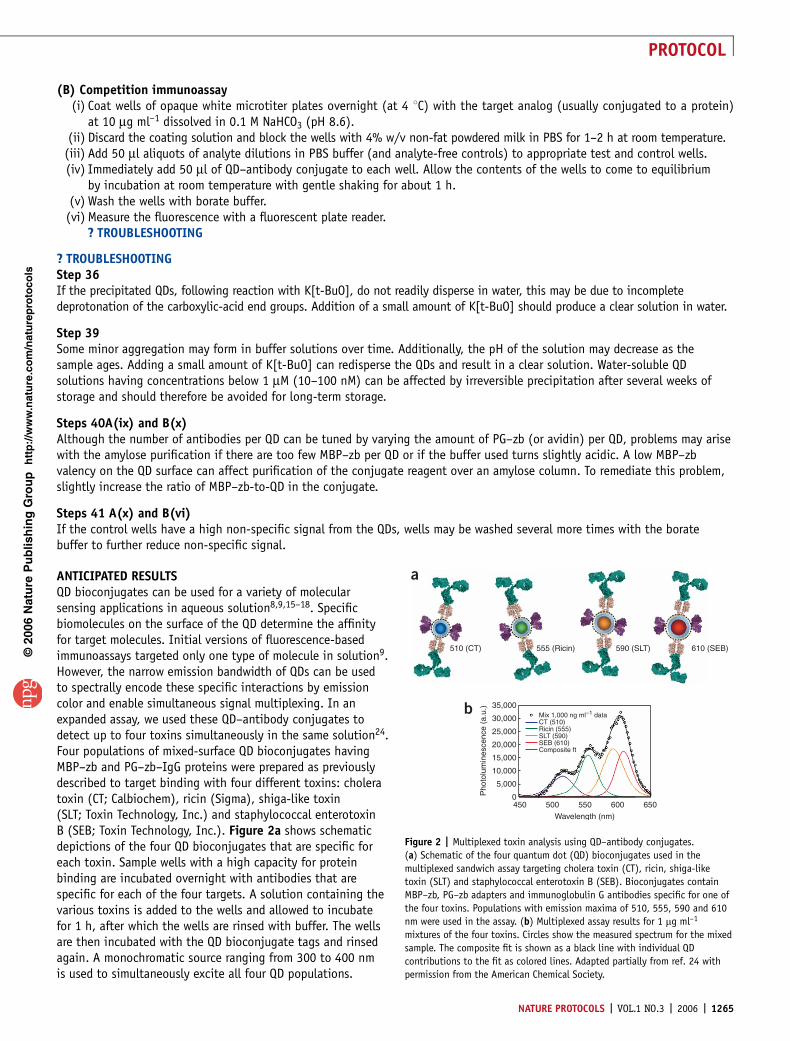
ANTICIPATED RESULTSQD bioconjugates can be used for a variety of molecularsensing applications in aqueous solution8,9,15–18. Specificbiomolecules on the surface of the QD determine the affinityfor target molecules. Initial versions of fluorescence-basedimmunoassays targeted only one type of molecule in solution9.However, the narrow emission bandwidth of QDs can be usedto spectrally encode these specific interactions by emissioncolor and enable simultaneous signal multiplexing. In anexpanded assay, we used these QD–antibody conjugates todetect up to four toxins simultaneously in the same solution24.Four populations of mixed-surface QD bioconjugates havingMBP–zb and PG–zb–IgG proteins were prepared as previouslydescribed to target binding with four different toxins: choleratoxin (CT; Calbiochem), ricin (Sigma), shiga-like toxin(SLT; Toxin Technology, Inc.) and staphylococcal enterotoxinB (SEB; Toxin Technology, Inc.). Figure 2a shows schematicdepictions of the four QD bioconjugates that are specific foreach toxin. Sample wells with a high capacity for proteinbinding are incubated overnight with antibodies that arespecific for each of the four targets. A solution containing thevarious toxins is added to the wells and allowed to incubatefor 1 h, after which the wells are rinsed with buffer. The wellsare then incubated with the QD bioconjugate tags and rinsedagain. A monochromatic source ranging from 300 to 400 nmis used to simultaneously excite all four QD populations.
p
uor
G g
n ih si l
bu
P eru ta
N 600 2©
nat
ure
pro
toco
ls/
moc.er
ut an.
ww
w//:ptt
h
4500
5,000
10,000
15,000
20,000
25,000
30,000 Mix 1,000 ng ml–1 dataCT (510)Ricin (555)SLT (590)SEB (610)Composite ft
35,000
500 550
Wavelength (nm)
Pho
tolu
min
esce
nce
(a.u
.)
600 650
a
b
555 (Ricin)510 (CT) 590 (SLT) 610 (SEB)
Figure 2 | Multiplexed toxin analysis using QD–antibody conjugates.
(a) Schematic of the four quantum dot (QD) bioconjugates used in the
multiplexed sandwich assay targeting cholera toxin (CT), ricin, shiga-like
toxin (SLT) and staphylococcal enterotoxin B (SEB). Bioconjugates contain
MBP–zb, PG–zb adapters and immunoglobulin G antibodies specific for one of
the four toxins. Populations with emission maxima of 510, 555, 590 and 610
nm were used in the assay. (b) Multiplexed assay results for 1 mg ml–1
mixtures of the four toxins. Circles show the measured spectrum for the mixed
sample. The composite fit is shown as a black line with individual QD
contributions to the fit as colored lines. Adapted partially from ref. 24 with
permission from the American Chemical Society.
NATURE PROTOCOLS | VOL.1 NO.3 | 2006 | 1265
PROTOCOL

Detection of fluorescence emission at a certain color (i.e.,wavelength range) following signal deconvolution indicatesthe presence of a sandwich-binding interaction and is apositive indicator for that particular toxin in solution.Figure 2b shows data from a multiplexed sandwichimmunoassay for CT, SEB, SLT and ricin. Goat anti-choleratoxin (Biogenesis), monoclonal anti-SEB 6B ascites (BioVeris)further purified by MEP Hypercel affinity chromotography(Ciphergen), a pool of monoclonal anti-SLT antibodies(9C9, 3C10, BB12; Toxin Technology, Inc.) and monoclonalanti-ricin RIC-03-A-G1 (a gift from the Naval MedicalResearch Center) were used as the capture antibodiesadsorbed onto the wells of plates. Rabbit anti-CT antibody(Biogenesis) was coupled to 510 nm emitting QDs, polyclonalrabbit anti-ricin (a gift from the Naval Medical ResearchCenter) was conjugated to 550 nm emitting QDs, the poolof monoclonal anti-SLT antibodies (9C9, 3C10, BB12;Toxin Technology, Inc.) was coupled to 590 nm emittingQDs, and polyclonal rabbit anti-SEB antibody (ToxinTechnology, Inc.) was coupled to 610 nm emitting QDs. The PG–zb conjugation strategy detailed in Step 41 (A) and (B)was used to prepare the various QD–antibody conjugates used in these experiments.
A competition assay specific for soluble 2,4,6-trinitrotoluene (TNT) is constructed using biomolecules that have beenengineered to express terminal polyhistidine that binds to QDs. Competition assays are frequently used in the detectionof small molecules. Figure 3 shows data from a competition immunoassay for TNT using QDs coated with both scFv andMBP–zb17. Ovalbumin derivatized by modification with trinitro-benzenesulfonic acid (TNB–ovalbumin) was used to coat wellsof a fluorescent microtiter plate. Varying amounts of soluble TNT were added to the wells, followed by the addition of anti-TNTscFv/MBP–zb QD bioconjugates. The wells were incubated for 1 h to achieve equilibrium and washed prior to fluorescencemeasurement in the plate reader.
ACKNOWLEDGMENTS This work was supported by grants from the Office of NavalResearch (ONR) and DARPA. A.R.C. was supported by a National Research Councilfellowship.
COMPETING INTERESTS STATEMENT The authors declare that they have nocompeting financial interests.
Published online at http://www.natureprotocols.comReprints and permissions information is available online at http://npg.nature.com/reprintsandpermissions
1. Gaponenko, S.V. Optical Properties of Semiconductor Nanocrystals (CambridgeUniversity Press Cambridge, UK, 1998).
2. Michalet, X. et al. Quantum dots for live cells, in vivo imaging, and diagnostics.Science 307, 538–544 (2005).
3. Medintz, I.L., Uyeda, H.T., Goldman, E.R. & Mattoussi, H. Quantum dotbioconjugates for imaging, labeling and sensing. Nature Materials 4, 435–446(2005).
4. Dubertret, B. et al. In vivo imaging of quantum dots encapsulated in phospholipidmicelles. Science 298, 1759–1762 (2002).
5. Wu, X.Y. et al. Immunofluorescent labeling of cancer marker Her2 and othercellular targets with semiconductor quantum dots. Nature Biotech. 21, 41–46(2003).
6. Mattoussi, H. et al. Self-assembly of CdSe-ZnS quantum dot bioconjugatesusing an engineered recombinant protein. J. Am. Chem. Soc. 122, 12142–12150(2000).
7. Kim, S. & Bawendi, M.G. Oligomeric ligands for luminescent and stablenanocrystal quantum dots. J. Am. Chem. Soc. 125, 14652–14653 (2003).
8. Goldman, E.R. et al. Conjugation of luminescent quantum dots with antibodiesusing an engineered adaptor protein to provide new reagents forfluoroimmunoassays. Anal. Chem. 74, 841–847 (2002).
9. Goldman, E.R. et al. Avidin: a natural bridge for quantum dot-antibodyconjugates. J. Am. Chem. Soc. 124, 6378–6382 (2002).
10. Murray, C.B., Norris, D.J. & Bawendi, M.G. Synthesis and characterization ofnearly monodisperse CdE (E ¼ sulfur, selenium, tellurium) semiconductornanocrystallites. J. Am. Chem. Soc. 115, 8706–8715 (1993).
11. Peng, Z.A. & Peng, X. Formation of high-quality CdTe, CdSe, and CdS nanocrystalsusing CdO as precursor. J. Am. Chem. Soc. 123, 183–184 (2001).
12. Hines, M.A. & Guyot-Sionnest, P. Synthesis and characterization of stronglyluminescing ZnS-capped CdSe nanocrystals. J. Phys. Chem. 100, 468–471 (1996).
13. Dabbousi, B.O. et al. CdSe-ZnS core-shell quantum dots: synthesis andcharacterization of a size series of highly luminescent materials.J. Phys. Chem. B 101, 9463–9475 (1997).
14. Peng, X., Schlamp, M.C., Kadavanich, A.V. & Alivisatos, A.P. Epitaxial growth ofhighly luminescent CdSe/CdS core/shell nanocrystals with photostability andelectronic accessibility. J. Am. Chem. Soc. 119, 7019–7029 (1997).
15. Medintz, I.L. et al. Self-assembled nanoscale biosensors based on quantumdot FRET donors. Nature Mat. 2, 630–638 (2003).
16. Clapp, A.R. et al. Fluorescence resonance energy transfer between quantumdot donors and dye-labeled protein acceptors. J. Am. Chem. Soc. 126,301–310 (2004).
17. Goldman, E.R. et al. Self-assembled luminescent CdSe-ZnS quantum dotbioconjugates prepared using engineered poly-histidine terminated proteins.Anal. Chim. Acta 534, 63–67 (2005).
18. Goldman, E.R. et al. A hybrid quantum dot-antibody fragment fluorescenceresonance energy transfer-based TNT sensor. J. Am. Chem. Soc. 127,6744–6751 (2005).
19. Lakowicz, J.R. Principles of Fluorescence Spectroscopy 2nd edn (Kluwer Academic/Plenum Publishers, New York, 1999).
20. Goldman, E.R., Mattoussi, H., Anderson, G.P., Medintz, I.L. & Mauro, J.M.in Methods in Molecular Biology Vol. 303: NanoBiotechnology Protocols(eds. Rosenthal, S.J. & Wright, D.W.) 19–33 (Humana Press Inc., Totowa, NJ, 2005).
21. Chang, H.C. et al. A general method for facilitating heterodimeric pairing between2 proteins — applications to expression of a and b-T-cell receptor extracellularsegments. Proc. Natl. Acad. Sci. USA 91, 11408–11412 (1994).
22. Goldman, E.R. et al. 2,4,6-trinitrotoluene detection using recombinantantibodies. J. Environ. Monit. 5, 380–383 (2003).
23. Gunsalus, I.C., Barton, L.S. & Gruber, W. Biosynthesis and structure of lipoic acidderivatives. J. Am. Chem. Soc. 78, 1763–1768 (1956).
24. Goldman, E.R. et al. Multiplexed toxin analysis using four colors of quantum dotfluororeagents. Anal. Chem. 76, 684–688 (2004).
p
uor
G g
n ih si l
bu
P eru ta
N 600 2©
nat
ure
pro
toco
ls/
moc.er
ut an.
ww
w//:ptt
h
120
100
80
60
Per
cent
sig
nal
TNT concentration (µg ml–1)
40
20
0
0.001 0.01 0.1 1 10 100
Figure 3 | Results from a competition assay using QD–anti-TNT scFv conjugate
to detect soluble TNT. Alone, quantum dot (QD) bioconjugates with attached
anti-TNT (4,6-trinitrotoluene) scFv bind to surface-immobilized trinitro-
benzenesulfonic acid (TNB). As the concentration of free TNT added to the
solution increases, fewer QD bioconjugates bind to the surface-immobilized
TNB (due to competition with free TNT) and the measured fluorescence signal
decreases. Adapted partially from ref. 17 with permission from Elsevier.
1266 | VOL.1 NO.3 | 2006 | NATURE PROTOCOLS
PROTOCOL

ERRATUM
Erratum: Capping of CdSe–ZnS quantum dots with DHLA and subsequent conjugation with proteinsAaron R Clapp, Ellen R Goldman and Hedi Mattoussi
Nat. Protocols doi:10.1038/nprot.2006.184; published online 28 September; corrected online 30 November 2006.
In the version of this article initially published online, the article’s page numbers should have been 1258–1266. This error has been corrected in the PDF version of the article.