ultra sensitive detection of influenza a virus based on ... · introduction influenza viruses ......
TRANSCRIPT

*Corresponding author email: [email protected] GroupSymbiosis Group
Symbiosis ISSN: 2376-4589DOI: http://dx.doi.org/10.15226/2376-4589/2/3/00108
Ultra Sensitive Detection of Influenza A Virus Based on Cdse/Zns Quantum Dots
ImmunoassayFeng Wu1,2#, Mao Mao1#, Qian Liu2, Lei Shi3, Yu Cen2, Zhifeng Qin3 and Lan Ma2*
1Key Laboratory for Special Functional Materials, Henan University, Kaifeng 475004, P. R. China2Division of Life Science and Health, Graduate School at Shenzhen, Tsinghua University, Shenzhen, 518055, P. R. China
3Shenzhen Entry-Exit Inspection and Quarantine Bureau of the People’s Republic of China (SZCIQ), Shenzhen, 518045, P.R. China
#Authors contributed equally to this article
SOJ Biochemistry Open AccessResearch article
severe outbreaks disease, and they can cause serious infections among people.
To date, numerous analytical methods have being used to detect influenza A virus. According to the type of detection target, these methods could be categorized into, for example, virus isolation and identification, nucleic acid-based detection, antigen detection, and antibody detection [3-8]. Most of these methods need demanding conditions and professional operations, and some of the detection processes is time-consuming. For these reasons, some new biosensors based on nano materials such as gold nanoparticles, magnetic nano beads and quantum dots (QDs)have been coming to the forefront [9-12].Developing a sensitive, specific and fast biosensor for diagnosing the influenza virus at the early stages of infection is a challenge. Lateral flow immune assay method has been widely used in detection of infectious diseases with the advantages of rapid, easy to operate, and low cost [13]. However, traditional method such as colloidal gold lateral flow tests has limitations when high sensitivity is needed [14].QDs have been developed to replace colloidal gold owing to their excellent optical properties such as high fluorescence efficiency, wide range of excitation wavelength, narrow and symmetric emission spectra, and QDs based lateral flow tests have been proved higher sensitivity and reproducibility for detection of pathogen, characteristic protein and even nucleic acid [15-18].
In this paper, we present an ultrasensitive lateral flow immunoassay (LFIA) for detecting influenza A virus. This immune sensor is based on site-specific covalent binding with the Fc end of influenza A virus antibodies to CdSe/ ZnS QDs. The carboxyl-functionalized CdSe/ ZnS QDs conjugated with antibodies via an amide bond often result in random links of the antibody structure in the QD conjugates and block certain antigen-binding sites. In order to obtain site-specific linking, we modified carboxyl-functionalized QDs with adipicdihydrazide (ADH) and oxidized the carbohydrate groups on the antibody’s Fc region
AbstractAn ultrasensitive lateral flow immunoassay system (LFIAS)
was established for the detection of influenza A virus. In this LFIAS, hydrophilic dihydrazide-modified CdSe/ZnSquantom dots (QDs) were conjugated with specific antibodies and used as fluorescent labels, and a pair of matched anti-nucleoprotein of influenza A virus antibodies were used to form a sandwich immunoassay. The QDs were in conjugation with the fragment crystallizable region (Fc region) of specific influenza A virus antibodies through aldehyde-hydrazide covalent chemistry, conferring high sensitivity. The antibodies used for detection are specific for the most conserved and popular nucleoprotein of influenza A virus and ensure the accuracy and specificity. The QDs-LFIAS can analyze the nasal-pharyngeal swab samples through simple steps and get results within 15 min. Detection of nasal-pharyngeal swab samples makes it more rapid and convenient, and it is highly efficient for identification of influenza infection and improves influenza patients’ management. The limit of detection of this QDs-LFIAS for recombinant nucleoprotein of influenza A virus was 0.01 ng/ mL, which was 1000-fold higher than the sensitivity of colloidal gold method. The detection of actual patient samples indicated that the QDs-LFIAS had a high compliance with real-time PCR.
Keywords: Lateral flow immunoassay; Influenza A virus; CdSe/ZnS; Quantum dots
Received: 24 November, 2016; Accepted: 16 December, 2016; Published: 28 December, 2016
*Corresponding author: Lan Ma, Division of Life Science and Health, Graduate School at Shenzhen, Tsinghua University, Shenzhen, 518055, P. R. China, E-mail: [email protected]
IntroductionInfluenza viruses circulate each year and cause mild to severe
illness and even death in humans. Type A influenza can outbreaks in seasonal and regional, and the viruses are susceptible to mutation. On the basis of the antigenic nature of the surface glycoproteins, hemagglutinin (HA) and neuraminidase (NA), type A influenza viruses are subdivided into several subtypes [1]. At present, the seasonal influenza A virus subtypes caused by human infection is influenza A (H1N1) and A (H3N2) [2]. Avian influenza viruses such as A (H5N1) A (H5N6), A (H7N9) and A (H9N2) can sometimes spread to domestic poultry and cause
Page 2 of 6Citation: Wu F, Mao M, Liu Q, Shi L, Ma L, et al. (2016) Ultra Sensitive Detection of Influenza A Virus Based on Cdse/Zns Quantum Dots Immunoassay. SOJ Biochem 2(3), 6.
Ultra Sensitive Detection of Influenza A Virus Based on Cdse/Zns Quantum Dots Immunoassay
Copyright: © 2016 Ma et al.
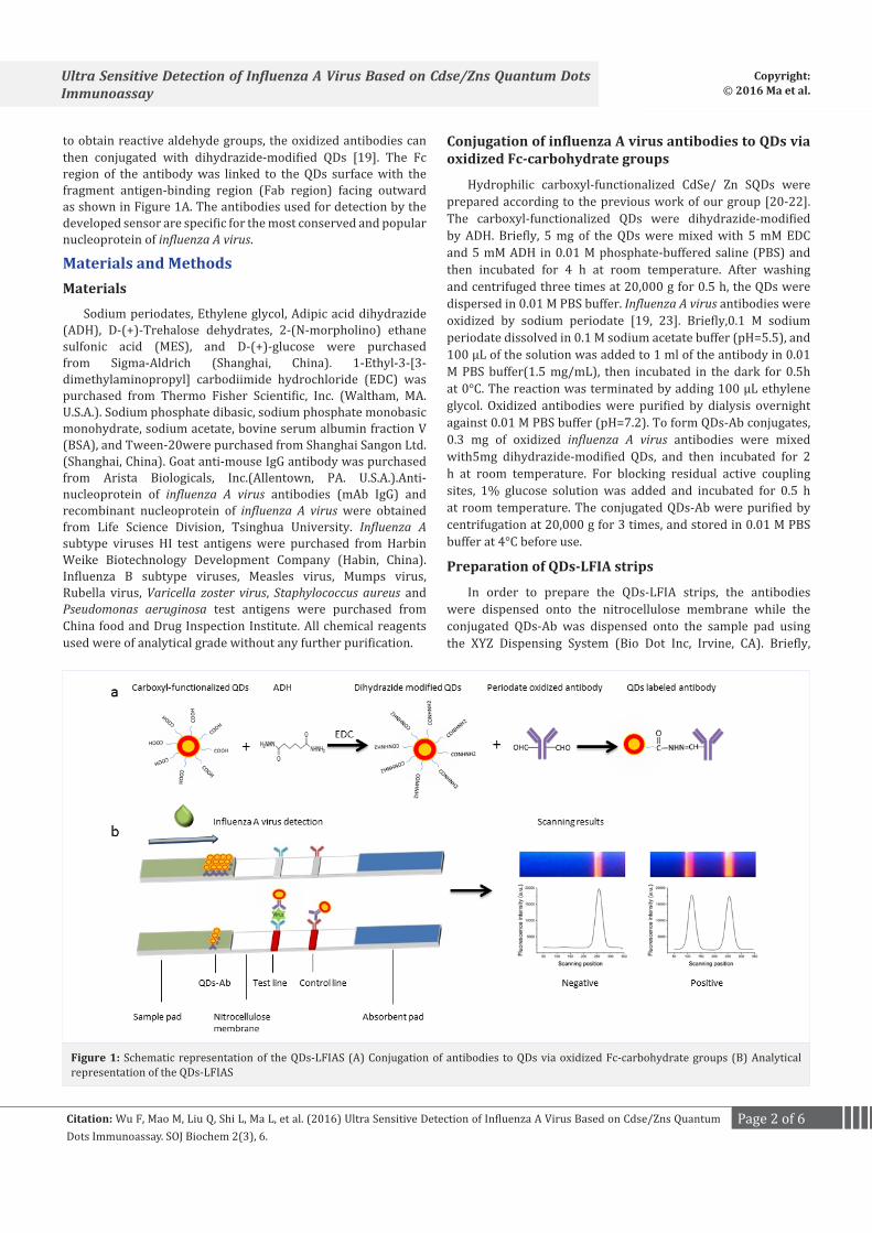
to obtain reactive aldehyde groups, the oxidized antibodies can then conjugated with dihydrazide-modified QDs [19]. The Fc region of the antibody was linked to the QDs surface with the fragment antigen-binding region (Fab region) facing outward as shown in Figure 1A. The antibodies used for detection by the developed sensor are specific for the most conserved and popular nucleoprotein of influenza A virus.
Materials and MethodsMaterials
Sodium periodates, Ethylene glycol, Adipic acid dihydrazide (ADH), D-(+)-Trehalose dehydrates, 2-(N-morpholino) ethane sulfonic acid (MES), and D-(+)-glucose were purchased from Sigma-Aldrich (Shanghai, China). 1-Ethyl-3-[3-dimethylaminopropyl] carbodiimide hydrochloride (EDC) was purchased from Thermo Fisher Scientific, Inc. (Waltham, MA. U.S.A.). Sodium phosphate dibasic, sodium phosphate monobasic monohydrate, sodium acetate, bovine serum albumin fraction V (BSA), and Tween-20were purchased from Shanghai Sangon Ltd. (Shanghai, China). Goat anti-mouse IgG antibody was purchased from Arista Biologicals, Inc.(Allentown, PA. U.S.A.).Anti-nucleoprotein of influenza A virus antibodies (mAb IgG) and recombinant nucleoprotein of influenza A virus were obtained from Life Science Division, Tsinghua University. Influenza A subtype viruses HI test antigens were purchased from Harbin Weike Biotechnology Development Company (Habin, China).Influenza B subtype viruses, Measles virus, Mumps virus, Rubella virus, Varicella zoster virus, Staphylococcus aureus and Pseudomonas aeruginosa test antigens were purchased from China food and Drug Inspection Institute. All chemical reagents used were of analytical grade without any further purification.
Conjugation of influenza A virus antibodies to QDs via oxidized Fc-carbohydrate groups
Hydrophilic carboxyl-functionalized CdSe/ Zn SQDs were prepared according to the previous work of our group [20-22]. The carboxyl-functionalized QDs were dihydrazide-modified by ADH. Briefly, 5 mg of the QDs were mixed with 5 mM EDC and 5 mM ADH in 0.01 M phosphate-buffered saline (PBS) and then incubated for 4 h at room temperature. After washing and centrifuged three times at 20,000 g for 0.5 h, the QDs were dispersed in 0.01 M PBS buffer. Influenza A virus antibodies were oxidized by sodium periodate [19, 23]. Briefly,0.1 M sodium periodate dissolved in 0.1 M sodium acetate buffer (pH=5.5), and 100 µL of the solution was added to 1 ml of the antibody in 0.01 M PBS buffer(1.5 mg/mL), then incubated in the dark for 0.5h at 0°C. The reaction was terminated by adding 100 µL ethylene glycol. Oxidized antibodies were purified by dialysis overnight against 0.01 M PBS buffer (pH=7.2). To form QDs-Ab conjugates, 0.3 mg of oxidized influenza A virus antibodies were mixed with5mg dihydrazide-modified QDs, and then incubated for 2 h at room temperature. For blocking residual active coupling sites, 1% glucose solution was added and incubated for 0.5 h at room temperature. The conjugated QDs-Ab were purified by centrifugation at 20,000 g for 3 times, and stored in 0.01 M PBS buffer at 4°C before use.
Preparation of QDs-LFIA strips
In order to prepare the QDs-LFIA strips, the antibodies were dispensed onto the nitrocellulose membrane while the conjugated QDs-Ab was dispensed onto the sample pad using the XYZ Dispensing System (Bio Dot Inc, Irvine, CA). Briefly,
Figure 1: Schematic representation of the QDs-LFIAS (A) Conjugation of antibodies to QDs via oxidized Fc-carbohydrate groups (B) Analytical representation of the QDs-LFIAS

Page 3 of 6Citation: Wu F, Mao M, Liu Q, Shi L, Ma L, et al. (2016) Ultra Sensitive Detection of Influenza A Virus Based on Cdse/Zns Quantum Dots Immunoassay. SOJ Biochem 2(3), 6.
Ultra Sensitive Detection of Influenza A Virus Based on Cdse/Zns Quantum Dots Immunoassay
Copyright: © 2016 Ma et al.
anti-influenza A virus coating antibody was dispensed onto the nitrocellulose membrane at 2 mg/ mL as the test line, and the goat anti-mouse IgG was dispensed at 0.5 mg/ mL as the control line. The dispensed nitrocellulose membranes were dried at 37°C in a vacuum oven for 4 h. The sample pad was pretreated by PBS buffer containing D-(+)-trehalose dehydrate (1%, w/ v), BSA (1%, w/v) and Tween-20 (0.1%, w/v), and dried at 37°C for 3 h. The conjugated QDs-Ab was dispensed at a ratio of 10 µL/ cm onto the pretreated sample pad and dried at 37°C for 3 h. The QDs-LFIA strip was assembled in its standard configuration as shown in Figure 1B. The completed QDs-LFIA was cut into individual 3.5 mm strips and stored in sealed and dry condition.
Analytical procedure
Sixty micro liter of analyte was added onto the sample port of QDs-LFIA strip, the captured fluorescence intensity of test line and control line were scanned by a fluorescence test strip scanning device after a 15 min reaction. The captured fluorescence QDs on the test line and control line produced a bright fluorescent band in response to 365 nm ultraviolet excitation, and the fluorescence intensity at 620 nm were detected by the device [22]. As shown in Figure 1B, once influenza A virus was in the sample, the QDs-Abbound the influenza A virus specifically and were later captured by the second anti-influenza A virus antibody at the test line, and QDs-Ab without influenza A virus bound were captured by the goat anti-mouse IgG at the control line, the captured QDs-Ab aggregated into a fluorescent band under 365 nm ultra violet excitation. The fluorescence intensity of test line was closely correlated with the concentration of captured QDs-Ab and virus complex..The cutoff value was obtained by detecting 50 negative samples, the average of fluorescence intensity was calculated, and two times of the average value was defined as the cutoff value. The calculated cut off value is 102 a.u. Fluorescence intensity of 102 a.u. or above was defined as positive, while less than 102 a.u. was negative. The limit of detection (LOD) was estimated by analyzing samples at various concentrations. Briefly, the recombinant nucleoprotein of influenza A virus was ten-fold diluted by 20 mM PBS (1000, 100, 10, 1, 0.1, 0.01 and 0.001 ng/ mL) and test separately, 20 mM PBS was test as a negative control. The fluorescence intensity of test line was detected while the LOD was calculated. The LOD was defined as the lowest concentration of recombinant nucleoprotein whose fluorescence intensity was the minimum above the cutoff value. We also test the reproducibility of QDs-LFIA strip by analyzing 20 replicates of recombinant nucleoprotein sample sat different concentrations (1, 10 and 100 ng/ mL). The specificity and cross-reactivity was analyzed by detecting influenza A virus subtypes(H1N1, H3N2, H5N1 re-4/6, H7N9, H9N2 re-2,)HI test antigens, influenza B virus subtypes(1704 strain, Victoria strain, Yamagata strain),Measles virus, Mumps virus, Rubella virus, Varicella zoster virus, Staphylococcus aureus and Pseudomonas aeruginosa.
Practical field sample tests
Sixty samples (human throat swabs collected into sterile Hanks’ balanced salt solution viral transport media)—which
were collected and preserved by Shenzhen International Travel Health Care Center, Shenzhen Entry-Exit Inspection and Quarantine Bureau—were assayed using QDs-LFIA strips. Commercial influenza A antigen rapid diagnostic test kit (colloidal gold) was conducted in parallel, and real-time PCR assay was used as a reference method to evaluate the accuracy of QDs- LFIAS. A pair of specific primers (Forward Primer 5’-GACCRATCCTGTCACCTCTGAC-3’, Reverse Primer 5’-AGGGCATTYTGGACAAAKCGTCTA-3’) and a probe (FAM-TGCAGTCCTCGCTCACTGGGCACG-BHQ1) for detecting influenza A virus was used in real-time PCR assay.
Results and discussionConjugation of antibodies to QDs via oxidized Fc-carbohydrate groups
The reaction mechanism of hydrophilic carboxyl-functionalized CdSe/ ZnS QDs has been previously described in the literature [24, 25]. The dynamic light scattering analysis in Figure 2 shows that the average hydrodynamic size of carboxyl-functionalized CdSe/ Zn SQDs was 42.86 nm with Stdev 18.72 nm, and this size increased to 109.5 nm with Stdev 52.16 nm after dihydrazide modified and conjugation with antibodies, which indicated that antibodies were successful conjugated.
Limit of detection of QDs-LFIA strip
The LOD was estimated by analyzing recombinant nucleoprotein of influenza A virus samples at various concentrations, the fluorescence intensity of test line was scanned by fluorescence test strip scanner after 15 min of addition of the samples. The diluted recombinant nucleoprotein of influenza A virus samples was also tested by commercial influenza A antigen rapid diagnostic test kit (colloidal gold). As demonstrated by Figure3A, with the recombinant nucleoprotein concentration increasing, the fluorescent band of test line at 0.1 ng/ mL was still visible. The commercial influenza A antigen rapid diagnostic test kit (colloidal gold) could only detect the recombinant nucleoprotein at 10 ng/ mL concentration through Figure 3B. As show in Figure 3C, employing the fluorescence test strip scanning device, the recombinant nucleoprotein could be detected at 0.01 ng/ mL by QDs-LFIA strips, which was 1000-fold higher than the sensitivity of colloidal gold method.
Figure 2: DLS data of QDs and conjugated QDs-Ab. (A) Carboxyl-func-tionalized QDs. (B) Antibody conjugated QDs. Average hydrodynamic size of CdSe/ZnS QDs was 42.86 nm and this size increased to 109.5 nm after conjugation with antibodies

Page 4 of 6Citation: Wu F, Mao M, Liu Q, Shi L, Ma L, et al. (2016) Ultra Sensitive Detection of Influenza A Virus Based on Cdse/Zns Quantum Dots Immunoassay. SOJ Biochem 2(3), 6.
Ultra Sensitive Detection of Influenza A Virus Based on Cdse/Zns Quantum Dots Immunoassay
Copyright: © 2016 Ma et al.
Reproducibility of QDs-LFIA stripThe reproducibility of this QDs-LFIA strip was analysed
by detecting 20 replicates of the recombinant nucleoprotein at various concentrations (1, 10 and 100 ng/mL). The fluorescence intensity of test line was detected while the relative standard deviations (RSD) were later calculated. Table 1 shows the RSD results and the RSD values were below 8%, demonstrating the QDs-LFIA strip had good reproducibility.
Specificity and cross-reactivity of QDs-LFIA stripThe specificity and cross-reactivity of the QDs-LFIA strip was
analyzed by detecting influenza A virus subtypes (H1N1, H3N2, H5N1 re-4/6, H7N9, H9N2 re-2,) HI test antigens, influenza B virus subtypes(1704 strain, Victoria strain, Yamagata strain), Measles virus, Mumps virus, Rubella virus, Varicella zoster virus, Staphylococcus aureus and Pseudomonas aeruginosa. Figure 4A shows that the QDs-LFIA strip could detect all the subtypes of influenza A virus used but none of other type antigens. We also tested the influenza A virus subtypes with concentration ranging from 1/512 to 16HAU. As demonstrated in Figure4B, the QDs-LFIA could detect influenza A virus subtype H1N1 at 1/256 HAU, influenza A virus subtype H5N1 at 1/32 HAU, influenza A virus subtype H3N2, H7N9 and H9N2 at 1/128 HAU. It demonstrated that the QD-LFIAS could detect influenza A subtype viruses with high sensitivity and specificity.
Practical field sample testsSixty samples (human throat swabs collected into sterile
Hanks’ balanced salt solution viral transport media) were detected using QDs-LFIAS. Table 2 shows that all positive samples with low real-time PCR threshold cycle (Ct ≤ 30) were detected by QDs- LFIAS with a high accuracy. For the positive samples with high real-time PCR threshold cycle (Ct ˃ 30), three were detected as negative by QDs-LFIAS, but all were detected as negative by commercial influenza A antigen rapid diagnostic test kit (colloidal gold). The results indicated that the QDs-LFIAS had an accuracy of 95%, while that of the commercial influenza A antigen rapid diagnostic test kit (colloidal gold) was 56.7% compared with real-time PCR.
At the early stage of influenza A virus infection, the symptoms of patients were similar to those of common cold and it was difficult and insufficient to use these common symptoms for the diagnosis of type A influenza virus infection [26]. Besides, the influenza A virus titer was very low during the early infection. So, high sensitivity is the key for diagnosing the influenza virus at the early stages of infection. Real-time PCR method has ultra-high accuracy but time consuming, as point-of-care testing
Figure 3: (A) Images of tested QDs-LFIAS in response to excitation with 365 nm ultraviolet light. (B) Images of tested commercial influenza A antigen rapid diagnostic test strip (colloidal gold). (C). Fluorescence in-tensity scans at different concentrations of recombinant nucleoprotein of influenza A virus measured by the fluorescence strip scanning device. Shows the fluorescence from test line of QDs-LFIA strips at 0.1ng/mL was still visible. B shows the test line of commercial influenza A anti-gen rapid diagnostic test kit (colloidal gold) at 10 ng/mL was visible. C shows the test line of QDs-LFIA strips at 0.01 ng/mL can be detected by fluorescence test strip scanning device.
Table 1: Reproducibility Tests of QDs-LFIAS. 20 replicates of the recombinant nucleoprotein at various concentrations (1, 10 and 100 ng/mL) were test by QDs-LFIAS. The relative standard deviations (RSD) were then calculated accordingly.
Concentrations (ng/mL)
Fluorescence intensity average
(a.u.)
Stdev RSD/ %
(a.u.) n=201 650.5 42.5 6.5
10 2957.8 217.8 7.4100 13827.8 820.7 5.9
Figure 4: (A) Specificity tests of QDs-LFIAS. (B) Fluorescence intensity scans at different concentrations of influenzaA virus subtypes. Shows QDs-LFIAS could detect all the subtypes of influenza A virus used but none of other type antigens. B shows QDs-LFIAS could detect the sub-types of influenza A virus with high sensitivity.
Table 2: Practical field sample Tests using QDs-LFIAS. Sixty human throat swab samples were detected using QDs- LFIAS, commercial influenza A antigen rapid diagnostic test kit and real-time PCR. Compared with real-time PCR, the QDs-LFIAS had an accuracy of 95%, while that of the commercial infl uenzaA antigen rapid diagnostic test kit (colloidal gold) was 56.7%.
Samples Number of samples
QDs-LFIAS result(P/N)
Commercial rapid
diagnostic test kit
result(P/N)
Real-time PCR
result(P/N)
Real-time PCR positive
result20 17/3 0/20 20/0
Ct > 30Real-
time PCR positive
result20 20/0 14/6 20/0
Ct ≤ 30Negative sample 20 0/20 0/20 0/20
Page 5 of 6Citation: Wu F, Mao M, Liu Q, Shi L, Ma L, et al. (2016) Ultra Sensitive Detection of Influenza A Virus Based on Cdse/Zns Quantum Dots Immunoassay. SOJ Biochem 2(3), 6.
Ultra Sensitive Detection of Influenza A Virus Based on Cdse/Zns Quantum Dots Immunoassay
Copyright: © 2016 Ma et al.
method, the QDs-LFIAS was proved to detect human throat swab samples more sensitive and accurate than colloidal gold method. Detection of nasal-pharyngeal swab samples makes it more rapid and convenient, and it is highly efficient for monitoring and prevention of influenza outbreak in the hospital emergency, port quarantine, schools and also in-home health care. QDs-LFIAS is a low-cost technique with small amount of immunoassay reagents consumed, and results can be objectively determined by a handheld device. According to our results above, the QDs-LFIAS only requires one step and provides results in 15 min. This simple and less time consuming method could be used in on-site tests, clinical diagnosis and early treatments of influenza virus infection.
ConclusionsThe purpose of this study was to develop an ultrasensitive,
rapid and low cost lateral flow immune sensor for influenza A virus preliminary screening. We developed a QDs-LFIAS method, which rapidly analyzed the sample through one steps. The results were objectively analyzed by an inexpensive, portable device within 15min. The LOD of this QDs-LFIAS for recombinant nucleoprotein of influenza A virus was 0.01ng/ mL, which was 1000-fold higher than the sensitivity of colloidal gold method. The QDs-LFIAS could detect influenza A virus subtype H1N1 at a concentration of 1/ 256 HAU, influenza A virus subtype H5N1 at 1/ 32 HAU, influenza A virus subtype H3N2, H7N9 and H9N2 at 1/128 HAU. It demonstrated that the QD-LFIAS could detect influenza A virus subtypes with high sensitivity and specificity. This was more sensitive than that of traditional point-of-care testing methods. The specificity and reproducibility were shown to be good. Real patient samples demonstrated that the QDs-LFIAS had high accuracy, and detection of nasal-pharyngeal swab samples makes it more rapid and efficient for identification of influenza infection and improves influenza patients’ management.
AcknowledgmentsThis work was supported by the following sources:
the National High Technology Research and Development Program of China (863 Program, NO. 2013AA032204), Science and Technology Planning Project of Guangdong Province (2012B031500003), Shenzhen strategic emerging industry development special funds (JSGG20140716144254155)
Conflict of interest
The authors declare that there is no conflict of interest regarding the publication of this paper.
References1. Hinshaw VS, Air GM, Gibbs AJ, Graves L, Prescott B, Karunakaran D.
Antigenic and Genetic-Characterization of a Novel Hemagglutinin Subtype of Influenza-a Viruses from Gulls. Journal of Virology. 1982;42(3):865-872.
2. The World Health Organization Media Center Page,2014.
3. Chen HT, Zhang J, Sun DH, Ma LN, Liu XT, Cai XP, et al. Development of reverse transcription loop-mediated isothermal amplification for rapid detection of H9 avian influenza virus. Journal of virological methods. 2008;151(2):200-203. doi:10.1016/j.jviromet.2008.05.009.
4. Alberini I, Del Tordello E, Fasolo A, Temperton NJ, Galli G, Gentile C, et al. Pseudoparticle neutralization is a reliable assay to measure immunity and cross-reactivity to H5N1 influenza viruses. Vaccine. 2009;27(43):5998-6003. doi: 10.1016/j.vaccine.2009.07.079.
5. Moore C, Telles JN, Corden S, Gao RB, Vernet G, Van Aarle P, et al. Development and validation of a commercial real-time NASBA assay for the rapid confirmation of influenza A H5N1 virus in clinical samples. Journal of virological methods. 2010;170(1-2):173-176. doi:10.1016/j.jviromet.2010.09.014.
6. Yang SY, Chieh JJ, Wang WC, Yu CY, Lan CB, Chen JH, et al. Ultra-highly sensitive and wash-free bio-detection of H5N1 virus by immunomagnetic reduction assays. Journal of virological methods. 2008;153(2):250-252. doi:10.1016/j.jviromet.2008.07.025.
7. Xie Z, Pang YS, Liu J, Deng X, Tang X, Sun J, et al. A multiplex RT-PCR for detection of type A influenza virus and differentiation of avian H5, H7, and H9 hemagglutinin subtypes. Molecular and cellular probes. 2006;20(3-4):245-249. doi:10.1016/j.mcp.2006.01.003.
8. Payungporn S, Chutinimitkul S, Chaisingh A, Damrongwantanapokin S, Buranathai C, Amonsin A et al. Single step multiplex real-time RT-PCR for H5N1 influenza A virus detection. Journal of virological methods. 2006;131(2):143-147. doi:10.1016/j.jviromet.2005.08.004.
9. Kamikawa TL, Mikolajczyk MG, Kennedy M, Zhang P, Wang W, Scott DE, et al. Nanoparticle-based biosensor for the detection of emerging pandemic influenza strains. Biosensors & bioelectronics. 2010;26(4):1346-1352. doi: 10.1016/j.bios.2010.07.047.
10. Krejcova L, Nejdl L, Rodrigo MA, Zurek M, Matousek M, Hynek D, et al. 3D printed chip for electrochemical detection of influenza virus labeled with CdS quantum dots. Biosensors & bioelectronics. 2014;54:421-427. doi: 10.1016/j.bios.2013.10.031.
11. Lee C, Gaston MA, Weiss AA, Zhang P. Colorimetric viral detection based on sialic acid stabilized gold nanoparticles. Biosensors & bioelectronics. 2013;42:236-241. doi: 10.1016/j.bios.2012.10.067.
12. Wu Z, Zhou CH, Chen JJ, Xiong C, Chen Z, Pang DW, et al. Bifunctional magnetic nanobeads for sensitive detection of avian influenza A (H7N9) virus based on immunomagnetic separation and enzyme-induced metallization. Biosensors & bioelectronics. 2015;68:586-592. doi:10.1016/j.bios.2015.01.051.
13. Ngom B, Guo Y, Wang X, Bi D. Development and application of lateral flow test strip technology for detection of infectious agents and chemical contaminants: a review. Analytical and bioanalytical chemistry. 2010;397(3):1113-1135. doi:10.1007/s00216-010-3661-4.
14. Xie QY, Wu YH, Xiong QR, Xu HY, Xiong YH, Liu K, et al. Advantages of fluorescent microspheres compared with colloidal gold as a label in immunochromatographic lateral flow assays. Biosensors &bioelectronics. 2014;54:262-265. doi:10.1016/j.bios.2013.11.002.
15. Shen H, Yuan H, Niu JZ, Xu S, Zhou C, Ma L, et al. Phosphine-free synthesis of high-quality reverse type-I ZnSe/CdSe core with CdS/Cd(x)Zn(1 - x)S/ZnS multishell nanocrystals and their application for detection of human hepatitis B surface antigen. Nanotechnology. 2011;22(37):375602. doi:10.1088/0957-4484/22/37/375602.
16. Sapountzi EA, Tragoulias SS, Kalogianni DP, Ioannou PC, Christopoulos TK. Lateral flow devices for nucleic acid analysis exploiting quantum dots as reporters. Analytica chimica acta. 2015;864:48-54. doi:10.1016/j.aca.2015.01.020.
17. Li X, Lu D, Sheng Z, Chen K, Guo X, Jin M, et al. A fast and sensitive immunoassay of avian influenza virus based on label-free quantum

Page 6 of 6Citation: Wu F, Mao M, Liu Q, Shi L, Ma L, et al. (2016) Ultra Sensitive Detection of Influenza A Virus Based on Cdse/Zns Quantum Dots Immunoassay. SOJ Biochem 2(3), 6.
Ultra Sensitive Detection of Influenza A Virus Based on Cdse/Zns Quantum Dots Immunoassay
Copyright: © 2016 Ma et al.
dot probe and lateral flow test strip. Talanta. 2012;100:1-6. doi: 10.1016/j.talanta.2012.08.041.
18. Zhaohui L, Ying W, Jun W, Zhiwen T, Pounds JG, Yuehe L. Rapid and sensitive detection of protein biomarker using a portable fluorescence biosensor based on quantum dots and a lateral flow test strip. Analytical Chemistry. 2010;82(16):7008-7014. doi: 10.1021/ac101405a.
19. Xing Y, Chaudry Q, Shen C, Kong KY, Zhau HE, Chung LW, et al. Bioconjugated quantum dots for multiplexed and quantitative immunohistochemistry. Nature protocols. 2007;2(5):1152-1165. doi:10.1038/nprot.2007.107.
20. Shen H, Wang H, Tang Z, Niu JZ, Lou S, Du Z, et al. High quality synthesis of monodisperse zinc-blende CdSe and CdSe/ZnS nanocrystals with a phosphine-free method. CrystEngComm. 2009;11(8):1733-1738. doi:10.1039/b909063k.
21. Zhou C, Shen H, Guo Y, Xu L, Niu J, Zhang Z, et al. A versatile method for the preparation of water-soluble amphiphilic oligomer-coated semiconductor quantum dots with high fluorescence and stability. Journal of colloid and interface science. 2010;344(2):279-285. doi: 10.1016/j.jcis.2010.01.015.
22. Wu F, Yuan H, Zhou C, Mao M, Liu Q, Shen H, et al. Multiplexed detection of influenza A virus subtype H5 and H9 via quantum dot-based immunoassay. Biosensors & bioelectronics. 2016;77:464-470. doi:10.1016/j.bios.2015.10.002.
23. Gideon Fleminger, Eran Hadas, Tamar Wolf, Solomon B. Oriented Immobilization of Periodate-Oxidized Monoclonal Antibodies on Amino and Hydrazide Derivatives of Eupergit C. Applied Biochemistry and Biotechnology. 1990;23(2):123-137. doi:10.1007/BF02798382.
24. Shen H, Wang H, Zhou C, Niu JZ, Yuan H, Ma L, et al. Large scale synthesis of stable tricolor Zn1− xCdxSe core/multishell nanocrystals via a facile phosphine-free colloidal method. Dalton Transactions. 2011;40(36):9180-9188. doi: 10.1039/C1DT10865D.
25. Shen H, Yuan H, Wu F, Bai X, Zhou C, Wang H, et al. Facile synthesis of high-quality CuInZnxS2+x core/shell nanocrystals and their application for detection of C-reactive protein. Journal of Materials Chemistry. 2012;22(35):18623-18630. doi:10.1039/c2jm33763k.
26. Van dD, C., Hak E, Wallinga J, Van Loon AM, Lammers JWJ, Bonten MJM. Symptoms of influenza virus infection in hospitalized patients. Infection Control & Hospital Epidemiology the Official Journal of the Society of Hospital Epidemiologists of America. 2008;29(4):314-319.