carbohydrates and mucins · 2019-11-28 · carbohydrates provide cells with energy by converting to...
TRANSCRIPT

Carbohydrates and mucins
Dr Phil Bryant, Wales, UK

Carbohydrates
Provide cells with energy by converting to glucose
Excess glucose stored in liver and muscle as glycogen
Residual unstored glycogen is turned into fat
Carbohydrates comprise carbon, oxygen and hydrogen and include sugars, starch and cellulose
Chemically they are ketones or aldehydes
They are classified as follows:
Monosaccharides = one sugar unit (glucose, ribose, fructose) Oligosaccharides = 2-10 sugar units (sucrose, lactose, maltose) Polysaccharides = many sugar units (amylose, cellulose)

Classification of carbohydrates
1. Glycans Group I - Neutral polysaccharides
2. Glycosaminoglycans (GAGs) Group II - Acid mucopolysaccharides
3. Glycoproteins Group III – Glycoproteins and Group IV – Glycolipids

1. Glycans
Carbohydrates that contain glucose – eg glycogen, starch and cellulose
Those that contain N-acetylglucosamine - eg chitin
All PAS positive
All negative with Alcian Blue and Mucicarmine stains

2. Glycosaminoglycans (GAGs)
A. Carboxylated (COOH) – hyaluronic acid (found in connective
tissue)
B. Sulphated and carboxylated – chondroitin sulphates 4 and 6 (found in cartilage, blood vessels)
C. Chondroitin sulphate B – dermatan sulphate (found in skin, connective tissues, aorta, lung)
D. Heparin (found in mast cells and arteries)
E. Sulphated only (found in aorta)
1. All polysaccharides in this group are acidic and attached to protein 2. Acid mucopolysaccharides are also called connective tissue mucins
3. Glycoproteins
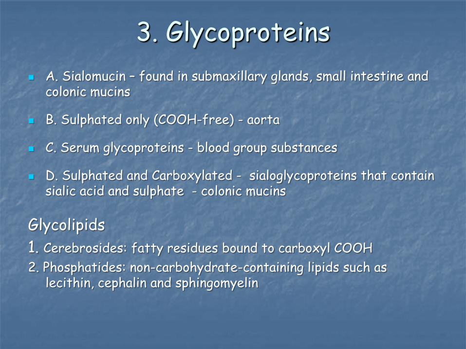
A. Sialomucin – found in submaxillary glands, small intestine and colonic mucins
B. Sulphated only (COOH-free) - aorta
C. Serum glycoproteins - blood group substances
D. Sulphated and Carboxylated - sialoglycoproteins that contain sialic acid and sulphate - colonic mucins
Glycolipids 1. Cerebrosides: fatty residues bound to carboxyl COOH 2. Phosphatides: non-carbohydrate-containing lipids such as
lecithin, cephalin and sphingomyelin

Staining methods for carbohydrates
Periodic acid Schiff (PAS)
Based on the oxidation of 1,2 glycol groups to aldehydes using periodic acid
Aldehydes are detected using Schiff reagent
The sugar units from which aldehydes can be formed by reaction with periodic acid are glucose, galactose, mannose, fucose and some sialic acids
Glycogen and some mucins are PAS +
Sites rich in GAGs (such as cartilage and mast cells) stain weakly or negative

Periodic acid Schiff (PAS)
0.5% periodic
acid
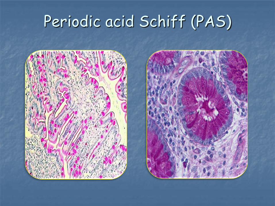
Periodic acid Schiff (PAS)

Periodic acid Schiff (PAS)
High concentrations of hexose sugars, including glucose, in the collagen of basement membranes
The collagen fibres of connective tissue stain pink with PAS
Glomerular basement membranes become thickened and irregular in glomerulonephritis

PAS - Diastase

Periodic acid Schiff - Diastase
No digestion After digestion PAS + PAS -

Alcian blue pH 1.0 (sulphated mucopolysaccharides)
Alcian blue is a basic dye
When used in a 0.1N hydrochloric acid (pH 1.0), alcian blue stains only sulphated acid muco-polysaccharides and glycoproteins
Acid mucopolysaccharides that are carboxylated only will not stain

Alcian blue pH 1.0
Sulphated MPS

Alcian blue pH 2.5 (acid mucopolysaccharides)
When used in a 3% acetic acid solution (pH 2.5), it stains sulphated and carboxylated acid mucopolysaccharides and glycoproteins
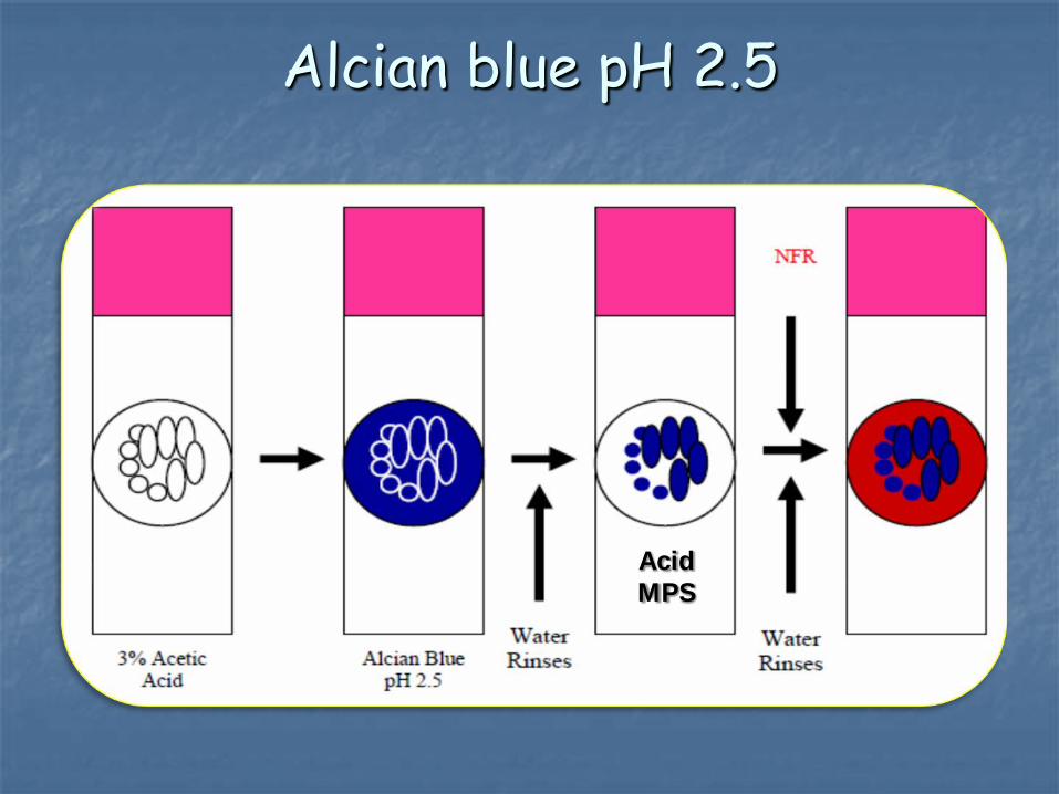
Alcian blue pH 2.5
Acid MPS
Alcian blue with hyaluronidase
This stain differentiates between epithelial and connective tissue mucins
Staining will disappear or be dramatically reduced when tissue sections containing hyaluronic acid, chondroitin sulphate A or chondroitin sulphate C (connective tissue mucins) are digested with testicular hyaluronidase
Glycoproteins (epithelial mucins) will not be affected
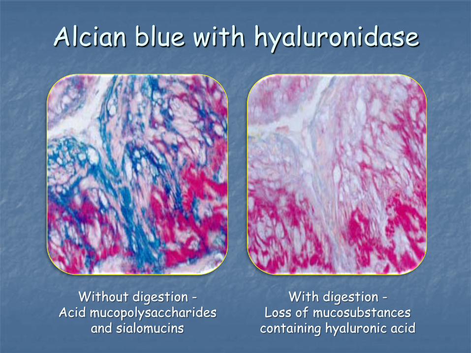
Alcian blue with hyaluronidase
Without digestion - Acid mucopolysaccharides
and sialomucins
With digestion - Loss of mucosubstances
containing hyaluronic acid

Alcian blue / PAS
Differentiate between neutral and acidic mucosubstances
Staining for the acidic mucosubstances will be done with the Alcian Blue pH 2.5 technique and the neutral muco-substances by the PAS reaction
Acid mucopolysaccharides - Blue Neutral polysaccharides - Magenta Other substances - Purple

Alcian blue / PAS
Alcian blue / PAS results
Alcian blue performed before PAS
Combined stain shows neutral mucin red (notably that of the oesophagus and stomach)
Mucous cells of gastric glands have sialic acid mucins and stain purple with alcian blue pH 2.5 and PAS
Secretory cells of the duodenum and jejunum are both neutral and sialic acid mucins and also stain purple
Useful for Barrett’s oesophagus - reflux of acid from the stomach causes the normal squamous epithelium to change to a columnar intestinal form with goblet cells containing stainable sialic acids

Mucicarmine
Aluminum in the haematoxylin solution forms a chelating complex with carmine
The resulting compound has a net positive (+) charge and attaches to the acid groups of mucins

Mucicarmine

An alkaline solution containing an aluminium complex of carminic acid in 20% methanol
This technique is used only to detect glycogen
The dye is held in contact with glycogen by hydrogen bonding to the numerous hydroxyl groups present
Best’s carmine for glycogen

Colloidal iron
Demonstrates sulphated and carboxylated muco- polysaccharides
At low pH, colloidal ferric iron is absorbed by both mucosubstances
After excess reagent is washed out, the Prussian blue detects the iron bound to the tissue

The oxidant chromium trioxide over-oxidizes most of the carbohydrate content of connective tissue to yield carboxyl groups
However it generates abundant aldehydes in the cell walls of fungal hyphae (chitin) and these are then stained with methenamine- silver nitrate (or the PAS)
Grocott’s methenamine silver

Amyloid
A fibrillar protein that deposits in many tissues under certain pathologic conditions
Amyloid contains 1-2% carbohydrate, mostly acid mucopolysaccharides
Amyloidosis is a disease characterized by an amorphous, eosinophilic, extracellular deposit
It gradually replaces cellular elements of vital organs and causes progressive loss of function and death

Alkaline Congo Red
Amyloid chemically resembles cellulose and stains with Congo Red
Using a polariser, green birefringence is most specific for amyloid demonstration
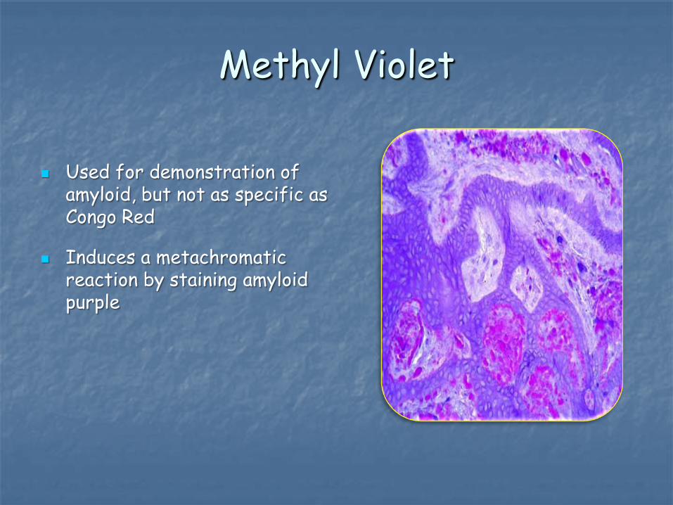
Methyl Violet
Used for demonstration of amyloid, but not as specific as Congo Red
Induces a metachromatic reaction by staining amyloid purple

Thioflavine T
Thioflavine T is a fluorescent dye that attaches non-specifically to amyloid
Background nuclear staining is quenched by staining with haemalum

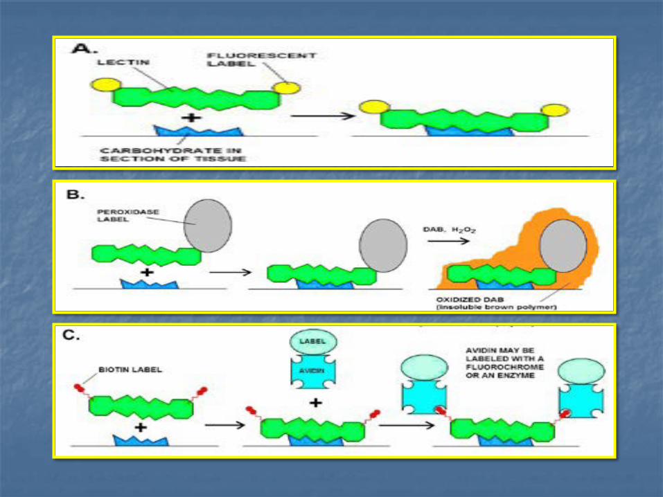
Lectin histochemistry
Proteins (mostly derived from plants) that resemble an antibody in having at least two sites that can bind to specific sugar receptors in a carbohydrate
Applications:
Show specific cell-types in organs such as the stomach and kidney
Stain nerve cells and processes in the CNS + PNS
Many uses in tumour biology and pathology
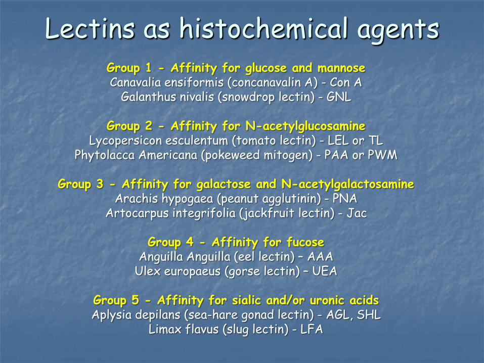
Group 1 - Affinity for glucose and mannose Canavalia ensiformis (concanavalin A) - Con A
Galanthus nivalis (snowdrop lectin) - GNL
Group 2 - Affinity for N-acetylglucosamine Lycopersicon esculentum (tomato lectin) - LEL or TL
Phytolacca Americana (pokeweed mitogen) - PAA or PWM
Group 3 - Affinity for galactose and N-acetylgalactosamine Arachis hypogaea (peanut agglutinin) - PNA
Artocarpus integrifolia (jackfruit lectin) - Jac
Group 4 - Affinity for fucose Anguilla Anguilla (eel lectin) – AAA
Ulex europaeus (gorse lectin) – UEA
Group 5 - Affinity for sialic and/or uronic acids Aplysia depilans (sea-hare gonad lectin) - AGL, SHL
Limax flavus (slug lectin) - LFA
Lectins as histochemical agents

Microglial cells in the brain, stained by virtue of their affinity for the lectin RCA-1 from Ricinus communis