care of the child with a gastrostomy tube: what the school
TRANSCRIPT

Gwen Spector RN, BSN, COCN, CGRN
Ostomy Nurse Specialist
Pediatric Gastroenterology
Care of the Child with a Gastrostomy Tube: What The School Nurse Should Know
06/27/12

Today’s Lesson:1. Gastrostomy Tubes2. Daily Care and Use of the G-tube3. Common Problems4. Resources5. Skills

The Why, When, Where, How, & What of Gastrostomy Tubes

Indications for Gastrostomy
6/29/2012
• A gastrostomy device is placed for the purpose of feeding, administering medications and/or decompressing the stomach.
• Feedings can be given directly into the stomach or small intestine.
• The gastrostomy device may be used as a supplement to oral feeds or the patient may be solely fed this way
• A gastrostomy is generally placed when enteral feedings are necessary for longer than 6-12 weeks time. It may be needed for the rest of the patient’s life or for a shorter period depending on the patient’s circumstances.

Indications for GastrostomyAspiration/swallowing problemsFailure to thrive/poor growth-sometimes ? causeHigh caloric needs-like Cystic FibrosisMinimal or no oral intake, poor suck/nipplingMedically necessary medications and/or fluids-for patients w/ seizures/diabetes insipidus, etc. Need for specialized diets Trauma/anomaly of the mouth or esophagus

Intro to GastrostomyA gastrostomy is a surgically created opening in the stomach.
This opening is also referred to as the stoma. The gastrostomy tube enters the stomach through this stoma and tract.The tract is the channel-like formation between the stomach and the skin where the G-tube/button is located.

The Method of Placement:The gastrostomy is created and the gastrostomy tube placed during one of these methods:
1. Surgicala. Open procedure
b. Laparoscopic
2. Endoscopic3. Radiological
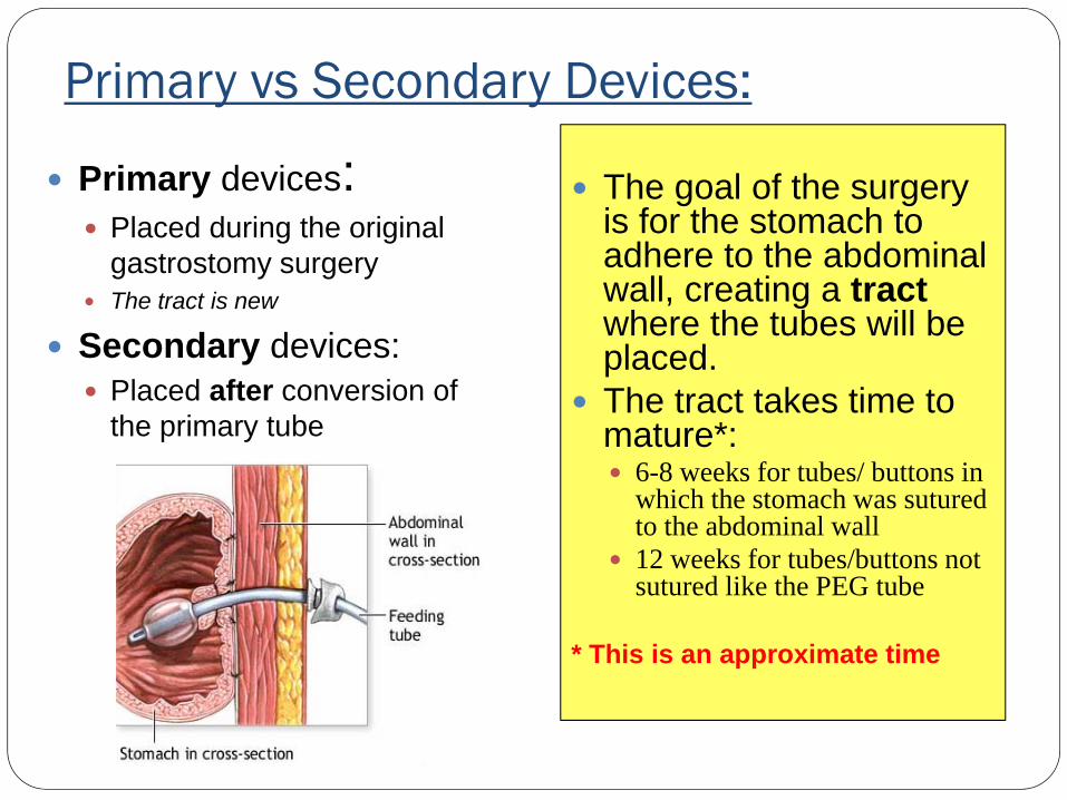
Primary vs Secondary Devices:
Primary devices:Placed during the original gastrostomy surgeryThe tract is new
Secondary devices:Placed after conversion of the primary tube
The goal of the surgery is for the stomach to adhere to the abdominal wall, creating a tractwhere the tubes will be placed.The tract takes time to mature*:
6-8 weeks for tubes/ buttons in which the stomach was sutured to the abdominal wall 12 weeks for tubes/buttons not sutured like the PEG tube
* This is an approximate time

Gastrostomy Primary TubesSurgical tube
Placed by a surgeon during open surgery
The stomach is sutured to the abdominal wall
Often done w/ NissenFundoplication
Tube needs stabilization
Changed to the button in 6-8 weeks
PEG tubePlaced generally by Gastroenterologist (sometimes with a surgeon)
Has an external bolster
Stomach not sutured
Changed to a G-button in 3-6 months
1Drainage Tube Attachment Device2Percutaneous Endoscopic Gastrostomy Tubes
Pezzar tube w/ DTAD1
• Primary G-button• Laparoscopic or endoscopic
placement
• Limited manipulation for the 1st 2 weeks
PEG tube

Gastrostomy ButtonAKA: low profile tube, skin level device
2 main types:BalloonNon-balloon
Placement:Primary-placed with initial gastrostomy surgery
Laparoscopic
PEG (One step)
Secondary to a longer G-tube after the tract has matured 1
4-6 weeks after open surgical tube placement
3 -6 months after PEG tube placement
Needs feeding extension to administer feedings/medications
1 these are approximate times

Comparison of Types of G-Buttons:Balloon Non-balloon
Advantages Easily replaced @ home
Comes in multiple sizes
Lasts longer (sometimes up to several years)
No balloon to break
Not as likely to be accidentally dislodged
Disadvantages Balloon breaks, needs more frequent replacement
Easier to dislodge
Has to be changed by provider or nurse w/ advanced training
Not available in as many sizes as balloon type
May need sedation to place
Some devices need size specific extensions and separate decompression extension (BARD button)
Physician preference and availability of buttons may direct what is placed

Balloon-Type G-ButtonsBrands:
Mic-key® Mini®, Mini ONE balloon® Nutriport™
Multiple Sizes: Mini ONE balloon®
Fr: 12,14,16,18,20,24Cm: 0.8 – 5.0 cm
Balloon 5 mL on most buttons (4 mL for neonates or 12 Fr . buttons)*
over inflation can obstruct pylorus or cause gastric erosions
Use sterile /distilled water and preferably 6-10 mL slip tip syringe
Extensions are not size specific within each brand.
Mic-key®
Internal lock system(‘key”)
*Read manufacturer guidelines Mini ONE balloon G-button

Non-balloon Type G-ButtonHas a silicone mushroom or basket-type tip inside to keep the G-button from coming outPlaced with an obturator1 or special insertion device to stretch out the internal bolsterBrands:
BARD ® or Microvasive®,Mini ONE ® non-balloonEntristar®
Sizes:Varies, more limited than balloon-type
• BARD/Microvasive® buttons have separate decompression tubes, size-specific extensions
BARD®
1

G-J tubes/buttonsThe (G-J) tube is actually 2 tubes in one and is placed through a gastrostomy tract.
• The jejunal portion is advanced into the intestine to bypass the stomach for feedings. (under fluoro)
• The feedings go through the jejunalport while the medications are to be administered through the gastric port
• Continuous feedings are given for usually 18-24 hours. Bolus feedings are not usually given in order to prevent dumping syndrome. There are 3 ports:
1. Jejunal (intestine)2. Gastric (stomach)3. Balloon
11
1
2
2
2
3
3
3

Special UsesSometimes a button is placed in the cecum in the first part of the colon for the administration of laxatives in a child with bowel problems.
This is called a cecostomy button. The same feeding buttons are used as are used with gastric feedings.
The button will usually be located in the RLQ.
Care is similar to the gastrostomy button.

Anatomy of a G-tube/button1. External bolster prevents tube/button
from migrating into stoma
2. Internal bolster/retention balloon secures device internally
3. Balloon valve allows access to inflate/deflate the balloon
4. Anti-reflux valve within the feeding port of button prevents reflux of formula
5. Flap/cap over access port6. Feeding adaptor
5 4
22
2
1
11 5
6
4
3
Daily Care and Use of the G-tube
Daily CareRoutine Care:
Assess the site for abnormalitiesStabilize/secure the tubeClean the skin around the tube with mild soap & water if neededApply topical treatment(s) if orderedChange dressings when soiled (if applicable)
Post-op Period: Sutures may be present. Some of these are dissolvable and some will need to be removed in approximately 1 week by a HCP. There may be a mid-line incision and steri-strips in place. Let the steri-strips fall off on their own.Primary G-buttons have specific care instructions: Minimal manipulation for the first 2 weeks.
Reusable G-tube pads

Nursing AssessmentAssess the gastrostomy site
Is there redness? Is it streaky, bumpy or uniform?Is there swelling/induration?Is there drainage? How much? What color? Does it happen more at certain times?Is there an odor?Is there pain? With touching, with feeds, all the time or intermittent? Does the gastrostomy device fit well? Not too loose or too tight.
Assess the abdomenIs there distension and/or firmness?Is there pain?

Stabilizing the G-tube Extremely important!
Prevents migration of tube
Prevents rocking motion*
Goal: keep tube from being pulled and maintain a 90 degree angle
Don’t tape down too tight, no tension*
Some tubes have a stabilization device already
Some tubes need an external stabilizer
i.e. drainage tube attachment device, sausage roll, baby nipple
Sausage roll
Hollister® drainage tubeattachment device
* Dressing the tube flat against the abdomen causes enlargement of the stoma
baby nipple & hydrocolloid dressing

Protecting the Gastrostomy Tube/button
The G-tube/button may need to be protected if the patient tries to pull on it or is very active and bumps it. It can be challenging to find the method that works for that patient. Some things that work are:
Stretchy gauze/nettingACE bandageAbdominal binderClothing like Onsies, overalls Tape/Coban

Feeding through the G-tube/buttonFeedings are given continuous and/or bolus per pump, syringe or gravity bag.The patient should be on the right side with the HOB elevated 300 during and 30-60 minutes after feedings.Bolus/syringe feedings should not be given too fast. The more elevated the syringe is –the faster the formula will run in. Always stabilize the G-button with your fingers when attaching extensionsSyringe feeds should be to gravity, do NOT force-ok to gently push.Signs of formula intolerance include abdominal distension,cramping, diarrhea and/or vomiting. Some children have a Nissen and can’t vomit. You may need to stop feedings or slow the rate down.

Flushing Gastrostomy DevicesFlush the gastrostomy devices with the appropriate amount of water before and after feedings and medications:
10 mL unless the patient is fluid restricted then 3 – 5 mL is appropriate unless otherwise ordered per the provider.Jejunal tubes require a greater amount of flush in order to prevent the tube from clogging1. Any gastrostomy device should also be flushed every 6 hours when receiving continuous feedings.
Your Child’sWeight
Amount of flush
< 20 pounds 10 ml20-75 pounds 15 ml
>75 pounds 20 ml1 Flush for jejunal tubes

Giving Medication through the G-tube/buttonDon’t crush pills that are time releasedDo not give medications that dissolve under the tongue or are sustained release through the tube.Don’t mix medications with formula or in formula bag unless ordered by the providerGive medications separately from each other to avoid drug interactions. Phenytoin should be given 1 hour before or 2 hours after enteral feeding.
It is preferred that medications that are given through a G-button should be given through the medication port of the feeding extension instead of directly into the button because the valve can be damaged.Give medications through the correct port – be careful not give medications through the balloon port.
Medication port of Mic-key extension

Venting the TubeYou may need to vent the patient’s tube to remove excess air or fluid. You can do this a few different ways:
Use a catheter-tip syringe and aspirate the excess air from stomach or hold the syringe above the stomaSome non-balloon G-buttons have specific extensions for decompression (size specific), others like the balloon-type G-buttons use the regular feeding extension to vent.
Farrell Valve ® Gastric Pressure Relief Device is recommended to use with continuous feeds.
It’s designed to help patients who suffer from poor gastric motility, pain and bloating. New bag every 24 hourswww.corpakmedsystems.com/Product_Main/enteral_main.html#Farrell
Farrell Valve

Cleaning the EquipmentFeeding extensions:
Wash with warm soapy water or half white vinegar/half water. Flush through with warm waterHang to dry or flush air through.May reuse for 2 weeks*
Rinse feeding bags/ syringes after each use
*per manufacturer’s guidelines
Mic-key/Mini ONE extension set
BARD ® or Microvasive®,
Bolus extension
Bolus extension
Pump extension
Decompression extension

Common Problems You May See
1. Stoma2. Skin Problems3. Tube Problems

Stoma: LeakageAll gastrostomies leak some
There are multiple causes of leakage including:
Any increase in intra-abdominal pressure:
constipation, coughing, heavy breathing, ventilated kids, crying, vomiting, change in weight or abdominal girth
Balloon has deflatedIncorrect size, improper stabilizationUnderlying disorder like slow motilityTube displacementPoor wound healing Body structure: scoliosisPositioningSpasticity Inability to decompress stomachFeeding intolerance Recent illness/new medications

Stoma: LeakageTreatment:
Treat the underlying causeChange to correct size and/or new button
Add more water to the balloon
Medication to suppress the acid in the stomach per provider
Protect skinBarrier Products: Powder, Creams
Dressings: gauze, foam
Pouch/attach to drainage bag if leakage extreme
Change rate/route of feeds

Skin: Irritant DermatitisCause:
Primarily from leakage of gastric contents 1
Can also be from harsh cleansers, antibacterial & other topical medications, external bolster too tightSometimes skin conditions such as eczema can mimic this •Treatment:
•Correct the cause•Barrier products: creams, powders to protect and heal the skin•Oral/topical medications per provider to reduce acid•Absorptive dressings
1
1

Skin Protection: Building a BarrierPurpose:
Keeps good moisture in and bad moisture outProtects the skin from caustic fluidsTechnique as important as the products are
Products:zinc oxide, petrolatum , skin prep, barrier powder
Technique:Put medications on firstIf using powder apply before barrier creams and spraysSkin prep/film helps seal in powder and provide a light barrierApply creams thick like icing Layer if needed Don’t wipe completely off each time, blot and reapply
Barrier Products1. powder 2. cream 3. prep
1
2
3

Skin: Hypergranulation Tissue
One of the most common problems seen-the body is trying to healExtra growth of tissue; pink-red. Sometimes beefy1 looking.Yellow, “snotty” and/or brown drainageSometimes friable – may bleedMay be painfulOften mistaken for an infection
Cause: incorrect stabilization, tube moving around in the stoma a lot, excessive moisture, Peroxide use, Dilantin, occlusive dressings Treatment:
silver nitrate application per provider/trained nurse
Tissue will turn gray/black and slough off2. May discolor clothing and skin.
Steroid creams: TriamcinoloneStabilize the tube, change size of button, don’t leave extensions on when not is use, if extensions are needed for long periods-tape down to limit movement in the stoma.Barrier powder to control moistureFoam dressings
21

Skin: Bacterial InfectionAppearance:
Red streaking, spreading erythemaSwelling around the siteAbnormal bump
Other symptoms:Increased tendernessMay or may not have fever, odor or green/purulent drainage Cause:
MRSA common sourcePoor hygieneTight fit of device/tension on the stoma
Treatment: Clean w/ sterile water or saline 2-3x/dAntibiotics per provider Silver dressings
Cellulitis

Skin: Fungal InfectionAppearance:
Red papular rash, often has satellite lesions.
Causes: Trapped moisture Hot, humid environmentG-tube located deep in a skin foldchronic moisture, immuno-suppression, cortico-steroids and diabetes
TX:Keep area clean and dry Antifungal medication as prescribed per provider

Tube: malfunctionSigns:
Formula leaking from the button- a small amount is acceptableBalloon leaks/has a holeNeed to fill the balloon frequently Piece of the tube/button falls off or won’t work appropriatelyCrack or hole on G-tube
TX:Replace device as soon as possible*For GJ tubes/buttons they need replacement under fluoroscopy

Tube: ObstructionCauses:
Inappropriate med administration, thick formulas, failure to flush, pill fragments, solutabs, viscous medications, defective tubing
PreventionFlush well as recommended/ordered Liquid medication administration
TX:Check for kinks, make sure clamp is openFlush with warm water, use 30-60 cc syringeTry Push-pull methodMilk the tubingChange out extensions/button if possibleDe-clogging methods per providerReplacement if possible*

Tube: DislodgementCauses:
Balloon deflatesG-button is pulled out:
Purposefully
Accidentally
EARLY Dislodgement6-8 weeks
Tract not well formed
Forcing a tube may disrupt the stomach from the abdominal wall
LATE Dislodgement> 8-12 weeks safer but complications can still occurFYI: The stoma can
start to close in 1-2 hr

Texas BNE Statement15.24 Nurses Engaging In Reinsertion of Permanently Placed Feeding Tubes
1. The nurse should complete training designed specifically for the type or types of permanent feeding tubes the nurse may need to replace, including overall patient assessment, verification of proper tube placement, and assessment of the tube insertion site.
2. A registered nurse or a physician who has the necessary expertise with regard to the specific feeding tube provides supervision during the training process.
3. The nurse demonstrates competency in all appropriate aspects (knowledge, decision-making, and psycho-motor skills) of performing the procedure.
4. The patient has an established tract. The established tract is not determined by the nurse.
5. The facility has resources available to develop an educational program for initial instruction of LVNs and/or RNs, as well as for ongoing competency validation.
6. Documentation of each nurse's initial education and ongoing competency validation should be maintained by the nurse and/or the employer in accordance with facility policies.
7. Regardless of training, policies and procedures of the facility must also permit the nurse to engage in the procedure.

Red FlagsSigns of Peritonitis/Device in incorrect place:
Difficulty with the flow of formula or medicationsAbdominal pain with and/or after feeding Abdominal tenderness and or rigidity; fever
Signs of feeding intolerance or aspiration:
Choking/gagging with feedingsVomiting/abdominal pain with feedings
STOP FEEDING
Signs of an Infection within 2 weeks of initial surgery:
Redness with swelling around the gastrostomy
Signs of dehydration if patient having leakage from the gastrostomy site
Signs of dumping syndrome:Nausea/Vomiting
Sweating, Heart palpitations, rapid heart rate
Weakness, fatigue , fainting or passing out
Dizziness, lightheadedness
Shakiness, feelings of anxiety, nervousness

When & Who to CallProblem Who to Call
•Tube comes out/gets dislodged•Tube is clogged
•Parent/caretaker•HCP or parent
•Red Flags are present •Call the HCP and parent /send the patient to the ER if warranted
•Abnormal findings: •Call the HCP and notify the parent
•Equipment needs •Need more supplies- MD who writes orders for the supplies; Broken supplies- DME company
•Questions/concerns about the patient •Speak to the HCP taking care of the patient
•Education needs for nurse/parent •Nurse/Educator/Manufacturer resources

Important to Know:1. Know when the original gastrostomy surgery was done
and special instructions related to the care2. Know who & when to contact3. Stabilize/secure the G-tube4. Know what each port is for 5. Flush with water at least after feedings and medications and
during continuous feedings to prevent clogging6. Not all G-tube sites look perfect and this does not always
reflect the quality of care, know what the patient’s baseline is or call the HCP.
7. See Do’s & Don’ts Handout

ResourcesAmerican Pediatric Surgical Nurses www.apsna.orgComplex Child http://complexchild.com/Feeding Tube Awareness www.feedingtubeawareness.comMic-key G-button www.mic-key.comwww.youtube.com/user/mymickeytubeThe Oley Foundationwww.oley.orgParent: www.parent-2-parent.comSpecial Childwww.specialchild.com
Reusable G-tube pads:
Julia’s G-tube pads: http://store.juliasgtubepads.com/
My Button Buddies: www.mybuttonbuddies.com

Questions?

Play Time!