caries management by risk assessment putting it …...caries management by risk assessment –...
TRANSCRIPT

Caries Management by Risk
Assessment – Putting it into
Practice
NNOHA October 2012 John D.B. Featherstone
Professor and Dean
E-mail [email protected]
School of Dentistry
University of California San Francisco

Disclosure
I have no personal financial interest in any
company relevant to this presentation.
I have consulted for, or have done
research funded or supported by:
Arm and Hammer, Beecham, Cadbury, GSK,
KaVo, Novamin, Omnii Oral
Pharmaceuticals, Oral B, Philips Oralcare,
Procter and Gamble, 3M ESPE Preventive
Care, Wrigley, and the National Institutes
of Health.

What is Dental Caries?
Dental caries is tooth decay
Specific bacteria (Streptococcus mutans,
Streptococcus sobrinus, lactobacilli and more)
on the tooth surface feed on carbohydrates
and make acids as waste products
Acids travel into the tooth and dissolve mineral
- if mineral loss is not halted or reversed a
cavity is formed
Dental caries is a transmissible bacterial
infection

Protective Factors
“White spot” lesion

Protective Factors
Frank occlusal
cavity

Protective Factors
Childhood
Caries

Protective Factors
Root caries

Protective Factors
Courtesy Dr Don Curtis

The Caries Balance
Protective Factors • Saliva flow and components
• Fluoride, Calcium, Phosphate:
remineralization
• Antibacterials:-
chlorhexidine, xylitol, new?
No Caries Caries
Pathological Factors
• Acid-producing bacteria
• Frequent eating/drinking of
fermentable carbohydrates
•Sub-normal saliva flow and
function
Featherstone, Community Dent Oral Epidem, 1999

Protective Factors
Stay in
balance to
survive

Pathological Factors
Cariogenic bacteria: mutans streptococci (S. mutans and S. sobrinus), lactobacillus species and others
Frequency of ingestion of fermentable carbohydrates: sucrose, glucose, fructose, cooked starch
Reduced salivary function (medication induced; radiation therapy; disease; genetic)

Acid producing bacteria are usually
less than 1 percent of the total flora in
the plaque
Scanning
Electron
Micrograph
of bacteria on
a tooth
surface

Streptococcus mutans culture showing active cell
division. S. sobrinus is similar. Sucrose leads to
extracellular polysaccharides that stick the
plaque together

Mutans Streptococci
This group of bacteria contains two
primary species that appear in humans
Streptococcus mutans - almost universal
Streptococcus sobrinus - virulent, high risk
Both species produce acids and can live in
acid

Lactobacillus culture. Lactobacilli species produce
predominantly lactic acid from fermentable carbohydrates

Additional Species strongly
related to high caries in children
Tanner and co-workers 2010 and 2011
Bifidobacteriaceae
Veillonella
Scardovia wiggsiae
Watch this space
Any bacteria that produce acid as a
byproduct of metabolism
Biofilm is a cooperative city

Additional Species strongly related
to occlusal caries in dentin
Lima et al, Caries Res, 2011
Lactobacillus casei and Lactobacillus
fermentum
Veillonella species
Actinomyces species
Bifidobacterium species
Any bacteria that produce acid as a
byproduct of metabolism
Biofilm is a cooperative city

What about
the clinical
relevance?
Does drilling
and filling
really fix
caries?


Caries Management Study
S1
S3 S7 S2
Baseline
Observations
Saliva Sample
MS, LB and F
Radiographs
DMFS
1-7 cavities
Co
ntr
ol
Inte
rve
nti
on
Final Observations
Radiographs
DMFS
Randomization
Restorations
+Anti-bacterial
and Fluoride
Treatment
All
Restorations
Complete
S2
S4-S6
Conventional
Treatment Plan
Restorations
S3 S7
Final Observations
Radiographs
DMFS
All
Restorations
Complete
S4-S6
high low
2 Years
N=116
(CHX + F)
N=115

Decayed Surfaces vs. log MS and log LB
(Revised bacterial classifications 1-07)
High
Bacterial
Challenge
Baseline Bacterial
Levels vs Decay
Existing Cavity = High Risk


Chlorhexidine plus Fluoride
Restorations

Patients With Frank cavities
One or more frank cavities indicates high risk for future new carious lesions
Moderate to high levels of mutans streptococci
Moderate to high levels of lactobacilli
Patients who have a high bacterial challenge require antibacterial treatment as well as fluoride
Placing restorations does not reduce the bacterial loading in the rest of the mouth
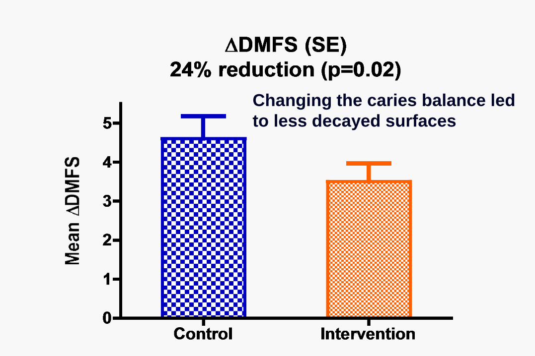
Changing the caries balance led
to less decayed surfaces
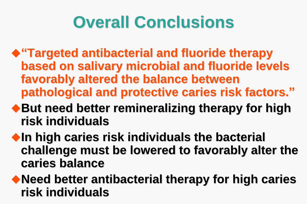
Overall Conclusions
“Targeted antibacterial and fluoride therapy based on salivary microbial and fluoride levels favorably altered the balance between pathological and protective caries risk factors.”
But need better remineralizing therapy for high risk individuals
In high caries risk individuals the bacterial challenge must be lowered to favorably alter the caries balance
Need better antibacterial therapy for high caries risk individuals

Pathological Factors
Cariogenic bacteria: mutans streptococci (S. mutans and S. sobrinus) and lactobacillus species
Frequency of ingestion of fermentable carbohydrates: sucrose, glucose, fructose, cooked starch
Reduced salivary function (medication induced; radiation therapy; disease; genetic)

+
Demineralization:-
Step 1
Cariogenic
Bacteria
S. Mutans
S. Sobrinus
Lactobacilli
++?
Fermentable
Carbohydrates
Sucrose
Glucose
Fructose
Cooked starch
Organic Acids
Which penetrate enamel and
dentin
Dissolve tooth mineral

Protective Factors
Cariogenic foods contain fermentable
carbohydrates such as sucrose,
glucose, fructose and cooked starch

Pathological Factors
Cariogenic bacteria: mutans streptococci (S. mutans and S. sobrinus) and lactobacillus species
Frequency of ingestion of fermentable carbohydrates: sucrose, glucose, fructose, cooked starch
Reduced salivary function (medication induced; radiation therapy; disease; genetic)

Male, 55 years old, before
radiation to the head and neck
for cancer treatment. Causes
saliva flow and function to be
cut by at least 90%
Same male, after radiation
to the head and neck. Six
months later, showing
rampant decay and massive
destruction of the teeth

The Caries Balance
Protective Factors • Saliva flow and components
• Fluoride, Calcium, Phosphate:
remineralization
• Antibacterials:-
chlorhexidine, xylitol, new?
No Caries Caries
Pathological Factors
• Acid-producing bacteria
• Frequent eating/drinking of
fermentable carbohydrates
•Sub-normal saliva flow and
function
Featherstone, Community Dent Oral Epidem, 1999


Protective factors
Salivary components and flow
Fluoride, calcium and phosphate:
remineralization
Antibacterials from extrinsic sources

Saliva Contains Numerous
Important Components
Calcium, phosphate and fluoride
Proteins and lipids that form the pellicle that protects the tooth surface
Proteins that keep calcium in solution - they maintain supersaturation
Buffers: bicarbonate, phosphate, peptides
Antibacterial substances & immunoglobulins

Protective factors
Salivary components and flow
Fluoride, calcium and phosphate:
remineralization
Antibacterials from extrinsic sources

+
Demineralization:-
Step 2
If fluoride is present in the
solution between the
crystals it inhibits mineral
loss
Organic
Acids
Dental Mineral =
Carbonated
Hydroxyapatite
Acid soluble
Demineralization
Calcium and phosphate into
solution

Crystal of dental mineral
Ca2+ PO43- Ca2+ PO4
3- Ca2+ PO43- Ca2+
F -
Schematic adsorption of
fluoride on crystal surfaces
by fluoride
F -
F- F
-


+
Remineralization/Tooth Repair
Fluoride speeds up remineralization ->
less soluble mineral
Calcium
in tooth
water
(from
saliva)
Phosphate
in tooth
water (from
saliva)
Remineralization
Builds on existing crystal remnants
New mineral less soluble
Fluoride helps

Crystal of dental mineral
Ca2+ PO43- Ca2+ PO4
3- Ca2+ PO43- Ca2+
Ca2+ Ca2+
F -
PO43-
Schematic
enhancement of
remineralization by
fluoride
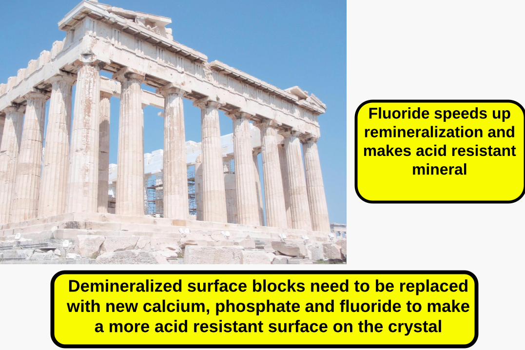
Fluoride speeds up
remineralization and
makes acid resistant
mineral
Demineralized surface blocks need to be replaced
with new calcium, phosphate and fluoride to make
a more acid resistant surface on the crystal

Enamel/dentin
crystal =
Carbonated apatite
Partly dissolved
crystal
Crystal
nucleus
ACID
Acid resistant
Ca10 (PO4)6 (F)2 =
fluorapatite-like
coating on crystals
Remineralization Calcium +
Phosphate
+ Fluoride

Protective Factors
Fluoride can not enter bacteria in its ionic form,
but as the bacteria produce acid HF is formed,
which diffuses readily into the cells

Fluoride inhibits demineralization
Fluoride enhances remineralization
Fluoride can inhibit plaque bacteria
Fluoride works primarily via
topical (surface) mechanisms (Fluoride in water, foods, beverages, products)

What can we do with
this knowledge?

Caries Management by Risk
Assessment (CAMBRA):

Caries Management Step by Step
Dental/medical history
Clinical exam
Detect caries lesions early enough to reverse or prevent progression
Assess caries risk
Treatment plan including chemical therapy
Use fluoride and/or antibacterial therapy based on observations
Use minimally invasive restorative procedures to conserve tooth structure
Recall and review

Protective Factors
Detecting Dental Caries, Tactile, Visual, X-ray,
Laser, Impedance and Fluorescence Methods
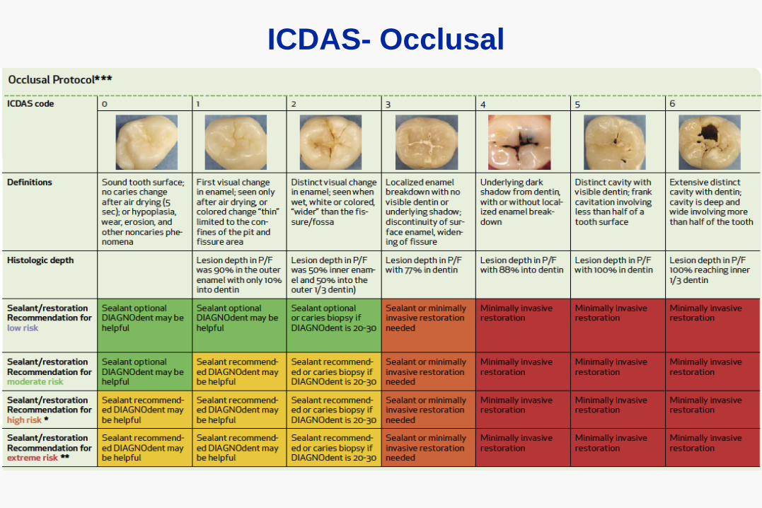
ICDAS- Occlusal


Fluorescence assisted detection
Courtesy of Peter Rechmann, UCSF

Caries Management Step by Step
Dental/medical history
Clinical exam
Detect caries lesions early enough to reverse or prevent progression
Assess caries risk
Treatment plan including chemical therapy
Use fluoride and/or antibacterial therapy based on observations
Use minimally invasive restorative procedures to conserve tooth structure
Recall and review

Risk Assessment
Assessing the
risk for caries in
the future

Putting into practice
the results of many
years of research.
“Caries Management
by Risk Assessment”
October, November
2007. On line, free
California Dental
Association Journal
based upon the
“Caries Balance”
http://www.cdafoundation.org/journal

Caries Risk Assessment
Form
Featherstone et al,
CDAJ, 2007
Circle Yes’s
Visualize the Balance
Decide on caries risk
level: Low, Moderate,
High, Extreme
Used in UCSF student
clinics and �beyond
Used in many private
practices, with
modifications

California Dental
Association Journal:
30(10), 2011
Sophie Domejean, Joel M White,
John D Featherstone

Caries Risk Assessment 12,954 Patients over 6 years
70% identified as high risk progressed to new cavities
88% identified as extreme risk progressed to new cavities
76% identified as low risk did not progress to cavities
Caries risk assessment method validated and can be improved even further
Need effective remineralizing and antibacterial therapy to deal with high and extreme risk patients

Caries Risk assessment (Age 6 years and older/adult) - 1
1. Disease Indicators = Clinical Observations
a) Visible cavities present
b) Caries restored in last 3 years
c) Interproximal caries lesions/radiolucencies
d) White spots on enamel surfaces
Any one of these signals a bacteria test for MS and LB or overall bacterial activity (ATP test)
These are all clinical observations that tell us nothing about the cause of the disease - they indicate presence of disease
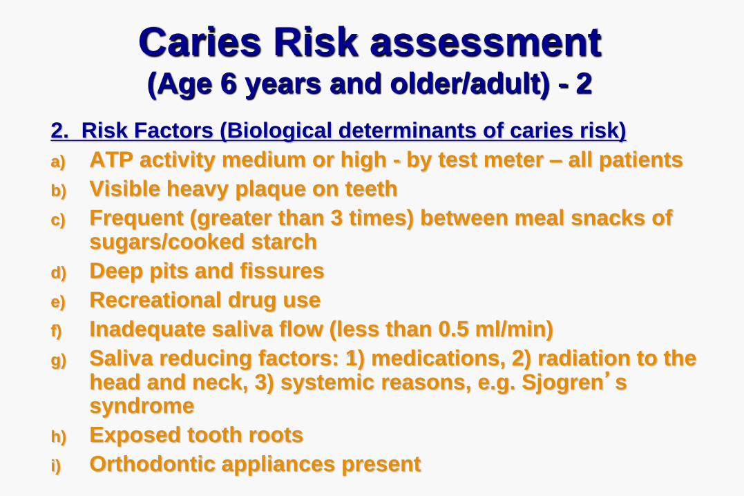
Caries Risk assessment (Age 6 years and older/adult) - 2
2. Risk Factors (Biological determinants of caries risk)
a) ATP activity medium or high - by test meter – all patients
b) Visible heavy plaque on teeth
c) Frequent (greater than 3 times) between meal snacks of sugars/cooked starch
d) Deep pits and fissures
e) Recreational drug use
f) Inadequate saliva flow (less than 0.5 ml/min)
g) Saliva reducing factors: 1) medications, 2) radiation to the head and neck, 3) systemic reasons, e.g. Sjogren’s syndrome
h) Exposed tooth roots
i) Orthodontic appliances present

Caries Risk assessment (Age 6 years and older/adult) - 3
3. Protective Factors
a) Lives/works/school in community with fluoridated water
b) Uses fluoride toothpaste once daily
c) Use fluoride toothpaste at least twice daily
d) Uses fluoride rinse/gel daily
e) Uses 5000 ppm F toothpaste daily
f) Fluoride varnish in last 6 months
g) Office F topical in last 6 months
h) Chlorhexidine rinse prescribed/used daily for 1 week every month last 6 months
i) Xylitol gum/candies 4 times daily last 6 months
j) Calcium/phosphate paste last 6 months
k) Saliva flow visibly adequate or > 1 ml/min by test

CariScreenTM ATP Test
Real time (15 second )
inexpensive screening
test for quantitative
measure of bacterial
activity
>4,000 high bacteria
>1,500 moderate
<1,500 low
CariScreen score had a strong positive correlation r =
0.76 with total cell count, a positive correlation with MS
counts r = 0.69. OHSRU

Caries Risk Assessment (Age 6 years - adult)-4
4. Bacterial test for (ATP) for all as a baseline measure. High ATP signals high bacterial challenge
5. Count the yes’s. Assess caries risk and check risk as extreme, high, moderate or low
6. Treatment Plan
Includes home care, office preventive treatments and restorative work
7. Home Care Recommendations
8. Recall and Re-assessment of Caries Risk
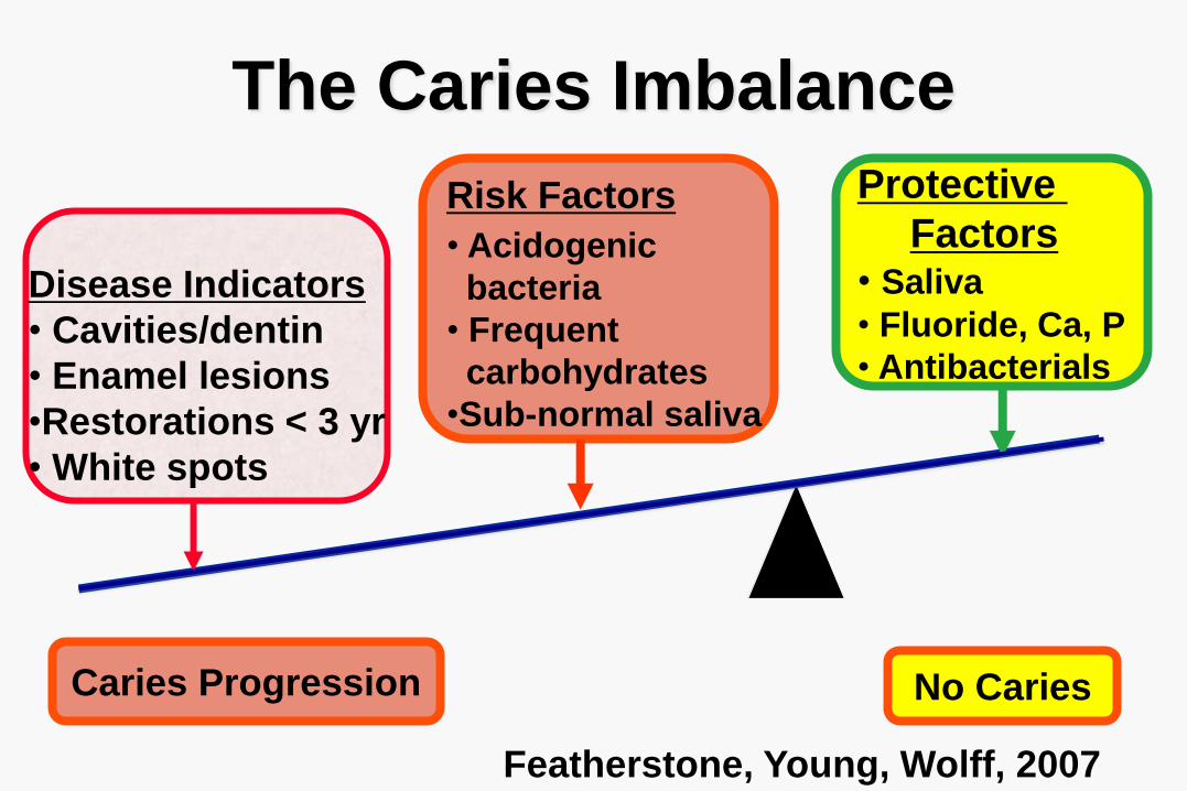
The Caries Imbalance
Protective
Factors • Saliva
• Fluoride, Ca, P
• Antibacterials
No Caries Caries Progression
Risk Factors
• Acidogenic
bacteria
• Frequent
carbohydrates
•Sub-normal saliva
Disease Indicators
• Cavities/dentin
• Enamel lesions
•Restorations < 3 yr
• White spots
Featherstone, Young, Wolff, 2007

Caries Risk Assessment
Form
Featherstone et al,
CDAJ, 2007
Circle Yes’s
Visualize the Balance
Decide on caries risk
level: Low, Moderate,
High, Extreme
Used in UCSF student
clinics and �beyond
Used in many private
practices, with
modifications
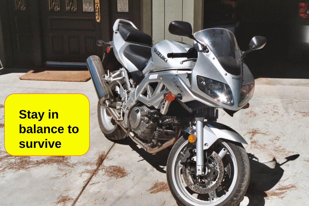
Protective Factors
Stay in
balance to
survive

Caries Management Step by Step
Dental/medical history
Clinical exam
Detect caries lesions early enough to reverse or prevent progression
Assess caries risk
Treatment plan including chemical therapy
Use fluoride and/or antibacterial therapy based on observations
Use minimally invasive restorative procedures to conserve tooth structure
Recall and review

What can we do with this
knowledge?
1. Enhance remineralization with
fluoride, calcium and phosphate
2. Use antibacterial therapy for high
and extreme risk individuals

Fluoride inhibits demineralization
Fluoride enhances remineralization
Fluoride can inhibit plaque bacteria
Fluoride works primarily via
topical (surface) mechanisms (Fluoride in water, foods, beverages, products)

Protective Factors
Fluoride levels in the mouth are
sufficient to enhance remineralization

Brushing twice daily with
a fluoride-containing
dentifrice is one of the
most effective ways to
control dental decay.
High bacterial challenge
overcomes the
therapeutic effects of
fluoride.

Protective Factors
Numerous clinical trials showed ~30% reduction
with fluoride dentifrice 1000-2800 ppm F.
Curnow, Pine, et al, 2002 reported 56% reduction
with supervised brushing twice daily

Over the counter fluoride rinses (0.05% NaF) are
very effective in high caries risk patients when
used once or twice daily for one minute, plus a
fluoride-containing dentifrice. O’Reilly and
Featherstone, 1987

Office-Applied Fluoride Products
Gel (> 5,000 ppm F)
and Fluoride Varnish Do not require continuing patient
compliance
Forms slowly soluble calcium fluoride-like
deposits in lesions and the plaque
Gives slow release fluoride for several
weeks
Three times a year for high risk patients
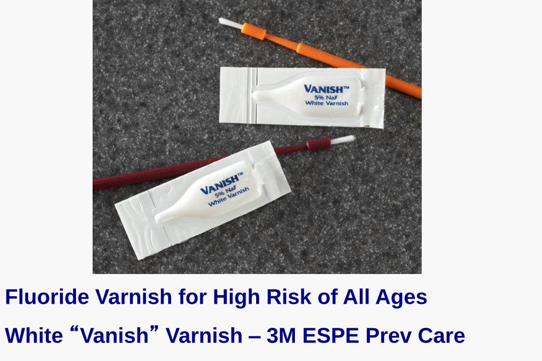
Fluoride Varnish for High Risk of All Ages
White “Vanish” Varnish – 3M ESPE Prev Care

501 Children, 9.1 years old, China, using fluoride
toothpaste
Four groups: a) sealant, b) F varnish 6 mths,
c) Silver Diamine F, d) placebo control (water)
Pit/fissure sites with dentin caries at 24 months
Sealant 1.6 %
NaF varnish 2.4 %
Silver DF 2.2 %
Control 4.6 % - significantly different
J Dent Res 91:753-758, 2012

• 5000 ppm F vs 1450 ppm F (as NaF) toothpaste
• Caries incidence and caries progression
• Compliance assessed
• Prevented fraction 40%: 5,000 ppm versus 1450 ppm F
• Caries still progressed in many, even with high
concentration fluoride

High concentration fluoride products for high
risk patients. Proven effective for root caries.

Clinpro™ 5000 1.1% NaF
Dentifrice
3M ESPE
Contains added calcium

Conclusions - Fluoride
The anti-caries effects of fluoride are
primarily topical (surface) in plaque
The systemic benefits of fluoride are
minimal
Therapeutic levels of F can be achieved
from drinking water and fluoride products
Fluoride therapy may not overcome a high
bacterial challenge

Calcium Phosphopeptide:
CPP/ACP
Laboratory studies: Three decades
Clinical Studies: clinical evidence

Representation of a proposed
CPP-ACP complex
Cross et al. 2007 Curr Pharm Des,


Protective Factors
Biofilm Modification is necessary as part
of our therapy for high bacterial challenge
individuals. Caries is a transmissible
bacterial infection


Chlorhexidine Gluconate 0.12%, 10 ml, daily for 1
week reduces MS markedly and LB somewhat after
restorations completed. Repeat every month.

Chlorhexidine was effective at reducing
the bacterial challenge in high caries risk
individuals even when compliance was
problematic
Preferred regimen is once a day rinse for
one week every month for a year
Monitor success by bacterial testing
Ideally we need a better antibacterial
therapy
Must combine with remin/fluoride

CariFree Treatment Rinse?
Sodium hypochlorite
and sodium fluoride in
separate bottles mixed
before use
Clinical trial -
unpublished, indicates
very good clinical
efficacy
No microbiological data
published

Conclusions: Antibacterial
Anticaries Agents
Several avenues are being explored that show
promise for improved antibacterial clinical applications in the future.
It will take years for clinical studies and regulatory approvals

Manage caries based upon risk status
and the caries balance

Low Risk Patient Protective Factors
• No new caries in 5 years
• Saliva normal
• Fluoride, calcium, phosphate
- remineralization:-
• 2 x daily F toothpaste
• Antibacterials:- No need
No Caries Caries
Pathological Factors
• Low Acid-producing bacteria
• Saliva normal
• Carbohydrates o.k.
X

Moderate Risk Patient Protective Factors
• Saliva flow and components
adequate, calcium, phosphate
• Fluoride, calcium, phosphate
- remineralization
F toothpaste, F mouthrinse
• Antibacterials:- chlorhexidine,
xylitol.
No Caries Caries
Lies Dormant
Pathological Factors
• Acid-producing bacteria:-
time for another bacterial test?
• Saliva flow measured normal
• snacking controlled
• Caries 4 years ago - no new

High Risk Patient
Protective Factors • Office applied Fluoride Varnish
• Chlorhexidine 10 ml daily one
week a month for 6 months
• Brush with 5000 ppm F
toothpaste 2x daily - enhance
remineralization
• Xylitol gum 4x daily
• Recall 4-6 months
No New Caries
Caries
On Hold
Pathological and Risk
Factors
• Bacteria high
• Cavities
• Snacking
• Hyposalivation?

One week a month
10 ml once daily
2-3 x daily
3-4 x daily
2-3 x daily
Initial and
every recall

Extreme High Risk
Patient Protective Factors
• Office applied Fluoride Varnish
• Chlorhexidine 10 ml daily one
week a month for 6 months
• Brush with high 5000 ppm F
toothpaste daily - enhance
remineralization
• Xylitol gum 4x daily
•Baking soda rinse
•Consider MI paste
• Recall 3-6 months
No New Caries
Caries
On Hold

Caries Management Step by Step
Dental/medical history
Clinical exam
Detect caries lesions early enough to reverse or prevent progression
Assess caries risk
Treatment plan including chemical therapy
Use fluoride and/or antibacterial therapy based on observations
Use minimally invasive restorative procedures to conserve tooth structure
Recall and review

CDA Journal
November 2010

1. Modification of the oral flora
2. Patient education
3. Remineralization of non-cavitated lesions of enamel
and dentin
4. Minimal operative intervention of cavitated lesions
5. Repair of defective restorations
FDI statement 2002
Minimal Intervention in the Management of
Dental Caries
