case 35 clinical history - virtualpathology.leeds.ac.uk 35.pdf · ‡san diego pathologists medical...
TRANSCRIPT

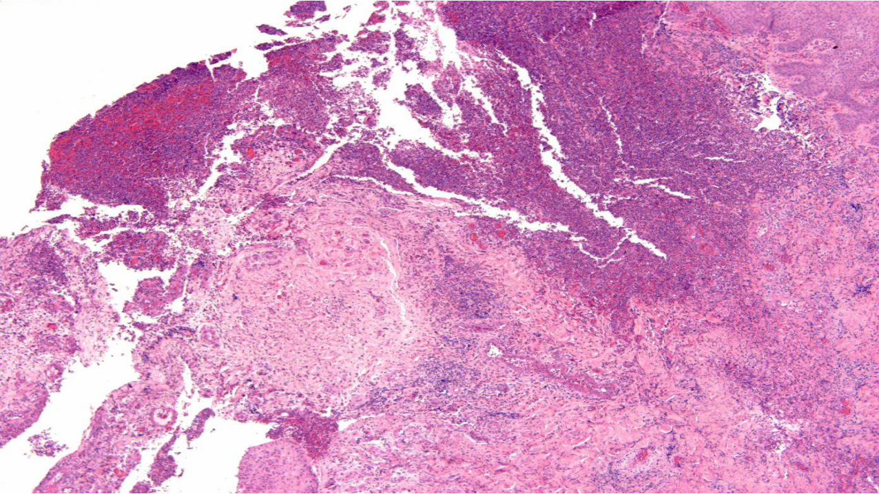
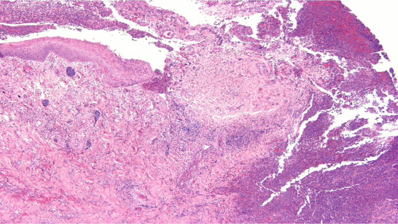
CASE 35 CLINICAL HISTORY
bull Female 24
bull Painful ulcerated lesion
bull Left buttock
bull Developed over a few weeks
bull Abscess
bull Excision
bull Two months later developed a similar lesion on right buttock
CD30
CD3
CD4
CD30
CD30
CD30
PODOPLANIN
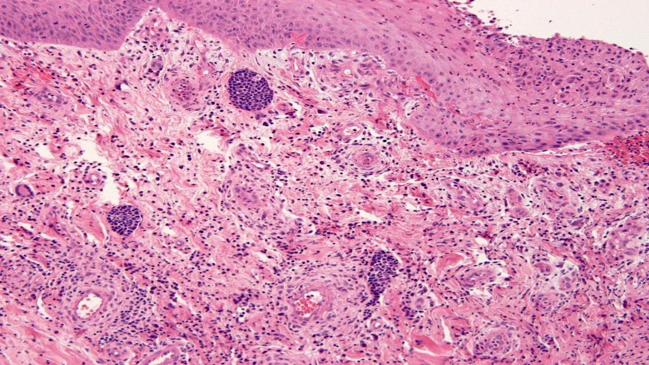
CASE 35 The main diagnosis to consider is A Pyoderma gangrenosum B Cutaneous vasculitis C Intravascular CD30 positive T-cell lymphoma (intralymphatic CD30 positive lymphoproliferative disorder) D Lymphomatoid papulosis type A E Peripheral T cell lymphoma NOS
CASE 35 The main diagnosis to consider is A Pyoderma gangrenosum B Cutaneous vasculitis C Intravascular CD30 positive T-cell lymphoma (intralymphatic CD30 positive lymphoproliferative disorder) D Lymphomatoid papulosis type A E Peripheral T cell lymphoma NOS
CASE 35 DIAGNOSIS
INTRAVASCULAR CD30 POSITIVE ANAPLASTIC LARGE T CELL LYMPHOMA
OR
BENIGN ATYPICAL INTRAVASCULAR CD30(+) T-CELL PROLIFERATION
(INTRALYMPHATIC CD30+ LYMPHOPROLIFERATIVE DISORDER
AM J SURG PATHOL MAY 2014 EPUB AHEAD OF PRINT )
INTRAVASCULAR LYMPHOMAS
bull Most have a B cell lineage and are associated with very poor prognosis
bull T cell NK (EBV positive) intravascular lymphomas are very rare
bull CD30 positive intralymphatic lymphoproliferative disorder has only recently been recognized as a distinctive entity
INTRAVASCULAR LARGE B CELL LYMPHOMA
CLINICAL FINDINGS
bull F = M
bull Adults (elderly)
bull Most common in the Asia
bull Frequent involvement of the CNS and skin
bull Hepatosplenic involvement (26) bone marrow involvement (32) lymph node involvement (only 11)
bull Usually sistemic involvement by the time cutaneous lesions develop
bull Violaceous ill-defined lesions on trunk and limbs (inverted livedo reticularis pattern)
bull B symptoms frequent
bull Poor prognosis
bull In cases with cutaneous involvement only (around 26) prognosis appears to be better
INTRAVASCULAR LARGE B CELL LYMPHOMA
HISTOLOGICAL FINDINGS
bull Dilated dermal and subcutaneous vascular channels including venules capillaries and arterioles
bull Blood vessels appear dilated and contain numerous large pleomorphic lymphoid cells
bull Most cases have a pan-B cell phenotype (CD20 and CD79a positive)
Am J Surg Pathol 2013 Apr
bull A case of localized cutaneous intravascular anaplastic lymphoma kinase-negative ALCL (cIALCL) with a very indolent clinical course The patient experienced a single cutaneous relapse and remains alive without disease 4 years after diagnosis Our index case of cIALCL and 1 other tested case were immunohistochemically confirmed to be intralymphatic (contained within D2-40+vessels) as compared with the blood vessel localization of cITNKL Recognition of cIALCLs as a distinct clinicopathologic entity and in particular their distinction from aggressive usually EBV cITNKLs may be possible on the basis of a combination of clinicopathologic criteria allowing for localized therapy in a subset of patients
Intravascular ALK-negative anaplastic large cell lymphoma with localized cutaneous involvement and an indolent clinical course toward recognition of a distinct clinicopathologic entity
Metcalf RA Bashey S Wysong A Kim J Kim YH Gratzinger D
Department of Pathology Stanford University School of Medicine Stanford CA 94305 USA
PROPOSAL
bull Large T-cell lymphoma with typical ALCL morphology and immunophenotype
bull as well as an intralymphatic localization
bull represents a distinct clinicopathologic entity that
bull should be distinguished from intravascular usually EBV+ aggressive T-cell or NK-cell lymphomas involving blood vasculature
Am J Dermatopathol 2011 Dec
Abstract
bull We present a case of a rare intravascular large T-cell lymphoma in a 59-year-old man with an unusual CD3+ CD4+ CD5- CD30+ CD56- TIA-1-negative and EBER-negative phenotype This T helper or CD30 phenotype is particularly uncommon To our knowledge it has only been described once before and never in the absence of the cytotoxic marker TIA-1 This case exemplifies the particular diagnostic challenges raised by intravascular large cell lymphomas generally and should encourage the use of endothelial immunohistochemical staining in questionable cases While evaluating skin punch biopsies it is critical to keep this rare entity on the differential diagnosis along with the relatively more common intravascular large B-cell lymphoma and epithelial malignancies Additionally our understanding of intravascular large natural killerT-cell lymphoma as a heterogeneous phenotypic entity continues to evolve This case demonstrates that the degree of this phenotypic heterogeneity may be even greater than previously thought
A rare case of intravascular large T-cell lymphoma with an unusual T helper phenotype
Deetz CO Gilbertson KG 2nd Anadkat MJ Dehner LP Lu D
Lauren V Ackerman Laboratory of Surgical Pathology Washington University Medical Center St Louis MO 63110 USA
Cutan Pathol 2011 Feb
bull The patient was a 47-year-old woman who had developed multiple erythematous patches and plaques on her back
bull The lesions responded well to CHOP (cyclophosphamide hydroxydoxorubicin oncovin prednisone) chemotherapy but relapsed shortly after therapy The patient was surviving with the disease for eight years but was ultimately lost to follow up
bull Histopathologically the neoplasm evolved from IL to extravascular lymphoma This was showed in biopsies obtained at different stages of the disease The lymphoma cells stained positively for CD30 CD45 CD3 CD4 CD5 and Ki67 and lacked expression of anaplastic lymphoma kinase (ALK) CD8 CD45RA CD45RO CD20 CD79 CD56 perforin and granzyme B
bull Our results suggest that IALCL represents a distinct subtype of IL and is histopathologically and biologically different from IL with B NK or T cell phenotype
Cutaneous intravascular anaplastic large cell lymphoma
Wang L Li C Gao T
Department of Dermatology Xijing Hospital Fourth Military Medical University Xian China
Am J Dermatopathol 2013
bull Report of 2 patients with skin lesions showing an atypical intravascular CD30 T-cell proliferation Both the patients did not present systemic disease and therefore exhibit a favorable outcome
Benign atypical intravascular CD30(+) T-cell proliferation a reactive condition mimicking intravascular lymphoma
Riveiro-Falkenbach E Fernaacutendez-Figueras MT Rodriacuteguez-Peralto JL
Department of Pathology Hospital Universitario 12 de Octubre Instituto de Investigacioacuten i+12 Universidad Complutense Madrid Spain
bull Am J Surg Pathol 2014 May 6 [Epub ahead of print]
bull Intralymphatic Cutaneous Anaplastic Large Cell LymphomaLymphomatoid Papulosis Expanding the Spectrum of CD30-positive Lymphoproliferative Disorders
bull Samols MA1 Su A Ra S Cappel MA Louissant A Jr Knudson RA Ketterling RP Said J Binder S Harris NL Feldman AL Kim J Kim YH Gratzinger D
bull Author information
bull 1Departments of Pathology Dermatology Stanford University School of Medicine Stanford daggerUCLA Medical Center Los Angeles DaggerSan Diego Pathologists Medical Group San Diego CA sectDepartment of Dermatology Mayo Clinic Jacksonville FL ∥Department of Pathology Massachusetts General Hospital Boston MA paraDepartment of Laboratory Medicine and Pathology Mayo Clinic Rochester MN
bull Abstract
bull Intravascular large B-cell lymphomas and EBV NKT-cell lymphomas commonly follow an aggressive clinical course We recently reported an entirely intravascular anaplastic large cell lymphoma (ALCL) in the skin with a surprisingly indolent clinical course interestingly this lymphoma involved the lymphatic rather than the blood vasculature We hypothesized that intravascular skin-limited ALCL is distinct from aggressive systemic intravascular lymphomas in its intralymphatic localization and clinical course We now describe 18 cases of cutaneous intravascular large cell lymphoproliferations from 4 institutions All 12 intravascular large T-cell lesions were intralymphatic the majority (9) were CD30 T-cell lymphoproliferative disorders (TLPDs) 5 further classified as intravascular ALK ALCL One ALK ALCL and 2 benign microscopic intravascular T-cell proliferations were also intralymphatic A single case of otherwise typical cutaneous follicle center lymphoma contained intralymphatic centroblasts The clinical and pathologic characteristics of the CD30 TLPDs were similar to those of their extravascular counterparts including extralymphatic dermal involvement in a subset DUSP22-IRF4 translocations in half of tested ALK ALCLs and associated mycosis fungoides in 1 most were skin-limited at baseline and remained so at relapse All 5 cases of intravascular large B-cell lymphoma involved the blood vasculature and behaved in a clinically aggressive manner the ALK ALCL although intralymphatic was systemic and clinically aggressive We propose that cutaneous ALK ALCL and related CD30 ALK TLPDs involving the lymphatics are part of an expanding spectrum of CD30 TLPDs The identification of intralymphatic as distinct from blood vascular localization may provide critical prognostic and therapeutic information
CD3CD30(+) intravascular lesions with an indolent clinical course
bull Few cases all with good behavior
bull However cases that are ALK positive are associated with involvement elsewhere including lymph nodes and are associated with poor prognosis
CONCLUSION
bull Newly described entity
bull Indolent and part of the spectrum of CD30 positive lymphoproliferative disorders
bull Exclude other lymphomas (systemic ALK1 negative anaplastic lymphomaB T cellNK intravascular lymphoma)
References
1 Riveiro-Falkenbach E Fernaacutendez-Figueras MT Rodriacuteguez-Peralto JL Benign atypical intravascular CD30(+) T-cell proliferation a reactive condition mimicking intravascular lymphoma Am J Dermatopathol 2013 Apr35(2)
2 Metcalf RA Bashey S Wysong A Kim J Kim YH Gratzinger D Intravascular ALK-negative anaplastic large cell lymphoma with localized cutaneous involvement and an indolent clinical course toward recognition of a distinct clinicopathologic entity Am J Surg Pathol 2013 Apr
3 Plaza JA Feldman AL Magro C Cutaneous CD30-positive lymphoproliferative disorders with CD8 expression a clinicopathologic study of 21 cases J Cutan Pathol 2013 Feb40(2)236-47
4 Deetz CO Gilbertson KG 2nd Anadkat MJ Dehner LP Lu D A rare case of intravascular large T-cell lymphoma with an unusual T helper phenotype Am J Dermatopathol 2011 Dec33(8)e99-102
5 Wang L Li C Gao T Cutaneous intravascular anaplastic large cell lymphoma J Cutan Pathol 2011 Feb38(2)221-6
6 Zizi-Sermpetzoglou A Petrakopoulou N Tepelenis N Savvaidou V Vasilakaki T Intravascular T-cell lymphoma of the vulva CD30 positive a case report Eur J Gynaecol Oncol 200930(5)586-8
7 Baum CL Stone MS Liu V Atypical intravascular CD30+ T-cell proliferation following trauma in a healthy 17-year-old male first reported case of a potential diagnostic pitfall and literature review J Cutan Pathol 2009 Mar36(3)350-4
8 Kuo TT Chen MJ Kuo MC Cutaneous intravascular NK-cell lymphoma report of a rare variant associated with Epstein-Barr virus Am J Surg Pathol 2006 Sep30(9)1197-201
9 Takahashi E Kajimoto K Fukatsu T Yoshida M Eimoto T Nakamura S Intravascular large T-cell lymphoma a case report of CD30-positive and ALK-negative anaplastic type with cytotoxic molecule expression Virchows Arch 2005 Dec

CD30
CD3
CD4
CD30
CD30
CD30
PODOPLANIN
CASE 35 The main diagnosis to consider is A Pyoderma gangrenosum B Cutaneous vasculitis C Intravascular CD30 positive T-cell lymphoma (intralymphatic CD30 positive lymphoproliferative disorder) D Lymphomatoid papulosis type A E Peripheral T cell lymphoma NOS
CASE 35 The main diagnosis to consider is A Pyoderma gangrenosum B Cutaneous vasculitis C Intravascular CD30 positive T-cell lymphoma (intralymphatic CD30 positive lymphoproliferative disorder) D Lymphomatoid papulosis type A E Peripheral T cell lymphoma NOS
CASE 35 DIAGNOSIS
INTRAVASCULAR CD30 POSITIVE ANAPLASTIC LARGE T CELL LYMPHOMA
OR
BENIGN ATYPICAL INTRAVASCULAR CD30(+) T-CELL PROLIFERATION
(INTRALYMPHATIC CD30+ LYMPHOPROLIFERATIVE DISORDER
AM J SURG PATHOL MAY 2014 EPUB AHEAD OF PRINT )
INTRAVASCULAR LYMPHOMAS
bull Most have a B cell lineage and are associated with very poor prognosis
bull T cell NK (EBV positive) intravascular lymphomas are very rare
bull CD30 positive intralymphatic lymphoproliferative disorder has only recently been recognized as a distinctive entity
INTRAVASCULAR LARGE B CELL LYMPHOMA
CLINICAL FINDINGS
bull F = M
bull Adults (elderly)
bull Most common in the Asia
bull Frequent involvement of the CNS and skin
bull Hepatosplenic involvement (26) bone marrow involvement (32) lymph node involvement (only 11)
bull Usually sistemic involvement by the time cutaneous lesions develop
bull Violaceous ill-defined lesions on trunk and limbs (inverted livedo reticularis pattern)
bull B symptoms frequent
bull Poor prognosis
bull In cases with cutaneous involvement only (around 26) prognosis appears to be better
INTRAVASCULAR LARGE B CELL LYMPHOMA
HISTOLOGICAL FINDINGS
bull Dilated dermal and subcutaneous vascular channels including venules capillaries and arterioles
bull Blood vessels appear dilated and contain numerous large pleomorphic lymphoid cells
bull Most cases have a pan-B cell phenotype (CD20 and CD79a positive)
Am J Surg Pathol 2013 Apr
bull A case of localized cutaneous intravascular anaplastic lymphoma kinase-negative ALCL (cIALCL) with a very indolent clinical course The patient experienced a single cutaneous relapse and remains alive without disease 4 years after diagnosis Our index case of cIALCL and 1 other tested case were immunohistochemically confirmed to be intralymphatic (contained within D2-40+vessels) as compared with the blood vessel localization of cITNKL Recognition of cIALCLs as a distinct clinicopathologic entity and in particular their distinction from aggressive usually EBV cITNKLs may be possible on the basis of a combination of clinicopathologic criteria allowing for localized therapy in a subset of patients
Intravascular ALK-negative anaplastic large cell lymphoma with localized cutaneous involvement and an indolent clinical course toward recognition of a distinct clinicopathologic entity
Metcalf RA Bashey S Wysong A Kim J Kim YH Gratzinger D
Department of Pathology Stanford University School of Medicine Stanford CA 94305 USA
PROPOSAL
bull Large T-cell lymphoma with typical ALCL morphology and immunophenotype
bull as well as an intralymphatic localization
bull represents a distinct clinicopathologic entity that
bull should be distinguished from intravascular usually EBV+ aggressive T-cell or NK-cell lymphomas involving blood vasculature
Am J Dermatopathol 2011 Dec
Abstract
bull We present a case of a rare intravascular large T-cell lymphoma in a 59-year-old man with an unusual CD3+ CD4+ CD5- CD30+ CD56- TIA-1-negative and EBER-negative phenotype This T helper or CD30 phenotype is particularly uncommon To our knowledge it has only been described once before and never in the absence of the cytotoxic marker TIA-1 This case exemplifies the particular diagnostic challenges raised by intravascular large cell lymphomas generally and should encourage the use of endothelial immunohistochemical staining in questionable cases While evaluating skin punch biopsies it is critical to keep this rare entity on the differential diagnosis along with the relatively more common intravascular large B-cell lymphoma and epithelial malignancies Additionally our understanding of intravascular large natural killerT-cell lymphoma as a heterogeneous phenotypic entity continues to evolve This case demonstrates that the degree of this phenotypic heterogeneity may be even greater than previously thought
A rare case of intravascular large T-cell lymphoma with an unusual T helper phenotype
Deetz CO Gilbertson KG 2nd Anadkat MJ Dehner LP Lu D
Lauren V Ackerman Laboratory of Surgical Pathology Washington University Medical Center St Louis MO 63110 USA
Cutan Pathol 2011 Feb
bull The patient was a 47-year-old woman who had developed multiple erythematous patches and plaques on her back
bull The lesions responded well to CHOP (cyclophosphamide hydroxydoxorubicin oncovin prednisone) chemotherapy but relapsed shortly after therapy The patient was surviving with the disease for eight years but was ultimately lost to follow up
bull Histopathologically the neoplasm evolved from IL to extravascular lymphoma This was showed in biopsies obtained at different stages of the disease The lymphoma cells stained positively for CD30 CD45 CD3 CD4 CD5 and Ki67 and lacked expression of anaplastic lymphoma kinase (ALK) CD8 CD45RA CD45RO CD20 CD79 CD56 perforin and granzyme B
bull Our results suggest that IALCL represents a distinct subtype of IL and is histopathologically and biologically different from IL with B NK or T cell phenotype
Cutaneous intravascular anaplastic large cell lymphoma
Wang L Li C Gao T
Department of Dermatology Xijing Hospital Fourth Military Medical University Xian China
Am J Dermatopathol 2013
bull Report of 2 patients with skin lesions showing an atypical intravascular CD30 T-cell proliferation Both the patients did not present systemic disease and therefore exhibit a favorable outcome
Benign atypical intravascular CD30(+) T-cell proliferation a reactive condition mimicking intravascular lymphoma
Riveiro-Falkenbach E Fernaacutendez-Figueras MT Rodriacuteguez-Peralto JL
Department of Pathology Hospital Universitario 12 de Octubre Instituto de Investigacioacuten i+12 Universidad Complutense Madrid Spain
bull Am J Surg Pathol 2014 May 6 [Epub ahead of print]
bull Intralymphatic Cutaneous Anaplastic Large Cell LymphomaLymphomatoid Papulosis Expanding the Spectrum of CD30-positive Lymphoproliferative Disorders
bull Samols MA1 Su A Ra S Cappel MA Louissant A Jr Knudson RA Ketterling RP Said J Binder S Harris NL Feldman AL Kim J Kim YH Gratzinger D
bull Author information
bull 1Departments of Pathology Dermatology Stanford University School of Medicine Stanford daggerUCLA Medical Center Los Angeles DaggerSan Diego Pathologists Medical Group San Diego CA sectDepartment of Dermatology Mayo Clinic Jacksonville FL ∥Department of Pathology Massachusetts General Hospital Boston MA paraDepartment of Laboratory Medicine and Pathology Mayo Clinic Rochester MN
bull Abstract
bull Intravascular large B-cell lymphomas and EBV NKT-cell lymphomas commonly follow an aggressive clinical course We recently reported an entirely intravascular anaplastic large cell lymphoma (ALCL) in the skin with a surprisingly indolent clinical course interestingly this lymphoma involved the lymphatic rather than the blood vasculature We hypothesized that intravascular skin-limited ALCL is distinct from aggressive systemic intravascular lymphomas in its intralymphatic localization and clinical course We now describe 18 cases of cutaneous intravascular large cell lymphoproliferations from 4 institutions All 12 intravascular large T-cell lesions were intralymphatic the majority (9) were CD30 T-cell lymphoproliferative disorders (TLPDs) 5 further classified as intravascular ALK ALCL One ALK ALCL and 2 benign microscopic intravascular T-cell proliferations were also intralymphatic A single case of otherwise typical cutaneous follicle center lymphoma contained intralymphatic centroblasts The clinical and pathologic characteristics of the CD30 TLPDs were similar to those of their extravascular counterparts including extralymphatic dermal involvement in a subset DUSP22-IRF4 translocations in half of tested ALK ALCLs and associated mycosis fungoides in 1 most were skin-limited at baseline and remained so at relapse All 5 cases of intravascular large B-cell lymphoma involved the blood vasculature and behaved in a clinically aggressive manner the ALK ALCL although intralymphatic was systemic and clinically aggressive We propose that cutaneous ALK ALCL and related CD30 ALK TLPDs involving the lymphatics are part of an expanding spectrum of CD30 TLPDs The identification of intralymphatic as distinct from blood vascular localization may provide critical prognostic and therapeutic information
CD3CD30(+) intravascular lesions with an indolent clinical course
bull Few cases all with good behavior
bull However cases that are ALK positive are associated with involvement elsewhere including lymph nodes and are associated with poor prognosis
CONCLUSION
bull Newly described entity
bull Indolent and part of the spectrum of CD30 positive lymphoproliferative disorders
bull Exclude other lymphomas (systemic ALK1 negative anaplastic lymphomaB T cellNK intravascular lymphoma)
References
1 Riveiro-Falkenbach E Fernaacutendez-Figueras MT Rodriacuteguez-Peralto JL Benign atypical intravascular CD30(+) T-cell proliferation a reactive condition mimicking intravascular lymphoma Am J Dermatopathol 2013 Apr35(2)
2 Metcalf RA Bashey S Wysong A Kim J Kim YH Gratzinger D Intravascular ALK-negative anaplastic large cell lymphoma with localized cutaneous involvement and an indolent clinical course toward recognition of a distinct clinicopathologic entity Am J Surg Pathol 2013 Apr
3 Plaza JA Feldman AL Magro C Cutaneous CD30-positive lymphoproliferative disorders with CD8 expression a clinicopathologic study of 21 cases J Cutan Pathol 2013 Feb40(2)236-47
4 Deetz CO Gilbertson KG 2nd Anadkat MJ Dehner LP Lu D A rare case of intravascular large T-cell lymphoma with an unusual T helper phenotype Am J Dermatopathol 2011 Dec33(8)e99-102
5 Wang L Li C Gao T Cutaneous intravascular anaplastic large cell lymphoma J Cutan Pathol 2011 Feb38(2)221-6
6 Zizi-Sermpetzoglou A Petrakopoulou N Tepelenis N Savvaidou V Vasilakaki T Intravascular T-cell lymphoma of the vulva CD30 positive a case report Eur J Gynaecol Oncol 200930(5)586-8
7 Baum CL Stone MS Liu V Atypical intravascular CD30+ T-cell proliferation following trauma in a healthy 17-year-old male first reported case of a potential diagnostic pitfall and literature review J Cutan Pathol 2009 Mar36(3)350-4
8 Kuo TT Chen MJ Kuo MC Cutaneous intravascular NK-cell lymphoma report of a rare variant associated with Epstein-Barr virus Am J Surg Pathol 2006 Sep30(9)1197-201
9 Takahashi E Kajimoto K Fukatsu T Yoshida M Eimoto T Nakamura S Intravascular large T-cell lymphoma a case report of CD30-positive and ALK-negative anaplastic type with cytotoxic molecule expression Virchows Arch 2005 Dec

CD30
CD30
CD30
PODOPLANIN
CASE 35 The main diagnosis to consider is A Pyoderma gangrenosum B Cutaneous vasculitis C Intravascular CD30 positive T-cell lymphoma (intralymphatic CD30 positive lymphoproliferative disorder) D Lymphomatoid papulosis type A E Peripheral T cell lymphoma NOS
CASE 35 The main diagnosis to consider is A Pyoderma gangrenosum B Cutaneous vasculitis C Intravascular CD30 positive T-cell lymphoma (intralymphatic CD30 positive lymphoproliferative disorder) D Lymphomatoid papulosis type A E Peripheral T cell lymphoma NOS
CASE 35 DIAGNOSIS
INTRAVASCULAR CD30 POSITIVE ANAPLASTIC LARGE T CELL LYMPHOMA
OR
BENIGN ATYPICAL INTRAVASCULAR CD30(+) T-CELL PROLIFERATION
(INTRALYMPHATIC CD30+ LYMPHOPROLIFERATIVE DISORDER
AM J SURG PATHOL MAY 2014 EPUB AHEAD OF PRINT )
INTRAVASCULAR LYMPHOMAS
bull Most have a B cell lineage and are associated with very poor prognosis
bull T cell NK (EBV positive) intravascular lymphomas are very rare
bull CD30 positive intralymphatic lymphoproliferative disorder has only recently been recognized as a distinctive entity
INTRAVASCULAR LARGE B CELL LYMPHOMA
CLINICAL FINDINGS
bull F = M
bull Adults (elderly)
bull Most common in the Asia
bull Frequent involvement of the CNS and skin
bull Hepatosplenic involvement (26) bone marrow involvement (32) lymph node involvement (only 11)
bull Usually sistemic involvement by the time cutaneous lesions develop
bull Violaceous ill-defined lesions on trunk and limbs (inverted livedo reticularis pattern)
bull B symptoms frequent
bull Poor prognosis
bull In cases with cutaneous involvement only (around 26) prognosis appears to be better
INTRAVASCULAR LARGE B CELL LYMPHOMA
HISTOLOGICAL FINDINGS
bull Dilated dermal and subcutaneous vascular channels including venules capillaries and arterioles
bull Blood vessels appear dilated and contain numerous large pleomorphic lymphoid cells
bull Most cases have a pan-B cell phenotype (CD20 and CD79a positive)
Am J Surg Pathol 2013 Apr
bull A case of localized cutaneous intravascular anaplastic lymphoma kinase-negative ALCL (cIALCL) with a very indolent clinical course The patient experienced a single cutaneous relapse and remains alive without disease 4 years after diagnosis Our index case of cIALCL and 1 other tested case were immunohistochemically confirmed to be intralymphatic (contained within D2-40+vessels) as compared with the blood vessel localization of cITNKL Recognition of cIALCLs as a distinct clinicopathologic entity and in particular their distinction from aggressive usually EBV cITNKLs may be possible on the basis of a combination of clinicopathologic criteria allowing for localized therapy in a subset of patients
Intravascular ALK-negative anaplastic large cell lymphoma with localized cutaneous involvement and an indolent clinical course toward recognition of a distinct clinicopathologic entity
Metcalf RA Bashey S Wysong A Kim J Kim YH Gratzinger D
Department of Pathology Stanford University School of Medicine Stanford CA 94305 USA
PROPOSAL
bull Large T-cell lymphoma with typical ALCL morphology and immunophenotype
bull as well as an intralymphatic localization
bull represents a distinct clinicopathologic entity that
bull should be distinguished from intravascular usually EBV+ aggressive T-cell or NK-cell lymphomas involving blood vasculature
Am J Dermatopathol 2011 Dec
Abstract
bull We present a case of a rare intravascular large T-cell lymphoma in a 59-year-old man with an unusual CD3+ CD4+ CD5- CD30+ CD56- TIA-1-negative and EBER-negative phenotype This T helper or CD30 phenotype is particularly uncommon To our knowledge it has only been described once before and never in the absence of the cytotoxic marker TIA-1 This case exemplifies the particular diagnostic challenges raised by intravascular large cell lymphomas generally and should encourage the use of endothelial immunohistochemical staining in questionable cases While evaluating skin punch biopsies it is critical to keep this rare entity on the differential diagnosis along with the relatively more common intravascular large B-cell lymphoma and epithelial malignancies Additionally our understanding of intravascular large natural killerT-cell lymphoma as a heterogeneous phenotypic entity continues to evolve This case demonstrates that the degree of this phenotypic heterogeneity may be even greater than previously thought
A rare case of intravascular large T-cell lymphoma with an unusual T helper phenotype
Deetz CO Gilbertson KG 2nd Anadkat MJ Dehner LP Lu D
Lauren V Ackerman Laboratory of Surgical Pathology Washington University Medical Center St Louis MO 63110 USA
Cutan Pathol 2011 Feb
bull The patient was a 47-year-old woman who had developed multiple erythematous patches and plaques on her back
bull The lesions responded well to CHOP (cyclophosphamide hydroxydoxorubicin oncovin prednisone) chemotherapy but relapsed shortly after therapy The patient was surviving with the disease for eight years but was ultimately lost to follow up
bull Histopathologically the neoplasm evolved from IL to extravascular lymphoma This was showed in biopsies obtained at different stages of the disease The lymphoma cells stained positively for CD30 CD45 CD3 CD4 CD5 and Ki67 and lacked expression of anaplastic lymphoma kinase (ALK) CD8 CD45RA CD45RO CD20 CD79 CD56 perforin and granzyme B
bull Our results suggest that IALCL represents a distinct subtype of IL and is histopathologically and biologically different from IL with B NK or T cell phenotype
Cutaneous intravascular anaplastic large cell lymphoma
Wang L Li C Gao T
Department of Dermatology Xijing Hospital Fourth Military Medical University Xian China
Am J Dermatopathol 2013
bull Report of 2 patients with skin lesions showing an atypical intravascular CD30 T-cell proliferation Both the patients did not present systemic disease and therefore exhibit a favorable outcome
Benign atypical intravascular CD30(+) T-cell proliferation a reactive condition mimicking intravascular lymphoma
Riveiro-Falkenbach E Fernaacutendez-Figueras MT Rodriacuteguez-Peralto JL
Department of Pathology Hospital Universitario 12 de Octubre Instituto de Investigacioacuten i+12 Universidad Complutense Madrid Spain
bull Am J Surg Pathol 2014 May 6 [Epub ahead of print]
bull Intralymphatic Cutaneous Anaplastic Large Cell LymphomaLymphomatoid Papulosis Expanding the Spectrum of CD30-positive Lymphoproliferative Disorders
bull Samols MA1 Su A Ra S Cappel MA Louissant A Jr Knudson RA Ketterling RP Said J Binder S Harris NL Feldman AL Kim J Kim YH Gratzinger D
bull Author information
bull 1Departments of Pathology Dermatology Stanford University School of Medicine Stanford daggerUCLA Medical Center Los Angeles DaggerSan Diego Pathologists Medical Group San Diego CA sectDepartment of Dermatology Mayo Clinic Jacksonville FL ∥Department of Pathology Massachusetts General Hospital Boston MA paraDepartment of Laboratory Medicine and Pathology Mayo Clinic Rochester MN
bull Abstract
bull Intravascular large B-cell lymphomas and EBV NKT-cell lymphomas commonly follow an aggressive clinical course We recently reported an entirely intravascular anaplastic large cell lymphoma (ALCL) in the skin with a surprisingly indolent clinical course interestingly this lymphoma involved the lymphatic rather than the blood vasculature We hypothesized that intravascular skin-limited ALCL is distinct from aggressive systemic intravascular lymphomas in its intralymphatic localization and clinical course We now describe 18 cases of cutaneous intravascular large cell lymphoproliferations from 4 institutions All 12 intravascular large T-cell lesions were intralymphatic the majority (9) were CD30 T-cell lymphoproliferative disorders (TLPDs) 5 further classified as intravascular ALK ALCL One ALK ALCL and 2 benign microscopic intravascular T-cell proliferations were also intralymphatic A single case of otherwise typical cutaneous follicle center lymphoma contained intralymphatic centroblasts The clinical and pathologic characteristics of the CD30 TLPDs were similar to those of their extravascular counterparts including extralymphatic dermal involvement in a subset DUSP22-IRF4 translocations in half of tested ALK ALCLs and associated mycosis fungoides in 1 most were skin-limited at baseline and remained so at relapse All 5 cases of intravascular large B-cell lymphoma involved the blood vasculature and behaved in a clinically aggressive manner the ALK ALCL although intralymphatic was systemic and clinically aggressive We propose that cutaneous ALK ALCL and related CD30 ALK TLPDs involving the lymphatics are part of an expanding spectrum of CD30 TLPDs The identification of intralymphatic as distinct from blood vascular localization may provide critical prognostic and therapeutic information
CD3CD30(+) intravascular lesions with an indolent clinical course
bull Few cases all with good behavior
bull However cases that are ALK positive are associated with involvement elsewhere including lymph nodes and are associated with poor prognosis
CONCLUSION
bull Newly described entity
bull Indolent and part of the spectrum of CD30 positive lymphoproliferative disorders
bull Exclude other lymphomas (systemic ALK1 negative anaplastic lymphomaB T cellNK intravascular lymphoma)
References
1 Riveiro-Falkenbach E Fernaacutendez-Figueras MT Rodriacuteguez-Peralto JL Benign atypical intravascular CD30(+) T-cell proliferation a reactive condition mimicking intravascular lymphoma Am J Dermatopathol 2013 Apr35(2)
2 Metcalf RA Bashey S Wysong A Kim J Kim YH Gratzinger D Intravascular ALK-negative anaplastic large cell lymphoma with localized cutaneous involvement and an indolent clinical course toward recognition of a distinct clinicopathologic entity Am J Surg Pathol 2013 Apr
3 Plaza JA Feldman AL Magro C Cutaneous CD30-positive lymphoproliferative disorders with CD8 expression a clinicopathologic study of 21 cases J Cutan Pathol 2013 Feb40(2)236-47
4 Deetz CO Gilbertson KG 2nd Anadkat MJ Dehner LP Lu D A rare case of intravascular large T-cell lymphoma with an unusual T helper phenotype Am J Dermatopathol 2011 Dec33(8)e99-102
5 Wang L Li C Gao T Cutaneous intravascular anaplastic large cell lymphoma J Cutan Pathol 2011 Feb38(2)221-6
6 Zizi-Sermpetzoglou A Petrakopoulou N Tepelenis N Savvaidou V Vasilakaki T Intravascular T-cell lymphoma of the vulva CD30 positive a case report Eur J Gynaecol Oncol 200930(5)586-8
7 Baum CL Stone MS Liu V Atypical intravascular CD30+ T-cell proliferation following trauma in a healthy 17-year-old male first reported case of a potential diagnostic pitfall and literature review J Cutan Pathol 2009 Mar36(3)350-4
8 Kuo TT Chen MJ Kuo MC Cutaneous intravascular NK-cell lymphoma report of a rare variant associated with Epstein-Barr virus Am J Surg Pathol 2006 Sep30(9)1197-201
9 Takahashi E Kajimoto K Fukatsu T Yoshida M Eimoto T Nakamura S Intravascular large T-cell lymphoma a case report of CD30-positive and ALK-negative anaplastic type with cytotoxic molecule expression Virchows Arch 2005 Dec
CD30
CD30
PODOPLANIN
CASE 35 The main diagnosis to consider is A Pyoderma gangrenosum B Cutaneous vasculitis C Intravascular CD30 positive T-cell lymphoma (intralymphatic CD30 positive lymphoproliferative disorder) D Lymphomatoid papulosis type A E Peripheral T cell lymphoma NOS
CASE 35 The main diagnosis to consider is A Pyoderma gangrenosum B Cutaneous vasculitis C Intravascular CD30 positive T-cell lymphoma (intralymphatic CD30 positive lymphoproliferative disorder) D Lymphomatoid papulosis type A E Peripheral T cell lymphoma NOS
CASE 35 DIAGNOSIS
INTRAVASCULAR CD30 POSITIVE ANAPLASTIC LARGE T CELL LYMPHOMA
OR
BENIGN ATYPICAL INTRAVASCULAR CD30(+) T-CELL PROLIFERATION
(INTRALYMPHATIC CD30+ LYMPHOPROLIFERATIVE DISORDER
AM J SURG PATHOL MAY 2014 EPUB AHEAD OF PRINT )
INTRAVASCULAR LYMPHOMAS
bull Most have a B cell lineage and are associated with very poor prognosis
bull T cell NK (EBV positive) intravascular lymphomas are very rare
bull CD30 positive intralymphatic lymphoproliferative disorder has only recently been recognized as a distinctive entity
INTRAVASCULAR LARGE B CELL LYMPHOMA
CLINICAL FINDINGS
bull F = M
bull Adults (elderly)
bull Most common in the Asia
bull Frequent involvement of the CNS and skin
bull Hepatosplenic involvement (26) bone marrow involvement (32) lymph node involvement (only 11)
bull Usually sistemic involvement by the time cutaneous lesions develop
bull Violaceous ill-defined lesions on trunk and limbs (inverted livedo reticularis pattern)
bull B symptoms frequent
bull Poor prognosis
bull In cases with cutaneous involvement only (around 26) prognosis appears to be better
INTRAVASCULAR LARGE B CELL LYMPHOMA
HISTOLOGICAL FINDINGS
bull Dilated dermal and subcutaneous vascular channels including venules capillaries and arterioles
bull Blood vessels appear dilated and contain numerous large pleomorphic lymphoid cells
bull Most cases have a pan-B cell phenotype (CD20 and CD79a positive)
Am J Surg Pathol 2013 Apr
bull A case of localized cutaneous intravascular anaplastic lymphoma kinase-negative ALCL (cIALCL) with a very indolent clinical course The patient experienced a single cutaneous relapse and remains alive without disease 4 years after diagnosis Our index case of cIALCL and 1 other tested case were immunohistochemically confirmed to be intralymphatic (contained within D2-40+vessels) as compared with the blood vessel localization of cITNKL Recognition of cIALCLs as a distinct clinicopathologic entity and in particular their distinction from aggressive usually EBV cITNKLs may be possible on the basis of a combination of clinicopathologic criteria allowing for localized therapy in a subset of patients
Intravascular ALK-negative anaplastic large cell lymphoma with localized cutaneous involvement and an indolent clinical course toward recognition of a distinct clinicopathologic entity
Metcalf RA Bashey S Wysong A Kim J Kim YH Gratzinger D
Department of Pathology Stanford University School of Medicine Stanford CA 94305 USA
PROPOSAL
bull Large T-cell lymphoma with typical ALCL morphology and immunophenotype
bull as well as an intralymphatic localization
bull represents a distinct clinicopathologic entity that
bull should be distinguished from intravascular usually EBV+ aggressive T-cell or NK-cell lymphomas involving blood vasculature
Am J Dermatopathol 2011 Dec
Abstract
bull We present a case of a rare intravascular large T-cell lymphoma in a 59-year-old man with an unusual CD3+ CD4+ CD5- CD30+ CD56- TIA-1-negative and EBER-negative phenotype This T helper or CD30 phenotype is particularly uncommon To our knowledge it has only been described once before and never in the absence of the cytotoxic marker TIA-1 This case exemplifies the particular diagnostic challenges raised by intravascular large cell lymphomas generally and should encourage the use of endothelial immunohistochemical staining in questionable cases While evaluating skin punch biopsies it is critical to keep this rare entity on the differential diagnosis along with the relatively more common intravascular large B-cell lymphoma and epithelial malignancies Additionally our understanding of intravascular large natural killerT-cell lymphoma as a heterogeneous phenotypic entity continues to evolve This case demonstrates that the degree of this phenotypic heterogeneity may be even greater than previously thought
A rare case of intravascular large T-cell lymphoma with an unusual T helper phenotype
Deetz CO Gilbertson KG 2nd Anadkat MJ Dehner LP Lu D
Lauren V Ackerman Laboratory of Surgical Pathology Washington University Medical Center St Louis MO 63110 USA
Cutan Pathol 2011 Feb
bull The patient was a 47-year-old woman who had developed multiple erythematous patches and plaques on her back
bull The lesions responded well to CHOP (cyclophosphamide hydroxydoxorubicin oncovin prednisone) chemotherapy but relapsed shortly after therapy The patient was surviving with the disease for eight years but was ultimately lost to follow up
bull Histopathologically the neoplasm evolved from IL to extravascular lymphoma This was showed in biopsies obtained at different stages of the disease The lymphoma cells stained positively for CD30 CD45 CD3 CD4 CD5 and Ki67 and lacked expression of anaplastic lymphoma kinase (ALK) CD8 CD45RA CD45RO CD20 CD79 CD56 perforin and granzyme B
bull Our results suggest that IALCL represents a distinct subtype of IL and is histopathologically and biologically different from IL with B NK or T cell phenotype
Cutaneous intravascular anaplastic large cell lymphoma
Wang L Li C Gao T
Department of Dermatology Xijing Hospital Fourth Military Medical University Xian China
Am J Dermatopathol 2013
bull Report of 2 patients with skin lesions showing an atypical intravascular CD30 T-cell proliferation Both the patients did not present systemic disease and therefore exhibit a favorable outcome
Benign atypical intravascular CD30(+) T-cell proliferation a reactive condition mimicking intravascular lymphoma
Riveiro-Falkenbach E Fernaacutendez-Figueras MT Rodriacuteguez-Peralto JL
Department of Pathology Hospital Universitario 12 de Octubre Instituto de Investigacioacuten i+12 Universidad Complutense Madrid Spain
bull Am J Surg Pathol 2014 May 6 [Epub ahead of print]
bull Intralymphatic Cutaneous Anaplastic Large Cell LymphomaLymphomatoid Papulosis Expanding the Spectrum of CD30-positive Lymphoproliferative Disorders
bull Samols MA1 Su A Ra S Cappel MA Louissant A Jr Knudson RA Ketterling RP Said J Binder S Harris NL Feldman AL Kim J Kim YH Gratzinger D
bull Author information
bull 1Departments of Pathology Dermatology Stanford University School of Medicine Stanford daggerUCLA Medical Center Los Angeles DaggerSan Diego Pathologists Medical Group San Diego CA sectDepartment of Dermatology Mayo Clinic Jacksonville FL ∥Department of Pathology Massachusetts General Hospital Boston MA paraDepartment of Laboratory Medicine and Pathology Mayo Clinic Rochester MN
bull Abstract
bull Intravascular large B-cell lymphomas and EBV NKT-cell lymphomas commonly follow an aggressive clinical course We recently reported an entirely intravascular anaplastic large cell lymphoma (ALCL) in the skin with a surprisingly indolent clinical course interestingly this lymphoma involved the lymphatic rather than the blood vasculature We hypothesized that intravascular skin-limited ALCL is distinct from aggressive systemic intravascular lymphomas in its intralymphatic localization and clinical course We now describe 18 cases of cutaneous intravascular large cell lymphoproliferations from 4 institutions All 12 intravascular large T-cell lesions were intralymphatic the majority (9) were CD30 T-cell lymphoproliferative disorders (TLPDs) 5 further classified as intravascular ALK ALCL One ALK ALCL and 2 benign microscopic intravascular T-cell proliferations were also intralymphatic A single case of otherwise typical cutaneous follicle center lymphoma contained intralymphatic centroblasts The clinical and pathologic characteristics of the CD30 TLPDs were similar to those of their extravascular counterparts including extralymphatic dermal involvement in a subset DUSP22-IRF4 translocations in half of tested ALK ALCLs and associated mycosis fungoides in 1 most were skin-limited at baseline and remained so at relapse All 5 cases of intravascular large B-cell lymphoma involved the blood vasculature and behaved in a clinically aggressive manner the ALK ALCL although intralymphatic was systemic and clinically aggressive We propose that cutaneous ALK ALCL and related CD30 ALK TLPDs involving the lymphatics are part of an expanding spectrum of CD30 TLPDs The identification of intralymphatic as distinct from blood vascular localization may provide critical prognostic and therapeutic information
CD3CD30(+) intravascular lesions with an indolent clinical course
bull Few cases all with good behavior
bull However cases that are ALK positive are associated with involvement elsewhere including lymph nodes and are associated with poor prognosis
CONCLUSION
bull Newly described entity
bull Indolent and part of the spectrum of CD30 positive lymphoproliferative disorders
bull Exclude other lymphomas (systemic ALK1 negative anaplastic lymphomaB T cellNK intravascular lymphoma)
References
1 Riveiro-Falkenbach E Fernaacutendez-Figueras MT Rodriacuteguez-Peralto JL Benign atypical intravascular CD30(+) T-cell proliferation a reactive condition mimicking intravascular lymphoma Am J Dermatopathol 2013 Apr35(2)
2 Metcalf RA Bashey S Wysong A Kim J Kim YH Gratzinger D Intravascular ALK-negative anaplastic large cell lymphoma with localized cutaneous involvement and an indolent clinical course toward recognition of a distinct clinicopathologic entity Am J Surg Pathol 2013 Apr
3 Plaza JA Feldman AL Magro C Cutaneous CD30-positive lymphoproliferative disorders with CD8 expression a clinicopathologic study of 21 cases J Cutan Pathol 2013 Feb40(2)236-47
4 Deetz CO Gilbertson KG 2nd Anadkat MJ Dehner LP Lu D A rare case of intravascular large T-cell lymphoma with an unusual T helper phenotype Am J Dermatopathol 2011 Dec33(8)e99-102
5 Wang L Li C Gao T Cutaneous intravascular anaplastic large cell lymphoma J Cutan Pathol 2011 Feb38(2)221-6
6 Zizi-Sermpetzoglou A Petrakopoulou N Tepelenis N Savvaidou V Vasilakaki T Intravascular T-cell lymphoma of the vulva CD30 positive a case report Eur J Gynaecol Oncol 200930(5)586-8
7 Baum CL Stone MS Liu V Atypical intravascular CD30+ T-cell proliferation following trauma in a healthy 17-year-old male first reported case of a potential diagnostic pitfall and literature review J Cutan Pathol 2009 Mar36(3)350-4
8 Kuo TT Chen MJ Kuo MC Cutaneous intravascular NK-cell lymphoma report of a rare variant associated with Epstein-Barr virus Am J Surg Pathol 2006 Sep30(9)1197-201
9 Takahashi E Kajimoto K Fukatsu T Yoshida M Eimoto T Nakamura S Intravascular large T-cell lymphoma a case report of CD30-positive and ALK-negative anaplastic type with cytotoxic molecule expression Virchows Arch 2005 Dec
CD30
PODOPLANIN
CASE 35 The main diagnosis to consider is A Pyoderma gangrenosum B Cutaneous vasculitis C Intravascular CD30 positive T-cell lymphoma (intralymphatic CD30 positive lymphoproliferative disorder) D Lymphomatoid papulosis type A E Peripheral T cell lymphoma NOS
CASE 35 The main diagnosis to consider is A Pyoderma gangrenosum B Cutaneous vasculitis C Intravascular CD30 positive T-cell lymphoma (intralymphatic CD30 positive lymphoproliferative disorder) D Lymphomatoid papulosis type A E Peripheral T cell lymphoma NOS
CASE 35 DIAGNOSIS
INTRAVASCULAR CD30 POSITIVE ANAPLASTIC LARGE T CELL LYMPHOMA
OR
BENIGN ATYPICAL INTRAVASCULAR CD30(+) T-CELL PROLIFERATION
(INTRALYMPHATIC CD30+ LYMPHOPROLIFERATIVE DISORDER
AM J SURG PATHOL MAY 2014 EPUB AHEAD OF PRINT )
INTRAVASCULAR LYMPHOMAS
bull Most have a B cell lineage and are associated with very poor prognosis
bull T cell NK (EBV positive) intravascular lymphomas are very rare
bull CD30 positive intralymphatic lymphoproliferative disorder has only recently been recognized as a distinctive entity
INTRAVASCULAR LARGE B CELL LYMPHOMA
CLINICAL FINDINGS
bull F = M
bull Adults (elderly)
bull Most common in the Asia
bull Frequent involvement of the CNS and skin
bull Hepatosplenic involvement (26) bone marrow involvement (32) lymph node involvement (only 11)
bull Usually sistemic involvement by the time cutaneous lesions develop
bull Violaceous ill-defined lesions on trunk and limbs (inverted livedo reticularis pattern)
bull B symptoms frequent
bull Poor prognosis
bull In cases with cutaneous involvement only (around 26) prognosis appears to be better
INTRAVASCULAR LARGE B CELL LYMPHOMA
HISTOLOGICAL FINDINGS
bull Dilated dermal and subcutaneous vascular channels including venules capillaries and arterioles
bull Blood vessels appear dilated and contain numerous large pleomorphic lymphoid cells
bull Most cases have a pan-B cell phenotype (CD20 and CD79a positive)
Am J Surg Pathol 2013 Apr
bull A case of localized cutaneous intravascular anaplastic lymphoma kinase-negative ALCL (cIALCL) with a very indolent clinical course The patient experienced a single cutaneous relapse and remains alive without disease 4 years after diagnosis Our index case of cIALCL and 1 other tested case were immunohistochemically confirmed to be intralymphatic (contained within D2-40+vessels) as compared with the blood vessel localization of cITNKL Recognition of cIALCLs as a distinct clinicopathologic entity and in particular their distinction from aggressive usually EBV cITNKLs may be possible on the basis of a combination of clinicopathologic criteria allowing for localized therapy in a subset of patients
Intravascular ALK-negative anaplastic large cell lymphoma with localized cutaneous involvement and an indolent clinical course toward recognition of a distinct clinicopathologic entity
Metcalf RA Bashey S Wysong A Kim J Kim YH Gratzinger D
Department of Pathology Stanford University School of Medicine Stanford CA 94305 USA
PROPOSAL
bull Large T-cell lymphoma with typical ALCL morphology and immunophenotype
bull as well as an intralymphatic localization
bull represents a distinct clinicopathologic entity that
bull should be distinguished from intravascular usually EBV+ aggressive T-cell or NK-cell lymphomas involving blood vasculature
Am J Dermatopathol 2011 Dec
Abstract
bull We present a case of a rare intravascular large T-cell lymphoma in a 59-year-old man with an unusual CD3+ CD4+ CD5- CD30+ CD56- TIA-1-negative and EBER-negative phenotype This T helper or CD30 phenotype is particularly uncommon To our knowledge it has only been described once before and never in the absence of the cytotoxic marker TIA-1 This case exemplifies the particular diagnostic challenges raised by intravascular large cell lymphomas generally and should encourage the use of endothelial immunohistochemical staining in questionable cases While evaluating skin punch biopsies it is critical to keep this rare entity on the differential diagnosis along with the relatively more common intravascular large B-cell lymphoma and epithelial malignancies Additionally our understanding of intravascular large natural killerT-cell lymphoma as a heterogeneous phenotypic entity continues to evolve This case demonstrates that the degree of this phenotypic heterogeneity may be even greater than previously thought
A rare case of intravascular large T-cell lymphoma with an unusual T helper phenotype
Deetz CO Gilbertson KG 2nd Anadkat MJ Dehner LP Lu D
Lauren V Ackerman Laboratory of Surgical Pathology Washington University Medical Center St Louis MO 63110 USA
Cutan Pathol 2011 Feb
bull The patient was a 47-year-old woman who had developed multiple erythematous patches and plaques on her back
bull The lesions responded well to CHOP (cyclophosphamide hydroxydoxorubicin oncovin prednisone) chemotherapy but relapsed shortly after therapy The patient was surviving with the disease for eight years but was ultimately lost to follow up
bull Histopathologically the neoplasm evolved from IL to extravascular lymphoma This was showed in biopsies obtained at different stages of the disease The lymphoma cells stained positively for CD30 CD45 CD3 CD4 CD5 and Ki67 and lacked expression of anaplastic lymphoma kinase (ALK) CD8 CD45RA CD45RO CD20 CD79 CD56 perforin and granzyme B
bull Our results suggest that IALCL represents a distinct subtype of IL and is histopathologically and biologically different from IL with B NK or T cell phenotype
Cutaneous intravascular anaplastic large cell lymphoma
Wang L Li C Gao T
Department of Dermatology Xijing Hospital Fourth Military Medical University Xian China
Am J Dermatopathol 2013
bull Report of 2 patients with skin lesions showing an atypical intravascular CD30 T-cell proliferation Both the patients did not present systemic disease and therefore exhibit a favorable outcome
Benign atypical intravascular CD30(+) T-cell proliferation a reactive condition mimicking intravascular lymphoma
Riveiro-Falkenbach E Fernaacutendez-Figueras MT Rodriacuteguez-Peralto JL
Department of Pathology Hospital Universitario 12 de Octubre Instituto de Investigacioacuten i+12 Universidad Complutense Madrid Spain
bull Am J Surg Pathol 2014 May 6 [Epub ahead of print]
bull Intralymphatic Cutaneous Anaplastic Large Cell LymphomaLymphomatoid Papulosis Expanding the Spectrum of CD30-positive Lymphoproliferative Disorders
bull Samols MA1 Su A Ra S Cappel MA Louissant A Jr Knudson RA Ketterling RP Said J Binder S Harris NL Feldman AL Kim J Kim YH Gratzinger D
bull Author information
bull 1Departments of Pathology Dermatology Stanford University School of Medicine Stanford daggerUCLA Medical Center Los Angeles DaggerSan Diego Pathologists Medical Group San Diego CA sectDepartment of Dermatology Mayo Clinic Jacksonville FL ∥Department of Pathology Massachusetts General Hospital Boston MA paraDepartment of Laboratory Medicine and Pathology Mayo Clinic Rochester MN
bull Abstract
bull Intravascular large B-cell lymphomas and EBV NKT-cell lymphomas commonly follow an aggressive clinical course We recently reported an entirely intravascular anaplastic large cell lymphoma (ALCL) in the skin with a surprisingly indolent clinical course interestingly this lymphoma involved the lymphatic rather than the blood vasculature We hypothesized that intravascular skin-limited ALCL is distinct from aggressive systemic intravascular lymphomas in its intralymphatic localization and clinical course We now describe 18 cases of cutaneous intravascular large cell lymphoproliferations from 4 institutions All 12 intravascular large T-cell lesions were intralymphatic the majority (9) were CD30 T-cell lymphoproliferative disorders (TLPDs) 5 further classified as intravascular ALK ALCL One ALK ALCL and 2 benign microscopic intravascular T-cell proliferations were also intralymphatic A single case of otherwise typical cutaneous follicle center lymphoma contained intralymphatic centroblasts The clinical and pathologic characteristics of the CD30 TLPDs were similar to those of their extravascular counterparts including extralymphatic dermal involvement in a subset DUSP22-IRF4 translocations in half of tested ALK ALCLs and associated mycosis fungoides in 1 most were skin-limited at baseline and remained so at relapse All 5 cases of intravascular large B-cell lymphoma involved the blood vasculature and behaved in a clinically aggressive manner the ALK ALCL although intralymphatic was systemic and clinically aggressive We propose that cutaneous ALK ALCL and related CD30 ALK TLPDs involving the lymphatics are part of an expanding spectrum of CD30 TLPDs The identification of intralymphatic as distinct from blood vascular localization may provide critical prognostic and therapeutic information
CD3CD30(+) intravascular lesions with an indolent clinical course
bull Few cases all with good behavior
bull However cases that are ALK positive are associated with involvement elsewhere including lymph nodes and are associated with poor prognosis
CONCLUSION
bull Newly described entity
bull Indolent and part of the spectrum of CD30 positive lymphoproliferative disorders
bull Exclude other lymphomas (systemic ALK1 negative anaplastic lymphomaB T cellNK intravascular lymphoma)
References
1 Riveiro-Falkenbach E Fernaacutendez-Figueras MT Rodriacuteguez-Peralto JL Benign atypical intravascular CD30(+) T-cell proliferation a reactive condition mimicking intravascular lymphoma Am J Dermatopathol 2013 Apr35(2)
2 Metcalf RA Bashey S Wysong A Kim J Kim YH Gratzinger D Intravascular ALK-negative anaplastic large cell lymphoma with localized cutaneous involvement and an indolent clinical course toward recognition of a distinct clinicopathologic entity Am J Surg Pathol 2013 Apr
3 Plaza JA Feldman AL Magro C Cutaneous CD30-positive lymphoproliferative disorders with CD8 expression a clinicopathologic study of 21 cases J Cutan Pathol 2013 Feb40(2)236-47
4 Deetz CO Gilbertson KG 2nd Anadkat MJ Dehner LP Lu D A rare case of intravascular large T-cell lymphoma with an unusual T helper phenotype Am J Dermatopathol 2011 Dec33(8)e99-102
5 Wang L Li C Gao T Cutaneous intravascular anaplastic large cell lymphoma J Cutan Pathol 2011 Feb38(2)221-6
6 Zizi-Sermpetzoglou A Petrakopoulou N Tepelenis N Savvaidou V Vasilakaki T Intravascular T-cell lymphoma of the vulva CD30 positive a case report Eur J Gynaecol Oncol 200930(5)586-8
7 Baum CL Stone MS Liu V Atypical intravascular CD30+ T-cell proliferation following trauma in a healthy 17-year-old male first reported case of a potential diagnostic pitfall and literature review J Cutan Pathol 2009 Mar36(3)350-4
8 Kuo TT Chen MJ Kuo MC Cutaneous intravascular NK-cell lymphoma report of a rare variant associated with Epstein-Barr virus Am J Surg Pathol 2006 Sep30(9)1197-201
9 Takahashi E Kajimoto K Fukatsu T Yoshida M Eimoto T Nakamura S Intravascular large T-cell lymphoma a case report of CD30-positive and ALK-negative anaplastic type with cytotoxic molecule expression Virchows Arch 2005 Dec

CASE 35 The main diagnosis to consider is A Pyoderma gangrenosum B Cutaneous vasculitis C Intravascular CD30 positive T-cell lymphoma (intralymphatic CD30 positive lymphoproliferative disorder) D Lymphomatoid papulosis type A E Peripheral T cell lymphoma NOS
CASE 35 The main diagnosis to consider is A Pyoderma gangrenosum B Cutaneous vasculitis C Intravascular CD30 positive T-cell lymphoma (intralymphatic CD30 positive lymphoproliferative disorder) D Lymphomatoid papulosis type A E Peripheral T cell lymphoma NOS
CASE 35 DIAGNOSIS
INTRAVASCULAR CD30 POSITIVE ANAPLASTIC LARGE T CELL LYMPHOMA
OR
BENIGN ATYPICAL INTRAVASCULAR CD30(+) T-CELL PROLIFERATION
(INTRALYMPHATIC CD30+ LYMPHOPROLIFERATIVE DISORDER
AM J SURG PATHOL MAY 2014 EPUB AHEAD OF PRINT )
INTRAVASCULAR LYMPHOMAS
bull Most have a B cell lineage and are associated with very poor prognosis
bull T cell NK (EBV positive) intravascular lymphomas are very rare
bull CD30 positive intralymphatic lymphoproliferative disorder has only recently been recognized as a distinctive entity
INTRAVASCULAR LARGE B CELL LYMPHOMA
CLINICAL FINDINGS
bull F = M
bull Adults (elderly)
bull Most common in the Asia
bull Frequent involvement of the CNS and skin
bull Hepatosplenic involvement (26) bone marrow involvement (32) lymph node involvement (only 11)
bull Usually sistemic involvement by the time cutaneous lesions develop
bull Violaceous ill-defined lesions on trunk and limbs (inverted livedo reticularis pattern)
bull B symptoms frequent
bull Poor prognosis
bull In cases with cutaneous involvement only (around 26) prognosis appears to be better
INTRAVASCULAR LARGE B CELL LYMPHOMA
HISTOLOGICAL FINDINGS
bull Dilated dermal and subcutaneous vascular channels including venules capillaries and arterioles
bull Blood vessels appear dilated and contain numerous large pleomorphic lymphoid cells
bull Most cases have a pan-B cell phenotype (CD20 and CD79a positive)
Am J Surg Pathol 2013 Apr
bull A case of localized cutaneous intravascular anaplastic lymphoma kinase-negative ALCL (cIALCL) with a very indolent clinical course The patient experienced a single cutaneous relapse and remains alive without disease 4 years after diagnosis Our index case of cIALCL and 1 other tested case were immunohistochemically confirmed to be intralymphatic (contained within D2-40+vessels) as compared with the blood vessel localization of cITNKL Recognition of cIALCLs as a distinct clinicopathologic entity and in particular their distinction from aggressive usually EBV cITNKLs may be possible on the basis of a combination of clinicopathologic criteria allowing for localized therapy in a subset of patients
Intravascular ALK-negative anaplastic large cell lymphoma with localized cutaneous involvement and an indolent clinical course toward recognition of a distinct clinicopathologic entity
Metcalf RA Bashey S Wysong A Kim J Kim YH Gratzinger D
Department of Pathology Stanford University School of Medicine Stanford CA 94305 USA
PROPOSAL
bull Large T-cell lymphoma with typical ALCL morphology and immunophenotype
bull as well as an intralymphatic localization
bull represents a distinct clinicopathologic entity that
bull should be distinguished from intravascular usually EBV+ aggressive T-cell or NK-cell lymphomas involving blood vasculature
Am J Dermatopathol 2011 Dec
Abstract
bull We present a case of a rare intravascular large T-cell lymphoma in a 59-year-old man with an unusual CD3+ CD4+ CD5- CD30+ CD56- TIA-1-negative and EBER-negative phenotype This T helper or CD30 phenotype is particularly uncommon To our knowledge it has only been described once before and never in the absence of the cytotoxic marker TIA-1 This case exemplifies the particular diagnostic challenges raised by intravascular large cell lymphomas generally and should encourage the use of endothelial immunohistochemical staining in questionable cases While evaluating skin punch biopsies it is critical to keep this rare entity on the differential diagnosis along with the relatively more common intravascular large B-cell lymphoma and epithelial malignancies Additionally our understanding of intravascular large natural killerT-cell lymphoma as a heterogeneous phenotypic entity continues to evolve This case demonstrates that the degree of this phenotypic heterogeneity may be even greater than previously thought
A rare case of intravascular large T-cell lymphoma with an unusual T helper phenotype
Deetz CO Gilbertson KG 2nd Anadkat MJ Dehner LP Lu D
Lauren V Ackerman Laboratory of Surgical Pathology Washington University Medical Center St Louis MO 63110 USA
Cutan Pathol 2011 Feb
bull The patient was a 47-year-old woman who had developed multiple erythematous patches and plaques on her back
bull The lesions responded well to CHOP (cyclophosphamide hydroxydoxorubicin oncovin prednisone) chemotherapy but relapsed shortly after therapy The patient was surviving with the disease for eight years but was ultimately lost to follow up
bull Histopathologically the neoplasm evolved from IL to extravascular lymphoma This was showed in biopsies obtained at different stages of the disease The lymphoma cells stained positively for CD30 CD45 CD3 CD4 CD5 and Ki67 and lacked expression of anaplastic lymphoma kinase (ALK) CD8 CD45RA CD45RO CD20 CD79 CD56 perforin and granzyme B
bull Our results suggest that IALCL represents a distinct subtype of IL and is histopathologically and biologically different from IL with B NK or T cell phenotype
Cutaneous intravascular anaplastic large cell lymphoma
Wang L Li C Gao T
Department of Dermatology Xijing Hospital Fourth Military Medical University Xian China
Am J Dermatopathol 2013
bull Report of 2 patients with skin lesions showing an atypical intravascular CD30 T-cell proliferation Both the patients did not present systemic disease and therefore exhibit a favorable outcome
Benign atypical intravascular CD30(+) T-cell proliferation a reactive condition mimicking intravascular lymphoma
Riveiro-Falkenbach E Fernaacutendez-Figueras MT Rodriacuteguez-Peralto JL
Department of Pathology Hospital Universitario 12 de Octubre Instituto de Investigacioacuten i+12 Universidad Complutense Madrid Spain
bull Am J Surg Pathol 2014 May 6 [Epub ahead of print]
bull Intralymphatic Cutaneous Anaplastic Large Cell LymphomaLymphomatoid Papulosis Expanding the Spectrum of CD30-positive Lymphoproliferative Disorders
bull Samols MA1 Su A Ra S Cappel MA Louissant A Jr Knudson RA Ketterling RP Said J Binder S Harris NL Feldman AL Kim J Kim YH Gratzinger D
bull Author information
bull 1Departments of Pathology Dermatology Stanford University School of Medicine Stanford daggerUCLA Medical Center Los Angeles DaggerSan Diego Pathologists Medical Group San Diego CA sectDepartment of Dermatology Mayo Clinic Jacksonville FL ∥Department of Pathology Massachusetts General Hospital Boston MA paraDepartment of Laboratory Medicine and Pathology Mayo Clinic Rochester MN
bull Abstract
bull Intravascular large B-cell lymphomas and EBV NKT-cell lymphomas commonly follow an aggressive clinical course We recently reported an entirely intravascular anaplastic large cell lymphoma (ALCL) in the skin with a surprisingly indolent clinical course interestingly this lymphoma involved the lymphatic rather than the blood vasculature We hypothesized that intravascular skin-limited ALCL is distinct from aggressive systemic intravascular lymphomas in its intralymphatic localization and clinical course We now describe 18 cases of cutaneous intravascular large cell lymphoproliferations from 4 institutions All 12 intravascular large T-cell lesions were intralymphatic the majority (9) were CD30 T-cell lymphoproliferative disorders (TLPDs) 5 further classified as intravascular ALK ALCL One ALK ALCL and 2 benign microscopic intravascular T-cell proliferations were also intralymphatic A single case of otherwise typical cutaneous follicle center lymphoma contained intralymphatic centroblasts The clinical and pathologic characteristics of the CD30 TLPDs were similar to those of their extravascular counterparts including extralymphatic dermal involvement in a subset DUSP22-IRF4 translocations in half of tested ALK ALCLs and associated mycosis fungoides in 1 most were skin-limited at baseline and remained so at relapse All 5 cases of intravascular large B-cell lymphoma involved the blood vasculature and behaved in a clinically aggressive manner the ALK ALCL although intralymphatic was systemic and clinically aggressive We propose that cutaneous ALK ALCL and related CD30 ALK TLPDs involving the lymphatics are part of an expanding spectrum of CD30 TLPDs The identification of intralymphatic as distinct from blood vascular localization may provide critical prognostic and therapeutic information
CD3CD30(+) intravascular lesions with an indolent clinical course
bull Few cases all with good behavior
bull However cases that are ALK positive are associated with involvement elsewhere including lymph nodes and are associated with poor prognosis
CONCLUSION
bull Newly described entity
bull Indolent and part of the spectrum of CD30 positive lymphoproliferative disorders
bull Exclude other lymphomas (systemic ALK1 negative anaplastic lymphomaB T cellNK intravascular lymphoma)
References
1 Riveiro-Falkenbach E Fernaacutendez-Figueras MT Rodriacuteguez-Peralto JL Benign atypical intravascular CD30(+) T-cell proliferation a reactive condition mimicking intravascular lymphoma Am J Dermatopathol 2013 Apr35(2)
2 Metcalf RA Bashey S Wysong A Kim J Kim YH Gratzinger D Intravascular ALK-negative anaplastic large cell lymphoma with localized cutaneous involvement and an indolent clinical course toward recognition of a distinct clinicopathologic entity Am J Surg Pathol 2013 Apr
3 Plaza JA Feldman AL Magro C Cutaneous CD30-positive lymphoproliferative disorders with CD8 expression a clinicopathologic study of 21 cases J Cutan Pathol 2013 Feb40(2)236-47
4 Deetz CO Gilbertson KG 2nd Anadkat MJ Dehner LP Lu D A rare case of intravascular large T-cell lymphoma with an unusual T helper phenotype Am J Dermatopathol 2011 Dec33(8)e99-102
5 Wang L Li C Gao T Cutaneous intravascular anaplastic large cell lymphoma J Cutan Pathol 2011 Feb38(2)221-6
6 Zizi-Sermpetzoglou A Petrakopoulou N Tepelenis N Savvaidou V Vasilakaki T Intravascular T-cell lymphoma of the vulva CD30 positive a case report Eur J Gynaecol Oncol 200930(5)586-8
7 Baum CL Stone MS Liu V Atypical intravascular CD30+ T-cell proliferation following trauma in a healthy 17-year-old male first reported case of a potential diagnostic pitfall and literature review J Cutan Pathol 2009 Mar36(3)350-4
8 Kuo TT Chen MJ Kuo MC Cutaneous intravascular NK-cell lymphoma report of a rare variant associated with Epstein-Barr virus Am J Surg Pathol 2006 Sep30(9)1197-201
9 Takahashi E Kajimoto K Fukatsu T Yoshida M Eimoto T Nakamura S Intravascular large T-cell lymphoma a case report of CD30-positive and ALK-negative anaplastic type with cytotoxic molecule expression Virchows Arch 2005 Dec
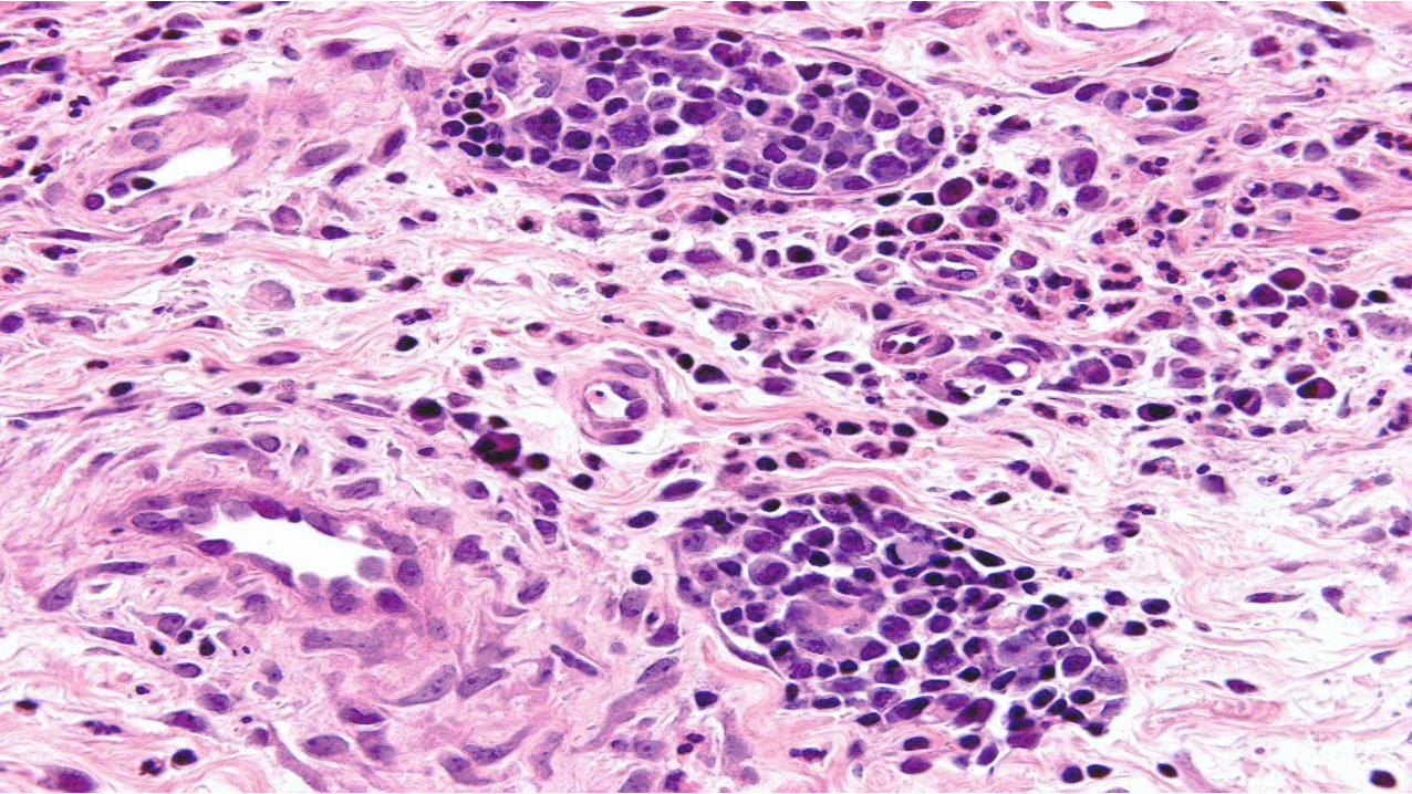
CASE 35 The main diagnosis to consider is A Pyoderma gangrenosum B Cutaneous vasculitis C Intravascular CD30 positive T-cell lymphoma (intralymphatic CD30 positive lymphoproliferative disorder) D Lymphomatoid papulosis type A E Peripheral T cell lymphoma NOS
CASE 35 DIAGNOSIS
INTRAVASCULAR CD30 POSITIVE ANAPLASTIC LARGE T CELL LYMPHOMA
OR
BENIGN ATYPICAL INTRAVASCULAR CD30(+) T-CELL PROLIFERATION
(INTRALYMPHATIC CD30+ LYMPHOPROLIFERATIVE DISORDER
AM J SURG PATHOL MAY 2014 EPUB AHEAD OF PRINT )
INTRAVASCULAR LYMPHOMAS
bull Most have a B cell lineage and are associated with very poor prognosis
bull T cell NK (EBV positive) intravascular lymphomas are very rare
bull CD30 positive intralymphatic lymphoproliferative disorder has only recently been recognized as a distinctive entity
INTRAVASCULAR LARGE B CELL LYMPHOMA
CLINICAL FINDINGS
bull F = M
bull Adults (elderly)
bull Most common in the Asia
bull Frequent involvement of the CNS and skin
bull Hepatosplenic involvement (26) bone marrow involvement (32) lymph node involvement (only 11)
bull Usually sistemic involvement by the time cutaneous lesions develop
bull Violaceous ill-defined lesions on trunk and limbs (inverted livedo reticularis pattern)
bull B symptoms frequent
bull Poor prognosis
bull In cases with cutaneous involvement only (around 26) prognosis appears to be better
INTRAVASCULAR LARGE B CELL LYMPHOMA
HISTOLOGICAL FINDINGS
bull Dilated dermal and subcutaneous vascular channels including venules capillaries and arterioles
bull Blood vessels appear dilated and contain numerous large pleomorphic lymphoid cells
bull Most cases have a pan-B cell phenotype (CD20 and CD79a positive)
Am J Surg Pathol 2013 Apr
bull A case of localized cutaneous intravascular anaplastic lymphoma kinase-negative ALCL (cIALCL) with a very indolent clinical course The patient experienced a single cutaneous relapse and remains alive without disease 4 years after diagnosis Our index case of cIALCL and 1 other tested case were immunohistochemically confirmed to be intralymphatic (contained within D2-40+vessels) as compared with the blood vessel localization of cITNKL Recognition of cIALCLs as a distinct clinicopathologic entity and in particular their distinction from aggressive usually EBV cITNKLs may be possible on the basis of a combination of clinicopathologic criteria allowing for localized therapy in a subset of patients
Intravascular ALK-negative anaplastic large cell lymphoma with localized cutaneous involvement and an indolent clinical course toward recognition of a distinct clinicopathologic entity
Metcalf RA Bashey S Wysong A Kim J Kim YH Gratzinger D
Department of Pathology Stanford University School of Medicine Stanford CA 94305 USA
PROPOSAL
bull Large T-cell lymphoma with typical ALCL morphology and immunophenotype
bull as well as an intralymphatic localization
bull represents a distinct clinicopathologic entity that
bull should be distinguished from intravascular usually EBV+ aggressive T-cell or NK-cell lymphomas involving blood vasculature
Am J Dermatopathol 2011 Dec
Abstract
bull We present a case of a rare intravascular large T-cell lymphoma in a 59-year-old man with an unusual CD3+ CD4+ CD5- CD30+ CD56- TIA-1-negative and EBER-negative phenotype This T helper or CD30 phenotype is particularly uncommon To our knowledge it has only been described once before and never in the absence of the cytotoxic marker TIA-1 This case exemplifies the particular diagnostic challenges raised by intravascular large cell lymphomas generally and should encourage the use of endothelial immunohistochemical staining in questionable cases While evaluating skin punch biopsies it is critical to keep this rare entity on the differential diagnosis along with the relatively more common intravascular large B-cell lymphoma and epithelial malignancies Additionally our understanding of intravascular large natural killerT-cell lymphoma as a heterogeneous phenotypic entity continues to evolve This case demonstrates that the degree of this phenotypic heterogeneity may be even greater than previously thought
A rare case of intravascular large T-cell lymphoma with an unusual T helper phenotype
Deetz CO Gilbertson KG 2nd Anadkat MJ Dehner LP Lu D
Lauren V Ackerman Laboratory of Surgical Pathology Washington University Medical Center St Louis MO 63110 USA
Cutan Pathol 2011 Feb
bull The patient was a 47-year-old woman who had developed multiple erythematous patches and plaques on her back
bull The lesions responded well to CHOP (cyclophosphamide hydroxydoxorubicin oncovin prednisone) chemotherapy but relapsed shortly after therapy The patient was surviving with the disease for eight years but was ultimately lost to follow up
bull Histopathologically the neoplasm evolved from IL to extravascular lymphoma This was showed in biopsies obtained at different stages of the disease The lymphoma cells stained positively for CD30 CD45 CD3 CD4 CD5 and Ki67 and lacked expression of anaplastic lymphoma kinase (ALK) CD8 CD45RA CD45RO CD20 CD79 CD56 perforin and granzyme B
bull Our results suggest that IALCL represents a distinct subtype of IL and is histopathologically and biologically different from IL with B NK or T cell phenotype
Cutaneous intravascular anaplastic large cell lymphoma
Wang L Li C Gao T
Department of Dermatology Xijing Hospital Fourth Military Medical University Xian China
Am J Dermatopathol 2013
bull Report of 2 patients with skin lesions showing an atypical intravascular CD30 T-cell proliferation Both the patients did not present systemic disease and therefore exhibit a favorable outcome
Benign atypical intravascular CD30(+) T-cell proliferation a reactive condition mimicking intravascular lymphoma
Riveiro-Falkenbach E Fernaacutendez-Figueras MT Rodriacuteguez-Peralto JL
Department of Pathology Hospital Universitario 12 de Octubre Instituto de Investigacioacuten i+12 Universidad Complutense Madrid Spain
bull Am J Surg Pathol 2014 May 6 [Epub ahead of print]
bull Intralymphatic Cutaneous Anaplastic Large Cell LymphomaLymphomatoid Papulosis Expanding the Spectrum of CD30-positive Lymphoproliferative Disorders
bull Samols MA1 Su A Ra S Cappel MA Louissant A Jr Knudson RA Ketterling RP Said J Binder S Harris NL Feldman AL Kim J Kim YH Gratzinger D
bull Author information
bull 1Departments of Pathology Dermatology Stanford University School of Medicine Stanford daggerUCLA Medical Center Los Angeles DaggerSan Diego Pathologists Medical Group San Diego CA sectDepartment of Dermatology Mayo Clinic Jacksonville FL ∥Department of Pathology Massachusetts General Hospital Boston MA paraDepartment of Laboratory Medicine and Pathology Mayo Clinic Rochester MN
bull Abstract
bull Intravascular large B-cell lymphomas and EBV NKT-cell lymphomas commonly follow an aggressive clinical course We recently reported an entirely intravascular anaplastic large cell lymphoma (ALCL) in the skin with a surprisingly indolent clinical course interestingly this lymphoma involved the lymphatic rather than the blood vasculature We hypothesized that intravascular skin-limited ALCL is distinct from aggressive systemic intravascular lymphomas in its intralymphatic localization and clinical course We now describe 18 cases of cutaneous intravascular large cell lymphoproliferations from 4 institutions All 12 intravascular large T-cell lesions were intralymphatic the majority (9) were CD30 T-cell lymphoproliferative disorders (TLPDs) 5 further classified as intravascular ALK ALCL One ALK ALCL and 2 benign microscopic intravascular T-cell proliferations were also intralymphatic A single case of otherwise typical cutaneous follicle center lymphoma contained intralymphatic centroblasts The clinical and pathologic characteristics of the CD30 TLPDs were similar to those of their extravascular counterparts including extralymphatic dermal involvement in a subset DUSP22-IRF4 translocations in half of tested ALK ALCLs and associated mycosis fungoides in 1 most were skin-limited at baseline and remained so at relapse All 5 cases of intravascular large B-cell lymphoma involved the blood vasculature and behaved in a clinically aggressive manner the ALK ALCL although intralymphatic was systemic and clinically aggressive We propose that cutaneous ALK ALCL and related CD30 ALK TLPDs involving the lymphatics are part of an expanding spectrum of CD30 TLPDs The identification of intralymphatic as distinct from blood vascular localization may provide critical prognostic and therapeutic information
CD3CD30(+) intravascular lesions with an indolent clinical course
bull Few cases all with good behavior
bull However cases that are ALK positive are associated with involvement elsewhere including lymph nodes and are associated with poor prognosis
CONCLUSION
bull Newly described entity
bull Indolent and part of the spectrum of CD30 positive lymphoproliferative disorders
bull Exclude other lymphomas (systemic ALK1 negative anaplastic lymphomaB T cellNK intravascular lymphoma)
References
1 Riveiro-Falkenbach E Fernaacutendez-Figueras MT Rodriacuteguez-Peralto JL Benign atypical intravascular CD30(+) T-cell proliferation a reactive condition mimicking intravascular lymphoma Am J Dermatopathol 2013 Apr35(2)
2 Metcalf RA Bashey S Wysong A Kim J Kim YH Gratzinger D Intravascular ALK-negative anaplastic large cell lymphoma with localized cutaneous involvement and an indolent clinical course toward recognition of a distinct clinicopathologic entity Am J Surg Pathol 2013 Apr
3 Plaza JA Feldman AL Magro C Cutaneous CD30-positive lymphoproliferative disorders with CD8 expression a clinicopathologic study of 21 cases J Cutan Pathol 2013 Feb40(2)236-47
4 Deetz CO Gilbertson KG 2nd Anadkat MJ Dehner LP Lu D A rare case of intravascular large T-cell lymphoma with an unusual T helper phenotype Am J Dermatopathol 2011 Dec33(8)e99-102
5 Wang L Li C Gao T Cutaneous intravascular anaplastic large cell lymphoma J Cutan Pathol 2011 Feb38(2)221-6
6 Zizi-Sermpetzoglou A Petrakopoulou N Tepelenis N Savvaidou V Vasilakaki T Intravascular T-cell lymphoma of the vulva CD30 positive a case report Eur J Gynaecol Oncol 200930(5)586-8
7 Baum CL Stone MS Liu V Atypical intravascular CD30+ T-cell proliferation following trauma in a healthy 17-year-old male first reported case of a potential diagnostic pitfall and literature review J Cutan Pathol 2009 Mar36(3)350-4
8 Kuo TT Chen MJ Kuo MC Cutaneous intravascular NK-cell lymphoma report of a rare variant associated with Epstein-Barr virus Am J Surg Pathol 2006 Sep30(9)1197-201
9 Takahashi E Kajimoto K Fukatsu T Yoshida M Eimoto T Nakamura S Intravascular large T-cell lymphoma a case report of CD30-positive and ALK-negative anaplastic type with cytotoxic molecule expression Virchows Arch 2005 Dec

INTRAVASCULAR LARGE B CELL LYMPHOMA
CLINICAL FINDINGS
bull F = M
bull Adults (elderly)
bull Most common in the Asia
bull Frequent involvement of the CNS and skin
bull Hepatosplenic involvement (26) bone marrow involvement (32) lymph node involvement (only 11)
bull Usually sistemic involvement by the time cutaneous lesions develop
bull Violaceous ill-defined lesions on trunk and limbs (inverted livedo reticularis pattern)
bull B symptoms frequent
bull Poor prognosis
bull In cases with cutaneous involvement only (around 26) prognosis appears to be better
INTRAVASCULAR LARGE B CELL LYMPHOMA
HISTOLOGICAL FINDINGS
bull Dilated dermal and subcutaneous vascular channels including venules capillaries and arterioles
bull Blood vessels appear dilated and contain numerous large pleomorphic lymphoid cells
bull Most cases have a pan-B cell phenotype (CD20 and CD79a positive)
Am J Surg Pathol 2013 Apr
bull A case of localized cutaneous intravascular anaplastic lymphoma kinase-negative ALCL (cIALCL) with a very indolent clinical course The patient experienced a single cutaneous relapse and remains alive without disease 4 years after diagnosis Our index case of cIALCL and 1 other tested case were immunohistochemically confirmed to be intralymphatic (contained within D2-40+vessels) as compared with the blood vessel localization of cITNKL Recognition of cIALCLs as a distinct clinicopathologic entity and in particular their distinction from aggressive usually EBV cITNKLs may be possible on the basis of a combination of clinicopathologic criteria allowing for localized therapy in a subset of patients
Intravascular ALK-negative anaplastic large cell lymphoma with localized cutaneous involvement and an indolent clinical course toward recognition of a distinct clinicopathologic entity
Metcalf RA Bashey S Wysong A Kim J Kim YH Gratzinger D
Department of Pathology Stanford University School of Medicine Stanford CA 94305 USA
PROPOSAL
bull Large T-cell lymphoma with typical ALCL morphology and immunophenotype
bull as well as an intralymphatic localization
bull represents a distinct clinicopathologic entity that
bull should be distinguished from intravascular usually EBV+ aggressive T-cell or NK-cell lymphomas involving blood vasculature
Am J Dermatopathol 2011 Dec
Abstract
bull We present a case of a rare intravascular large T-cell lymphoma in a 59-year-old man with an unusual CD3+ CD4+ CD5- CD30+ CD56- TIA-1-negative and EBER-negative phenotype This T helper or CD30 phenotype is particularly uncommon To our knowledge it has only been described once before and never in the absence of the cytotoxic marker TIA-1 This case exemplifies the particular diagnostic challenges raised by intravascular large cell lymphomas generally and should encourage the use of endothelial immunohistochemical staining in questionable cases While evaluating skin punch biopsies it is critical to keep this rare entity on the differential diagnosis along with the relatively more common intravascular large B-cell lymphoma and epithelial malignancies Additionally our understanding of intravascular large natural killerT-cell lymphoma as a heterogeneous phenotypic entity continues to evolve This case demonstrates that the degree of this phenotypic heterogeneity may be even greater than previously thought
A rare case of intravascular large T-cell lymphoma with an unusual T helper phenotype
Deetz CO Gilbertson KG 2nd Anadkat MJ Dehner LP Lu D
Lauren V Ackerman Laboratory of Surgical Pathology Washington University Medical Center St Louis MO 63110 USA
Cutan Pathol 2011 Feb
bull The patient was a 47-year-old woman who had developed multiple erythematous patches and plaques on her back
bull The lesions responded well to CHOP (cyclophosphamide hydroxydoxorubicin oncovin prednisone) chemotherapy but relapsed shortly after therapy The patient was surviving with the disease for eight years but was ultimately lost to follow up
bull Histopathologically the neoplasm evolved from IL to extravascular lymphoma This was showed in biopsies obtained at different stages of the disease The lymphoma cells stained positively for CD30 CD45 CD3 CD4 CD5 and Ki67 and lacked expression of anaplastic lymphoma kinase (ALK) CD8 CD45RA CD45RO CD20 CD79 CD56 perforin and granzyme B
bull Our results suggest that IALCL represents a distinct subtype of IL and is histopathologically and biologically different from IL with B NK or T cell phenotype
Cutaneous intravascular anaplastic large cell lymphoma
Wang L Li C Gao T
Department of Dermatology Xijing Hospital Fourth Military Medical University Xian China
Am J Dermatopathol 2013
bull Report of 2 patients with skin lesions showing an atypical intravascular CD30 T-cell proliferation Both the patients did not present systemic disease and therefore exhibit a favorable outcome
Benign atypical intravascular CD30(+) T-cell proliferation a reactive condition mimicking intravascular lymphoma
Riveiro-Falkenbach E Fernaacutendez-Figueras MT Rodriacuteguez-Peralto JL
Department of Pathology Hospital Universitario 12 de Octubre Instituto de Investigacioacuten i+12 Universidad Complutense Madrid Spain
bull Am J Surg Pathol 2014 May 6 [Epub ahead of print]
bull Intralymphatic Cutaneous Anaplastic Large Cell LymphomaLymphomatoid Papulosis Expanding the Spectrum of CD30-positive Lymphoproliferative Disorders
bull Samols MA1 Su A Ra S Cappel MA Louissant A Jr Knudson RA Ketterling RP Said J Binder S Harris NL Feldman AL Kim J Kim YH Gratzinger D
bull Author information
bull 1Departments of Pathology Dermatology Stanford University School of Medicine Stanford daggerUCLA Medical Center Los Angeles DaggerSan Diego Pathologists Medical Group San Diego CA sectDepartment of Dermatology Mayo Clinic Jacksonville FL ∥Department of Pathology Massachusetts General Hospital Boston MA paraDepartment of Laboratory Medicine and Pathology Mayo Clinic Rochester MN
bull Abstract
bull Intravascular large B-cell lymphomas and EBV NKT-cell lymphomas commonly follow an aggressive clinical course We recently reported an entirely intravascular anaplastic large cell lymphoma (ALCL) in the skin with a surprisingly indolent clinical course interestingly this lymphoma involved the lymphatic rather than the blood vasculature We hypothesized that intravascular skin-limited ALCL is distinct from aggressive systemic intravascular lymphomas in its intralymphatic localization and clinical course We now describe 18 cases of cutaneous intravascular large cell lymphoproliferations from 4 institutions All 12 intravascular large T-cell lesions were intralymphatic the majority (9) were CD30 T-cell lymphoproliferative disorders (TLPDs) 5 further classified as intravascular ALK ALCL One ALK ALCL and 2 benign microscopic intravascular T-cell proliferations were also intralymphatic A single case of otherwise typical cutaneous follicle center lymphoma contained intralymphatic centroblasts The clinical and pathologic characteristics of the CD30 TLPDs were similar to those of their extravascular counterparts including extralymphatic dermal involvement in a subset DUSP22-IRF4 translocations in half of tested ALK ALCLs and associated mycosis fungoides in 1 most were skin-limited at baseline and remained so at relapse All 5 cases of intravascular large B-cell lymphoma involved the blood vasculature and behaved in a clinically aggressive manner the ALK ALCL although intralymphatic was systemic and clinically aggressive We propose that cutaneous ALK ALCL and related CD30 ALK TLPDs involving the lymphatics are part of an expanding spectrum of CD30 TLPDs The identification of intralymphatic as distinct from blood vascular localization may provide critical prognostic and therapeutic information
CD3CD30(+) intravascular lesions with an indolent clinical course
bull Few cases all with good behavior
bull However cases that are ALK positive are associated with involvement elsewhere including lymph nodes and are associated with poor prognosis
CONCLUSION
bull Newly described entity
bull Indolent and part of the spectrum of CD30 positive lymphoproliferative disorders
bull Exclude other lymphomas (systemic ALK1 negative anaplastic lymphomaB T cellNK intravascular lymphoma)
References
1 Riveiro-Falkenbach E Fernaacutendez-Figueras MT Rodriacuteguez-Peralto JL Benign atypical intravascular CD30(+) T-cell proliferation a reactive condition mimicking intravascular lymphoma Am J Dermatopathol 2013 Apr35(2)
2 Metcalf RA Bashey S Wysong A Kim J Kim YH Gratzinger D Intravascular ALK-negative anaplastic large cell lymphoma with localized cutaneous involvement and an indolent clinical course toward recognition of a distinct clinicopathologic entity Am J Surg Pathol 2013 Apr
3 Plaza JA Feldman AL Magro C Cutaneous CD30-positive lymphoproliferative disorders with CD8 expression a clinicopathologic study of 21 cases J Cutan Pathol 2013 Feb40(2)236-47
4 Deetz CO Gilbertson KG 2nd Anadkat MJ Dehner LP Lu D A rare case of intravascular large T-cell lymphoma with an unusual T helper phenotype Am J Dermatopathol 2011 Dec33(8)e99-102
5 Wang L Li C Gao T Cutaneous intravascular anaplastic large cell lymphoma J Cutan Pathol 2011 Feb38(2)221-6
6 Zizi-Sermpetzoglou A Petrakopoulou N Tepelenis N Savvaidou V Vasilakaki T Intravascular T-cell lymphoma of the vulva CD30 positive a case report Eur J Gynaecol Oncol 200930(5)586-8
7 Baum CL Stone MS Liu V Atypical intravascular CD30+ T-cell proliferation following trauma in a healthy 17-year-old male first reported case of a potential diagnostic pitfall and literature review J Cutan Pathol 2009 Mar36(3)350-4
8 Kuo TT Chen MJ Kuo MC Cutaneous intravascular NK-cell lymphoma report of a rare variant associated with Epstein-Barr virus Am J Surg Pathol 2006 Sep30(9)1197-201
9 Takahashi E Kajimoto K Fukatsu T Yoshida M Eimoto T Nakamura S Intravascular large T-cell lymphoma a case report of CD30-positive and ALK-negative anaplastic type with cytotoxic molecule expression Virchows Arch 2005 Dec
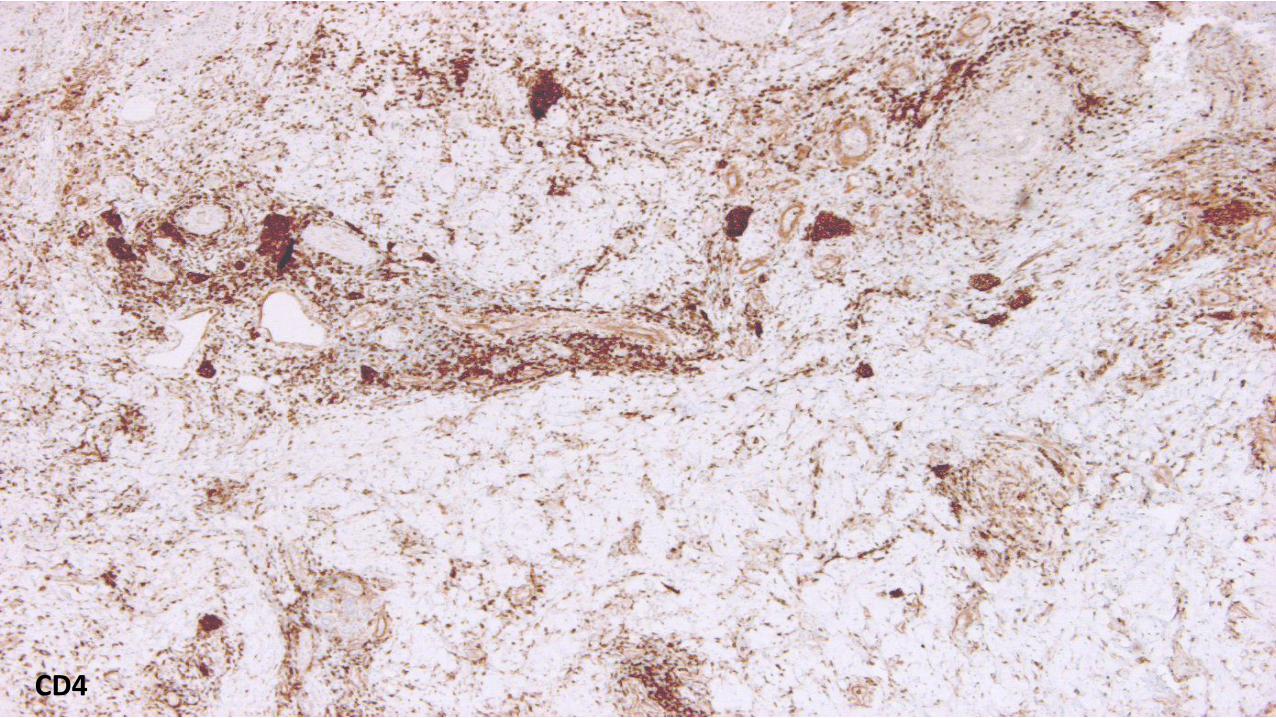
INTRAVASCULAR LARGE B CELL LYMPHOMA
HISTOLOGICAL FINDINGS
bull Dilated dermal and subcutaneous vascular channels including venules capillaries and arterioles
bull Blood vessels appear dilated and contain numerous large pleomorphic lymphoid cells
bull Most cases have a pan-B cell phenotype (CD20 and CD79a positive)
Am J Surg Pathol 2013 Apr
bull A case of localized cutaneous intravascular anaplastic lymphoma kinase-negative ALCL (cIALCL) with a very indolent clinical course The patient experienced a single cutaneous relapse and remains alive without disease 4 years after diagnosis Our index case of cIALCL and 1 other tested case were immunohistochemically confirmed to be intralymphatic (contained within D2-40+vessels) as compared with the blood vessel localization of cITNKL Recognition of cIALCLs as a distinct clinicopathologic entity and in particular their distinction from aggressive usually EBV cITNKLs may be possible on the basis of a combination of clinicopathologic criteria allowing for localized therapy in a subset of patients
Intravascular ALK-negative anaplastic large cell lymphoma with localized cutaneous involvement and an indolent clinical course toward recognition of a distinct clinicopathologic entity
Metcalf RA Bashey S Wysong A Kim J Kim YH Gratzinger D
Department of Pathology Stanford University School of Medicine Stanford CA 94305 USA
PROPOSAL
bull Large T-cell lymphoma with typical ALCL morphology and immunophenotype
bull as well as an intralymphatic localization
bull represents a distinct clinicopathologic entity that
bull should be distinguished from intravascular usually EBV+ aggressive T-cell or NK-cell lymphomas involving blood vasculature
Am J Dermatopathol 2011 Dec
Abstract
bull We present a case of a rare intravascular large T-cell lymphoma in a 59-year-old man with an unusual CD3+ CD4+ CD5- CD30+ CD56- TIA-1-negative and EBER-negative phenotype This T helper or CD30 phenotype is particularly uncommon To our knowledge it has only been described once before and never in the absence of the cytotoxic marker TIA-1 This case exemplifies the particular diagnostic challenges raised by intravascular large cell lymphomas generally and should encourage the use of endothelial immunohistochemical staining in questionable cases While evaluating skin punch biopsies it is critical to keep this rare entity on the differential diagnosis along with the relatively more common intravascular large B-cell lymphoma and epithelial malignancies Additionally our understanding of intravascular large natural killerT-cell lymphoma as a heterogeneous phenotypic entity continues to evolve This case demonstrates that the degree of this phenotypic heterogeneity may be even greater than previously thought
A rare case of intravascular large T-cell lymphoma with an unusual T helper phenotype
Deetz CO Gilbertson KG 2nd Anadkat MJ Dehner LP Lu D
Lauren V Ackerman Laboratory of Surgical Pathology Washington University Medical Center St Louis MO 63110 USA
Cutan Pathol 2011 Feb
bull The patient was a 47-year-old woman who had developed multiple erythematous patches and plaques on her back
bull The lesions responded well to CHOP (cyclophosphamide hydroxydoxorubicin oncovin prednisone) chemotherapy but relapsed shortly after therapy The patient was surviving with the disease for eight years but was ultimately lost to follow up
bull Histopathologically the neoplasm evolved from IL to extravascular lymphoma This was showed in biopsies obtained at different stages of the disease The lymphoma cells stained positively for CD30 CD45 CD3 CD4 CD5 and Ki67 and lacked expression of anaplastic lymphoma kinase (ALK) CD8 CD45RA CD45RO CD20 CD79 CD56 perforin and granzyme B
bull Our results suggest that IALCL represents a distinct subtype of IL and is histopathologically and biologically different from IL with B NK or T cell phenotype
Cutaneous intravascular anaplastic large cell lymphoma
Wang L Li C Gao T
Department of Dermatology Xijing Hospital Fourth Military Medical University Xian China
Am J Dermatopathol 2013
bull Report of 2 patients with skin lesions showing an atypical intravascular CD30 T-cell proliferation Both the patients did not present systemic disease and therefore exhibit a favorable outcome
Benign atypical intravascular CD30(+) T-cell proliferation a reactive condition mimicking intravascular lymphoma
Riveiro-Falkenbach E Fernaacutendez-Figueras MT Rodriacuteguez-Peralto JL
Department of Pathology Hospital Universitario 12 de Octubre Instituto de Investigacioacuten i+12 Universidad Complutense Madrid Spain
bull Am J Surg Pathol 2014 May 6 [Epub ahead of print]
bull Intralymphatic Cutaneous Anaplastic Large Cell LymphomaLymphomatoid Papulosis Expanding the Spectrum of CD30-positive Lymphoproliferative Disorders
bull Samols MA1 Su A Ra S Cappel MA Louissant A Jr Knudson RA Ketterling RP Said J Binder S Harris NL Feldman AL Kim J Kim YH Gratzinger D
bull Author information
bull 1Departments of Pathology Dermatology Stanford University School of Medicine Stanford daggerUCLA Medical Center Los Angeles DaggerSan Diego Pathologists Medical Group San Diego CA sectDepartment of Dermatology Mayo Clinic Jacksonville FL ∥Department of Pathology Massachusetts General Hospital Boston MA paraDepartment of Laboratory Medicine and Pathology Mayo Clinic Rochester MN
bull Abstract
bull Intravascular large B-cell lymphomas and EBV NKT-cell lymphomas commonly follow an aggressive clinical course We recently reported an entirely intravascular anaplastic large cell lymphoma (ALCL) in the skin with a surprisingly indolent clinical course interestingly this lymphoma involved the lymphatic rather than the blood vasculature We hypothesized that intravascular skin-limited ALCL is distinct from aggressive systemic intravascular lymphomas in its intralymphatic localization and clinical course We now describe 18 cases of cutaneous intravascular large cell lymphoproliferations from 4 institutions All 12 intravascular large T-cell lesions were intralymphatic the majority (9) were CD30 T-cell lymphoproliferative disorders (TLPDs) 5 further classified as intravascular ALK ALCL One ALK ALCL and 2 benign microscopic intravascular T-cell proliferations were also intralymphatic A single case of otherwise typical cutaneous follicle center lymphoma contained intralymphatic centroblasts The clinical and pathologic characteristics of the CD30 TLPDs were similar to those of their extravascular counterparts including extralymphatic dermal involvement in a subset DUSP22-IRF4 translocations in half of tested ALK ALCLs and associated mycosis fungoides in 1 most were skin-limited at baseline and remained so at relapse All 5 cases of intravascular large B-cell lymphoma involved the blood vasculature and behaved in a clinically aggressive manner the ALK ALCL although intralymphatic was systemic and clinically aggressive We propose that cutaneous ALK ALCL and related CD30 ALK TLPDs involving the lymphatics are part of an expanding spectrum of CD30 TLPDs The identification of intralymphatic as distinct from blood vascular localization may provide critical prognostic and therapeutic information
CD3CD30(+) intravascular lesions with an indolent clinical course
bull Few cases all with good behavior
bull However cases that are ALK positive are associated with involvement elsewhere including lymph nodes and are associated with poor prognosis
CONCLUSION
bull Newly described entity
bull Indolent and part of the spectrum of CD30 positive lymphoproliferative disorders
bull Exclude other lymphomas (systemic ALK1 negative anaplastic lymphomaB T cellNK intravascular lymphoma)
References
1 Riveiro-Falkenbach E Fernaacutendez-Figueras MT Rodriacuteguez-Peralto JL Benign atypical intravascular CD30(+) T-cell proliferation a reactive condition mimicking intravascular lymphoma Am J Dermatopathol 2013 Apr35(2)
2 Metcalf RA Bashey S Wysong A Kim J Kim YH Gratzinger D Intravascular ALK-negative anaplastic large cell lymphoma with localized cutaneous involvement and an indolent clinical course toward recognition of a distinct clinicopathologic entity Am J Surg Pathol 2013 Apr
3 Plaza JA Feldman AL Magro C Cutaneous CD30-positive lymphoproliferative disorders with CD8 expression a clinicopathologic study of 21 cases J Cutan Pathol 2013 Feb40(2)236-47
4 Deetz CO Gilbertson KG 2nd Anadkat MJ Dehner LP Lu D A rare case of intravascular large T-cell lymphoma with an unusual T helper phenotype Am J Dermatopathol 2011 Dec33(8)e99-102
5 Wang L Li C Gao T Cutaneous intravascular anaplastic large cell lymphoma J Cutan Pathol 2011 Feb38(2)221-6
6 Zizi-Sermpetzoglou A Petrakopoulou N Tepelenis N Savvaidou V Vasilakaki T Intravascular T-cell lymphoma of the vulva CD30 positive a case report Eur J Gynaecol Oncol 200930(5)586-8
7 Baum CL Stone MS Liu V Atypical intravascular CD30+ T-cell proliferation following trauma in a healthy 17-year-old male first reported case of a potential diagnostic pitfall and literature review J Cutan Pathol 2009 Mar36(3)350-4
8 Kuo TT Chen MJ Kuo MC Cutaneous intravascular NK-cell lymphoma report of a rare variant associated with Epstein-Barr virus Am J Surg Pathol 2006 Sep30(9)1197-201
9 Takahashi E Kajimoto K Fukatsu T Yoshida M Eimoto T Nakamura S Intravascular large T-cell lymphoma a case report of CD30-positive and ALK-negative anaplastic type with cytotoxic molecule expression Virchows Arch 2005 Dec
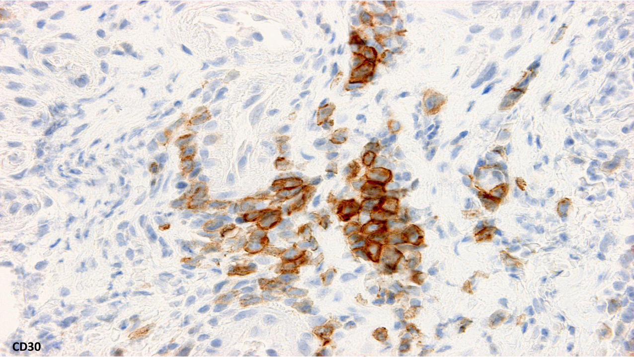
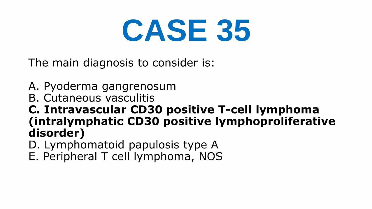