case - fisiokinesiterapia · case y31yo m who ... alpine skiing american football rugby basketball...
TRANSCRIPT

Case31yo M who sustained an injury to L knee while playing Basketball approximately 2 weeks ago. He describes pivoting and hyperextending his knee, which swelled over the next few days. He now presents to clinic with pain on pivoting.L Knee PE on presentation:
2+ L knee effusion.Skin intact.Active range of motion 0-120 degrees. Increased pain with flexion past 120 degrees.Severe tenderness to palpation over medial joint line down to posterior horn of medial meniscus. Stable to varus and valgus stress, with pain on varus stress localized over medial meniscus.Lachman negative – inadequate test because of significant patient guarding.Posterior drawer negative.Positive McMurray.Positive Apley.Soft and compressible L calf compartment.Normal toe flexion and extension.
Rest of PE WNL.

Differential DiagnosisLigament injury:
Knee dislocationCruciate injuryCollateral injury
Meniscal injuryPatellar injury Fracture
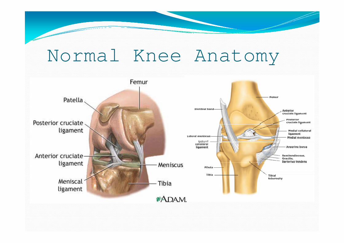
Normal Knee Anatomy

Normal ACL Function
Primal Pictures

Normal Knee Plain Films

Normal Knee MRI

Typical ACL InjuryNon-contact injury occurs in 70% of cases.
Running or jumping with sudden deceleration and direction change, or pivoting while rotating or lateral bending (valgus stress).
Contact injuryLateral blow leading to hyperextension or lateral stress while foot is planted.

In pictures…
Culprit sports:Alpine skiingAmerican footballRugbyBasketballSoccerGymnasticsTennis.

Closer look…

Classical Plain Film Findings
Segond Fracture: avulsion fracture of anterolateraltibial plateau at the site of attachment of the lateral capsular ligament.
Avulsion fractures from femoral or tibial ACL attachments.
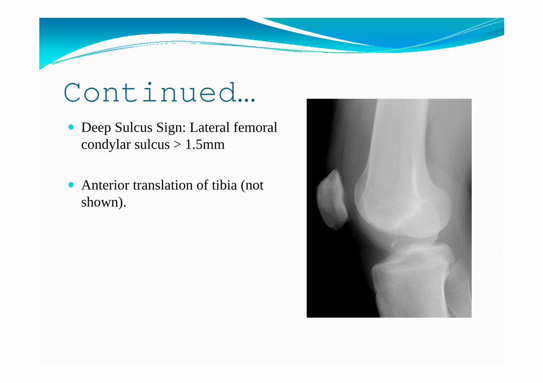
Continued…Deep Sulcus Sign: Lateral femoral condylar sulcus > 1.5mm
Anterior translation of tibia (not shown).

Classic MRI FindingsIrregular/wavy ACL contour with decreased angulation.Widespread increased signal intensity on the region of ACL (all sequences).Posterior displacement of lateral meniscus.Loss of normal curvature with increased angulation of PCL.Undulation of patellar tendon.Medullary osseous contusions of posterolateral tibial plateau and id ti f l t l f l d l
Aunt Minnie's Atlas and Imaging-Specific Diagnosis


Radiologic ResultsPlain Films
Moderate joint effusion.?Deep sulcus sign.
MRIAcute full thickness L knee midsubstanceACL tear w/ moderate joint effusion and bone contusions in lateral femoral condyle and lateral tibial plateau.Medial meniscocapsular injury with medial tibial plateau edema.Posterolateral soft tissue and posterior capsule edema suggesting sprain.

Case Plain Films
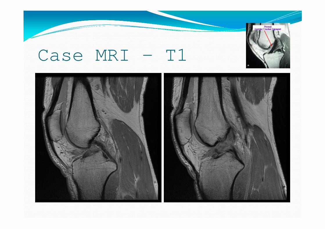
Case MRI – T1

Case MRI – T2

10-Day Follow-Up…Paient returned with increased ability to bear weight and reduced pain. Repeat L knee PE:
Markedly diminished effusion.Positive anterior draw and Lachman (no guarding).Persistent tenderness over lateral femoral condyle.Calf is soft and non-tender.Good motion from 0 to 130 degrees.
Patient referred to Orthopedic Sports for assessment by knee specialist (surgery vs. non-surgical management)