case report medical treatment with steroids and interferon ... desai s, deb r, kane s, kurkure p,...
TRANSCRIPT

PBOncology, Gastroenterology and Hepatology Reports| Jan-Jun 2015 | Vol 4 | Issue 1 43 Oncology, Gastroenterology and Hepatology Reports| Jan-Jun 2015 | Vol 4 | Issue 1
Richa D. Patel, Manisha Khare, Nikhil Majethia, Alka Kalgutkar
Department of Pathology, Lokmanya Tilak Municipal Medical
College and Hospital, Sion, Mumbai, Maharashtra, India
Address for the Correspondence:Dr. Richa D. Patel,
Department of Pathology, Lokmanya Tilak Municipal
Medical College and Hospital, Sion, Mumbai ‑ 400 021,
Maharashtra, India. E‑mail: [email protected]
Infantile hepatic hemangioendothelioma presenting as early heart failure: An autopsy case report
INTRODUCTION
Hepatic tumors in children are relatively rare, accounting for 1-4% of all pediatric solid tumors. Infantile hemangioendothelioma (IHE) of the liver is a rare mesenchymal tumor, but it is the most common vascular tumor of the liver in children, accounting for 12% of all childhood hepatic tumors. Almost 85% of patients with infantile hepatic hemangioendothelioma (IHHE) are diagnosed during the first 6 months of life, and is the most common symptomatic tumor occurring during this time period.[1-3] IHE appears to be a histologically benign tumor that may have a poor outcome because of severe complications like congestive heart failure (CHF) seen in 15% and liver failure in 2% of infants. A noncomplicated tumor may spontaneously regress, but most fatalities occur in patients whose initial presentation is intractable heart failure.[4]
Here, we reviewed an autopsy study of IHHE in a 20-day-old female patient. The clinical, pathological, radiological features along with the differential diagnosis of this tumor are being discussed.
CASE REPORT
A female baby was born after 39 weeks of gestation through a normal spontaneous delivery with weight of 3200 g. Baby cried well after birth. At the age of 20 days, she got admitted to our hospital for abdominal distention, tachypnea, and tachycardia. Physical examination revealed a mildly cyanotic infant with respiratory distress and palpable liver of 5 cm below the right costal margin (normal range [N]: 3.5 cm); no cutaneous hemangiomas were noted. The chest X-ray showed cardiomegaly and the electrocardiogram disclosed biventricular hypertrophy.
Laboratory studies, including liver function tests (aspartate aminotransferase [N: 0-60 IU/L] and alanine aminotransferase [N: 0-50 IU/L]) and coagulation profile (platelet counts [N: 1.5-4.5 lac/cmm], prothrombin time [N: 11-13 s] and partial thromboplastin time [N: 20-30 s]) were within the normal range. At 22 days old, serum alpha-fetoprotein (AFP) was 2026 µg/L (N: 9452-12610 µg/L). Abdominal ultrasound revealed enlarged liver with multiple lesions throughout parenchyma showing mild vascularity with largest measuring 4.8 cm in segment 7 and 8, features suggestive of neoplastic
Case Repor t
Access this article online
Website: www.oghr.org
DOI: 10.4103/2348-3113.139649
Quick response code:
Infantile hepatic hemangioendothelioma (IHHE) is the most common type of hepatic vascular tumor in infancy, but is rarely reported because of the low incidence estimated to be about 1/20,000. We report an autopsy case of IHHE in a 20-day-old female who came with an initial manifestation of abdominal distension and congestive heart failure (CHF). Symptoms of cardiac decompensation gradually worsened in spite of treatment and child died within 2 days. On autopsy, grossly enlarged liver showed microscopy of large ecstatic vascular channels lined by single layer of plump endothelial cells and diagnosis was given as Type I IHHE. CHF is common complication due to arteriovenous shunts within the tumor and contributes to high morbidity and mortality in up to 70% of untreated infants with median age of presentation of 1-month. Therefore, if symptoms develop, aggressive treatments are warranted.
Key words: Autopsy, heart failure, hemangioendothelioma, hepatic tumor, infantile tumor
Abs
trac
t

Patel, et al.: Infantile hepatic hemangioendothelioma presenting as early heart failure
44Oncology, Gastroenterology and Hepatology Reports| Jan-Jun 2015 | Vol 4 | Issue 1 45 Oncology, Gastroenterology and Hepatology Reports| Jan-Jun 2015 | Vol 4 | Issue 1
etiology. Symptoms of cardiac decompensation gradually worsened in spite of treatment with digoxin and prednisolone and patient died within 2 days. Autopsy was performed. Internal examination of the abdomen showed markedly enlarged liver, covering whole of the upper quadrant of abdomen [Figure 1]. Approximately, 800 ml of pale colored fluid was found. The liver measured 17.5 × 10.5 × 5 cm (N: 5.27-7.73 cm largest dimension) and weighed 1200 g (N: 78 g). External surface of the liver showed multiple nodular lesions of sizes ranging 2-5 cm in diameter and firm to spongy soft consistency. Cut surface showed multiple greyish white to red nodular, well-encapsulated lesions uniformly distributed in the liver with intervening normal parenchyma [Figure 2]. Heart showed marked right ventricular dilation. Other organs were unremarkable. Provisional cause of death was given as cardiorespiratory failure in a case of hepatic neoplasm.
On microscopy, the liver showed multiple nodules composed of large ecstatic vascular channels lined by single layer of plump endothelial cells separated by loose stroma [Figure3]. A diagnosis of Type I hemangioendothelioma was given.
DISCUSSION
Infantile hepatic hemangioendothelioma is a type of capillary hemangioma, which consists of a network of capillary-sized endothelium-lined vessels. IHHE is the third most common type of hepatic tumor and the most common benign vascular tumor of the liver in infants. Tumors show a female predominance, with a female to male ratio of 1.3-2:1.[5]
Clinical manifestations of IHH are variable depending on the tumor size and location, and include hepatomegaly (83%), an abdominal mass (66%), skin hemangioma (65%), anorexia, vomiting (25%), and failure to thrive (25%).[6] Hematologic abnormalities are anemia and thrombocytopenia caused by trapping of thrombocytes within the hemangioendothelioma with consumptive coagulopathy, also known as Kasabach-Merritt syndrome. Less common presentations include splenomegaly, jaundice, ascites, gastrointestinal bleeding and rarely, spontaneous rupture and malignant transformation to angiosarcoma extensive arteriovenous shunting may be detected within these lesions, resulting in decreased peripheral vascular resistance. The maintenance of vascular bed perfusion may require increases in blood volume and cardiac output, which may lead to high cardiac output and CHF, observed in up to 50-60% of patients with IHHE.[7]
Serum AFP is an important tumor marker for the evaluation of pediatric hepatic masses. Increased serum AFP concentrations are rarely observed in patients with IHHE. An increase in serum AFP is not directly related to treatment outcome.[8]
On abdominal ultrasound, IHH show variable echogenicity but is predominantly hypoechoic with well-defined margins. Computed tomography demonstrates focal areas of low attenuation, which show early peripheral enhancement and delayed central filling-in with contrast. Magnetic resonance imaging identify IHHE as low-signal
Figure 1: Gross photograph of autopsy showing massively enlarged liver with multiple nodular lesions (arrows) of varying sizes involving whole liver
Figure 2: Gross photograph of cut surface of formalin fixed liver showing multiple well encapsulated nodular areas (arrows) of variable size uniformly distributed in whole liver
Figure 3: Photomicrograph showing large ectatic vascular channels (arrows) lined by single layer of plump endothelial cells separated by loose stroma (H and E, original magnification ×100)
Patel, et al.: Infantile hepatic hemangioendothelioma presenting as early heart failure
44Oncology, Gastroenterology and Hepatology Reports| Jan-Jun 2015 | Vol 4 | Issue 1 45 Oncology, Gastroenterology and Hepatology Reports| Jan-Jun 2015 | Vol 4 | Issue 1
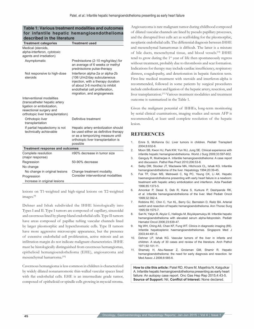
Angiosarcoma is rare malignant tumor during childhood composed of dilated vascular channels are lined by pseudo papillary processes, and the disrupted liver cells act as scaffolding for the pleomorphic, neoplastic endothelial cells. The differential diagnosis between IHHE and mesenchymal hamartomas is difficult. The latter is a mixture of bile ducts, mesenchymal tissue, and blood vessels.[10] IHHE tend to grow during the 1st year of life then spontaneously regress without treatment, probably due to thrombosis and scar formation. Indications for therapy may include cardiac insufficiency, respiratory distress, coagulopathy, and deterioration in hepatic function tests. First-line medical treatment with steroids and interferon-alpha is recommended, followed in some patients by surgical procedures include embolization and ligation of the hepatic artery, resection, and liver transplantation.[11] Various treatment modalities and treatment outcome is summarized in the Table 1.
Given the malignant potential of IHHEs, long-term monitoring by serial clinical examinations, imaging studies and serum AFP is recommended, at least until complete resolution of the hepatic lesion.
REFERENCES1. Emre S, McKenna GJ. Liver tumors in children. Pediatr Transplant
2004;8:632‑8.2. Moon SB, Kwon HJ, Park KW, Yun WJ, Jung SE. Clinical experience with
infantile hepatic hemangioendothelioma. World J Surg 2009;33:597‑602.3. Ganguly R, Mukherjee A. Infantile hemangioendothelioma: A case report
and discussion. Pathol Res Pract 2010;206:53‑8.4. Selby DM, Stocker JT, Waclawiw MA, Hitchcock CL, Ishak KG. Infantile
hemangioendothelioma of the liver. Hepatology 1994;20:39‑45.5. Fok TF, Chan MS, Metreweli C, Ng PC, Yeung CK, Li AK. Hepatic
haemangioendothelioma presenting with early heart failure in a newborn: treatment with hepatic artery embolization and interferon. Acta Paediatr 1996;85:1373‑5.
6. Amonkar P, Desai S, Deb R, Kane S, Kurkure P, Deshpande RK, et al. Infantile hemangioendothelioma of the liver. Med Pediatr Oncol 1999;32:392‑4.
7. Robbins RC, Chin C, Yun KL, Berry GJ, Bernstein D, Reitz BA. Arterial switch and resection of hepatic hemangioendothelioma. Ann Thorac Surg 1995;59:1575‑7.
8. Sari N, Yalçin B, Akyüz C, Haliloglu M, Büyükpamukçu M. Infantile hepatic hemangioendothelioma with elevated serum alpha‑fetoprotein. Pediatr Hematol Oncol 2006;23:639‑47.
9. Ng WH, Ching AS, Chan KF, Fung WT. Clinics in diagnostic imaging (89). Infantile hepatosplenic haemangioendotheliomas. Singapore Med J 2003;44:491‑5.
10. Dehner LP, Ishak KG. Vascular tumors of the liver in infants and children. A study of 30 cases and review of the literature. Arch Pathol 1971;92:101‑11.
11. Shamaly H, Abu‑Nassar Z, Groisman GM, Shamir R. Hepatic hemangloendothelioma: the need for early diagnosis and resection. Isr Med Assoc J 2006;8:585‑6.
Table 1: Various treatment modalities and outcomes for infantile hepatic hemangioendothelioma described in the literatureTreatment categories Treatment used
Medical (steroids, alpha‑interferon, cytotoxic agents and irradiation)
Asymptomatic Prednisolone (2‑10 mg/kg/day) for an average of 6 weeks or methyl prednisolone pulse‑therapy
Not responsive to high‑dose steroids
Interferon alpha‑2a or alpha‑2b (106 U/m2/day subcutaneous injection, with a therapy duration of about 3‑6 months) to inhibit endothelial cell proliferation, migration, and angiogenesis
Interventional modalities (transcatheter hepatic artery ligation or embolization, resectional surgery and orthotopic liver transplantation)
Orthotopic liver transplantation
Definitive treatment
If partial hepatectomy is not technically achievable
Hepatic artery embolization should be used either as definitive therapy or as a temporizing measure until orthotopic liver transplantation is possible
Treatment response and outcomesComplete resolution (major response)
≥90% decrease in tumor size
Regression 50-90% decreaseNo change
No change in original lesions Change treatment modality. Consider interventional modalitiesProgression
Increase in original lesions
lesions on T1-weighted and high-signal lesions on T2-weighted images.[9]
Dehner and Ishak subdivided the IHHE histologically into Types I and II. Type I tumors are composed of capillary, sinusoidal and cavernous lined by plump bland endothelial cells. Type II tumors have areas composed of papillae tufting vascular channels lined by larger pleomorphic and hyperchromatic cells. Type II tumors have more aggressive microscopic appearance, but the presence of extensive endothelial cell proliferation, active mitosis and an infiltration margin do not indicate malignant characteristics. IHHE must be histologically distinguished from cavernous hemangiomas, epithelioid hemangioendothelioma (EHE), angiosarcoma and mesenchymal hamartoma.[10]
Cavernous hemangioma is less common in children is characterized by widely dilated nonanastomotic thin-walled vascular spaces lined with flat endothelial cells. EHE is an intermediate grade tumor, composed of epithelioid or spindle cells growing in myxoid stroma.
How to cite this article: Patel RD, Khare M, Majethia N, Kalgutkar A. Infantile hepatic hemangioendothelioma presenting as early heart failure: An autopsy case report. Onc Gas Hep Rep 2015;4:43‑5.Source of Support: Nil, Conflict of Interest: None declared.