case report diffuse ocular and orbital inflammation after
TRANSCRIPT
Case ReportDiffuse ocular and orbital inflammation after zoledronate infusion—case report and review of the literatureObi C. Umunakwe, MD, PhD, David Herren, MD, Stephen J. Kim, MD, and Sahar Kohanim, MD, MPHAuthor affiliations: Department of Ophthalmology and Visual Sciences, Vanderbilt University Medical Center, Nashville,Tennessee
SummaryBisphosphonates have become a commonly used class of medications to treat osteoporosis and other bonediseases. Zoledronate (zoledronic acid) can be dosed annually via intravenous infusion, making it anappealing option for patients and physicians. We report the case of a 68-year-old woman who developedsevere, unilateral, ocular inflammation, including corneal endotheliitis, anterior uveitis with hyphema,scleritis, and orbital inflammation beginning 12 hours after receiving her first zoledronate infusion. Symp-toms escalated but ultimately resolved with topical steroids and high-dose systemic corticosteroids. To ourknowledge, this is the first report of unilateral diffuse inflammation of the eye and orbit, including cornealinflammation developing within 12 hours of a first zoledronate infusion.
IntroductionBisphosphonates have become the most commonly usedmedication for treatment of osteoporosis.1 They are alsoused in conditions such as hypercalcemia, Paget diseaseof bone, and bony metastatic disease.2 Zoledronate isamong the most frequently used bisphosphonatesbecause it permits an annual dosing regimen via intrave-nous infusion. Ocular complications related tobisphosphonates have previously been described and arerare. Most of these occurrences are limited to nonspe-cific conjunctivitis, anterior uveitis, or blurred visionwith no long-term sequelae.3 However, bisphosphonateshave also been reported to cause scleritis, their mostvision-threatening side effect.3,4
Case ReportA 68-year-old woman presented at an outside hospitalwith a 24-hour history of right eye pain, redness, andphotophobia. Her past medical history was notable forhypertension, hyperlipidemia, depression/anxiety,migraine headaches, and osteoporosis. Her family his-tory was noncontributory. Symptoms of right eye pain,redness, and photophobia began within 12 hours of herfirst-ever intravenous infusion of 4 mg of zoledronate totreat osteoporosis. These symptoms were associated
with fever, myalgias, and arthralgias. She was evaluatedat her local emergency department and treated with atopical antibiotic. The next day, she was examined by anophthalmologist at the Vanderbilt Eye Institute, whofound decreased visual acuity (20/50), conjunctivalinjection, 4+ cell and 2+ flare in the anterior chamber,layered hypopyon with hyphema, globe tenderness, andactive scleritis. A diagnosis of bisphosphonate-associ-ated anterior uveitis and scleritis was presumed, and shewas started on topical steroids, cycloplegics, and oralnonselective anti-inflammatory drugs (NSAIDs).
Two days later, she returned with worsening symptomsand was found to have visual acuity of 20/400, conjunc-tival injection, 3+ to 4+ cell and flare, scleral inflamma-tion, and globe tenderness, with the addition of ptosis,corneal edema, posterior synechiae, and Descemet’sfolds. B-scan ultrasonography was normal, and she wasstarted on oral prednisone 40 mg daily in addition to herprevious regimen. The left eye remained uninvolved.
On day 5, she noted some improvement in her symp-toms after starting the oral steroids. Her visual acuityimproved to 20/150, and she had developed a hyphema.The remainder of her examination was stable.
Published December 28, 2017.Copyright ©2017. All rights reserved. Reproduction in whole or in part in any form or medium without expressed written permission of theDigital Journal of Ophthalmology is prohibited.doi:10.5693/djo.02.2017.08.002Correspondence: Sahar Kohanim, MD, MPH, 2311 Pierce Avenue, Nashville, TN 37232 (email: [email protected]).
Digital Journal of O
phthalmology, Vol. 23
Digital Journal of O
phthalmology, Vol. 23
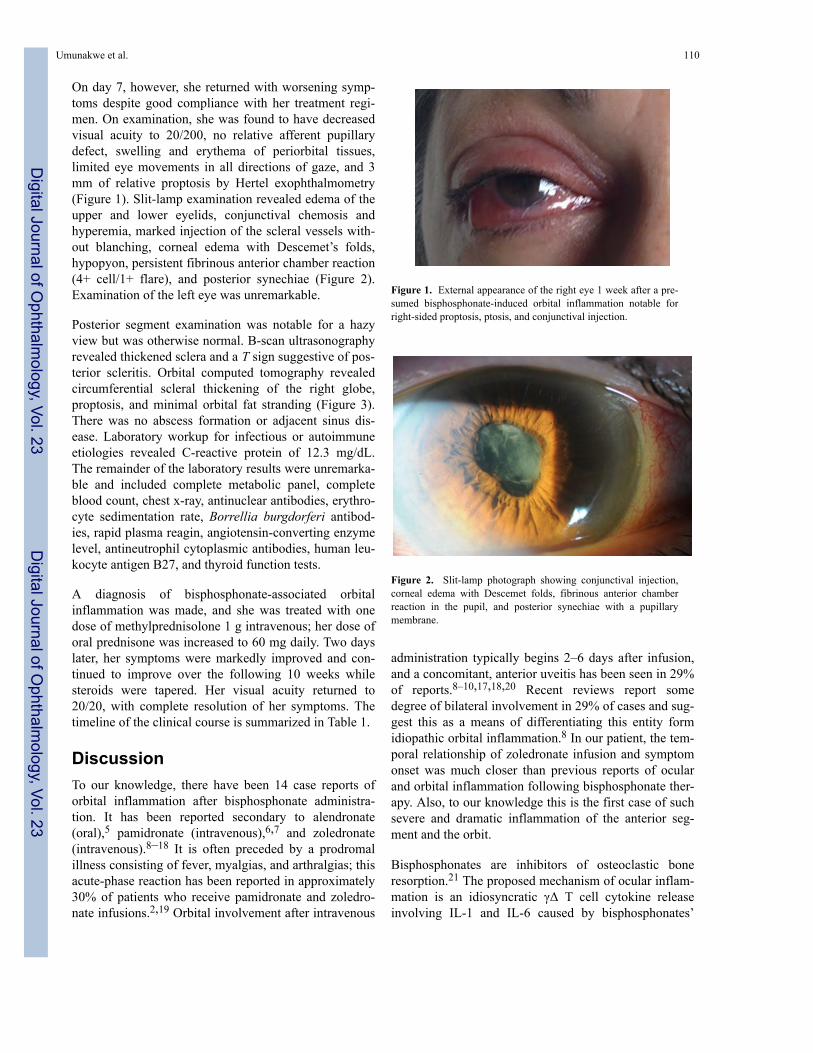
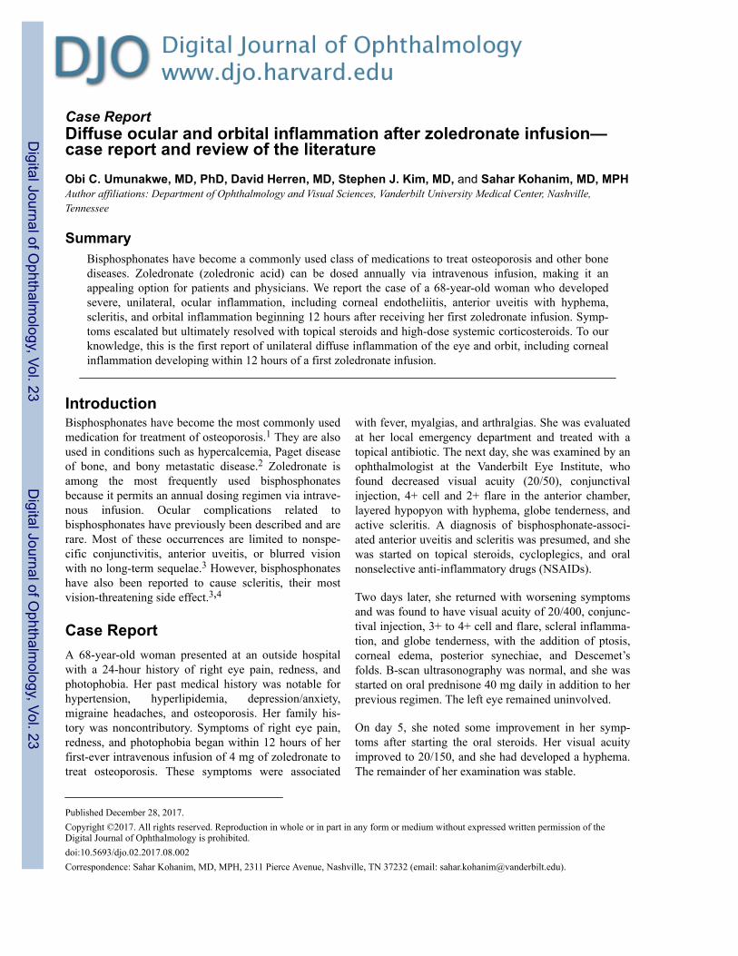
On day 7, however, she returned with worsening symp-toms despite good compliance with her treatment regi-men. On examination, she was found to have decreasedvisual acuity to 20/200, no relative afferent pupillarydefect, swelling and erythema of periorbital tissues,limited eye movements in all directions of gaze, and 3mm of relative proptosis by Hertel exophthalmometry(Figure 1). Slit-lamp examination revealed edema of theupper and lower eyelids, conjunctival chemosis andhyperemia, marked injection of the scleral vessels with-out blanching, corneal edema with Descemet’s folds,hypopyon, persistent fibrinous anterior chamber reaction(4+ cell/1+ flare), and posterior synechiae (Figure 2).Examination of the left eye was unremarkable.
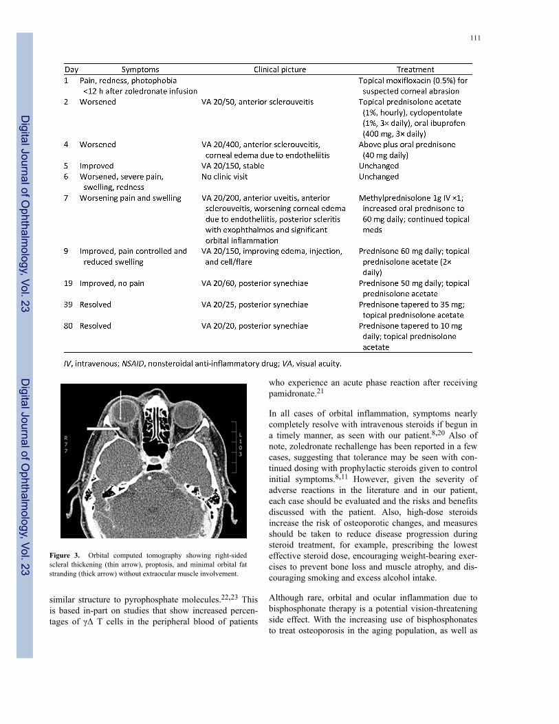
Posterior segment examination was notable for a hazyview but was otherwise normal. B-scan ultrasonographyrevealed thickened sclera and a T sign suggestive of pos-terior scleritis. Orbital computed tomography revealedcircumferential scleral thickening of the right globe,proptosis, and minimal orbital fat stranding (Figure 3).There was no abscess formation or adjacent sinus dis-ease. Laboratory workup for infectious or autoimmuneetiologies revealed C-reactive protein of 12.3 mg/dL.The remainder of the laboratory results were unremarka-ble and included complete metabolic panel, completeblood count, chest x-ray, antinuclear antibodies, erythro-cyte sedimentation rate, Borrellia burgdorferi antibod-ies, rapid plasma reagin, angiotensin-converting enzymelevel, antineutrophil cytoplasmic antibodies, human leu-kocyte antigen B27, and thyroid function tests.
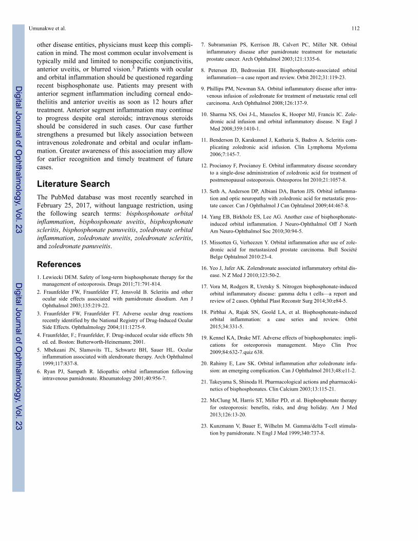
A diagnosis of bisphosphonate-associated orbitalinflammation was made, and she was treated with onedose of methylprednisolone 1 g intravenous; her dose oforal prednisone was increased to 60 mg daily. Two dayslater, her symptoms were markedly improved and con-tinued to improve over the following 10 weeks whilesteroids were tapered. Her visual acuity returned to20/20, with complete resolution of her symptoms. Thetimeline of the clinical course is summarized in Table 1.
DiscussionTo our knowledge, there have been 14 case reports oforbital inflammation after bisphosphonate administra-tion. It has been reported secondary to alendronate(oral),5 pamidronate (intravenous),6,7 and zoledronate(intravenous).8–18 It is often preceded by a prodromalillness consisting of fever, myalgias, and arthralgias; thisacute-phase reaction has been reported in approximately30% of patients who receive pamidronate and zoledro-nate infusions.2,19 Orbital involvement after intravenous
administration typically begins 2–6 days after infusion,and a concomitant, anterior uveitis has been seen in 29%of reports.8–10,17,18,20 Recent reviews report somedegree of bilateral involvement in 29% of cases and sug-gest this as a means of differentiating this entity formidiopathic orbital inflammation.8 In our patient, the tem-poral relationship of zoledronate infusion and symptomonset was much closer than previous reports of ocularand orbital inflammation following bisphosphonate ther-apy. Also, to our knowledge this is the first case of suchsevere and dramatic inflammation of the anterior seg-ment and the orbit.
Bisphosphonates are inhibitors of osteoclastic boneresorption.21 The proposed mechanism of ocular inflam-mation is an idiosyncratic γΔ T cell cytokine releaseinvolving IL-1 and IL-6 caused by bisphosphonates’
Figure 1. External appearance of the right eye 1 week after a pre-sumed bisphosphonate-induced orbital inflammation notable forright-sided proptosis, ptosis, and conjunctival injection.
Figure 2. Slit-lamp photograph showing conjunctival injection,corneal edema with Descemet folds, fibrinous anterior chamberreaction in the pupil, and posterior synechiae with a pupillarymembrane.
Umunakwe et al. 110
Digital Journal of O
phthalmology, Vol. 23
Digital Journal of O
phthalmology, Vol. 23
similar structure to pyrophosphate molecules.22,23 Thisis based in-part on studies that show increased percen-tages of γΔ T cells in the peripheral blood of patients
Figure 3. Orbital computed tomography showing right-sidedscleral thickening (thin arrow), proptosis, and minimal orbital fatstranding (thick arrow) without extraocular muscle involvement.
who experience an acute phase reaction after receivingpamidronate.21
In all cases of orbital inflammation, symptoms nearlycompletely resolve with intravenous steroids if begun ina timely manner, as seen with our patient.8,20 Also ofnote, zoledronate rechallenge has been reported in a fewcases, suggesting that tolerance may be seen with con-tinued dosing with prophylactic steroids given to controlinitial symptoms.8,11 However, given the severity ofadverse reactions in the literature and in our patient,each case should be evaluated and the risks and benefitsdiscussed with the patient. Also, high-dose steroidsincrease the risk of osteoporotic changes, and measuresshould be taken to reduce disease progression duringsteroid treatment, for example, prescribing the lowesteffective steroid dose, encouraging weight-bearing exer-cises to prevent bone loss and muscle atrophy, and dis-couraging smoking and excess alcohol intake.
Although rare, orbital and ocular inflammation due tobisphosphonate therapy is a potential vision-threateningside effect. With the increasing use of bisphosphonatesto treat osteoporosis in the aging population, as well as
111
Digital Journal of O
phthalmology, Vol. 23
Digital Journal of O
phthalmology, Vol. 23
other disease entities, physicians must keep this compli-cation in mind. The most common ocular involvement istypically mild and limited to nonspecific conjunctivitis,anterior uveitis, or blurred vision.3 Patients with ocularand orbital inflammation should be questioned regardingrecent bisphosphonate use. Patients may present withanterior segment inflammation including corneal endo-theliitis and anterior uveitis as soon as 12 hours aftertreatment. Anterior segment inflammation may continueto progress despite oral steroids; intravenous steroidsshould be considered in such cases. Our case furtherstrengthens a presumed but likely association betweenintravenous zoledronate and orbital and ocular inflam-mation. Greater awareness of this association may allowfor earlier recognition and timely treatment of futurecases.
Literature SearchThe PubMed database was most recently searched inFebruary 25, 2017, without language restriction, usingthe following search terms: bisphosphonate orbitalinflammation, bisphosphonate uveitis, bisphosphonatescleritis, bisphosphonate panuveitis, zoledronate orbitalinflammation, zoledronate uveitis, zoledronate scleritis,and zoledronate panuveitis.
References1. Lewiecki DEM. Safety of long-term bisphosphonate therapy for the
management of osteoporosis. Drugs 2011;71:791-814.2. Fraunfelder FW, Fraunfelder FT, Jensvold B. Scleritis and other
ocular side effects associated with pamidronate disodium. Am JOphthalmol 2003;135:219-22.
3. Fraunfelder FW, Fraunfelder FT. Adverse ocular drug reactionsrecently identified by the National Registry of Drug-Induced OcularSide Effects. Ophthalmology 2004;111:1275-9.
4. Fraunfelder, F.; Fraunfelder, F. Drug-induced ocular side effects 5thed. ed. Boston: Butterworth-Heinemann; 2001.
5. Mbekeani JN, Slamovits TL, Schwartz BH, Sauer HL. Ocularinflammation associated with alendronate therapy. Arch Ophthalmol1999;117:837-8.
6. Ryan PJ, Sampath R. Idiopathic orbital inflammation followingintravenous pamidronate. Rheumatology 2001;40:956-7.
7. Subramanian PS, Kerrison JB, Calvert PC, Miller NR. Orbitalinflammatory disease after pamidronate treatment for metastaticprostate cancer. Arch Ophthalmol 2003;121:1335-6.
8. Peterson JD, Bedrossian EH. Bisphosphonate-associated orbitalinflammation—a case report and review. Orbit 2012;31:119-23.
9. Phillips PM, Newman SA. Orbital inflammatory disease after intra-venous infusion of zoledronate for treatment of metastatic renal cellcarcinoma. Arch Ophthalmol 2008;126:137-9.
10. Sharma NS, Ooi J-L, Masselos K, Hooper MJ, Francis IC. Zole-dronic acid infusion and orbital inflammatory disease. N Engl JMed 2008;359:1410-1.
11. Benderson D, Karakunnel J, Kathuria S, Badros A. Scleritis com-plicating zoledronic acid infusion. Clin Lymphoma Myeloma2006;7:145-7.
12. Procianoy F, Procianoy E. Orbital inflammatory disease secondaryto a single-dose administration of zoledronic acid for treatment ofpostmenopausal osteoporosis. Osteoporos Int 2010;21:1057-8.
13. Seth A, Anderson DP, Albiani DA, Barton JJS. Orbital inflamma-tion and optic neuropathy with zoledronic acid for metastatic pros-tate cancer. Can J Ophthalmol J Can Ophtalmol 2009;44:467-8.
14. Yang EB, Birkholz ES, Lee AG. Another case of bisphosphonate-induced orbital inflammation. J Neuro-Ophthalmol Off J NorthAm Neuro-Ophthalmol Soc 2010;30:94-5.
15. Missotten G, Verheezen Y. Orbital inflammation after use of zole-dronic acid for metastasized prostate carcinoma. Bull SociétéBelge Ophtalmol 2010:23-4.
16. Yeo J, Jafer AK. Zolendronate associated inflammatory orbital dis-ease. N Z Med J 2010;123:50-2.
17. Vora M, Rodgers R, Uretsky S. Nitrogen bisphosphonate-inducedorbital inflammatory disease: gamma delta t cells—a report andreview of 2 cases. Ophthal Plast Reconstr Surg 2014;30:e84-5.
18. Pirbhai A, Rajak SN, Goold LA, et al. Bisphosphonate-inducedorbital inflammation: a case series and review. Orbit2015;34:331-5.
19. Kennel KA, Drake MT. Adverse effects of bisphosphonates: impli-cations for osteoporosis management. Mayo Clin Proc2009;84:632-7.quiz 638.
20. Rahimy E, Law SK. Orbital inflammation after zoledronate infu-sion: an emerging complication. Can J Ophthalmol 2013;48:e11-2.
21. Takeyama S, Shinoda H. Pharmacological actions and pharmacoki-netics of bisphosphonates. Clin Calcium 2003;13:115-21.
22. McClung M, Harris ST, Miller PD, et al. Bisphosphonate therapyfor osteoporosis: benefits, risks, and drug holiday. Am J Med2013;126:13-20.
23. Kunzmann V, Bauer E, Wilhelm M. Gamma/delta T-cell stimula-tion by pamidronate. N Engl J Med 1999;340:737-8.
Umunakwe et al. 112
Digital Journal of O
phthalmology, Vol. 23
Digital Journal of O
phthalmology, Vol. 23