case report grievous temporal and occipital injury...
TRANSCRIPT
Hindawi Publishing CorporationCase Reports in OtolaryngologyVolume 2013, Article ID 957251, 5 pageshttp://dx.doi.org/10.1155/2013/957251
Case ReportGrievous Temporal and Occipital Injury Caused by a Bear Attack
Sampath Chandra Prasad,1 Nikhil Dinaker Thada,1 Pallavi Rao,2
Smitha Rani Thada,3 and Kishore Chandra Prasad1
1 Department of Otolaryngology, Head & Neck Surgery, Kasturba Medical College, Manipal University, First Floor,Nethravathi Building, Balmatta, Mangalore, Karnataka 575001, India
2Department of Radiodiagnosis, Kasturba Medical College, Manipal University, Attavar, Mangalore, Karnataka 575002, India3 Department of Oral Medicine & Radiology, Manipal College of Dental Sciences, Manipal University, Madhava Nagar,Manipal, Karnataka 576104, India
Correspondence should be addressed to Sampath Chandra Prasad; [email protected]
Received 27 October 2013; Accepted 21 November 2013
Academic Editors: J. I. De Diego and A. Rapoport
Copyright © 2013 Sampath Chandra Prasad et al. This is an open access article distributed under the Creative CommonsAttribution License, which permits unrestricted use, distribution, and reproduction in any medium, provided the original work isproperly cited.
Bear attacks are reported from nearly every part of the world. The chance of a human encountering a bear increases as the remotebear territory diminishes.The sloth bear is one of the three species of bears found in India, which inhabits the forests of India and itsneighboring countries. Here we describe a teenager who came to us with a critical injury involving the face, temporal and occipitalbones inflicted by a sloth bear attack. He underwent a temporal exploration, facial nerve decompression, pinna reconstruction, andoccipital bone repair to save him from fatality.
1. Introduction
Bear attacks on humans are rare and are even more rarelyreported in medical literature. Each year people have numer-ous accidental interactions with bears. A very small fractionof this results in human injury. The chance of a humanencountering a bear increases as the remote bear territorydiminishes. A search of scientific literature reveals very fewarticles detailing case reports or an in-depth analysis ofinjuries due to bearmauling.Herewe discuss the presentationand subsequent management of an 18-year-old young manwho came to us in a state of shock with a history of an assaultby a sloth bear which led to an avulsion of his temporal andoccipital bones associated with a facial nerve paralysis.
2. Case Report
An 18-year old teenager, a shepherd by occupation, presentedto the emergency room in a state of shock with laceration ofthe ear and head, bleeding from the ear and the back of thehead. A history by the relatives of the boy revealed that he hadbeen mauled by a sloth bear in the forests of his native village
where he had gone shepherding with his brother-in-law. Toelaborate the account, which in itself is interesting, the twoboys unexpectedly ran into a sloth bear in the forest, whichattacked the brother-in-law. Seeing this, our boy ran to hisrescue and in turn incurred the wrath of the bear on himself.While being mauled himself, the boy showed great presenceof mind when, just as the bear was about to take a bite athis face, he hurled a rock piece from nearby into the bear’smouth and kicked it in its belly. The bear was frightened andran way.The boy started bleeding profusely from his right earand the back of his head and following that he collapsed intounconsciousness. He was taken to the village health centerwhere he was given first aid but no fluid replacement orwound inspection was done. The boy was referred to ourdepartment after a 3-hour journey.
There was no history of vomiting or nasal bleed. On ex-amination, we found the patient to be semiconscious, dis-oriented, and in a state of hypovolemic shock. His GlasgowComa Scale was 10/15. BP was 80/40; pulse was feeble withtachycardia, tachypnea, and cool clammy skin. There was ac-tive bleeding from the left ear wound and the scalp over theoccipital bone. The patient had a painful spasm of the neck
2 Case Reports in Otolaryngology
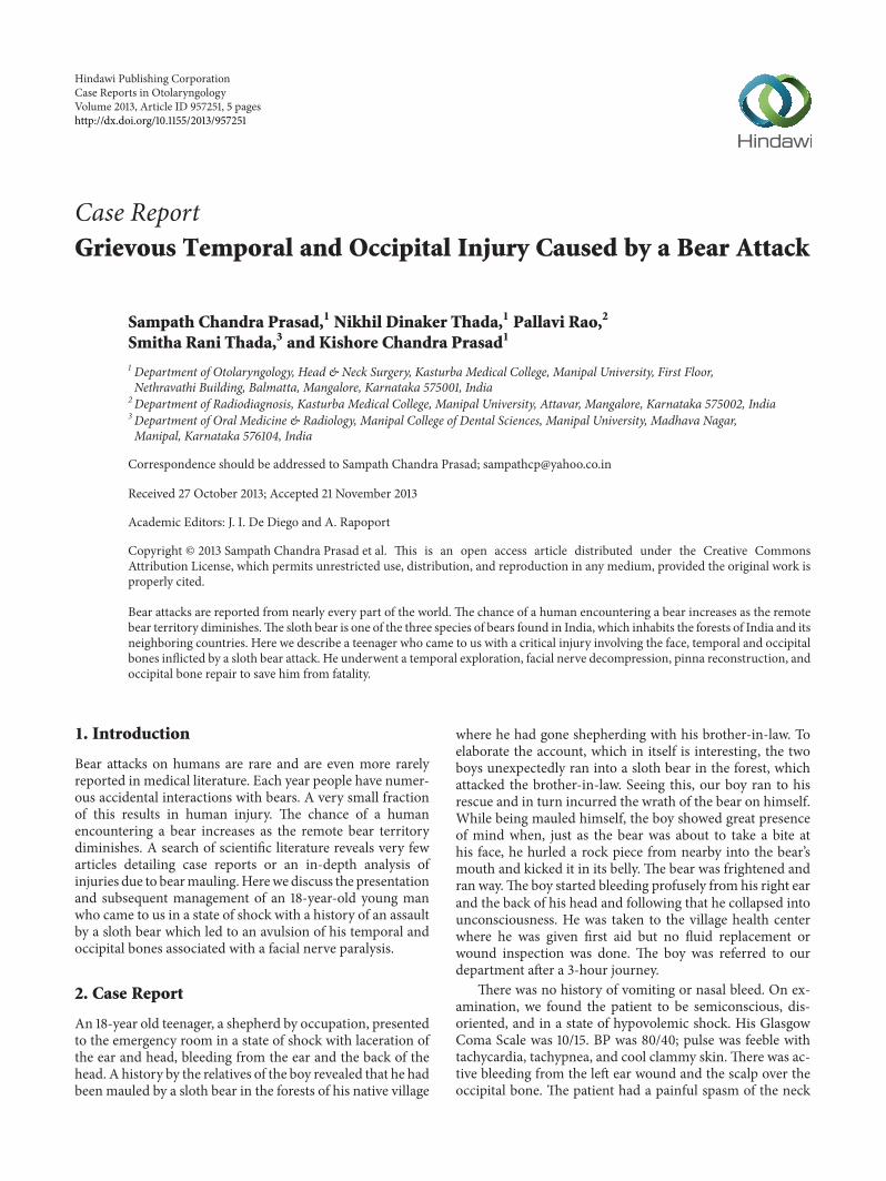
Figure 1: HB Grade IV facial nerve palsy.
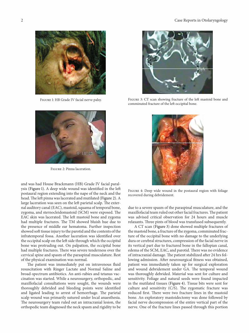
Figure 2: Pinna laceration.
and was had House Brackmann (HB) Grade IV facial paral-ysis (Figure 1). A deep wide wound was identified in the leftpostaural region extending into the nape of the neck and thehead.The left pinna was lacerated andmutilated (Figure 2). Alarge laceration was seen on the left parietal scalp. The exter-nal auditory canal (EAC),mastoid, squama of temporal bone,zygoma, and sternocleidomastoid (SCM) were exposed. TheEAC skin was lacerated. The left mastoid bone and zygomahad multiple fractures. The TM showed bluish hue due tothe presence of middle ear hematoma. Further inspectionshowed soft tissue injury to the parotid and the contents of theinfratemporal fossa. Another laceration was identified overthe occipital scalp on the left side through which the occipitalbone was protruding out. On palpation, the occipital bonehad multiple fractures. There was severe tenderness over thecervical spine and spasm of the paraspinal musculature. Restof the physical examination was normal.
The patient was immediately put on intravenous fluidresuscitation with Ringer Lactate and Normal Saline andbroad-spectrum antibiotics. An anti-rabies and tetanus vac-cination was started. While a neurosurgery, orthopedic, andmaxillofacial consultations were sought, the wounds werethoroughly debrided and bleeding points were identifiedand ligated leading to arrest of hemorrhage. The parietalscalp wound was primarily sutured under local anaesthesia.The neurosurgery team ruled out an intracranial lesion, theorthopedic team diagnosed the neck spasm and rigidity to be
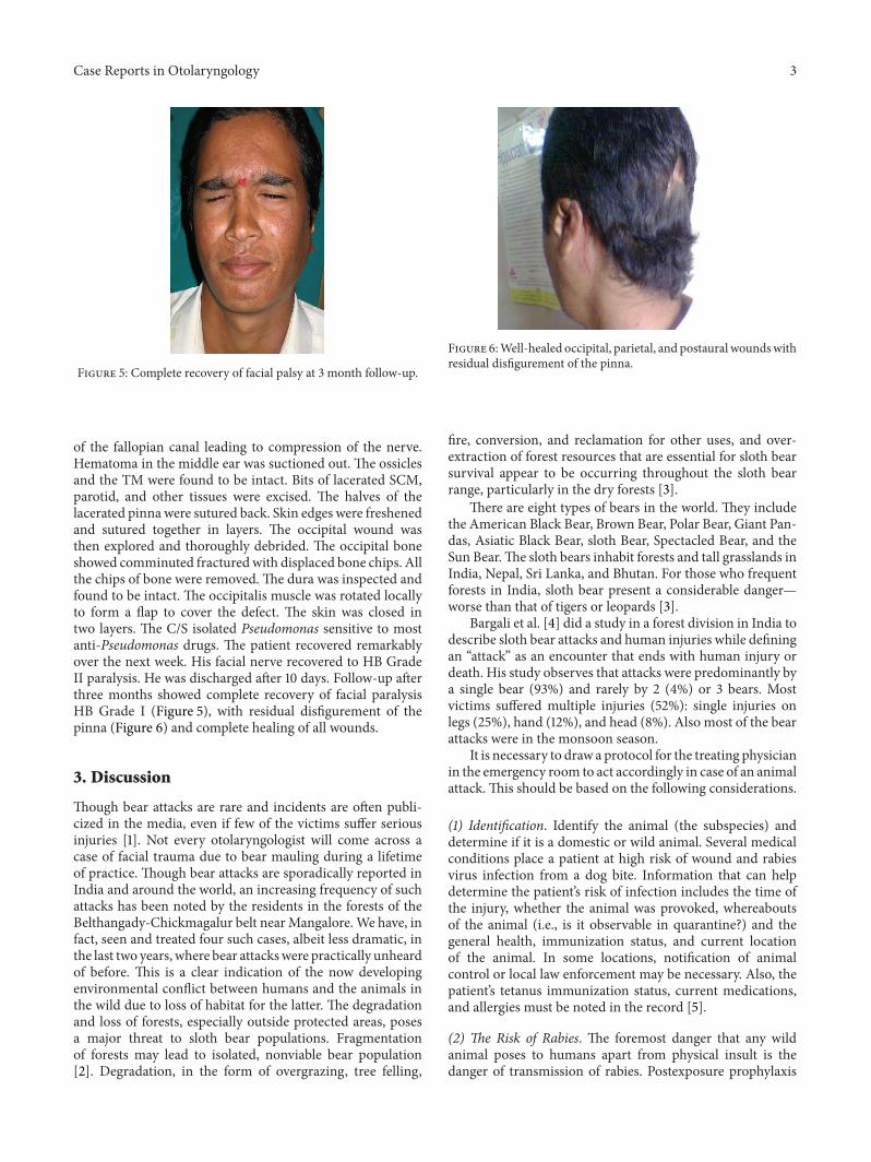
Figure 3: CT scan showing fracture of the left mastoid bone andcomminuted fracture of the left occipital bone.
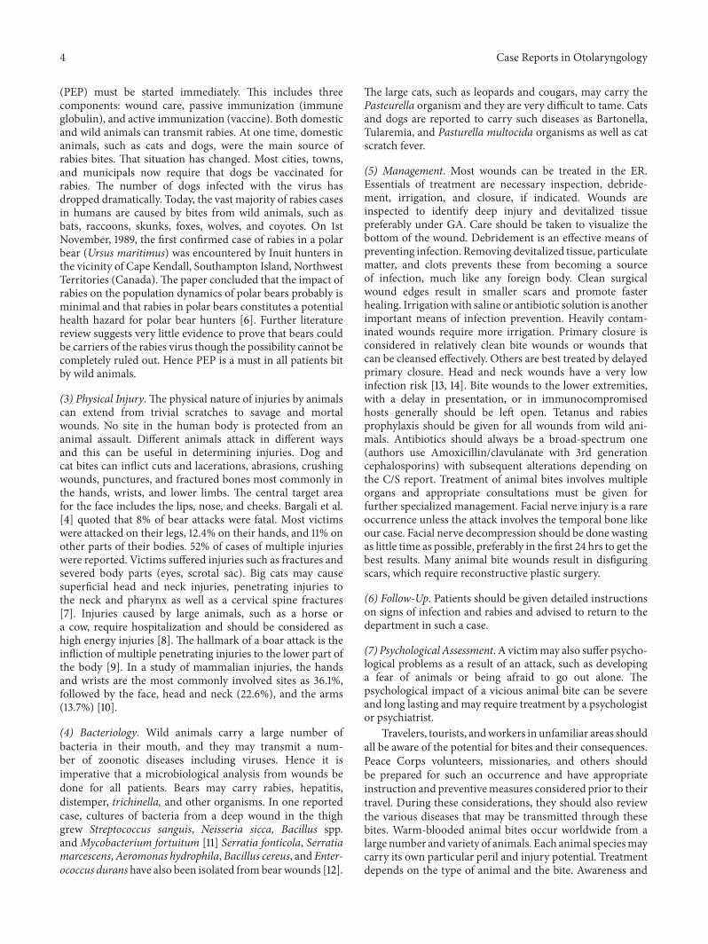
Figure 4: Deep wide wound in the postaural region with foliagerecovered during debridement.
due to a severe spasm of the paraspinal musculature, and themaxillofacial team ruled out other facial fractures.Thepatientwas advised critical observation for 24 hours and musclerelaxants. Three pints of blood was transfused subsequently.
A CT scan (Figure 3) done showed multiple fractures ofthemastoid bone, a fracture of the zygoma, comminuted frac-ture of the occipital bone with no damage to the underlyingdura or cerebral structures, compression of the facial nerve inits vertical part due to fractured bone in the fallopian canal,edema of the SCM, EAC, and parotid.There was no evidenceof intracranial damage.The patient stabilized after 24 hrs fol-lowing admission. After neurosurgical fitness was obtained,patient was immediately taken up for surgical explorationand wound debridement under GA. The temporal woundwas thoroughly debrided. Material was sent for culture andsensitivity. Foliage and natural seeds were found impactedin the mutilated tissues (Figure 4). Tissue bits were sent forculture and sensitivity (C/S). The zygomatic fracture wasreduced first. There were two fracture lines in the mastoidbone. An exploratory mastoidectomy was done followed byfacial nerve decompression of the entire vertical part of thenerve. One of the fracture lines passed through this portion
Case Reports in Otolaryngology 3
Figure 5: Complete recovery of facial palsy at 3 month follow-up.
of the fallopian canal leading to compression of the nerve.Hematoma in the middle ear was suctioned out. The ossiclesand the TM were found to be intact. Bits of lacerated SCM,parotid, and other tissues were excised. The halves of thelacerated pinna were sutured back. Skin edges were freshenedand sutured together in layers. The occipital wound wasthen explored and thoroughly debrided. The occipital boneshowed comminuted fracturedwith displaced bone chips. Allthe chips of bone were removed. The dura was inspected andfound to be intact. The occipitalis muscle was rotated locallyto form a flap to cover the defect. The skin was closed intwo layers. The C/S isolated Pseudomonas sensitive to mostanti-Pseudomonas drugs. The patient recovered remarkablyover the next week. His facial nerve recovered to HB GradeII paralysis. He was discharged after 10 days. Follow-up afterthree months showed complete recovery of facial paralysisHB Grade I (Figure 5), with residual disfigurement of thepinna (Figure 6) and complete healing of all wounds.
3. Discussion
Though bear attacks are rare and incidents are often publi-cized in the media, even if few of the victims suffer seriousinjuries [1]. Not every otolaryngologist will come across acase of facial trauma due to bear mauling during a lifetimeof practice. Though bear attacks are sporadically reported inIndia and around the world, an increasing frequency of suchattacks has been noted by the residents in the forests of theBelthangady-Chickmagalur belt nearMangalore.We have, infact, seen and treated four such cases, albeit less dramatic, inthe last two years, where bear attackswere practically unheardof before. This is a clear indication of the now developingenvironmental conflict between humans and the animals inthe wild due to loss of habitat for the latter. The degradationand loss of forests, especially outside protected areas, posesa major threat to sloth bear populations. Fragmentationof forests may lead to isolated, nonviable bear population[2]. Degradation, in the form of overgrazing, tree felling,
Figure 6:Well-healed occipital, parietal, and postaural woundswithresidual disfigurement of the pinna.
fire, conversion, and reclamation for other uses, and over-extraction of forest resources that are essential for sloth bearsurvival appear to be occurring throughout the sloth bearrange, particularly in the dry forests [3].
There are eight types of bears in the world. They includethe American Black Bear, Brown Bear, Polar Bear, Giant Pan-das, Asiatic Black Bear, sloth Bear, Spectacled Bear, and theSun Bear.The sloth bears inhabit forests and tall grasslands inIndia, Nepal, Sri Lanka, and Bhutan. For those who frequentforests in India, sloth bear present a considerable danger—worse than that of tigers or leopards [3].
Bargali et al. [4] did a study in a forest division in India todescribe sloth bear attacks and human injuries while definingan “attack” as an encounter that ends with human injury ordeath. His study observes that attacks were predominantly bya single bear (93%) and rarely by 2 (4%) or 3 bears. Mostvictims suffered multiple injuries (52%): single injuries onlegs (25%), hand (12%), and head (8%). Also most of the bearattacks were in the monsoon season.
It is necessary to draw a protocol for the treating physicianin the emergency room to act accordingly in case of an animalattack. This should be based on the following considerations.
(1) Identification. Identify the animal (the subspecies) anddetermine if it is a domestic or wild animal. Several medicalconditions place a patient at high risk of wound and rabiesvirus infection from a dog bite. Information that can helpdetermine the patient’s risk of infection includes the time ofthe injury, whether the animal was provoked, whereaboutsof the animal (i.e., is it observable in quarantine?) and thegeneral health, immunization status, and current locationof the animal. In some locations, notification of animalcontrol or local law enforcement may be necessary. Also, thepatient’s tetanus immunization status, current medications,and allergies must be noted in the record [5].
(2) The Risk of Rabies. The foremost danger that any wildanimal poses to humans apart from physical insult is thedanger of transmission of rabies. Postexposure prophylaxis
4 Case Reports in Otolaryngology
(PEP) must be started immediately. This includes threecomponents: wound care, passive immunization (immuneglobulin), and active immunization (vaccine). Both domesticand wild animals can transmit rabies. At one time, domesticanimals, such as cats and dogs, were the main source ofrabies bites. That situation has changed. Most cities, towns,and municipals now require that dogs be vaccinated forrabies. The number of dogs infected with the virus hasdropped dramatically. Today, the vast majority of rabies casesin humans are caused by bites from wild animals, such asbats, raccoons, skunks, foxes, wolves, and coyotes. On 1stNovember, 1989, the first confirmed case of rabies in a polarbear (Ursus maritimus) was encountered by Inuit hunters inthe vicinity of Cape Kendall, Southampton Island, NorthwestTerritories (Canada).The paper concluded that the impact ofrabies on the population dynamics of polar bears probably isminimal and that rabies in polar bears constitutes a potentialhealth hazard for polar bear hunters [6]. Further literaturereview suggests very little evidence to prove that bears couldbe carriers of the rabies virus though the possibility cannot becompletely ruled out. Hence PEP is a must in all patients bitby wild animals.
(3) Physical Injury. The physical nature of injuries by animalscan extend from trivial scratches to savage and mortalwounds. No site in the human body is protected from ananimal assault. Different animals attack in different waysand this can be useful in determining injuries. Dog andcat bites can inflict cuts and lacerations, abrasions, crushingwounds, punctures, and fractured bones most commonly inthe hands, wrists, and lower limbs. The central target areafor the face includes the lips, nose, and cheeks. Bargali et al.[4] quoted that 8% of bear attacks were fatal. Most victimswere attacked on their legs, 12.4% on their hands, and 11% onother parts of their bodies. 52% of cases of multiple injurieswere reported. Victims suffered injuries such as fractures andsevered body parts (eyes, scrotal sac). Big cats may causesuperficial head and neck injuries, penetrating injuries tothe neck and pharynx as well as a cervical spine fractures[7]. Injuries caused by large animals, such as a horse ora cow, require hospitalization and should be considered ashigh energy injuries [8]. The hallmark of a boar attack is theinfliction of multiple penetrating injuries to the lower part ofthe body [9]. In a study of mammalian injuries, the handsand wrists are the most commonly involved sites as 36.1%,followed by the face, head and neck (22.6%), and the arms(13.7%) [10].
(4) Bacteriology. Wild animals carry a large number ofbacteria in their mouth, and they may transmit a num-ber of zoonotic diseases including viruses. Hence it isimperative that a microbiological analysis from wounds bedone for all patients. Bears may carry rabies, hepatitis,distemper, trichinella, and other organisms. In one reportedcase, cultures of bacteria from a deep wound in the thighgrew Streptococcus sanguis, Neisseria sicca, Bacillus spp.and Mycobacterium fortuitum [11] Serratia fonticola, Serratiamarcescens,Aeromonas hydrophila,Bacillus cereus, andEnter-ococcus duranshave also been isolated frombearwounds [12].
The large cats, such as leopards and cougars, may carry thePasteurella organism and they are very difficult to tame. Catsand dogs are reported to carry such diseases as Bartonella,Tularemia, and Pasturella multocida organisms as well as catscratch fever.
(5) Management. Most wounds can be treated in the ER.Essentials of treatment are necessary inspection, debride-ment, irrigation, and closure, if indicated. Wounds areinspected to identify deep injury and devitalized tissuepreferably under GA. Care should be taken to visualize thebottom of the wound. Debridement is an effective means ofpreventing infection. Removing devitalized tissue, particulatematter, and clots prevents these from becoming a sourceof infection, much like any foreign body. Clean surgicalwound edges result in smaller scars and promote fasterhealing. Irrigationwith saline or antibiotic solution is anotherimportant means of infection prevention. Heavily contam-inated wounds require more irrigation. Primary closure isconsidered in relatively clean bite wounds or wounds thatcan be cleansed effectively. Others are best treated by delayedprimary closure. Head and neck wounds have a very lowinfection risk [13, 14]. Bite wounds to the lower extremities,with a delay in presentation, or in immunocompromisedhosts generally should be left open. Tetanus and rabiesprophylaxis should be given for all wounds from wild ani-mals. Antibiotics should always be a broad-spectrum one(authors use Amoxicillin/clavulanate with 3rd generationcephalosporins) with subsequent alterations depending onthe C/S report. Treatment of animal bites involves multipleorgans and appropriate consultations must be given forfurther specialized management. Facial nerve injury is a rareoccurrence unless the attack involves the temporal bone likeour case. Facial nerve decompression should be done wastingas little time as possible, preferably in the first 24 hrs to get thebest results. Many animal bite wounds result in disfiguringscars, which require reconstructive plastic surgery.
(6) Follow-Up. Patients should be given detailed instructionson signs of infection and rabies and advised to return to thedepartment in such a case.
(7) Psychological Assessment. A victimmay also suffer psycho-logical problems as a result of an attack, such as developinga fear of animals or being afraid to go out alone. Thepsychological impact of a vicious animal bite can be severeand long lasting and may require treatment by a psychologistor psychiatrist.
Travelers, tourists, andworkers in unfamiliar areas shouldall be aware of the potential for bites and their consequences.Peace Corps volunteers, missionaries, and others shouldbe prepared for such an occurrence and have appropriateinstruction and preventivemeasures considered prior to theirtravel. During these considerations, they should also reviewthe various diseases that may be transmitted through thesebites. Warm-blooded animal bites occur worldwide from alarge number and variety of animals. Each animal speciesmaycarry its own particular peril and injury potential. Treatmentdepends on the type of animal and the bite. Awareness and
Case Reports in Otolaryngology 5
prevention are primary in the avoidance of major injury andhealthcare risks [14].
4. Conclusion
While it is true that bears have the potential to be dangerousto humans and that a number of people are injured by bearsevery year, in reality, the incidence of attacks, on humans isrelatively rare. Wild-animal attacks, though rare, remind usthat humans can still be food or prey. Characteristic patternsof injury and wound infection should be appropriatelyidentified and treated.Though bear bites causing rabies is notdefinitively reported and researched, it is important to realizethat all mammals have the potential to carry and transmitrabies and PEPmust be started immediately. Primary closureof wounds gives good results, especially in the head andneck.The conflict of human and animal environments due toman’s excessive needs and greed will lead to more situationslike this and it is necessary that the treating doctor beaware of such conditions and the treatment protocols toprovide optimum care in such cases. Awareness, education,knowledge, and prevention, rather than the elimination ofanimal populations, may be the best way to control wild-animal attacks on humans in the future.
Conflict of Interests
All authors hereby declare that they have no conflict ofinterests including financial and personal relationships withother people or organizations that could inappropriatelyinfluence (bias) their work.
References
[1] R. A. Dieter Jr., D. L. Dieter, R. A. Diete III, and B. Forbes,“Bear mauling: a descriptive review,” International Journal ofCircumpolar Health, vol. 60, no. 4, pp. 696–704, 2001.
[2] D. L. Garshelis, A. R. Joshi, J. L. D. Smith, and C. G. Rice, “Slothbear conservation action plan,” in Bears: Status Survey andConservation Action Plan, C. Servheen and B. Peyton, Eds., p.309, IUCN/SSC Bear and Polar Bear Specialist Groups. IUCN,Gland, Switzerland, 1999.
[3] K. Yoganand and C. G. Rice, Evaluating Panna National Parkwith Special Reference to Ecology of Sloth Bear (Melursus ursi-nus). Final Project Report,Wildlife Institute of India, Dehradun,India, 2005.
[4] H. S. Bargali, N. Akhtar, and N. P. S. Chauhan, “Characteristicsof sloth bear attacks and human casualties in North BilaspurForest Division, Chhattisgarh, India,” Ursus, vol. 16, no. 2, pp.263–267, 2005.
[5] K. T. Lewis and M. Stiles, “Management of cat and dog bites,”American Family Physician, vol. 52, no. 2, pp. 479–485, 489–490,1995.
[6] M. Taylor, B. Elkin, N. Maier, and M. Bradley, “Observation ofa polar bear with rabies,” Journal of Wildlife Diseases, vol. 27, no.2, pp. 337–339, 1991.
[7] M. B. Wiens and P. B. Harrison, “Big cat attack: a case study,”Journal of Trauma, vol. 40, no. 5, pp. 829–831, 1996.
[8] A. Nogalski, L. Jankiewicz, G. Cwik, J. Karski, and Ł.Matuszewski, “Animal related injuries treated at the Depart-ment of Trauma and Emergency Medicine, Medical Universityof Lublin,” Annals of Agricultural and Environmental Medicine,vol. 14, no. 1, pp. 57–61, 2007.
[9] S. Manipady, R. G. Menezes, and B. K. Bastia, “Death by attackfrom a wild boar,” Journal of Clinical Forensic Medicine, vol. 13,no. 2, pp. 89–91, 2006.
[10] C. E. MacBean, D. M. Taylor, and K. Ashby, “Animal andhuman bite injuries in Victoria, 1998–2004,”Medical Journal ofAustralia, vol. 186, no. 1, pp. 38–40, 2007.
[11] V. A. Lehtinen, T. Kaukonen, I. Ikaheimo, S. Mahonen, M.Koskela, and P. Ylipalosaari, “Mycobacterium fortuitum infec-tion after a brown bear bite,” Journal of Clinical Microbiology,vol. 43, no. 2, p. 1009, 2005.
[12] D. Kunimoto, R. Rennie, D. M. Citron, and E. J. C. Goldstein,“Bacteriology of a bear bite wound to a human: case report,”Journal of Clinical Microbiology, vol. 42, no. 7, pp. 3374–3376,2004.
[13] S. E. Kountakis, S. A. Chamblee, A. A. J. Maillard, and C. M.Stiernberg, “Animal bites to the head and neck,” Ear, Nose andThroat Journal, vol. 77, no. 3, pp. 216–219, 1998.
[14] K. D. Wolff, “Management of animal bite injuries of the face:experience with 94 patients,” Journal of Oral and MaxillofacialSurgery, vol. 56, no. 7, pp. 838–843, 1998.

Submit your manuscripts athttp://www.hindawi.com
Stem CellsInternational
Hindawi Publishing Corporationhttp://www.hindawi.com Volume 2014
Hindawi Publishing Corporationhttp://www.hindawi.com Volume 2014
MEDIATORSINFLAMMATION
of
Hindawi Publishing Corporationhttp://www.hindawi.com Volume 2014
Behavioural Neurology
EndocrinologyInternational Journal of
Hindawi Publishing Corporationhttp://www.hindawi.com Volume 2014
Hindawi Publishing Corporationhttp://www.hindawi.com Volume 2014
Disease Markers
Hindawi Publishing Corporationhttp://www.hindawi.com Volume 2014
BioMed Research International
OncologyJournal of
Hindawi Publishing Corporationhttp://www.hindawi.com Volume 2014
Hindawi Publishing Corporationhttp://www.hindawi.com Volume 2014
Oxidative Medicine and Cellular Longevity
Hindawi Publishing Corporationhttp://www.hindawi.com Volume 2014
PPAR Research
The Scientific World JournalHindawi Publishing Corporation http://www.hindawi.com Volume 2014
Immunology ResearchHindawi Publishing Corporationhttp://www.hindawi.com Volume 2014
Journal of
ObesityJournal of
Hindawi Publishing Corporationhttp://www.hindawi.com Volume 2014
Hindawi Publishing Corporationhttp://www.hindawi.com Volume 2014
Computational and Mathematical Methods in Medicine
OphthalmologyJournal of
Hindawi Publishing Corporationhttp://www.hindawi.com Volume 2014
Diabetes ResearchJournal of
Hindawi Publishing Corporationhttp://www.hindawi.com Volume 2014
Hindawi Publishing Corporationhttp://www.hindawi.com Volume 2014
Research and TreatmentAIDS
Hindawi Publishing Corporationhttp://www.hindawi.com Volume 2014
Gastroenterology Research and Practice
Hindawi Publishing Corporationhttp://www.hindawi.com Volume 2014
Parkinson’s Disease
Evidence-Based Complementary and Alternative Medicine
Volume 2014Hindawi Publishing Corporationhttp://www.hindawi.com