case report ovarian cystic tumor composed of … · torsion of the right ovarian cyst. case report...
TRANSCRIPT
Introduction Ovarian tumor composed only of Brenner tumor and struma ovarii is very rare; only 6 cases have been reported in the English literature, to the best of the author’s knowledge [1-6]. Herein reported is a 66-year-old woman with Brunner tumor and struma ovarii of the right ovary. This patient presented with acute abdomen due to torsion of the right ovarian cyst. Case report A 65-year-old woman was found to have right ovarian cyst by gynecologic routine check. She was followed up. One year later, when she was 66-year-old, she suddenly complained of right abdominal pain. Physical examination and imag-ing techniques are suggestive of the torsion of the right ovarian cyst. Therefore, emergency right oophorectomy was performed. Macroscopically, the ovarian cyst was hemor-rhagic and red, and measured 7 x 6 x 6 cm
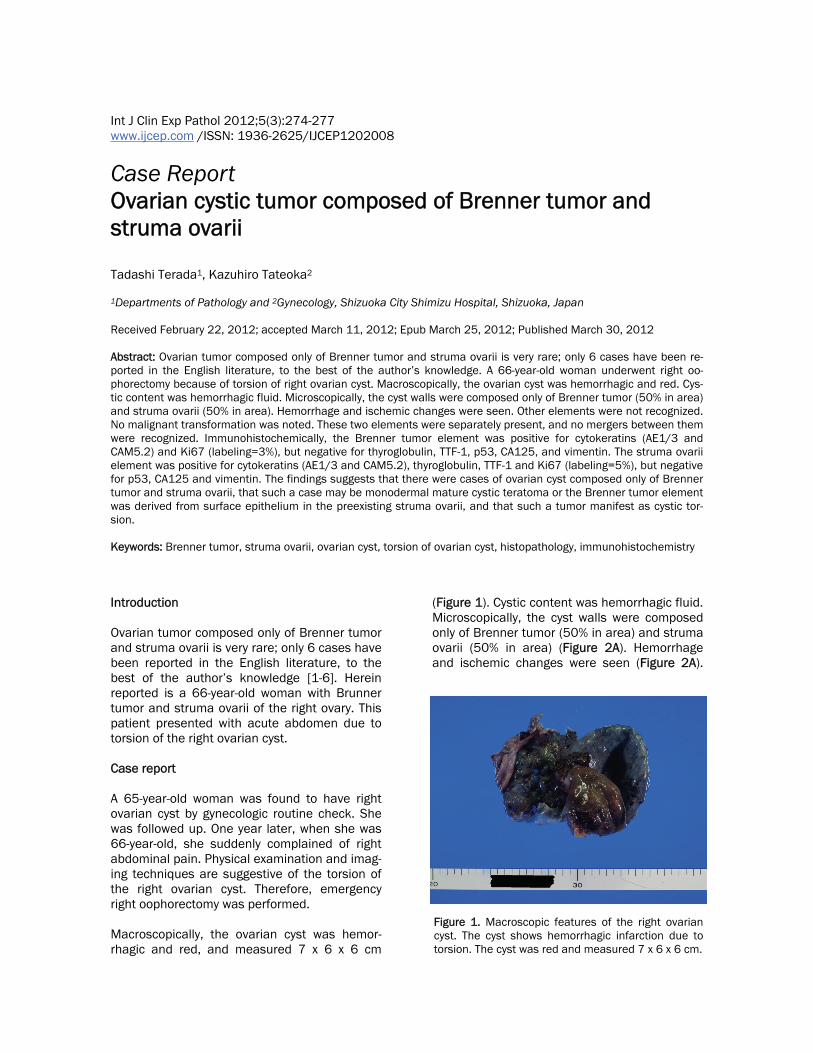
(Figure 1). Cystic content was hemorrhagic fluid. Microscopically, the cyst walls were composed only of Brenner tumor (50% in area) and struma ovarii (50% in area) (Figure 2A). Hemorrhage and ischemic changes were seen (Figure 2A).
Int J Clin Exp Pathol 2012;5(3):274-277 www.ijcep.com /ISSN: 1936-2625/IJCEP1202008
Case Report Ovarian cystic tumor composed of Brenner tumor and struma ovarii Tadashi Terada1, Kazuhiro Tateoka2 1Departments of Pathology and 2Gynecology, Shizuoka City Shimizu Hospital, Shizuoka, Japan Received February 22, 2012; accepted March 11, 2012; Epub March 25, 2012; Published March 30, 2012 Abstract: Ovarian tumor composed only of Brenner tumor and struma ovarii is very rare; only 6 cases have been re-ported in the English literature, to the best of the author’s knowledge. A 66-year-old woman underwent right oo-phorectomy because of torsion of right ovarian cyst. Macroscopically, the ovarian cyst was hemorrhagic and red. Cys-tic content was hemorrhagic fluid. Microscopically, the cyst walls were composed only of Brenner tumor (50% in area) and struma ovarii (50% in area). Hemorrhage and ischemic changes were seen. Other elements were not recognized. No malignant transformation was noted. These two elements were separately present, and no mergers between them were recognized. Immunohistochemically, the Brenner tumor element was positive for cytokeratins (AE1/3 and CAM5.2) and Ki67 (labeling=3%), but negative for thyroglobulin, TTF-1, p53, CA125, and vimentin. The struma ovarii element was positive for cytokeratins (AE1/3 and CAM5.2), thyroglobulin, TTF-1 and Ki67 (labeling=5%), but negative for p53, CA125 and vimentin. The findings suggests that there were cases of ovarian cyst composed only of Brenner tumor and struma ovarii, that such a case may be monodermal mature cystic teratoma or the Brenner tumor element was derived from surface epithelium in the preexisting struma ovarii, and that such a tumor manifest as cystic tor-sion. Keywords: Brenner tumor, struma ovarii, ovarian cyst, torsion of ovarian cyst, histopathology, immunohistochemistry
Figure 1. Macroscopic features of the right ovarian cyst. The cyst shows hemorrhagic infarction due to torsion. The cyst was red and measured 7 x 6 x 6 cm.
Coexistence of Brenner tumor and struma ovarii
275 Int J Clin Exp Pathol 2012;5(3):274-277
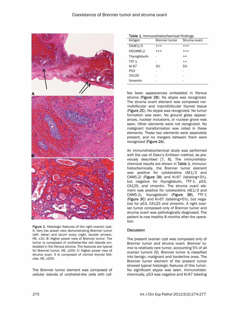
The Brenner tumor element was composed of cellular islands of urothelial-like cells with cof-
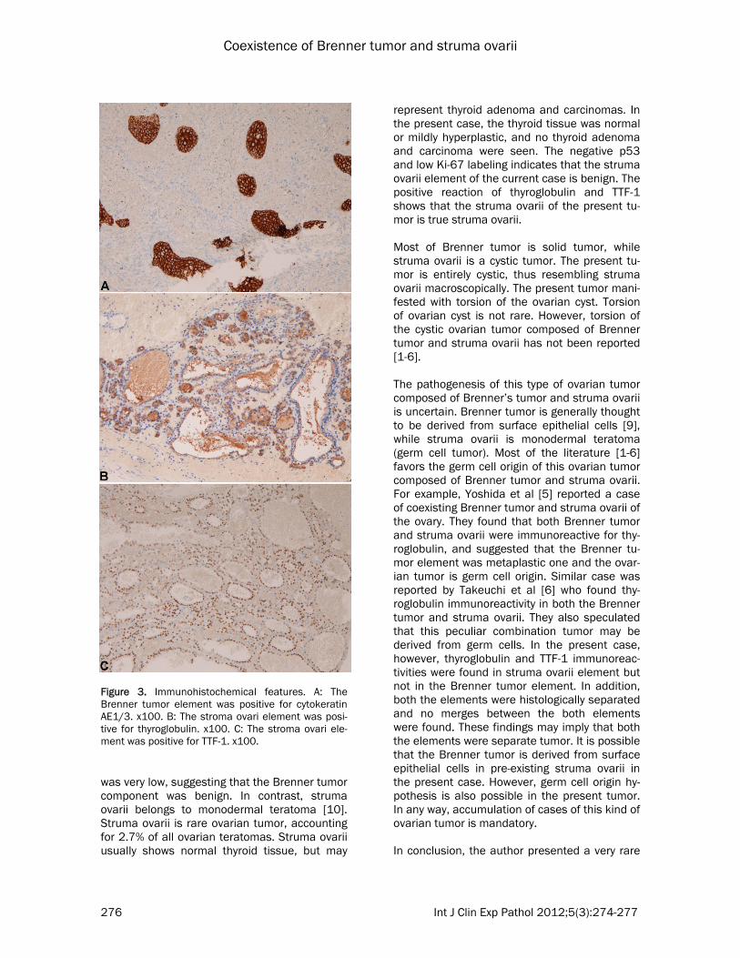
fee bean appearances embedded in fibrous stroma (Figure 2B). No atypia was recognized. The struma ovarii element was composed nor-mofollicular and macrofollicular thyroid tissue (Figure 2C). No atypia was recognized. No tumor formation was seen. No ground glass appear-ances, nuclear inclusions, or nuclear grove was seen. Other elements were not recognized. No malignant transformation was noted in these elements. These two elements were separately present, and no mergers between them were recognized (Figure 2A). An immunohistochemical study was performed with the use of Dako’s EnVision method, as pre-viously described [7, 8]. The immunohisto-chemical results are shown in Table 1. Immuno-histochemically, the Brenner tumor element was positive for cytokeratins (AE1/3 and CAM5.2) (Figure 3A) and Ki-67 (labeling=3%), but negative for thyroglobulin, TTF-1, p53, CA125, and vimentin. The struma ovarii ele-ment was positive for cytokeratins (AE1/3 and CAM5.2), thyroglobulin (Figure 3B), TTF-1 (Figure 3C) and Ki-67 (labeling=5%), but nega-tive for p53, CA125 and vimentin. A right ovar-ian tumor composed only of Brenner tumor and struma ovarii was pathologically diagnosed. The patient is now healthy 8 months after the opera-tion. Discussion The present ovarian cyst was composed only of Brenner tumor and struma ovarii. Brenner tu-mor is relatively rare tumor, accounting 5% of all ovarian tumors [9]. Brenner tumor is classified into benign, malignant and borderline ones. The Brenner tumor element of the present tumor showed typical histologic features of this tumor. No significant atypia was seen. Immunohisto-chemically, p53 was negative and Ki-67 labeling
Figure 2. Histologic features of the right ovarian cyst. A: Very low power view demonstrating Brenner tumor (left, allow) and strum ovary (right, double arrows). HE, x10. B: Higher power view of Brenner tumor. The tumor is composed of urothelial-like cell islands em-bedded in the fibrous stroma. The features are typical for Brenner tumor. HE, x200. C: Higher power view of struma ovari. It is composed of normal thyroid folli-cles. HE, x200.
Table 1. Immunohistochemical findings Antigen Brenner tumor Struma ovarii
CKAE1/3 +++ +++
CKCAM5.2 +++ +++
Thyroglobulin - ++
TTF-1 - ++ Ki-67 3% 5% P53 - - CA125 - - Vimentin - -
Coexistence of Brenner tumor and struma ovarii
276 Int J Clin Exp Pathol 2012;5(3):274-277
was very low, suggesting that the Brenner tumor component was benign. In contrast, struma ovarii belongs to monodermal teratoma [10]. Struma ovarii is rare ovarian tumor, accounting for 2.7% of all ovarian teratomas. Struma ovarii usually shows normal thyroid tissue, but may
represent thyroid adenoma and carcinomas. In the present case, the thyroid tissue was normal or mildly hyperplastic, and no thyroid adenoma and carcinoma were seen. The negative p53 and low Ki-67 labeling indicates that the struma ovarii element of the current case is benign. The positive reaction of thyroglobulin and TTF-1 shows that the struma ovarii of the present tu-mor is true struma ovarii. Most of Brenner tumor is solid tumor, while struma ovarii is a cystic tumor. The present tu-mor is entirely cystic, thus resembling struma ovarii macroscopically. The present tumor mani-fested with torsion of the ovarian cyst. Torsion of ovarian cyst is not rare. However, torsion of the cystic ovarian tumor composed of Brenner tumor and struma ovarii has not been reported [1-6]. The pathogenesis of this type of ovarian tumor composed of Brenner’s tumor and struma ovarii is uncertain. Brenner tumor is generally thought to be derived from surface epithelial cells [9], while struma ovarii is monodermal teratoma (germ cell tumor). Most of the literature [1-6] favors the germ cell origin of this ovarian tumor composed of Brenner tumor and struma ovarii. For example, Yoshida et al [5] reported a case of coexisting Brenner tumor and struma ovarii of the ovary. They found that both Brenner tumor and struma ovarii were immunoreactive for thy-roglobulin, and suggested that the Brenner tu-mor element was metaplastic one and the ovar-ian tumor is germ cell origin. Similar case was reported by Takeuchi et al [6] who found thy-roglobulin immunoreactivity in both the Brenner tumor and struma ovarii. They also speculated that this peculiar combination tumor may be derived from germ cells. In the present case, however, thyroglobulin and TTF-1 immunoreac-tivities were found in struma ovarii element but not in the Brenner tumor element. In addition, both the elements were histologically separated and no merges between the both elements were found. These findings may imply that both the elements were separate tumor. It is possible that the Brenner tumor is derived from surface epithelial cells in pre-existing struma ovarii in the present case. However, germ cell origin hy-pothesis is also possible in the present tumor. In any way, accumulation of cases of this kind of ovarian tumor is mandatory. In conclusion, the author presented a very rare
Figure 3. Immunohistochemical features. A: The Brenner tumor element was positive for cytokeratin AE1/3. x100. B: The stroma ovari element was posi-tive for thyroglobulin. x100. C: The stroma ovari ele-ment was positive for TTF-1. x100.
Coexistence of Brenner tumor and struma ovarii
277 Int J Clin Exp Pathol 2012;5(3):274-277
case of ovarian cystic tumor composed only of Brenner tumor and struma ovarii. Address correspondence to: Dr. Tadashi Terada, De-partment of Pathology, Shizuoka City Shimizu Hospi-tal, Miyakami 1231 Shimizu-Ku, Shizuoka 424-8636, Japan Tel: +81-54-336-1111; Fax: +81-54-334-1173; E-mail: [email protected] References [1] Klein HZ, Strauss SH, Unger AM. Coexisting Bren-
ner tumor and stroma ovarii (mature gonado-blastoma): report of a case. Obstet Gynecol 1968; 31: 779-784.
[2] Chiarelli SM, Onnis GL. Coexisting Brenner tumor and struma ovarii: case report. Eur J Gynecol Oncol 1980; 1: 108-111.
[3] Elemenoglou A, Zizi-Serbetzoglou A, Trihia H, Vasilakaki T, Boumia E. Mixed ovarian neoplasm composed of struma ovarii and Brenner tumor: report of a case. Eur J Gynecol Oncol 1994; 15: 138-141.
[4] Burg J, Kommoss F, Bittinger F, Moll R, Kirkpatrick CJ. Mature cystic teratoma of the ovary with struma ovarii and benign Brenner tumor: a case report with immunohistochemical characterization. Int J Gynecol Pathol 2002; 21: 74-77.
[5] Yoshida M, Okabayashi C, Tachibana M, Minami R. Coexisting Brenner tumor and struma ovarii in the right ovary: case report and review of the literature. Pathol Int 2004; 54: 793-797.
[6] Takeuchi K, Ohbayashi C, Kitazawa S, Ohara N, Maruo T. Coexistence of Brenner tumor and struma ovarii: case report. Eur J Gynecol Oncol 2005; 26: 109-110.
[7] Terada, T, Kawaguchi M, Furukawa K, Sekido Y, Osamura Y. Minute mixed ductal-endocrine car-cinoma of the pancreas with predominant intra-ductal growth. Pathol Int 2002; 52: 740-746.
[8] Terada T, Kawaguchi M. Primary clear cell ade-nocarcinoma of the peritoneum. Tohoku J Exp Med 2005: 206: 271-275.
[9] Lee KR, Russell P, Tavassoli FA et al. Surface epithelial-strumal tumor. In: Tavassoli FA and Devilee P eds. World Health Organization Classi-fication od Tumours. Pathology and genetics of tumours of the breast and female genital or-gans. IARC press, Lyon. 2003; pp: 142-143.
[10] Nogales F, Talerman A, Kubick-Huch RA, Tavas-soli FA, Devouassouz-Shisheboran M. Germ cell tumor. Tavassoli FA and Devilee P eds. World Health Organization Classification od Tumours. Pathology and genetics of tumours of the breast and female genital organs. IARC press, Lyon. 2003; pp: 163-175.