case report short and long term outcomes associated...
TRANSCRIPT

Case ReportShort and Long Term Outcomes Associated withFetal Cholelithiasis: A Report of Two Cases withAntenatal Diagnosis and Postnatal Follow-Up
Juan Troyano-Luque,1 Ana Padilla-Pérez,1
Ingrid Martínez-Wallin,1 Margarita Álvarez de la Rosa,1 Salvatore Andrea Mastrolia,1,2
José Luis Trujillo,1 and Tirso Pérez-Medina3
1 Department of Obstetrics and Gynecology, Ultrasound and Fetal Medicine Unit, University Hospital of Canary Islands,Ctra. Ofra, s/n, Santa Cruz de Tenerife, 38320 San Cristobal de La Laguna, Spain
2Department of Obstetrics and Gynecology, School of Medicine, University Hospital Policlinico of Bari andUniversity of Bari “Aldo Moro”, Piazza Giulio Cesare 11, 70124 Bari, Italy
3 Department of Obstetrics and Gynecology, University Hospital Puerta de Hierro, Calle Manuel de Falla,1 Majadahonda, 28222 Madrid, Spain
Correspondence should be addressed to Juan Troyano-Luque; [email protected]
Received 11 August 2014; Accepted 19 September 2014; Published 30 September 2014
Academic Editor: Giovanni Monni
Copyright © 2014 Juan Troyano-Luque et al. This is an open access article distributed under the Creative Commons AttributionLicense, which permits unrestricted use, distribution, and reproduction in any medium, provided the original work is properlycited.
The aims of this study were to present and discuss ultrasound findings of prenatal fetal cholelithiasis in two cases with differentetiology and evolution. Case 1: a pregnant woman from sub-Saharan Africa, suffering from Lyme disease, was treated withceftriaxone sodium. Six weeks later, biliary sludge associated with polyhydramnios was detected in the fetus and the fetal growthpercentile was 14. Emergency caesarean was performed at 36 weeks of gestation due to fetal distress. Biliary sludge persists inthe two-and-a-half-year-old child. Case 2: the fetus of a Caucasian woman with normal pregnancy showed multiple cholelithiasisassociated with polyhydramnios at 31 weeks of gestation. At 39 weeks and 4 days, cesarean section was performed due to lackof dilation. The biliary disease resolved spontaneously at seven months of age, with no associated abnormalities. In conclusion,prenatal diagnosis of cholelithiasis is straightforward, but prognosis cannot be defined yet. Serious complications do not arise in70% of cases, but severe diseases may ensue in 20%. Persistence of cholelithiasis after one year of age results in cholelithiasis inchildhood and beyond. Biliary sludge is associated with worse prognosis than cholelithiasis when it appears before 28 weeks ofgestation.
1. Introduction
Prenatal diagnosis of cholelithiasis can be readily performedwith sufficient experience and when the genetic sonogram isused.
The gallbladder should be examined routinely using high-resolution ultrasound.This is done primarily from the secondtrimester, since gallbladder changes are often prenatallyunderdiagnosed. Importantly, such changes may be a markerof disease in some severe cases [1–3].
2. Case 1
The first case involved a 31-year-old pregnant illegal immi-grant from Mali with a history of a caesarean birth due topreeclampsia. At 20 weeks of gestation, she was admitted toour department for fever of 38.8∘C of unknown origin. Thepathologywas initially attributed to a nonspecific viral illness.The genetic sonogram showed a fetuswithout signs ofmalfor-mation, except for increased gallbladder volume (appropriatefor gestational age) and polyhydramnios. Genetic analysis of
Hindawi Publishing CorporationCase Reports in Obstetrics and GynecologyVolume 2014, Article ID 714271, 5 pageshttp://dx.doi.org/10.1155/2014/714271

2 Case Reports in Obstetrics and Gynecology
(a) (b)
(c) (d)
(e) (f)
Figure 1: Case 1: ultrasound images of the distended gallbladder (a) containing biliary sludge (b) and 3D ultrasound of the same finding(arrow) (c). Case 2: ultrasound images of the distended gallbladder (d) showing biliary calculi (e) and 3D ultrasound of the same finding(arrow) (f).
the amniotic fluid extracted by amniocentesis showed theresulting karyotype was 46 XX.
The patient’s symptoms exacerbated rapidly: her feverincreased to 40∘C and she presented general malaise, facialparesis, cardiac arrhythmia, respiratory distress, joint pain,and stiff neck. Laboratory tests showed slightly elevatederythrocyte sedimentation rate (ESR) and mild leukocytosisas well as lymphocytic pleocytosis with increased IgG andspecific oligoclonal bands. Lumbar puncture was performedas the woman had retronucal pain and stiffness. Lymphocyticpleocytosis was detected in cerebrospinal fluid (CSF) witha high concentration of protein and glucose and specificoligoclonal bands. These results and the geographical originof the patient suggested severe sepsis. Cell cultures such
as BSK-Hrevealed infection by Borrelia spp., confirmed bypolymerase chain reaction (PCR).
Thepatientwas diagnosedwith Lymedisease, stages 2 and3. Intravenous ceftriaxone sodium was administered at dosesof 500mg/mL every 24 hours for 16 days, which effectivelycured the infection.
At week 26 a distended gallbladder with echogeniccontent of lumpy consistency was observed (Figure 1(a)). Inaddition, the fetal growth percentile decreased to percentile14.
Gestation continued with no significant events but fetalgrowth almost reached a plateau and gallbladder findingspersisted. At week 36, urgent caesarean was required dueto fetal distress after fetal growth stopped and pathological

Case Reports in Obstetrics and Gynecology 3
hemodynamic data were identified. The female newbornweighed 2090 g and had an Apgar score of 9/9 and umbilicalcord pH was 7.25.
In the first hours after birth, the newborn experiencedbilious vomiting, so, in addition to being breastfed, shereceived neonatal serotherapy for three days. The infantshowed gradually increasing tolerance to oral feeding, begin-ning with artificial milk, although there was some sporadicvomiting due to reflux. Positive health developments anda ponderal growth in the 25th percentile allowed hospitaldischarge at 25 days of birth with a weight of 2,450 grams.
Currently, at two years and six months of age, the infantstill suffers from cholelithiasis and has suffered severalepisodes of typical food intolerance. During this time she hasbeen admitted twice to the hospital emergency department,once for viral meningitis without sequelae and on anotheroccasion for dehydration after a period of acetonemic vomit-ing. Ponderal index percentile remains low. Cholecystectomyand other possibilities are being considered to treat theinfant, depending on short term developments. The infant iscurrently being studied in the pediatric ward of the hospital.
3. Case 2
The second case involved a 31-year-old patient in her secondpregnancy, with unremarkablemedical history except for onespontaneous miscarriage one year before.
Pregnancy control was performed from 6 weeks of gesta-tion with no significant events and micronized progesteronewas vaginally administered at a dose of 200mg every 24 hoursuntil week 11.
At week 20, moderate polyhydramnios was detectedin the fetus and, remarkably, increased gallbladder sizewas observed, shown in Figure 1(b). Cytogenetic study ofamniotic fluid obtained by amniocentesis revealed a 46XY karyotype. Standard gestational control was performed,focused on regularmonitoring of the gallbladder findings. Allmaternal laboratory results were normal.
At week 32, amniotic fluid volume had normalizedbut echogenic structures in the distended gallbladder wereobserved. No pathological intrahepatic signs were detected.
Scheduled ultrasound showed a gradual increase ofechogenic structures in the gallbladder. At 39 + 3 weeks ofgestation, the patient spontaneously initiated labor whichwas terminated by cesarean due to fetal distress. The malenewborn weighed 3200 g and had an Apgar score of 9/9 andumbilical cord pH was 7.27.
Neonatal ultrasound examination confirmed the pres-ence of cholelithiasis that did not affect the walls of the gall-bladder and associated condensed biliary sludge (Figure 2).
Neonatal monitoring showed intolerance to breast milkso feeding with low-fat formula milk was initiated. Breastmilk intolerance was associated with low weight gain in thefirst months, with a ponderal index in the 14th percentile.At 7 months, the cholelithiasis disappeared together with allcondensed bile, and the gallbladder presented clear walls.
Figure 2: Case 2: neonatal ultrasound image showing fetal gallblad-der sludge obstructing the bile duct.
Currently, the child of 2 years and 6months is completelyasymptomatic and has adequate food tolerance withoutassociated disorders.
4. Discussion
The first ultrasound report of prenatal cholelithiasis datesfrom 1983 [4], but it was Potter in 1928 [5] who firstpublished the presence of fetal cholelithiasis in a neonatalautopsy. Over the past 18 years, we have detected 16 casesof prenatal echogenic material in the gallbladder. In 10 caseswhere cholelithiasis was detected after 30 weeks of gesta-tion, it resolved in the first year of life without subsequentcomplications. In two cases it was necessary to performcholecystectomy at 11 and 14 years of age, respectively, due toacute cholecystitis. The remaining four cases presented com-plete atrioventricular canal, polycystic kidney disease withcorticomedullary dysplasia, primary hyperoxaluria (neona-tal diagnosis), and genodermatosis (neonatal diagnosis). Inthese cases, the diagnoses were made by ultrasound beforeweek 24 and neonatal exitus occurred.
Based on the ultrasound diagnosis, cholelithiasis canoccur at any time during pregnancy, but early gestationalage and maternal or underlying family-specific diseasesmust be considered when establishing criteria for prenatalcholelithiasis severity. On the one hand, the appearance ofbiliary sludge before 26weekswas a factor of neonatal severityand chronicity in one of the cases. On the other hand,the appearance of cholelithiasis after 30 weeks of gestation,without any risk factors, was associated with good prenataland neonatal clinical course.
Genetic disorders, racial and environmental variables,socioeconomic status, poor nutrition, maternal dehydration,and sepsis should be considered as risk factors affecting theseverity of fetal cholelithiasis [6].
About 70% of cholelithiasis cases are diagnosed postna-tally [7]; fortunatelymost are not significantly linked to severeperinatal conditions [1–3], but based on our experience atleast the relationship of cholelithiasis with child choledo-cholithiasis should be considered, as noted by other authors[8].
4 Case Reports in Obstetrics and Gynecology
Remarkably, in our two cases, increased gallbladdervolume and polyhydramnios appeared before the echogenicmaterial inside was detected; therefore we could considerthese as signs appearingbefore the onset of cholelithiasis.
From a clinical point of view, a better outcome wasobserved in the male fetus (Case 2) with later onset ofcholelithiasis and whose mother had no significant medicalhistory.
The only repeated event of some significance in bothpregnancies of the sub-Saharanpatient, noting the previousmiscarriage, was the transvaginal micronized progesteroneadministered from the 6th to the 11th week of gestation.
In the first case the mother had multiple risk factors,including the absence of obstetric control until week 20of pregnancy, being an illegal immigrant from sub-SaharanAfrica, and presenting fever upon hospital admission withsubsequent worsening of her general health. At the begin-ning, the pathogen responsible for the deterioration of hergeneral condition could not be identified by standard cellculture techniques. However, one of the special culturemediaused, BSK-H, suggested it could be a Borrelia spp. infection.
PCR and reverse line blot hybridization assay were usedto diagnose Lyme disease in this patient. Lyme disease iscaused by the bacterium Borrelia burgdorferi and transferredto humans through a tick vector such as blacklegged ticks thathave previously bitten rodent carriers. It was first detectedin Old Lyme in CT, USA, hence its name [7]. The diseasehas three evolutionary stages: stage 1, early localized Lymedisease; stage 2, early disseminated infection; and stage3, late persistent Lyme disease. The latter is regarded asthe acute phase of the disease. The factors associated withincreased likelihood of infection are living outdoors, in ruralsettings, and in close contact with domestic animals or pets(such as primates), essentially in those places where it ispossible to be bitten by a tick of this type. Poor hygiene orlow socioeconomic status increases the probability of Lymedisease infection.
A striking finding in the first case was the presenceof dense biliary sludge in the 26th week, undetected inprevious examinations. Besides the described infection, theonly probable causal factor of the fetal disease was theadministration of ceftriaxone sodium for 14 days at doses of500mg/mL every 24 hours. This drug belongs to the groupof cephalosporins and there are clear contraindications to itsuse in cases of jaundice, acidosis, or hypoalbuminemia: underthese conditions the drug prevents bilirubin from binding toserum albumin.This action is enhanced by drugs that containcalcium. Treatment duration of longer than 14 days, togetherwith renal failure or dehydration, can lead to precipitation ofceftriaxone calcium in the gallbladder. Cholelithiasis usuallydisappears when treatment is stopped [9]. It is therefore notadvisable to administer doses higher than 80mg/kg, as thisincreases the risk of biliary precipitation. Pancreatitis is oneof the disease complications in these patients [10].
The incidence of prenatal cholelithiasis is one case per260 [11], but despite technological advances, 70% are notdiagnosed. Fortunately this condition is not life-threatening
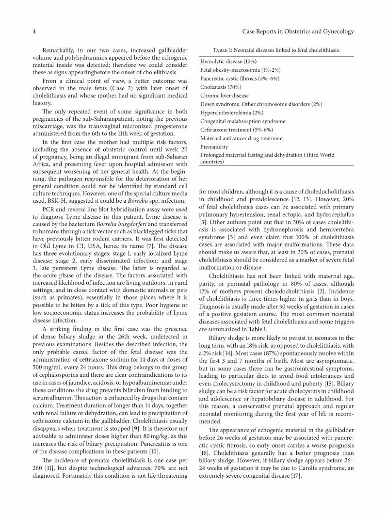
Table 1: Neonatal diseases linked to fetal cholelithiasis.
Hemolytic disease (10%)Fetal obesity-macrosomia (1%-2%)Pancreatic cystic fibrosis (4%–6%)Cholestasis (70%)Chronic liver diseaseDown syndrome. Other chromosome disorders (2%)Hypercholesterolemia (2%)Congenital malabsorption syndromeCeftriaxone treatment (5%-6%)Maternal anticancer drug treatmentPrematurityProlonged maternal fasting and dehydration (Third Worldcountries)
formost children, although it is a cause of choledocholithiasisin childhood and preadolescence [12, 13]. However, 20%of fetal cholelithiasis cases can be associated with primarypulmonary hypertension, renal ectopia, and hydrocephalus[3]. Other authors point out that in 30% of cases cholelithi-asis is associated with hydronephrosis and hemivertebrasyndrome [3] and even claim that 100% of cholelithiasiscases are associated with major malformations. These datashould make us aware that, at least in 20% of cases, prenatalcholelithiasis should be considered as a marker of severe fetalmalformation or disease.
Cholelithiasis has not been linked with maternal age,parity, or perinatal pathology in 80% of cases, although12% of mothers present choledocholithiasis [2]. Incidenceof cholelithiasis is three times higher in girls than in boys.Diagnosis is usually made after 30 weeks of gestation in casesof a positive gestation course. The most common neonataldiseases associated with fetal cholelithiasis and some triggersare summarized in Table 1.
Biliary sludge is more likely to persist in neonates in thelong term, with an 18% risk, as opposed to cholelithiasis, witha 2% risk [14]. Most cases (87%) spontaneously resolve withinthe first 3 and 7 months of birth. Most are asymptomatic,but in some cases there can be gastrointestinal symptoms,leading to particular diets to avoid food intolerances andeven cholecystectomy in childhood and puberty [15]. Biliarysludge can be a risk factor for acute cholecystitis in childhoodand adolescence or hepatobiliary disease in adulthood. Forthis reason, a conservative prenatal approach and regularneonatal monitoring during the first year of life is recom-mended.
The appearance of echogenic material in the gallbladderbefore 26 weeks of gestation may be associated with pancre-atic cystic fibrosis, so early onset carries a worse prognosis[16]. Cholelithiasis generally has a better prognosis thanbiliary sludge. However, if biliary sludge appears before 26–24 weeks of gestation it may be due to Caroli’s syndrome, anextremely severe congenital disease [17].

Case Reports in Obstetrics and Gynecology 5
5. Conclusion
Although fetal cholelithiasis does not imply a severe disease,20% of cases can be associated to genetic disorders, malfor-mations, or other diseases.
Biliary sludge is associated with worse prognosis thancholelithiasis, and its early onset may signal the presence ofCaroli’s disease and epidermal nevus syndromes.
Polyhydramnios and increased gallbladder volume oftenappear at the beginning of the 2nd trimester, before theappearance of cholelithiasis. Certain drugs including ceftri-axone sodium can induce the appearance of biliary echogenicmaterial.
The association of sepsis and poormaternal dietary intakemay be predisposing factors for long term cholelithiasis inchildren. About 11% of prenatal cholelithiasis cases precedecholelithiasis and cholecystitis in children and adults. Over70% of cases of cholelithiasis are not detected prenatally, sothis disorder should be included in the genetic sonogramassessment.
Conflict of Interests
The authors declare that there is no conflict of interestsregarding the publication of this paper.
References
[1] B. Broussin and E. Daube, “La lithiase vesiculaire foetale. Apropos de trois observations et revue de la litterature,” Journalde Gynecologie, Obstetrique et Biologie de la Reproduction, vol.19, no. 1, pp. 90–95, 1990.
[2] D. L. Brown, R. L. Teele, P. M. Doubilet, D. N. DiSalvo, C. B.Benson, and G. A. van Alstyne, “Echogenic material in the fetalgallbladder: sonographic and clinical observations,” Radiology,vol. 182, no. 1, pp. 73–76, 1992.
[3] T. Kiserud, K. Gjelland,H. Bognø,M.Waardal, H. Reigstad, andK. Rosendahl, “Echogenic material in the fetal gallbladder andfetal disease,” Ultrasound in Obstetrics and Gynecology, vol. 10,no. 2, pp. 103–106, 1997.
[4] I. Beretsky and D. H. Lankin, “Diagnosis of fetal cholelithia-sis using real-time high-resolution imaging employing digitaldetection,” Journal of Ultrasound in Medicine, vol. 2, no. 8, pp.381–383, 1983.
[5] H. A. Potter, “Gallbladder disease in young subjects,” Surgery,Gynecology and Obstetrics , vol. 46, p. 795, 1928.
[6] S. Reif, D. G. Sloven, and E. Lebenthal, “Gallstones in children.Characterization by age, etiology, and outcome,”The AmericanJournal of Diseases of Children, vol. 145, no. 1, pp. 105–108, 1991.
[7] S. K. Sood, “Effective retrieval of Lyme disease information ontheWeb,”Clinical Infectious Diseases, vol. 35, no. 4, pp. 451–464,2002.
[8] J. J. Roslyn, W. E. Berquist, H. A. Pitt et al., “Increased riskof gallstones in children receiving total parenteral nutrition,”Pediatrics, vol. 71, no. 5, pp. 784–789, 1983.
[9] C. Blais andR.Duperval, “Biliary pseudolithiasis in a child asso-ciated with 2 days of ceftriaxone therapy,” Pediatric Radiology,vol. 24, no. 3, pp. 218–219, 1994.
[10] J. C. Lopez Gutierrez, Z. Ros Mar, M. Lopez Santamarıa, A.Gonzalez Gomzalez, I. Pastor Abascal, and J. Gonzalez Utrilla,
“Fetal cholelithiasis: a clinical case and review of the literature,”Anales Espanoles de Pediatria, vol. 32, no. 5, pp. 468–469, 1990.
[11] A. Agnifili, R. Verzaro, G. Carducci et al., “Fetal cholelithiasis:a prospective study of incidence, predisposing factors, andultrasonographic and clinical features,” Clinical Pediatrics, vol.38, no. 6, pp. 371–373, 1999.
[12] E. Roberts, “The jaundiced baby,” in Disease of the Liver andBiliary System in Children, D. Kelly, Ed., pp. 35–73, BlackwellPublishing, Oxford, UK, 2nd edition, 2004.
[13] F. Santamaria, P. Sarnelli, L. Celentano et al., “Noninvasiveinvestigation of hepatopulmonary syndrome in children andadolescents with chronic cholestasis,” Pediatric Pulmonology,vol. 33, no. 5, pp. 374–379, 2002.
[14] C. W. Ko, J. H. Sekijima, and S. P. Lee, “Biliary sludge,” Annalsof Internal Medicine, vol. 130, no. 4, pp. 301–311, 1999.
[15] J. F. R. Robertson, R. Carachi, E. M. Sweet, and P. A. M.Raine, “Cholelithiasis in childhood: a follow-up study,” Journalof Pediatric Surgery, vol. 23, no. 3, pp. 246–249, 1988.
[16] M. Stuhrmann, M. Macek Jr., A. Reis et al., “Genotype analysisof cystic fibrosis patients in relation to pancreatic sufficiency,”The Lancet, vol. 335, no. 8691, pp. 738–739, 1990.
[17] J. Lendoire, P. B. Schelotto, J. A. Rodrıguez et al., “Bile duct cysttypeV (Caroli’s disease): surgical strategy and results,”HPB, vol.9, no. 4, pp. 281–284, 2007.

Submit your manuscripts athttp://www.hindawi.com
Stem CellsInternational
Hindawi Publishing Corporationhttp://www.hindawi.com Volume 2014
Hindawi Publishing Corporationhttp://www.hindawi.com Volume 2014
MEDIATORSINFLAMMATION
of
Hindawi Publishing Corporationhttp://www.hindawi.com Volume 2014
Behavioural Neurology
EndocrinologyInternational Journal of
Hindawi Publishing Corporationhttp://www.hindawi.com Volume 2014
Hindawi Publishing Corporationhttp://www.hindawi.com Volume 2014
Disease Markers
Hindawi Publishing Corporationhttp://www.hindawi.com Volume 2014
BioMed Research International
OncologyJournal of
Hindawi Publishing Corporationhttp://www.hindawi.com Volume 2014
Hindawi Publishing Corporationhttp://www.hindawi.com Volume 2014
Oxidative Medicine and Cellular Longevity
Hindawi Publishing Corporationhttp://www.hindawi.com Volume 2014
PPAR Research
The Scientific World JournalHindawi Publishing Corporation http://www.hindawi.com Volume 2014
Immunology ResearchHindawi Publishing Corporationhttp://www.hindawi.com Volume 2014
Journal of
ObesityJournal of
Hindawi Publishing Corporationhttp://www.hindawi.com Volume 2014
Hindawi Publishing Corporationhttp://www.hindawi.com Volume 2014
Computational and Mathematical Methods in Medicine
OphthalmologyJournal of
Hindawi Publishing Corporationhttp://www.hindawi.com Volume 2014
Diabetes ResearchJournal of
Hindawi Publishing Corporationhttp://www.hindawi.com Volume 2014
Hindawi Publishing Corporationhttp://www.hindawi.com Volume 2014
Research and TreatmentAIDS
Hindawi Publishing Corporationhttp://www.hindawi.com Volume 2014
Gastroenterology Research and Practice
Hindawi Publishing Corporationhttp://www.hindawi.com Volume 2014
Parkinson’s Disease
Evidence-Based Complementary and Alternative Medicine
Volume 2014Hindawi Publishing Corporationhttp://www.hindawi.com