casein phosphopeptides as possible nutraceuticals for functional
TRANSCRIPT
CCAASSEEIINN PPHHOOSSPPHHOOPPEEPPTTIIDDEESS AASS PPOOSSSSIIBBLLEE NNUUTTRRAACCEEUUTTIICCAALLSS FFOORR FFUUNNCCTTIIOONNAALL FFOOOODDSS
Gabriella Pinto
Dottorato in Scienze Biotecnologiche – XXII ciclo
Indirizzo Biotecnologie industriale Università di Napoli Federico II

2

3
Dottorato in Scienze Biotecnologiche – XXII ciclo Indirizzo Biotecnologie industriale
Università di Napoli Federico II
CCAASSEEIINN PPHHOOSSPPHHOOPPEEPPTTIIDDEESS AASS PPOOSSSSIIBBLLEE NNUUTTRRAACCEEUUTTIICCAALLSS FFOORR FFUUNNCCTTIIOONNAALL FFOOOODDSS
Gabriella Pinto
Dottoranda: Gabriella Pinto Relatore: Prof. Francesco Addeo Coordinatore: Prof. Ettore Benedetti

4

5
“La scienza deve abbandonare il terreno
dell'incontrollata genialità individuale, del
caso, dell'arbitrario, della sintesi affrettata e
procedere sulla base di uno sperimentalismo
fondato sulla consapevolezza della natura
strumentale delle facoltà conoscitive…la
conquista di verità nuove non può essere
opera del singolo, ma solo di una collettività di
scienziati organizzata a questo scopo”.
Francis Bacon

6

7
INDICE
1. Riassunto pag. 1 2. Summary pag. 6 3. Introduction pag. 7 4. Bioactive Peptides pag. 8 4.1 Antithrombotic peptides pag. 8 4.2 Antihypertensive peptides pag. 9 4.3 Opioid peptides pag. 11 4.4 Immunomodulating peptides pag. 12 4.5 Antimicrobial peptides pag. 13 4.6 Glycomacropeptide pag. 14 5. The object of study of this PhD work. Bioactive casein phosphopeptides pag. 15 6. Possible use of CPP for industrial applications pag. 17 7.Techniques for phosphopeptide enrichment for industrial applications pag. 19 8. Development of a new enrichment technique pag. 22 9. Experimental procedure 1 pag. 23 10. Results and discussion pag. 25 11. Formulation of a functional drink in semi-industrial scale pag. 35 12. Experimental procedure 2 pag. 36 13. Results and discussion pag. 37 14. Quantification of CPP in HAS-CPP preparation to be added to milk pag. 43 15. Evaluation of the quality of used milk for CPP preparation pag. 49 16. Experimental procedure 3 pag. 50 17 Results and discussions pag. 51 17.1 Dephosphorylation kinetic pag. 51 17.2. Analysis of proteins and CPP occurring in mastitic milk pag. 58 18. Use of CPP as process markers pag. 68 19. Experimental procedure 4 pag. 69 20. Results and discussion pag. 69 21. Conclusions pag. 73 22. References pag. 74

8

1
1. Riassunto Nella sequenza delle proteine del latte sono criptati peptidi cosiddetti ‘bioattivi’, di lunghezza variabile tra 3 e 20 residui amminoacidici, rilasciati per digestione enzimatica in vivo od in vitro.
È stato dimostrato che, a seconda della sequenza amminoacidica, essi possono esplicare attività specifiche in diverse classi di prodotti naturali o sintetici del tipo antitrombotico, antiipertensivo, oppioide, immunomodulante ed antimicrobico (1). Oggetto della tesi di dottorato è la preparazione, a livello di laboratorio ed a livello di impianto pilota, di una classe di peptidi bioattivi, suscettibili di utilizzazione industriale come ingrediente nutraceutico. Nutraceutico è un termine, derivante dalla unione di “nutrizione" e "farmaceutico", col quale si designano alimenti o nutrienti estratti dagli alimenti che svolgono sia azione di prevenzione di patologie sia azione benefica sulla salute umana. Questa categoria di peptidi bioattivi comprende i fosfopeptidi della caseina bovina. I fosfopeptidi (CaseinoPhosphoPeptides, CPP) sono rilasciati dalla caseina, a seguito di idrolisi enzimatica delle frazioni caseiniche κ-, αs1-, αs2-, e β-CN (2). Essi sono caratterizzati da una sequenza consenso del tipo SerP-X-SerP/ThrP/Glu/Asp, dove X è un qualsiasi residuo amminoacidico eccetto Pro e non contengono residui di tirosina e di istidina fosforilati. Una sequenza comune alle frazioni αs1-, αs2-, e β-CN è -Ser(P)-Ser(P)-Ser(P)-Glu-Glu- (3). I CPP sono interessanti perché sopravvivono al passaggio gastrointestinale (4) e si ritrovano nello stomaco, nel duodeno e nell’ileo distale dopo ingestione di latte (5). Fungono da trasportatori di cationi minerali di- o trivalenti, aumentandone la biodisponibilità per l’assorbimento nel piccolo intestino (6). É stato, infatti, dimostrato che i CPP, in particolare i peptidi (1-25)4P della β-caseina e (59-79)5P dell’αs1-caseina, aumentano l’uptake di calcio in cellule tumorali umane differenziate in senso enterocitico (HT-29) (7), nelle cellule Caco2 (8), e negli osteoblasti (9). Questi dati ci hanno suggerito la possibilità di utilizzare i CPP come vettori di Ca2+ in alimenti funzionali per aumentarne la biodisponibilità nello sviluppo dell’organismo, nella calcificazione delle ossa e nella prevenzione dell’osteoporosi. E’ stato poi dimostrato che i CPP sono capaci di stabilizzare le soluzioni di fosfato di calcio amorfo (ACP), formando piccoli cluster del tipo CPP-ACP, fino ad una taglia-limite, oltrepassata la quale il fosfato di calcio comincia a precipitare con formazione di nuclei cristallini (10). Il meccanismo di azione sarebbe il seguente: i CPP trasportano l’ACP sulla superficie del dente, inibendo la demineralizzazione dello smalto dentale e promuovono la rimineralizzazione delle lesioni superficiali (11). Essi svolgono anche un ruolo indiretto nel contrasto alla carie, impedendo l’adesione alla superficie del dente di Streptococcus mutans o di altri streptococci (12). Il complesso CPP-ACP, ideato da Eric Reynolds dell’Università di Melbourne, è commercializzato come prodotto per la cura orale (RecaldentTM) e viene aggiunto come ingrediente nutraceutico in gomma masticante (Trident White Gum), paste dentifricie (GC Tooth Mousse) ed in altri prodotti.
Esistendo sul mercato preparazioni commerciali di CPP (Arla, Danimarca, Tatua, Nuova Zelanda), è stata fatta un’indagine conoscitiva tesa a stabilirne la composizione in CPP. L’analisi mediante MALDI ha rivelato che i due prodotti commerciale sono costituiti in effetti da una miscela di CPP e di peptidi non fosforilati. Non essendo conosciuto il sistema di preparazione di questi prodotti, è probabile che l’innovazione operata da queste industrie a livello di prodotto sia stata

2
l’utilizzazione di uno o più enzimi in sequenza per generare CPP. Le preparazioni commerciali contengono infatti peptidi a diversa taglia molecolare cosa che indica una procedura differente di preparazione. Non esistendo sul mercato un preparazione di CPP esente da peptidi non fosforilati, l’obiettivo della tesi è stato quello di elaborare un procedimento per ottenere CPP ad elevata concentrazione, cioè peptidi a diverso grado di fosforilazione, presenti nella caseina nativa, esenti da peptidi non fosforilati. La prima parte del lavoro è stata, quindi, focalizzata sull’individuazione di una metodologia di frazionamento degli idrolizzati enzimatici di caseina per il raggiungimento dell’obiettivo prefissato. Sono stati considerati diversi procedimenti di arricchimento dei CPP: a) precipitazione come sali di bario insolubili (13); b) derivatizzazione chimica (14); c) cromatografia di affinità su c1) ioni metallici immobilizzati (IMAC) (15); c2) su ossidi di metallo (MOC) (16). La derivatizzazione chimica dà basse rese di prodotto ed un prodotto finale più complesso di quello iniziale (17). Le tecniche cromatografiche IMAC e MOC consentono di arricchire la preparazione in CPP, ma recuperano anche peptidi non fosforilati di natura acida (18). L’accoppiamento di resine con ossidi di metallo modificati da idrossiacidi alifatici (19), l’impiego di tamponi di caricamento su TiO2 contenenti acido 2,5-p-idrossibenzoico (DHB) (18), la precipitazione con fosfato di calcio accoppiato all’arricchimento IMAC (20), la combinazione di cromatografia a scambio cationico (SCX) e scambio anionico (SAX) (21) si sono dimostrati capaci di arricchire, su base analitica, le preparazioni di CPP, ma non sono scalabili allo stato industriale. Tuttavia, nessuno dei sistemi analitici sperimentati potrebbe trovare applicazione alimentare, sostanzialmente per tre ragioni: a) la matrice a cui sono legati i CPP non è edibile; b) l’eluizione di CPP dalla matrice richiede sistemi di purificazione molto lunghi e costosi; c) l’aggiunta all’alimento dei CPP eluiti altera il gusto conferente una marcata nota di amarognolo, anche per la capacità dei CPP di chelare gli ioni minerali con cui viene a contatto. La seconda parte del lavoro è stata rivolta alla messa a punto di una tecnica di arricchimento specifica per fosfoproteine e peptidi fosforilati, allo scopo di disporre di un procedimento preparativo di CPP da utilizzare successivamente come ingrediente in alimenti funzionali. L’idea è stata quella di sviluppare una tecnica analitica da adattare successivamente a fini preparativi. Essa prevede l’impiego come matrice cromatografica dell’idrossiapatite (HA), una forma cristallina di fosfato di calcio [Ca10(PO4)6(OH)2], rinvenibile nel tessuto osseo e dentale, largamente impiegata in medicina per protesi ortopediche e dentali, per favorire la completa integrazione del metallo nel tessuto osseo umano. Il meccanismo di funzionamento dell’HA prevede che i gruppi carichi negativamente delle proteine (gruppi carbossilici e fosfato) interagiscano con gli ioni Ca2+ (C-sites) del reticolo cristallino formando legami di tipo elettrostatico (22). Le proteine fosforilate vengono in tal modo immobilizzate sulla resina, mentre quelle non fosforilate vengono allontanate mediante lavaggio con tamponi di opportuna composizione. L’affinità delle frazioni caseiniche per l’adsorbente è tanto maggiore quanto maggiore è il grado di fosforilazione della proteina (22). Abbiamo dimostrato che l’affinità dei CPP nei riguardi dell’HA è simile a quello esibita dalle fosfoproteine. In dettaglio, si è trovato che l’HA lega i fosfopeptidi presenti, in quantità anche sub-stechiometrica, in miscele complesse di peptidi. Se si dispone di fosfoproteine genitrici legate all’HA, si possono generare in situ, a mezzo di un’opportuna idrolisi enzimatica, i corrispondenti fosfopeptidi. I CPP rimangono legati alla resina, mentre tutti quelli non fosforilati, ad esempio αs1-CN (f91-100) (m/z 1266.7) e αs1-CN (f8-22) (m/z 1758.9), presi come esempi di peptidi

3
acidi non fosforilati, vengono allontanati con i tamponi di lavaggio. Per sviluppare il corrispondente procedimento industriale è stato ideato un metodo a livello di laboratorio. Si produce un’idrolisi triptica della caseina fresca legata all’HA. I CPP rimangono legati all’HA, mentre i peptidi non fosforilati vengono allontanati con lavaggi, fino ad ottenere un prodotto lavato con acqua distillata. Il prodotto viene, poi, essiccato per la conservazione della polvere per tempi dell’ordine di anni a temperatura ambiente.
È stato, poi, affrontato il problema di stabilire quali peptidi rimangano legati alla resina. Per questo è stato elaborato un sistema di controllo di qualità della preparazione, utilizzando tecniche di spettrometria di massa quali MALDI-TOF, LC-ESI/MS e LC-ESI-MS/MS. Abbiamo dimostrato che ognuna di queste tecniche consente di caratterizzare i CPP legati alla resina. Dallo studio è risultato che la resina immobilizza una pletora di peptidi, tra cui i CPP con fosforilazione multipla del tipo αs1-CN (f59-79)5P (2720.91 Da), β-CN (f1-25)4P (3122.26 Da) e αs2-CN (f46-70)5P (3087.99 Da), ognuno caratterizzato dalla sequenza cluster (Ser(P)-Ser(P)-Ser(P)-Glu-Glu), funzionale al trasporto di ioni metallici. La preparazione del complesso HA-CPP, in scala semi-industriale, ha suggerito di sintetizzare l’HA, a causa degli elevati costi di questo materiale, invece che utilizzare il costoso prodotto commerciale. Pertanto, l’HA è stata sintetizzata per via umida a partire da Ca(OH)2 e H3PO4, utilizzando condizioni di temperatura e pH ottimali, per l’ottenimento di cristalli di apatite pura. La capacità legante della resina sintetizzata è stata valutata confrontando la capacità legante dei CPP in 3 diverse miscele proteiche: caseina isoelettrica, proteine del latte (caseine + sieroproteine non fosforilate) e latte. L’analisi MALDI-TOF ha evidenziato la cattura, da parte dell’HA sintetica, degli stessi CPP, in quantità simili, nonostante la diversa complessità della matrice di partenza. La prova ha, quindi, dimostrato che il procedimento è scalabile per fini industriali. Una volta ottenuto il complesso HA-CPP su scala pilota, si è passati alla progettazione di un alimento funzionale. È stato scelto come alimento funzionale il latte poiché l’aggiunta al latte di quantità variabili tra 2.0 e 5.0g del complesso CPP-ACP⁄litro aumenta la capacità dei CPP a ripristinare la mineralizzazione delle lesioni dello smalto dentale (23), facendo aumentare rispettivamente dell’ 81% e del 164% il contenuto in minerali (calcio e fosfato) del latte “nativo” (24). Naturalmente, data la maggiore densità, i granuli di HA-CPP, prima dell’aggiunta al latte, sono stati finemente triturati per evitare l’immediata sedimentazione nel latte. Tuttavia, prima del consumo, la bevanda deve essere agitata, per rendere omogenea la sospensione. Una volta agitata, la bevanda arricchita di CPP ha mostrato le stesse caratteristiche sensoriali del latte di partenza, in quanto i gruppi fosfato dei CPP sono neutralizzati dagli ioni calcio dei cristalli di HA. Il complesso HA-CPP è inodore ed insapore. Nelle prove di recupero dei granuli di HA aggiunti al latte mediante centrifugazione, l’analisi MALDI dei granuli ha mostrato che l’HA è capace di legare i CPP liberi e la caseina solubile del latte. In altre prove, i granuli di HA-CPP aggiunti al latte si sono rivelati capaci di mantenere legati i CPP della preparazione e di chelare anche i CPP liberi del latte. Per stabilire la quantità di CPP aggiunta nel latte, è stato affrontato il problema di un dosaggio quantitativo applicabile ad una qualsiasi matrice alimentare. Sono stati scelti come modello per la valutazione quantitativa i CPP del tipo monofosforilato, αs1-CN (f106-119)1P con sequenza VPQLEIVPNpSAEER, e difosforilato, αs1-CN (f43-58)2P con sequenza DIGpSEpSTEDQAMEDIK. Il metodo quantitativo si basa sull’utilizzo di standard interni a base di fosfopeptidi sintetici analoghi a quelli naturali. E’ stato verificato che il peptide naturale e quello analogo

4
sintetico, modificato in un solo sito amminoacidico, ionizzano allo stesso modo. I peptidi sintetici utilizzati si sono rivelati, dunque, idonei a fungere da standard interno per l’analisi quantitativa dei CPP monofosforilati e difosforilati. La misura delle aree sottese dalle coppie di picchi MALDI/MS dei peptidi monoP e diP ha consentito di costruire delle rette di calibrazione. Utilizzando tale sistema analitico si è potuto stabilire sia la quantità di CPP presente nel complesso HA-CPP, sia quella aggiunta nel latte da rendere funzionale. Nella terza parte del lavoro di tesi, è stato affrontato il problema della valutazione della qualità della preparazione sperimentale a base di CPP. Com’è noto, la presenza di proteasi attive nel latte, a seguito di stati patologici della ghiandola mammaria, si riflette sia sulla integrità (proteolisi) sia sul livello di fosforilazione della caseina. La defosforilazione è una delle modifiche post-traslazionali più rilevanti dal punto di vista biologico perché, tra l’altro, altera la funzionalità della caseina, rendendola meno adatta a caseificazione ed a fungere da ingrediente per la preparazione di CPP. In un latte con elevato numero di cellule somatiche, è stata dimostrata una spinta degradazione proteolitica ad opera degli enzimi idrolitici sia endogeni che derivati dalle stesse cellule somatiche. Abbiamo potuto stabilire su base molecolare che latte proveniente da animali affetti da mastite è caratterizzato da un profilo caseinico alterato per l’aumento di attività della fosfatasi (25) e della plasmina (26), responsabili rispettivamente della defosforilazione e dell’ idrolisi delle frazioni caseiniche. Questi tipi di alterazione delle micelle di caseina abbassano la qualità del latte alimentare e la resa casearia. Per valutare la qualità della caseina in un latte mastitico, abbiamo utilizzato l’HA per valutare la chelazione delle fosfoproteine e dei CPP da parte dell’adsorbente. Lo studio con tecniche MALDI-MS ed ESI-Q-TOF ha messo in evidenza la comparsa di frazioni caseiniche con un ampio intervallo di fosforilazione. Ad esempio, la β-CN 6P e 5P, allo stato nativo, si trasformava in proteina con grado di fosforilazione variabile da 6 a 3P. Inoltre, è stata rilevata la comparsa di frammenti proteici prodotti dall’azione della plasmina e da altri enzimi associati con le cellule somatiche (catepsine, elastasi, etc). I CPP possono essere anche fungere da marcatori di processo. In prodotti stagionati come il formaggio Grana Padano, infatti, i CPP subiscono una degradazione più o meno spinta ad opera di proteinasi/peptidasi e fosfatasi alcalina ed acida, a seconda della zona in cui il campione analizzato è stato prelevato, parte esterna o parte interna della forma. La parte centrale del formaggio, durante la lavorazione, subisce un’auto-pastorizzazione, mentre la parte esterna, raffreddandosi più velocemente dalla temperatura di lavorazione (circa 55° C), contiene livelli di fosfatasi residua di circa 10-100 volte superiori. Poiché il Disciplinare di produzione del formaggio prevede l’uso di latte crudo, ogni lavorazione con latte pastorizzato (attività fosfatasica negativa) produrrà un’attività di fosfatasi uguale, indipendentemente dalla zona di prelievo del campione nella forma. In un lavoro precedente si era messo in evidenza che nel Grana Padano DOP, le fosfatasi, aminopeptidasi e carbossipeptidasi del formaggio producono una progressiva defosforilazione e idrolisi dei CPP derivati dall’azione idrolitica della plasmina, fino a fornire peptidi contenenti il cluster Ser(P)-Ser(P)-Ser(P)-Glu-Glu, più resistente alla digestione enzimatica in vivo di altri peptidi mono-P e di-P (27). La nostra tecnica di arricchimento dei CPP mediante l’utilizzazione di HA per lo studio dei CPP nella frazione solubile a pH 4.6, ha confermato l’esistenza di un profilo dipendente da una diversa attività enzimatica tra parti interne ed zona esterna nelle forme di formaggio Grana Padano (28). Abbiamo trovato che il profilo dei CPP riflette il gradiente termico centripeto con differenza di raffreddamento tra zone esterne ed interne della forma,
5
con inattivazione centripeta degli enzimi del formaggio. Nella parte esterna della forma abbiamo dimostrato che i CPP subiscono una defosforilazione più spinta che nelle zone interne. In conclusione, nel corso del dottorato, è stata sviluppata una selettiva tecnica di arricchimento di componenti fosforilati, a partire da miscele complesse di proteine e peptidi. I peptidi fosforilati della caseina sonostati, poi, utilizzati per la formulazione di un latte funzionale contenente microgranuli di HA-CPP, utili per la cura della carie dentaria e per veicolare microelementi (ioni rame, ferro, selenio, etc.). La tecnica analitica elaborata è suscettibile di applicazione generale per lo studio del fosfoproteoma. Lo studio della fosforilazione proteica ha consentito, inoltre, di affrontare uno degli aspetti della qualità delle proteine, sulla base del loro grado di fosforilazione. In questa direzione, il metodo analitico si presta a fornire informazioni dettagliate sulla valutazione della genuinità di latte alimentare e di formaggio. Bibliografia 1. FitzGerald, R. J., et al. 2003 In: P.F. Fox, & P.L.H. McSweeney (Eds.), Advanced dairy chemistry, Vol. 1: Proteins (3rd ed.), 675–698. 2. Kitts, D. et al. 1994 J. Physiol. Pharmacol. 72, 423–434 3. Meisel, H. et al. 1995 Nahrung 39, 1-20 4. Chabance, B. et al. 1998 Biochimie 80(2), 155–16 5. Meisel, H., et al. 2003 British Journal of Nutrition 89(3), 351–359 6. FitzGerald, R. J. et al. 1998 Int. Dairy J. 8, 451–457 7. Ferraretto, A. et al. 2001 Journal of Nutrition 131(6), 1655–1661. 8. Ferraretto A. et al. 2009 Journal of Nutritional Biochemistry, article in press. 9. Ferraretto A. et al. 2009 Peptides, article in Press. 10. Holt, C. et al. 1998 J. Dairy Sci. 81, 2994-3003. 11. Reynolds, E.C. et al. 1997 J. Dent. Res. 76, 1587-1595. 12. Rose, R. K. et al. 2000 Arch. Oral. Biol. 45, 569-57. 13. Manson, W. et al. 1971 Archives of Biochemistry and Biophysics 145, 16-26. 14. Oda Y. et al. 2001 Nat. Biotechnol.19, 379–382. 15. Posewitz, M. C. et al. 1999 Anal. Chem. 71, 2883–2892. 16. Pinkse, M. W. et al. 2004 Anal. Chem. 76, 3935–3943. 17. Zhou, H. et al. 2001 Nat. Biotechnol. 19, 375–378. 18. Roepstorff P. et al. 2005 Mol Cell Proteomics 4(7), 873-86. 19. Sugiyama N. et al. 2007 Mol Cell Proteomics 6(6), 1103-1109. 20. Roepstorff P. et al. 2007 Mol Cell Proteomics 6(11), 2032-2042. 21. Guanghui Han et al. 2008 Analyst. 133, 1128–1138. 22. Schmidt S. R. et al. 2007 Journal of Chrom. B 849, 154–162. 23. Walker G, et al. 2006 J. Dairy Res. 73, 74–78. 24. Walker GD et al. 2009 Australian Dental J. 54, 245–249. 25. Babaei H. et al. 2007 Veterinary Research Communications 31, 419-425. 26. Leitner G. et al. 2004 J. Dairy Sci. 87, 46-52. 27. Addeo et al. 1997 J. Dairy Res. 64, 601-615 28. Addeo et al. 1997 Lait 77, 217-228.

6
2. Summary In the present PhD work, the results affording the formulation of a new functional drink are presented using as nutraceutical component a preparation of caseinophosphopeptides (CPP). CPP as well as other source bioactive peptides are encrypted within the sequence of a protein. The CPP, produced in vivo or in vitro by enzymatic hydrolysis of casein, are phosphopeptides containing SerP or ThrP residues occurring mainly in the SerP-X-SerP/ThrP/Glu/Asp clusters where X is any amino acid residue except Pro. The phosphorylated cluster sequence Ser(P)-Ser(P)-Ser(P)-Glu-Glu occurs in most of the CPP, serving as the binding site for di- or trivalent minerals.
We were interested to CPP for their ability to form soluble organophosphate salts and may function as mineral carriers, especially for calcium, preventing calcium precipitation as insoluble calcium phosphate. This suggests the possibility that CPP, increasing solubility of calcium, may enhance absorption of calcium. It has been demonstrated that CPP play a key role in bone mineralization and dental enamel recalcification. In the first part of PhD work, various chromatographic adsorbents for immobilizing metal ion (IMAC) and metal oxide affinity chromatography (MOAC) have been evaluated for obtaining phosphorylated peptides in mixtures. However, none of the above indicated analytical system is suitable for milk fortification with CPP. In the second part of the thesis, a new selective technique for CPP enrichment based on hydroxyapatite (HA) chromatography has been developed for obtaining functional milk. This technique is the most appropriate for CPP enrichment using casein as source of highly phosphorylated protein. Therefore, casein is particularly suitable for the CPP extraction from complex mixtures of phosphocaseins. The structure of CPP was determined by MALDI-TOF and LC-MS analysis and confirmed by LC-ESI-MS/MS sequencing. Once confirmed the purity of the mixture, CPP in semi-industrial scale preparation was realized. In order to reduce production costs, HA (HAS) was synthesized and used for the enrichment of CPP using 3 different protein mixtures as substrate. The selection of casein, milk proteins or raw milk was chosen on the basis of the specific industrial needs. In any case, the HAS-CPP complex needed to be finely ground for milk fortifying. Whatever the source of casein, the sensory properties of CCP-complex were compatible with the use of the novel formulation as functional ingedient. To know the amount of CPP produced in various experimental different souces of casein, some CPP were used as internal standards. The latter included one-site modified synthetic monophosphorylated αs1-CN (106-119) and diphosphorylated, αs1-CN (43-58)2P peptides. Using these internal standards, the two natural phosphopeptide counterparts were quantified. In the third part of this PhD thesis, the quality of CPP preparation has been evaluated. The study has been focused on the casein phosphorylation as index of the protein quality. The phosphoproteomic approach has allowed to detect abnomalous milk such as the mastitic milk and mechanisms of dephosporylation in Protected Denomination of Origin Grana Padano (GP) cheese. In both the cases, the composition of pH 4.6 soluble CPP or pH 4.6 insoluble caseins was suitable to provide information on milk and cheese genuineness. The phosphoproteomics in the presented version should be applicable to a variety of biological systems mainly in medicine and biology.

7
3. Introduction Milk is rich in a variety of essential nutrients and is considered as a basic food. The major bovine milk proteins are classified into two classes: casein and whey proteins. The main group of milk proteins are the caseins accounting for ca. 80% total proteins milk subdivided into four families named �� αs1-casein (34%), β-casein (34%), αs2-casein (15%), and κ-casein (14%). Each fractions show genetic polymorphism and post-translational modification with phosphorylation and/or glycosylation (only κ-casein). All the other milk proteins (ca. 20%) are grouped under the name of whey proteins. The major bovine whey proteins are β-lactoglobulin and α-lactalbumin. Whey, the pH 4.6 soluble fraction of milk, also contains substantial amounts of immunoglobulins (IgGs), serum albumin, which are filtered by the mammary gland and secreted into milk. Due to the presence of plasmin, an indigeneous endopeptidase of milk, β-casein is processed throughout the N- and C-terminal end. The C-terminal peptides, designed in the nomenclature as γ-casein, co-precipitate with native casein by lowerg milk pH to 4.6. The N-terminal peptides, called proteose-peptones are heat-stable to heat treatments at variance with whey proteins which are very sensitive to heating. Moreover, they are acid-insensitive being the proteose-peptones for the main part phosphorylated peptides.
Milk has long been considered only as food protein source for young mammals because milk proteins are the principal source of amino acids. However, milk proteins, in addition to a nutritional role have physiological importance and source of biologically active peptides. Bioactive peptides are defined those peptides produced in vivo or in vitro by enzymatic hydrolysis exerting in vivo biological functions or physiological effects. During the last two decades, the presence of peptides with biological activity has been demonstrated. These peptides, which are inactive within the sequence of the parent protein, can be released by enzymatic hydrolysis, for example, during the gastrointestinal digestion or in the food manufacturing. In fermented milk or ripened cheese, proteolysis leads to the formation of various peptides, some of which can exhibit themselves a biological activity or as precursors cas release active forms of biological peptides. Once they are liberated in the body, bioactive peptides may act as regulatory compounds with hormone-like activity. These peptides usually contain 3-20 amino acid residues per molecule and their function is generated by their particular primary sequence. Thus, these peptides represent health enhancer nutraceuticals potentially functional to foods. Although other animal, as well as plant, protein contain potential bioactive sequences, milk proteins are currently the main source of a range of biologically active peptides1. Each milk proteins can be degraded into numerous peptide fragments by enzymatic proteolysis and serve as source of bioactive peptides. Increased awareness of the diet-health relationship in many countries has stimulated a trend in nutrition science whereby more attention is given to the health effects of individual foods. The role of diet and specific foods in the prevention and treatment of diseases and improved body functions has become more prominent and active. With today’s sophisticated analytical, and biochemical research tools, the presence of many other compounds with biological activity has been demonstrated. Improvements in separation techniques, in the dairy industry and enzyme technology, offer the opportunity to isolate, concentrate or modify biologically active compounds (or others), so that their application in functional, dietary supplements, nutraceuticals and medical foods has become possible. Potentially, the addition of bioactive peptides to the food products could improve consumer’s safety as a result

8
of the antimicrobial property, for example. Lastly, bioactive peptides may function as health care products, providing therapeutic value for either treatment of infection or prevention of disease2.
Biologically active peptides are recommended as constituents of the so-called ‘functional food’ (i.e., food designed in order to obtain the desired functional and biological properties) 3,4. Functional foodstuffs are a source of nutrients responsible for the physiological aspects of the proper functioning of the body.
4. Bioactive peptides It is now well established that physiologically active peptides are produced during gastrointestinal digestion and fermentation of food proteins by lactic acid bacteria. Bioactive peptides have been defined as specific protein fragments that have a positive impact on body functions or conditions and may ultimately influence health5. Protein-derived peptides can affect a decrease in blood pressure, inhibit the activity of proline endopeptidases, stimulate the functions of the immune system, demonstrate opioid agonist and antagonistic activity, induce contractions of smooth muscles, inhibit the process of thrombocyte aggregation, exhibit antibacterial, fungicidal and surface activity, bind metal ions and participate in mineral transport, determine the sensory properties, and improve the nutritive value of foods. A growing interest has been observed recently in the use of bioactive peptides for therapeutic purposes, especially in treatment with antibiotics and antimycotic agents, as well as in therapy of the following pathological states: neoplasms, viral inflammations (infections), disorders of the immune systems, neurological and cardiologic disorders6. Structural motifs occurring in peptides may serve as a source of information to be used while designing new compounds, the so-called ‘peptidemimetics’ with a similar mechanism of interactions with receptors7 For this reason, the potential of distinct dietary peptide sequences to promote human health by reducing the risk of chronic diseases or boosting natural immune protection has aroused a lot of scientific interest over the past few years. The beneficial health effects may be attributed to numerous known peptide sequences exhibiting, e.g., antimicrobial, antioxidative, antithrombotic, antihypertensive and immunomodulatory activities8,9,10.
4.1 Antithrombotic peptides It was discovered that the mechanisms involved in milk clotting, defined by the interaction of κ-CN with chymosin and blood clotting processes, defined by the interaction of fibrinogen with thrombin, are for several aspects comparable. A large number of molecular similarities have been previously reported between these two clotting phenomena11. In addition, structural homologies between the undecapeptide (residues 106-116, with MAIPPKKNQDK sequence) from bovine κ-casein, and the C-terminal dodecapeptide (residues 400-411 with HHLGGAKQAGDV sequence) of human fibrinogen γ-chain have been reported12. Three amino acid residues (Ile108, Lys112, Asp115) of the undecapeptide of κ-casein are in homologous positions as compared with the γ-chain sequence of human fibrinogen13. Fibrinogen has a bifunctional role in the blood clotting: it participates both in platelet aggregation [fibrinogen binds to a specific receptor on the platelet surface: the glycoprotein IIb-IIIa complex (GP Ilb-IIIa)] and in fibrin formation14. Casoplatelins, which are κ-casein

9
derived peptides such as f106–116 but also the smaller fragments such as f106-112, f112-116, f113–11615, are inhibitors of both the aggregation of ADP-activated platelets and the binding of human fibrinogen γ-chain to a specific receptor region on the platelet surface16. Furthermore, the κ-casein fragment f103–111 can also prevent blood clotting through inhibition of platelet aggregation, but it is ineffective in the fibrinogen binding to ADP-treated platelets17. These residues seem to be important for the inhibitory effect which is due to the competition between antithrombotic peptides and the γ-chain for the platelet receptors. The potential physiological effects of these antithrombotic peptides have not been established, but such peptides have been detected in the plasma of newborn children after breastfeeding or ingestion of cow milk-based infant formulae 18.
4.2 Antihypertensive peptides
Blood pressure regulation is partially dependent on the renin–angiotensin system; renin acts on angiotensinogen and releases angiotensin I, an inactive decapeptide that is further converted into the hormone angiotensin II, an active octapeptide with a potent vasoconstrictor action19, by the angiotensin-converting enzyme (peptidyldipeptide hydrolase, ACE) action which removes two amino acids from the inactive form. Furthermore this enzyme inactivates bradykinin, which has hypotensive activity. (Figure1).
Figure 1. The renin–angiotensin system
Angiotensin II has a central role in the regulation of blood pressure and vascular structure. Reducing the levels of angiotensin II by ACE inhibition results in decreased vasoconstriction, as well as diminished aldosterone secretion which decreases the renal output while increasing water retention20. Therefore, inhibition of ACE is considered a useful therapeutic approach in the treatment of high blood pressure. Much research has been done related to bioactive peptides, and some of these studies have been focused on ACE-inhibitory peptides. Several food protein sources contain ACE-inhibitory peptides but the main ACE inhibitory peptides derive from milk proteins. The peptides obtained from casein are known as casokinins while those peptides derived from whey proteins termed lactokinins. The first observation about their activity is that the peptides being studied have little or no effect on blood pressure of normotensive subjects suggesting that they exert no acute hypotensive

10
effect. Therefore, ACE inhibitory peptides could be applied as initial treatment in mildly hypertensive individuals or as supplemental treatment. Another important consideration resulted by several in vitro and in vivo assays that is peptides with an ACE-inhibitory activity in vitro do not necessarily possess an antihypertensive effect after ingestion; the results of these tests have highlighted an important lack of correlation between the in vitro ACE inhibitory activity and the in vivo action. In order to produce antihypertensive effects in vivo the peptides have to be able to survive gastrointestinal digestion, be absorbed intact through the intestine and finally reach the cardiovascular system in an active form. It has been reported an increase in ACE inhibitory activity by the action of digestive enzymes on fermented casein solution21,22 In fact it was found that the sequence KVLPVPE (β-casein f169-175), with a low in vitro ACE-inhibitory activity was hydrolysed in vivo by pancreatic digestion to the potent ACE inhibitor KVLPVP, which was probably responsible for the high antihypertensive activity of KVLPVPE in spontaneously hypertensive rats (SHR)23. On the contrary, peptides that apparently exhibit in vitro ACE-inhibitory activity can fail to show in vivo antihypertensive activity as they are hydrolysed during the gastrointestinal digestion. Most of the ACE inhibitory peptides are short sized fomed by only two to nine amino acid residues. It has been demonstrated that di- or tri-peptides, especially those with C-terminal proline or hydroxyproline, are generally resistant to degradation by digestive enzymes24,25. Proline is known to be actually resistant to degradation by digestive enzymes26. In addition, short peptides consisting of two or three amino acids are absorbed more rapidly than free amino acids27. These peptides can pass the intestinal tract, and after absorption, inhibit the production of angiotensin-II in blood. The ACE inhibitory tripeptides Val-Pro-Pro (VPP) or Ile-Pro-Pro (IPP), for example, were detected in the aorta of SHR, following oral administration of fermented milk28. Nakamura et al.29 studied the antihypertensive effect of orally administered doses of Calpis sour milk. Calpis (Calpis Food Industry Co., Ltd., Tokyo, Japan) is a Japanese soft drink made from skim milk fermented by Calpis sour milk starter containing Lactobacillus helveticus and Saccharomyces cerevisiae. From this sour milk two tripeptides (VPP or IPP) were purified having each a antihypertensive activity in SHR rats. The sour milk or tripeptides decreased systolic blood pressure 6–8h postadministration. Gobbetti et al. 30 demonstrated the formation of ACE-inhibitory peptides with two dairy strains, Lb. delbrueckii ssp. bulgaricus and Lc. lactis ssp. cremoris, after fermentation of milk separately with each strain for 72h. The most inhibitory fractions of the fermented milk mainly contained β-casein derived peptides. The milk fermented by L. delbrueckii subsp. bulgaricus SS1 contained the fragments of β-casein f6-14, f7-14, f73-82, f74-82, and f75-82 while the milk fermented by L. lactis subsp. cremoris FT4 contained the sequences of β-CN f7-14, f47-52, and f169-175 and κ-CN f155-160 and f152-160. The structure-activity relationships of the ACE inhibitory peptides has not well studied. However, some general features have been highlighted 31,32,33. The binding to ACE is strongly influenced by the C-terminal sequence. In fact ACE appears to prefer substrates or competitive inhibitors containing hydrophobic (aromatic or branched side chains) amino acid residues at each of the three C-terminal positions. It is known that the presence of Pro or lysine or arginine as a C-terminal or antepenultimate residue appears to enhance binding. In contrast, ACE binds only weakly competitive peptide inhibitors that have penultimate Pro residues34,35. In

11
addition, the presence of the positive charge of Lys (ε-amino group) and Arg (guanidine group) as the C-terminal residue may contribute to the inhibitory potency. Furthermore, it has been postulated that the mechanism of ACE inhibition may involve the interaction of the inhibitor with subsites not normally occupied by substrates or with an anionic inhibitor binding site that is different from the catalytic site of the enzyme32. Peptides can adopt different configurations depending on the environmental conditions, which determine their bioactivity. Gómez-Ruiz J. A. et al have reported the relevance of the conformational structure of the peptide on the interaction with the active site of ACE. It seems that peptides containing trans-Pro in the C-terminal position are substrate for ACE better than those carrying cis-Pro. The carboxyl groups of both trans-Pro and the penultimate residue lie on the same side of the peptide chain. In contrast, if proline is in the cis-configuration, these two groups are forced to move to opposite sides of the chain. This could lead to the loss of interactions with the active site and, in consequence, to a decreased (if any) binding to the enzyme and inhibitory activity36. Recently, three novel casein-derived peptides, obtained by pepsin hydrolysis of the isoelectric casein have been identified. They corresponded to αs1-CN (f90–94) (RYLGY), αs1-CN (f143–149) (AYFYPEL), and αs2-CN (f89–95) (YQKFPQY). They showed both potent ACE-inhibitory and antioxidant activity; in particular two of them exerted high antihypertensive activity in SHR and their activity was similar to that of tripeptide VPP when orally administered at the same dose37.
4.3 Opioid peptides The first, most studied, biologically active casein peptides were the opioid peptides with opiate activity. The major opioid peptides are fragments of β-CN, called β-casomorphins, due to their exogenous origin and morphine-like properties38. They are fragments of β-casein between the 60th and the 70th residues, mainly f60–63, f60–64, f60–65, f60–66 and f60–7039. The possess the same N-terminal sequence (Tyr-Pro-Phe-Pro) and are characterized as μ-type ligands40. They have been obtained from pepsin hydrolysis of bovine αs1-CN 41,42,43. Similar peptides have been reported from human β-CN fractions44 and the Y-P-F sequence, which is common to bovine β-casomorphin, was also found to be present in the primary structure of human β-CN. In general, the αs- and β-CN fragments produce agonist responses, while those derived from κ-CN, called casoxins, elicit antagonist effects. In fact they suppress the agonistic activity of opioid peptides like enkephalin. Casoxins, found in both bovine and human κ-casein as well as in αs1-casein45, act as specific ligands of μ- and κ-receptor. The common structural feature among endogenous and exogenous opioid peptide (except αs-casein opioids) is the presence of a Tyr residue at the N-terminus, and the presence of another aromatic residue, e.g. Phe or Tyr, in the third or fourth position. This is an important structural feature, that ensure that the peptide fits into the binding site of opioid receptors. The negative potential, localized in the vicinity of the phenolic hydroxyl group of Tyr, seem to be essential for opioid activity. Lack of the Tyr residue results in a total absence of bioactivity46 while the Pro in the second N-terminal position seems to maintain the proper orientation of Tyr and Phe chains 47. Chabance et al.48 showed that many peptides from αs1-, β-, κ-casein and κ-caseinomacropeptide exist in the stomach of adults following consumption of milk or

12
yoghurt, and some casein fragments were also found in the duodenum. Studies have suggested that opioid peptide are formed in the gut as a result of in vivo hydrolysis of milk after ingesting of this but, once they are liberated, they are resistant to enzymes of the gastrointestinal tract and have been detected in vivo in the intestinal chyme of minipigs49 and human small intestines50. Because their absorption in the gut has not been observed in adults, it is generally concluded that the physiological influences are limited to the gastrointestinal tract with important effects on intestinal transit time, amino acid uptake, and water balance51.
Thus, orally administered opioid peptides may modulate absorption processes in the gut and influence the gastrointestinal function in two ways: first, by affecting smooth muscles, which reduces the transit time, and second, by affecting the intestinal transport of electrolytes, which explains their anti-secretory properties52. In contrast, passive transport of β-caseinomorphins across intestinal mucosal membranes does occur in neonates, which may experience physiological responses such as an analgesic effect on the nervous system resulting in calmness and sleep in infants after breast or bottle feeding53.
4.4 Immunomodulating peptides Milk protein hydrolysates and peptides derived from caseins and major whey proteins can enhance immune cell functions, measured as lymphocyte proliferation, antibody synthesis and cytokine regulation54. Breast feeding facilitates physical transmission of passive immunity via a number of multifunctional factors, which have a direct effect on the neonate’s resistance to bacterial and viral infections. The most important compounds are immunoglobulins, but, in addition, casein are included among these factors. In fact, during enzymatic digestion of human and bovine milk caseins, peptides with immunomodulating capacities are released. Jolles et al.55 were the first to report that trypsinised human milk possesses immunostimulating activity; in particular, the human milk hexapeptide Val-Glu-Pro-Ile-Pro-Tyr, corresponding to β-casein (f54-59) was isolated from its tryptic hydrolyzate56 and proven to account for such an activity. It is noteworthy that the hexapeptide represents the C-terminal part of β-casomorphin-11. Casein-derived immunopeptides including fragments of αs1-casein (residues 194-199; Thr-Thr-Met-Pro-Leu-Trp) and β-casein (residues 63-68; Pro-Gly-Pro-Ile-Pro-Asn and 191-193; Leu-Leu-Tyr) stimulate phagocytic activity of murine and human macrophages in vitro. Then, the pH 4.6-soluble products from the hydrolysis of whole bovine casein by chymosin encompass peptides possessing immunomodulatory activity, i.e. αs1-casein f1–23 and β-casein f193–20957. Furthermore, immunopeptides formed during milk fermentation have been shown to contribute to the antitumor effects observed in many studies with fermented milks. The peptides released by bacterial proteolysis might have important implications in modulation of the host’s immune response and have an impact on inhibition of tumor development.58.
In addition to this, a commercially available caseinophosphopeptide preparation (CPP-III), mainly consisting of αs2-casein f1–32 and β-casein f1–28, which exert an immunostimulatory action, attributed to the o-phospho-L-serine residue has been described, hence suggesting that such a bioactivity is relatively stable to proteinase action in the intestinal tract59. Nowadays, mechanism, structure and activity of the immunomodulating peptides is still debated. However, an Arg residue at the N- or C- terminal region of a

13
peptide has been suggested to be the leading motif recognizable by specific surface membrane receptors60.
4.5 Antimicrobial peptides The antibacterial properties of milk have been known for a long time. In fact, the incidence of disease like diarrhoea or respiratory infections are significantly lower in breast-fed infants than in formula-fed infants and a variety of protective factors in human milk have been claimed to be responsible for this effect. In addition to the naturally occurring antimicrobial proteins present in milk, there are also a variety of antibacterial peptides encrypted within the sequence of milk proteins that are released upon suitable hydrolysis of the parent protein. Lahov et al. (1971) isolated antibacterial glycopeptides released upon cow's milk heating. They showed that milk heating followed by chymosin digestion produced similar, basic high molecular weight polypeptides called casecidins which inhibited the growth of pathogenic bacteria as well as of lactobacilli. Casecidin was among the first amongst purified defense peptides and exhibited in vitro activity against Staphylococcus, Sarcina, Bacillus subtilis, Diplococcus pneumoniae, and Streptococcus pyogenes61. The peptide consisted of the αs1-casein (f1-23) segment, named 'isracidin', and was significantly effective in vivo at concentrations that were competitive with known antibiotics, as seen in the protection of mice against lethal infection by Staphylococcus aureus Smith strain. Field trials showed that injection of isracidin into the udder gave protection against mastitis in sheep and cows. Isracidin was both therapeutic and prophylactic and produced long-term immune resistance. Casocidin-I (bovine milk), a cationic αs2-CN derived peptide, inhibited Escherichia coli and Staphylococcus carnosus62 growth. Two other peptides were in the meantime isolated from a peptic hydrolysate of αs2-casein, namely f183–207 and f164–179; the former exhibited higher antimicrobial activity than the latter, although both possessed comparable hemolytic effects63. The search for antibacterial activity from αs2-casein has been extended to milk from other species. Recently, McCann et al.64 have isolated and identified a novel fragment from bovine αs1-casein. This cationic peptide (with a theoretical pI 10.46) corresponded to residues 99–109 of bovine αs1-casein. This peptide was obtained by submitting casein to pepsin hydrolysis and subsequent purification by several steps of preparative RP-HPLC. The latter peptide showed activity against the Gram-positive bacteria Bacillus subtilis and Listeria innocua. With respect to Gram-negative bacteria, (f99–109) presented activity against Salmonella typhimurium, E. coli, Sal. enteritidis and Citrobacter freundii. The κ-casein fraction also originated several antibacterial fragments, such as kappacin. It is another example of an antimicrobial peptide derived from κ-casein65. Kappacin corresponds to the nonglycosylated, phosphorylated form of caseinmacropeptide (CMP). In order to characterize the active region of kappacin, the peptide was subjected to hydrolysis with endoproteinase Glu-C, seeing that the peptide κ-casein A (f138-158) Ser(P)149 was the active form with antimicrobial activity against Str. mutans, E. coli and Porphyromonas gingivalis. It is important to emphasize that the active form is the phosphorylated and non-glycosylated form, since it has been demonstrated that non phosphorylated and glycosylated forms do not have any activity against Str. mutans. Other small peptide fragments from κ-casein have also demonstrated antibacterial activity.
14
A peptide, called κ-casecidin, has been identified in κ-casein tryptic digests and corresponded to κ-casein (f17-21). Chemically synthesized k-casecidin inhibited growth of some pathogenic bacteria such as S. aureus, E. coli and Sal. typhimurium66. In addition to these antimicrobial peptides, several human and rabbit β-casein sequences have been reported to elicit antimicrobial activity. A peptide raising from human β-casein was obtained after hydrolysis of human milk with a purified proteinase of Lactobacillus helveticus PR467. The peptide corresponded to human β-casein f(184-210) and showed a large inhibition spectrum against Gram-positive and Gram-negative bacteria, including species of potential clinical interest. Trypsin digestion of rabbit casein yielded several peptide fragments with antibacterial activity against Gram-positive bacteria68. Of the peptides identified, rabbit β-casein (f64-77) was the most active. In general, the mechanisms of action of this kind of peptides is not well known. An amphiphilic and a positive net charge are recognised as major structural motifs determining the interaction with bacterial membranes, which has been accepted as a common target in their mechanism of action. It is recognized that the antibacterial activity starts with the electrostatic interaction of cationic peptide with the anionic molecules orientated toward the exterior of the cell. This would provide a ready explanation for their specificity for bacterial membranes. However, there is uncertainty about how these peptides perturb the membrane and whether this membrane perturbation is related to their antimicrobial activity69.
4.6 Glycomacropeptide The glycomacropeptide (GMP) is formed during the enzymatic cheesemaking process. In fact, it is released from casein during the enzymatic (rennin or chymosin) κ-casein clotting. Chymosin is an aspartyl protease that is secreted in the fourth stomach of young mammals having the function of coagulating milk. This enzyme hydrolyses the κ-casein at the Phe105-Met106 bond into two fragments, e.g para-κ-casein (CMT, residues 1-105) and glycomacropeptide (GMP, residues 106-169). Since the C-terminal portion of glycosylated κ-casein molecules is more hydrophilic, for O-linked threonine and serine oligosaccharides, GMP is lost in the whey. Many studies over the last 10 years have attempted to establish the potential role of GMP and its non glycosylated form, CMP, in regulating the intestinal functions70. GMP is known to have many biological functions such as promoting bifidobacterium growth71, modulating immune responses72, inhibiting gastric secretions and slow down stomach contractions73. It has been suggested that CMP stimulates the release of cholecystokinin (CKK), the satiety hormone involved in controlling food intake and digestion in the duodenum of animals and humans74. In fact some peptides obtained from pepsin hydrolysis induce a satiety effect75 when administered to starving animals; the latter effect was similar to that exhibited following cholecystokinin injection. Intact CMP has been detected in the stomach during digestion76 but, on the other hand, it has also been observed that GMP can be absorbed as intact and partially digested into the circulating blood of human adults after milk or yoghurt ingestion77. Based on the above studies, commercial products containing GMP have

15
been launched on the market claiming appetite control and weight management effect. Moreover, in the recent years the preventive effects of GMP against intestinal infection have been also investigated. It is known that specific oligosaccharides are involved when infections of patients with bacteria, bacterial toxins and viruses. The initial phase of infection is brought about by the adhesion of lectins on the surface of bacteria to specific receptors on the intestinal epithelial cells. Mannose and sialic acids have been found to be involved in specific receptors78. Hence, such infection could be prevented by blocking the adhesion of pathogenic bacteria to the intestinal epithelial cells with food components. Since GMP contains sialic acid, it has been shown that GMP would be able to bind to pathogenic bacteria thus preventing intestinal infection79
5. The object of study of this PhD thesis. Bioactive casein phosphopeptides The term phosphopeptide was pioneered by Mellander (1950)80 and it means a casein-derived phosphorylated peptide which enhances vitamin D-independent bone calcification in rachitic infants. The CPP, produced in vivo or in vitro by enzymatic hydrolysis of casein, are casein-derived phosphopeptides containing SerP or ThrP residues that occur mainly in the SerP-X-SerP/ThrP/Glu/Asp clusters where X is any amino acid residue except Pro. They have different molecular size and most of them contain a common cluster sequence of three phosphoseryl groups followed by two residues of glutamic acid, Ser(P)-Ser(P)-Ser(P)-Glu-Glu, serving as binding sites for di- or trivalent mineral ions. The phosphate residues, corresponding to ca. 30% of the phosphorus content in milk, is present as Ser monoesters and the high concentration of negative charges of phosphorylated peptides makes them resistant to further proteolysis81. Chabance et al.82 have proven the occurrence of CPP in the stomach and duodenum following milk ingestion. Certain CPP were for the first time detected in ileostomy fluid by Meisel et al. which confirms the ability of such peptides to survive gastrointestinal passage to the human distal ileum83. It is also proved that the in vivo formation of CPP derived from bovine casein occurred in small intestine chyme of minipigs after ingestion of a diet containing casein84. Furthermore, the negatively charged phosphate groups of CPP represent the binding site for different minerals. CPP have been shown to bind to different macroelements and oligoelements. Phosphopeptides can form soluble organophosphate salts and may function as carriers for these minerals, especially calcium85. In vitro studies demonstrated that CPP can prevent the precipitation of calcium ions as insoluble salts like calcium phosphate86. According to Meisel87, binding of Ca involves Ser-bound phosphate groups, as well as the free carboxyl groups of Glu; the hydrophobic tail protects this complex from further interactions, and hence prevents formation of insoluble calcium phosphate. This suggested the possibility that CPP may enhance the soluble calcium amount in the intestinal lumen, thereby increasing the mineral availability for absorption in the small intestine88,89,90. The possible influence of CPP on calcium uptake by cultured HT-29 tumor cells was assessed by Ferraretto, et al. 91 that directly measured the intracellular free calcium concentration, [Ca2+]i, after exposure to different CPP preparations. Chemically synthesized CPP, β-casein (f1-25)4P and αs1-CN(f59-79)5P, carrying the characteristic ‘cluster SerP motif Ser(P)-Ser(P)-Ser(P)-Glu-Glu, were found to cause

16
an increase of Ca2+ concentration, due to influx of extracellular Ca2+. Moreover, the CPP-promoting effect on calcium concentration was proven to depend on the structural conformation conferred by both the phosphorylated ‘‘acidic motif’’ and the preceding N-terminal portion92. Notably, calcium binding to β-CN(f1-25)4P was reported to cause conformational changes to the peptide backbone resulting in a loop-type structure of the residues 1-4 (Arg1 to Glu4) and β-turn structure of residues 8-11 (Val8 to Glu11), 17-20 (Ser(P)17 to Glu20, the ‘acidic motif’), and 21-24 (Glu21 to Thr24)93. In the case of αs1-CN(59-79)5P calcium association leads to a β-turn structure of residues Pro73 to Val76, and a loop-like structure of residues Glu61 to Ser(P)67
94 (Figure 2).
Figure 2. Primary structure of casein phosphopeptides
Here, it was demonstrated that the rise of intracellular [Ca2+], is more pronounced with the β-casein-derived peptide than with those from αs1-casein . Presumably, steric factors due to the amino acids flanking the ‘acidic motif’ contribute to the calcium binding ability, as well as other biological properties of CPP, like the [Ca2+] intracellular rise effect and immunogenicity95. This tends to suggest that the interaction between CPP and the plasma membrane of HT-29 cells, presumably instrumental to favour a Ca2+ influx inside the cells, requires a precise peptide structure and conformation, where not only the ‘acidic motif’ but also some additional polypeptide portions could play pivotal roles96. Moreover, a recent work by Farrell et al.97 showed that the dephosphorylated form of β-CN(1-25)4P assumes a much more flexible and dynamic structure, which facilitates self-aggregation of the peptide. As a consequence, some motifs on the casein phosphopeptide might become cryptic, compromising the functionality of the peptide itself. It has been shown by Ferrarretto et al that the ability to take up extracellular calcium ions under CPP stimulation is exhibited also by both HT-29 and Caco2 cells, but only upon cell differentiation. This evidence adds novel support to the notion that CPP favour calcium absorption.98. Furthermore CPP are also capable to effectively bind Mg and Fe, as well as as Zn, Ba, Cr, Ni, Co and Se. Iron deficiency, a major worldwide nutritional problem, can be reduced by CPP; in fact, binding of Fe to phosphopeptides prevents formation of high-molecular weight ferric hydroxides, which are poorly absorbed. In vitro studies with rats99 have demonstrated that Fe bound to the phosphoserine residues of low-molecular weight CPP, as β-casein (f1–25), improved their ability to treat anaemia and restore Fe storage tissues, when compared with Fe bound to whole casein and inorganic salts. Zinc absorption can also be enhanced if that metal is bound to CCPs, in particular β-casein (f1–25)100. CPP have also the ability to stabilize calcium phosphate in solution through binding amorphous calcium phosphate with their multiple phosphoserine residues. This allows the formation of small CPP-ACP clusters, but without allowing growth to the critical size required for nucleation of crystals and subsequent precipitation of
17
calcium phosphate101,102. CPP-ACP have been demonstrated to have anticariogenic potential in laboratory, animal, and human in situ experiments103. The complex CPP-ACP can significantly enhance the availability of calcium in plaque; in fact CPP localize ACP at the tooth surface, inhibiting enamel demineralization and promoting enamel remineralization. Tooth enamel is a polymeric substance consisting of crystalline calcium phosphate, embedded in a protein matrix. Dental caries is initiated via the demineralization of tooth hard tissue by organic acids directly assumed with the diet or produced from fermentable carbohydrate by dental plaque cariogenic bacteria. Fluoride ions, in the presence of calcium and phosphate ions, can help to replace the lost minerals of early caries lesions by remineralization. In the development of teeth and bone, it has also been shown that phosphopeptides act as nucleators of hydroxyapatite and control the growth of the crystals, resulting in a unique crystal morphology characteristic of the biological systems. A new calcium phosphate remineralization technology has been developed by Reynolds104 based on CPP-ACP [RecaldentTM CASRN691364-49-5]. This CPP preparation is claimed to stabilize high concentrations of calcium and phosphate ions, together with fluoride ions, at the tooth surface by binding to pellicle and plaque. Reynolds (1997) has demonstrated that the complex CPP-ACP can remineralize sub-surface lesions in human enamel. In several different studies, the CPP-ACP technology has been demonstrated to increase the levels of calcium and phosphate ions in supragingival plaque significantly when delivered in a mouthrinse and to promote the remineralization of enamel subsurface lesions in situ105. Then, it has been also shown that CPP-ACP inhibited the adhesion of cariogenic streptococci as Streptococcus mutans to the tooth surface and produced a copious reservoir of bioavailable calcium ions106,107. Moreover, it been revealed that yogurt and cheese are also a consistent source of bioavailable CPP. Shaw et al.108 observed that ice cream, and cheese lowered incidence of dental caries in rats. Elderly people that eat cheese several times per week had a lower incidence of root surface caries development109. Interestingly, the concentration of CPP in yogurt is higher than that in milk due to the proteolytic activity of enzyme micro-organisms.110
Recently, it has demonstrated that CPP of yogurt have an inhibitory effect on demineralization and promote the remineralization of dental enamel111. Since CPP can be incorporated into foodstuffs as well as therapeutic agents and demonstrate none of the adverse effects of fluoride overuse (fluorosis at moderate doses and toxicity at higher doses), it is possible to exploit CPP as powerful anticaries agents. Hence, it comes our interest for these products as nutraceutical ingredients for the formulation of ‘functional foods’.
6. Possible use of CPP for industrial applications The “nutriceutical” term is a portmanteau of nutrition and pharmaceuticals and it has been coined the first time by Dr. Stephen De Felice (the founder and chairman of Foundation for Innovation in Medicine) in 1989. He defined nutraceuticals as “food, or parts of food, that provide medical or health benefits including the prevention and treatment of diseases”. The term functional food is also used to refer nutraceuticals. In the states of Canada and Great Britain, a functional food is essentially a food, but a nutraceutic is an isolated form or concentrated form. Achievements in separation

18
techniques in the dairy industry and enzyme technology offer the opportunity to isolate, concentrate or modify these compounds, so that their application in functional foods, dietary supplements, nutraceuticals and medical foods became possible. CPP represent functional substances or bioactive milk components that are potential ingredients of functional foods in conjunction with a certain “health claim”. These peptides are potential health-enhancing nutraceuticals for food and pharmaceutical applications. Numerous claims have been made with respect to the application of CPP to enhance mineral bioavailability. For examples, an adequate Ca2+supply is required for development or ricalcification following bone fracturing, in the prevention of osteoporosis and during the treatment of rickets. Recent studies have confirmed the possibility that CPP play a role as modulator of bone cell activity, probably sustained by the ability as calcium carriers, for their stimulatory activity on osteoblast differentiation and matrix mineralization112. Inadequate Ca2+ levels are also implicated in the development of hypertension, colon cancer and kidney stones. Lack of adequate dietary Fe2+ can, for example, lead to anaemia. Currently, several multinational companies, particularly in Europe and Japan, market CPP containing products aimed at enhancing the bioavailability of mineral functional foods. Moreover, calcium CPP are palatable and can be used as an anticariogenic additive113. The addition of CPP to toothpaste formulas has been suggested to have anticariogenic effects and prevent enamel demineralization114. Laboratory, animal and human in situ experiments have demonstrated that synthetic CPP-ACP nanocomplexes contained in mouthrinses and sugar-free chewing gums are anticariogenic115,116.In fact, CPP have been incorporated into a wide range of products such as toothpaste, toothpowder, topical gels, dental filling material, mouthwashed, chewing gum, lozenges, tablets, mineral drinks, nutritional supplements for children, confectionery, and products for dental care117. CPP-ACP is already being used commercially as an ingredient (RecaldentTM) (Table 1) in oral care products. Recaldent is the first product that has been claimed to strengthen teeth by increasing remineralisation and protect against decay (dental caries). It is an invention by Professor Eric Reynolds from the University of Melbourne, Australia and contains CPP and ACP. The complex (CPP-ACP) has been incorporated into sugar-free chewing gum as Trident White Gum that was marketed in the USA, Japan and four European countries. Apart from gums, Recaldent™ (CPP-ACP) is used in paste. In fact the GC Corporation has developed a concentrated paste, containing Recaldent™ (CPP-ACP) that is known as GC Tooth Mousse in most parts of the world and as MI (Minimum Intervention™) Paste in the USA and Japan.
19
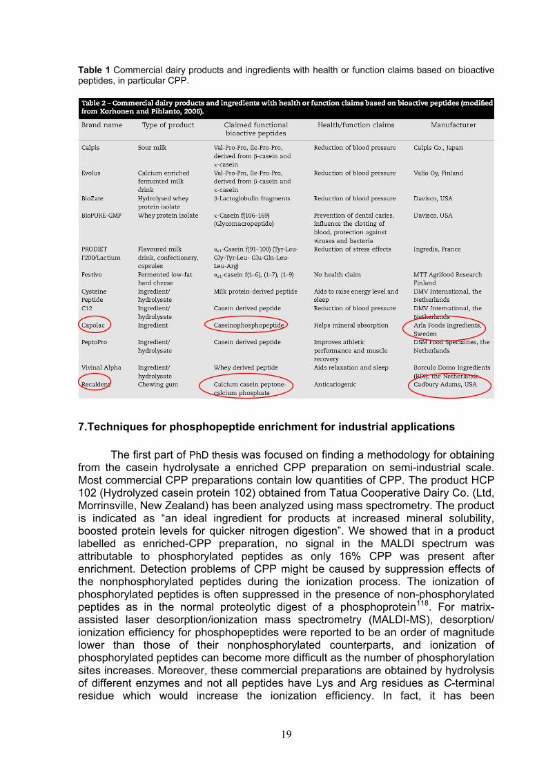
Table 1 Commercial dairy products and ingredients with health or function claims based on bioactive peptides, in particular CPP.
7.Techniques for phosphopeptide enrichment for industrial applications The first part of PhD thesis was focused on finding a methodology for obtaining from the casein hydrolysate a enriched CPP preparation on semi-industrial scale. Most commercial CPP preparations contain low quantities of CPP. The product HCP 102 (Hydrolyzed casein protein 102) obtained from Tatua Cooperative Dairy Co. (Ltd, Morrinsville, New Zealand) has been analyzed using mass spectrometry. The product is indicated as “an ideal ingredient for products at increased mineral solubility, boosted protein levels for quicker nitrogen digestion”. We showed that in a product labelled as enriched-CPP preparation, no signal in the MALDI spectrum was attributable to phosphorylated peptides as only 16% CPP was present after enrichment. Detection problems of CPP might be caused by suppression effects of the nonphosphorylated peptides during the ionization process. The ionization of phosphorylated peptides is often suppressed in the presence of non-phosphorylated peptides as in the normal proteolytic digest of a phosphoprotein118. For matrix-assisted laser desorption/ionization mass spectrometry (MALDI-MS), desorption/ ionization efficiency for phosphopeptides were reported to be an order of magnitude lower than those of their nonphosphorylated counterparts, and ionization of phosphorylated peptides can become more difficult as the number of phosphorylation sites increases. Moreover, these commercial preparations are obtained by hydrolysis of different enzymes and not all peptides have Lys and Arg residues as C-terminal residue which would increase the ionization efficiency. In fact, it has been
20
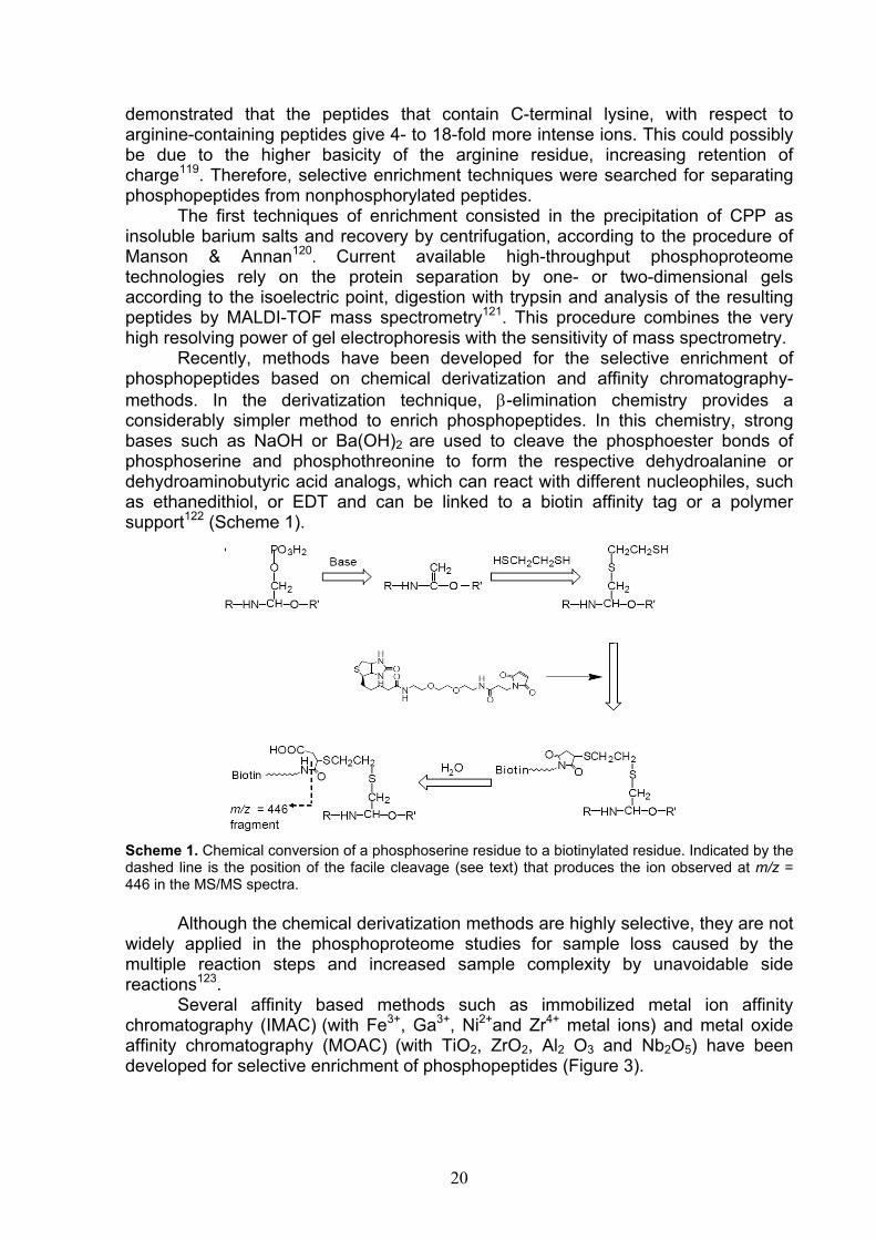
demonstrated that the peptides that contain C-terminal lysine, with respect to arginine-containing peptides give 4- to 18-fold more intense ions. This could possibly be due to the higher basicity of the arginine residue, increasing retention of charge119. Therefore, selective enrichment techniques were searched for separating phosphopeptides from nonphosphorylated peptides. The first techniques of enrichment consisted in the precipitation of CPP as insoluble barium salts and recovery by centrifugation, according to the procedure of Manson & Annan120. Current available high-throughput phosphoproteome technologies rely on the protein separation by one- or two-dimensional gels according to the isoelectric point, digestion with trypsin and analysis of the resulting peptides by MALDI-TOF mass spectrometry121. This procedure combines the very high resolving power of gel electrophoresis with the sensitivity of mass spectrometry. Recently, methods have been developed for the selective enrichment of phosphopeptides based on chemical derivatization and affinity chromatography- methods. In the derivatization technique, β-elimination chemistry provides a considerably simpler method to enrich phosphopeptides. In this chemistry, strong bases such as NaOH or Ba(OH)2 are used to cleave the phosphoester bonds of phosphoserine and phosphothreonine to form the respective dehydroalanine or dehydroaminobutyric acid analogs, which can react with different nucleophiles, such as ethanedithiol, or EDT and can be linked to a biotin affinity tag or a polymer support122 (Scheme 1).
Scheme 1. Chemical conversion of a phosphoserine residue to a biotinylated residue. Indicated by the dashed line is the position of the facile cleavage (see text) that produces the ion observed at m/z = 446 in the MS/MS spectra. Although the chemical derivatization methods are highly selective, they are not widely applied in the phosphoproteome studies for sample loss caused by the multiple reaction steps and increased sample complexity by unavoidable side reactions123. Several affinity based methods such as immobilized metal ion affinity chromatography (IMAC) (with Fe3+, Ga3+, Ni2+and Zr4+ metal ions) and metal oxide affinity chromatography (MOAC) (with TiO2, ZrO2, Al2 O3 and Nb2O5) have been developed for selective enrichment of phosphopeptides (Figure 3).

21
Figure 3. Schematic diagram of phosphopeptide isolation by IMAC or MOAC.
IMAC beads immobilized with Fe(III) or Ga(III) are commonly used for the purification of phosphopeptides124,125. In this technique, iminodiacetic acid (IDA, a tridentate metal-chelator) and nitrilotriacetic acid (NTA, a quadradentate metal-chelator) are often used as functional reagent that reacts with multivalent metal ions to form chelated ions with positive charges. Phosphate groups of phosphopeptides with negative charges are attracted by the chelated ions owing to static interaction, leading to phosphopeptides retained on the immobilized chelated ions. The phosphate group interacts through nonbonding ion pair electron coordination with metal ions which have been chelated to a multidentate ligand immobilized onto a support material. There is a number of challenges associated to IMAC. First, because of the metal ions are not covalently bound to the substrate, there is a possibility to leach these ions from the column, during the enrichment steps, which might lead to loss of phosphopeptides or to a contamination of peptides with metal ions. A second challenge is nonspecific binding of peptides that contain the acidic glutamic and aspartic acid residues. Methyl esterification of carboxyl groups has been demonstrated to decrease the nonspecific adsorption of acidic peptides on IMAC126. Instead of using IMAC with Fe3+, higher specificity and lower sample loss was observed for MOAC, using some metal oxides such as TiO2 and ZrO2. The negatively charged phosphopeptides selectively interact with titansphere via bidentate binding at the dioxide surface127,128,129. However, the selectivity of this method was somewhat compromised by the detection of several acidic non-phosphorylated peptides that were also retained by their TiO2 column. Considerable efforts have been made to improve this technique. Significant improvement was obtained in MOC with titania when benzoic acid derivatives such as 2,5-dihydroxybenzoic acid (DHB)130
and phthalic acid131 were used in the sample loading buffer to eliminate acidic nonphosphorylated peptides. Recently, it has been found that aliphatic hydroxyl acid-modified metal oxide works more efficiently and more specifically than aromatic modifiers such as DHB and phthalic acid in titania and zirconia MOC132. In several studies the combination of different enrichment methods has been found to be advantageous for selective phosphopeptide enrichment as the coupling of two complementary enrichment methods as calcium phosphate precipitation and

22
IMAC133, or as SCX and SAC where protein digests were first loaded on an SCX column, and the flow-through peptides from SCX were collected and further loaded onto a SAX column134. However, none of the tested analytical systems could be applied in industrial field for three reasons: a) the resin to which CPP are linked is not edible and barium chloride, in the procedure of Manson & Annan, is hazardous reagent and large quantity of expensive ethyl alcohol makes the procedure unrealiable, b) the elution of CPP from the resin requires very long and expensive purification systems, c) fortification of foods with eluted CPP alter the taste, making it more bitter. This is principally due to the ability by CPP to chelate mineral ions from contacting matrix.
8. Development of a new enrichment technique In the second part of the PhD work, a new technique for specific phosphoproteins and phosphopeptides enrichment from complex mixtures have been developed. The objective is to set out an analytical method for CPP enrichment to use for preparative purpose of nutraceuticals in functional foods. Using inexpensive, raw materials free of harmful Ba, Fe, Ga, Ni, Zr and Ti compounds, a large scale CPP isolation was obtained. Chromatography on industrial scale is based on ion exchange columns separation of great diameter or, to avoid the problems of clogging, on batch processes. The used material consists of a resin having the capacity of binding anion compounds such as CPP carrying negative charges in a wide pH range. This high selectivity technique is based on hydroxyapatite (HA) chromatography. HA is a form of calcium phosphate with the formula [Ca10(PO4)6(OH)2] which can be used as chromatographic matrix. HA chromatography is considered to work as a “mixed mode” ion exchange or a “pseudo-affinity” chromatography. The functional groups comprise positively charged pairs of crystal calcium ions (C-sites) and clusters of six negatively charged oxygen atoms associated with triplets of crystal phosphates (P-sites). The hydroxyapatite/biomolecule interactions are complex. Generally, it is thought that amino groups are attracted to negatively charged P-sites and repelled by positively charged C-sites. The opposite is true for negatively charged, phosphorylated residues of proteins. (Figure 4) .
Figure 4. Protein binding to hydroxyapatite. Double parentheses indicate repulsion. Dotted lines indicate ionic bonds. Triangular linkages indicate coordination bonds. The more phosphates are present, the more dominating is the calcium site binding and thus the tighter is the binding of the protein to the matrix135
PO4
COO
COO
+H3N
+H3N
+H3N
+H3N
PO4((
((
))
))
Protein

23
Phosphorylated groups on proteins and other solutes interact even more strongly with C-sites than do carboxyls136. This is reflected in extremely strong binding by proteins and peptide with high degree of phosphorylation. Then several studies demonstrated that phosphorylated proteins bind more strongly to HAP than their unphosphorylated counterparts137. The developed method employs ceramic hydroxyapatite (HA) as solid-phase adsorbent to separate nonphosphorylated from resin-bound phosphorylated components. Casein was chosen as phosphorylated protein model. Phosphopeptide was obtained by tryptic digestion in situ on HA-casein complexes.
9. Experimental procedure 1 Materials Raw whole milk was collected from local dairy farms. Milk was skimmed by centrifugation at 4,000 rpm at 4° C for 30 min. Isoelectric casein was prepared by adding to skim milk 10% (v/v) acetic acid, allowing to stand 30 min at 35° C and then adding 1M NaOAc to 4.6 final pH. After standing for 30 min, suspension was centrifuged (4,000 rpm, 5° C for 30min), the supernatant discarded, casein washed twice with buffer diluted 1:4, twice with water Milli-Q, and subsequently freeze dried. HA (Macro-Prep Ceramic Hydroxyapatite TYPE I, catalog number 157-0040) was purchased from Bio-Rad (Milan, Italy). Tris (hydroxymethyl) aminomethane hydrochloride (Tris-HCl), potassium chloride (KCl), urea, trifluoroacetic acid (TFA), acetonitrile (ACN) for HPLC Plus, orthophosphoric acid 85% were from Carlo Erba (Milan, Italy).Trypsin TPCK treated from bovine pancreas was from Sigma (St. Louis, MO, USA). Sodium acetate trihydrate, 2,5-dihydroxybenzoic acid (DHB) were from Fluka (St. Louis, MO, USA). Acetic acid was purchased from Baker Chemicals B.V. (Deventer, The Netherlands). Dithiothreitol (DTT) was from Applichem (Darmstadt, Germany). Water was prepared using a Milli-Q system (Millipore, Bedford, MA, USA). HA-based phosphoprotein/peptide enrichment Casein (10 mg) was dissolved in 80μl buffer at pH 8.0 containing TrisHCl 50 mM, KCl 0.2 M, urea 4.5 M, DTT 10 mM. The casein solution was loaded on HA (10 mg), previously equilibrated in the loading buffer. The HA-bound casein was incubated for 15 min at room temperature and centrifuged for 5 min at 4,000 rpm. The resin was washed in succession with three buffers, i.e. the loading buffer (1ml); washing buffer at pH 7.8 containing Tris-HCl 50mM (1 ml); and 20mM Tris-HCl (pH 7.8) buffer containing 20% ACN (v/v)(1ml). The resin washed with water Milli-Q (1ml) and finally freeze-dried with a SpeedVac concentrator system (Thermo Electron, Milford, MA). HA-based CPP enrichment The HA-bound phosphoproteins were hydrolyzed in situ with trypsin added to the suspension at an enzyme/substrate ratio of 1:50 (w/w) in 50mM Tris-HCl buffer, pH 8.0, containing 0.2 M KCl, 4.5M urea, and 10 mM DTT. The reaction was carried out at 37° C overnight and stopped by centrifuging the HA-CPP microgranules for 5 min at 4,000 rpm. Then, the microgranules were washed as described above for

24
phosphoproteins. After washing with water Milli-Q, they were dried by using a SpeedVac apparatus. MALDI-TOF MS Analysis MALDI-TOF mass spectra were recorded using a Voyager DE-PRO mass spectrometer (Applied Biosystem, Framingham, USA), in positive linear mode. All spectra were acquired in the range of 15–30 kDa (for proteins) and 1–5 kDa (for peptides) with the following settings: an accelerating voltage 25 kV (for proteins), and 20 kV (for peptides) and grid voltage 93% (for proteins) and 95% (for peptides) of accelerating voltage, a guide wire of 0.15% (for proteins) and 0.05% (for peptides), delayed ion extraction time of 485 (for proteins) and 175 ns (for peptides). The laser power was set just above the ion generation threshold to obtain peaks with highest possible signal to noise ratio. All spectra were acquired with 200 shots in 3 replicate. Two different matrix solutions were freshly made each day using stocks of the required solvents and sonicated for 15 min in an ultrasonic water bath prior to use. The sample/wash (SW) method was based on the sample/matrix/wash (SMW) method described by Zhang et al.138 with the exclusion of the wash step. The matrix for phosphoprotein analysis was constituted by sinapinic acid (SA) solution at 10 mg/mL in 50% ACN and 0.1% TFA. 1 μL of sample was added to spot and allowed to dry. The matrix for phosphopeptides contained 2,5-dihydroxybenzoic acid (2,5-DHB), 10 mg/mL in 1 ml of H2O/ACN/PA (49/50/1). 1 μL of sample was added to spot and allowed to dry. The HA-phosphoprotein microgranules were deposited onto the MALDI plate and covered with the SA matrix (10 mg/ml in 50% ACN) to promote analyte/matrix co-crystallization in presence of increasing TFA concentration (0.01%, 0.05%, 0.1% and 0.5%) for proteins. For CPP, 1% phosphoric acid (PA) was included in the DHB solution (10 mg/ml SA in 50% ACN)139. Microscopic observation The HA-CPP microgranules deposited onto a 96-well MALDI target were observed by stereomicroscopy before and after co-crystallization with the DHB matrix. The microscope (PBI International, Milan, Italy) was equipped with a binocular head illuminated with a 6 V, 20 W halogen lamp and 100-230 V, 50/60 Hz power supply LC/MS Analysis LC/MS spectra were obtained by means of an Agilent 1100LC/MSD single quadrupole instrument. HA-CPP complexes (1 mg) were solubilized with 120 μl of solution containing 0.5% PA and 100 μl of this solution were then injected. For this analysis, HA-CPP complexes of caseins were fractionated using a Vydac C18 218TP52 column (250mm-2.1mm i.d.), at a constant flow rate of 0.2 mL/min. Elution was carried out with a linear gradient from 5% to 90% of solvent B (0.1% TFA in acetonitrile, v/v) over 100 min after 15 min of isocratic elution at 5% B; solvent A was 0.1% TFA in water (v/v). Liquid effluents from the column were directly injected into the source of a LC/MSD SL (Agilent Technologies) single quadrupole mass spectrometer. The mass spectra were scanned from m/z 1600 to 400 at a scan cycle of 4.90 s/scan and 0.1 s inter-scan delay. The source temperature was 180°C. Spectra were acquired in positive ion mode, where the capillary voltage and the cone

25
voltage were 3.6 kV and 40 V, respectively. N2 was used as both the drying and nebulising gas. LC-ESI-MS/MS Analysis HA-CPP complexes (1 mg) were solubilized with 120 μl of 0.5% PA and analyzed by a CapLC nano-flow high-pressure pump system (Waters) interfaced with a QTOF Micro mass spectrometer (Waters/Micromass) operating in the positive ion mode. Chromatographic separations were performed on a reverse phase Atlantis dC18 capillary column (75 μm i.d.). The mobile phase was water (A) and ACN (B) with 0.1% FA. A linear gradient from 5 to 70% of B was applied over 45 min at a flow-rate of approximately 300 nL/min using a precolumn split with a pump operating at 5 µl min-1. The source conditions were as follows: capillary voltage, 2.6 kV; cone voltage, 100 V; and RF1 lens, 40 V. In the ESI-MS/MS experiments for peptide sequencing, argon was used as the collision gas. Raw data were processed using MassLynx 3.5 ProteinLynx software.
10. Results and discussion The analytical method which finally was adopted can be summarized in the following six consecutive steps: (1) selective enrichment of the phosphoproteins/phosphopeptides on HA microgranules (resin:protein 1:1, w/w) from protein mixtures, (2) flow-through of the unphosphorylated components using the dilution/washing buffer, (3) drying the protein/peptide-HA microgranules, (4) loading a few dried HA microgranules (~1000) on the MALDI target plate; (5) covering with the sinapinic acid or DHB matrix with acid solution (TFA) or (PA) respectively which co-crystallise with HA microgranules and (6) in situ desorption of the bound HA-phosphoproteins/phosphopeptides by direct MALDI/MS analysis (Figure 5).
= phosphate group It resulted a highly sensitive and selective method with good reproducibility for extraction and enrichment of phosphorylated proteins and peptides. The unphosphorylated components including γ-CN and nonphosphorylated peptide like αs1-CN (f91-100) (m/z 1267), and αs1-CN (f8-22) (m/z 1759) were removed from the resin in the washing buffer. Inclusion of 20% ACN in the Tris-HCl buffers greatly
Figure 5. Schematic representation of the technique of HAP-based phosphoproteins/peptides enrichment. Phosphoproteins/peptides were bound to the HAP through their phosphate groups, the non phosphorylated components were removed using washing buffers The dried HAP-phosphoproteins were analysed directly by MALDI–TOF after co-crystallization of the sinapinic acid with an appropriate quantity of trifluoroacetic acid.
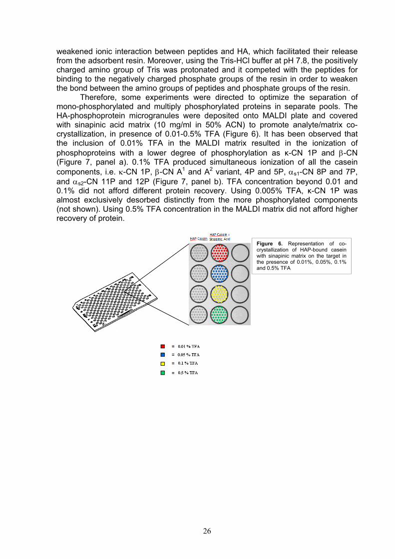
26
weakened ionic interaction between peptides and HA, which facilitated their release from the adsorbent resin. Moreover, using the Tris-HCl buffer at pH 7.8, the positively charged amino group of Tris was protonated and it competed with the peptides for binding to the negatively charged phosphate groups of the resin in order to weaken the bond between the amino groups of peptides and phosphate groups of the resin. Therefore, some experiments were directed to optimize the separation of mono-phosphorylated and multiply phosphorylated proteins in separate pools. The HA-phosphoprotein microgranules were deposited onto MALDI plate and covered with sinapinic acid matrix (10 mg/ml in 50% ACN) to promote analyte/matrix co-crystallization, in presence of 0.01-0.5% TFA (Figure 6). It has been observed that the inclusion of 0.01% TFA in the MALDI matrix resulted in the ionization of phosphoproteins with a lower degree of phosphorylation as κ-CN 1P and β-CN (Figure 7, panel a). 0.1% TFA produced simultaneous ionization of all the casein components, i.e. κ-CN 1P, β-CN A1 and A2 variant, 4P and 5P, αs1-CN 8P and 7P, and αs2-CN 11P and 12P (Figure 7, panel b). TFA concentration beyond 0.01 and 0.1% did not afford different protein recovery. Using 0.005% TFA, κ-CN 1P was almost exclusively desorbed distinctly from the more phosphorylated components (not shown). Using 0.5% TFA concentration in the MALDI matrix did not afford higher recovery of protein.
Figure 6. Representation of co-crystallization of HAP-bound casein with sinapinic matrix on the target in the presence of 0.01%, 0.05%, 0.1% and 0.5% TFA

27
Analysis of phosphopeptides For the isolation of CPP, well rinsed HA-casein microgranules in suspension were submitted to hydrolysis in situ by overnight trypsin treatment. The resulting microgranules were separated by centrifugation and cleansed through mixing the equilibration/washing buffer to ensure that the enzyme action was stopped. The nonphosphorylated peptides were eliminated by washing buffers as illustrated in the Figure 8.
= phosphate group
We raised the problem of determining which peptides remained tightly bound to the resin. Hence, a system of quality control of CPP preparation was developed, using mass spectrometry techniques such as MALDI/MS, LC-ESI/MS and LC-ESI-MS/MS. We demonstrated that each of these techniques allows to characterize the CPP bound to the resin. The study showed that the resin immobilizes a plethora of peptides, including CPP with multiple phosphorylation site of the type αs1-CN (f59-79) 5P (f2720.91 Da), β-CN (f1-25) 4P (3122.26 Da) and αs2-CN (f46-70) 5P (3087.99
κ-CN 1P β-CN 5P
κ-CN 1P
β-CN A1 5Pαs1-CN 8P
αs2-CN 11P
β -CN A2 5P
αs1-CN 7P
β -CN A1 e A2 4P
αs2-CN 12P
a
b
Figure 7. MALDI MS spectra of casein eluted with 0.01%TFA (a) and 0.1%TFA (b)
Figure 8. Schematic representation of the HAP-based CPP enrichment technique. HAP-bound phosphoproteins were hydrolyzed in situ by trypsin. Non phosphorylated peptides were washed out in the flow-through. The dried HAP-CPP microgranules included in the crystallime matrix were analysed by MALDI-TOF directly on the MALDI plate.

28
Da), each containing the sequence cluster Ser(P)-Ser(P)-Ser(P)-Glu-Glu, functional to metal ion transport. A rapid analytical technique has been developed for the identification of the CPP. The HA-CPP microgranules were co-crystallised onto the well MALDI plate included into DHB matrix solution and phosphoric acid. Inclusion of 1% phosphoric acid allowed to achieve the maximum ionization efficiency by MALDI and minimize binding of CPP to HA. Kjellstrom, S and Jensen, O. N.139 reported that phosphoric acid (PA) was proved to be the matrix additive chosen for the sensitive detection of phosphopeptides. PA actually worked well in enhancing the phosphopeptide signals. The microscope images of HA-CPP on well MALDI plate before and after crystallisation are shown in Figure 9.
Figure 9. The microscopic images of HA-CPP on well MALDI plate before (a) and after crystallisation (b) of HA-CPP microgranules with DHB matrix in PA solution. The MALDI spectrum of HA-CPP complexes is shown below (Figure 10). An enlargement in two spectra has been obtained for marking the corresponding CPP to every peak (Figure 10).
a b

29
Figure 10. MALDI mass spectrum of HA-CPP complexes loaded directly on the well MALDI plate after enrichment. An enlargement in two spectra has been underlined. The overnight incubation and high trypsin-to-substrate ratio (resin/enzyme=1/50) produced the maximum yield of tryptic peptides. Protein binding via Lys and Arg residues to negatively charged P-sites of HA would mask most of the casein sites to trypsin. In contrast, binding of phosphate groups to HA was chosen as

30
casein remained as susceptible to trypsin and produced tryptic CPP of ideal size for mass spectrometry analysis. The presence of basic amino acids Arg and Lys in C-terminal position also facilitates subsequent mass spectrometric analysis. In spite of this, from the proteolytic digests of casein about thirty CPP were identified, including some phosphopeptides with missed cleavages (Table 2). The list of the theoretical tryptic CPP annotated in the Swiss Prot archives and their molecular masses is shown in the Table 2. Tryptic phosphopeptides were identified by comparing the measured mass with that derived from the standard casein sample. Table 2. Identification of CPP bound to the HA microgranules loaded directly on the well MALDI plate after enrichment.
Molecular Mass (Da) NATIVE CPP
Expected Measured MH+ Peptide identification Missed cleavages CPP 1410.5 1411.6 αs2(f126-136)2P
1466.5 1467.6 αs2(f138-149)1P
1538.6 1540.3
αs2(f126-137)2P
1594.7 1595.6 αs2(f138-150)1P
1659.8 1661.6 αs1(f106-119)1P
1846.7 1847.1 αs1(f43-58)1P
1926.1 1928.3 αs1(f43-58)2P
1952.0 1952.3 αs1(f104-119)1P
2060.8 2062.8 β(f33-48)1P
2080.4 2079.4 αs1(f103-119)1P
2539.4 2538.6 αs2(f2-21)3P
2598.0 2598.1 αs1(f37-58)2P
2618.9 2619.3 αs2(f2-21)4P
2678.0 2678.3 αs1(f37-58)3P
2703.9 2703.8 αs1(f59-79)5P pyroQ
2720.9 2720.9 αs1(f59-79)5P
2746.9 2747.6
αs2(f1-21)4P
2855.2 2855.4 αs1(f35-58)2P
2886.2 2886.0 β(f2-25)3P
2935.2 2935.2 αs1(f35-58)3P
2966.2 2966.0 β(f2-25)4P
3088.0 3087.0 αs2(f46-70)5P
3007.9 3007.3 αs2(f46-70)4P
3122.3 3122.1
β(f1-25)4P
3042.3 3043.8 β(f1-25)3P
3132.1 3131.4 αs2(f1-24)4P
3477.5 3477.0 β(f1-28)4P
3605.6 3605.1 β(f1-29)4P

31
LC/MS analysis of samples enriched in phosphopeptides was also carried out to verify purity of the preparation and the correctness of peptide assignment. The LC-ESI-MS TIC profile (Figure 11) and identification of the chromatographic peaks are reported in Table 3.
Figure 11. LC-MS chromatogram of CPP bound to the HA microgranules dissolved with an aqueous solution containing 0.5% PA
Dissolution of HA in 5% PA (w/v) solution has been carried out to ensure that all CPP bound were actually eluted from the resin. Table 3. Peptide identification of each chromatographic peak of CPP bound to the HA microgranules dissolved with an aqueous solution containing 0.5% PA.
Peak Number Theoretical mass (Da) Measured mass (Da) Peptide identification
1 1410.49 1410.82 αs2-CN (f126-136)2P
2 1538.59 1538.04 αs2-CN (f126-137)2P
3 1942.70 1943.00 αs1-CN (f43-58)2P M(0)
4 2061.01 2061.59 β-CN (f33-48)1P
2720.90 2720.90 αs1-CN (f59-79)5P
5 2720.90 2720.98 αs1-CN (f59-79)5P
3007.90 3008.04 αs2-CN (f46-70)4P
6 3007.90 3008.04 αs2-CN (f46-70)4P
7 3004.09 3004.26 αs2-CN (f2-24)4P
2618.89 2618.97 αs2-CN (f2-21)4P
1 2 3
4
56
7
8
9
11
10
12
13
14
15
16
1718
19
20
mAU
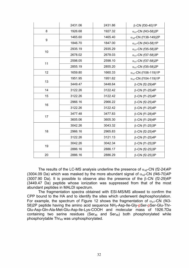
32
2431.06 2431.86 β-CN (f30-40)1P
8 1926.68 1927.32 αs1-CN (f43-58)2P
9 1465.60 1465.40 αs2-CN (f138-149)2P
1846.70 1847.00 αs1-CN (f43-58)1P
10 2935.19 2935.29 αs1-CN (f35-58)3P
2678.02 2678.03 αs1-CN (f37-58)3P
11 2598.05 2598.10 αs1-CN (f37-58)2P
2855.19 2855.20 αs1-CN (f35-58)2P
12 1659.80 1660.33 αs1-CN (f106-119)1P
13
1951.95 1951.62 αs1-CN (f104-119)1P
3449.47 3448.64 β-CN (f2-29)4P
14 3122.26 3122.42 β-CN (f1-25)4P
15 3122.26 3122.42 β-CN (f1-25)4P
16 2966.16 2966.22 β-CN (f2-25)4P
3122.26 3122.42 β-CN (f1-25)4P
17 3477.48 3477.83 β-CN (f1-28)4P
3605.06 3605.30 β-CN (f1-29)4P
18
3042.26 3043.32 β-CN (f1-25)3P
2966.16 2965.83 β-CN (f2-25)4P
3122.26 3121.13 β-CN (f1-25)4P
19 3042.26 3042.34 β-CN (f1-25)3P
2886.16 2886.17 β-CN (f2-25)3P
20 2886.16 2886.29 β-CN (f2-25)3P
The results of the LC-MS analysis underline the presence of αs2-CN (f2-24)4P (3004.09 Da) which was masked by the more abundant signal of αs2-CN (f46-70)4P (3007.90 Da). It is possible to observe also the presence of the β-CN (f2-29)4P (3449.47 Da) peptide whose ionization was suppressed from that of the most abundant peptides in MALDI spectrum. The fragmentation spectra obtained with ESI-MS/MS allowed to confirm the CPP bound to the HA and to identify the sites which underwent dephosphorylation. For example, the spectrum of Figure 12 shows the fragmentation of αs1-CN (f43-58)2P peptide having the amino acid sequence NH2-Asp-Ile-Gly-pSer-pSer-Glu-Thr-Glu-Asp-Gln-Ala-Met-Glu-Asp-Ile-Lys-COOH and molecular mass of 1926.7Da containing two serine residues (Ser46 and Ser48) both phosphorylated while phosphorytable Thr49 was unphosphorylated.

33
Figure 12. MS/MS spectrum from fragmentation of the doubly charged precursor ion at m/z 964.31. The product ions belonging to the y series is indicated. The fragmentation spectra were processed by the MaxEnt 3 program to a singly charged spectra and were elaborated using the ProteinLynx software. The ions y10 (1179.51 Da) and y11 (1346.51 Da) with molecular mass variation (∆m) of 167 Da (Ser + H3P04) indicate the fragmentation of phosphorylated Ser48 (11th position from the C-terminal), the ions y12 (1475.55 Da) and y13 (1642.55 Da) with ∆m of 167 Da (Ser + H3P04) indicate the fragmentation of phosphorylated Ser46 (13 th amino acids from the C-terminal). The fragmentation of the peptide α s1-CN (f43-58)1P with amino acid sequence NH2-Asp-Ile-Gly-Ser-Glu-pSer-Thr-Glu-Asp-Gln-Ala-Met-Glu-Asp-Ile-Lys-COOH and molecular mass of 1846.74 Da included only one phosphate group on Ser48, whereas Ser46 remained free (Figure 13).
% In
tens
ity
Mass m/z
MH2+

34
Figure 13. MS/MS spectrum from fragmentation of the doubly charged precursor ion at m/z 924.38. The product ions belonging to the y series is indicated. The fragmentation spectra were processed by the MaxEnt 3 program to a singly charged spectra and were elaborated using the ProteinLynx software.
The ions y10 (1179.51 Da) and y11 (1346.51 Da) with ∆m of 167 Da (Ser + H3P04) indicate the fragmentation of phosphorylated Ser48 (11th amino acids from the C-terminal), the ions y12 (1475.55 Da) and y13 (1562.59 Da) with ∆m of 87Da (Ser) indicate the fragmentation of not phosphorylated Ser46 residue (13th position from the C-terminal) The ESI Q-TOF analysis of the phosphorylated mixture confirmed the presence of the peptide αs1CN (f43-58) with one degree of phosphorylation. Thus this peptide confirms the copresence of native casein αs1-CN 7P and αs1-CN 8P. These experiments allowed to confirm the selectivity and specificity of the resin and the purity of the mixture obtained by applying the outlined analytical technique. At that time, it was assayed to transfer from the laboratory to pilot scale the HA-CPP complex for functional milk formulation.
MH2+

35
11. Formulation of a functional drink in semi-industrial scale The laboratory technique was transferred to semi-industrial scale. First at all the development of an industrial process implies the use of inexpensive raw materials. Thus, we planned the preparation of the complex HA-CPP in semi-industrial scale by de novo synthesizing HA, owing to the expensive commercial product. Therefore, HA was synthesized by wet-chemical precipitation reactions from Ca(OH)2 and H3PO4 solution, adopting temperature and pH optimum for obtaining pure crystals of apatite. The absorbent activity of the synthesized resin (HAS) was evaluated by comparing the binding capacity of CPP in 3 different proteins mxtures, e.g. isoelectric casein, milk proteins (casein + whey proteins) and raw milk. Milk proteins, commonly used in the dairy industry or raw milk were directly loaded on HA. After opportune washings, casein-HA and tryptic derived CPP were compared to those recovered by using isoelectric casein. We demonstrated that protein material loaded on HA could be variable depending on specific needs because the same CPP were linked to HAS from isoelectric casein, milk proteins and raw milk. For functional milk preparation, tryptic hydrolysis of in situ bound protein was operated on the HAS-casein suspension. There were CPP tightly bound to HA, and non-phosphorylated peptides removed from the resin by repeated washing buffers, until a washed product in milliQ water was obtained. The HA-CPP microgranules were then dried for the storage of the powder for times of the order of years at room temperature. Dried microgranules of HA-CPP complex were also analysed by MALDI. Then, dried microgranules were ground before addition to milk. The flow diagram of preparation of functional milk is shown below (Scheme 2).
Scheme 2. Flow diagram of functional milk preparation
HAS Synthesis Loading
protein on HASTryptic digestion
in situ
Drying
Rapid MALDI Analysis on well plate
Grinding Addition the
suspension to milk
Washing out of nonphosphorylated
peptides

36
12. Experimental procedure 2 Materials
Raw whole milk was collected from local dairy farms. The skimmed pasteurized milk was supplied by YMA industry. Milk proteins powder were from Sacco industry (Milan, Italy). Calcium hydroxide was purchased from Carlo Erba (Milan, Italy). The source of the other raw materials has already been above indicated. HA Synthesis
HA was precipitated using this following reaction scheme.
5Ca(OH)2 + 3H3PO4 Ca5(PO4)3(OH) + 9H2O 12.5 mmol or 925 mg of Ca(OH)2 powder in 125 mL of milliQ water and 7.5 mmol or 865 mg of 85 wt%H3PO4 in 125 mL of milliQ water were used for the HA precipitation. The orthophosphoric acid solution was added in a dropwise manner to a dilute solution/suspension of calcium hydroxide. Prior to PO4
3- ion addition to the Ca2+ ion solution, the latter was heated to 90°C and stirred for 1h at 400 rpm. The acid was added at a rate approximately equal to 3.5 mL/min using a peristaltic pump. The reaction was maintained under constant stirring to ~400 rpm at room temperature for 2h. The precipitation reaction was slow and proceeded at 4°C overnight. The precipitate was filtered, washed with milliQ H2O for 3 times and dried in an oven at 60° C. Application of the synthetic HAS to isoelectric casein, milk protein and raw milk
The binding capacity of the synthesized HAS was evaluated by comparing the MALDI spectra of CPP from 3 different proteins mixture, e.g. isoelectric casein (sample 1), milk proteins (sample 2) and raw milk (sample 3). Sample 1: 1 g casein was dissolved in 8 ml of buffer at pH 8.0 containing Tris-HCl 50 mM, KCl 0.2 M, urea 4.5 M, DTT 10 mM. The protein solution was loaded on 1 g of synthesized HA (ratio HA/protein=1/1), previously washed with loading buffer. Sample 2: 1g milk proteins was dissolved in 8 ml of pH 8.0 buffer containing Tris-HCl 50mM, KCl 0.2 M, urea 4.5 M, DTT 10 mM. The protein solution was loaded on 1g of synthesized HA (ratio HA/protein=1/1), previously washed with loading buffer. Sample 3: 31.2 ml of raw milk was loaded on 1 g of synthesized HAS (ratio HA/casein=1/1), previously washed with loading buffer. The next steps and MALDI analysis conditions were the same as those already reported. The MALDI analysis of milk peptides was conducted by loading on the spot 1 μl of milk solution diluted 1:100 with a solution containing H2O/ACN (50/50). 1 μL of the DHB solution (10 mg/mL) containing H2O/ACN/PA (49/50/1) was loaded on dried sample and allowed to dry.

37
Adding the suspension to skimmed pasteurized milk Aliquots of finely ground HAS-CPP microgranules (100 and 250 mg) could derived from each of the above prepared samples. To avoid the immediate sedimentation of HAS-CPP in milk, grinding was carried out using the Ultra-Turrax T25 apparatus for 3min at 24,000 rpm. 100 μl (0.2% v/v) and 250 μl (0.5% v/v) of the suspension aliquots were added to 50 ml of raw milk. The HAS-CPP microgranules added to the milk were recovered by centrifugation, washed and analyzed by MALDI-TOF to assess the residual binding capacity of the resin.
13. Results and discussion Hydroxyapatite [HA, Ca5(PO4)3OH] is a versatile biomaterial and when synthesized from wet-chemical precipitation reactions often forms nanoparticles with varying particle size and morphology, depending on the reactants and reaction conditions. Furthermore, the morphology, crystallite size, purity, stoichiometry, and structure of HA influence its bioactivity, bio-compatibility, chemical and physical stability, and mechanical properties, hence, its in vitro and in vivo performance after processing140. The synthesis of HA can take place through a variety of processes. HA synthesis includes the sol-gel technique141, solid-state reactions at elevated temperatures142, and biosynthesis routes143. Among the different synthesis methods, the wet precipitation method for synthesizing HA was proved to be the most suitable to industrial application. Among the various syntheses, the wet-chemical precipitation reactions from Ca(OH)2 and H3PO4 were chosen for simplicity, rapidity of reaction and high level of repeatability. The morphology of the HA precipitates changed from needlelike to nearly spherical. The aspect ratio (length-to-diameter ratio) decreases as the reaction temperature is increased when it is synthesized using Ca(OH)2 and H3PO4
144,145 The crystallinity of apatites prepared at higher precipitation temperatures was higher than those prepared at lower precipitation temperatures (T). In particular T=90°C produced a higher purity and crystallinity product.146. For this reason, the conditions describe above were used. To assess the binding capacity of the resin, three protein mixture a) isoelectric casein, b) milk protein and c) raw milk were loaded on HAS. These proteins were linked to the resin, and in situ hydrolyzed by trypsin. The spectra of the three preparations are compared in Figure 14.

38
Figure 14. MALDI mass spectra of CPP recovered by HAS from isoelectric casein (a), milk protein (b) and raw milk (c). Although the protein mixture loaded on HAS was of different complexity, the CPP profile revealed by MALDI-TOF analysis was similar, indicating that binding of the casein fractions to HAS was the same, with respect to the sample composition
a
b
c

39
and its form. This assays showed that the process was scalable for industrial purposes by using the most common protein source as ingredient depending on the specific industrial needs. Once obtained the HAS-CPP complexes on pilot scale, a functional food was formulated. It has been demonstrated that the addition of 2.0–5.0 g CPP-ACP⁄l to milk increases its ability to remineralize enamel subsurface lesions147
and the remineralizing effect of CPP-ACP in milk was dose-dependent with milk containing 0.2% CPP-ACP and 0.3% CPP-ACP producing an increase in mineral content of 81% and 164%, respectively, in comparison to the control milk148. The percentage of HAS-CPP was added to the functional milk. As the HAS-CPP suspension have greater density than milk, before addition to milk, the granules of HAS-CPP were finely ground to avoid their fast sedimentation. However, before drinking, functional milk should be shaken to homogenize the suspension. Before analysis of microgranules recovered by milk, control CPP from skimmed pasteurized milk was carried out by MALDI analysis (Figure 15, panel a). This analysis disclosed the affinity of the milk proteins and peptides for the resin. The spectra of the HA-CPP showed casein in soluble sodium salts (not shown spectrum) and CPP bound HA (Figure 15, panel b).
Figure 15. MALDI spectra of peptides from skimmed pasteurized milk before HAS enrichment (a) and after HAS enrichment (b).
a
b

40
In the spectra of the untreated skimmed pasteurized milk, four non-phosphorylated peptides such as β-CN (f184-204), αs1-CN (f2-22), αs2-CN (f189-207) and αs1-CN (f179-199) together with a number of CPP (Figure 15, panel a) was detected in the wider mass range 2200-4200 Da. Sample treated with HAS microgranules specifically captured CPP, while those non-phosphorylated contaminating the resin were removed by extensive washings in batch with buffer as detailed in the experimental procedure 1. Furthermore, the HAS-CPP microgranules added to milk were recovered by centrifugation and bound CPP were detected by MALDI-TOF, by loading microgranules on the well MALDI plate (Figure 16, panel c). Comparison of the MALDI spectra amongst HA-CPP before addition to milk (a), peptides in untreated skimmed pasteurized milk (b) and HA-CPP fixed after addition to milk (c) are shown in Figure 16 (panel a-c). The microgranules of HAS-CPP added to milk (Figure 16, panel a) were able to chelate further free CPP in milk (Figure 16, panel b).

41
Figure 16. MALDI spectra of comparison between HA-CPP before addition to milk (a), peptides in untreated skimmed pasteurized milk (b) and HA-CPP after addition to milk and after recovery by centrifugation (c). The HA-CPP microgranules added to milk were able to chelate further free CPP of milk as demonstrated in the spectrum (Figure 16, panel c) by the increased signals of peptides marked in purple. The number of CPP present in skimmed pasteurized milk sample (Figure 16, panel b) increased the level of CPP binding HAS-CPP complexes after addition to milk compared to the control (Figure 16, panel a). This means that some C-sites of HAS yet remained accessible to phosphate groups while the P-sites were able to chelate metal ions.
c
b
a

42
Finally, the same sensory characteristics of functional milk have been evaluated. Milk enriched with CPP showed the same sensory characteristics as starting milk, as CPP were neutralized by the calcium ions of the HAS crystals. The HAS-CPP complex is odourless and tasteless. The HAS-CPP complex has been added to milk but it could be added to a wide range of products as toothpaste, toothpowder, topical gels, dental filling material, mouthrinse, chewing gum, lozenges, tablets, mineral drinks, nutritional supplements for children, confectionery, and products for dental care mouthrinse. In conclusion, the preparation of a functional drink in semi-industrial settlement allowed to obtain a product with inexpensive raw materials and with nutraceutical ingredients (HAS-CPP) that could induce beneficial effects on human health especially for teeth.

43
14. Quantification of CPP in HAS-CPP preparation to be added to milk To understand the amount of CPP addition in the milk, the problem of quantitative determination of CPP linked to HAS has been faced. Matrix-assisted laser desorption ionization time-of-flight mass spectrometry (MALDI-TOFMS) is used to obtain fast and accurate determination of molecular mass, but quantitative determinations are generally made by other techniques. While there have been reports of quantitative MALDI-TOFMS applications, there are many problems inherent to the MALDI ionization process that have restricted its widespread use149. These limitations primarily stem from factors as the limited dynamic range due to detector saturation, difficulties associated with coupling MALDITOFMS to on-line separation techniques as liquid chromatography and especially the sample/matrix heterogeneity that are believed to contribute to the large variability in observed signal intensities for analytes. A major issue is the non-homogeneous distribution of the analyte in the cocrystallite. For example, especially non homogeneous needle-shaped crystals were observed when DHB was used as a matrix. This leads to the observation that, at several points of the sample, no analyte signals can be found whereas at other positions strong signals can be monitored (hot-spot formation). The varying ion response on different positions on the sample spots leads to poor “shot-to-shot” reproducibility and poor “point-to-point” repeatability and this is therefore one of the main reasons hampering quantitative MALDI MS150. Because of these difficulties, practical examples of quantitative applications of MALDI-TOFMS have been limited. Internal standards have been used successfully to overcome these problems.151,152. For quantification, in fact, internal standards are necessary to compensate for the poor shot-to-shot reproducibility inherent in the use of MALDI analysis. The selection of an appropriate internal standard (IS) for quantification is critical for the successful application of quantitative MALDI. There are several properties of the ideal internal standard153:1) It must be completely resolved from the analyte; 2) It must be chemically stable during the analysis; 3) It should be chemically similar to the analyte (including ionization and extraction efficiencies) and should not react with the analyte; 4) It should be close to the analyte in mass and concentration to avoid instrumental errors. Ideal internal standards are substances showing a high physico-chemical similarity to the analyte. Standards labelled with stable isotopes like 2H, 13C, 14N or 18O deliver best results in terms of accuracy of the quantification154. Another strategy is the use of non-isotope labelled internal standards155, applying structurally modified compounds. For example, peptides with high molecular similarity156 were successfully used as internal standards for the relative quantitation of peptides. Moreover, it has been reported that MALDI-TOF MS can be used for the quantification of proteins and other bioactive molecules by evaluating the ratio of the peak heights between the analyte and a correctly selected.internal standard157,158 . In this PhD work, synthetic internal standards were used to quantify tryptic CPP enriched by HAS and used as ingredient for the functional milk. The synthetic peptide analogues which were chosen varied for a single amino acid in the peptide sequence to achieve a ionization efficiency equivalent to that of tryptic phosphopeptide in the CPP mixture. Two CPP with different degree of phosphorylation as monophosphorylated peptide αs1-CN (f106-119) 1P (1659.8 Da) and diphosphorylated peptide, αs1-CN (f43-58) 2P (1926.7 Da) were chosen for the quantification. Four synthetic peptides were synthesized by solid-phase Fmoc synthesis without further purifications by Dr.
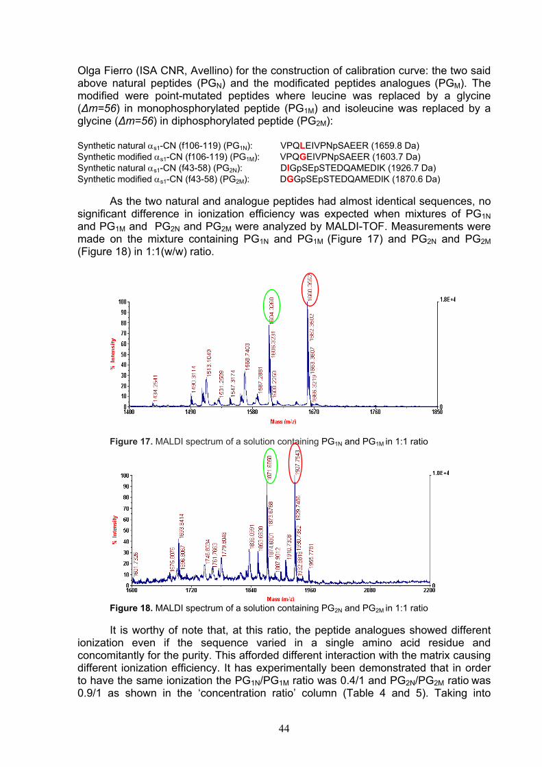
44
Olga Fierro (ISA CNR, Avellino) for the construction of calibration curve: the two said above natural peptides (PGN) and the modificated peptides analogues (PGM). The modified were point-mutated peptides where leucine was replaced by a glycine (∆m=56) in monophosphorylated peptide (PG1M) and isoleucine was replaced by a glycine (∆m=56) in diphosphorylated peptide (PG2M): Synthetic natural αs1-CN (f106-119) (PG1N): VPQLEIVPNpSAEER (1659.8 Da) Synthetic modified αs1-CN (f106-119) (PG1M): VPQGEIVPNpSAEER (1603.7 Da) Synthetic natural αs1-CN (f43-58) (PG2N): DIGpSEpSTEDQAMEDIK (1926.7 Da) Synthetic modified αs1-CN (f43-58) (PG2M): DGGpSEpSTEDQAMEDIK (1870.6 Da) As the two natural and analogue peptides had almost identical sequences, no significant difference in ionization efficiency was expected when mixtures of PG1N and PG1M and PG2N and PG2M were analyzed by MALDI-TOF. Measurements were made on the mixture containing PG1N and PG1M (Figure 17) and PG2N and PG2M (Figure 18) in 1:1(w/w) ratio.
Figure 17. MALDI spectrum of a solution containing PG1N and PG1M in 1:1 ratio
Figure 18. MALDI spectrum of a solution containing PG2N and PG2M in 1:1 ratio
It is worthy of note that, at this ratio, the peptide analogues showed different ionization even if the sequence varied in a single amino acid residue and concomitantly for the purity. This afforded different interaction with the matrix causing different ionization efficiency. It has experimentally been demonstrated that in order to have the same ionization the PG1N/PG1M ratio was 0.4/1 and PG2N/PG2M ratio was 0.9/1 as shown in the ‘concentration ratio’ column (Table 4 and 5). Taking into

45
account these ratios, three solutions containing concentrations of PGN and PGM in ratios of 1:1, 0.5:1 and 0.1:1 were compared (Figure 19 and 20).
Figure 19. MALDI spectra of solutions containing PG1N and PG1M in ratios of 1:1, 0.5:1 and 0.1:1 to detect the area of the underlying peak.
Figure 20. MALDI spectra of solutions containing PG1N and PG1M in ratios of 1:1, 0.5:1 and 0.1:1 to detect the area of the underlying peak. The peptide mixture was analysed by MALDI-TOF, operating in reflector mode. The spectra were recorded by acquiring about 500 shots/spectrum to compensate for uneven ionization within the sample spots. Laser intensity was kept constant during the acquisition of data points. Each data point represents the average of five different spectra. The calibration curve was obtained by measuring the area of the underlying peak of synthetic natural peptide and its corresponding modified counterparts. The value of each area was obtained by the summation of areas of the whole isotopic cluster. For each ratio, the average area of different five spectra was considered (Table 4 and 5). In these tables, the concentrations of peptides were also reported.
1:1 0.5:1 0.1:1
0.5:1 0.1:1 1:1

46
Table 4. Measured values for the construction of the calibration curve using αs1 (106-119) 1P and its modified peptide form PG1N
Expected N/M ratio
Peak AreaN
Peak AreaM
Area ratio
ng/ml of PG1N
ng/ml of PG1M
Concentration ratio
1/1 263792.25 276173.54 0.96 0.4 1 0.4 0.5/1 97657.95 194804.78 0.50 0.2 1 0.2 0.1/1 35621.34 234596.16 0.15 0.04 1 0.04
Table 5. Measured values for the construction of the calibration curve using αs1 (43-58) 2P peptide (PG2N)
Expected N/M ratio
Peak AreaN
Peak AreaM
Area ratio
ng/ml of PG2N
ng/ml of PG2M
Concentration ratio
1/1 239694.67 226015.81 1.06 0.9 1 0.9 0.5/1 104187.91 149248.32 0.70 0.45 1 0.45 0.1/1 25628.06 174475.81 0.15 0.09 1 0.09
The peak area ratio (Area PGN/Area PGM) was proportional to the measured concentrations of the corresponding peptides as shown by equation of the calibration curve (Figure 21 and 22).

47
Figure 21. Calibration curve of αs1 (43-58) 2P peptide
Figure 22. Calibration curve of αs1 (43-58) 2P peptide
The combined data yielded the two linear calibration curves having the equation y = 0.4478x – 0.0267 for PG1N and y = 0.8674x – 0.071 for PG2N. A linear relationship (correlation R² = 0,9999 for PG1N and R² = 0.967 for PG2N) was observed between the peak area ratio and the concentration ratio of peptides. The calibration curve was used to measure the amount of CPP (PG1N and PG2N) in a mixture containing HAS-CPP complexes to measure the amount of CPP added in functional milk. For this objective, known amounts of modified peptides were added to the phosphopeptide mixture. PG1M (0.1 ng/ml) and PG2M (1 ng/ml) were added to each solution containing HAS-CPP (1 mg/ml) dissolved in the minimum amount of acid (12 μl 5% PA) and diluited to a final volume of 1 ml of H2O/ACN (50/50). The MALDI spectra are shown below (Figure 23 and 24).

48
Figure 23. MALDI spectrum of HA-CPP mixture with PG1M added to the mixture for quantification PG1N.
Figure 24. MALDI spectrum of HA-CPP mixture with PG2M added to the mixture for quantification PG2N. From areas detected by MALDI and applying the linear equation, it is possible to know the amount of PG1N and PG2N peptide bound to the HAS. In 1 mg of HAS, there are 0.07 ng (40% of 0.18) of αs1 (106-119)1P and 1.26 ng (90% of 1.4) of αs1 (43-58)2P. As 100 mg and 250 mg of HAS-CPP were added to the functional milk, 7 ng and 17.5 ng of αs1 (106-119)1P and 126 ng and 315 ng of αs1 (43-58)2P were added to 50 ml of milk.

49
15. Evaluation of the quality of used milk for CPP preparation The third part of the PhD work has been dedicated to the study for the evaluation of milk quality for CPP preparation. If abnormal milk is used for the formulation of a functional drink, the quality of CPP preparation could substantially change. Endogenous milk enzymes are technologically significant for the deterioration of milk through proteolytic modification of native casein. Many of the these enzymes originate from somatic cells (neutrophils, macrophages, lymphocytes and a smaller number of epithelial cells)159. The neutrophils, also called polymorphonuclear leukocytes (PMN), dominate in milk during mastitis160. This pathology is an illness characterized by inflammation of the mammary gland and that causes the increase in somatic cell counts (SCC) and in proteolytic enzyme activities. Plasmin, the active enzyme derived from plasminogen activation, is usually considered as the principal proteolytic milk enzyme. Both plasmin and plasminogen-derived activities in milk increase with SCC and severity of mastitic infection, due to a weakened blood–milk barrier, and therefore contribute to the proteolytic systems in mastitic milk161. Plasmin activity was found to increase more than the plasminogen derived activity and it suggested that the increased plasmin activity in mastitic milk was due to both increased influx of plasminogen from blood to milk and increased activation162. In bovine milk, the proteose-peptone fraction was investigated in relation to health status of the animals163. β-CN-5P (f1-105/7) peptides should increase in subclinical mastitic milk because these components are breakdown products of β-casein by plasmin. Indeed, these peptides actually resulted a mixture of transient intermediate proteolytic products, as the β-CN5P (f1-105) also occurred in control milk. Therefore, they were considered as inappropriate indicators of subclinical mastitis. In contrast, the components β-CN (f29-105/7)1P as terminal products of plasmin-like activity were correlated with the plasmin activity as indicators of the endogenous proteolysis in milk from cows with subclinical mastitis. In addition to plasmin, there are also other native proteinases in milk that may be secreted from somatic cells or leaked out from damaged somatic cells164. Proteinases, including elastase and cathepsin G, are the predominant enzymes associated to somatic cells of mastitic milk165 whose specificity towards αs1- and β-casein is well documented166,167,168. Other proteinases found in mastitic milk include cathepsin B and the acid protease cathepsin D169. Recently, the avtivity of alkaline phosphatase (ALP) in association with subclinical mastitis (SCM) has been evidenced. The mean activities of ALP were higher in the milk from udders with SCM than in the milk from healthy udders, since the blood–milk barrier is damaged with infection and it is possible that ALP could be transferred from blood to milk. In this study, results have shown that the ALP presence was consistent in the early diagnosis of subclinical mastitis170. The casein phosphorylation is the most important posttranslational modification which declines the effect of the increased activity of phosphatase, associated with the somatic cells. Hence, it is important to extend the knowledge on the phosphorylation level of casein to better understand the milk technological properties. According to the casein model, phosphorylation is important since phosphate groups are directly involved in the micelle-micelle interactions during coagulation and curd syneresis171 . Moreover, casein breakdown in mastitic milk reduces the cheese yield as casein is partially lost in whey172. Furthermore, from a mastitic milk, CPP with lower degree of phosphorylation were obtained so that the Ser(P)-Ser(P)-Ser(P)-Glu-Glu cluster important for transporting minerals was altered.

50
The objective of the first part of this work was to study the phosphorylation kinetic of casein, via the dephosphorylation in vitro of bovine casein to understand the potential of the HA-based enrichment technique. In the second part, the casein and milk phosphopeptides with high level of SCC were studied, applying the same enrichment procedure. As model was assumed ovine casein isolated from the same animal with left udder with low and right one with higher level of SCC. Another important difference between the casein samples was the different ALP activity. Furthermore, the identity of the casein fractions and tryptic derived casein phosphopeptides with the exact number of phosphate group was unambiguously determined by mass spectrometry (MS). At the end of the study, the conclusive remark is that testing the presence of partly dephosphorylated caseins directly or indirectly through derived CPP is a suitable indicator for mastitic milk.
16. Experimental procedure 3 Materials
Raw ovine milk was collected from local dairy farms. Milk from the left udder showed low SCC, instead milk from right showed high level of SCC (4.6 x10⁶ cells/ml). Alkaline phosphatase (Grade I, 4,000U) from calf intestine was supplied by Roche (Roche Diagnostics, GmbH, Mannheim, Germany). Ammonium bicarbonate buffer (AMBIC) was from Carlo Erba (Milan, Italy). The other material have already been described above. Dephosphorylation kinetic
3 samples with different degree of phosphorylation of casein fractions were prepared. Sample1: Bovine casein (10 mg) was dissolved in 100 μL 0.4% AMBIC at pH 8 and dephosphorylated by alkaline phosphatase (100 μg) at an enzyme/substrate ratio of 1:100. The reaction was carried out at 37° C for 5 min. Samples 2,and 3: Bovine casein (10 mg) was dissolved in 100 μL 0.4% AMBIC at pH 8 and dephosphorylated by alkaline phosphatase (200 μg) at an enzyme/substrate ratio of 1:50. The reaction was carried out at 37° C for 15 min (sample 2) and 24h (sample 3). Each solution was analysed by MALDI-TOF, loading on the spot 1μl of 1:100 dilution with a solution containing H2O/ACN (50/50). 1 μL of the sinapinic acid solution (10mg/mL) containing H2O/ACN/PA (49.9/50/0.1) was loaded on dried sample and allowed to dry. The MALDI and ESI-MS analysis conditions are the same as those already reported above. The three solutions were freeze-dried with a SpeedVac concentrator system, before loading on the HA. The partially dephosphorylated casein solutions were dissolved in 80 μl buffer at pH 8.0 containing TrisHCl 50 mM, KCl 0.2 M, urea 4.5 M, DTT 10 mM and were loaded on 10 mg of HA. The next steps of phosphorylated protein enrichment and tryptic hydrolysis in situ were the same as those already reported. The rapid analysis of HA-phosphoprotein/CPP microgranules deposition was conducted for phosphoprotein/CPP enriched by HA. The microgranules were deposited onto the MALDI plate and covered by the SA matrix in presence of 0.1% TFA for proteins and by DHB solution in presence of 1% PA for CPP.

51
HA-based phosphoprotein/peptide enrichment of milk mastitic Ovine casein (10 mg) from right udder with high level of SCC was dissolved in 80 μl buffer at pH 8.0 containing TrisHCl 50 mM, KCl 0.2 M, urea 4.5 M, DTT 10 mM and loaded on HA (10 mg), previously washed with loading buffer. The next steps of phosphorylated protein enrichment and tryptic hydrolysis in situ were the same as those already reported. The MALDI analysis conditions are the same as those already reported.
17 Results and discussion
17.1 Dephosphorylation kinetic The HA enrichment technique was applied to partially dephosphorylated casein. The objective was to understand the affinity of the resin for a protein mixture with different phosphorylation degree. The enzyme-to-casein ratios and reaction time chosen were consistent with the optimum reaction kinetics.
After 24h, all proteins were dephosphorylated and no protein remained bound to the resin after loading on HA. From the comparison between the MALDI spectra of the casein fractions before (Figure 25 and 26, panel a) and after HA enrichment (Figure 25 and 26, panel b), it is evident that the resin has a greater affinity for proteins with a higher phosphorylation degree. In fact, the more phosphorylated proteins (green colored phosphoprotein) (Figure 25 and 26, panel b) appeared only after the HA enrichement. Before enrichment, the signals of more phosphorylated component were suppressed by that of the less phosphorylated components. Moreover, αs2-CN signals significantly increased (Figure 25 and 26, panel b) allowing a more correct identification of CPP.

52
Figure 25. Spectra MALDI of thecasein fractions (sample 1) after partially dephosphorylation at enzyme/substrate ratio of 1/100 for 5min (a) before and (b) after enrichment on HA. Signals of αs2CN have been indicated with a colour for a better visual identification of the fractions.
a
b

53
Figure 26. MALDI spectra of casein fractions (sample 2) after partially dephosphorylation at enzyme/substrate ratio of 1/50 for 15min (a) before and (b) after enrichment on HA. The identification of totally and partially phosphorylated casein fractions at different dephosphorylation reaction time (t=0, t=5 min and t=15 min) before and after casein enrichment are compared in Tables 6 and 7.
a
b

54
Table 6. Dephosphorylation kinetics before casein enrichment by HA
tAP=0 min tAP=5 min AP/protein=1/100
tAP=15 min AP/protein=1/50
Phosphoprotein Theoretical mass (Da)
Measured mass (Da)
Measured mass (Da)
Measured mass (Da)
k-CN 1P 19022.59 19028.54 19025.56 19026.89 k-CN 0P 18942.59 — 18943.56 18946.89 γ1-CN 1P 20562.01 20554.78 20514.38 20452.03
αs1-CN 8P 23614.87 23627.56 — — αs1-CN 7P 23534.87 23534.56 23525.10 — αs1-CN 6P 23454.87 — 23444.31 — αs1-CN 5P 23374.87 — 23363.95 — αs1-CN 4P 23294.87 — 23284.31 — αs1-CN 3P 23214.97 — 23205.13 23206.74 αs1-CN 2P 23134.87 — 23124.54 23126.74 αs1-CN 1P 23054.87 — 23044.85 23052.19 β-CN 5P 24022.39 24036.26 — —
β-CN 4P 23942.39 24955.27 — —
β-CN 3P 23862.39 — 23891.92 —
β-CN 2P 23782.39 — 23811.92 —
β-CN 1P 23702.39 — 23733.73 23700.40 β-CN 0P 23622.39 — 23651.73 23611.37
αs2-CN 12P 25308.48 — — —
αs2-CN 11P 25228.46 — — —
αs2-CN 9P 25068.46 — 25050.02 —
αs2-CN 6P 24828.46 — 24809.74 —
αs2-CN 4P 24668.46 — 24656.68 24661.11 αs2-CN 3P 24588.46 — 24576.68 24590.79 αs2-CN 2P 24508.46 — — 24510.79 αs2-CN 1P 24428.46 — — 24430.05 αs2-CN 0P 24348.46 — — 24353.04
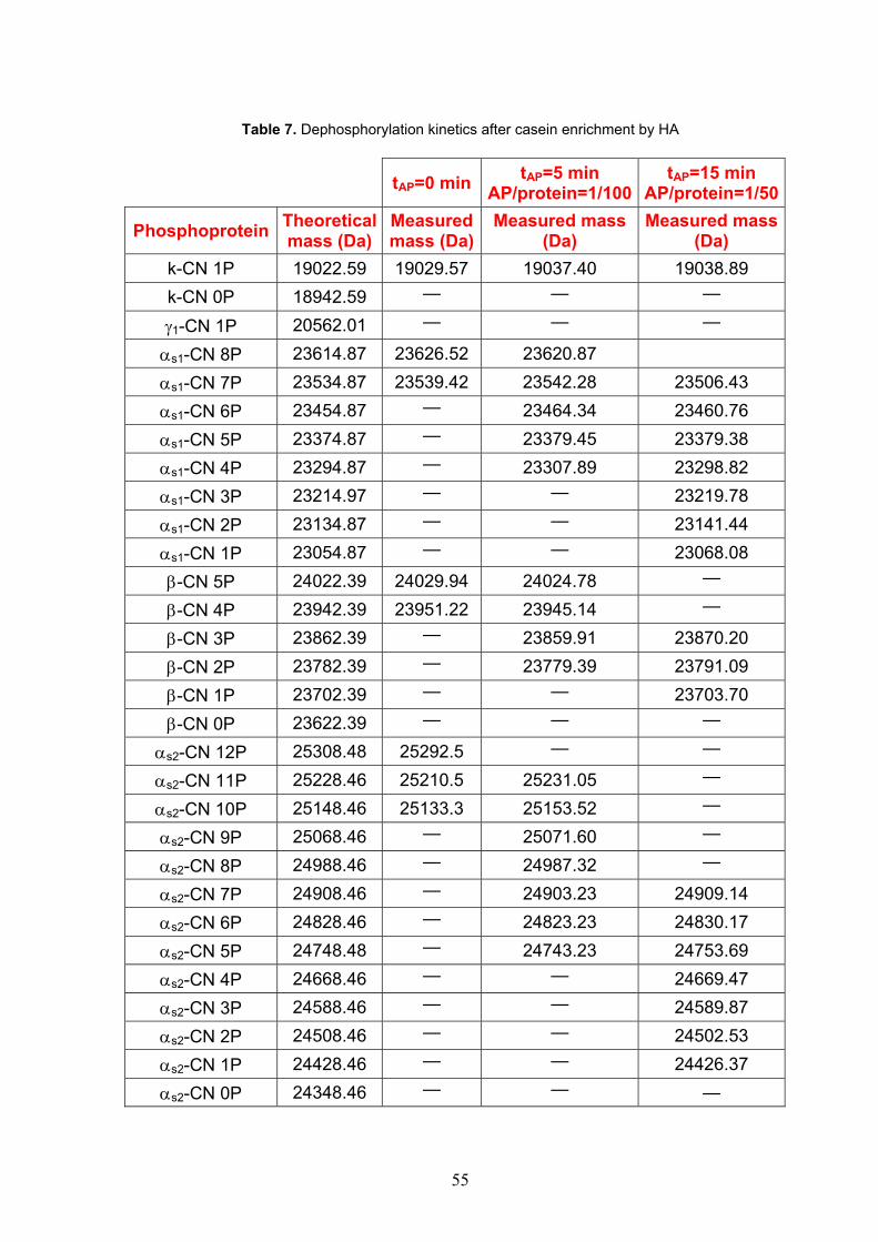
55
Table 7. Dephosphorylation kinetics after casein enrichment by HA
tAP=0 min tAP=5 min AP/protein=1/100
tAP=15 min AP/protein=1/50
Phosphoprotein Theoretical mass (Da)
Measured mass (Da)
Measured mass (Da)
Measured mass (Da)
k-CN 1P 19022.59 19029.57 19037.40 19038.89 k-CN 0P 18942.59 — — —
γ1-CN 1P 20562.01 — — —
αs1-CN 8P 23614.87 23626.52 23620.87 αs1-CN 7P 23534.87 23539.42 23542.28 23506.43 αs1-CN 6P 23454.87 — 23464.34 23460.76 αs1-CN 5P 23374.87 — 23379.45 23379.38 αs1-CN 4P 23294.87 — 23307.89 23298.82 αs1-CN 3P 23214.97 — — 23219.78 αs1-CN 2P 23134.87 — — 23141.44 αs1-CN 1P 23054.87 — — 23068.08 β-CN 5P 24022.39 24029.94 24024.78 —
β-CN 4P 23942.39 23951.22 23945.14 —
β-CN 3P 23862.39 — 23859.91 23870.20 β-CN 2P 23782.39 — 23779.39 23791.09 β-CN 1P 23702.39 — — 23703.70 β-CN 0P 23622.39 — — —
αs2-CN 12P 25308.48 25292.5 — —
αs2-CN 11P 25228.46 25210.5 25231.05 —
αs2-CN 10P 25148.46 25133.3 25153.52 —
αs2-CN 9P 25068.46 — 25071.60 —
αs2-CN 8P 24988.46 — 24987.32 —
αs2-CN 7P 24908.46 — 24903.23 24909.14 αs2-CN 6P 24828.46 — 24823.23 24830.17 αs2-CN 5P 24748.48 — 24743.23 24753.69 αs2-CN 4P 24668.46 — — 24669.47 αs2-CN 3P 24588.46 — — 24589.87 αs2-CN 2P 24508.46 — — 24502.53 αs2-CN 1P 24428.46 — — 24426.37 αs2-CN 0P 24348.46 — — —

56
The partially dephosphorylated casein fractions bound to the HA were hydrolyzed, in situ, under conditions indicated in Method section. By this means, peptides containing the Ser(P)-Ser(P)-Ser(P)-Glu-Glu cluster sequence were evaluated.
Figure 27. MALDI spectra of tryptic CPP hydrolyzed in situ from partially dephosphorylated casein bound to HA. The dephosphorylated reaction was conducted at the enzyme/substrate ratio of 1/100 for 5min (a) and with enzyme/substrate ratio of 1/50 for 15 min (b). Comparing the MALDI spectra (Fig. 27, panel a and b), the following considerations can be made: 1) totally phosphorylated peptides were still present in
a
b

57
the mixtures after 5 and 15min of the dephosphorylation reaction but the intensity ratio between β-CN (f1-25)4P (3122.3 Da) and β-CN (f1-25) 3P (3042.3 Da) and between αs1-CN (f59-79)4P (2640.9 Da) and αs1-CN (f59-79)3P (2560.9 Da) tended to decrease; 2) after 5min dephosphorylation reaction, αs2-CN (f46-70) 3P (2927.8 Da) was already detectable 3) after 15min dephosphorylation reaction the αs1-CN (f59-79)3P (2560.1 Da), αs2-CN (f2-21)3P (2459.4 Da), αs2-CN (f46-70)2P �(2848.7)� , β-CN (f1-25)2P (2963.0 Da) and β-CN (f1-25)1P (2883.0 Da) phosphopeptides were recorded. The partially phosphorylated peptides issued for different time of dephosphorylation (t=0, t=5 min and t=15 min) are compared in Table 8. Table 8. Dephosphorylation kinetic of in situ hydrolyzed and enriched phosphopeptides. The peptides of new formation at tAP=5 min are marked in green and The peptides of new formation at tAP=15 min are marked in blue.
tAP=0 min tAP=5 min
ratio=1/100 tAP=15 min ratio=1/50
Monophospho peptide
Theoretical mass (Da)
Measured mass (Da)
Measured mass (Da)
Measured mass (Da)
αs1(f106-119)1P 1659.8 1661.6 1660.9 1660.2
αs1(f104-119)1P 1952.0 1952.3 1952.8 1951.6
β(f33-48)1P 2060.8 2062.8 2061.7 2061,3
αs1(f103-119)1P 2080.4 2079.4 2081.6 2078.3
β(1-25)1P 2883.0 — — 2883.9
Diphosphopeptide Theoretical mass (Da)
Measured mass (Da)
Measured mass (Da)
Measured mass (Da)
αs2(f126-136)2P 1410.5 1411.0 1411.9 1411.4
αs2(f126-137)2P 1538.6 1540.3 1540.2 1539.7
αs1(f43-58)2P 1926.1 1928.3 1927.7 1927.1
αs1(f43-58)1P 1846.7 1848.2 1847.8 1847.1
αs2(f2-21)2P 2459.4 — — 2459.6
αs1(f37-58)2P 2598.1 2598.1 2600.5 2595.8
αs2(f46-70)2P 2848.7 — — 2849.8
αs1(f35-58)2P 2855.2 2855.3 2855.6 2852.9
β(f1-25)2P 2963.0 — — 2963.7
Triphosphopeptide Theoretical mass (Da)
Measured mass (Da)
Measured mass (Da)
Measured mass (Da)
αs2(f2-21)3P 2539.4 2538.6 2539.4 2540.0
αs1(f59-79)3P 2560.1 — — 2559.6
αs1(f37-58)3P 2678.1 2677.8 2677.5 2676.7

58
β(f2-25)3P 2886.2 2885.9 2885.1 2885.4
αs2(f46-70)3P 2928.0 — 2927.9 2927.7
αs1(f35-58)3P 2935.2 2935,0 2933.9 2932.6
β(f1-25)3P 3042.3 3043.8 3044.0 3043.2
Tetraphosphopeptide Theoretical mass (Da)
Measured mass (Da)
Measured mass (Da)
Measured mass (Da)
αs2(f2-21)4P 2618.9 2619.2 2619.9 —
αs1(f59-79)4P pyroQ 2623.9 — 2621.0 2621.4
αs1(f59-79)4P 2640.9 2640.7 2640.5 2639.4
β(f2-25)4P 2966.2 2965.9 2965.0
αs2(f46-70)4P 3007.9 3007.2 3006.8 3005.8
β(f1-25)4P 3122.3 3122.1 3122.1 3121.0
Pentaphosphopeptide Theoretical mass (Da)
Measured mass (Da)
Measured mass (Da)
Measured mass (Da)
αs1(f59-79)5P pyroQ 2703.9 2703.7 2703.3 2701.9
αs1(f59-79)5P 2720.9 2720.7 2720.4 2719.0
αs2(f46-70)5P 3088.0 3086.7 3086.8 3085.0
17.2. Analysis of proteins and CPP occurring in mastitic milk
The enrichment of mixtures dephosphorylated in vitro has confirmed the ability of the resin to bind phosphproteins and CPP with different degrees of phosphorylation. At this point, to evaluate the milk quality, our method for the phosphoprotein/phosphopeptide enrichment was tested on mastitic casein. In order to examine whether the phosphorylated proteins were enriched by HA, MALDI analysis was carried out on the casein sample before and after resin treatment (Figure 28)

59
Figure 28. MALDI spectra of casein sample before (a) and after (b) resin treatment in the mass range of 10-13 kDa. The partial MALDI-TOF spectrum of the original casein sample (Figure 28, panel a) was compared to the same sample after the enrichment cycle by HA (Figure 28, panel b). Original casein sample signals could be assigned to γ2-, γ3-CN, and β-CN (f98-207). These non-phosphorylated plasmin-mediated peptides of β-CN were washed out when native casein sample was treated with HA. HA-treated samples did not contain these peptides consistently with the function of HAP in capturing phosphopeptides. In contrast, HA allowed phosphoproteins/phosphopeptides be enriched from high SCC casein as clearly shown in Figure 28, panel b. Here, in the partial view of the MALDI spectra, the m/z 10-13 kDa range contained a series of phosphopeptides with various degree of phosphorylation. In particular, together with the intact β-CN (not shown), the derived peptides β-CN (f1-97) (6P÷2P) and β-CN (f1-105) (6P÷2P) occurred. The signals of the peptide β-CN (f1-107) formed simultaneously with the β-CN (f1-105) counterpart were slightly detectable as β-CN (f1-107) (6P÷4P). The β-CN (f29-105/7)1P fragments considered by Le Roux Y. et al.163 as possible indicators of the endogenous proteolysis in milk from cows with subclinical mastitis were not found perhaps because of the dephosphorylation activity. It must be remembered that unphosphorylated fragments didn’t bind to the
a
b

60
HA. In contrast, the new fragment β-CN (f1-97) was released when the plasmin activity increased in mastitic milks. Consistently, the presence of the partly dephosphorylated fragments is connected to the higher ALP activity of mastitic milk. The casein derived from high SCC milk was also analysed using LC-ESI-MS to confirm the identity of proteins and phosphopeptides of mastitic milk. Four phosphorylated β-CN (6P÷3P) were detected in high SCC milk instead of the two, i.e. β-CN 6P and 5P occurring in control milk. Four phosphorylated αs1-CN (10P÷7P) were detected in high SCC instead of the two, i.e. αs1-CN 10P and 9P of control milk. In contrast, αs1-CN (f141-148) (10P÷8P) and αs1-CN (Gln78) (10÷8P) deleted forms occurred at similar degree of phosphorylation as shown in Table 9. Moreover, we recorded that αs1-CN (f141-148) was the prominent internally deleted form consistently with that occurs in milk from healthy animal for which (110-117) form was found. This is in agreement with the previous results173. αs2-CN shows the highest number of possible phosphorylation sites compared to the other casein fractions and naturally occurs under two molecular species both plasmin-sensitive substrates, a major full-length, and an internal 9-residue domain deleted. Both proteins show the same phosphorylation degree (Table 9).

61
Table 9. Identification of casein fractions in mastitic ovine milk obtained by HA enrichment and ESI-MS analysis
Measured molecular mass
(Da)
Calculated molecular mass
(Da) Protein
αs2-Casein 25623.09± 4.7 25621.71 αs2(1-208)12P
25542.38± 1.2 25541.73 αs2(1-208)11P
25466.38± 6.2 25461.75 αs2(1-208)10P
25378.64± 3.6 25381.77 αs2(1-208)9P
25304.80± 5.0 25301.79 αs2(1-208)8P
25224.71± 3.9 25221.81 αs2(1-208)7P �
24546.7±0.8 24546.6 αs2(1-208)-(34-42)11P
24466.1±0.5 24466.6 αs2(1-208)-(34-42)10P
24386.2±0.8 24386.6 αs2(1-208)-(34-42)9P
24305.5±0.3 24306.7 αs2(1-208)-(34-42)8P
24225.9.7±0.1 24226.7 αs2(1-208)-(34-42)7P
αs1-Casein
23564.2± 3.0 23561.6 αs1(1-199)10P
23481.3± 0.8 23481.7 αs1(1-199)9P
23400.7± 1.8 23401.7 αs1(1-199)8P
23323.2±2.8 23321.7 αs1(1-199)7P
23435.0±0.8 23433.5 αs1(1-199)-(Gln78) 10P
23354.7±0.8 23353.5 αs1(1-199) -(Gln78) 9P
23275.4±0.4 23273.6 αs1(1-199)-(Gln78) 8P �
22546.70±3.5 22549.5 αs1(1-199)-(141-148)10P 22466.53±3.1 22469.5 αs1(1-199)-(141-148)9P 22386.15±3.5 22389.5 αs1(1-199)-(141-148)8P 22306.20±3.5 22309.5 αs1(1-199)-(140-148)7P
β-Casein 23830.98±1.1 23831.14 β(1-207)6P 23747.04±4.7 23751.16 β(1-207)5P 23667.23±3.7 23671.18 β(1-207)4P 23587.32±4.2 23591.19 β(1-207)3P
The ESI-MS analysis allowed the identification of a wide variety of phosphorylated peptides of different size and wide phosphorylation degree derived from the intense enzymatic degradation associated with high SCC (Table 10).

62
Table 10. Identification of peptides in mastitic milk obtained by HA enrichment and LC-ESI-MS analysis. N-terminal and C-terminal cleavage sites are highlighted. The degree as well as maximum degree of phosphorylation are compared.
Fragment N-terminal
cleavage site C-terminal
cleavage siteDegrees of
Phosphorylation Maximum degree of
phosphorylation β-Casein (1-207)
β(f1-29) — Lys29-Ile30 5P, 4P, 3P, 2P 5P
β(f29-60) Lys28-Lys29 Tyr60-Pro61 1P 1P
β(f30-60) Lys29-Ile30 Tyr60-Pro61 1P 1P
β(f1-97) — Lys97-Val98 6P, 5P, 4P, 3P, 2P 6P
β(f1-105) — Lys105-His106 6P, 5P, 4P, 3P, 2P 6P
β(f1-107) — Lys107-Glu108 6P, 5P, 4P 6P
αs2-Casein (1-208) αs2(f1-25) — Lys25-Asn26 4P, 3P 4P
αs2(f36-69) Lys35-Leu36 Glu69-Val70 4P, 3P, 2P 4P
αs2(f36-71) Lys35-Leu36 Lys71-Ile72 3P, 2P 4P
αs2(f36-77) Lys35-Leu36 Lys77-His78 3P, 2P 4P
αs2(f1-53) — Tyr53-Ser54 4P, 3P, 2P, 1P 4P
αs2(f54-115) Tyr53-Ser54 Arg115-Asn116 4P,3P 4P
αs2(f54-204) Tyr53-Ser54 Tyr204-Val205 7P, 6P, 5P, 4P 7P
αs2(f54-208) Tyr53-Ser54 — 7P, 6P, 5P, 4P 7P
αs2(f116-204) Arg115-Asn116 Tyr204-Val205 3P, 2P, 1P 3P
αs2(f116-208) Arg115-Asn116 — 3P, 2P, 1P 3P
αs2(f115-151) Lys114-Arg115 Lys151-Thr152 3P, 2P 3P
αs1-Casein (1-199) αs1(f4-58) Lys3-His4 Lys58-Gln59 3P, 2P, 1P 3P
αs1(f1-60) — Met60-Lys61 3P, 2P, 1P 3P
αs1(f2-60) Arg1-Pro2 Met60-Lys61 3P, 2P, 1P 3P
αs1(f61-174) Met60-Lys61 Thr174-Asp175 6P, 5P, 4P 7P
αs1(f62-174) Lys61-Ala62 Thr174-Asp176 6P, 5P, 4P 7P
αs1(f62-199) Lys61-Ala62 — 7P, 6P 7P
αs1(f62-90) Lys61-Ala62 Arg90-Tyr91 4P, 3P, 2P 6P
αs1(f43-102) Lys42-Asp43 Lys102-Lys103 5P, 4P, 3P, 2P 8P
αs1(f43-103) Lys42-Asp43 Lys103-Tyr104 5P, 4P, 3P, 2P 8P

63
The specific cleavage of Lys-X and Arg-X at C-terminal position was clearly due to the increased activity of plasmin. In complement with other bonds, Tyr-X cleavage was caused by cathepsin G. All peptides showed a wide phosphorylation range, suggesting that proteolytic cleveage occurred concurrently with the intense dephosphorylation enzyme correlated with increased phosphatase activity. Additionally, the tryptic in situ hydrolysis of mastitic casein bound to HA provided additional information about the milk quality to be used as nutraceutical. The identification of these CPP could represent easier indicators about the nature of milk, mastitic or not-mastitic. As dephosphorylation produces a characteristic mass decrease of 80 Da or multiple of 80 Da, CPP with this mass variation confirmed the phosphatase activity. This procedure was found very effective and actually represents one of the most powerful tool for the identification of parent phosphorylated proteins. The partial view of the MALDI spectra in two mass ranges taken by direct analysis of the crystallized matrix containing HA and phosphoric acid is shown in Figure 29 (panel a and b).

64
Figure 29. MALDI spectrum of tryptic CPP HA-enriched from mastitic milk in different mass range: 1-2.9 kDa (a) and 2.8-3.7 kDa (b). Adducts of CPP with K+ are encircled in green.
All the tryptic phosphopeptides identified in the HA-enriched fraction are listed in Table 11.
a
b

65
Table 11. Tryptic CPP enriched from mastitic ovine milk
User mass
(Da)
DB mass
(Da) Peptide Position Modifications
β-Casein
3542.83 3544.27 REQEELNVVGETVESLSSSE ESITHINK/(K) β-CN (f1-28)
PHOS PHOS PHOS PHOS
PHOS
3462.83 3465.28 REQEELNVVGETVESLSSSE ESITHINK/(K) β-CN (f1-28) PHOS PHOS
PHOS PHOS
3385.43 3385.30 REQEELNVVGETVESLSSSE ESITHINK/(K) β-CN (f1-28) PHOS PHOS
PHOS
3306.42 3304.33 REQEELNVVGETVESLSSSE ESITHINK/(K) β-CN (f1-28) PHOS PHOS
3226.04 3225.34 REQEELNVVGETVESLSSSE ESITHINK/(K) β-CN (f1-28) PHOS
2062.01 2062.99 (K)/FQSEEQQQTEDELQDK/(I) β-CN (f33-48) PHOS
αs1-Casein
2905.49 2905.78 (K)/ENINELSKDIGSESIEDQAM EDAK/(Q) αs1-CN (f35-58) PHOS PHOS
PHOS
2825.08 2826.79 (K)/ENINELSKDIGSESIEDQAM EDAK/(Q) αs1-CN (f35-58) PHOS PHOS
1898.54 1898.78 (K)/DIGSESIEDQAMEDAK/(Q) αs1-CN (f43-58) PHOS PHOS
1818.40 1798.92 (K)/DIGSESIEDQAMEDAK/(Q) αs1-CN (f43-58) PHOS
2158.69 2155.97 K)/DIGSESIEDQAMEDAKQM/(K) αs1-CN (f43-60)2P PHOS PHOS
Aspecific cleavage
1239.82 1238.50 (K)/ SAEQLHSMK /(E) αs1-CN (f115-124) PHOS
2287.34 2287.75 (K)/AGSSSSSEEIVPNSAEQK/(Y) αs1 CN (f62-79) PHOS PHOS PHOS PHOS PHOS PHOS
2208.60 2207.77 (K)/AGSSSSSEEIVPNSAEQK/(Y) αs1-CN (f62-79) PHOS PHOS PHOS PHOS
PHOS
2127.85 2127.79 (K)/AGSSSSSEEIVPNSAEQK/(Y) αs1-CN (f62-79) PHOS PHOS PHOS PHOS
αs2-Casein
3187.93 3192.10 (K)/HKMEHVSSSEEPINISQEIY KQEK/(N) α s2-CN (f2-25) PHOS PHOS
PHOS PHOS
3105.60 3111.13 (K)/HKMEHVSSSEEPINISQEIY KQEK/(N) α s2-CN (f2-25) PHOS PHOS
PHOS
3031.89 3032.14 (K)/HKMEHVSSSEEPINISQEIY KQEK/(N) α s2-CN (f2-25) PHOS PHOS

66
2951.98 2952.16 (K)/HKMEHVSSSEEPINISQEIY KQEK/(N) αs2-CN (f2-25) PHOS
2934.22 2934.85 KHKMEHVSSSEEPINISQEI YK/(Q) αs2-CN (f1-22) PHOS PHOS
PHOS PHOS
2854.40 2854.87 KHKMEHVSSSEEPINISQEI YK/(Q) αs2-CN (f1-22) PHOS PHOS
PHOS
2775.72 2774.89 KHKMEHVSSSEEPINISQEI YK/(Q) α s2-CN (f1-22) PHOS PHOS
2693.81 2694.91 KHKMEHVSSSEEPINISQEI YK/(Q) α s2-CN (f1-22) PHOS
2540.45 2541.36 (K)/MEHVSSSEEPINISQEIYK/(Q) αs2-CN (f4-22) PHOS PHOS PHOS PHOS
2461.48 2461.38 (K)/MEHVSSSEEPINISQEIYK/(Q) α s2-CN (f4-22) PHOS PHOS PHOS
1540.62 1540.41 (R)/EQLSTSEENSKK/(T) αs2-CN (f126-137) PHOS PHOS
1412.48 1412.24 (R)/EQLSTSEENSK/(K) αs2-CN (f126-136) PHOS PHOS
αs2-Casein aspecific cleavage of tryptic CPP
2806.00 2806.68 KHKMEHVSSSEEPINISQEI Y(K) αs2-CN (f1-21)
C-termin. Tyr21-Lys22
PHOS PHOS PHOS PHOS
2677.00 2678.50 (K)HKMEHVSSSEEPINISQEIY(K) αs2-CN (f2-21)
C-termin. Tyr21-Lys22
PHOS PHOS PHOS PHOS
2412.00 2413.19 (K)MEHVSSSEEPINISQEIY(K) αs2-CN (f4-21)
C-termin. Tyr21-Lys22
PHOS PHOS PHOS PHOS
2254.42 2253.97 (Y)SIRSSSEESAEVAPEEVK/(I) αs2-CN (f54-71) N-termin. Tyr53-
Ser54
PHOS PHOS PHOS PHOS
2174.12 2174.99 (Y)SIRSSSEESAEVAPEEVK/(I) αs2-CN (f54-71) N-termin. Tyr53-
Ser54
PHOS PHOS PHOS
It is possible to note the absence of non-phosphorylated peptides confirming the specificity of the HA-based procedure while the presence of partially dephosphorylated forms validated casein from high SCC. From native β-casein, five partly dephosphorylated CPP species were identified, corresponding to loss of 1, 2, 3, 4 and 5 phosphate groups. The presence of β-CN (f1-28) (5P÷1P) is an additional indicator to the previous finding. A lower phosphorylation degree (6P÷3P) was observed before trypric hydrolysis (Table 9). This means the partial dephosphorylation of β-casein in the presence of anomalous SSC. From the native αs1-casein, tryptic hydrolysis produced αs1-CN (f62-79)6P (6P÷4P) and CPP as αs1-CN (f43-60)2P due to aspecific cleavage. From αs2-casein, αs2-CN (f2-25)4P (4P÷2P) and aspecific αs2-CN (f54-71)4P (4P÷3P) were released. Three CPP having different N-terminal extension, i.e. 1-22, 4-22 and 2-25, raise from αs2CN (Table 11). All CPP containing the cluster Ser(P)-Ser(P)-Ser(P)-Glu-Glu, essential to bind minerals, showed a wide degree of phosphorylation due to the loss of phosphate groups.

67
In conclusion, high plasmin (or other proteases) activity associated with high cell count, and especially alkaline phosphatase activity may represent mastitis indicator. The enrichment by HA of phosphoproteins and CPP is fundamental for evaluating milk quality.

68
18. Use of CPP as process markers CPP can also represent process markers. Proteolysis and casein and phosphopeptide dephosphorylation is of utmost interest in the cheese ripening. We considered Grana Padano (GP) cheese where the biochemical changes which occur during ripening are caused by the synergistic action of a variety of enzymes, such as chymosin, indigenous milk enzyme proteinase, starter bacteria enzymes, and secondary microorganisms enzymes174. Then, it has been suggested that acid phosphatases from both milk and lactic acid bacteria contribute to dephosphorylation of phosphopeptides in cheese, which are produced from casein by enzymatic activity during cheese ripening175. In cheese made from pasteurized milk, indigenous acid phosphatase or bacterial phosphatases are probably responsible for dephosphorylation of caseins, but in raw milk cheese, e.g. Parmigiano Reggiano or Grana Padano, ALP may play an important role176. Hence, in a cheese made from raw milk, the enzymatic activity is more intense than that occurring in a cheese made from pasteurized milk. Pasteurization, in fact, inactivates several enzymes in milk, including lipase and alkaline phosphatase. Lack of alkaline phosphatase activity indicates that the milk has been properly pasteurized. As Grana Padano is a D.O.P. cheese and its Production Disciplinary states that it must be produced with raw milk, it is important to have a technique for controlling the use of raw milk. It has been demonstrated that a centripetal temperature gradient and a consequent gradient of alkaline phosphatase inactivation between the outer part and the core of GP cheese takes place during moulding. The traditional cheesemaking technology also includes milk clotting at 32° C, curd cooking up to 55° C, and cheese moulding at room temperature for 48 h. It has been able to note that the temperature decreases rapidly to 35-40° C at the surface, but because of the slow heat-transfer within the cheese, the core remains at 52-56 °C for 8-10 h. Such condition promotes the formation of a centripetal gradient of ALP inactivation which remains unmodified in moulded forms at room temperature throughout ripening177. The outside of the Grana contain levels of residual phosphatase about 10-100 times higher than the core of cheese. Moreover, the significant heat load on the inner zones of the cheese together with the low pH value may reduce the activity of other indigenous and heat-sensitive enzymes, like acid protease178. Also an increased proteolytic activity was observed mainly in the outer zone of cheese. More free amino acids (FAA) were detected in the outer parts of GP cheese179 just caused by the higher enzyme activities than in the cheese core. The peptide degradation involves firstly dephosphorylation of the SerP N-terminal and then release of Ser by aminopeptidases177. This mechanism was confirmed by the extremely low levels of SerP found in the free amino acid (FAA) containing fraction of GP whereas free Ser accumulated as a consequence of the simultaneous presence of both active phosphatase and aminopeptidase. As well known, constitutive alkaline phosphatase is inactivated by milk pasteurization which can affect splitting of SerP during cheese ripening: If SerP wasn’t dephosphorylated by residual phosphatase, free Ser cannot be released by the bacterial aminopeptidase activity. Hence, milk pasteurization originates an equal distribution of FAA between the outer and the core part of the ripened cheese form. In fact, no free Ser gradient depending on the unequal activity of ALP within the cheese was observed but FAA content was depressed in comparison to control raw-milk cheese177 together with longer size CPP.

69
In conclusion, the detection of a different peptide profile associated to the thermal gradient can be diagnostic for the use of raw milk as stated by the Decree of the Regulatory Production.
19. Experimental procedure 4 Materials 0.1N Hydrochloric acid solution was Carlo Erba. 3 samples of Grana Padano cheese, at 15 months ripening, were supplied from GranBiraghi factory obtained according to the traditional cheesemaking technology. For the cheese samples, a cylindric portion with 5cm diameter from the round side near the outer layer and from the core of the cheese was recovered. Each portion was grated to obtain an uniform sample. The other materials have been already reported. Preparation of pH 4.6-soluble fraction of cheese Finely grated cheese (10 mg) was suspended in 45 mL of 0.1 N HCl. The suspension was homogenized for 1 min by an Ultra Turrax T50 and then centrifuged at 4,000 rpm for 30 min at 4° C. The resulting supernatant solution was freeze dried (Lyolab A, LSL Secfroid, Aclens, Switzerland). HA-based CPP enrichment of the pH 4.6 soluble fraction Lyophilized soluble fraction (10 mg) was dissolved in 80 μl buffer at pH 8.0 containing TrisHCl 50 mM, KCl 0.2 M, urea 4.5 M, DTT 10 mM and loaded on HA (10mg), previously washed with loading buffer. The next steps of phosphorylated protein enrichment and tryptic hydrolysis in situ were the same as those already reported above. LC/MS Analysis The LC-MS analysis was carried out under the conditions above reported. The only difference was that, instead of TFA, formic acid (FA) was used in the elution buffer to reduce signal suppression.
20. Results and discussion After enrichment by HA of the pH 4.6-soluble fraction of the GP inner and outer part, a lot of phospopeptides bound to HA, while nonphosphorylated compounds were washed out with buffers. From the analysis of recovered CPP, it is possible to note that, during cheese ripening, several CPP derived by a intense enzymatic hydrolysis were present in the core and the inner part of the ripened cheese (Tables 12 and 13). All peptides had in common the N-terminal or C-terminal residues. Plasmin appeared to be the main responsible enzyme for casein degradation during hard cheese ripening. β-Casein was extensively degraded by this enzyme with the concomitant formation of plasmin-derived peptides as β-CN (f1-28), β-CN (f1-29), β-CN (f1-32), found only in the inner part of GP (Table 12).

70
Table 12. Identification of CPP by LC-ESI-MS from the inner part of a 15-month-old cheese. Measured mass (Da)
(MH+)
Measured mass (Da)
(MH2+)
Measured mass (Da)
(MH3+)
Measured mass (Da)
(MH4+) Amino acid sequence CPP
identification
β-casein 1348.4 674.7
LpSpSpSEESITR β-CN (f16-25)3P 1435.4 718.2 479.1 pSLpSpSpSEESITR β-CN (f15-25)3P 1591.1 796.1 531.0 pSpSpSEESITRINK β-CN (f17-28)4P 1644.6 822.8 548.9 EpSLpSpSpSEESITR β-CN(f14-25)4P 1704.1 852.6 568.7 LpSpSpSEESITRINK β-CN (f16-28)3P 1743.4 872.2 581.8 VEpSLpSpSpSEESITR β-CN (f13-25)4P 1790.6 895.8 597.5 pSLpSpSpSEESITRINK β-CNf(f15-28)3P 1857.1 929.1 619.7 IVEpSLpSpSpSEESITR β-CN(f12-25)4P 1870.0 935.5 624.0 pSLpSpSpSEESITRINK β-CN (f15-28)4P 1871.0 936.0 624.3 EpSLpSpSpSEESITRIN β-CN (f14-27)4P 1999.0 1000.0 667.0 EpSLpSpSpSEESITRINK β-CN (f14-28)4P 2098.0 1049.5 700.0 VEpSLpSpSpSEESITRINK β-CN(f13-28)4P 2211.5 1106.3 737.8 IVEpSLpSpSpSEESITRINK β-CN (f12-28)4P 2341.0 1171.0 781.0 EIVEpSLpSpSpSEESITRINK β-CN (f11-28)4P 2594.2 1297.6 865.4 VPGEIVEpSLpSpSpSEESITRINK β-CN (f8-28)4P 2708.8 1354.9 903.6 678.0 NVPGEIVEpSLpSpSpSEESITRINK β-CN (f7-28)4P
2967.0 1484.0 989.7 742.5 ELEEL NVPGEIVEpSLpSpSpSEESITR
β-CN (f2-25)4P
3079.0 1540.0 1027.0 770.5 EEL NVPGEIVEpSLpSpSpSEESITRINK
β-CN (f4-28)4P
3123.5 1562.3 1041.8 781.6 RELEEL NVPGEIVEpSLpSpSpSE ESITR
β-CN (f1-25)4P
3193.0
1065.0 799.0 LEEL NVPGEIVEpSLpSpSpSEESITR INK
β-CN (f3-28)4P
3322.0 1108.0 831.3 ELEEL NVPGEIVEpSLpSpSpSEESITR INK
β-CN (f2-28)4P
3450.0 1150.7 863.3 ELEEL NVPGEIVEpSLpSpSpSEESITRINKK
β-CN (f2-29)4P
3478.6 1160.2 870.4 RELEEL NVPGEIVEpSLpSpSpSEESITRINK
β-CN (f1-28)4P
3607.0 1203.0 902.5 RELEEL NVPGEIVEpSLpSpSpSEESITRINKK
β-CN (f1-29)4P
3978.0 1326.7 995.3 RELEEL NVPGEIVEpSLpSpSpSEESITRINKK IEK
β-CN (f1-32)4P
αs1-casein 1314.2 657.5
IpSpSpSEEIVPN αs1-CN (f65-74)3P 1401.4 701.2 467.8 pSIpSpSpSEEIVPN αs1-CN (f64-74)3P 1481.2 741.1 494.4 pSIpSpSpSEEIVPN αs1-CN (f64-74)4P 1610.6 805.8 537.5 EpSIpSpSpSEEIVPN αs1-CN (f63-74)4P 1681.4 841.2 561.1 AEpSIpSpSpSEEIVPN αs1-CN (f62-74)4P 1810.6 905.8 604.2 EAEpSIpSpSpSEEIVPN αs1-CN(f61-74)4P 1965.3 983.2 655.8 IpSpSpSEEIVPNpSVEQK αs1-CN (f65-79)4P 2132.0 1066.5 711.3 pSIpSpSpSEEIVPNpSVEQK αs1-CN (f64-79)5P

71
2261.0 1131.0 754.3 EpSIpSpSpSEEIVPNpSVEQK αs1-CN(f63-79)5P 2333.5 1167.3 778.5 AEpSIpSpSpSEEIVPNpSVEQK αs1-CN (f62-79)5P 2462.0 1231.5 821.3 EAEpSIpSpSpSEEIVPNpSVEQK αs1-CN(f61-79)5P
αs2-casein 1515.4 758.2 505.8
pSpSpSEESIIpSQE αs2-CN (f8-18)4P
1614.4 807.7 538.8 VpSpSpSEESIIpSQE αs2-CN (f7-18)4P 2006.6 1003.8 669.5 VpSpSpSEESIIpSQETYK αs2-CNf(f7-21)4P
In the outer parts of cheese, the higher intense enzymatic hydrolysis with respect to the core caused the degradation of these precursors and their disappearance (Table 13). Figure 13. Identification of CPP by LC-ESI-MS from the outer portion of a 15-month-old cheese sample. Measured mass (Da)
(MH+)
Measured mass (Da)
(MH2+)
Measured mass (Da)
(MH3+)
Measured mass (Da)
(MH4+) Amino acid sequence CPP
identification
β-casein 1192.2 596.8
LpSpSpSEESIT β-CN (f16-24)3P
1348.4 674.8 450.2 LpSpSpSEESITR β-CN (f16-25)3P
1359.2 680.2 SLpSpSpSEESIT β-CN (f15-24)4P
1435.4 718.0 479.0 SLpSpSpSEESITR β-CN (f15-25)3P
1703.8 852.4 568.6 LpSpSpSEESITRINK β-CN (f16-28)3P
1742.0 872.2 582.0 VEpSLpSpSpSEESITR β-CN (f13-25)4P
1870.6 935.8 624.0 pSLpSpSpSEESITRINK β-CN (f15-28)4P
1871.0 936.0 624.4 EpSLpSpSpSEESITRIN β-CN (f14-27)4P
1999.0 1000.4 667.4 EpSLpSpSpSEESITRINK β-CN (f14-28)4P
2211.8 1106.6 738.0 553.8 IVEpSLpSpSpSEESITRINK β-CN (f12-28)4P
2341.6 1171.3 781.2 EIVEpSLpSpSpSEESITRINK β-CN (f11-28)4P
2708.8 903.6 677.8 NVPGEIVEpSLpSpSpSEESITRINK β-CN (f7-28)4P
αs1-casein 1314.2 657.8
IpSpSpSEEIVPN αs1-CN (f65-74)3P
1320.4 660.8 EpSIpSpSpSEEIV αs1CN (f63-72)3P
1401.4 701.2 468.0 pSIpSpSpSEEIVPN αs1-CN (f64-74)3P
1481.4 741.2 494.4 pSIpSpSpSEEIVPN αs1-CN (f64-74)4P
1610.6 805.5 537.3 EpSIpSpSpSEEIVPN αs1-CN (f63-74)4P
1681.4 841.0 561.0 AEpSIpSpSpSEEIVPN αs1-CN (f62-74)4P
1965.4 983.4 656.0 IpSpSpSEEIVPNpSVEQK αs1-CN (f65-79)4P
2132.4 1067.0 711.6 pSIpSpSpSEEIVPNpSVEQK αs1-CN (f64-79)5P
2333.0 1166.8 778.4 AEpSIpSpSpSEEIVPNpSVEQK αs1-CN (f62-79)5P
αs2-casein 1515.4 758.2 505.6
pSpSpSEESIIpSQE αs2-CN (f8-18)4P
1614.4 807.8 538.8 VpSpSpSEESIIpSQE αs2-CN (f7-18)4P
Plasmin hydrolysed αs1-casein producing the αs1-CN (f61-79) peptide further attacked at the Met60-Glu61 peptide bond, originated by action of an unknown

72
endopeptidase. The consequent formation of the three peptides such as αs1-CN(f1-60)2P, αs1-CN (f23-60)2P and αs1-CN (f61-79) 5P182 was recorded. Plasmin hydrolysed αs2-casein producing the αs2-CN (f1-21) fragment. Then, several shorter CPP were released from this precursor through the combined action of proteases and phosphatases; each peptide was devoid of one or more residues at the N- and/or C-terminus (Table 12 and 13). Following the specific cleveage of plasmin-derived CPP, it is possible to deduce the activity of general and specific aminopeptidases (which release the N-terminal amino acid from oligopeptides), phosphatase and carboxypeptidases A and B (which release the C-terminal amino acid from oligopeptides). In particular, the synergistic activity of phosphatase and aminopeptidases allows to obtain shorter CPP with cleavage in the cluster Ser(P)-Ser(P)-Ser(P)-Glu-Glu, otherwise resistant to any enzymatic activity180. As already mentioned, the presence of CPP such as αs1-CN (f65-74)3P, αs1-CN (f65-79)4P and β-CN (f16-25)3P is possibly due to the formation of dephosphorylated intermediate peptides such as αs1-CN (f64-74)3P and β-CN (f15-25)3P. This synergistic enzyme activity was also present in the inner part of GP. Here, the residual activity of phosphatases was very low as demonstrated by the higher number of CCPs in the GP core. This was sign of a lower enzyme activity than in the outer part of cheese form. All larger CPP such as β-CN (f2-25)4P, β-CN (f4-28)4P, β-CN (f1-25)4P, β-CN (f3-28)4P, β-CN (f2-28)4P, β-CN (f2-29)4P, β-CN (f1-28)4P, β-CN (f1-29)4P, β-CN (f1-32)4P, αs1-CN(f61-79)5P, αs1-CN(f63-79)5P, αs2-CN(f7-21)4P were absent in the outer parts of the cheese form while they occurred in the inner one. In GP made using pasteurized milk, the different peptide profile was not observed owing to the phosphatase inactivation and lower proteolytic activity. Peptides as αs1-CN (f65-74)3P, αs1-CN (f65-79)4P and β-CN (f16-25)3P were absent because CPP were not dephosphorylated by alkaline phosphatase (data not shown). Since indigenous ALP is fully inactivated in pasteurized milk, no gradient in free Ser depending on the unequal activity of the enzyme within the cheese form was observed and a diminutive amount of FAA was found in the core cheese form. In conclusion, the analysis of CPP of the inner and outer parts of the GP cheese form has provided some comprehensive information on the nature of the milk used in processing.

73
21. Conclusions
In conclusion, during the PhD work, a technique for selective enrichment of phosphorylated components has been developed starting from complex mixtures of proteins and peptides. The high selectivity technique of CPP enrichment based on hydroxyapatite (HA) chromatography was then transferred in semi-industrial scale for the formulation of a functional milk containing HA-CPP microgranules. HAS was synthesized by wet-chemical precipitation reactions from Ca(OH)2 and H3PO4 to reduce production costs and 3 different protein mixtures as casein, milk proteins and raw milk were used to recover tryptic CPP. It was thus possible to use several initial raw materials for preparing the new ingredient, according to the specific industrial needs. Once CPP were bound to the resin, the complex HAS-CPP was finely ground and added to milk. The sensory characteristics of this new formulation were compatible for milk fortification. Hence, in this PhD work, a new drink enriched with functional components of high biotechnological interest as phosphopeptides has been created. CPP preparation was useful for the treatment of dental caries and vehiculing trace elements (copper, iron, selenium, etc). The quality of the CPP preparation was also important. The evaluation of milk quality was conducted through the study of phosphoproteome of a mastitic milk sample. In this kind of milk, the developed analytical technique revealed new phosphorylated components in a wide range of dephosphorylation due to the increased phosphatase(s) activity associated to the high somatic cells and a number of plasmin-derived casein peptides. Dephosphorylation of proteins and CPP correlated well to high SCC having a negative impact on the functionality of the bioactive peptides. Furthermore, the study of CPP dephosphorylation in the inner and outer parts of a 15-month-old cheese has allowed to find CPP as process markers. In a milk made by raw milk, a gradient of alkaline phosphatase and protease inactivation was associated to a thermal gradient between the core and outer parts of GP, while no gradient of inactivation was evident for a cheese made by pasteurized milk. In this direction, the analytical method is suitable to provide detailed information on the evaluation of the genuineness of liquid milk and cheese. The development of such a specific strategy opens up the possibility for further application in phosphoproteomics and structure-function studies.

74
22. References 1 Hans Meisel 1997, Livestock Production Science 50, 125-138 2 Clare DA, Swaisgood HE 2000 J Dairy Sci 83, 1187-1195 3 Korhonen H, Pihlanto-Leppala A, Rantamaki P, Tupasela T 1998. Trends in Food Science and Technology 9, 307-319. 4 Hilliam M 2003. Australian Journal of Dairy Technology 58, 98-103. 5 Kitts DD, Weiler K 2003 Current Pharmaceutical Design 9, 1309-1323. 6 Latham, PW 1999. Nature Biotechnology 17, 755-758 7 Dziuba M, Darewicz M. 2007 Food Science and Technology International 13; 393-404. 8 FitzGerald RJ, Meisel H 2003. In: P.F. Fox, & P.L.H. McSweeney Eds., Advanced dairy chemistry, Vol. 1: Proteins (3rd ed.) 675-698.\ 9 Korhonen H, Pihlanto-Leppala A 2003. Current Pharmaceutical Design 9, 1297-1308. 10 Shimizu M 2004. BioFactors 21, 43-47. 11 Jollès P, Henschen A 1982. Trends Biochem. Sci. 7, 325-328. 12 Jollès P, Loucheux-Lefebvre MH, Henschen A 1978. J. Mol. Evol. 11, 271-277. 13 Fiat AM, Jollès P 1989. Mol. Cell. Biochem. 87, 5-30. 14 Jollès P, Levy-Toledano S, Fiat AM, Soria C, Gillesen D, Thomaidis A, Dunn FW, Caen J 1986. European Journal of Biochemistry 158(2), 379-382. 15 Bouhalla S, Mollè D, Leonil J 1992. Biotechnology Letters 14, 805-810. 16 Schlimme E, Meisel H 1995. Die Nahrung 39(1), 1-20. 17 Fiat AM, Migliore D, Jollès P 1993. Journal of Dairy Science 76(1), 301-310. 18 Chabance B, Jollès P, Izquierdo C, Mazoyer E, Francoual C, Drouet L 1995. British Journal of Nutrition 73(4), 583-590. 19 Ondetti MA, Rubin B, Cushman DW 1977. Science, 196, 441-444. 20 Fyhrquist F, Metsarinne K, Tikkanen I 1995 J Human Hypertens; 9 (Suppl. 5):S19-S24 21 Pihlanto-Leppälä A, Rokka T, Korhonen H 1998. International Dairy Journal, 8, 325-331. 22 Vermeirssen V, Van Camp J, Decroos K, Van Wijmelbeke L, Verstraete W 2003. Journal of Dairy Science 86(2), 429-438. 23 Maeno M, Yamamoto N, Takano T 1996. Journal of Dairy Science 79(8), 1316-1321. 24 Matsufuji H, Matsui T, Seki E, Osajima K, Nakashima M, Osajima Y. 1994. Biosci Biotechnol. Biochem. 58(12) 2244-2245. 25 Vermeirssen V, Van Camp J 2004. Br J Nutr. 92(3), 357-366. 26 Yamamoto N, Ejiri M, Mizuno S 2003. Current Pharmaceutical Design 9, 1345-1355. 27 Webb KE, Jr 1990 J Anim Sci. 68, 3011–3022. 28 Masuda O, Nakamura Y, Takano T. 1996. J Nutr. 126, 3063-3068. 29 Nakamura Y, Yamamoto N, Kumi S, Takano T 1995. Journal of Dairy Science, 78:1253–1257. 30 Gobbetti M, Ferranti P, Smacchi E, Goffredi F, Addeo F 2000. Applied and Environmental Microbiology 66, 3898-3904. 31 FitzGerald RJ, Murray BA, Walsh GJ 2004. Journal of Nutrition 134, 980S-988S. 32 Meisel H 1997. Biopolymers 43, 119-128 33 Meisel H 1997. Livestock Production Science 50, 125-138. 34 Rohrbach MS, Williams EB, Jr Rolstad RA 1981. J. Biol. Chem. 256, 225-230. 35 Cheung HS, Wang FL, Ondetti MA, Sabo EF, Cushman DW 1980. J. Biol. Chem. 255, 401-407. 36 Gómez-Ruiz JA, Recio I, Belloque J 2004. J. Agric. Food Chem. 52, 6315-6319. 37 Del Mar Contreras M, Carron R, Montero MJ, Ramos M, Recio I 2009. International Dairy Journal 19(10) 566–573. 38 Brantl V, Teschemacher H, Hemschem A, Lottspeich F 1979. Hoppe Seylers Z. Physiol. Chem. 360, 1211–1216 39 Smacchi E, Gobbetti M 2000. Food Microbiology 17(2), 129-141. 40 Teschemacher H, Brantl V 1994. In V. Brantl, & H. Teschemacher (Eds.), β-Casomorphin and related peptides: Recent developments 3-17. Weinheim, VCM, Germany 41 Meisel H, Schlimme E 1990. Trends Food Sci. Technol. 1, 41-43. 42 Paroli E 1988. World Rev. Nutr. Diet 55, 58–97. 43 Zioudrou C, Streaty RA, Klee WA 1979. J. Biol. Chem. 254, 2446-2449. 44 Greenberg R, Groves ML, Dower HJ 1984. J. Biol. Chem. 259, 5132-5138.

75
45 Yoshikawa M, Tani F, Shiota H, Usui H, Kurahashi K, Chiba H 1994. In V. Brantl, & H. Teschemacher (Eds.), β-Casomorphins and related peptides: Recent developments 43–48. Weinheim, VCH, Germany 46 Chang, KJ, Killian A, Hazum E, Cuatrecasas P 1981. Science 212, 75-77. 47 Mierke DF, Nobner G, Shiller PW, Goodman M 1990. Int. J. Peptide Res. 35, 34-45 48 Chabance B, Marteau P, Rambaud JC, Migliore-Samour D, Boynard M, Perrotin P, Guillet R, Jollès P, Fiat AM 1998. Biochimie 80(2), 155-165 49 Meisel H 1986. FEBS Lett. 196, 223-227. 50 Svedberg J, de Haas J, Leimenstoll G, Paul F, Teschemacher H 1985. Peptides 6, 825-830. 51 Schulte-Frohlinde E, Schimd R, Brantl V, Schusdziarra V 1994. β-Casomorphins and Related Peptides: Recent Developments 155-160 52 Teschemacher H, Koch G, Brantl V 1997. Biopolymers 43(2), 99-117. 53 Sturner RA, Chang KJ 1988. Pediatric Research 23, 4-10. 54 Gill HS, Doull F, Rutherfurd KJ, Cross ML 2000. British Journal of Nutrition 84(Suppl. 1), S111–S117. 55 Jollès P, Parker F, Floch F, Migliore D, Alliel P, Zerial A, Werner GH 1981. Journal of Immunopharmacology 3(3–4), 363–369. 56 Parker F, Migliore-Samour D, Floch F, Zerial A, Werner GH, Jollès J, Casaretto M, Zahn H, Jollès P 1984. European Journal of Biochemistry 145(3), 677-682. 57 Minkiewicz P, Slangen CJ, Dziuba J, Visser S, Mioduszewska H 2000. Milchwissenschaft, 55(1), 14-17. 58 Matar C, LeBlanc JG, Martin L, Perdigón G. 2003. In E. R. Farnworth (Ed., Handbook of fermented functional foods. Functional foods and nutraceuticals series 177-201. 59 Hata I, Ueda J, Otani, H 1999. Milchwissenschaft, 54(1), 3-7. 60 Silva SV, Malcata FX 2005. International Dairy Journal 15, 1-15. 61 Lahov E, Regelson W 1996. Food and Chemical Toxicology, 34(1), 131-145. 62 Zucht HD, Raida M, Adermann K, Magert HJ, Forssman WG 1995. FEBS Lett. 372, 185-188 63 Recio I, Visser S 1999. Biochimica et Biophysica Acta, 1428(2-3), 314-326 McCann KB, Shiell BJ, Michalski WP, Lee A, Wan J, Roginski H 2006. International Dairy Journal 16, 316–323.. 65 Malkoski M, Dashper SG, O‘Brien-Simpson NM, Talbo GH, Macris M, Cross KJ 2001. Antimicrobial Agents and Chemotherapy 45, 2309-2315. 66 Matin MA, Monnai M, Otani H 2000. Animal Science Journal 71, 197-207. 67 Minervini F, Algaron F, Rizzello CG, Fox PF, Monnet V, Gobetti M 2003. Applied and Environmental Microbiology 69, 5297-5305. 68 Baranyi M, Thomas U, Pellegrini A 2003. Journal of Dairy Research 70, 189-197. 69 Epand RM, Vogel HJ 1999. Biochimica et Biophysica Acta 1462,11-28. 70 Manso MA, López-Fandino R 2004. Food Reviews International 20, 329-355 71 Bezkorovainy A. Grohlich D, Nichols JH 1979. American J. Clin.Nutri. 32(7), 1428-1432 72 Otani H, Monnai M, Kawasaki Y, Kawakami H, Tanimoto M 1995 Journal of Dairy Research, Volume 62(2), 349-357 73 Guilloteau P, Le Huërou-Luron I, Chayvialle JA, Toullec R, Legeas M, Bernard C, Roger L, Mendy F 1994. Reproduction nutrition Development 34, 612-613. 74 Yvon M, Beucher S, Guilloteau P, Le Huerou-Luron I, Corring T 1994. Reproduction, Nutrition & Development 34, 527-537. 75 Stan Stan EI, Ekimovskii AP, Aleinik SI, Zhuravlev BV 1988. Voprosy Pitaniia 1, 39-43. 76 Fosset S, Fromentin G, Gietzen DW, Dubarry M, Huneau JF, Antoine JM 2002. Peptides 23, 1773-1781. 77 Chabance B, Marteau P, Rambaud JC, Migliore-Samour D, Boynard M, Perrotin P 1998. Biochimie, 80, 155-165. 78 Ajit V 1997. FASEB J., 11, 248-255. 79 Nakajima K, Tamura N, Kobayashi-Hattori K, Yoshida T; Hara-Kudo Y; Ikedo M, Sugita-Konishi Y, Hattori M 2005. Biosci. Biotechnol. Biochem., 69(12), 2294-2301. 80 Mellander O 1950. Acta of the Society of Medicine (Uppsala), 55, 247-255. 81 Kasai T, Honda T, Kiriyama S 1992. Bioscience, Biotechnology and Biochemistry 56, 1150-1151. 82 Chabance B, Marteau P, Rambaud JC, Migliore-Samour D, Boynard M, Perrotin P, Guillet R, Jollès P, Fiat AM 1998. Biochimie, 80(2), 155–166. 83 Meisel H, Bernard H, Fairweather-Tait S, FitzGerald RJ, Hartmann R, Lane CN, McDonagh D, Teucher B, Wal JM 2003. British Journal of Nutrition 89(3), 351-359.

76
84 Meisel H, Frister H 1989. J. Dairy Sci. 56, 343-349. 85 Meisel H. 1998. International Dairy Journal 8(5-6), 363-373. 86 Meisel H, Olieman C 1998. Anal. Chim. Acta 372, 291-297. 87 Meisel H 1997. Biopolymers 43(2), 119-128. 88 Sato R, Naguchi T, Naito H 1986. Journal of Nutritional Science and Vitaminology, 32(1), 67-76. 89 FitzGerald RJ 1998. Int. Dairy J. 8, 451-457. 90 Kitts DD, Yuan YV 1992 Caseinophosphopeptides and calcium bioavailability. Trends Food Sci. Technol. 3: 31–35 91 Ferraretto A, Signorile A, Gravaghi C, Fiorilli A 2001. Journal of Nutrition 131(6), 1655-1661. 92 Ferraretto A, Gravaghi C, Fiorilli A, Tettamanti G 2003. FEBS Letters, 551(1–3), 92-98. 93 Cross KJ, Huq NL, Bicknell W, Reynolds EC 2001 Biochem. J. 356, 277-286. 94 Huq NL, Cross KJ, Reynolds EC 1995 Biochim. Biophys. Acta 1247, 201-208 95 Bernard H, Meisel H, Creminon C, Wal JM 2000. FEBS Lett. 467, 239-244. 96 Ferraretto A, Gravaghi C, Fiorilli A, Tettamanti G 2003. FEBS Letters, 551(1–3), 92-98. 97 Farrell Jr., HM Qi PX, Wickham ED, Unruh JJ 2002. J. Protein Chem. 21, 307-321. 98 Cosentino S, Gravaghia C, Donetti E,. Donida BM, Lombardi G, Bedonic M, Fiorilli A, Tettamanti G, Ferraretto A 2009. Journal of Nutritional Biochemistry article in press. 99 Ait-Oukhatar N, Bouhalla S, Arhan P, Maubois JL, Drosdowsky M, Bougle D 1999. Journal of Agricultural and Food Chemistry, 47(7), 2786-2790. 100 Peres JM, Bouhalla S, Petit C, Bureau F, Maubois JL, Arhan P, Bougle D 1998. Reproduction, Nutrition & Development 38(4), 465-472. 101 Holt C, Wahlgren NM, Drakenberg T 1996. Biochem. J. 314, 1035-1039. 102 Holt C, Timmins PA, Errington N, Leaver J 1998. Eur. J. Biochem. 252, 73-78. 103 Reynolds EC, Black CL, Cai F, Cross KJ, Eakins D, Huq NL 1999. J Clin Dent X(2):86-88. 104 Reynolds EC 2009. Advances in Dental Research 21; 25-29. 105 Reynolds EC, Cai F, Shen P, Walker GD 2003. J Dent Res 82, 206-211. 106 Rose RK 2000. Caries Res 34, 427-431. 107 Rose RK 2000. Arch Oral Biol 45, 569-575. 108 Shaw JH, Ensfield BJ, Wollman DH 1959. J. Nutr. 67, 253-273. 109 Papas AS, Joshi A, Belanger AJ, Kent RL, Palmer CA, DePaola PF 1995. Am. J. Clin. Nutr. 61, 417S-422S. 110 Rasić JL, Kurmann JA 1983. Experientia Suppl. 39, 1–295. 111 Ferrazzano GF, Cantile T, Quarto M, Ingenito A, Chianese L, Addeo F 2008. Australian Dental Journal 53, 314-319. 112 Donida BM, Mrak E, Gravaghi C, Villa I, Cosentino S, Zacchi E, Perego S, Rubinacci A, Fiorilli A, Tettamanti G, Ferraretto A 2009. Peptides article in press. 113 Scholz-Ahrens KE, Schrezenmeir J 2000. British Journal of Nutrition, 84, S147-S153. 114 Grenby, TH, Andrews AT, Mistry, M, Williams, RJH 2001. Journal of Dentistry, 29, 83-92. 115 Reynolds EC, Cai F, Shen P, Walker GD. 2003. J Dent Res. 82, 206-211. 116 Iijima Y, Cai F, Shen P, Walker G, Reynolds C, Reynolds EC. 2004. Caries Res. 38, 551-556. 117 Luo SJ, Wong LL 2000. International Patent Application No. PCT/US00/02142. 118 Craig AG, Hoeger CA, Miller CL, Goedken T, Rivier JE, Fischer WH. 1994. Biol Mass Spectrom 23, 519-528. 119 Krause E, Wenschuh H, Jungblut PR 1999. Anal Chem 71, 4160-4165. 120 Manson W, Annan WD 1971. Archives of Biochemistry and Biophysics 145, 16-26. 121 Gygi SP, Corthals GL, Zhang Y, Rochon Y, Aebersold R 2000. PNAS, 97(17), 9390-9395. 122 Oda Y, Nagasu T, Chait, BT 2001. Nat. Biotechnol.19, 379-382. 123 Zhou H, Watts, JD, Aebersold R 2001. Nat. Biotechnol. 19, 375-378. 124 Posewitz MC, Tempst P 1999. Anal. Chem. 71, 2883-2892. 125 Kokubu M, Ishihama Y, Sato T, Nagasu T, Oda Y 2005. Anal. Chem. 77, 5144-5154. 126 Moser K, White FM 2006 J. Proteome Res. 5, 98-104. 127 Pinkse MW, Uitto PM, Hilhorst MJ, Ooms B, Heck AJ 2004. Anal. Chem. 76, 3935-3943. 128 Rinalducci, S.; Larsen, M. R.; Mohammed, S.; Zolla, L. 2006. J. Proteome Res. 5, 973-982. 129 Thingholm TE, Jorgensen TJ, Jensen ON, Larsen MR 2006. Nat Protoc. 1 (4), 1929-1935. 130 Larsen MR, Thingholm TE,. Jensen ON, Roepstorff P, Jorgensen TJD 2005. Mol. Cell. Proteomics 4, 873-886. 131 Bodenmiller B, Mueller LN, Mueller M, Domon B, Aebersold R 2007. Nat. Methods 4, 231-237. 132 Sugiyama N, Masuda T, Shinoda K, Nakamura A, Tomita M, Ishihama Y 2007. Mol. Cell. Proteomics 6 (6), 1103–1109.

77
133 Zhang X, Ye J, Jensen ON , Roepstorff P 2007. Mol. Cell. Proteomics 6(11), 2032-2042. 134 Guanghui Han G, Ye M, Zou H 2008. Analyst 133, 1128-1138. 135 Schmidt SR, Schweikart F, Andersson ME 2007. Journal of Chrom. B 849, 154-162. 136 Kawasaki T 1991. J Chromatogr. 544, 147-184. 137 Tashima Y, Terui M, Itoh H, Mizunuma H, Kobayashi R, Marumo F 1990. J. Biochem. 108 (2) 271-217. 138 Zhang X, Shi L, Shu S, Wang Y, Zhao K, Xu N, Liu S, Roepstorff P 2007. Proteomics 7, 2340-2349. 139 Kjellstrom S, Jensen ON 2004. Anal. Chem. 76, 5109-5117. 140 Suchanek W, Yoshimura MJ 1998. Mater. Res. 13, 94-117. 141 Chai CS, Ben-Nissan, B 1999. J Mater. Sci. Mater. Med. 10, 465-469. 142 Xu Y, Wang D, Yang L, Tang H 2001. Mater. Charact. 47, 83-87. 143 Yadav KL, Brown PW 2003. J. Biomed. Mater. Res. 65A, 158-163. 144 Asaoka N, Best S, Knowles JC, Bonfield W 1995. Bioceramics 8, 331-337. 145 Kumar R, Prakash KH, Cheang P, Khor KA 2004. Langmuir 20, 5196-5200. 146 Jha LJ, Best SM, Knowles JC, Rehman I, Santos JD, Bonfield W 1997. Journal of materials science: materials in medicine 8, 185-191. 147 Walker G, Cai F, Shen P 2006. J Dairy Res. 73, 74-78. 148 Walker GD, Cai F, Shen P, Bailey DL, Yuan Y, Cochrane NJ, Reynolds C, Reynolds EC 2009. Australian Dental Journal 54, 245-249. 149 Kazmaier T, Roth S, Zapp J, Harding M, Kuhn R 1998. Fres. J. Anal. Chem. 361, 473-478. 150 Gusev AI, Wilkinson WR, Proctor A, Hercules DM 1993. Appl. Spectrosc. 47(8), 1091-1092. 151 Wilkinson WR, Gusev AI, Proctor A, Houalla M, Hercules DM 1997. J. Anal Chem. 357, 241-248. 152 Tang K, Allman SL, Jones RB, Chen CH 1993. Anal. Chem. 65, 2164-2166. 153 Boyd RK 1993 Rapid Commun Mass Spectrom. 7, 257-271. 154 Kang MJ, Tholey A, Heinzle E. 2001. Rapid Commun Mass Spectrom. 15, 1327-1333. 155 Bungert D, Heinzle E, Tholey A 2004. Anal Biochem. 326, 167-175. 156 Helmke SM, Yen CY, Cios KJ, Nunley K, Bristow MR, Duncan MW, Perryman MB 2004. Anal. Chem. 76 1683-1689. 157 Jespersen S, Niessen, WMA, Tjaden, UR, van der Greef J 1995 J. Mass Spectrom. 30, 357-364. 158 Bucknall M, Fung KY, Duncan MW 2002. J. Am. Soc. Mass Spectrom. 13, 1015-1027. 159 Sordillo LM, Shafer-Weaver K, DeRosa D 1997. Journal of Dairy Science 80, 1851-1865. 160 Azzara CD, Dimick PS 1985 Journal of Dairy Science 68, 3171-3175. 161 Politi I, Lachance E, Block E, Turner JD 1989. Journal of Dairy Science 72, 900-906. 162 Heegaard CW, Christensen T, Rasmussen MD, Benfeldt C, Jensen NE, Sejrsen K, Petersen TE, Andreasen PA 1994. Fibrinolysis 8, 22-30. 163 Le Roux Y, Girardet JM, Humbert G, Laurent F, Linden G 1995 J Dairy Sci 78, 1298-1305. 164 Larsen LB, McSweene PLH, Hayes MG, Andersen JB, Ingvartsen KL, Kelly AL 2006. International Dairy Journal 16, 1–8. 165 Travis J 1988. The American Journal of Medicine 84(6A) 37-42. 166 Considine T, Healy A, Kelly AL, McSweeney PLH 1999. Food Chemistry 66, 463-470. 167 Considine T, Healy A, Kelly AL, McSweeney PLH 2000. Food Chemistry 69 19-26. 168 Considine T, Geary S, Kelly AL, McSweeney PLH 2002. Food Chemistry 76, 59-67. 169 Baggiolini M, Bretz U, Dewald B 1978. In K. Havemann, A. Janoff (Eds., Neutral proteases of human polymorphonuclear leucocytes. 3–17. Baltimore, MD: Urban and Schwarzenburg Inc). 170 Babaei H, Mansouri-Najand L, Molaei MM, Kheradmand A, Sharifan M 2007. Veterinary Research Communications 31, 419-425. 171 Muir DD, Griffiths WM, Phillips JD,. Sweetsur AWM, West IG 1986. J. Soc. Dairy Technol. 39, 115-118. 172 Fox PF, Kelly AL 2006. Int. Dairy J. 16, 500-516. 173 Mamone G, Caira S, Garro G, Nicolai A, Ferranti P, Picariello G, Malorni A, Chianese L, Addeo F 2003. Electrophoresis 24, 2824–2837 174 Fox PF 2002. In: Roginski H, Fuquay JW, Fox PF (eds, Encyclopedia of Dairy Sciences, 252-268. Academic Press, London). 175 Akuzawa R, Fox PF 2004. Animal Science Journal 75, 385-391. 176 De Noni I,. Pellegrino L, Resmini P, Ferranti P 1997. Nahrung 41(5), S268 -273. 177 Pellegrino L, Battelli G, Resmini P, Ferranti P, Barone F, Addeo F 1997 Lait 77, 217-228. 178 Kaminogawa S, Mizobuchi H, Yamauchi K 1972 Agrie Biol. Chem. 36, 2163-2167.

78
179 Resmini P, Hogenboom JA, Pazzaglia C, Pellegrino L 1993. Scienza e Tecnica Lattiero-Casearia 44, 7-19. 180 Ferranti P, Barone F, Chianese L, Addeo F, Scaloni A, Pellegrino L, Resmini P 1997. Journal of Dairy Research 64, 601-615.
79
Ringrazio di cuore il Prof. Addeo per avermi affettuosamente accolta nel suo
gruppo di ricerca e per il suo prezioso apporto scientifico. Un grazie per il per
il supporto sperimentale e didattico alle dottoresse con cui ho lavorato ed in
particolare modo a Simonetta Caira, Marina Cuollo, Letizia Petrucci, Sabrina
De pascale e Maria Adalgisa Nicolai. Infine, ringrazio la Dott.ssa Olga Fierro e
il dott. G. Picariello dell’ISA (CNR Avellino) per la loro disponibilità e il loro
sostegno scientifico.

80
Publications 1. Caputo R, Capone S, Della Greca M, Longobardo L, Pinto G. Novel selenium-containing non-natural diamino acids (2007). Tetrahedron Letters 48, 1425–1427. 2. Caputo R, Capone S, Della Greca M, Longobardo L, Pinto G. Chiral Aminoalkyl Cation Equivalents. Part 2. Novel Selenium-Containing Non-Natural Diamino Acids (2007). ChemInform 38, (22).

Tetrahedron Letters 48 (2007) 1425–1427
Novel selenium-containing non-natural diamino acidsI
Romualdo Caputo,* Stefania Capone,� Marina Della Greca,Luigi Longobardo and Gabriella Pinto
Dipartimento di Chimica Organica e Biochimica, Universita di Napoli Federico II, Via Cynthia 4, I-80126 Napoli, Italy
Received 21 November 2006; revised 12 December 2006; accepted 15 December 2006Available online 20 December 2006
Abstract—The general synthesis of a new class of non-natural diamino acids, 2-amino-3-[(2 0-aminoalkyl)seleno]propanoic acids, orSe-(aminoalkyl)selenocysteines, is reported. Under the conditions devised, enantiopure N-Boc-protected b-LL-iodoamines, which arereadily generated from proteinogenic a-amino acids, were treated with the selenolate anion obtained from NaBH4 splitting of theSe–Se bond in commercial LL-selenocystine. The Se-alkylation products were enantiomerically pure and the reaction is high yielding(92–98%), without any detectable traces of accompanying by-products.� 2007 Elsevier Ltd. All rights reserved.
1 2
R
NH2
Se
NH2
CO2H HO2C
NH2
XCO2H
NH2
a (X = S), b (X = Se)
Selenium is an essential micronutrient for animals andhumans: to date, its bio-availability seems to dependupon the naturally occurring selenium-containing aminoacids selenocysteine (Sec) and selenomethionine,although other selenoamino acids, such as Se-methyl-selenocysteine, selenohomocysteine and selenocystathio-nine, are also involved in selenoamino acid metabolicpathways.1
In recent years, organoselenium chemistry has emergedas an exceptional class of structures, due to its pivotalrole in the synthesis of a large number of biological com-pounds and important therapeutic products rangingfrom antiviral and anticancer agents to naturally occur-ring food supplements.2
Simple organochalcogenide compounds have beenreported to display antioxidant activity in vitro andin vivo. It has been suggested that the exploitation ofthe redox activity of selenium, as in the case of tellu-rium, could provide antioxidants of considerablepotency, which would be suitable tools in free radicalbiology, as the scavengers of reactive oxidizing agents.3
0040-4039/$ - see front matter � 2007 Elsevier Ltd. All rights reserved.doi:10.1016/j.tetlet.2006.12.096
Keywords: Organoselenium; Selenolanthionine; Selenocystine; b-Iodo-amines; Diamino acids.q Chiral aminoalkyl cation equivalents, 2.* Corresponding author. Tel.: +39 081 674117; fax: +39 081 674
102; e-mail: [email protected]�Present address: Laboratorium fur Organische Chemie, ETH Zurich,
HCI Honggerberg, Wolfgang-Pauli-Strasse 10, CH-8093 Zurich,Switzerland.
Exciting areas of research using chiral organoseleniumcompounds also include catalytic asymmetric reactionsto provide enantiomerically enriched compounds, repre-senting a new trend in this field of organometallicchemistry. In this context, chiral selenide- and diselenide-containing ligands have been employed as useful cata-lysts in various asymmetric transformations includingenantioselective addition of diethylzinc to aldehydes,4,5
the 1,4-addition of Grignard reagents to enones,6 andpalladium-catalyzed asymmetric allylic substitution.7
Here we report the synthesis of new enantiopure non-natural diamino acids (1) containing selenium. Theyrepresent a new family of chalcogenide diamino acidsparalleling their sulfurated analogues that we haverecently reported.8 The interest of both classes of suchnovel non-natural diamino acids depends essentiallyupon their structural similarities with natural meso-lan-thionine (2a), key-residue of lanthibiotic peptides andmeso-selenolanthionine (2b), respectively. Even moreinteresting is their inclusion in peptides8 that performsformal ‘bioconjugation’ processes,9,10 mimicking theresults of the alkylation, by proteinogenic a-amino acidmoieties, at the chalcogen atom of selenocysteines(Sec), or cysteines (Cys) already present in peptide
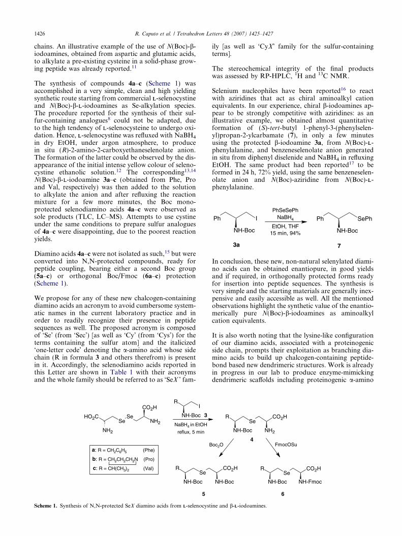
NH-Boc
IPh
NH-Boc
SePhPh
3a 7
PhSeSePhNaBH4
EtOH, THF15 min, 94%
1426 R. Caputo et al. / Tetrahedron Letters 48 (2007) 1425–1427
chains. An illustrative example of the use of N(Boc)-b-iodoamines, obtained from aspartic and glutamic acids,to alkylate a pre-existing cysteine in a solid-phase grow-ing peptide was already reported.11
The synthesis of compounds 4a–c (Scheme 1) wasaccomplished in a very simple, clean and high yieldingsynthetic route starting from commercial LL-selenocystineand N(Boc)-b-LL-iodoamines as Se-alkylation species.The procedure reported for the synthesis of their sul-fur-containing analogues8 could not be adapted, dueto the high tendency of LL-selenocysteine to undergo oxi-dation. Hence, LL-selenocystine was refluxed with NaBH4
in dry EtOH, under argon atmosphere, to producein situ (R)-2-amino-2-carboxyethaneselenolate anion.The formation of the latter could be observed by the dis-appearance of the initial intense yellow colour of seleno-cystine ethanolic solution.12 The corresponding13,14
N(Boc)-b-LL-iodoamine 3a–c (obtained from Phe, Proand Val, respectively) was then added to the solutionto alkylate the anion and after refluxing the reactionmixture for a few more minutes, the Boc mono-protected selenodiamino acids 4a–c were observed assole products (TLC, LC–MS). Attempts to use cystineunder the same conditions to prepare sulfur analoguesof 4a–c were disappointing, due to the poorest reactionyields.
Diamino acids 4a–c were not isolated as such,15 but wereconverted into N,N-protected compounds, ready forpeptide coupling, bearing either a second Boc group(5a–c) or orthogonal Boc/Fmoc (6a–c) protection(Scheme 1).
We propose for any of these new chalcogen-containingdiamino acids an acronym to avoid cumbersome system-atic names in the current laboratory practice and inorder to readily recognize their presence in peptidesequences as well. The proposed acronym is composedof ‘Se’ (from ‘Sec’) [as well as ‘Cy’ (from ‘Cys’) for theterms containing the sulfur atom] and the italicized‘one-letter code’ denoting the a-amino acid whose sidechain (R in formula 3 and others therefrom) is presentin it. Accordingly, the selenodiamino acids reported inthis Letter are shown in Table 1 with their acronymsand the whole family should be referred to as ‘SeX ’ fam-
HO2C
NH2
Se
CO2H
NH2Se
R
NH-Boc
I
SeR
NH-Boc
B
3
5
a: R = CH2C6H5 (Phe)
b: R = CH2CH2CH2N (Pro)
c: R = CH(CH3)2 (Val)
NaBH4 in EtOH
reflux, 5 min
Scheme 1. Synthesis of N,N-protected SeX diamino acids from LL-selenocyst
ily [as well as ‘CyX’ family for the sulfur-containingterms].
The stereochemical integrity of the final productswas assessed by RP-HPLC, 1H and 13C NMR.
Selenium nucleophiles have been reported16 to reactwith aziridines that act as chiral aminoalkyl cationequivalents. In our experience, chiral b-iodoamines ap-pear to be strongly competitive with aziridines: as anillustrative example, we obtained almost quantitativeformation of (S)-tert-butyl 1-phenyl-3-(phenylselen-yl)propan-2-ylcarbamate (7), in only a few minutesusing the protected b-iodoamine 3a, from N(Boc)-LL-phenylalanine, and benzeneselenolate anion generatedin situ from diphenyl diselenide and NaBH4 in refluxingEtOH. The same product had been reported17 to beformed in 24 h, 72% yield, using the same benzeneselen-olate anion and N(Boc)-aziridine from N(Boc)-LL-phenylalanine.
In conclusion, these new, non-natural selenylated diami-no acids can be obtained enantiopure, in good yieldsand if required, in orthogonally protected forms readyfor insertion into peptide sequences. The synthesis isvery simple and the starting materials are generally inex-pensive and easily accessible as well. All the mentionedobservations highlight the synthetic value of the enantio-merically pure N(Boc)-b-iodoamines as aminoalkylcation equivalents.
It is also worth noting that the lysine-like configurationof our diamino acids, associated with a proteinogenicside chain, prompts their exploitation as branching dia-mino acids to build up chalcogen-containing peptide-bond based new dendrimeric structures. Work is alreadyin progress in our lab to produce enzyme-mimickingdendrimeric scaffolds including proteinogenic a-amino
CO2H
NH2
SeR
NH-Boc
CO2H
NH-Boc
CO2H
NH-Fmoc
SeR
NH-Boc
oc2O FmocOSu4
6
ine and b-LL-iodoamines.

Table 1. New N,N-protected SeX diamino acids
N(Boc)-b-LL-Iodoamine SeX diamino acid Yielda (%) Mp (�C) ½a�25D ðCHCl3Þ
3a Boc–SeF(Boc)–OH (5a) 98 132–133 45.13a Fmoc–SeF(Boc)–OH (6a) 95 138–139 40.23b Boc–SeP(Boc)–OH (5b) 92 Foam 23.83b Fmoc–SeP(Boc)–OH (6b) 94 97–98 31.33c Boc–SeV(Boc)–OH (5c) 97 Foam 35.73c Fmoc–SeV(Boc)–OH (6c) 95 88–89 38.5
a Yield of diprotected selenodiamino acid, referred to the starting b-iodoamine.
R. Caputo et al. / Tetrahedron Letters 48 (2007) 1425–1427 1427
acids, such as Ser, His and Asp, that are commonlyinvolved in the enzymatic active sites.18
References and notes
1. Kyoto Encyclopedia of Genes and Genomes, KEGG,Release 40.0, October 2006. <http://www.genome.jp/kegg/>.
2. (a) Mugesh, G.; du Mont, W.-W.; Sies, H. Chem. Rev.2001, 101, 2125–2180; (b) Nogueira, C. W.; Zeni, G.;Rocha, J. B. T. Chem. Rev. 2004, 104, 6255–6286.
3. Rossato, J. I.; Ketzer, L. A.; Centuriao, F. B.; Silva, S. J.N.; Ludtke, D. S.; Zeni, G.; Braga, A. L.; Rubin, M. A.;Rocha, J. B. T. Neurochem. Res. 2002, 27, 297–303.
4. (a) Wirth, T. Tetrahedron Lett. 1995, 36, 7849–7852; (b)Wirth, T.; Kulieke, K. J.; Fragale, G. Helv. Chim. Acta1996, 79, 1957–1966; (c) Santi, C.; Wirth, T. Tetrahedron:Asymmetry 1999, 10, 1019–1023.
5. Braga, A. L.; Paixao, M. W.; Ludtke, D. S.; Silveira, C.C.; Rodrigues, O. E. D. Org. Lett. 2003, 5, 2635–2638.
6. Braga, A. L.; Silva, S. J. N.; Ludtke, D. S.; Drekener, R.L.; Silveira, C. C.; Rocha, J. B. T.; Wessjohann, L. A.Tetrahedron Lett. 2002, 43, 7329–7331.
7. Braga, A. L.; Paixao, M. W.; Marin, G. Synlett 2005, 11,1675–1678.
8. Bolognese, A.; Fierro, O.; Guarino, D.; Longobardo, L.;Caputo, R. Eur. J. Org. Chem. 2006, 167–173.
9. Davis, B. G. Curr. Opin. Biotechnol. 2003, 14, 379–386.10. Qi, D. F.; Tann, C. M.; Haring, D.; Distefano, M. D.
Chem. Rev. 2001, 101, 3081–3112.11. Campiglia, P.; Gomez-Monterrey, I.; Longobardo, L.;
Lama, T.; Novellino, E.; Grieco, P. Tetrahedron Lett.2004, 45, 1453–1456.
12. Block, E.; Birringer, M.; Jiang, W.; Nakaodo, T.;Thompson, H. J.; Toscano, P. J.; Uzar, H.; Zhang, X.;Zhu, Z. J. Agric. Food Chem. 2001, 49, 458–470.
13. Caputo, R.; Cassano, E.; Longobardo, L.; Palumbo, G.Tetrahedron Lett. 1995, 36, 167–168.
14. Caputo, R.; Cassano, E.; Longobardo, L.; Palumbo, G.Tetrahedron 1995, 51, 12337–12550.
15. Commercial LL-selenocystine (0.3 g, 1 mmol) was sus-pended in anhydrous EtOH (10 mL) under argon atmo-sphere. Solid NaBH4 (0.2 g, 5 mmol) was added in oneportion and the mixture refluxed for 5 min, until clear andcolourless. tert-Butyl (S)-1-iodo-3-methylbutan-2-ylcarba-mate 3c (0.6 g, 2 mmol) dissolved in anhydrous THF
(4 mL) was then added. After 10 min under reflux, thereaction mixture was cooled to room temperature and splitin two portions. The first one, after evaporation of thesolvents under vacuum, was redissolved in THF (10 mL)and treated with FmocOSu (0.3 g, 1 mmol) under standardconditions to afford eventually (R)-2-{[(9H-fluoren-9-yl)-methoxy]carbonylamino}-3-[(S)-2-(tert-butoxycarbon-ylamino)-3-methylbutylselenyl]propanoic acid, Fmoc–SeV(Boc)–OH (6c), (96%). An analytical sample: mp 88–89 �C (from CH2Cl2–hexane), ½a�25
D +38.5 (c 1.0, CHCl3).1H NMR (500 MHz, CD3OD, J in Hz): d = 0.87 (d, 3H,J = 6.8), 0.90 (d, 3H, J = 6.8), 1.44, (s, 9H), 1.77 (dq, 1H,J = 6.8), 2.67 (dd, 1H, J = 9.3 and 12.2), 2.82 (dd, 1H,J = 4.4 and 12.2), 2.97 (dd, 1H, J = 7.8 and 12.7), 3.03(dd, 1H, J = 4.9 and 12.7), 3.50 (m, 1H), 4.25 (t, 1H,J = 6.8), 4.34 (m, 2H), 4.44 (m, 1H), 7.31 (t, 2H, J = 7.8),7.39 (t, 2H, J = 7.8), 7.69 (d, 2H, J = 7.8), 7.79 (d, 2H,J = 7.8); 13C NMR (125 MHz, CD3OD): d = 18.8, 20.5,27.0, 29.3, 29.9, 33.9, 48.7, 56.3, 58.0, 68.7, 80.4, 121.4,126.8, 128.6, 129.3, 142.9, 145.6, 158.8, 174.6. HR-MS(EI): calcd for C28H36N2O6Se [M+H+] 576.5626, found576.5630.The second portion, after evaporation of the solventsunder vacuum, was also redissolved in THF (10 mL) andtreated with Boc2O (0.2 g, 1 mmol) under standard con-ditions to afford (R)-2-(tert-butoxycarbonylamino)-3-[(S)-2-(tert-butoxycarbonylamino)-3-methylbutylselenyl]prop-anoic acid, Boc–SeV(Boc)–OH (5c) (97%). An analyticalsample: foam, ½a�25
D +35.7 (c 1.6, CHCl3). 1H NMR(500 MHz, CD3OD, J in Hz): d = 0.89 (d, 3H, J = 6.8),0.92 (d, 3H, J = 6.8), 1.45 (s, 18H), 1.78 (dq, 1H, J = 6.8),2.65 (dd, 1H, J = 12.7 and 8.9), 2.83 (dd, 1H, J = 12.7 and3.9), 2.93 (dd, 1H, J = 12.7 and 6.8), 3.0 (dd, 1H, J = 12.7and 4.9), 3.50 (m, 1H), 4.37 (br t, 1H); 13C NMR(125 MHz, CD3OD): d = 18.7, 20.5, 27.1, 29.3, 29.7, 34.0,58.5, 80.3, 81.2, 158.2, 158.9, 174.5. HR-MS (EI): calcd forC18H34N2O6Se [M+H+] 454.4397, found 454.4402.
16. (a) Braga, A. L.; Ludtke, D. S.; Paixao, M. W.; Alberto,E. E.; Stefani, H. A.; Juliano, L. Eur. J. Org. Chem. 2005,4260–4264; (b) Braga, A. L.; Schneider, P. H.; Paixao, M.W.; Deobald, A. M.; Peppe, C.; Bottega, D. P. J. Org.Chem. 2006, 71, 4305–4307.
17. Mp 81–82 �C (from Et2O–hexane), ½a�25D 15.5 (c 1.0,
CH2Cl2); lit.16 1H NMR spectrum consistent withthe structure; mp 80.7–81.3 �C, ½a�25
D 14.18. Darbre, T.; Reymond, J.-L. Acc. Chem. Res. 2006, 39,
925–934.