ccd2: design constructs for protein expression, the easy way
TRANSCRIPT

research papers
992 https://doi.org/10.1107/S2059798321005891 Acta Cryst. (2021). D77, 992–1000
Received 10 March 2021
Accepted 7 June 2021
Edited by D. J. Rigden, University of Liverpool,
United Kingdom
Keywords: protein analysis; meta server; CCD2;
protein expression; expression construct design.
CCD2: design constructs for protein expression,the easy way
Andrea Giovanni Murachelli,a* George Damaskosb and Anastassis Perrakisa*
aOncode Institute and Department of Biochemistry, Netherlands Cancer Institute, Plesmanlaan 121, 1066 CX Amsterdam,
The Netherlands, and bDepartment of Biochemistry, Netherlands Cancer Institute, Plesmanlaan 121,
1066 CX Amsterdam, The Netherlands. *Correspondence e-mail: [email protected], [email protected]
Studying the function or structure of proteins usually requires the generation of
many protein-truncation constructs for recombinant expression, which is a
tedious and error-prone job. CCD2 is a software tool designed to facilitate and
automate this task. CCD2 helps scientists by aggregating the information
necessary to design protein-expression constructs. This information includes
sequence conservation, secondary structure prediction, domain(s) and disorder
detection, post-translational modifications and information on similar (domain)
structures that are available in the Protein Data Bank. CCD2 then allows users
to easily choose the boundaries for protein constructs and automatically
generates the primers necessary for construct amplification by polymerase chain
reaction. Finally, CCD2 provides a quick analysis of the properties of the chosen
constructs, together with their DNA vector maps for bookkeeping. The features
of CCD2 are discussed step by step, showing that it can be a useful tool for
laboratories that engage in recombinant protein production for any type of
experiment, and in particular for structural biology studies.
1. Introduction
Proteins, especially from eukaryotes, are modular machines
comprising multiple domains, often connected by flexible
regions. Most structural biology projects require the genera-
tion of multiple truncation constructs in order to explore
recombinant expression, solubility, crystallizability or the
functional properties of a target protein or macromolecular
complex. Generation of a protein construct has two phases.
Firstly, the constructs must be designed based on features of
the sequence of the protein of interest. The aim here is to find
suitable cutting points that are most likely to preserve protein
folding and solubility while retaining the desired functional
properties. Secondly, once the truncation constructs are
known, amplification primers must be designed to amplify the
relevant DNA sequence, which will then be cloned into a
suitable recombinant expression vector. Although all of the
information necessary for protein-construct design is available
online, aggregating it and mapping it onto the protein of
interest is rather tedious. Furthermore, truncation points are
decided on the protein sequence, but primer design requires
working with the DNA sequence. Mapping protein residues to
the DNA sequence and designing primers with suitable
chemical properties and appropriate cloning adaptors is
trivial, but is error-prone and time-consuming. ProteinCCD
(Crystallization Construct Designer; Mooij et al., 2009), a Java,
browser-based tool that we previously designed, aggregated
many sequence-analysis tools in a single interface, allowing
the user to generate PCR primers automatically starting from
the protein sequence. However, technological changes have
ISSN 2059-7983

rendered ProteinCCD inoperable in modern browsers and
obsolete.
CCD2 (Crystallization Construct Designer 2) is the successor
to ProteinCCD, using modern technology, but most impor-
tantly offering a largely expanded set of functionalities,
features and tools.
2. Methods
2.1. Architecture
CCD2 comprises of two parts: a user-facing, graphical user
interface (GUI) and a server-side backend that is responsible
for data gathering and manipulation. The GUI (whose func-
tional core is also used for LAHMA; https://lahma.rhpc.nki.nl;
van Beusekom et al., 2021) consists of an interactive web page
written in JavaScript/jQuery and styled with the Bootstrap
CSS/html libraries. Such an arrangement allows easy extensi-
bility and compatibility with all modern browsers. The
backend is written in Python 3.6 and uses Flask (Ronacher,
2010) to expose a RESTful API to the frontend. To improve
network performance, requests to external servers are asyn-
chronously parallelized by both the frontend (AJAX) and the
backend (AIOHTTP).
CCD2 requires several third-party tools for data analysis.
The software programs IUPred (Dosztanyi et al., 2005) and
NCOILS (Lupas et al., 1991) and the multiple sequence-
alignment tool MUSCLE (Edgar, 2004) are executed locally
and can be installed using operating-system-specific package
managers and repositories (for example apt-get on
Ubuntu). Predator (Frishman & Argos, 1996) can be obtained
from http://ftp.ebi.ac.uk/pub/software/unix/predator/. The
DisEMBL disorder-prediction tool (Linding, Jensen et al.,
research papers
Acta Cryst. (2021). D77, 992–1000 Andrea Giovanni Murachelli et al. � Designing constructs for protein expression 993
Figure 1Flowchart of CCD2. This flowchart shows the decision path/pipeline implemented by CCD2. Points where user input is required are indicated by goldellipses. Light-blue shaded boxes or highlighting show features that were present in the old ProteinCCD (Mooij et al., 2009). Coloured boxes indicatewhich modules of the backend are responsible for each operation.

2003) has been reimplemented for Python 3.6+ using the
original code distributed by the DisEMBL authors as a
starting point. The Savitsky–Golay smoothing algorithm is
copyright by SciPy Developers and is distributed under its
own licence. Predictions from GlobPlot (Linding, Russell et
al., 2003), the Pfam database (El-Gebali et al., 2019), SMART
(Letunic et al., 2021), NLS Mapper (Kosugi et al., 2009),
UniProt (The UniProt Consortium, 2019) and Phosphosite
(Hornbeck et al., 2004) are gathered through the respective
web servers. Secondary-structure predictions using HNN
(Guermeur, 1997), DPM (Deleage & Roux, 1987) and MLRC
(Guermeur et al., 1999) are gathered via the NPS@ server
(Combet et al., 2000). Similarity searches against the PDB
(Berman et al., 2003) and SwissProt (The UniProt Consortium,
2019) databases are performed against a local copy of these
databases using NCBI BLAST+ (Camacho et al., 2009;
Altschul et al., 1990). A CCD2 script (update_db.py) takes
care of installing and/or updating these local database
repositories.
Internally, CCD2 implements a pipeline that is summarized
in Fig. 1. To improve parallelization and maintainability,
different tasks are fulfilled by different modules of the
backend (coloured boxes in Fig. 1), each accessible through a
separate REST call.
2.2. Web-server implementation, installation and codeavailability
CCD2 is accessible at the URL https://ccd.rhpc.nki.nl hosted
by the Netherlands Cancer Institute Research High-Perfor-
mance Computing Facility.
CCD2 can also be run locally on a Linux-based machine
(tested on Ubuntu version 20.04). The source code of CCD2
is available at https://github.com/ProteinCCD2/ProteinCCD2
and is free for noncommercial use (licencing terms are avail-
able at the code repository site). Some external tools are
covered by different licencing arrangement and should be
obtained by the user in accordance with the term of the
respective licences.
research papers
994 Andrea Giovanni Murachelli et al. � Designing constructs for protein expression Acta Cryst. (2021). D77, 992–1000
Figure 2CCD2 can automatically match UniProt protein isoforms with their encoding DNA. An annotated screenshot of the user interface of CCD2 is depicted,showing the data retrieved for UniProt entry Q9UJ41 (human RABGEF5). The three existing isoforms of Q9UJ41 are aligned to show their differences.For each isoform, the length and matching coding DNA sequences are shown. DNA cross-references link to the entry in the respective database.Choosing an isoform (by clicking on its radio button) will automatically paste the coding DNA into the DNA sequence window, allowing CCD2 to use itfor primer design. The user can also paste their own DNA sequence, if necessary. For ease of display, some white space in this figure has been trimmedcompared with the normal CCD2 display.
Installation of CCD2 is straightforward and is explained in
the README.MD file provided with the distribution. Python
environment consistency is maintained using Anaconda
virtual environments.
3. Results
In this section, we will describe the functions of CCD2
following the natural order of user interaction schematized in
Fig. 1. We will also provide some general tips about protein-
construct design.
In our experience, a successful protein construct fulfils three
related requirements: (i) it is recombinantly expressed (ideally
at high levels), (ii) it is (highly) soluble and (iii) it is confor-
mationally stable or at least constrained. (i) and (ii) are
requirements for recombinant expression and biochemical,
biophysical and many other functional assays, whereas (iii) is a
requirement for a high-resolution structure by X-ray crystal-
lography or by single-particle cryoEM. CCD2 can help effi-
cient recombinant expression by providing a quick and easy
way to clone the user’s construct with different tags and in
different hosts (see Section 3.4) and by facilitating relative
bookkeeping (see Section 3.5). The solubility requirement (ii)
is achieved when proteins are correctly folded and do not
expose an excessive hydrophobic surface to the solvent. Due
to the modular nature of proteins, this is true when a trun-
cation cuts between, but not within, protein domains and
structural elements. For structural biology and requirement
(iii), one would also prune unstructured regions (i.e. regions
that are not part of a folded domain) to limit the conforma-
tional freedom of the construct. If one is interested in intrin-
sically disordered proteins, and expressing disordered regions
is instead the target, unstructured regions would be cloned
instead. Either way, protein-construct design boils down to
identifying domains and disorder in proteins. A main goal of
CCD2 is to collate and display at a glance all of the informa-
tion useful for domain identification.
3.1. Identifying the DNA sequence of the protein of interest
The first step required for construct design is to retrieve and
analyse the sequence of the protein of interest (POI).
However, since the final objective is to generate cloning
primers, CCD2 needs to start by knowing the DNA sequence
that codes for the POI (Fig. 2). Two options are available. The
user can paste their own DNA sequence into the GUI and
start the workflow from there. This is the only option available
in the case where the POI is coded by an ORF that is non-
natural (i.e. codon-optimized) or by an ORF that is not
present in the UniProt database. However, if the POI is coded
by a natural sequence whose translation has been deposited in
the UniProt database, CCD2 can query UniProt using a user-
provided identifier (for example Q9UJ41) or mnemonic
accession code (for example RABX5_HUMAN). From the
UniProt entry, CCD2 can automatically determine which
isoforms of the POI are reported and match them to appro-
priate DNA sequences (open reading frames; ORFs) by
querying the cross-referenced nucleotide databases. UniProt
protein sequences are determined by consensus and curation
(https://www.uniprot.org/help/canonical_nucleotide), meaning
that there is no one-to-one match between DNA primary
database accessions and protein isoforms. CCD2 simply
gathers all the cross-referenced DNA sequences, translates
them and matches them to isoforms at the protein level. No
attempt is made to compare the raw DNA sequences for silent
single-nucleotide polymorphisms, because these are rare and
are extremely unlikely to affect the eventually generated
primers. Imperfect protein sequence matches of up to three
single amino-acid substitutions are shown to the user if no
perfect match can be found for an isoform, along with a
detailed notice about the sequence differences. For bacterial
proteins, CCD2 can parse multicistronic genes and genomic
sequences (where the entry does not exceed 1 Mb in download
size).
Once isoform matching is complete, the user is prompted to
choose which isoform they wish to use for downstream
analysis and primer generation. Cross-references to the
primary DNA databases are provided for each isoform (Fig. 2).
For easier visual reference, an alignment of the different
isoforms is also provided. Inspection of the differences
between isoforms can suggest viable truncation positions and
hint at domains that might be swapped in or out among
isoforms.
3.2. Creating and visualizing a report on sequenceconservation
Domains are functionally and structurally constrained, and
are thus evolutionarily conserved. Disordered and linker
sequences are under looser evolutionary pressure and mutate
more frequently, unless they are of specific functional impor-
tance. In general, in a multiple sequence alignment, well
ordered domains will appear as contiguous stretches of
conserved residues, whereas linker and disordered regions will
show higher divergence. CCD2 attempts to find and display
a multiple sequence alignment of the POI using three
approaches. If a UniProt ID is provided by the user, and this
ID can be mapped to a pre-calculated Ensembl alignment
(Yates et al., 2020), this alignment is retrieved. If no UniProt
ID is available or provided, CCD2 performs a BLAST search
against a local copy of the SwissProt database and looks for up
to five hits that have an identity of >95% with the POI. CCD2
then queries Ensembl and looks for pre-calculated alignments
for any of these hits. If such a hit exists, it is considered a
homolog to the POI and the corresponding Ensembl align-
ment is retrieved. If this approach also fails, CCD2 displays
results of the BLAST search that (i) have an E-value of >0.001
and (ii) represent sequence coverage of the POI of �75%.
Requiring a high sequence coverage is likely to find and
display true orthologues of the POI (rather than showing
sequences of loosely related proteins that simply share a single
domain with the POI). Then, the POI isoform chosen by the
user is aligned with the homolog sequences using MUSCLE
(Edgar, 2004).
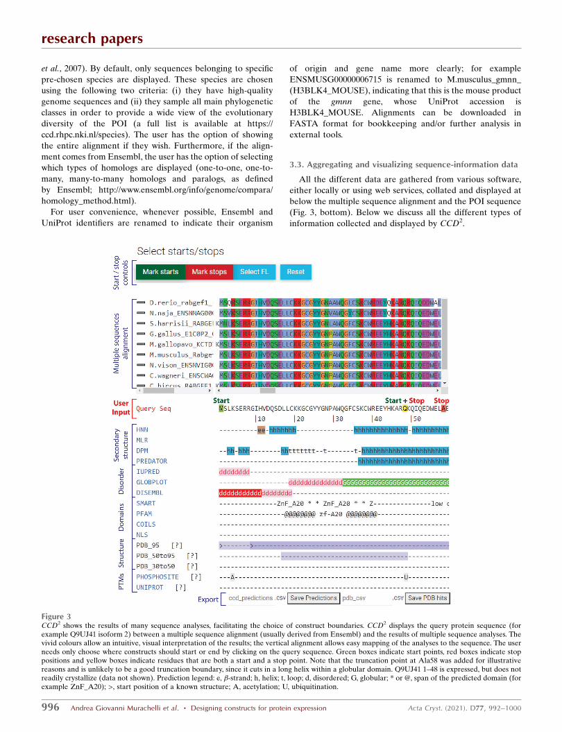
The Ensembl or on-the-fly constructed multiple sequence
alignment is then displayed in the GUI (Fig. 3, top) and
coloured by conservation using the ClustalX scheme (Larkin
research papers
Acta Cryst. (2021). D77, 992–1000 Andrea Giovanni Murachelli et al. � Designing constructs for protein expression 995
et al., 2007). By default, only sequences belonging to specific
pre-chosen species are displayed. These species are chosen
using the following two criteria: (i) they have high-quality
genome sequences and (ii) they sample all main phylogenetic
classes in order to provide a wide view of the evolutionary
diversity of the POI (a full list is available at https://
ccd.rhpc.nki.nl/species). The user has the option of showing
the entire alignment if they wish. Furthermore, if the align-
ment comes from Ensembl, the user has the option of selecting
which types of homologs are displayed (one-to-one, one-to-
many, many-to-many homologs and paralogs, as defined
by Ensembl; http://www.ensembl.org/info/genome/compara/
homology_method.html).
For user convenience, whenever possible, Ensembl and
UniProt identifiers are renamed to indicate their organism
of origin and gene name more clearly; for example
ENSMUSG00000006715 is renamed to M.musculus_gmnn_
(H3BLK4_MOUSE), indicating that this is the mouse product
of the gmnn gene, whose UniProt accession is
H3BLK4_MOUSE. Alignments can be downloaded in
FASTA format for bookkeeping and/or further analysis in
external tools.
3.3. Aggregating and visualizing sequence-information data
All the different data are gathered from various software,
either locally or using web services, collated and displayed at
below the multiple sequence alignment and the POI sequence
(Fig. 3, bottom). Below we discuss all the different types of
information collected and displayed by CCD2.
research papers
996 Andrea Giovanni Murachelli et al. � Designing constructs for protein expression Acta Cryst. (2021). D77, 992–1000
Figure 3CCD2 shows the results of many sequence analyses, facilitating the choice of construct boundaries. CCD2 displays the query protein sequence (forexample Q9UJ41 isoform 2) between a multiple sequence alignment (usually derived from Ensembl) and the results of multiple sequence analyses. Thevivid colours allow an intuitive, visual interpretation of the results; the vertical alignment allows easy mapping of the analyses to the sequence. The userneeds only choose where constructs should start or end by clicking on the query sequence. Green boxes indicate start points, red boxes indicate stoppositions and yellow boxes indicate residues that are both a start and a stop point. Note that the truncation point at Ala58 was added for illustrativereasons and is unlikely to be a good truncation boundary, since it cuts in a long helix within a globular domain. Q9UJ41 1–48 is expressed, but does notreadily crystallize (data not shown). Prediction legend: e, �-strand; h, helix; t, loop; d, disordered; G, globular; * or @, span of the predicted domain (forexample ZnF_A20); >, start position of a known structure; A, acetylation; U, ubiquitination.

3.3.1. Secondary-structure prediction. Domains have a
high content of secondary structure, while disordered regions
do not. CCD2 runs the sequence through four secondary-
structure prediction algorithms [HNN (Guermeur, 1997),
DPM (Deleage & Roux, 1987), MLRC (Guermeur et al., 1999)
and Predator (Frishman & Argos, 1996)]. These secondary-
structure prediction methods are reasonably reliable and
quick. Their results are displayed together, so that the user can
derive a consensus view. Consecutive stretches of consensus
secondary structure indicate domains.
3.3.2. Disorder prediction. Disordered regions often have
low-complexity, repetitive sequences. Additionally, polar and
charged amino acids are overrepresented in disordered
regions (Dyson, 2016). CCD2 gathers disorder and globular
region information using IUPred (Dosztanyi et al., 2005),
DisEMBL (Linding, Jensen et al., 2003) and GlobPlot
(Linding, Russell et al., 2003). The SMART database
(Letunic et al., 2021) is also used to display low-complexity
regions. Cuts in the constructs should encompass, but
not cut within, predicted globular regions. Trimming
terminal disordered regions is generally required for
crystallization, and might lead to more homogeneous
protein preparations owing to reduced proteolytic
degradation.
3.3.3. Domain detection. CCD2 highlights known domains
in the protein sequence by querying the SMART (Letunic et
al., 2021) and Pfam (El-Gebali et al., 2019) domain-fingerprint
databases. Additionally, CCD2 performs a BLAST search
(Altschul et al., 1990) against a local copy of the Protein Data
Bank (PDB), reporting hits at three different levels of simi-
larity. The prediction ‘PDB_95’ highlights the parts of the POI
sequence that have an identity of �95% to a solved structure
in the PDB, thus indicating that parts of the POI (or of a very
close homologue) have been experimentally determined. The
boundaries of the expression constructs deposited in the
corresponding PDB structures are also indicated on the POI
sequence. Hovering the cursor over the construct boundaries
(marked with ‘>’ or ‘<’ for a start or stop position, respec-
tively) will display the PDB code and chain of the matching
structures.
These are experimentally validated, effectual boundaries
for truncation constructs. The predictions ‘PDB50_to_95’ and
‘PDB30_to_50’ similarly highlight parts of the POI sequence
that have BLAST hits against the PDB with identities between
95% and 50% and between 50% and 30%, respectively. These
portions of the POI sequence are homologous to known
structures, indicating the likely existence and approximate
boundaries of a folded domain. All of the results of the search
against the PDB can be downloaded for further analysis by
clocking on the ‘Save PDB hits’ button. These include the
PDB code, sequence coverage and percentage identity for
each matching hit.
research papers
Acta Cryst. (2021). D77, 992–1000 Andrea Giovanni Murachelli et al. � Designing constructs for protein expression 997
Figure 4CCD2 offers multiple choices for primer generation. (a) CCD2 is (optionally) integrated with the pETNKI LIC series of vectors, which offer greatversatility, since the same construct fits multiple vectors. (b) CCD2 can design primers for conventional restriction cloning. (c) CCD2 allows the choice ofany custom overhang for primers. (d) CCD2 automatically generates primers based on user-chosen boundaries on the protein sequence, meltingtemperature and primer overhangs. Shown here are the primers for start positions 1 and 48 and stop positions 48 and 58 (from Fig. 3), with a Tm of 65�Cand overhangs for pETNKI LIC 1.1. The overhang portion of the primer is shown in lower case and the annealing portion is shown in upper case. Theprimers are named prefix_Fw/Rv_position, where the prefix is chosen by the user (for example RBX5), Fw stands for forward, Rv stands for reverse and‘position’ is the chosen start/stop position. Primers can be copied and pasted into a spreadsheet or saved in comma-separated value (csv) format. TheDNA sequences of the constructs resulting from all possible combinations of primers can also be saved in csv format.

3.3.4. Coiled-coil detection. Coiled coils are very common
structural domains that often mediate protein–protein inter-
actions. CCD2 searches for coiled coils by querying the
SMART database and by direct prediction with NCOILS
(Lupas et al., 1991). Truncation within coiled coils is possible
(see, for example, Ciferri et al., 2008), although trial and error
is necessary.
3.3.5. Detection of other functional elements. CCD2
further detects the presence of putative nuclear localization
signals (NLS) using NLS (Kosugi et al., 2009). The presence of
an NLS can influence expression in eukaryotic systems.
However, NLSs are low-complexity, generally disordered
sequences, so their removal can positively affect crystal-
lization.
If experimentally validated post-translational modifications
(PTMs) are recorded in the UniProt entry for the sequence of
interest, these are displayed. UniProt covers a wide variety of
possible PTMs, including glycosylation, disulfide bridges,
cross-links (intra-chain and to other proteins such as
ubiquitin), chemical modification of amino acids and more.
These modifications are indicated with a single-letter code
including, for example, ‘A’ for acetylation, ‘^’ for a disulfide
link, ‘+’ for multiple known modifications etc. (a full legend
can be found on the tutorial page at https://ccd.rhpc.nki.nl/
tutorial). Hovering the cursor over each letter will display
more precise information about each modification. Because
UniProt annotations always refer to the sequence of the
canonical isoform, these annotations are disabled if the user
has selected an alternative splicing variant, to avoid sequence
discrepancy.
When data are available (human, rat and mouse proteins),
CCD2 also queries the Phosphosite Plus database (Hornbeck
et al., 2004) for the presence of experimentally validated post-
translational modifications (PTMs) on the sequence. Anno-
tations follow the same notation as for UniProt above.
Hovering over each annotation provides further information
about the underlying data.
PTMs are added by enzymes and require physical accessi-
bility to be attached. Thus, the presence of PTMs can hint at
disordered, highly accessible linker regions or at least solvent-
exposed residues (Dyson, 2016). PTMs can also inform about
the functionality of truncation constructs.
3.4. Designing protein constructs and single-click generationof DNA primers
With all the necessary information available, the user can
choose where truncation constructs should start or stop by
clicking start and stop points on the sequence of the POI
(Fig. 3, middle). The clicked amino acid is always included in
the final construct. Start points will generate forward PCR
primers and stop points will generate reverse PCR primers. A
position can be marked as being both a start and a stop.
research papers
998 Andrea Giovanni Murachelli et al. � Designing constructs for protein expression Acta Cryst. (2021). D77, 992–1000
Figure 5Primer generation and protein-construct analysis. (a) The possible constructs resulting from the primers chosen in Fig. 3 and displayed in Fig. 4(e) areshown here, together with their predicted molecular wight (MW), isoelectric point (pI) and extinction coefficient " at 280 nm. (b) For pETNKI vectors(for example pETNKI 1.1), the tagged and protease-cleaved constructs are also shown, together with their predicted physical properties.

PCR amplificates typically need adapter sequences to be
cloned into recipient vectors. These sequences are added to
the primers as ‘overhangs’ that extend beyond the primer
sequence that anneals to the template DNA. CCD2 allows the
user to choose overhangs in three ways (Figs. 4a–4c). Firstly,
the version of CCD2 hosted on our servers is designed to work
in tandem with the pETNKI series (Luna-Vargas et al., 2011)
of ligation-independent cloning (LIC; Aslanidis & de Jong,
1990) vectors. These vectors are suitable for mammalian,
insect-cell or Escherichia coli expression and are designed for
maximum intercompatibility, so that the same PCR amplifi-
cate can be cloned in multiple targets. CCD2 can automatically
generate PCR primers with the correct overhangs for any
pETNKI vector chosen (Fig. 4a). Some pETNKI vectors can
be freely obtained from Addgene (https://www.addgene.org/,
catalogue Nos. 108703–108710); others, which are encumbered
by third-party patents, can be sourced from the Netherlands
Cancer Institute protein-production facility with a material
transfer agreement. When running a local copy of CCD2, user-
defined vectors can be integrated instead of the petNKI series
(not shown). Alternatively, CCD2 contains a utility to generate
primer overhangs for conventional restriction cloning (Fig. 4b).
Finally, CCD2 can accept user-provided custom overhangs,
which may contain nonstandard sequences (i.e. other than the
standard DNA bases ATCG; Fig. 4c). In all cases, the user is
notified of the final overhang sequence and of the presence of
start/stop codons in the overhang (not shown). The user can
also choose the properties of the primer by choosing a desired
melting temperature (Tm; default 65�C) or primer length.
Overhangs are not considered in determining the Tm. Finally,
the user can also choose a name for the primers.
Using these data, CCD2 automatically maps the user-chosen
start and stop positions from the protein to the DNA sequence
and generates a table with all of the primers that can be saved
in spreadsheet-compatible format for bookkeeping or copied
and pasted for quick ordering of the primers (Fig. 4d). The
amplified DNA sequences resulting from all possible combi-
nations of starts and stops (i.e. resulting from a start and stop
primer that amplifies any portion of the protein sequence) can
also be downloaded in spreadsheet-compatible format by
clicking on the ‘Save Construct DNA’ button.
3.5. Enabling data tracking and bookkeeping
CCD2 displays the sequence of the protein truncations that
are generated by all possible start and stop combinations in a
different panel, along with basic information about their
predicted molecular weight (MW), isoelectric point (pI) and
predicted extinction coefficient at 280 nm (Fig. 5a). These are
calculated with the same algorithm as used by ProtParam in
the Expasy portal (Gasteiger et al., 2005). If pETNKI vectors
are chosen as cloning targets (or custom vectors are integrated
in a local copy of CCD2), CCD2 also has the information about
the sequence of each construct prior to (Fig. 5b) and after
(Fig. 5c) proteolytic tag cleavage, and can provide further
provide the sequence, molecular weight, predicted isoelectric
point (pI) and expected 280 nm extinction coefficient for all
generated constructs, either with attached tag or after protease
digestion. All of these data can be saved in spreadsheet-
compatible format for bookkeeping and to assist in protein
expression and purification.
Finally, for pETNKI and custom vectors, CCD2
can generate and save annotated plasmid maps of the
chosen truncation constructs (GenBank format; https://
www.ncbi.nlm.nih.gov/genbank/samplerecord/). These can be
opened in any standard DNA-manipulation software and are
useful as a reference to check the success of cloning.
4. Conclusions
The design and cloning of constructs are frequent and time-
consuming tasks in any structural biology project, and often in
general biochemistry and biophysics. CCD2 streamlines these
tasks: it helps in the design of constructs by consolidating
multiple informative analyses of the sequence in a single place,
and it enables the user to make quick decisions about where
protein truncations should be placed. Then, once the bound-
aries have been chosen, CCD2 takes care of the nitty-gritty
details of primer design and plasmid mapping, also providing a
brief recombinant construct analysis. Overall, CCD2 allows the
user to save valuable time and reduce costly mistakes in any
structural biology project.
Acknowledgements
We would like to acknowledge Professor Titia Sixma for
support and feedback, Dr Wouter Touw for help in project
setup and helpful discussions, the Research High-Performance
Computing (RHPC) facility of the Netherlands Cancer Insti-
tute for hosting the CCD2 website, and the members of the
Sixma and Perrakis groups at NKI for helpful discussion, beta
testing and feedback.
References
Altschul, S. F., Gish, W., Miller, W., Myers, E. W. & Lipman, D. J.(1990). J. Mol. Biol. 215, 403–410.
Aslanidis, C. & de Jong, P. J. (1990). Nucleic Acids Res. 18, 6069–6074.Berman, H., Henrick, K. & Nakamura, H. (2003). Nat. Struct. Mol.
Biol. 10, 980.Beusekom, B. van, Damaskos, G., Hekkelman, M. L., Salgado-Polo,
F., Hiruma, Y., Perrakis, A. & Joosten, R. P. (2021). Acta Cryst. D77,28–40.
Camacho, C., Coulouris, G., Avagyan, V., Ma, N., Papadopoulos, J.,Bealer, K. & Madden, T. L. (2009). BMC Bioinformatics, 10, 421.
Ciferri, C., Pasqualato, S., Screpanti, E., Varetti, G., Santaguida, S.,Dos Reis, G., Maiolica, A., Polka, J., De Luca, J. G., De Wulf, P.,Salek, M., Rappsilber, J., Moores, C. A., Salmon, E. D. &Musacchio, A. (2008). Cell, 133, 427–439.
Combet, C., Blanchet, C., Geourjon, C. & Deleage, G. (2000). TrendsBiochem. Sci. 25, 147–150.
Deleage, G. & Roux, B. (1987). Protein Eng. Des. Sel. 1, 289–294.Dosztanyi, Z., Csizmok, V., Tompa, P. & Simon, I. (2005). J. Mol. Biol.
347, 827–839.Dyson, H. J. (2016). Biophys. J. 110, 1013–1016.Edgar, R. C. (2004). Nucleic Acids Res. 32, 1792–1797.El-Gebali, S., Mistry, J., Bateman, A., Eddy, S. R., Luciani, A., Potter,
S. C., Qureshi, M., Richardson, L. J., Salazar, G. A., Smart, A.,Sonnhammer, E. L. L., Hirsh, L., Paladin, L., Piovesan, D., Tosatto,
research papers
Acta Cryst. (2021). D77, 992–1000 Andrea Giovanni Murachelli et al. � Designing constructs for protein expression 999

S. C. E. & Finn, R. D. (2019). Nucleic Acids Res. 47, D427–D432.
Frishman, D. & Argos, P. (1996). Protein Eng. Des. Sel. 9, 133–142.
Gasteiger, E., Hoogland, C., Gattiker, A., Duvaud, S., Wilkins, M. R.,Appel, R. D. & Bairoch, A. (2005). The Proteomics ProtocolsHandbook, edited by J. M. Walker, pp. 571–607. Totowa: HumanaPress.
Guermeur, Y. (1997). Combinaison de classifieurs statistiques:application a la prediction de la structure secondaire des proteines.http://www.theses.fr/1997PA066667.
Guermeur, Y., Geourjon, C., Gallinari, P. & Deleage, G. (1999).Bioinformatics, 15, 413–421.
Hornbeck, P. V., Chabra, I., Kornhauser, J. M., Skrzypek, E. & Zhang,B. (2004). Proteomics, 4, 1551–1561.
Kosugi, S., Hasebe, M., Tomita, M. & Yanagawa, H. (2009). Proc. NatlAcad. Sci. USA, 106, 10171–10176.
Larkin, M. A., Blackshields, G., Brown, N. P., Chenna, R.,McGettigan, P. A., McWilliam, H., Valentin, F., Wallace, I. M.,Wilm, A., Lopez, R., Thompson, J. D., Gibson, T. J. & Higgins, D. G.(2007). Bioinformatics, 23, 2947–2948.
Letunic, I., Khedkar, S. & Bork, P. (2021). Nucleic Acids Res. 49,D458–D460.
Linding, R., Jensen, L. J., Diella, F., Bork, P., Gibson, T. J. & Russell,R. B. (2003). Structure, 11, 1453–1459.
Linding, R., Russell, R. B., Neduva, V. & Gibson, T. J. (2003). NucleicAcids Res. 31, 3701–3708.
Luna-Vargas, M. P. A., Christodoulou, E., Alfieri, A., van Dijk, W. J.,Stadnik, M., Hibbert, R. G., Sahtoe, D. D., Clerici, M., Marco, V. D.,Littler, D., Celie, P. H. N., Sixma, T. K. & Perrakis, A. (2011). J.Struct. Biol. 175, 113–119.
Lupas, A., Van Dyke, M. & Stock, J. (1991). Science, 252, 1162–1164.
Mooij, W. T. M., Mitsiki, E. & Perrakis, A. (2009). Nucleic Acids Res.37, W402–W405.
Ronacher, A. (2010). Flask. https://palletsprojects.com/p/flask/.The UniProt Consortium (2019). Nucleic Acids Res. 47, D506–
D515.Yates, A. D., Achuthan, P., Akanni, W., Allen, J., Allen, J., Alvarez-
Jarreta, J., Amode, M. R., Armean, I. M., Azov, A. G., Bennett, R.,Bhai, J., Billis, K., Boddu, S., Marugan, J. C., Cummins, C.,Davidson, C., Dodiya, K., Fatima, R., Gall, A., Giron, C. G., Gil, L.,Grego, T., Haggerty, L., Haskell, E., Hourlier, T., Izuogu, O. G.,Janacek, S. H., Juettemann, T., Kay, M., Lavidas, I., Le, T., Lemos,D., Martinez, J. G., Maurel, T., McDowall, M., McMahon, A.,Mohanan, S., Moore, B., Nuhn, M., Oheh, D. N., Parker, A., Parton,A., Patricio, M., Sakthivel, M. P., Abdul Salam, A. I., Schmitt, B. M.,Schuilenburg, H., Sheppard, D., Sycheva, M., Szuba, M., Taylor, K.,Thormann, A., Threadgold, G., Vullo, A., Walts, B., Winterbottom,A., Zadissa, A., Chakiachvili, M., Flint, B., Frankish, A., Hunt, S. E.,IIsley, G., Kostadima, M., Langridge, N., Loveland, J. E., Martin,F. J., Morales, J., Mudge, J. M., Muffato, M., Perry, E., Ruffier, M.,Trevanion, S. J., Cunningham, F., Howe, K. L., Zerbino, D. R. &Flicek, P. (2020). Nucleic Acids Res. 48, D682–D688.
research papers
1000 Andrea Giovanni Murachelli et al. � Designing constructs for protein expression Acta Cryst. (2021). D77, 992–1000