cd44-regulated intracellular proliferation of listeria ...iai.asm.org/content/71/7/4102.full.pdf ·...
TRANSCRIPT

INFECTION AND IMMUNITY, July 2003, p. 4102–4111 Vol. 71, No. 70019-9567/03/$08.00�0 DOI: 10.1128/IAI.71.7.4102–4111.2003Copyright © 2003, American Society for Microbiology. All Rights Reserved.
CD44-Regulated Intracellular Proliferation of Listeria monocytogenesEmma Eriksson,1 Lone Dons,2 Antonio Gigliotti Rothfuchs,1 Paraskevi Heldin,3
Hans Wigzell,1 and Martin E. Rottenberg1*Microbiology & Tumorbiology Center, Karolinska Institute, Stockholm,1 and Department of Medical Biochemistry and
Microbiology, Biomedical Center, Uppsala University, Upsala,3 Sweden, and Department of VeterinaryMicrobiology, The Royal Veterinary and Agricultural University, Frederiksberg, Denmark2
Received 9 December 2002/Returned for modification 11 February 2003/Accepted 14 April 2003
CD44 has been implicated in immune and inflammatory processes. We have analyzed the role of CD44 in theoutcome of Listeria monocytogenes infection in murine bone marrow-derived macrophages (BMM). Surpris-ingly, a dramatically decreased intracellular survival of L. monocytogenes was observed in CD44�/� BMM.CD44�/� heart or lung fibroblast cultures also showed reduced bacterial levels. Moreover, livers from CD44�/�-infected mice showed diminished levels of L. monocytogenes. In contrast, intracellular growth of Salmonellaenterica serovar Typhimurium was the same in CD44�/� and control BMM. The CD44-mediated increasedbacterial proliferation was not linked to altered BMM differentiation or to secretion of soluble factors. CD44did not mediate listerial uptake, and it played no role in bacterial escape from the primary phagosome orformation of actin tails. Furthermore, CD44-enhanced listerial proliferation occurred in the absence of intra-cellular bacterial spreading. Interestingly, coincubation of BMM with hyaluronidase or anti-CD44 antibodiesthat selectively inhibit hyaluronan binding increased intracellular listerial proliferation. Treatment of cellswith hyaluronan, in contrast, diminished listerial growth and induced proinflammatory transcript levels. Wesuggest that L. monocytogenes takes advantage of the CD44-mediated signaling to proliferate intracellularly,although binding of CD44 to certain ligands will inhibit such response.
CD44 glycoprotein is found on the surface of many celltypes, including lymphocytes, macrophages, and epithelialcells. Expression levels vary depending on origin and activationstatus of the cell. CD44-dependent processes are known toinclude organ development, neuronal axon guidance, hemato-poiesis, and numerous immune functions. Among the latter,CD44 participates in lymphocyte adhesion to inflamed endo-thelium, lymphocyte homing, and tumor metastasis (31). Hya-luronan (HA), a main carbohydrate component of the extra-cellular matrix, is the principal but not sole ligand of CD44.CD44 is also the major receptor for HA. HA is normally aglycosaminoglycan of high molecular weight. At sites of inflam-mation, low-molecular-weight (LMW) HA accumulates, mostlikely due to the presence of hyaluronidases (HA’ses) and/orreactive oxygen species. Binding of LMW HA to CD44 caninduce expression of cytokines, chemokines, and adhesion andeffector molecules and can induce translocation of transcrip-tion factors in cell lines or primary cell cultures (2, 22–24, 26,28, 29, 41). Thus, besides tethering cells to extracellular li-gands, CD44 has broader functions in cellular signaling cas-cades. CD44 also provides a link between the plasma mem-brane and the actin cytoskeleton. CD44 can have coreceptorfunctions mediating the signaling of receptor tyrosine kinases,such as Met.
The impact of CD44 in the regulation of immune responsesand inflammation has been broadly studied (27, 31, 40, 42), butfew studies have addressed the potential role of CD44 in thecontrol of pathogens (4, 12, 13, 15, 39).
The gram-positive bacterium Listeria monocytogenes is a hu-
man pathogen that causes severe disease in immunocom-promised individuals and will induce abortions in pregnantwomen. L. monocytogenes is known to invade a variety of cells,including macrophages. After cellular uptake, the bacteriumescapes from the primary phagosome into cytoplasm, where itstarts to multiply and then spread to nearby cells (45). Thepresence of an inducible listerial hexose phosphate transportermediating rapid intracellular replication has been recently de-scribed (17). In the cytoplasm L. monocytogenes expressesActA protein, a cofactor for the nucleation of actin filaments.The bacterium polymerizes actin filaments around itself, cre-ating a long actin tail. Such tails will propel listeria to the cellmembrane, where projections involved in listerial cell-to-cellspread will be formed (11).
Immune resistance to L. monocytogenes depends on the abil-ity of the host to mount a Th1-like immune response (43).Cytokines such as gamma interferon (IFN-�) will activate mac-rophage bactericidal mechanisms, which play a crucial role inthe control of listerial infection in vivo (20, 32).
We initially hypothesized that signals through HA and CD44could inhibit the intracellular growth of L. monocytogenes byupregulating the expression of inflammatory genes and by con-trolling the cytoskeleton rearrangements. Instead, our studiesrevealed that L. monocytogenes makes use of CD44 signaling togrow efficiently intracellularly.
MATERIALS AND METHODS
Reagents. Anti-CD44 (KM 703, KM 81), anti-CD4, and anti-major histocom-patibility complex (MHC) class I monoclonal antibodies were purified from thesupernatant of hybridomas CRL-1896, TIB-241, L3T4, and HB51, respectively(American Type Culture Collection, Manassas, Va.), by using protein G-Sepha-rose (Amersham-Pharmacia, Uppsala, Sweden). Hyaluronidase (HA’se) fromStreptomyces species was purchased from Calbiochem (San Diego, Calif.). HA’se
* Corresponding author. Mailing address: Nobelsvag 16, S 171 77Stockholm, Sweden. Phone: 46-8-728-67-11. Fax: 46-8-32-8878. E-mail:[email protected].
4102
on July 11, 2018 by guesthttp://iai.asm
.org/D
ownloaded from
type III from sheep testes, chondroitinase ABC from Proteus vulgaris, heparinase(HS’se) III from Flavobacterium heparinum, and heparan sulfate (HS) frombovine kidneys were all purchased from Sigma-Aldrich (St. Louis, Mo.). Hyalu-ronans (HA), with the sizes of 190 and 250 kDa, were kindly provided by Ove Vik(Q-Med AB, Uppsala, Sweden).
Mice. C57Bl/6 mice were bred under specific-pathogen-free conditions. Amutant mouse strain without CD44 was generated by homologous recombinationin embryonic stem cells and was backcrossed with C57Bl/6 (36).
Bacteria. L. monocytogenes wild-type (WT) strain EGD (BUG600, serotype1/2a) and the �plcB2 mutant (35) with a defective lecithinase were used.TheactA-defective mutants, deficient in actin polymerization strain LO28�ActA(BUG 875) (18) and the BUG314 strain, containing a Tn917 transposon insertedin actA (25) and the parental control strain LO28, all obtained from the PasteurInstitute (Paris, France), were used. To study intracellular bacterial localizationof L. monocytogenes NF-L357, which contains a transcriptional fusion betweenactA-plcB and the green fluorescent protein gene (gfp) was used (16). In all casesthe bacteria were grown in brain heart infusion (BHI) broth and BHI agar(Difco, Becton Dickinson, Md.) at 37°C. Before infection bacteria were grown at37°C in BHI broth to late-exponential phase (optical density at 600 nm, 0.8). Thebacteria were washed once with phosphate-buffered saline (PBS), resuspended inBHI broth with 15% glycerol, plated on duplicate BHI agar plates, and quanti-fied after an overnight incubation at 37°C. Bacterial suspensions were stored at�70°C until use.
The plasmid pAUL-A (8), which replicates at 30°C but not at 37°C andpossesses an erythromycin resistance gene, was used to distinguish replicationrate from death rate of L. monocytogenes in bone marrow-derived macrophages(BMM). L. monocytogenes EGD was transformed with pAUL-A by electropo-ration and was grown at 30°C in BHI medium containing 5 �g of erythromycinper ml overnight. To produce cultures containing less than one copy of theplasmid per bacterium, 20 ml of the culture was inoculated in 180 ml of BHI andwas grown at 37°C for 6 h. Numbers of total and plasmid-containing L. mono-cytogenes and the total number of plasmids were calculated during this incuba-tion by plating bacteria on BHI agar in the presence or absence of erythromycinor by further incubating bacteria at 42°C to stationary phase before plating onBHI agar in the presence of erythromycin. An initial 5.3-fold increase in thenumber of plasmid-containing bacteria during the first 4.5 h in culture wasdetermined. The mean copy number at start was calculated to be 3.6 plasmids per
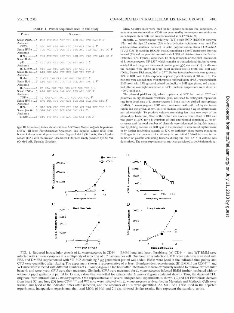
FIG. 1. Reduced intracellular growth of L. monocytogenes in CD44�/� BMM, lung, and heart fibroblasts. (A) CD44�/� and WT BMM wereinfected with L. monocytogenes at a multiplicity of infection of 0.2 bacteria per cell. One hour after infection BMM were extensively washed withPBS, and DMEM supplemented with 5% FCS containing 5 �g gentamicin per ml was added. BMM were lysed at the indicated time points, andCFU were quantified after plating. The experiment shown is representative of at least 10 independent experiments. (B) BMM from CD44�/� andWT mice were infected with different numbers of L. monocytogenes. One hour after infection cells were extensively washed to remove extracellularbacteria and were lysed; CFU were then measured. Similarly, CFU were measured for L. monocytogenes-infected BMM further incubated with orwithout 5 �g of gentamicin per ml for 15 min, a dose that was lethal for extracellular L. monocytogenes (data not shown). Thus, the depicted CFUoriginate from intracellular L. monocytogenes. One representative of several independent experiments is shown. (C and D) Fibroblasts derivedfrom heart (C) and lung (D) from CD44�/� and WT mice were infected with L. monocytogenes as described in Materials and Methods. Cells werewashed and lysed at the indicated times after infection, and the amounts of CFU were quantified. An MOI of 1:1 was used in the depictedexperiments. Independent experiments that used MOIs of 10:1 and 2:1 also showed similar results. Bars represent the standard errors.
TABLE 1. Primer sequences used in this study
Primer Sequence
Sense iNOS ......5� CCC TTC CGA AGT TTC TGG CAG CAG CAG C 3�Antisense
iNOS .............5� GGC TGT CAG AGC CTC GTG GCT TTG G 3�Sense IFN-� .....5� TGG ACC TGT GGG TTG TTG ACC TCA AAC TTG GC 3�Antisense
IFN-� ............5� TCG ATC TTG GCT TTG CAG CTC TTC CTC ATG GC 3�Sense IL-12
p40.................5� CGT GCT CAT GGC TGG TGC AAA G 3�Antisense
IL-12 p40 ......5� CTT CAT CTG CAA GTT CTT GGG C 3�Sense IL-1� ......5� ATG GCC AAA GTT CCT GAC TTG TTT 3�Antisense
IL-1� .............5� C CTT CAG CAA CAC GGG CTG GTC 3�Sense IL-6 ........5� ATG AAG TTC CTC TCT GCA AGA GAC T 3�Antisense
IL-6................5� CA CTA GGT TTG CCG AGT AGA TCT C 3�Sense TNF-�....5� ATG AGC ACA GAA AGC ATG ATC CGC 3�Antisense
TNF-� ...........5� CC AAA GTA GAC CTG CCC GGA CTC 3�Sense IFN-� .....5� GAC TCA TCT GCT GCT TGG AAT GCA ACC CTC 3�Antisense
IFN-� ............5� GAC TCA CTC CTT CTC CTC ACT CAG TCT TGC C 3�Sense �-actin....5� GTG GGC CGC TCT AGG CAC CAA 3�Antisense
�-actin...........5� CTC TTT GAT GTC ACG CAC GAT TTC 3�
VOL. 71, 2003 CD44-MEDIATED INTRACELLULAR LISTERIAL GROWTH 4103
on July 11, 2018 by guesthttp://iai.asm
.org/D
ownloaded from
bacterium, and growth of plasmid-containing bacteria represents the segregationof preexisting plasmids into daughter bacteria. After 4.5 h the total number ofplasmid-containing bacteria stopped increasing and was identical to the totalnumber of plasmids. Moreover, the total number of plasmids was not alteredduring the 5-h culture period. Thus, after 4.5 h each plasmid-containing bacteriahad one copy of the plasmid. At this time, 20 to 30% of total bacteria contained
pAUL-A. Aliquots of the culture grown for 4.5 h were frozen at �70°C and wereused later for infection of BMM. BMM were infected as described below, andCFU in lysates were measured in plates containing 5 �g of erythromycin/ml(measuring plasmid-containing bacteria) and in plates not containing erythro-mycin (measuring total number of bacteria). The relative proportion of pAUL-A-containing bacteria is a measurement of the growth rate, while the totalnumber of bacteria with pAUL-A is a measurement of killing by the host cell. Asimilar strategy has been previously used to evaluate toxicity during in vitro andin vivo infection with Salmonella enterica serovar Typhimurium (3, 21).
Differentiation of BMM. BMM were established as described previously (33).In brief, mice were killed by cervical dislocation, and femurs and tibias of thehind legs were dissected and flushed with 5 ml of ice-cold, sterile PBS. Bonemarrow cells (typically 2 107 to 3 107 per mouse) were washed and resus-pended in Dulbecco’s minimal essential medium (DMEM) containing glucoseand supplemented with 10% fetal calf serum (FCS), 20% L929 cell-conditionedmedium (as a source of macrophage-colony stimulating factor), 100 �g of strep-tomycin/ml, 100 U of penicillin/ml, and 10 mM HEPES buffer. The cells werecultured at 5 104 cells per well in 24-well plates for 7 days at 37°C, 5% CO2.BMM cultures were then created by washing vigorously with PBS to removenonadherent cells. Adherent cells were also harvested and counted by trypanblue exclusion. Typically, the yield of bone marrow cells was 2.0 105 to 4.0 105 BMM per well (24-well plates) after 7 days in culture, which is when theywere infected.
Fibroblast primary cultures. Fibroblasts were obtained by enzymatic digestionof hearts and lungs from 6- to 10-week-old CD44�/� or WT mice as describedpreviously (7). In brief, organs were minced in Iscove’s modified medium(IMDM) supplemented with 10% FCS, 100 �g of streptomycin/ml, 100 U ofpenicillin/ml, and 10 mM HEPES buffer and were incubated in IMDM contain-ing 1 mg of collagenase (type IV; Sigma, St. Louis, Mo.) per ml at 37°C for 10min. Released cells in the supernatant were collected, and undigested tissue wasfurther enzymatically treated for two more cycles. Recovered cells were pooledand plated, and highly enriched cultures of adherent cardiac and pulmonar cellswere recovered during the plating procedure. After the third passage, nearly allcells appeared to be fibroblasts by morphology, as also previously determined byimmunostaining (7). Such cultures were trypsinized and resuspended in IMDM,and 5 104 cells per well were seeded on a 24-well plate. Infection was per-formed 24 h after plating.
Infection and infectivity assay. BMM and fibroblasts cultured in 24-well plateswere cocultured with L. monocytogenes for 1 h at 37°C, 5% CO2. Cells were thenextensively washed with PBS to remove the extracellular bacteria. Triplicate wellswere used for all conditions. To prevent extracellular bacterial growth, cells werethen cultivated in DMEM containing 5% FCS and 5 �g of gentamicin per ml. Atdifferent time points after infection cells were washed with PBS and lysed with0.1% Triton X-100 in PBS, and aliquots of lysates were plated on BHI agarplates. Bacteria in two or three tenfold dilutions of the lysates were quantified.Plates were incubated overnight at 37°C and CFU were determined.
BMM were also infected in parallel with S. enterica serovar Typhimuriumstrain 14028 (American Type Culture Collection). S. enterica serovar Typhi-murium was grown in Luria broth (LB) agar plates overnight. For this purpose,bacterial colonies were diluted in PBS at a concentration of 5 108 bacteria perml. Ninety microliters of bacteria was coincubated with 10 �l of C57Bl/6 normalmouse serum for 30 min at 37°C. Bacteria were further diluted at 106 per ml inDMEM-5% FCS. BMM were cocultured with S. enterica serovar Typhimuriumand were centrifuged at 500 g for 5 min at room temperature. Cells wereincubated for 1 h at 37°C and were extensively washed with PBS, and DMEMcontaining 5 �g of gentamicin per ml was then added. Cells were lysed at theindicated time points after infection as described above, and CFU in cell lysateswere quantified on LB agar plates after overnight incubation.
Immunostaining and flow cytometry. BMM were phenotypically characterizedby fluorescence-activated cell sorter analysis. For this purpose, BMM were de-tached from plates by using a cell scraper and were incubated with PBS contain-ing 10% normal goat serum (Sigma) to block unspecific binding of the secondaryantibody. Cells were then incubated with purified monoclonal rat antibody toF4/80, Mac-3, CD80, or GR-1 for 30 min on ice. Cells were then washed andincubated with biotinylated goat anti-rat serum for 30 min on ice. Anti-CD44,-CD19, -CD3, and -CD8 antibodies were directly labeled with fluorescein iso-thiocyanate. Anti-CD4, -CD14, and -NK1.1 antibodies were directly labeled withphycoerythrin. Anti-MHC class II (Ia) and -ICAM-1 (MALA-2) antibodies werebiotinylated. Isotype-matched control rat immunoglobulins (Igs) directly labeledwith fluorescein isothiocyanate, phycoerythrin, or biotin were used as controls.When staining with biotinylated or directly labeled antibodies, BMM were pre-incubated with anti-CD16/CD32 antibodies to block Fc� receptors. After incu-bation with biotinylated antibodies, cells were washed, stained with neutroavidin
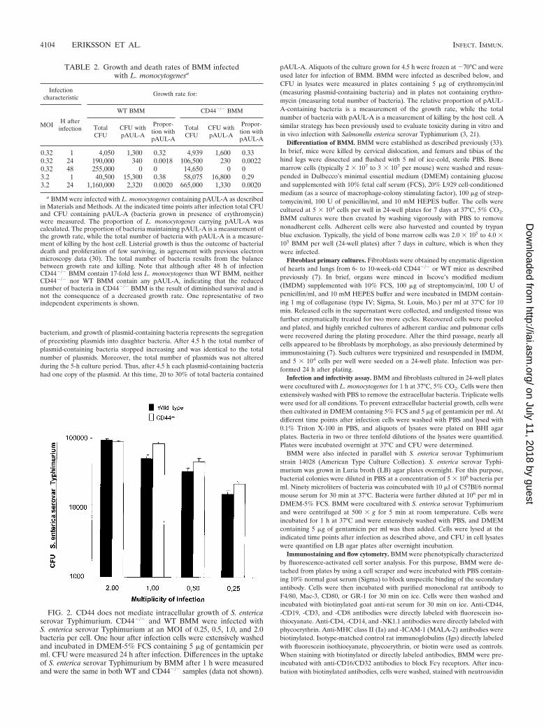
FIG. 2. CD44 does not mediate intracellular growth of S. entericaserovar Typhimurium. CD44�/� and WT BMM were infected withS. enterica serovar Typhimurium at an MOI of 0.25, 0.5, 1.0, and 2.0bacteria per cell. One hour after infection cells were extensively washedand incubated in DMEM-5% FCS containing 5 �g of gentamicin perml. CFU were measured 24 h after infection. Differences in the uptakeof S. enterica serovar Typhimurium by BMM after 1 h were measuredand were the same in both WT and CD44�/� samples (data not shown).
TABLE 2. Growth and death rates of BMM infectedwith L. monocytogenesa
Infectioncharacteristic Growth rate for:
MOI H afterinfection
WT BMM CD44�/� BMM
TotalCFU
CFU withpAUL-A
Propor-tion withpAUL-A
TotalCFU
CFU withpAUL-A
Propor-tion withpAUL-A
0.32 1 4,050 1,300 0.32 4,939 1,600 0.330.32 24 190,000 340 0.0018 106,500 230 0.00220.32 48 255,000 0 0 14,650 0 03.2 1 40,500 15,300 0.38 58,075 16,800 0.293.2 24 1,160,000 2,320 0.0020 665,000 1,330 0.0020
a BMM were infected with L. monocytogenes containing pAUL-A as describedin Materials and Methods. At the indicated time points after infection total CFUand CFU containing pAUL-A (bacteria grown in presence of erythromycin)were measured. The proportion of L. monocytogenes carrying pAUL-A wascalculated. The proportion of bacteria maintaining pAUL-A is a measurement ofthe growth rate, while the total number of bacteria with pAUL-A is a measure-ment of killing by the host cell. Listerial growth is thus the outcome of bacterialdeath and proliferation of few surviving, in agreement with previous electronmicroscopy data (30). The total number of bacteria results from the balancebetween growth rate and killing. Note that although after 48 h of infectionCD44�/� BMM contain 17-fold less L. monocytogenes than WT BMM, neitherCD44�/� nor WT BMM contain any pAUL-A, indicating that the reducednumber of bacteria in CD44�/� BMM is the result of diminished survival and isnot the consequence of a decreased growth rate. One representative of twoindependent experiments is shown.
4104 ERIKSSON ET AL. INFECT. IMMUN.
on July 11, 2018 by guesthttp://iai.asm
.org/D
ownloaded from
Alexa (Molecular Probes, Eugene, Oreg.), and analyzed with a FACScan (Bec-ton-Dickinson). All primary antibodies were purchased from Pharmingen (SanDiego, Calif.), except for F4/80 (Serotec, Raleigh, N.C.).
Immunostaining of intracellular bacteria. To analyze intracellular bacteriallocalization, BMM were infected with the L. monocytogenes NF-L357 strain,which contains a chromosomal actA-gfp-plcB transcriptional fusion, and wereimmunostained with anti-L. monocytogenes serum. actA/plcB expression (and,therefore, gfp) occurs shortly after L. monocytogenes gains access to the host cellcytosol (16). BMM grown on 13-mm2-diameter coverslides were infected withstrain NF-L357 as described above. At different time points after infection cellswere washed with PBS and fixed with 4% paraformaldehyde in PBS for 15 min,and the cell membrane was lysed with 0.5% Triton X-100 in PBS for 15 min atroom temperature. Thereafter cells were washed with PBS, and unspecific bind-
ing was blocked by incubation with PBS containing 5% bovine serum albumin(BSA) and 0.5% Triton X-100. The cultures were then incubated with rabbitanti-L. monocytogenes serum (Difco Laboratories, Detroit, Mich.) diluted 1:800in PBS supplemented with 1% BSA, 0.5% Triton X-100, and 0.01% NaN3 for 1 hat room temperature. The cells were then washed with PBS and were stainedwith a Texas-Red-conjugated donkey anti-rabbit IgG (H�L) (Jackson Immu-noResearch Laboratories, West Baltimore Pike, Pa.) diluted 1:100 in PBS con-taining 1% BSA for 1 h at room temperature and were washed again. Coverslides were analyzed with a fluorescent microscope.
Fluorescent staining of actin filaments. When residing in cytoplasm, L. mono-cytogenes generates an actin comet tail which confers movement to the bacteria,enabling it to enter adjacent cells. Listerial actin tails were stained with fluoro-chrome-labeled phalloidin. In brief, BMM grown on 13-mm2-diameter cover-
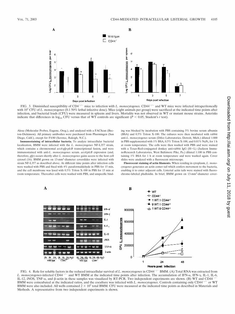
FIG. 3. Diminished susceptibility of CD4�/� mice to infection with L. monocytogenes. CD44�/� and WT mice were infected intraperitoneallywith 105 CFU of L. monocytogenes (0.1 50% lethal infective dose). Mice (eight animals per group) were sacrificed at the indicated time points afterinfection, and bacterial loads (CFU) were measured in spleens and livers. Mortality was not observed in WT or mutant mouse strains. Asterisksindicate that differences in log10 CFU versus that of WT controls are significant (P 0.05, Student’s t test).
FIG. 4. Role for soluble factors in the reduced intracellular survival of L. monocytogenes in CD44�/� BMM. (A) Total RNA was extracted fromL. monocytogenes-infected CD44�/� and WT BMM at the indicated time points after infection. The accumulation of IFN-�, IFN-�, IL-1, IL-6,IL-12, iNOS, TNF-�, and �-actin in these samples was visualized by RT-PCR. Two independent experiments are shown. (B) WT and CD44�/�
BMM were coincubated at the indicated ratios, and the coculture was infected with L. monocytogenes. Controls containing only CD44�/� or WTBMM were also included. All wells contained 2 105 total BMM. CFU were measured at the indicated time points as described in Materials andMethods. A representative from two independent experiments is shown.
VOL. 71, 2003 CD44-MEDIATED INTRACELLULAR LISTERIAL GROWTH 4105
on July 11, 2018 by guesthttp://iai.asm
.org/D
ownloaded from
slides and infected with L. monocytogenes were used. At indicated time pointsafter infection cells were washed with PBS and were fixed with PBS containing4% formaldehyde, and the cell membrane was lysed with PBS containing 0.5%Triton X-100. Cells were washed and incubated with anti-L. monocytogenesserum as described above, washed, and stained with Alexa Fluor 594-labeled goatanti-rabbit IgG (H�L) (Molecular Probes). The cells were then washed andstained with Alexa Fluor 488-labeled phalloidin (Molecular Probes) for 20 minat room temperature, and coverslides were read with a fluorescent microscope.
RT-PCR assay. Tumor necrosis factor alpha (TNF-�), IFN-�, IFN-�, interleu-kin-1 (IL-1), IL-6, IL-12p40, inducible nitric oxide synthase (iNOS), and �-actinmRNA in freshly extracted RNA from L. monocytogenes-infected or HA-treatedWT or CD44�/� BMM were visualized by reverse transcription (RT)-PCRassays as previously described (34).
Reactions were carried out for 37 cycles (38 cycles for IL-12 and 30 cycles for�-actin) in a thermal cycler with an annealing step at 60°C (67°C for IL-12).
The primer sequences for the amplification of the cDNA are listed in Table 1.IFN-� primer sequences were obtained from Clontech. These primers recognizea consensus sequence present in IFN-�1, IFN-�2, and IFN-�7 genes.
RESULTS
We first compared the outcome of L. monocytogenes infec-tion in CD44�/� and WT BMM. Surprisingly, CD44�/� BMMshowed a strikingly reduced growth of intracellular L. mono-cytogenes compared to that of WT cells (Fig. 1A). The reducednumbers of L. monocytogenes in CD44�/� BMM was not dueto a diminished bacterial uptake by the mutant cells, sincesimilar numbers of intracellular L. monocytogenes were mea-sured in mutant and WT BMM at 1 h after infection (Fig. 1B).By using L. monocytogenes containing one copy of the temper-
ature-sensitive, erythromycin-resistant gene containing plas-mid pAUL-A per bacteria, it was shown that the reducedintracellular number of L. monocytogenes in CD44�/� BMMwas due to increased rate of death rather than to reducedgrowth rate of the bacteria (Table 2).
To test for the presence of CD44-facilitated listerial growthin cell types other than BMM, we studied L. monocytogenesinfection in primary lung and heart fibroblast cultures fromCD44�/� and WT mice. Listerial number was again lower inCD44�/� cells from both sources (Fig. 1C and D).
Next we analyzed whether CD44 is a general supporter ofintracellular growth of bacterial pathogens. However, no dif-ferences in uptake (data not shown) or intracellular growth ofS. enterica serovar Typhimurium in CD44�/� and WT BMMwere found (Fig. 2).
We next explored whether reduced in vitro growth ofL. monocytogenes in CD44�/� BMM related to a diminishedsusceptibility of CD44�/� mice to bacterial infection in vivo.WT and CD44�/� mice were infected intraperitoneally with105 L. monocytogenes cells, and bacterial numbers were quan-tified in spleen and liver at different time points after infection.The livers from CD44�/� mice showed a significantly dimin-ished number of L. monocytogenes compared to that of WTcontrols, whereas bacterial numbers recovered from thespleens of both mouse strains were similar (Fig. 3).
We found no variation in phenotype of CD44�/� BMM
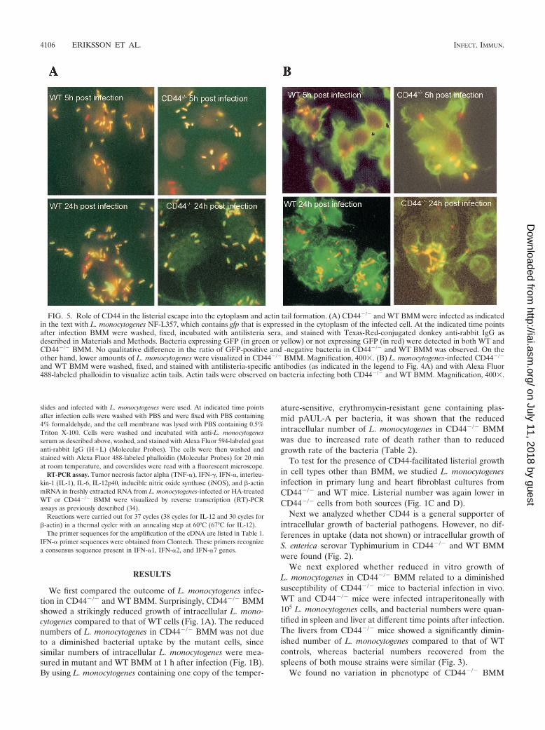
FIG. 5. Role of CD44 in the listerial escape into the cytoplasm and actin tail formation. (A) CD44�/� and WT BMM were infected as indicatedin the text with L. monocytogenes NF-L357, which contains gfp that is expressed in the cytoplasm of the infected cell. At the indicated time pointsafter infection BMM were washed, fixed, incubated with antilisteria sera, and stained with Texas-Red-conjugated donkey anti-rabbit IgG asdescribed in Materials and Methods. Bacteria expressing GFP (in green or yellow) or not expressing GFP (in red) were detected in both WT andCD44�/� BMM. No qualitative difference in the ratio of GFP-positive and -negative bacteria in CD44�/� and WT BMM was observed. On theother hand, lower amounts of L. monocytogenes were visualized in CD44�/� BMM. Magnification, 400. (B) L. monocytogenes-infected CD44�/�
and WT BMM were washed, fixed, and stained with antilisteria-specific antibodies (as indicated in the legend to Fig. 4A) and with Alexa Fluor488-labeled phalloidin to visualize actin tails. Actin tails were observed on bacteria infecting both CD44�/� and WT BMM. Magnification, 400.
4106 ERIKSSON ET AL. INFECT. IMMUN.
on July 11, 2018 by guesthttp://iai.asm
.org/D
ownloaded from
compared to that of WT BMM that could be related to thediminished growth of L. monocytogenes. CD44�/� and WTBMM both expressed high levels of Mac-3, GR-1, F4/80, andMHC class II, expressed moderate levels of CD80 and CD14,and were negative for T, B, or NK cell markers, such as CD3,CD4, CD8, CD19, and NK1.1 as measured by fluorescence-activated cell sorting (data not shown).
We then studied if different levels of cellular activation couldaccount for the diminished intracellular growth of L. monocy-togenes in CD44�/� BMM. For this purpose RNA was ex-tracted from BMM before and at different time points afterinfection, and the presence of transcripts for inflammatorycytokines was evaluated by RT-PCR. The levels of IFN-�,IFN-�, IL-12p40, IL-1, IL-6, iNOS, and TNF-� mRNA wereall increased in L. monocytogenes-infected WT and CD44�/�
BMM. CD44�/� BMM contained equal or lower levels of alltranscripts than WT BMM (Fig. 4A). Thus, the enhanceddeath rate of L. monocytogenes in CD44�/� BMM is not linkedto the presence of higher levels of inflammatory cytokine tran-scripts.
We next analyzed intracellular bacterial growth by usingvarious cocultures of CD44�/� and WT BMM. We hypothe-sized that an increased secretion of inflammatory cytokinesfrom CD44�/� BMM or the secretion of soluble factors pro-moting bacterial growth by WT BMM could modify the liste-rial numbers in either direction in the coculture. No shift in theexpected bacterial numbers in these cocultures was detected(Fig. 4B), suggesting that soluble factors account neither forthe enhanced listerial growth in the presence of CD44 nor forthe increased bacterial death rate in its absence.
CD44 might be necessary for escape of L. monocytogenesfrom phagosome to cytoplasm. To study this, BMM were in-
fected with the NF-L357 strain of L. monocytogenes containinga gfp gene only expressed when bacteria localize in the cyto-plasm (16). Staining gfp-expressing bacteria with anti-L. mono-cytogenes sera, we found similar ratios of gfp-expressing and-nonexpressing bacteria in CD44�/� and WT BMM. WT BMMshowed higher total numbers of bacteria than CD44�/� BMM.Thus, CD44 plays no detectable role in the escape of L. mono-cytogenes from the phagosome into the cytoplasm (Fig. 5A).
CD44 may participate in the formation of actin tails byL. monocytogenes, thereby reducing bacterial growth in CD44�/�
BMM. However, intracellular L. monocytogenes with actin tailswas observed both in CD44�/� and in WT BMM (Fig. 5B).
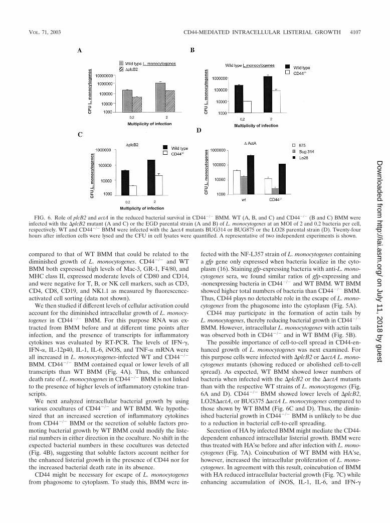
The possible importance of cell-to-cell spread in CD44-en-hanced growth of L. monocytogenes was next examined. Forthis purpose cells were infected with �plcB2 or �actA L. mono-cytogenes mutants (showing reduced or abolished cell-to-cellspread). As expected, WT BMM showed lower numbers ofbacteria when infected with the �plcB2 or the �actA mutantsthan with the respective WT strains of L. monocytogenes (Fig.6A and D). CD44�/� BMM showed lower levels of �plcB2,LO28�actA, or BUG375 �actA L. monocytogenes compared tothose shown by WT BMM (Fig. 6C and D). Thus, the dimin-ished bacterial growth in CD44�/� BMM is unlikely to be dueto a reduction in bacterial cell-to-cell spreading.
Secretion of HA by infected BMM might mediate the CD44-dependent enhanced intracellular listerial growth. BMM werethus treated with HA’se before and after infection with L. mono-cytogenes (Fig. 7A). Coincubation of WT BMM with HA’se,however, increased the intracellular proliferation of L. mono-cytogenes. In agreement with this result, coincubation of BMMwith HA reduced intracellular bacterial growth (Fig. 7C) whileenhancing accumulation of iNOS, IL-1, IL-6, and IFN-�
FIG. 6. Role of plcB2 and actA in the reduced bacterial survival in CD44�/� BMM. WT (A, B, and C) and CD44�/� (B and C) BMM wereinfected with the �plcB2 mutant (A and C) or the EGD parental strain (A and B) of L. monocytogenes at an MOI of 2 and 0.2 bacteria per cell,respectively. WT and CD44�/� BMM were infected with the �actA mutants BUG314 or BUG875 or the LO28 parental strain (D). Twenty-fourhours after infection cells were lysed and the CFU in cell lysates were quantified. A representative of two independent experiments is shown.
VOL. 71, 2003 CD44-MEDIATED INTRACELLULAR LISTERIAL GROWTH 4107
on July 11, 2018 by guesthttp://iai.asm
.org/D
ownloaded from

mRNA (Fig. 7D). Control experiments showed that HA assuch did not affect extracellular growth of L. monocytogenes(Fig. 7B). However, HA and HA’se also affected listerial pro-liferation in CD44�/� BMM in a manner similar to that of WTBMM (Fig. 7A and C). Thus, hyaloadherins other than CD44participate in HA-mediated control of listerial infection. Toanalyze whether CD44 plays a role in modulation of intracel-lular bacterial growth by HA, BMM were treated with mono-clonal antibodies neutralizing (KM 81) or not neutralizing(KM 703) the HA binding ability of CD44 (47). Anti-MHCclass I and anti-CD4 monoclonal antibodies were used as con-trols. Incubation of WT but not CD44�/� BMM with KM 81resulted in an increased intracellular proliferation of L. mono-cytogenes, whereas no such consequence was observed afterincubation of BMM with KM 703 or the control monoclonalantibodies (Fig. 8). Thus, whereas signaling via CD44 facili-tates listerial growth, addition of HA can restrict growth oc-curring in part via binding to CD44.
Proteoglycan forms of CD44 also exhibit affinity for mole-cules, such as chondroitin sulfate, HS, fibronectin, and osteo-
pontin (31). Treatment of BMM with chondroitin sulfatasealso enhanced intracellular listerial growth (Fig. 9A), whereasno effect on the outcome of infection was observed upon add-ing HS or HS’se (Fig. 9B and C).
DISCUSSION
Herein we demonstrate that the presence of CD44 facilitatesintracellular growth of L. monocytogenes in murine systems.Changes in bacterial proliferation did not relate to alteredbacterial phagocytosis or secretion of proinflammatory cyto-kines in CD44�/� BMM. The presence of CD44 increasedbacterial survival without altering growth rate and was notrequired for bacterial escape from primary phagosome intocytoplasm or for actin tail formation. CD44-facilitated listerialgrowth does not depend on functional cell-to-cell spreading byL. monocytogenes. These results altogether suggest that CD44signaling affects survival of L. monocytogenes in the cytoplasmof the primary infected cell. In agreement with this suggestion,inhibitors of tyrosine protein kinases, phosphatidylinositol 3-
FIG. 7. Effect of addition or depletion of HA in the intracellular growth of L. monocytogenes in BMM. (A) CD44�/� and WT BMM werecoincubated with 2 U of HA’se per ml of DMEM-5% FCS at 37°C, 5% CO2 1 h before and during infection with L. monocytogenes. The bacterialload (CFU) of treated and control BMM was quantified 24 h after infection. A representative of four independent experiments is depicted.Addition of HA (250 kDa) to cocultures of HA’se and BMM completely inhibited the enzyme-mediated enhancement of bacterial proliferation,confirming the specificity of the enzymatic reaction (data not shown). (B) L. monocytogenes was grown at 37°C in BHI medium in the presenceof 50 and 500 �g of HA (190 kDa) or in the absence of HA. CFU were recorded in culture aliquots at the indicated time points of incubation.(C) WT and CD44�/� BMM were treated with 10 and 100 �g of HA (190 kDa) per ml 1 day before and during infection with L. monocytogenes.The bacterial load (CFU) of treated and control BMM was quantified at the indicated time points after infection. A representative from twoindependent experiments is shown. (D) WT BMM were treated with 100 and 500 �g of HA/ml (190 kDa) diluted in DMEM-5% FCS. The presenceof IFN-�, iNOS, IL-6, IL-1�, and �-actin mRNA in treated and control BMM was determined by RT-PCR at the indicated hours after treatment.A representative from two independent experiments is shown.
4108 ERIKSSON ET AL. INFECT. IMMUN.
on July 11, 2018 by guesthttp://iai.asm
.org/D
ownloaded from

kinase, and actin polymerization enzymes that mediate CD44signaling all reduced listerial growth in WT macrophages (datanot shown). These inhibitors, however, do not uniquely affectCD44 activation. To our knowledge, such a role for CD44 indetermining the intracellular fate of bacteria has not beendescribed. This CD44-enhanced intracellular listerial survivalwas observed both in cultures of primary macrophages andfibroblasts and is likely to be directly related to diminishedL. monocytogenes numbers in livers of CD44�/�-infected mice.The presence of different immune effector cells (10) partici-pating in listerial control in spleen and liver could account forthe dissimilar involvement of CD44 in listerial control in theseorgans. Alternatively, CD44-mediated intracellular listerialproliferation could be more prevalent in liver compared to thatin spleen cell populations.
On the other hand, CD44 did not affect the intracellulargrowth of another intracellular pathogen, S. enterica serovarTyphimurium.
We plan to use cells transfected with CD44 lacking intracel-lular protein domains necessary for different signaling path-ways to further dissect the role of this cell receptor in listerialgrowth. CD44 undergoes alternative splicing (encompassing 10variant exons) giving rise to isoforms that probably show dif-ferences in their biological functions (31), and it could relate tothe regulatory function of the receptor during infection with
L. monocytogenes. Alternative splicing will not take place in thetransfected molecule. Affinity of CD44 for ligands is dependenton posttranslational modifications, such as glycosylation, thatare cell type and growth condition specific (38), and it could bealso altered by listerial infection. CD44 has been suggested toact as a functional coreceptor for hepatocyte growth factor/scatter factor by binding and presenting growth factor to thereceptor tyrosine kinase c-Met (44). The bacterial surface pro-tein InlB promotes not only internalization (5, 14) but also theescape into the cytoplasm of L. monocytogenes in mammaliancells (19). In turn, the Met receptor mediates InIB-dependentinternalization of L. monocytogenes (37). However, mutantslacking InlB (as well as InlA and InlA/B) showed diminishedintracellular growth in CD44�/� BMM compared to that ofWT BMM (data not shown).
There is indirect evidence that the ability to replicate withinhost cells requires specific adaptations in the microbe. A hex-ose phosphate transporter (hpt) has been identified as a viru-lence factor involved in the listerial replication phase (9, 17).Whether listerial hpt and other virulence factor(s) specificallyinteract with CD44 signaling and promote listerial growth willbe further studied.
CD44 signaling has been indicated to affect the outcome ofother extracellular and intracellular infections. Binding of theHA capsule of Streptococcus pyogenes to CD44 mediates cy-toskeletal rearrangements (12, 13). Such rearrangements dis-rupt the intercellular junctions and permit bacterial passagebetween epithelial cells (13). Interaction of the Shigella flexneriIpaB-IpaC-secreted complex with CD44 has also been sug-gested to be involved in the cellular uptake of shigella and inshigella-induced cytoskeletal rearrangements (39).
Inflammatory stimuli promote local accumulation of LMWHA fragments (6, 42). We confirmed previous reports (22, 23,26, 29, 41) indicating that coincubation of cells with LMW HAincreased proinflammatory transcripts and demonstrate fur-ther that such coincubation diminished listerial growth in mac-rophages. Eliminating HA via HA’se treatment acceleratedlisterial proliferation, which is in line with the possibility thatmacrophage-derived HA acts by binding to hyaloadherins anddiminishes listerial growth. A role for CD44 in HA-mediatedcontrol of listerial growth was further suggested by our findingof an accelerated listerial growth in WT BMM treated with amonoclonal antibody that selectively interferes with HA bind-ing to CD44 (47). However, and in line with the fact that CD44
FIG. 8. Increased intracellular listerial growth in BMM coincubat-ed with a monoclonal antibody specifically neutralizing the HA bindingability of CD44. CD44�/� and WT BMM were treated with differentconcentrations of the anti-CD44 monoclonal antibodies KM 81 andKM 703 1 h before and during infection with L. monocytogenes. Anti-CD4 (L3T4) and anti-MHC class I (HB51) antibodies were used ascontrols. The bacterial load (CFU) of treated and control BMM wasquantified after 24 h of infection.
FIG. 9. Effect of treatment with chondroitinase, HS’se, and HS on the intracellular growth of L. monocytogenes. CD44�/� and WT BMM werecoincubated with 0.01 of U chondroitinase (A), 0.5 U of HS’se (B), or 20 �g of HS (C) per ml of DMEM-5% FCS at 37°C 1 h before and duringinfection with L. monocytogenes. The bacterial load (CFU) of treated and control BMM was quantified at 24 h (A) and at the indicated time points(B and C) after infection. A representative from two independent experiments for each condition is shown.
VOL. 71, 2003 CD44-MEDIATED INTRACELLULAR LISTERIAL GROWTH 4109
on July 11, 2018 by guesthttp://iai.asm
.org/D
ownloaded from
is not the sole receptor reacting with HA (1, 42, 46), othercellular receptors also participate in the control of listerialinfection, since HA’se enhanced listerial growth in CD44�/�
BMM and coincubation with HA diminished listerial growth inthese cells. Adding complexity to this analysis is the fact thatCD44 not only binds HA but also binds HS, chondroitin sul-fate, collagen, fibronectin, and osteopontin (42). In this studywe found that treatment of BMM with chondroitin sulfatasebut not HS’se increased listerial intracellular growth, a findingwe will further explore.
Altogether our results suggest a dual role of CD44 in theoutcome of infection with L. monocytogenes; whereas a quies-cent receptor (or low levels of ligands) will deliver signals thatwill facilitate intracellular listerial growth, ligands binding toCD44 will diminish such response.
ACKNOWLEDGMENTS
This work was supported by the Karolinska Institute; The Founda-tion for Knowledge and Competence Development, Stockholm, Swe-den; The Swedish Cancer Society; The Swedish Medical ResearchCouncil; and Q-Med AB, Uppsala, Sweden.
We thank M. Rhen, Microbiology & Tumorbiology Center, Karo-linska Institute, for providing the S. enterica serovar Typhimuriumbacterial strain and for critical analysis of the manuscript; O. Wik,Q-Med AB, for providing the HA preparation used; and T. Mak,Amgen, Ontario, Canada, for providing the CD44�/� mice. We aregrateful to P. Cossart, Unite des Ineractions Bacteries-Cellules, Insti-tut Pasteur, Paris, France; N. Freitag, Wayne State University, Schoolof Medicine, Detroit, Mich.; and T. Chakraborty, Institut fur Mediz-inische Mikrobiologie, Justus-Liebig-Universitat Gie�en, Gie�en,Germany, for their kind gifts of the L. monocytogenes strains. We thankBerit Olsson for excellent technical assistance.
REFERENCES
1. Banerji, S., J. Ni, S. X. Wang, S. Clasper, J. Su, R. Tammi, M. Jones, andD. G. Jackson. 1999. LYVE-1, a new homologue of the CD44 glycoprotein,is a lymph-specific receptor for hyaluronan. J. Cell Biol. 144:789–801.
2. Beck-Schimmer, B., B. Oertli, T. Pasch, and R. P. Wuthrich. 1998. Hyalu-ronan induces monocyte chemoattractant protein-1 expression in renal tu-bular epithelial cells. J. Am. Soc. Nephrol. 9:2283–2290.
3. Benjamin, W. H., Jr., P. Hall, S. J. Roberts, and D. E. Briles. 1990. Theprimary effect of the Ity locus is on the rate of growth of Salmonella typhi-murium that are relatively protected from killing. J. Immunol. 144:3143–3151.
4. Blass, S. L., E. Pure, and C. A. Hunter. 2001. A role for CD44 in theproduction of IFN-gamma and immunopathology during infection with Toxo-plasma gondii. J. Immunol. 166:5726–5732.
5. Braun, L., H. Ohayon, and P. Cossart. 1998. The InIB protein of Listeriamonocytogenes is sufficient to promote entry into mammalian cells. Mol.Microbiol. 27:1077–1087.
6. Cao, H. J., H. S. Wang, Y. Zhang, H. Y. Lin, R. P. Phipps, and T. J. Smith.1998. Activation of human orbital fibroblasts through CD40 engagementresults in a dramatic induction of hyaluronan synthesis and prostaglandinendoperoxide H synthase-2 expression. Insights into potential pathogenicmechanisms of thyroid-associated ophthalmopathy. J. Biol. Chem. 273:29615–29625.
7. Castanos-Velez, E., S. Maerlan, L. M. Osorio, F. Aberg, P. Biberfeld, A. Orn,and M. E. Rottenberg. 1998. Trypanosoma cruzi infection in tumor necrosisfactor receptor p55-deficient mice. Infect. Immun. 66:2960–2968.
8. Chakraborty, T., M. Leimeister-Wachter, E. Domann, M. Hartl, W. Goebel,T. Nichterlein, and S. Notermans. 1992. Coordinate regulation of virulencegenes in Listeria monocytogenes requires the product of the prfA gene. J. Bac-teriol. 174:568–574.
9. Chico-Calero, I., M. Suarez, B. Gonzalez-Zorn, M. Scortti, J. Slaghuis, W.Goebel, and J. A. Vazquez-Boland. 2002. Hpt, a bacterial homolog of themicrosomal glucose-6-phosphate translocase, mediates rapid intracellularproliferation in Listeria. Proc. Natl. Acad. Sci. USA 99:431–436.
10. Conlan, J. W. 1999. Early host-pathogen interactions in the liver and spleenduring systemic murine listeriosis: an overview. Immunobiology 201:178–187.
11. Cossart, P., and H. Bierne. 2001. The use of host cell machinery in thepathogenesis of Listeria monocytogenes. Curr. Opin. Immunol. 13:96–103.
12. Cywes, C., I. Stamenkovic, and M. R. Wessels. 2000. CD44 as a receptor for
colonization of the pharynx by group A Streptococcus. J. Clin. Investig.106:995–1002.
13. Cywes, C., and M. R. Wessels. 2001. Group A Streptococcus tissue invasionby CD44-mediated cell signalling. Nature 414:648–652.
14. Dramsi, S., I. Biswas, E. Maguin, L. Braun, P. Mastroeni, and P. Cossart.1995. Entry of Listeria monocytogenes into hepatocytes requires expression ofinIB, a surface protein of the internalin multigene family. Mol. Microbiol.16:251–261.
15. Eugene, E., I. Hoffmann, C. Pujol, P. O. Couraud, S. Bourdoulous, and X.Nassif. 2002. Microvilli-like structures are associated with the internalizationof virulent capsulated Neisseria meningitidis into vascular endothelial cells.J. Cell Sci. 115:1231–1241.
16. Freitag, N. E., and K. E. Jacobs. 1999. Examination of Listeria monocyto-genes intracellular gene expression by using the green fluorescent protein ofAequorea victoria. Infect. Immun. 67:1844–1852.
17. Goetz, M., A. Bubert, G. Wang, I. Chico-Calero, J. A. Vazquez-Boland, M.Beck, J. Slaghuis, A. A. Szalay, and W. Goebel. 2001. Microinjection andgrowth of bacteria in the cytosol of mammalian host cells. Proc. Natl. Acad.Sci. USA 98:12221–12226.
18. Gouin, E., P. Dehoux, J. Mengaud, C. Kocks, and P. Cossart. 1995. iactA ofListeria ivanovii, although distantly related to Listeria monocytogenes actA,restores actin tail formation in an L. monocytogenes actA mutant. Infect.Immun. 63:2729–2737.
19. Gregory, S. H., A. J. Sagnimeni, and E. J. Wing. 1997. Internalin B promotesthe replication of Listeria monocytogenes in mouse hepatocytes. Infect. Im-mun. 65:5137–5141.
20. Guleria, I., and J. W. Pollard. 2001. Aberrant macrophage and neutrophilpopulation dynamics and impaired Th1 response to Listeria monocytogenes incolony-stimulating factor 1-deficient mice. Infect. Immun. 69:1795–1807.
21. Gulig, P. A., T. J. Doyle, M. J. Clare-Salzler, R. L. Maiese, and H. Matsui.1997. Systemic infection of mice by wild-type but not Spv- Salmonella typhi-murium is enhanced by neutralization of gamma interferon and tumor ne-crosis factor alpha. Infect. Immun. 65:5191–5197.
22. Haslinger, B., S. Mandl-Weber, A. Sellmayer, and T. Sitter. 2001. Hyaluro-nan fragments induce the synthesis of MCP-1 and IL-8 in cultured humanperitoneal mesothelial cells. Cell Tissue Res. 305:79–86.
23. Hodge-Dufour, J., P. W. Noble, M. R. Horton, C. Bao, M. Wysoka, M. D.Burdick, R. M. Strieter, G. Trinchieri, and E. Pure. 1997. Induction of IL-12and chemokines by hyaluronan requires adhesion-dependent priming ofresident but not elicited macrophages. J. Immunol. 159:2492–2500.
24. Horton, M. R., C. M. McKee, C. Bao, F. Liao, J. M. Farber, J. Hodge-DuFour, E. Pure, B. L. Oliver, T. M. Wright, and P. W. Noble. 1998.Hyaluronan fragments synergize with interferon-gamma to induce the C-X-C chemokines mig and interferon-inducible protein-10 in mouse macro-phages. J. Biol. Chem. 273:35088–35094.
25. Kocks, C., E. Gouin, M. Tabouret, P. Berche, H. Ohayon, and P. Cossart.1992. L. monocytogenes-induced actin assembly requires the actA geneproduct, a surface protein. Cell 68:521–531.
26. McKee, C. M., C. J. Lowenstein, M. R. Horton, J. Wu, C. Bao, B. Y. Chin,A. M. Choi, and P. W. Noble. 1997. Hyaluronan fragments induce nitric-oxide synthase in murine macrophages through a nuclear factor �B-depen-dent mechanism. J. Biol. Chem. 272:8013–8018.
27. Noble, P. W. 2002. Hyaluronan and its catabolic products in tissue injury andrepair. Matrix Biol. 21:25–29.
28. Noble, P. W., F. R. Lake, P. M. Henson, and D. W. Riches. 1993. Hyaluronateactivation of CD44 induces insulin-like growth factor-1 expression by atumor necrosis factor-alpha-dependent mechanism in murine macrophages.J. Clin. Investig. 91:2368–2377.
29. Oertli, B., B. Beck-Schimmer, X. Fan, and R. P. Wuthrich. 1998. Mecha-nisms of hyaluronan-induced up-regulation of ICAM-1 and VCAM-1 ex-pression by murine kidney tubular epithelial cells: hyaluronan triggers celladhesion molecule expression through a mechanism involving activation ofnuclear factor-kappa B and activating protein-1. J. Immunol. 161:3431–3437.
30. Portnoy, D. A., R. D. Schreiber, P. Connelly, and L. G. Tilney. 1989. Gammainterferon limits access of Listeria monocytogenes to the macrophage cyto-plasm. J. Exp. Med. 170:2141–2146.
31. Pure, E., and C. A. Cuff. 2001. A crucial role for CD44 in inflammation.Trends Mol. Med. 7:213–221.
32. Rosen, H., S. Gordon, and R. J. North. 1989. Exacerbation of murine liste-riosis by a monoclonal antibody specific for the type 3 complement receptorof myelomonocytic cells. Absence of monocytes at infective foci allows Lis-teria to multiply in nonphagocytic cells. J. Exp. Med. 170:27–37.
33. Rothfuchs, A. G., D. Gigliotti, K. Palmblad, U. Andersson, H. Wigzell, andM. E. Rottenberg. 2001. IFN-alpha beta-dependent, IFN-gamma secretionby bone marrow-derived macrophages controls an intracellular bacterialinfection. J. Immunol. 167:6453–6461.
34. Rottenberg, M. E., A. Gigliotti Rothfuchs, D. Gigliotti, M. Ceausu, C. Une,V. Levitsky, and H. Wigzell. 2000. Regulation and role of IFN-gamma in theinnate resistance to infection with Chlamydia pneumoniae. J. Immunol. 164:4812–4818.
35. Schluter, D., E. Domann, C. Buck, T. Hain, H. Hof, T. Chakraborty, and M.Deckert-Schluter. 1998. Phosphatidylcholine-specific phospholipase C from
4110 ERIKSSON ET AL. INFECT. IMMUN.
on July 11, 2018 by guesthttp://iai.asm
.org/D
ownloaded from

Listeria monocytogenes is an important virulence factor in murine cerebrallisteriosis. Infect. Immun. 66:5930–5938.
36. Schmits, R., J. Filmus, N. Gerwin, G. Senaldi, F. Kiefer, T. Kundig, A.Wakeham, A. Shahinian, C. Catzavelos, J. Rak, C. Furlonger, A. Zakarian,J. J. Simard, P. S. Ohashi, C. J. Paige, J. C. Gutierrez-Ramos, and T. W.Mak. 1997. CD44 regulates hematopoietic progenitor distribution, granu-loma formation, and tumorigenicity. Blood 90:2217–2233.
37. Shen, Y., M. Naujokas, M. Park, and K. Ireton. 2000. InIB-dependentinternalization of Listeria is mediated by the Met receptor tyrosine kinase.Cell 103:501–510.
38. Skelton, T. P., C. Zeng, A. Nocks, and I. Stamenkovic. 1998. Glycosylationprovides both stimulatory and inhibitory effects on cell surface and solubleCD44 binding to hyaluronan. J. Cell Biol. 140:431–446.
39. Skoudy, A., J. Mounier, A. Aruffo, H. Ohayon, P. Gounon, P. Sansonetti, andG. Tran Van Nhieu. 2000. CD44 binds to the Shigella IpaB protein andparticipates in bacterial invasion of epithelial cells. Cell Microbiol. 2:19–33.
40. Tammi, M. I., A. J. Day, and E. A. Turley. 2002. Hyaluronan and homeosta-sis: a balancing act. J. Biol. Chem. 277:4581–4584.
41. Termeer, C. C., J. Hennies, U. Voith, T. Ahrens, J. M. Weiss, P. Prehm, andJ. C. Simon. 2000. Oligosaccharides of hyaluronan are potent activators ofdendritic cells. J. Immunol. 165:1863–1870.
42. Turley, E. A., P. W. Noble, and L. Y. Bourguignon. 2002. Signaling propertiesof hyaluronan receptors. J. Biol. Chem. 277:4589–4592.
43. Unanue, E. R. 1997. Why listeriosis? A perspective on cellular immunity toinfection. Immunol. Rev. 158:5–9.
44. van der Voort, R., T. E. Taher, V. J. Wielenga, M. Spaargaren, R. Prevo, L.Smit, G. David, G. Hartmann, E. Gherardi, and S. T. Pals. 1999. Heparansulfate-modified CD44 promotes hepatocyte growth factor/scatter factor-induced signal transduction through the receptor tyrosine kinase c-Met.J. Biol. Chem. 274:6499–6506.
45. Vazquez-Boland, J. A., M. Kuhn, P. Berche, T. Chakraborty, G. Dominguez-Bernal, W. Goebel, B. Gonzalez-Zorn, J. Wehland, and J. Kreft. 2001. Lis-teria pathogenesis and molecular virulence determinants. Clin. Microbiol.Rev. 14:584–640.
46. Yang, B., B. L. Yang, R. C. Savani, and E. A. Turley. 1994. Identification ofa common hyaluronan binding motif in the hyaluronan binding proteinsRHAMM, CD44 and link protein. EMBO J. 13:286–296.
47. Zheng, Z., S. Katoh, Q. He, K. Oritani, K. Miyake, J. Lesley, R. Hyman, A.Hamik, R. M. Parkhouse, A. G. Farr, et al. 1995. Monoclonal antibodies toCD44 and their influence on hyaluronan recognition. J. Cell Biol. 130:485–495.
Editor: B. B. Finlay
VOL. 71, 2003 CD44-MEDIATED INTRACELLULAR LISTERIAL GROWTH 4111
on July 11, 2018 by guesthttp://iai.asm
.org/D
ownloaded from