cell cycles -...
TRANSCRIPT

Chapter 7 Cell Cycles
INTRODUCTION
Cell growth and division is essential to asexual reproduction and the development of multicellular organisms. The transmission of genetic information is accomplished in a cellular process called mitosis. This process
ensures that at cell division, each daughter cell inherits identical genetic material, i.e. exactly one copy of each chromosome present in the parental cell. An evolutionary adaptation of mitosis lead to a special type of cell di-vision that reduces the number of chromosomes from diploid to haploid: this is meiosis, and is an essential step in sexual reproduction to avoid doubling the number of chromosomes each time progeny are generated through fertilization.
Figure 7-1 Confocal micrograph of human cells showing the stages of cell division. DNA is stained blue, microtubules stained green and kinetochores stained pink. Starting from the top and going clockwise you see an interphase cell with DNA in the nucleus. In the next cell, the nucleus dissolves and chro-mosomes condense in prophase. The next is prometaphase where micro-tubules are starting to attach, but the chromosomes haven’t aligned. Next is metaphase where the chromosomes are all attached to microtubules and aligned on the metaphase plate. The next two are early and late anaphase, as the chromosomes start separating to their respective poles. Finally there is telophase where the cells are com-pleting division to be two daughter cells. (Flickr-M. Daniels; Wellcome Images- CC BY-NC-ND 2.0)
A BASIC STAGES OF A TYPICAL CELL CYCLEThe life cycle of eukaryotic cells can generally be di-
vided into four stages (and a typical cell cycle is shown in Figure 7-2). When a cell is produced through fertil-ization or cell division it normally goes through four main stages: G1, S, G2, and M. The first stage of inter-phase is a lag period is called Gap 1 (G1), and is the first part of interphase. This is where the cell does its normal cellular functions and it grows in size, particu-larly after mitosis when the daughters are half the size
of the mother cell. This stage ends with the onset of the DNA synthesis (S) phase, during which each chro-mosome is replicated (For more information on DNA replication, see the chapter on DNA and chromosome replication.) Though the chromosomes aren’t con-densed yet, because S phase is still part of interphase, they are replicated as two sister chromatids attached at the centromere. Still in interphase and following rep-lication, there is another lag phase, called Gap 2 (G2). In G2, the cell is continues to grow and building up the

MRU Open Genetics Chapter 760
Drosophila, undergo many rounds of DNA synthesis (S) without any mitosis or cell division, leading to en-doreduplication (doubling of chromosomes within the nucleus). Understanding the control of the cell cycle is an active area of research, particularly because of the relationship between cell division and cancer.B MITOSIS
During the S-phase of interphase the chromo-somes replicate so that each chromosome has two sister chromatids attached at the centromere. After S-phase and G2, the cell enters Mitosis. The first step in mitosis is prophase where the nucleus dissolves and the replicated chromosomes condense into the visible structures we associated with chromosomes. Next is metaphase, where the microtubules attach to the kinetochore and the chromosomes align along the middle of the dividing cell, known as the metaphase plate. The kinetochore is the region in the chromo-some where the microtubules attach, which contains the centromere and proteins that help the microtubules bind. Then in anaphase, each of the sister chromatids from each chromosome gets pulled towards opposite poles of the dividing cell. Finally in telophase, identical sets of unreplicated chromosomes (single chromatids) are completely separated from each other into the two daughter cells, and the nucleus re-forms around each of the two sets of chromosomes. Following this is the partitioning of the cytoplasm (cytokinesis) to com-plete the process and to make two identical daughter cells. Figure 7-1 on page 59 and Figure 7-3 show real pictures and a cartoon schematic of the process, respectively.
You should note that this is a dynamic and ongoing process, and cells don’t just jump from one stage to the
required proteins necessary for cell division to occur, and continuing to grow. This is a checkpoint stage, where if there are any problems with replication or acquiring the needed proteins for cell division doesn’t occur, the cell cycle will arrest until it can fix itself or choose to die. The final stage is mitosis (M), where the cell undergoes cell division as is described in the last section.
Many variants of this generalized cell cycle also exist. Cells undergoing meiosis do not usually have a G2 phase. Cells like hematopoietic stem cells, that are found in the bone marrow and produce all the other blood cells will consistently go through these phases as they are constantly replicating. Others, like some cells in the nervous system will no longer divide in their lifetime. These cells never leave G1 phase, and are said to enter a permanent, non-dividing stage called G0. On the other hand some cells, like the larval tissues in
Figure 7-2 Stages of the cell cycle. The outer ring identifies when a cell is in interphase (I) and when it is in mitosis (M). The inner ring identifies the four major stages. Cells can enter G0 if they are not actively undergoing cell division, and may re-enter the cell cycle at a later time.
Figure 7-3 A cartoon diagram showing the main stages of Mitosis.
(Original-M. Deyholos/L. Canham-CC:AN)
http://tinyurl.com/oog-mitosis
You can practice chromosome movements of mitosis online.

61
next. When looking at snapshots of real cells, you will more often see cells between two stages, like is seen in some of the images in Figure 7-1.
Meiosis is a process which is just as dynamic as mi-tosis and shares some similar steps. That said, meiosis had developed, through evolutionary processes, extra steps which reduce the number of chromosomes in the daughter cells without losing any required chromo-some types altogether. That is, the genetic instructions go from diploid to haploid in a carefully-controlled process.C MEIOSIS
Most eukaryotes reproduce sexually - a cell from one individual joins with a cell from another to create offspring. In order for this to be successful, the cells that fuse must contain half the number of chromo-somes as in the adult organism. Otherwise, the num-ber of chromosomes would double with each genera-tion, an unsustainable mechanism. The chromosome number is reduced through the process of meiosis. Meiosis is similar in many ways to (Figure 7-4), as the chromosomes are lined up along the metaphase plate and divided to the poles using microtubules. It also differs in many significant ways. Keep this in mind and try to note the differences as you read ahead. “N” refers to whether an organism is haploid or diploid; “C” is the quantity (mass) of DNA in each cell.
To begin, let’s review what you should already know about meiosis. Much of this information should be re-view. It’s fundamental to genetics, so keep in mind that we repeat it because it’s important you understand it!
Meiosis has two main stages, designated by the roman numerals I and II. In Meiosis I homologous chromosomes segregate, while in Meiosis II sister chromatids segregate (Figure 7-4). Most multicellular organisms use meiosis to produce gametes, the cells that fuse to make offspring. Some single celled eukary-otes such as yeast also use meiosis to enter the haploid part of their life cycle. Cells that will undergo meiosis are called meiocytes and are diploid (2N)(Figure 7-4). You will hear of cells that have not yet undergone meiosis to become egg or sperm cells called oocytes or spermatocytes respectively.
Meiosis begins similarly to mitosis in that a cell has grown large enough to divide and has replicated its chromosomes. It differs in that Meiosis requires two rounds of division. In the first, known as meiosis I, the replicated, homologous chromo-somes segregate.
During meiosis II the sister chromatids segregate. Note how meiosis I and II are both divided into prophase, metaphase, anaphase, and telophase, with those stages having similar features to mitosis (Figure 7-4). After two rounds of cytokinesis, four cells will be produced, each with a single copy of each chromosome in the set.
C.1 MEIOSIS I
Meiosis I is called a reductional division, because it reduces the number of chromosomes inherited in each of the daughter cells – the parent cell is 2N while the two daughter cells are each 1N. Meiosis I is further divided into Prophase I, Metaphase I, Anaphase I, and Telophase I, which are roughly similar to the corre-sponding stages of mitosis, except that in Prophase I and Metaphase I, homologous chromosomes pair up with each other, or synapse, and are called biva-lents (Figure 7-6), in contrast with mitosis where the chromosomes line up individually during metaphase. This is an important difference between mitosis and meiosis, because it affects the segregation of alleles, and also allows for recombination to occur through crossing-over, which will be described later. During Anaphase I, one member of each pair of homologous chromosomes migrates to each daughter cell (1N) (Figure 7-8).
In meiosis I replicated, homologous chromosomes pair up, or synapse, during prophase I, line up in the middle of the cell during metaphase I, and separate during anaphase I. For this to happen the homologous chromosomes need to be brought together while they condense during prophase I. During synapsis, proteins bind to both homologous chromosomes along their entire length and form the synaptonemal complex (synapse means junction). These proteins hold the chromosomes in a transient structure called a bivalent (Figure 7-6). The proteins are released when the cell enters anaphase I.
C.2 Stages of prophase I
In meiosis, Prophase I is divided up into five visual stages, that are steps along a continuum of events (Fig-ure 7-4 on page 62). Leptotene, zygotene, pachytene, diplotene and diakinesis. From interphase, a cell enters leptotene as the nuclear material begins to condense into long thin visible threads (chromosomes). During zygotene homologous chromosomes begin to pair up (synapse) and form an elaborate structure called the synaptonemal complex along their length. During zygotene the chromosomes are still quite long, but it

MRU Open Genetics Chapter 762
Figure 7-4 Stages of Prophase I and Meiosis with comparison to Mitosis. This example uses a diploid animal with 2 chromosome sets, so 4 chromosomes in total: Red, Maroon, Blue and Teal. Cross over events are shown between the two closest non-sister chromatids, but in reality can happen between all four chromatids.
Prophase I is divided into stages. Leptotene is defined by the beginning of chromosome condensation, though chromo-somes are still long. Zygotene chromosomes are still long, but you can readily identify chromosomes as they are start-ing to pair. Pachytene chromosomes are thickening and fully synapsed. Diplotene you can begin to see the individual chromatids and chiasmata. Diakinesis, chromosomes are fully condensed and nuclear membrane dissolves. Metaphase I, the synapsed chromosomes align along the metaphase plate and then the synapse breaks in Anaphase I. Meiosis I is completed with Telophase I and potentially interkinesis, completing the reductional division. Meiosis II is an equational division where the chromosomes align in Metaphase II similarly to Mitosis, and complete Anaphase II and Telophase II, leaving with 4 haploid gametes formed. (Original–L. Canham–CC BY-NC 3.0)
is more apparent that they are distinct now. At pachytene homologous chromosomes are thicker and fully syn-apsed (two chromosomes and four chromatids) to form bivalents. Crossing over (see section below) takes place in pachytene. After this, the pairing begins to loosen in diplotene. Remember that these are replicated chromo-somes, but before this point this isn’t apparent. This is also when the consequences of each crossing over event can be seen as a cross structure known as a chiasma (plural: chiasmata). Diakinesis follows as the chromosomes continue to fully condense and individualize. It is at this point that the nuclear membrane dissolves and the mi-crotubules begin to form. This is followed by metaphase I were the paired chromosomes orient on the metaphase plate in preparation for segregation (reductional).

63
C.3 Metaphase I, anaphase I and telophase I
Metaphase I is where the major difference between mitosis and meiosis occurs. The homologous pairs, or bivalents, orient themselves along the metaphase plate and the microtubules attach themselves to each chromosome’s centromere, one pole attaching to each respective homologous pair. Contrast to mitosis,
where the chromo-somes align individ-ually and the micro-tubules from both poles attach them to an individual chro-mosome in prepara-tion of separating the chromatids.
Anaphase I and Telophase I com-plete, separating the homologous pairs to their respective poles, but keeping the sister chromatids of each chromosome together. Telophase I
completes with cell division to create two cells. Different organisms and cells behave differently after telophase I. In some, the nuclear membrane reforms around the chromosomes in each pole, the chromosomes become elongated again. These cells may stay in this state, known as interkinesis for some time. Other organisms the chromosomes will stay condensed, no nuclear membrane will form, and it will go directly into meiosis II.
C.4 MEIOSIS II
At the completion of meiosis I there are two cells, each with one, replicat-ed copy of each chromosome (1N).
Figure 7-5 Changes in DNA and chromosome content during the cell cycle and mitosis. For simplici-ty, nuclear membranes are not shown, and all chromosomes are represented in a similar stage of condensation. (Original-M. Deyholos/L. Canham- CC BY-NC 3.0)
Figure 7-6 Diagram of a pair of homologous chromosomes during Prophase I.
Sister chromatids are chromatids found in one chromosome, so both blue chromatids are sister chroma-tids, and both green chromatids are sister chromatids. Non-sister chro-matids are between chromosomes, so the green’s non-sister chromatid is the blue.
When a pair of homologous chromo-somes synapse during Prophase I they form a bivalent. Proteins known as the synaptonemal complex form between both chromosomes and join them together. Crossovers form be-tween non-sister chromatids forming a cross-structure called a chiasma.
(Original-L. Canham- CC BY-NC 3.0)
Figure 7-7 Meiosis in Arabidopsis (n=5). Panels A-C show different stages of prophase I, each with an increasing degree of chromosome condensation. Subse-quent phases are shown: metaphase I (D), telophase I (E), metaphase II (F), anaphase II (G), and telophase II (H). (PLoS Genetics-Chelysheva, L. et al (2008) PLoS Genetics- CC BY 4.0)

MRU Open Genetics Chapter 764
As metaphase II start, the pairs of sister chromatids align themselves along the metaphase plate, each chromatid attached to a microtu-bule from each pole. Anaphase II splits the sister chromatids and the microtubules pull them to the op-posite poles. Telophase II reforms the nuclear membrane around the chromosomes, ending finally with cytokinesis and producing four cells with only one unreplicated chromosome of each type. There will be allelic differences among gametes based upon segregation of heterozygous alleles (Note the differences in colours of chromo-somes in each of the gametes in Figure 7-4).
C.5 CROSSING OVER (INTRA-CHROMOSOMAL RECOMBINATION)
During prophase I the homolo-gous chromosomes pair together and form a synaptonemal complex. Within the synaptonemal complex a second event, crossing over, oc-curs. These are places where DNA repair enzymes break the DNA of two non-sister chromatids in similar locations and then cova-lently reattach non-sister chroma-tids together to create a crossover between non-sister chromatids. This reorganization of chromatids will persist for the remainder of meiosis and result in recombina-
tion of alleles in the gametes. Crossover events can be seen as Chiasmata on the synapsed chromosomes in late Meiosis I.
Crossovers function to hold homologous chromo-somes together during meiosis I so they orient correct-ly and segregate successfully. They also cause the re-shuffling of gene/allele combinations to create genetic diversity, which can then be acted on in evolution.
C.6 GAMETE MATURATION
In animals and plants the cells produced by meiosis need to mature before they become functional gam-
Because the number of chromosomes per cell has decreased (2->1), meiosis I is called a reductional cell division. Meiosis II resembles mitosis, with one sis-ter chromatid from each chromosome separating to produce two daughter cells. Because Meiosis II, like mitosis, results in the segregation of sister chromatids, Meiosis II is called an equational division.
If, after telophase I, the cells enter interkinesis, then during prophase II the haploid chromosomes will con-dense and the nuclear membrane will dissolve again. If interkinesis did not happen, then the cell will con-tinue with meiosis II (Figure 1). Prophase II ends like in mitosis with the microtubules beginning to form.
Figure 7-8 Changes in DNA and chromosome content during the cell cycle. For simplicity, nuclear membranes are not shown, and all chromosomes are represented in a similar stage of condensation. (Original-Deyholos- CC BY-NC 3.0)

65
mans for example in Table 1. There are, however, many exceptions to this generalization, such as the human genome contains only 3.2 x 10⁹ DNA bases, while the wheat genome contains 17 x 109 DNA bases, almost 6 times as much. The Marbled Lungfish (Protopterus aethiopicus, Figure 7-9) contains ~133 x 109 DNA bas-es, (~45 times as much as a human) and a fresh water amoeboid, Polychaos dubium, which has as much as 670 x 109 bases (200x a human).
D.2 THE C-VALUE PARADOX
This apparent paradox (called the C-value paradox) can be explained by the fact that not all nuclear DNA encodes genes – much of the DNA in larger genomes is non-gene coding. In fact, in many organisms, genes are separated from each other by long stretches of DNA that do not code for genes or any other genetic information. Much of this “non-gene” DNA consists of transposable elements of various types, which are an interesting class of self-replicating DNA elements discussed later in this textbook. Other non-gene DNA includes short, highly repetitive sequences of various types. Together, this non-functional DNA is often referred to as “Junk DNA”.
D.3 THE “ONION TEST”.
This “test” deals with any proposed explanation for the function(s) of non-coding (junk) DNA. For any proposed function for the excess of DNA in eukaryote genomes (C-value paradox) can it “explain why an on-ion needs about five times more non-coding DNA for this function than a human?” The onion Allium cepa has a haploid genome size of ~17 pg, while humans have only ~3.5 pg. Why? Also, onion species range from 7 to 31.5 pg, so why is there this range of genome size in organisms of similar complexity?
See : (http://www.genomicron.evolver-zone.com/2007/04/onion-test/) for details.
etes. In male animals the four products of meiosis are called spermatids. They grow structures, like tails and become functional sperm cells. In female animals the gametes are eggs. In order that each egg contains the maximum amount of nutrients only one of the four products of meiosis becomes an egg. The other three cells end up as tiny disposable cells called polar bod-ies. In plants the products of meiosis reproduce a few times using mitosis as they develop into functional male or female gametes. D MEASURES OF DNA CONTENT AND
CHROMOSOME CONTENT
The amount of DNA within a cell changes during the following events: fertilization, DNA
synthesis and mitosis (Figure 7-5). We use “C” to represent the DNA content in a cell, and “N” to rep-resent the number of complete sets of chromosomes. In a gamete (i.e. sperm or egg), the amount of DNA is 1C, and the number of chromosomes is 1n. Upon fertilization, both the DNA content and the number of chromosomes doubles to 2C and 2N, respectively. Following DNA replication, the DNA content doubles again to 4C, but each pair of sister chromatids are still attached by the centromere, and so is still counted as a single chromosome (a replicated chromosome), so the number of chromosomes remains unchanged at 2N. If the cell undergoes mitosis, each daughter cell will return to 2C and 2N, because it will receive half of the DNA, and one of each pair of sister chromatids.
D.1 THE C-VALUE OF THE NUCLEAR GENOME
The complete set of DNA within the nucleus of any organism is called its nuclear genome and is measured as the C-value in units of either the number of base pairs or picograms of DNA. There is a general correla-tion between the nuclear DNA content of a genome (i.e. the C-value) and the physical size or complexity of an organism. Compare the size of E. coli and hu-
Figure 7-9 Marbled Lungfish (Pro-topterus aethiopicus) has a genome of ~133 x 109 base pairs, which is 200X that of a human. It is an example of the C-value paradox.
(Wikipedia-OpenCage- CC BY 2.5)
http://tinyurl.com/oog-meiosis
You can practice chromosome positions during phases of meiosis online.

MRU Open Genetics Chapter 766
Key terms:mitosis interphaseG1 PhaseS PhaseG2 PhaseM PhaseG0 Phasechromatidsprophase
Summary:• The asexual transmission of genetic information is accomplished in a process called Mitosis.• The process of mitosis can be divided into Prophase, Metaphase, Anaphase, and Telophase. • Mitosis reduces the C-number, but not the N-number of the daughter cells. • Not all the DNA in an organism codes for genes. In most higher eukaryotes most DNA is non-gene coding
and appears to have no specific function and is called “junk’ DNA. • Homologous chromosomes contain the same series of genes along their length, but not necessarily the same
alleles. Sister chromatids initially contain the same alleles.• Homologous chromosomes pair (synapse) with each other during meiosis, but not mitosis.• A diploid can have up to two different alleles at a single locus. The alleles segregate equally between gametes
during meiosis.• The c-value paradox refers to the observation that the amount of DNA is not necessarily related to the com-
plexity of the organism.
metaphasemicrotubuleskinetochoremetaphase plate anaphase telophase unreplicated chromosomesynaptonemal complex cytokinesis meiosis I
meiosis IIgametesmeiocytesreductional synapse bivalent leptotenezygotene pachytene crossing over diplotene
chiasma / chiasmatadiakinesis metaphase Ianaphase Itelophase Iinterkinesisprophase IImetaphase IIanaphase II
telophase IIN-valueC-valuereplicated chro-mosome
nuclear genomeC-value paradox
Study Questions:1. Species A has N=4 chromosomes and Species B has
N=6 chromosomes. Can you tell from this infor-mation which species has more DNA? Can you tell which species has more genes?
1. The answer to question 2 implies that not all DNA within a chromosome encodes genes. Can you name any examples of chromosomal regions that contain relatively few genes?
2. Consider the following:a) How many centromeres does a typical chromo-
some have? b) What would happen if there was more than one
centromere per chromosome? c) What if a chromosome had zero centromeres?
3. For a diploid with 2n=16 chromosomes, how many chromosomes and chromatids are per cell present in the gamete, the zygote, and immediately following
4. Refer to Table 7-1 on page 67.a) What is the relationship between DNA content
of a genome, number of genes, gene density, and chromosome number?What feature of genomes explains the c-value paradox?
b) Do any of the numbers in this Table show a cor-relation with organismal complexity?
5. You are working with a cell that has a genome of 2N=4 and is undergoing meiosis. Draw three large circles to represent the cell. The first circle should demonstrate the cell as a diploid in the G1 phase. The second circle should have the same cell after it reaches G2. The final circle should show the cell at prophase II (just draw one cell). Shade the chromo-somes so that homologous chromosomes are differ-ent from each other, and be sure to draw nonhomol-ogous chromosomes so they are also distinct from each other (you can make the chromosome arms larger or smaller, and move about the position of the centromere, for example. Assume no crossovers.
a) G1, b) S,
c) G2, d) Mitosis
67
6. Does equal segregation of alleles into daughter cells happen during mitosis, meiosis, or both?
7. What is the maximum number of alleles for a given locus in a normal gamete of a diploid species?
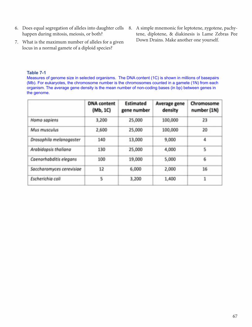
Table 7-1 Measures of genome size in selected organisms. The DNA content (1C) is shown in millions of basepairs (Mb). For eukaryotes, the chromosome number is the chromosomes counted in a gamete (1N) from each organism. The average gene density is the mean number of non-coding bases (in bp) between genes in the genome.
8. A simple mnemonic for leptotene, zygotene, pachy-tene, diplotene, & diakinesis is Lame Zebras Pee Down Drains. Make another one yourself.

MRU Open Genetics Chapter 768