cells - weeblyflemingbio.weebly.com/uploads/2/4/6/5/24658308/bio_chapter_3.pdf · recognize foreign...
TRANSCRIPT
CHAPTER 3
Cell Structure and Function 68
CHAPTER 4
Cells and Energy 98
CHAPTER 5
Cell Growth and Division 132
INTERNET MAGAZINE
Stem Cell Research—Potential Solutions, Practical Challenges 162
TECHNOLOGY Somatic Cell Nuclear Transfer
CAREER Cell Biologist
UNIT 2
Cells
67
C H A P T E R
KEY CONCE PTS
3 Cell Structure and Function
3.1 Cell TheoryCells are the basic unit of life.
3.2 Cell OrganellesEukaryotic cells share many similarities.
3.3 Cell MembraneThe cell membrane is a barrier that separates a cell from the external environment.
3.4 Diffusion and OsmosisMaterials move across membranes because of concentration differences.
3.5 Active Transport, Endocytosis, and ExocytosisCells use energy to transport materials that cannot diffuse across a membrane.
View animated chapterconcepts.• Cell Organelles• Get Through a Cell
Membrane
Keep current with biology news.• Featured stories• News feeds• Careers
Get more information on• Prokaryotic and Eukaryotic
Cells• Diffusion and Osmosis
BIOLOGYRESOURCE CENTER
BIOLOGY CLASSZONE .COM
68 Unit 2: Cells
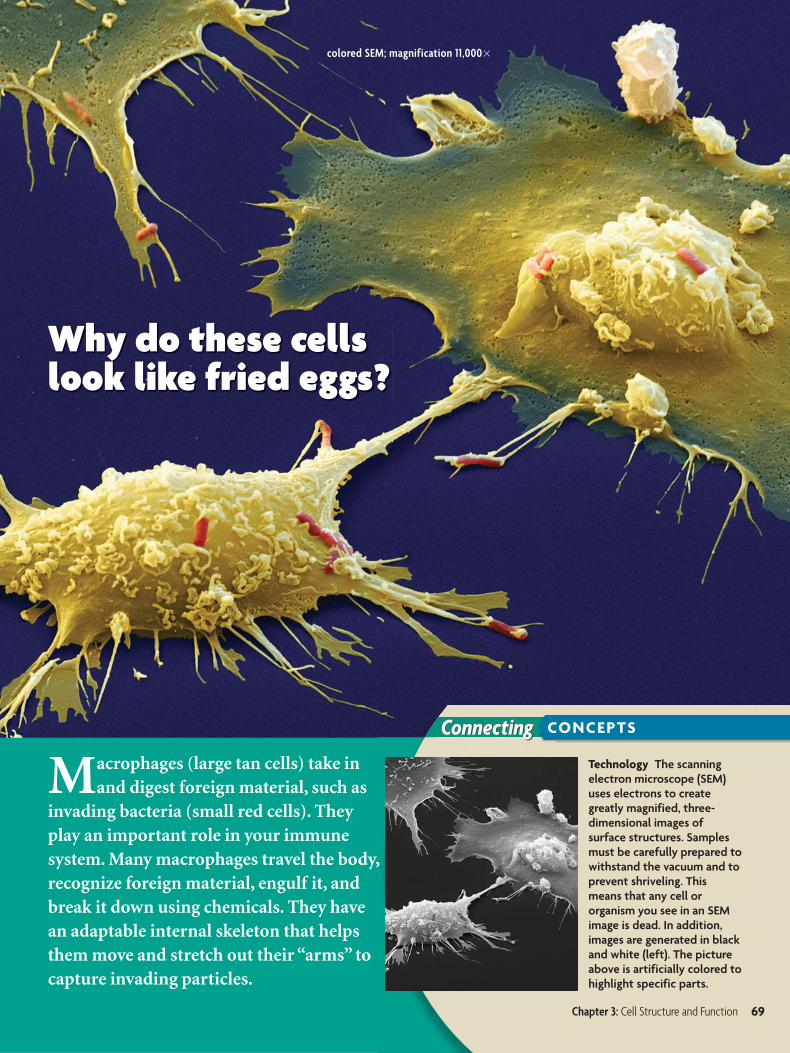
colored SEM; magnification 11,000�
Connecting CONCEPTS
Why do these cells look like fried eggs?
Macrophages (large tan cells) take in and digest foreign material, such as
invading bacteria (small red cells). They play an important role in your immune system. Many macrophages travel the body, recognize foreign material, engulf it, and break it down using chemicals. They have an adaptable internal skeleton that helps them move and stretch out their “arms” to capture invading particles.
Technology The scanning electron microscope (SEM) uses electrons to create greatly magnified, three-dimensional images of surface structures. Samples must be carefully prepared to withstand the vacuum and to prevent shriveling. This means that any cell ororganism you see in an SEM image is dead. In addition, images are generated in black and white (left). The picture above is artificially colored to highlight specific parts.
Chapter 3: Cell Structure and Function 69
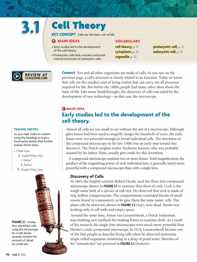
FIGURE 3.1 Hooke first identified cells using this microscope. Its crude lenses severely limited the amount of detail he could see.
I. Main Idea
A. Supporting idea
1. Detail
2. Detail
B. Supporting idea
TAKING NOTES
As you read, make an outline using the headings as topics. Summarize details that further explain those ideas.
3.1 Cell TheoryKEY CONCEPT Cells are the basic unit of life.
MAIN IDEAS
• Early studies led to the development of the cell theory.
• Prokaryotic cells lack a nucleus and most internal structures of eukaryotic cells.
VOCABULARY
cell theory,cell theory, p. 71
cytoplasm,cytoplasm, p. 72
organelle,organelle, p. 72
prokaryotic cell,prokaryotic cell, p. 72
eukaryotic cell,eukaryotic cell, p. 72
Connect You and all other organisms are made of cells. As you saw on theprevious page, a cell’s structure is closely related to its function. Today we knowthat cells are the smallest unit of living matter that can carry out all processesrequired for life. But before the 1600s, people had many other ideas about thebasis of life. Like many breakthroughs, the discovery of cells was aided by thedevelopment of new technology—in this case, the microscope.
MAIN IDEA
Early studies led to the development of thecell theory.
Almost all cells are too small to see without the aid of a microscope. Althoughglass lenses had been used to magnify images for hundreds of years, the earlylenses were not powerful enough to reveal individual cells. The invention ofthe compound microscope in the late 1500s was an early step toward thisdiscovery. The Dutch eyeglass maker Zacharias Janssen, who was probablyassisted by his father, Hans, usually gets credit for this invention.
A compound microscope contains two or more lenses. Total magnification, theproduct of the magnifying power of each individual lens, is generally much morepowerful with a compound microscope than with a single lens.
Discovery of CellsIn 1665, the English scientist Robert Hooke used the three-lens compoundmicroscope shown in FIGURE 3.1 to examine thin slices of cork. Cork is thetough outer bark of a species of oak tree. He observed that cork is made oftiny, hollow compartments. The compartments reminded Hooke of smallrooms found in a monastery, so he gave them the same name: cells. Theplant cells he observed, shown in FIGURE 3.2 (top), were dead. Hooke waslooking only at cell walls and empty space.
Around the same time, Anton van Leeuwenhoek, a Dutch tradesman,was studying new methods for making lenses to examine cloth. As a resultof his research, his single-lens microscopes were much more powerful thanHooke’s crude compound microscope. In 1674, Leeuwenhoek became oneof the first people to describe living cells when he observed numeroussingle-celled organisms swimming in a drop of pond water. Sketches ofhis “animalcules” are pictured in FIGURE 3.2 (bottom).
70 Unit 2: Cells
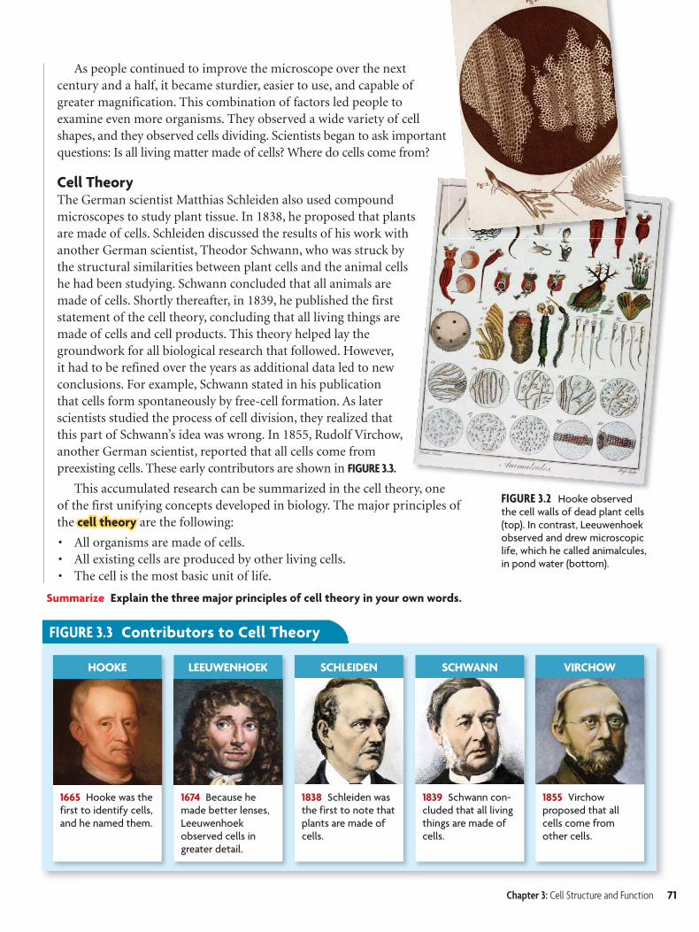
FIGURE 3.3 Contributors to Cell Theory
HOOKE LEEUWENHOEK SCHLEIDEN SCHWANN VIRCHOW
1665 Hooke was the first to identify cells, and he named them.
1674 Because he made better lenses, Leeuwenhoek observed cells in greater detail.
1838 Schleiden was the first to note that plants are made of cells.
1839 Schwann con-cluded that all living things are made of cells.
1855 Virchow proposed that all cells come from other cells.
FIGURE 3.2 Hooke observed the cell walls of dead plant cells (top). In contrast, Leeuwenhoek observed and drew microscopic life, which he called animalcules, in pond water (bottom).
As people continued to improve the microscope over the next century and a half, it became sturdier, easier to use, and capable of greater magnification. This combination of factors led people to examine even more organisms. They observed a wide variety of cell shapes, and they observed cells dividing. Scientists began to ask important questions: Is all living matter made of cells? Where do cells come from?
Cell TheoryThe German scientist Matthias Schleiden also used compound microscopes to study plant tissue. In 1838, he proposed that plants are made of cells. Schleiden discussed the results of his work with another German scientist, Theodor Schwann, who was struck by the structural similarities between plant cells and the animal cells he had been studying. Schwann concluded that all animals are made of cells. Shortly thereafter, in 1839, he published the first statement of the cell theory, concluding that all living things are made of cells and cell products. This theory helped lay the groundwork for all biological research that followed. However, it had to be refined over the years as additional data led to new conclusions. For example, Schwann stated in his publication that cells form spontaneously by free-cell formation. As later scientists studied the process of cell division, they realized that this part of Schwann’s idea was wrong. In 1855, Rudolf Virchow, another German scientist, reported that all cells come from preexisting cells. These early contributors are shown in FIGURE 3.3.
This accumulated research can be summarized in the cell theory, one of the first unifying concepts developed in biology. The major principles of the cell theorycell theory are the following:
• All organisms are made of cells.• All existing cells are produced by other living cells.• The cell is the most basic unit of life.
Summarize Explain the three major principles of cell theory in your own words.
Chapter 3: Cell Structure and Function 71
3.1 ASSESSMENT
Connecting
ONLINE QUIZClassZone.com
CONCEPTS
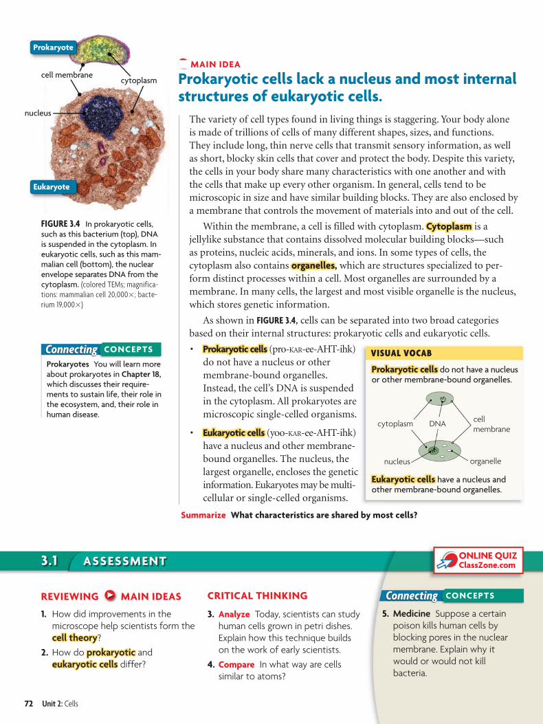
Prokaryotic cellsProkaryotic cells do not have a nucleus or other membrane-bound organelles.
VISUAL VOCAB
cell membrane
nucleus organelle
cytoplasm
Eukaryotic cellsEukaryotic cells have a nucleus and other membrane-bound organelles.
DNA
FIGURE 3.4 In prokaryotic cells, such as this bacterium (top), DNA is suspended in the cytoplasm. In eukaryotic cells, such as this mam-malian cell (bottom), the nuclear envelope separates DNA from the cytoplasm. (colored TEMs; magnifica-tions: mammalian cell 20,000�; bacte-rium 19,000�)
Eukaryote
cell membranecytoplasm
nucleus
Prokaryote
Prokaryotes You will learn more about prokaryotes in Chapter 18, which discusses their require-ments to sustain life, their role in the ecosystem, and, their role in human disease.
Connecting CONCEPTS
MAIN IDEA
Prokaryotic cells lack a nucleus and most internal structures of eukaryotic cells.
The variety of cell types found in living things is staggering. Your body aloneis made of trillions of cells of many different shapes, sizes, and functions.They include long, thin nerve cells that transmit sensory information, as wellas short, blocky skin cells that cover and protect the body. Despite this variety,the cells in your body share many characteristics with one another and withthe cells that make up every other organism. In general, cells tend to bemicroscopic in size and have similar building blocks. They are also enclosed bya membrane that controls the movement of materials into and out of the cell.
Within the membrane, a cell is filled with cytoplasm. CytoplasmCytoplasm is ajellylike substance that contains dissolved molecular building blocks—suchas proteins, nucleic acids, minerals, and ions. In some types of cells, thecytoplasm also contains organellesorganelles, which are structures specialized to per-form distinct processes within a cell. Most organelles are surrounded by amembrane. In many cells, the largest and most visible organelle is the nucleus,which stores genetic information.
As shown in FIGURE 3.4, cells can be separated into two broad categoriesbased on their internal structures: prokaryotic cells and eukaryotic cells.
• Prokaryotic cellsProkaryotic cells (pro-KAR-ee-AHT-ihk)do not have a nucleus or othermembrane-bound organelles.Instead, the cell’s DNA is suspendedin the cytoplasm. All prokaryotes aremicroscopic single-celled organisms.
• Eukaryotic cells Eukaryotic cells (yoo-KAR-ee-AHT-ihk)have a nucleus and other membrane-bound organelles. The nucleus, thelargest organelle, encloses the geneticinformation. Eukaryotes may be multi-cellular or single-celled organisms.
Summarize What characteristics are shared by most cells?
REVIEWING MAIN IDEAS
1. How did improvements in the
microscope help scientists form the
cell theorycell theory?
2. How do prokaryotic prokaryotic and
eukaryotic cellseukaryotic cells differ?
CRITICAL THINKING
3. Analyze Today, scientists can study
human cells grown in petri dishes.
Explain how this technique builds
on the work of early scientists.
4. Compare In what way are cells
similar to atoms?
5. Medicine Suppose a certain
poison kills human cells by
blocking pores in the nuclear
membrane. Explain why it
would or would not kill
bacteria.
72 Unit 2: Cells
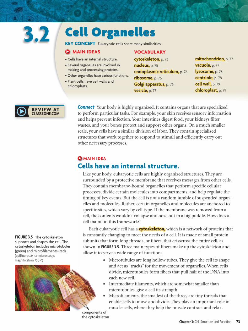
FIGURE 3.5 The cytoskeleton supports and shapes the cell. The cytoskeleton includes microtubules (green) and microfilaments (red). (epifluorescence microscopy; magnification 750�)
components of the cytoskeleton
3.2 Cell OrganellesKEY CONCEPT Eukaryotic cells share many similarities.
MAIN IDEAS
• Cells have an internal structure.
• Several organelles are involved in making and processing proteins.
• Other organelles have various functions.
• Plant cells have cell walls and chloroplasts.
VOCABULARY
cytoskeleton,cytoskeleton, p. 73
nucleus,nucleus, p. 75
endoplasmic reticulum,endoplasmic reticulum, p. 76
ribosome,ribosome, p. 76
Golgi apparatus,Golgi apparatus, p. 76
vesicle,vesicle, p. 77
mitochondrion,mitochondrion, p. 77
vacuole,vacuole, p. 77
lysosome,lysosome, p. 78
centriole,centriole, p. 78
cell wall,cell wall, p. 79
chloroplast,chloroplast, p. 79
Connect Your body is highly organized. It contains organs that are specializedto perform particular tasks. For example, your skin receives sensory informationand helps prevent infection. Your intestines digest food, your kidneys filterwastes, and your bones protect and support other organs. On a much smallerscale, your cells have a similar division of labor. They contain specializedstructures that work together to respond to stimuli and efficiently carry outother necessary processes.
MAIN IDEA
Cells have an internal structure.Like your body, eukaryotic cells are highly organized structures. They aresurrounded by a protective membrane that receives messages from other cells.They contain membrane-bound organelles that perform specific cellularprocesses, divide certain molecules into compartments, and help regulate thetiming of key events. But the cell is not a random jumble of suspended organ-elles and molecules. Rather, certain organelles and molecules are anchored tospecific sites, which vary by cell type. If the membrane was removed from acell, the contents wouldn’t collapse and ooze out in a big puddle. How does acell maintain this framework?
Each eukaryotic cell has a cytoskeleton,cytoskeleton, which is a network of proteins thatis constantly changing to meet the needs of a cell. It is made of small proteinsubunits that form long threads, or fibers, that crisscross the entire cell, asshown in FIGURE 3.5. Three main types of fibers make up the cytoskeleton andallow it to serve a wide range of functions.
• Microtubules are long hollow tubes. They give the cell its shapeand act as “tracks” for the movement of organelles. When cellsdivide, microtubules form fibers that pull half of the DNA intoeach new cell.
• Intermediate filaments, which are somewhat smaller thanmicrotubules, give a cell its strength.
• Microfilaments, the smallest of the three, are tiny threads thatenable cells to move and divide. They play an important role inmuscle cells, where they help the muscle contract and relax.
Chapter 3: Cell Structure and Function 73
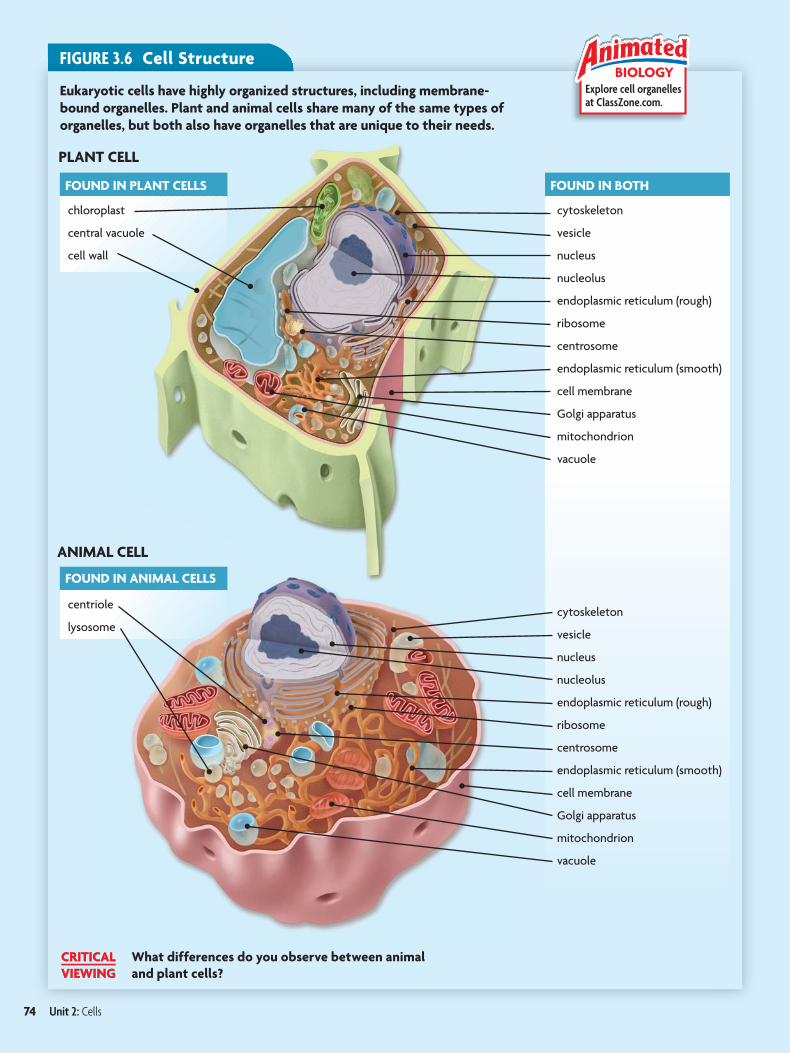
FOUND IN PLANT CELLS
chloroplast
central vacuole
cell wall
FOUND IN ANIMAL CELLS
centriole
lysosome
PLANT CELL
ANIMAL CELL
FOUND IN BOTH
cytoskeleton
vesicle
nucleus
nucleolus
endoplasmic reticulum (rough)
ribosome
centrosome
endoplasmic reticulum (smooth)
cell membrane
Golgi apparatus
mitochondrion
vacuole
cytoskeleton
vesicle
nucleus
nucleolus
endoplasmic reticulum (rough)
ribosome
centrosome
endoplasmic reticulum (smooth)
cell membrane
Golgi apparatus
mitochondrion
vacuole
Explore cell organelles at ClassZone.com.
BIOLOGYFIGURE 3.6 Cell Structure
Eukaryotic cells have highly organized structures, including membrane-
bound organelles. Plant and animal cells share many of the same types of
organelles, but both also have organelles that are unique to their needs.
What differences do you observe between animal
and plant cells?
CRITICAL
VIEWING
74 Unit 2: Cells
nucleus
pores
FIGURE 3.7 The nucleus stores and protects DNA. (colored SEM; magnification 90,000�)
ConnectingBiochemistry Recall from Chapter 2 that certain amino acids within a protein molecule may form hydrogen bonds with other amino acids. These bonds cause the protein to form a specific shape.
CONCEPTS
TAKING NOTES
Make a chart to correlate each organelle with its function.
Organelle Function
NucleusRibosome
stores DNA
Cytoplasm, which you read about in Section 3.1, is itself an importantcontributor to cell structure. In eukaryotes, it fills the space between thenucleus and the cell membrane. The fluid portion, excluding the organelles, iscalled cytosol and consists mostly of water. The makeup of cytoplasm showsthat water is necessary for maintaining cell structure. This is only one of manyreasons that water is an essential component for life, however. Many chemicalreactions occur in the cytoplasm, where water acts as an important solvent.
The remainder of this chapter highlights the structure and function of theorganelles found in eukaryotic cells. As FIGURE 3.6 shows, plant and animalcells use many of the same types of organelles to carry out basic functions.Both cell types also have organelles that are unique to their needs.
Infer What problems might a cell experience if it had no cytoskeleton?
MAIN IDEA
Several organelles are involved in making and processing proteins.
Much of the cell is devoted to making proteins. Proteins are made of 20 typesof amino acids that have unique characteristics of size, polarity, and acidity.They can form very long or very short protein chains that fold into differentshapes. And multiple protein chains can interact with each other. This almostlimitless variety of shapes and interactions makes proteins very powerful.Proteins carry out many critical functions, so they need to be made correctly.
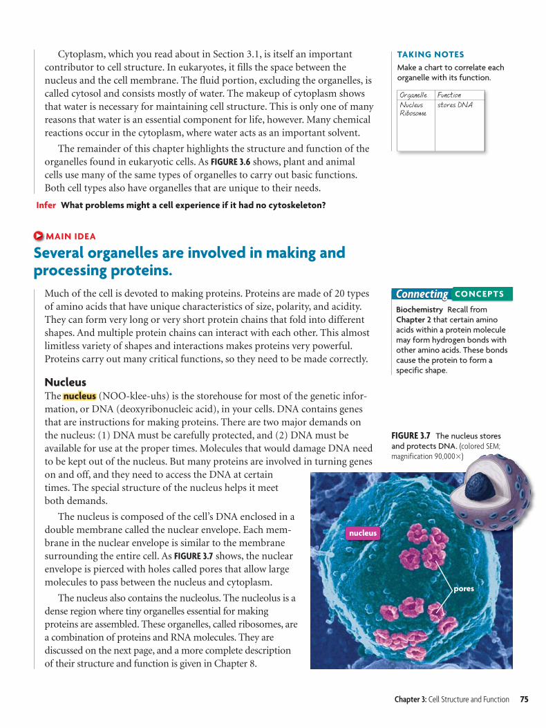
NucleusThe nucleusnucleus (NOO-klee-uhs) is the storehouse for most of the genetic infor-mation, or DNA (deoxyribonucleic acid), in your cells. DNA contains genesthat are instructions for making proteins. There are two major demands onthe nucleus: (1) DNA must be carefully protected, and (2) DNA must beavailable for use at the proper times. Molecules that would damage DNA needto be kept out of the nucleus. But many proteins are involved in turning geneson and off, and they need to access the DNA at certaintimes. The special structure of the nucleus helps it meetboth demands.
The nucleus is composed of the cell’s DNA enclosed in adouble membrane called the nuclear envelope. Each mem-brane in the nuclear envelope is similar to the membranesurrounding the entire cell. As FIGURE 3.7 shows, the nuclearenvelope is pierced with holes called pores that allow largemolecules to pass between the nucleus and cytoplasm.
The nucleus also contains the nucleolus. The nucleolus is adense region where tiny organelles essential for makingproteins are assembled. These organelles, called ribosomes, area combination of proteins and RNA molecules. They arediscussed on the next page, and a more complete descriptionof their structure and function is given in Chapter 8.
Chapter 3: Cell Structure and Function 75
endoplasmic reticulum
ribosomes
Golgi apparatus
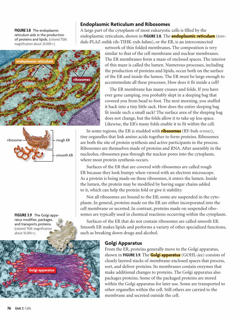
FIGURE 3.8 The endoplasmic reticulum aids in the production of proteins and lipids. (colored TEM; magnification about 20,000�)
FIGURE 3.9 The Golgi appa-ratus modifies, packages, and transports proteins. (colored TEM; magnification about 10,000�)
rough ER
smooth ER
ribosome
Endoplasmic Reticulum and RibosomesA large part of the cytoplasm of most eukaryotic cells is filled by the endoplasmic reticulum, shown in FIGURE 3.8. The endoplasmic reticulumendoplasmic reticulum (EHN-duh-PLAZ-mihk rih-TIHK-yuh-luhm), or the ER, is an interconnected
network of thin folded membranes. The composition is very similar to that of the cell membrane and nuclear membranes. The ER membranes form a maze of enclosed spaces. The interior of this maze is called the lumen. Numerous processes, including the production of proteins and lipids, occur both on the surface of the ER and inside the lumen. The ER must be large enough to accommodate all these processes. How does it fit inside a cell?
The ER membrane has many creases and folds. If you have ever gone camping, you probably slept in a sleeping bag that covered you from head to foot. The next morning, you stuffed it back into a tiny little sack. How does the entire sleeping bag fit inside such a small sack? The surface area of the sleeping bag does not change, but the folds allow it to take up less space. Likewise, the ER’s many folds enable it to fit within the cell.
In some regions, the ER is studded with ribosomesribosomes (RY-buh-SOHMZ), tiny organelles that link amino acids together to form proteins. Ribosomes are both the site of protein synthesis and active participants in the process. Ribosomes are themselves made of proteins and RNA. After assembly in the nucleolus, ribosomes pass through the nuclear pores into the cytoplasm, where most protein synthesis occurs.
Surfaces of the ER that are covered with ribosomes are called rough ER because they look bumpy when viewed with an electron microscope. As a protein is being made on these ribosomes, it enters the lumen. Inside the lumen, the protein may be modified by having sugar chains added to it, which can help the protein fold or give it stability.
Not all ribosomes are bound to the ER; some are suspended in the cyto-plasm. In general, proteins made on the ER are either incorporated into the cell membrane or secreted. In contrast, proteins made on suspended ribo-somes are typically used in chemical reactions occurring within the cytoplasm.
Surfaces of the ER that do not contain ribosomes are called smooth ER. Smooth ER makes lipids and performs a variety of other specialized functions, such as breaking down drugs and alcohol.
Golgi ApparatusFrom the ER, proteins generally move to the Golgi apparatus, shown in FIGURE 3.9. The Golgi apparatusGolgi apparatus (GOHL-jee) consists of closely layered stacks of membrane-enclosed spaces that process, sort, and deliver proteins. Its membranes contain enzymes that make additional changes to proteins. The Golgi apparatus also packages proteins. Some of the packaged proteins are stored within the Golgi apparatus for later use. Some are transported to other organelles within the cell. Still others are carried to the membrane and secreted outside the cell.
76 Unit 2: Cells
mitochondrion
vacuole
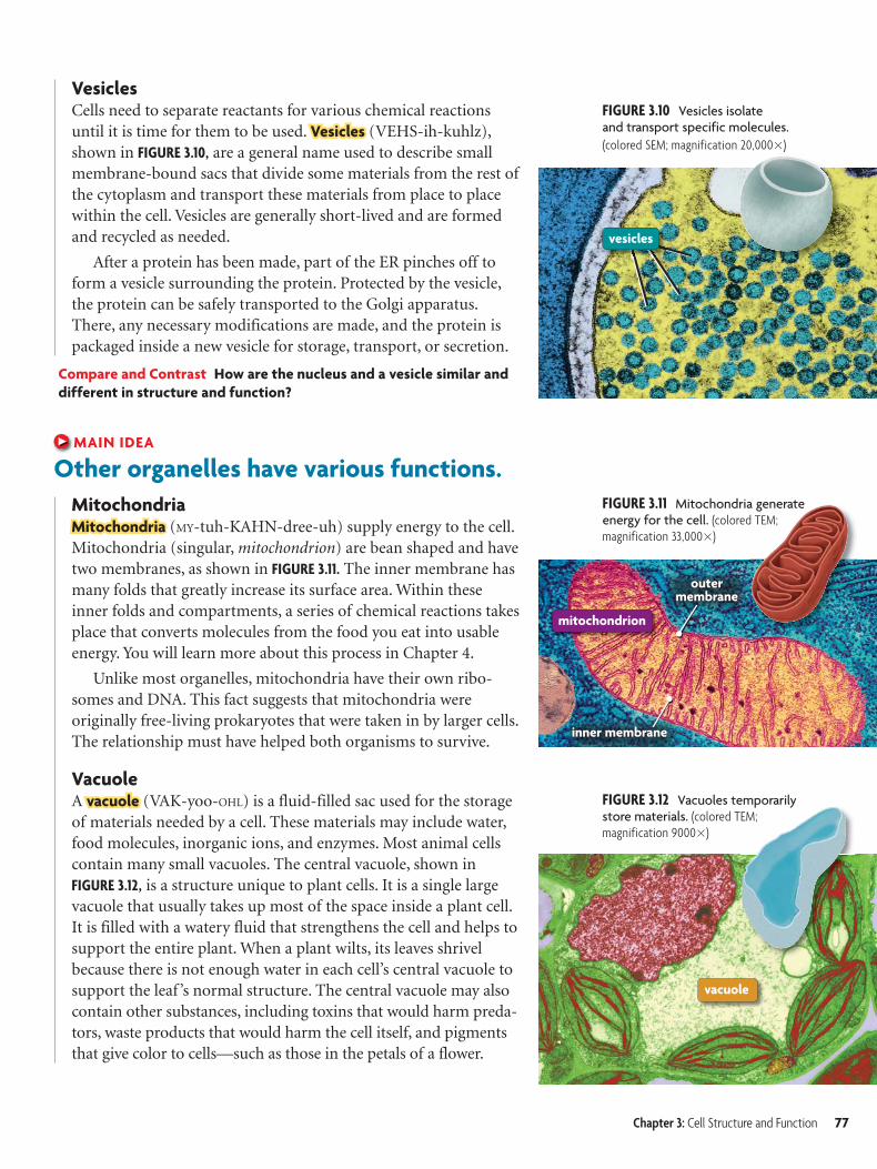
FIGURE 3.10 Vesicles isolate and transport specific molecules.
(colored SEM; magnification 20,000�)
vesicles
inner membraneinner membrane
outermembrane
outermembrane
FIGURE 3.11 Mitochondria generate energy for the cell. (colored TEM; magnification 33,000�)
FIGURE 3.12 Vacuoles temporarily store materials. (colored TEM;magnification 9000�)
VesiclesCells need to separate reactants for various chemical reactions until it is time for them to be used. VesiclesVesicles (VEHS-ih-kuhlz), shown in FIGURE 3.10, are a general name used to describe small membrane-bound sacs that divide some materials from the rest of the cytoplasm and transport these materials from place to place within the cell. Vesicles are generally short-lived and are formed and recycled as needed.
After a protein has been made, part of the ER pinches off to form a vesicle surrounding the protein. Protected by the vesicle, the protein can be safely transported to the Golgi apparatus. There, any necessary modifications are made, and the protein is packaged inside a new vesicle for storage, transport, or secretion.
Compare and Contrast How are the nucleus and a vesicle similar and
different in structure and function?
MAIN IDEA
Other organelles have various functions.
MitochondriaMitochondriaMitochondria (MY-tuh-KAHN-dree-uh) supply energy to the cell. Mitochondria (singular, mitochondrion) are bean shaped and have two membranes, as shown in FIGURE 3.11. The inner membrane has many folds that greatly increase its surface area. Within these inner folds and compartments, a series of chemical reactions takes place that converts molecules from the food you eat into usable energy. You will learn more about this process in Chapter 4.
Unlike most organelles, mitochondria have their own ribo-somes and DNA. This fact suggests that mitochondria were originally free-living prokaryotes that were taken in by larger cells. The relationship must have helped both organisms to survive.
VacuoleA vacuolevacuole (VAK-yoo-OHL) is a fluid-filled sac used for the storage of materials needed by a cell. These materials may include water, food molecules, inorganic ions, and enzymes. Most animal cells contain many small vacuoles. The central vacuole, shown in FIGURE 3.12, is a structure unique to plant cells. It is a single large vacuole that usually takes up most of the space inside a plant cell. It is filled with a watery fluid that strengthens the cell and helps to support the entire plant. When a plant wilts, its leaves shrivel because there is not enough water in each cell’s central vacuole to support the leaf ’s normal structure. The central vacuole may also contain other substances, including toxins that would harm preda-tors, waste products that would harm the cell itself, and pigments that give color to cells—such as those in the petals of a flower.
Chapter 3: Cell Structure and Function 77
lysosome
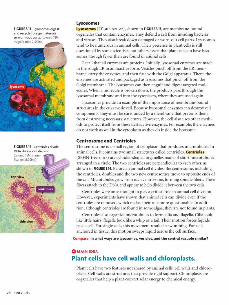
FIGURE 3.13 Lysosomes digest and recycle foreign materials or worn-out parts. (colored TEM; magnification 21,000�)
FIGURE 3.14 Centrioles divide DNA during cell division. (colored TEM; magni-fication 35,000�)
centrioles
side view
top view
LysosomesLysosomesLysosomes (LY-suh-SOHMZ), shown in FIGURE 3.13, are membrane-bound organelles that contain enzymes. They defend a cell from invading bacteria and viruses. They also break down damaged or worn-out cell parts. Lysosomes tend to be numerous in animal cells. Their presence in plant cells is still questioned by some scientists, but others assert that plant cells do have lyso-somes, though fewer than are found in animal cells.
Recall that all enzymes are proteins. Initially, lysosomal enzymes are made in the rough ER in an inactive form. Vesicles pinch off from the ER mem-brane, carry the enzymes, and then fuse with the Golgi apparatus. There, the enzymes are activated and packaged as lysosomes that pinch off from the Golgi membrane. The lysosomes can then engulf and digest targeted mol-ecules. When a molecule is broken down, the products pass through the lysosomal membrane and into the cytoplasm, where they are used again.
Lysosomes provide an example of the importance of membrane-bound structures in the eukaryotic cell. Because lysosomal enzymes can destroy cell components, they must be surrounded by a membrane that prevents them from destroying necessary structures. However, the cell also uses other meth-ods to protect itself from these destructive enzymes. For example, the enzymes do not work as well in the cytoplasm as they do inside the lysosome.
Centrosome and CentriolesThe centrosome is a small region of cytoplasm that produces microtubules. In animal cells, it contains two small structures called centrioles. Centrioles Centrioles
(SEHN-tree-OHLZ) are cylinder-shaped organelles made of short microtubules arranged in a circle. The two centrioles are perpendicular to each other, as shown in FIGURE 3.14. Before an animal cell divides, the centrosome, including the centrioles, doubles and the two new centrosomes move to opposite ends of the cell. Microtubules grow from each centrosome, forming spindle fibers. These fibers attach to the DNA and appear to help divide it between the two cells.
Centrioles were once thought to play a critical role in animal cell division. However, experiments have shown that animal cells can divide even if the centrioles are removed, which makes their role more questionable. In addi-tion, although centrioles are found in some algae, they are not found in plants.
Centrioles also organize microtubules to form cilia and flagella. Cilia look like little hairs; flagella look like a whip or a tail. Their motion forces liquids past a cell. For single cells, this movement results in swimming. For cells anchored in tissue, this motion sweeps liquid across the cell surface.
Compare In what ways are lysosomes, vesicles, and the central vacuole similar?
MAIN IDEA
Plant cells have cell walls and chloroplasts.
Plant cells have two features not shared by animal cells: cell walls and chloro-plasts. Cell walls are structures that provide rigid support. Chloroplasts are organelles that help a plant convert solar energy to chemical energy.
78 Unit 2: Cells
3.2 ASSESSMENT
Connecting CONCEPTS
ONLINE QUIZClassZone.com
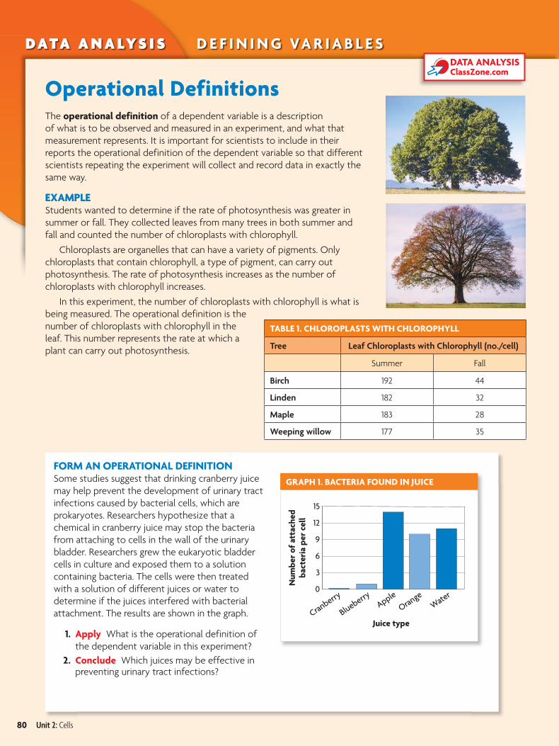
FIGURE 3.16 Chloroplasts convert solar energy into chemical energy through photosynthesis. (colored TEM; magnification 41,500�)
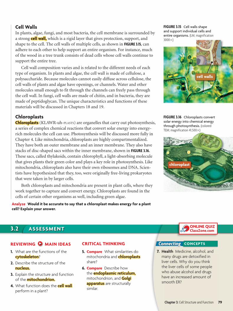
FIGURE 3.15 Cell walls shape and support individual cells and entire organisms. (LM; magnification 3000�)
chloroplast
cell walls
Cell WallsIn plants, algae, fungi, and most bacteria, the cell membrane is surrounded bya strong cell wall,cell wall, which is a rigid layer that gives protection, support, andshape to the cell. The cell walls of multiple cells, as shown in FIGURE 3.15, canadhere to each other to help support an entire organism. For instance, muchof the wood in a tree trunk consists of dead cells whose cell walls continue tosupport the entire tree.
Cell wall composition varies and is related to the different needs of eachtype of organism. In plants and algae, the cell wall is made of cellulose, apolysaccharide. Because molecules cannot easily diffuse across cellulose, thecell walls of plants and algae have openings, or channels. Water and othermolecules small enough to fit through the channels can freely pass throughthe cell wall. In fungi, cell walls are made of chitin, and in bacteria, they aremade of peptidoglycan. The unique characteristics and functions of thesematerials will be discussed in Chapters 18 and 19.
ChloroplastsChloroplasts Chloroplasts (KLAWR-uh-PLASTS) are organelles that carry out photosynthesis,a series of complex chemical reactions that convert solar energy into energy-rich molecules the cell can use. Photosynthesis will be discussed more fully inChapter 4. Like mitochondria, chloroplasts are highly compartmentalized.They have both an outer membrane and an inner membrane. They also havestacks of disc-shaped sacs within the inner membrane, shown in FIGURE 3.16.
These sacs, called thylakoids, contain chlorophyll, a light-absorbing moleculethat gives plants their green color and plays a key role in photosynthesis. Likemitochondria, chloroplasts also have their own ribosomes and DNA. Scien-tists have hypothesized that they, too, were originally free-living prokaryotesthat were taken in by larger cells.
Both chloroplasts and mitochondria are present in plant cells, where theywork together to capture and convert energy. Chloroplasts are found in thecells of certain other organisms as well, including green algae.
Analyze Would it be accurate to say that a chloroplast makes energy for a plant
cell? Explain your answer.
REVIEWING MAIN IDEAS
1. What are the functions of the
cytoskeletoncytoskeleton?
2. Describe the structure of the
nucleus.nucleus.
3. Explain the structure and function
of the mitochondrion.mitochondrion.
4. What function does the cell wallcell wall perform in a plant?
CRITICAL THINKING
5. Compare What similarities do
mitochondria and chloroplastschloroplasts
share?
6. Compare Describe how
the endoplasmic reticulum,endoplasmic reticulum, mitochondrion, and GolgiGolgi apparatusapparatus are structurally
similar.
7. Health Medicine, alcohol, and
many drugs are detoxified in
liver cells. Why do you think
the liver cells of some people
who abuse alcohol and drugs
have an increased amount of
smooth ER?
Chapter 3: Cell Structure and Function 79
DATA ANALYSISClassZone.com
D ATA A N A LY S I S D E F I N I N G VA R I A B L E S
Operational DefinitionsThe operational definition of a dependent variable is a description
of what is to be observed and measured in an experiment, and what that
measurement represents. It is important for scientists to include in their
reports the operational definition of the dependent variable so that different
scientists repeating the experiment will collect and record data in exactly the
same way.
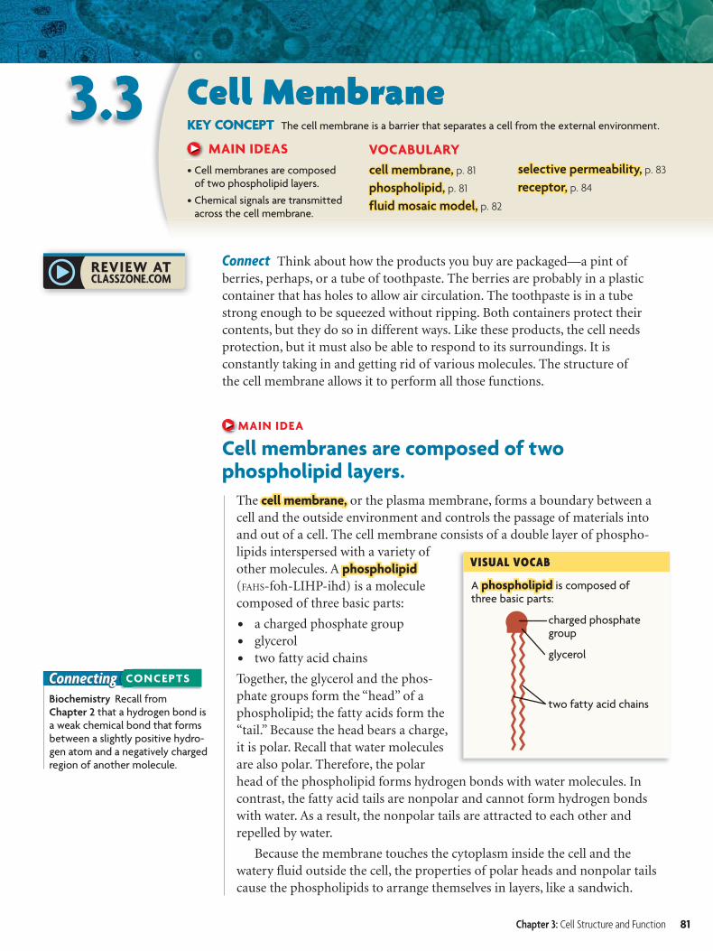
EXAMPLEStudents wanted to determine if the rate of photosynthesis was greater in
summer or fall. They collected leaves from many trees in both summer and
fall and counted the number of chloroplasts with chlorophyll.
Chloroplasts are organelles that can have a variety of pigments. Only
chloroplasts that contain chlorophyll, a type of pigment, can carry out
photosynthesis. The rate of photosynthesis increases as the number of
chloroplasts with chlorophyll increases.
In this experiment, the number of chloroplasts with chlorophyll is what is
being measured. The operational definition is the
number of chloroplasts with chlorophyll in the
leaf. This number represents the rate at which a
plant can carry out photosynthesis.
FORM AN OPERATIONAL DEFINITIONSome studies suggest that drinking cranberry juice
may help prevent the development of urinary tract
infections caused by bacterial cells, which are
prokaryotes. Researchers hypothesize that a
chemical in cranberry juice may stop the bacteria
from attaching to cells in the wall of the urinary
bladder. Researchers grew the eukaryotic bladder
cells in culture and exposed them to a solution
containing bacteria. The cells were then treated
with a solution of different juices or water to
determine if the juices interfered with bacterial
attachment. The results are shown in the graph.
1. Apply What is the operational definition of
the dependent variable in this experiment?
2. Conclude Which juices may be effective in preventing urinary tract infections?
TABLE 1. CHLOROPLASTS WITH CHLOROPHYLL
Tree Leaf Chloroplasts with Chlorophyll (no./cell)
Summer Fall
Birch 192 44
Linden 182 32
Maple 183 28
Weeping willow 177 35
GRAPH 1. BACTERIA FOUND IN JUICE
80 Unit 2: Cells
A phospholipidphospholipid is composed of three basic parts:
VISUAL VOCAB
glycerol
charged phosphate group
two fatty acid chains
ConnectingBiochemistry Recall from Chapter 2 that a hydrogen bond is a weak chemical bond that forms between a slightly positive hydro-gen atom and a negatively charged region of another molecule.
CONCEPTS
3.3 Cell MembraneKEY CONCEPT The cell membrane is a barrier that separates a cell from the external environment.
MAIN IDEAS
• Cell membranes are composed of two phospholipid layers.
• Chemical signals are transmitted across the cell membrane.
VOCABULARY
cell membrane,cell membrane, p. 81
phospholipid,phospholipid, p. 81
fluid mosaic model,fluid mosaic model, p. 82
selective permeability,selective permeability, p. 83
receptor,receptor, p. 84
Connect Think about how the products you buy are packaged—a pint ofberries, perhaps, or a tube of toothpaste. The berries are probably in a plasticcontainer that has holes to allow air circulation. The toothpaste is in a tubestrong enough to be squeezed without ripping. Both containers protect theircontents, but they do so in different ways. Like these products, the cell needsprotection, but it must also be able to respond to its surroundings. It isconstantly taking in and getting rid of various molecules. The structure ofthe cell membrane allows it to perform all those functions.
MAIN IDEA
Cell membranes are composed of two phospholipid layers.
The cell membrane, cell membrane, or the plasma membrane, forms a boundary between acell and the outside environment and controls the passage of materials intoand out of a cell. The cell membrane consists of a double layer of phospho-lipids interspersed with a variety ofother molecules. A phospholipidphospholipid
(FAHS-foh-LIHP-ihd) is a moleculecomposed of three basic parts:
• a charged phosphate group• glycerol• two fatty acid chains
Together, the glycerol and the phos-phate groups form the “head” of aphospholipid; the fatty acids form the“tail.” Because the head bears a charge,it is polar. Recall that water moleculesare also polar. Therefore, the polarhead of the phospholipid forms hydrogen bonds with water molecules. Incontrast, the fatty acid tails are nonpolar and cannot form hydrogen bondswith water. As a result, the nonpolar tails are attracted to each other andrepelled by water.
Because the membrane touches the cytoplasm inside the cell and thewatery fluid outside the cell, the properties of polar heads and nonpolar tailscause the phospholipids to arrange themselves in layers, like a sandwich.
Chapter 3: Cell Structure and Function 81
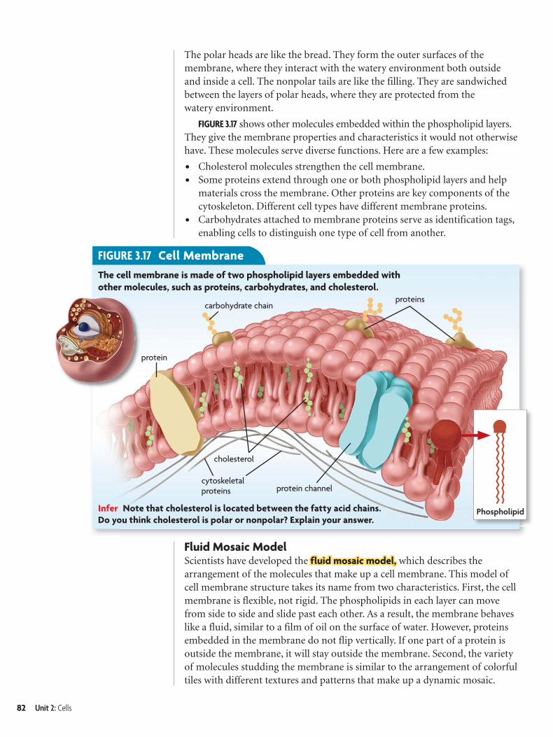
FIGURE 3.17 Cell Membrane
proteinscarbohydrate chain
protein channel
cholesterol
Phospholipid
cytoskeletal proteins
The cell membrane is made of two phospholipid layers embedded with
other molecules, such as proteins, carbohydrates, and cholesterol.
Infer Note that cholesterol is located between the fatty acid chains.
Do you think cholesterol is polar or nonpolar? Explain your answer.
protein
The polar heads are like the bread. They form the outer surfaces of themembrane, where they interact with the watery environment both outsideand inside a cell. The nonpolar tails are like the filling. They are sandwichedbetween the layers of polar heads, where they are protected from thewatery environment.
FIGURE 3.17 shows other molecules embedded within the phospholipid layers.They give the membrane properties and characteristics it would not otherwisehave. These molecules serve diverse functions. Here are a few examples:
• Cholesterol molecules strengthen the cell membrane.• Some proteins extend through one or both phospholipid layers and help
materials cross the membrane. Other proteins are key components of thecytoskeleton. Different cell types have different membrane proteins.
• Carbohydrates attached to membrane proteins serve as identification tags,enabling cells to distinguish one type of cell from another.
Fluid Mosaic ModelScientists have developed the fluid mosaic model,fluid mosaic model, which describes thearrangement of the molecules that make up a cell membrane. This model ofcell membrane structure takes its name from two characteristics. First, the cellmembrane is flexible, not rigid. The phospholipids in each layer can movefrom side to side and slide past each other. As a result, the membrane behaveslike a fluid, similar to a film of oil on the surface of water. However, proteinsembedded in the membrane do not flip vertically. If one part of a protein isoutside the membrane, it will stay outside the membrane. Second, the varietyof molecules studding the membrane is similar to the arrangement of colorfultiles with different textures and patterns that make up a dynamic mosaic.
82 Unit 2: Cells
Q U I C K L A B
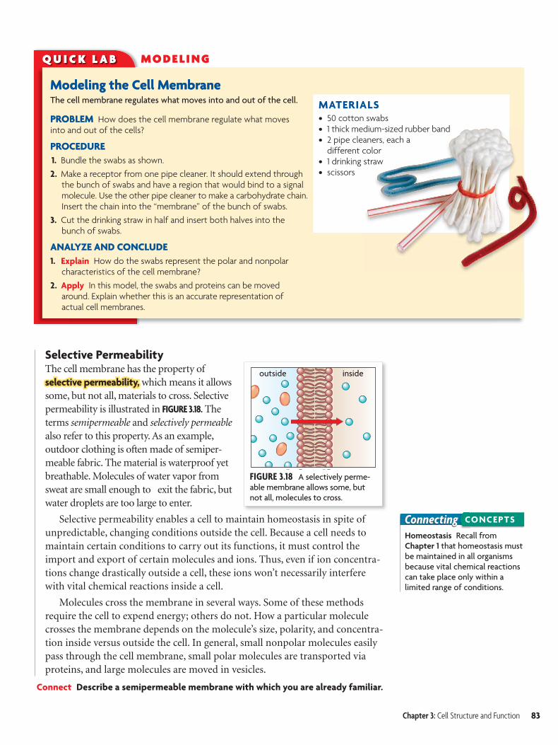
Modeling the Cell MembraneThe cell membrane regulates what moves into and out of the cell.
PROBLEM How does the cell membrane regulate what moves into and out of the cells?
PROCEDURE
1. Bundle the swabs as shown.
2. Make a receptor from one pipe cleaner. It should extend through the bunch of swabs and have a region that would bind to a signal molecule. Use the other pipe cleaner to make a carbohydrate chain.Insert the chain into the “membrane” of the bunch of swabs.
3. Cut the drinking straw in half and insert both halves into the bunch of swabs.
ANALYZE AND CONCLUDE
1. Explain How do the swabs represent the polar and nonpolar characteristics of the cell membrane?
2. Apply In this model, the swabs and proteins can be moved around. Explain whether this is an accurate representation of actual cell membranes.
MO D E LI N G
MATERIALS• 50 cotton swabs• 1 thick medium-sized rubber band• 2 pipe cleaners, each a
different color• 1 drinking straw• scissors
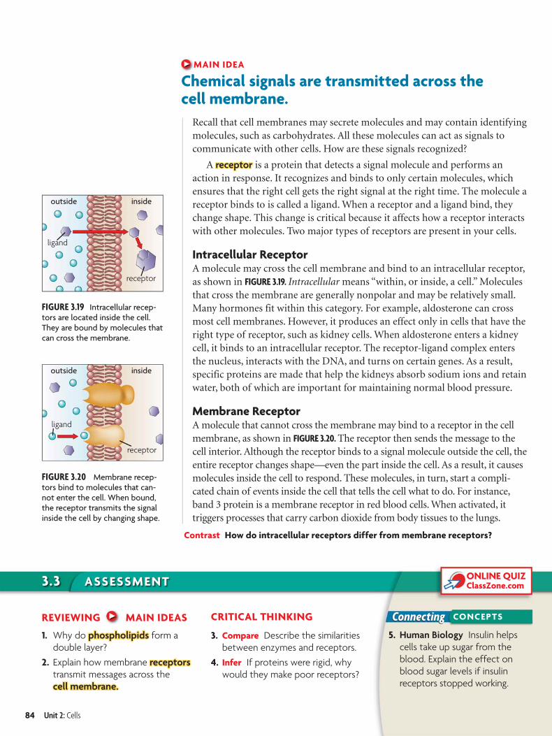
FIGURE 3.18 A selectively perme-able membrane allows some, but not all, molecules to cross.
outside inside
ConnectingHomeostasis Recall from Chapter 1 that homeostasis must be maintained in all organisms because vital chemical reactions can take place only within a limited range of conditions.
CONCEPTS
Selective Permeability The cell membrane has the property ofselective permeability,selective permeability, which means it allowssome, but not all, materials to cross. Selectivepermeability is illustrated in FIGURE 3.18. Theterms semipermeable and selectively permeablealso refer to this property. As an example,outdoor clothing is often made of semiper-meable fabric. The material is waterproof yetbreathable. Molecules of water vapor fromsweat are small enough to exit the fabric, butwater droplets are too large to enter.
Selective permeability enables a cell to maintain homeostasis in spite ofunpredictable, changing conditions outside the cell. Because a cell needs tomaintain certain conditions to carry out its functions, it must control theimport and export of certain molecules and ions. Thus, even if ion concentra-tions change drastically outside a cell, these ions won’t necessarily interferewith vital chemical reactions inside a cell.
Molecules cross the membrane in several ways. Some of these methodsrequire the cell to expend energy; others do not. How a particular moleculecrosses the membrane depends on the molecule’s size, polarity, and concentra-tion inside versus outside the cell. In general, small nonpolar molecules easilypass through the cell membrane, small polar molecules are transported viaproteins, and large molecules are moved in vesicles.
Connect Describe a semipermeable membrane with which you are already familiar.
Chapter 3: Cell Structure and Function 83
3.3 ASSESSMENTONLINE QUIZClassZone.com
Connecting CONCEPTS
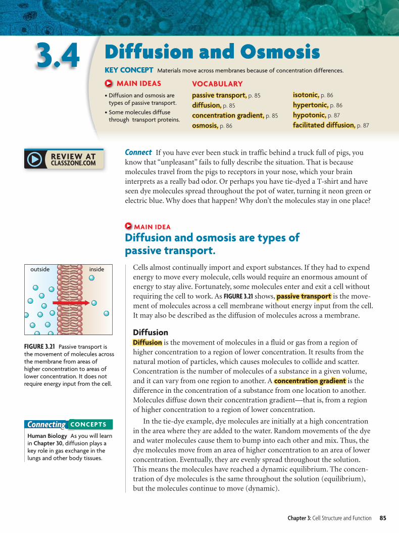
FIGURE 3.20 Membrane recep-tors bind to molecules that can-not enter the cell. When bound, the receptor transmits the signal inside the cell by changing shape.
outside
ligand
inside
receptor
FIGURE 3.19 Intracellular recep-tors are located inside the cell. They are bound by molecules that can cross the membrane.
outside inside
ligand
receptor
MAIN IDEA
Chemical signals are transmitted across the cell membrane.
Recall that cell membranes may secrete molecules and may contain identifyingmolecules, such as carbohydrates. All these molecules can act as signals tocommunicate with other cells. How are these signals recognized?
A receptorreceptor is a protein that detects a signal molecule and performs anaction in response. It recognizes and binds to only certain molecules, whichensures that the right cell gets the right signal at the right time. The molecule areceptor binds to is called a ligand. When a receptor and a ligand bind, theychange shape. This change is critical because it affects how a receptor interactswith other molecules. Two major types of receptors are present in your cells.
Intracellular ReceptorA molecule may cross the cell membrane and bind to an intracellular receptor,as shown in FIGURE 3.19. Intracellular means “within, or inside, a cell.” Moleculesthat cross the membrane are generally nonpolar and may be relatively small.Many hormones fit within this category. For example, aldosterone can crossmost cell membranes. However, it produces an effect only in cells that have theright type of receptor, such as kidney cells. When aldosterone enters a kidneycell, it binds to an intracellular receptor. The receptor-ligand complex entersthe nucleus, interacts with the DNA, and turns on certain genes. As a result,specific proteins are made that help the kidneys absorb sodium ions and retainwater, both of which are important for maintaining normal blood pressure.
Membrane ReceptorA molecule that cannot cross the membrane may bind to a receptor in the cellmembrane, as shown in FIGURE 3.20. The receptor then sends the message to thecell interior. Although the receptor binds to a signal molecule outside the cell, theentire receptor changes shape—even the part inside the cell. As a result, it causesmolecules inside the cell to respond. These molecules, in turn, start a compli-cated chain of events inside the cell that tells the cell what to do. For instance,band 3 protein is a membrane receptor in red blood cells. When activated, ittriggers processes that carry carbon dioxide from body tissues to the lungs.
Contrast How do intracellular receptors differ from membrane receptors?
REVIEWING MAIN IDEAS
1. Why do phospholipidsphospholipids form a
double layer?
2. Explain how membrane receptorsreceptors
transmit messages across the
cell membrane.cell membrane.
CRITICAL THINKING
3. Compare Describe the similarities
between enzymes and receptors.
4. Infer If proteins were rigid, why
would they make poor receptors?
5. Human Biology Insulin helps
cells take up sugar from the
blood. Explain the effect on
blood sugar levels if insulin
receptors stopped working.
84 Unit 2: Cells
outside inside
FIGURE 3.21 Passive transport is the movement of molecules across the membrane from areas of higher concentration to areas of lower concentration. It does not require energy input from the cell.
ConnectingHuman Biology As you will learn in Chapter 30, diffusion plays a key role in gas exchange in the lungs and other body tissues.
CONCEPTS
3.4 Diffusion and OsmosisKEY CONCEPT Materials move across membranes because of concentration differences.
MAIN IDEAS
• Diffusion and osmosis are types of passive transport.
• Some molecules diffuse through transport proteins.
VOCABULARY
passive transport,passive transport, p. 85
diffusion,diffusion, p. 85
concentration gradient,concentration gradient, p. 85
osmosis,osmosis, p. 86
isotonic,isotonic, p. 86
hypertonic,hypertonic, p. 86
hypotonic,hypotonic, p. 87
facilitated diffusion,facilitated diffusion, p. 87
Connect If you have ever been stuck in traffic behind a truck full of pigs, youknow that “unpleasant” fails to fully describe the situation. That is becausemolecules travel from the pigs to receptors in your nose, which your braininterprets as a really bad odor. Or perhaps you have tie-dyed a T-shirt and haveseen dye molecules spread throughout the pot of water, turning it neon green orelectric blue. Why does that happen? Why don’t the molecules stay in one place?
MAIN IDEA
Diffusion and osmosis are types of passive transport.
Cells almost continually import and export substances. If they had to expendenergy to move every molecule, cells would require an enormous amount ofenergy to stay alive. Fortunately, some molecules enter and exit a cell withoutrequiring the cell to work. As FIGURE 3.21 shows, passive transportpassive transport is the move-ment of molecules across a cell membrane without energy input from the cell.It may also be described as the diffusion of molecules across a membrane.
DiffusionDiffusion Diffusion is the movement of molecules in a fluid or gas from a region ofhigher concentration to a region of lower concentration. It results from thenatural motion of particles, which causes molecules to collide and scatter.Concentration is the number of molecules of a substance in a given volume,and it can vary from one region to another. A concentration gradient concentration gradient is thedifference in the concentration of a substance from one location to another.Molecules diffuse down their concentration gradient—that is, from a regionof higher concentration to a region of lower concentration.
In the tie-dye example, dye molecules are initially at a high concentrationin the area where they are added to the water. Random movements of the dyeand water molecules cause them to bump into each other and mix. Thus, thedye molecules move from an area of higher concentration to an area of lowerconcentration. Eventually, they are evenly spread throughout the solution.This means the molecules have reached a dynamic equilibrium. The concen-tration of dye molecules is the same throughout the solution (equilibrium),but the molecules continue to move (dynamic).
Chapter 3: Cell Structure and Function 85
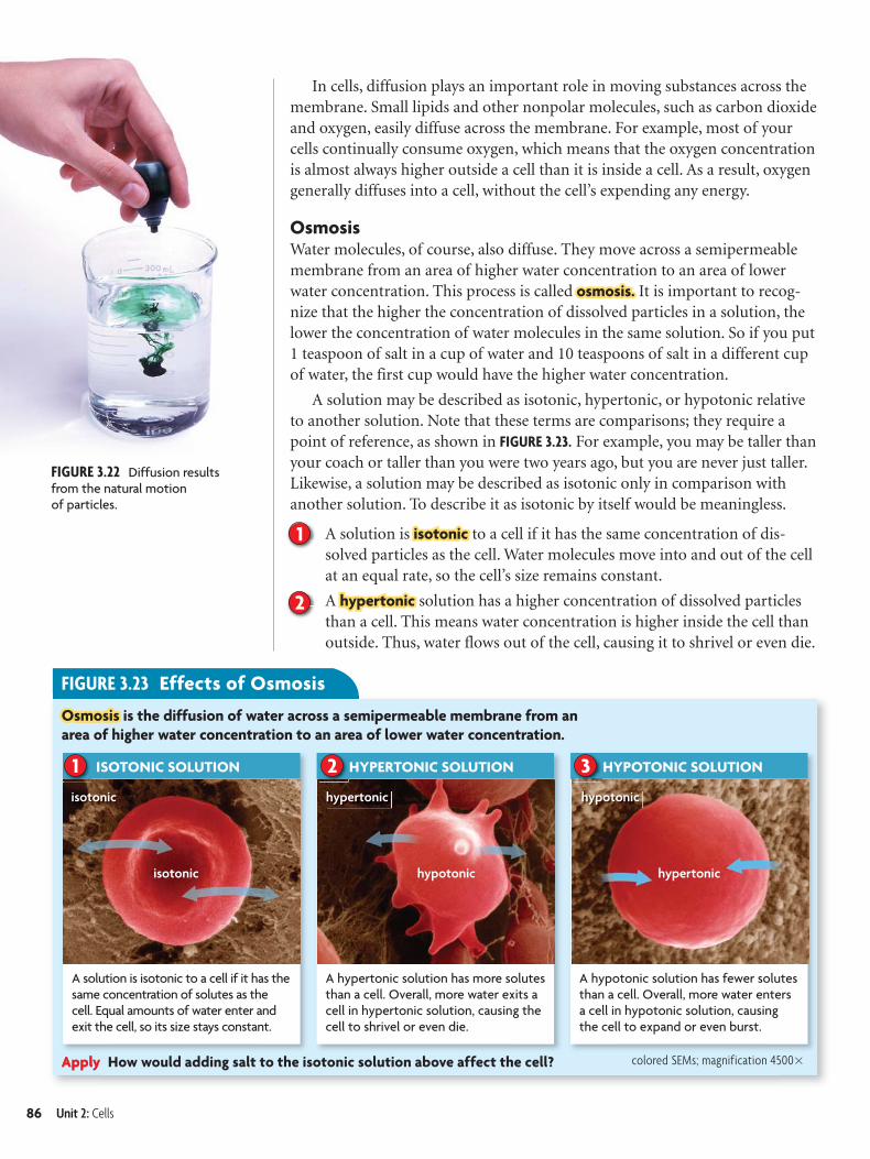
FIGURE 3.23 Effects of Osmosis
ISOTONIC SOLUTION
A solution is isotonic to a cell if it has the same concentration of solutes as the cell. Equal amounts of water enter and exit the cell, so its size stays constant.
isotonic
isotonic
HYPERTONIC SOLUTION
A hypertonic solution has more solutes than a cell. Overall, more water exits a cell in hypertonic solution, causing the cell to shrivel or even die.
hypertonic
hypotonic
2 HYPOTONIC SOLUTION
A hypotonic solution has fewer solutes than a cell. Overall, more water enters a cell in hypotonic solution, causing the cell to expand or even burst.
hypotonic
hypertonic
31
Apply How would adding salt to the isotonic solution above affect the cell?
OsmosisOsmosis is the diffusion of water across a semipermeable membrane from an
area of higher water concentration to an area of lower water concentration.
FIGURE 3.22 Diffusion results from the natural motion of particles.
colored SEMs; magnification 4500�
In cells, diffusion plays an important role in moving substances across themembrane. Small lipids and other nonpolar molecules, such as carbon dioxideand oxygen, easily diffuse across the membrane. For example, most of yourcells continually consume oxygen, which means that the oxygen concentrationis almost always higher outside a cell than it is inside a cell. As a result, oxygengenerally diffuses into a cell, without the cell’s expending any energy.
OsmosisWater molecules, of course, also diffuse. They move across a semipermeablemembrane from an area of higher water concentration to an area of lowerwater concentration. This process is called osmosis.osmosis. It is important to recog-nize that the higher the concentration of dissolved particles in a solution, thelower the concentration of water molecules in the same solution. So if you put1 teaspoon of salt in a cup of water and 10 teaspoons of salt in a different cupof water, the first cup would have the higher water concentration.
A solution may be described as isotonic, hypertonic, or hypotonic relativeto another solution. Note that these terms are comparisons; they require apoint of reference, as shown in FIGURE 3.23. For example, you may be taller thanyour coach or taller than you were two years ago, but you are never just taller.Likewise, a solution may be described as isotonic only in comparison withanother solution. To describe it as isotonic by itself would be meaningless.
A solution is isotonicisotonic to a cell if it has the same concentration of dis-solved particles as the cell. Water molecules move into and out of the cellat an equal rate, so the cell’s size remains constant.
A hypertonichypertonic solution has a higher concentration of dissolved particlesthan a cell. This means water concentration is higher inside the cell thanoutside. Thus, water flows out of the cell, causing it to shrivel or even die.
2
1
86 Unit 2: Cells
3.4 ASSESSMENT
Connecting
ONLINE QUIZClassZone.com
CONCEPTS
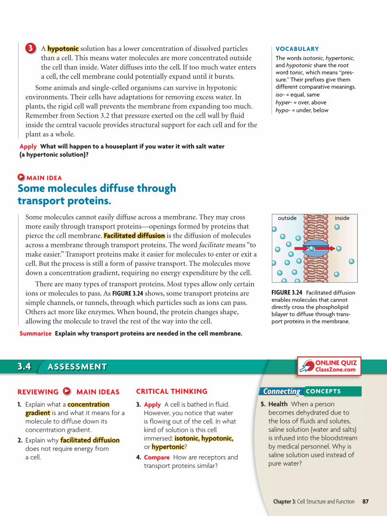
FIGURE 3.24 Facilitated diffusion enables molecules that cannot directly cross the phospholipid bilayer to diffuse through trans-port proteins in the membrane.
outside inside
VOCABULARY
The words isotonic, hypertonic, and hypotonic share the root word tonic, which means “pres-sure.” Their prefixes give them different comparative meanings.
iso- = equal, same
hyper- = over, above
hypo- = under, below
3 A hypotonic hypotonic solution has a lower concentration of dissolved particlesthan a cell. This means water molecules are more concentrated outsidethe cell than inside. Water diffuses into the cell. If too much water entersa cell, the cell membrane could potentially expand until it bursts.
Some animals and single-celled organisms can survive in hypotonicenvironments. Their cells have adaptations for removing excess water. Inplants, the rigid cell wall prevents the membrane from expanding too much.Remember from Section 3.2 that pressure exerted on the cell wall by fluidinside the central vacuole provides structural support for each cell and for theplant as a whole.
Apply What will happen to a houseplant if you water it with salt water
(a hypertonic solution)?
MAIN IDEA
Some molecules diffuse through transport proteins.
Some molecules cannot easily diffuse across a membrane. They may crossmore easily through transport proteins—openings formed by proteins thatpierce the cell membrane. Facilitated diffusionFacilitated diffusion is the diffusion of moleculesacross a membrane through transport proteins. The word facilitate means “tomake easier.” Transport proteins make it easier for molecules to enter or exit acell. But the process is still a form of passive transport. The molecules movedown a concentration gradient, requiring no energy expenditure by the cell.
There are many types of transport proteins. Most types allow only certainions or molecules to pass. As FIGURE 3.24 shows, some transport proteins aresimple channels, or tunnels, through which particles such as ions can pass.Others act more like enzymes. When bound, the protein changes shape,allowing the molecule to travel the rest of the way into the cell.
Summarize Explain why transport proteins are needed in the cell membrane.
REVIEWING MAIN IDEAS
1. Explain what a concentration concentration gradient gradient is and what it means for a
molecule to diffuse down its
concentration gradient.
2. Explain why facilitatedfacilitated diffusiondiffusion
does not require energy from
a cell.
CRITICAL THINKING
3. Apply A cell is bathed in fluid.
However, you notice that water
is flowing out of the cell. In what
kind of solution is this cell
immersed: isotonic, hypotonic,isotonic, hypotonic, or hypertonichypertonic?
4. Compare How are receptors and
transport proteins similar?
5. Health When a person
becomes dehydrated due to
the loss of fluids and solutes,
saline solution (water and salts)
is infused into the bloodstream
by medical personnel. Why is
saline solution used instead of
pure water?
Chapter 3: Cell Structure and Function 87
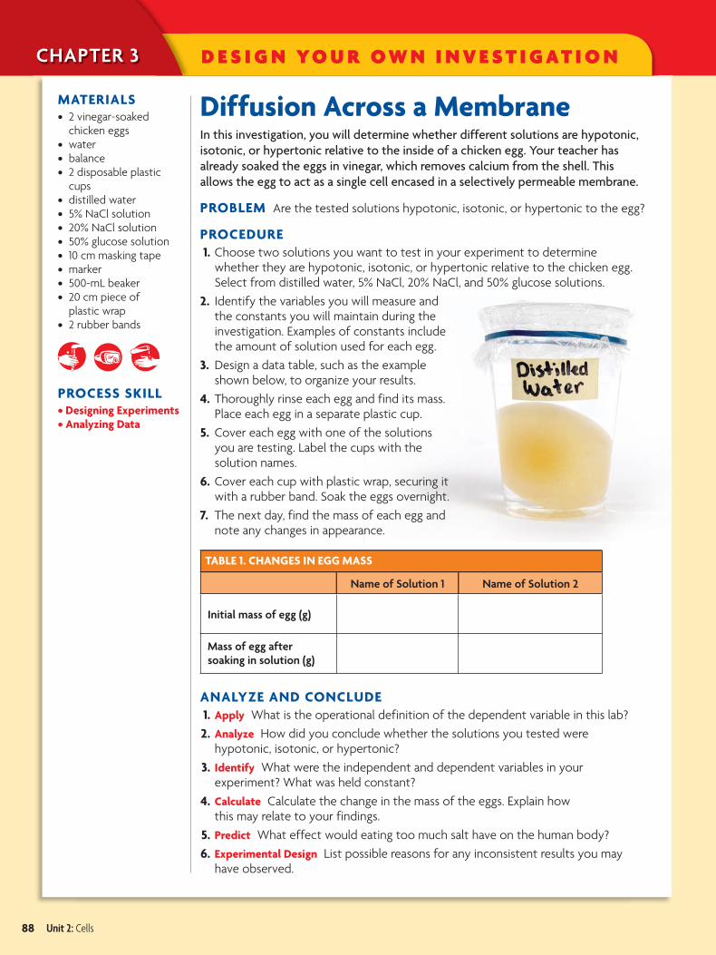
Diffusion Across a MembraneIn this investigation, you will determine whether different solutions are hypotonic,
isotonic, or hypertonic relative to the inside of a chicken egg. Your teacher has
already soaked the eggs in vinegar, which removes calcium from the shell. This
allows the egg to act as a single cell encased in a selectively permeable membrane.
PROBLEM Are the tested solutions hypotonic, isotonic, or hypertonic to the egg?
PROCEDURE
1. Choose two solutions you want to test in your experiment to determine whether they are hypotonic, isotonic, or hypertonic relative to the chicken egg. Select from distilled water, 5% NaCl, 20% NaCl, and 50% glucose solutions.
2. Identify the variables you will measure and the constants you will maintain during the investigation. Examples of constants include the amount of solution used for each egg.
3. Design a data table, such as the example shown below, to organize your results.
4. Thoroughly rinse each egg and find its mass. Place each egg in a separate plastic cup.
5. Cover each egg with one of the solutions you are testing. Label the cups with the solution names.
6. Cover each cup with plastic wrap, securing it with a rubber band. Soak the eggs overnight.
7. The next day, find the mass of each egg and note any changes in appearance.
TABLE 1. CHANGES IN EGG MASS
Name of Solution 1 Name of Solution 2
Initial mass of egg (g)
Mass of egg after soaking in solution (g)
ANALYZE AND CONCLUDE
1. Apply What is the operational definition of the dependent variable in this lab?
2. Analyze How did you conclude whether the solutions you tested were hypotonic, isotonic, or hypertonic?
3. Identify What were the independent and dependent variables in your experiment? What was held constant?
4. Calculate Calculate the change in the mass of the eggs. Explain how this may relate to your findings.
5. Predict What effect would eating too much salt have on the human body?
6. Experimental Design List possible reasons for any inconsistent results you may have observed.
MATERIALS• 2 vinegar-soaked
chicken eggs• water• balance• 2 disposable plastic
cups• distilled water• 5% NaCl solution• 20% NaCl solution• 50% glucose solution• 10 cm masking tape• marker• 500-mL beaker• 20 cm piece of
plastic wrap• 2 rubber bands
PROCESS SKILL• Designing Experiments• Analyzing Data
D E S I G N YO U R O W N I N V E S T I G AT I O NCHAPTER 3
88 Unit 2: Cells
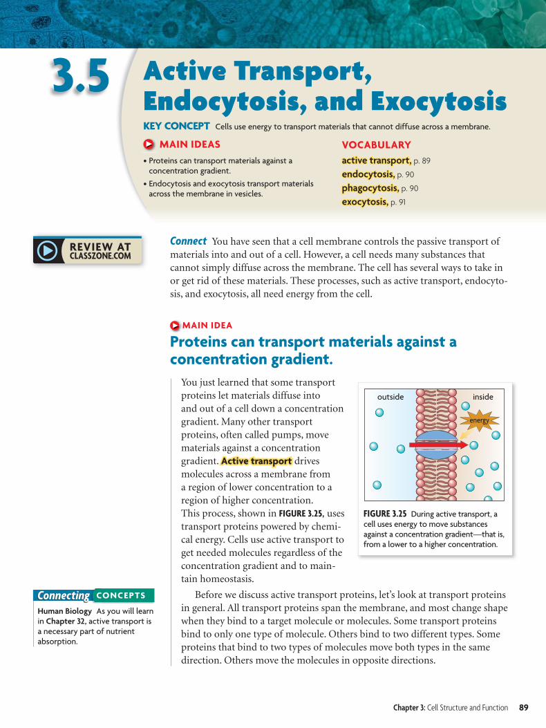
FIGURE 3.25 During active transport, a cell uses energy to move substances against a concentration gradient—that is, from a lower to a higher concentration.
outside inside
energy
ConnectingHuman Biology As you will learn in Chapter 32, active transport is a necessary part of nutrient absorption.
CONCEPTS
3.5 Active Transport, Endocytosis, and ExocytosisKEY CONCEPT Cells use energy to transport materials that cannot diffuse across a membrane.
MAIN IDEAS
• Proteins can transport materials against a concentration gradient.
• Endocytosis and exocytosis transport materials across the membrane in vesicles.
VOCABULARY
active transport,active transport, p. 89
endocytosis,endocytosis, p. 90
phagocytosis,phagocytosis, p. 90
exocytosis,exocytosis, p. 91
Connect You have seen that a cell membrane controls the passive transport ofmaterials into and out of a cell. However, a cell needs many substances thatcannot simply diffuse across the membrane. The cell has several ways to take inor get rid of these materials. These processes, such as active transport, endocyto-sis, and exocytosis, all need energy from the cell.
MAIN IDEA
Proteins can transport materials against a concentration gradient.
You just learned that some transportproteins let materials diffuse intoand out of a cell down a concentrationgradient. Many other transportproteins, often called pumps, movematerials against a concentrationgradient. Active transportActive transport drivesmolecules across a membrane froma region of lower concentration to aregion of higher concentration.This process, shown in FIGURE 3.25, usestransport proteins powered by chemi-cal energy. Cells use active transport toget needed molecules regardless of theconcentration gradient and to main-tain homeostasis.
Before we discuss active transport proteins, let’s look at transport proteinsin general. All transport proteins span the membrane, and most change shapewhen they bind to a target molecule or molecules. Some transport proteinsbind to only one type of molecule. Others bind to two different types. Someproteins that bind to two types of molecules move both types in the samedirection. Others move the molecules in opposite directions.
Chapter 3: Cell Structure and Function 89
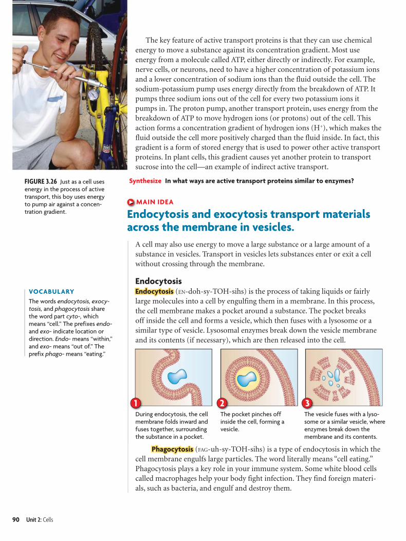
The pocket pinches off inside the cell, forming a vesicle.
During endocytosis, the cell membrane folds inward and fuses together, surrounding the substance in a pocket.
The vesicle fuses with a lyso-some or a similar vesicle, where enzymes break down the membrane and its contents.
31 2
FIGURE 3.26 Just as a cell uses energy in the process of active transport, this boy uses energy to pump air against a concen-tration gradient.
VOCABULARY
The words endocytosis, exocy-tosis, and phagocytosis share the word part cyto-, which means “cell.” The prefixes endo- and exo- indicate location or direction. Endo- means “within,” and exo- means “out of.” The prefix phago- means “eating.”
The key feature of active transport proteins is that they can use chemical energy to move a substance against its concentration gradient. Most use energy from a molecule called ATP, either directly or indirectly. For example, nerve cells, or neurons, need to have a higher concentration of potassium ions and a lower concentration of sodium ions than the fluid outside the cell. The sodium-potassium pump uses energy directly from the breakdown of ATP. It pumps three sodium ions out of the cell for every two potassium ions it pumps in. The proton pump, another transport protein, uses energy from the breakdown of ATP to move hydrogen ions (or protons) out of the cell. This action forms a concentration gradient of hydrogen ions (H+), which makes the fluid outside the cell more positively charged than the fluid inside. In fact, this gradient is a form of stored energy that is used to power other active transport proteins. In plant cells, this gradient causes yet another protein to transport sucrose into the cell—an example of indirect active transport.
Synthesize In what ways are active transport proteins similar to enzymes?
MAIN IDEA
Endocytosis and exocytosis transport materials across the membrane in vesicles.
A cell may also use energy to move a large substance or a large amount of a substance in vesicles. Transport in vesicles lets substances enter or exit a cell without crossing through the membrane.
EndocytosisEndocytosisEndocytosis (EN-doh-sy-TOH-sihs) is the process of taking liquids or fairly large molecules into a cell by engulfing them in a membrane. In this process, the cell membrane makes a pocket around a substance. The pocket breaks off inside the cell and forms a vesicle, which then fuses with a lysosome or a similar type of vesicle. Lysosomal enzymes break down the vesicle membrane and its contents (if necessary), which are then released into the cell.
PhagocytosisPhagocytosis (FAG-uh-sy-TOH-sihs) is a type of endocytosis in which the cell membrane engulfs large particles. The word literally means “cell eating.” Phagocytosis plays a key role in your immune system. Some white blood cells called macrophages help your body fight infection. They find foreign materi-als, such as bacteria, and engulf and destroy them.
90 Unit 2: Cells
3.5 ASSESSMENT
Connecting
ONLINE QUIZClassZone.com
CONCEPTS
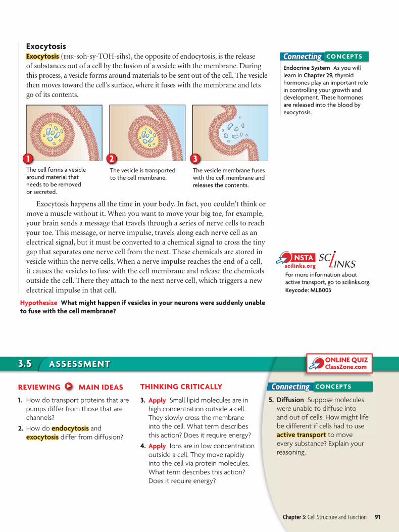
2 31The cell forms a vesicle around material that needs to be removed or secreted.
The vesicle is transported to the cell membrane.
The vesicle membrane fuses with the cell membrane and releases the contents.
ConnectingEndocrine System As you will learn in Chapter 29, thyroidhormones play an important role in controlling your growth and development. These hormones are released into the blood by exocytosis.
CONCEPTS
For more information about active transport, go to scilinks.org.
Keycode: MLB003
ExocytosisExocytosis Exocytosis (EHK-soh-sy-TOH-sihs), the opposite of endocytosis, is the releaseof substances out of a cell by the fusion of a vesicle with the membrane. Duringthis process, a vesicle forms around materials to be sent out of the cell. The vesiclethen moves toward the cell’s surface, where it fuses with the membrane and letsgo of its contents.
Exocytosis happens all the time in your body. In fact, you couldn’t think ormove a muscle without it. When you want to move your big toe, for example,your brain sends a message that travels through a series of nerve cells to reachyour toe. This message, or nerve impulse, travels along each nerve cell as anelectrical signal, but it must be converted to a chemical signal to cross the tinygap that separates one nerve cell from the next. These chemicals are stored invesicle within the nerve cells. When a nerve impulse reaches the end of a cell,it causes the vesicles to fuse with the cell membrane and release the chemicalsoutside the cell. There they attach to the next nerve cell, which triggers a newelectrical impulse in that cell.
Hypothesize What might happen if vesicles in your neurons were suddenly unable
to fuse with the cell membrane?
REVIEWING MAIN IDEAS
1. How do transport proteins that are
pumps differ from those that are
channels?
2. How do endocytosisendocytosis and
exocytosisexocytosis differ from diffusion?
THINKING CRITICALLY
3. Apply Small lipid molecules are in
high concentration outside a cell.
They slowly cross the membrane
into the cell. What term describes
this action? Does it require energy?
4. Apply Ions are in low concentration
outside a cell. They move rapidly
into the cell via protein molecules.
What term describes this action?
Does it require energy?
5. Diffusion Suppose molecules
were unable to diffuse into
and out of cells. How might life
be different if cells had to use
active transportactive transport to move
every substance? Explain your
reasoning.
Chapter 3: Cell Structure and Function 91
CHAPTER 3 O P T I O N S F O R I N Q U I RY
Use these inquiry-based labs and online activities to deepen yourunderstanding of cell structure.
I N V E S T I G AT I O N
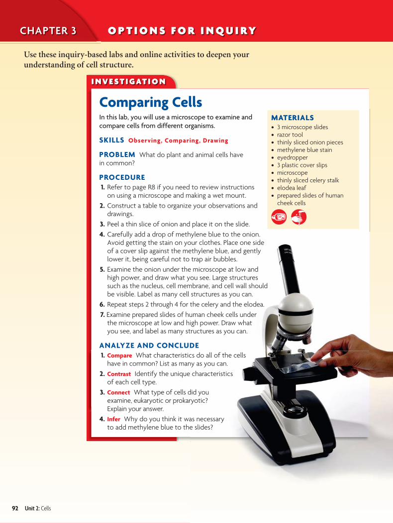
Comparing CellsIn this lab, you will use a microscope to examine and
compare cells from different organisms.
SKILLS Observing, Comparing, Drawing
PROBLEM What do plant and animal cells have in common?
PROCEDURE
1. Refer to page R8 if you need to review instructions on using a microscope and making a wet mount.
2. Construct a table to organize your observations and drawings.
3. Peel a thin slice of onion and place it on the slide.
4. Carefully add a drop of methylene blue to the onion. Avoid getting the stain on your clothes. Place one side of a cover slip against the methylene blue, and gently lower it, being careful not to trap air bubbles.
5. Examine the onion under the microscope at low and high power, and draw what you see. Large structures such as the nucleus, cell membrane, and cell wall should be visible. Label as many cell structures as you can.
6. Repeat steps 2 through 4 for the celery and the elodea.
7. Examine prepared slides of human cheek cells under the microscope at low and high power. Draw what you see, and label as many structures as you can.
ANALYZE AND CONCLUDE
1. Compare What characteristics do all of the cells have in common? List as many as you can.
2. Contrast Identify the unique characteristics of each cell type.
3. Connect What type of cells did you examine, eukaryotic or prokaryotic? Explain your answer.
4. Infer Why do you think it was necessary to add methylene blue to the slides?
MATERIALS• 3 microscope slides • razor tool• thinly sliced onion pieces• methylene blue stain• eyedropper• 3 plastic cover slips• microscope• thinly sliced celery stalk• elodea leaf• prepared slides of human
cheek cells
92 Unit 2: Cells
BIOLOGY
CLASSZONE .COMI N V E S T I G AT I O N
Modeling the CellThe diversity of life on Earth is enormous, although
all living things are made from the same basic
structural unit, the cell. In your body alone, there
are trillions of cells. In this activity, you will make a
model of a cell.
SKILL Modeling
PROBLEM What components make up a cell?
MATERIALS• 2 resealable plastic sandwich bags• gelatin jigglers• a small round balloon• a permanent marker• coffee stirring straws• drinking straws cut in half• different sizes of erasers• slices of two colors of sponges• tiny beads
PROCEDURE
1. Use the materials to construct a detailed model of a cell.
2. Be sure to include at least the following components in your model: cell membrane, cytoplasm, nucleus, cytoskeleton, ribosomes, mitochondria, Golgi apparatus, and centrioles.
3. Use both sandwich bags in constructing your model.
4. Tightly seal your cell after it has been completed.
ANALYZE AND CONCLUDE
1. Apply Make a table to list which materials you chose to represent the various cell structures and to explain your choices.
2. Analyze What is the significance of the double bag?
3. Connect What substance represents the cytoplasm? Explain why your choice is suitable.
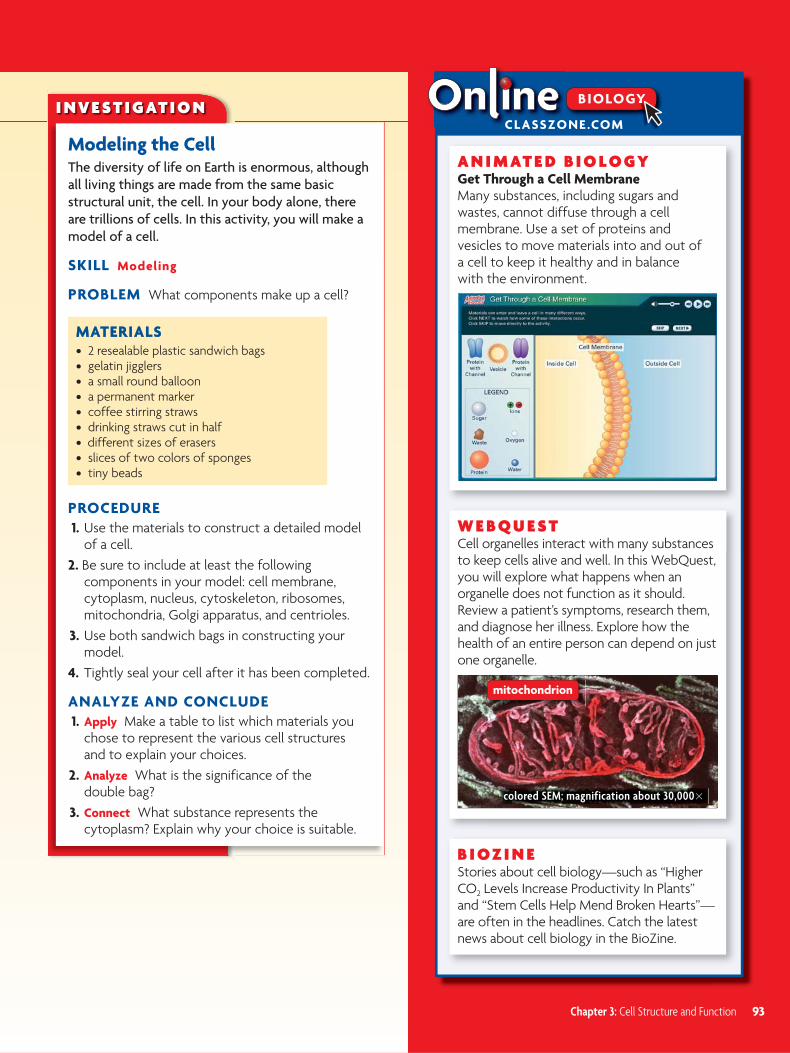
A N I M AT E D B I O L O G YGet Through a Cell MembraneMany substances, including sugars and wastes, cannot diffuse through a cell membrane. Use a set of proteins and vesicles to move materials into and out of a cell to keep it healthy and in balance with the environment.
W E B Q U E STCell organelles interact with many substances to keep cells alive and well. In this WebQuest, you will explore what happens when an organelle does not function as it should. Review a patient’s symptoms, research them, and diagnose her illness. Explore how the health of an entire person can depend on just one organelle.
B I O Z I N EStories about cell biology—such as “Higher CO2 Levels Increase Productivity In Plants” and “Stem Cells Help Mend Broken Hearts”—are often in the headlines. Catch the latest news about cell biology in the BioZine.
Chapter 3: Cell Structure and Function 93
colored SEM; magnification about 30,000�
mitochondrion
94 Unit
KEY CONCEPTS Vocabulary Games
3CHAPTER
@ CLASSZONE .COM
KEY CONCEPTS Vocabulary Games Concept Maps Animated Biology Online Quiz
Synthesize Your Notes
3.1 Cell TheoryCells are the basic unit of life. The contributions
of many scientists led to the discovery of cells
and the development of the cell theory. The cell
theory states that all organisms are made of cells,
all cells are produced by other living cells, and
the cell is the most basic unit of life.
3.2 Cell OrganellesEukaryotic cells share many similarities. They
have a nucleus and other membrane-bound
organelles that perform specialized tasks within
the cell. Many of these organelles are involved in
making proteins. Plant and animal cells share
many of the same types of organelles, but both
also have organelles that are specific to the cells’
unique functions.
3.3 Cell MembraneThe cell membrane is a barrier that separates a
cell from the external environment. It is made
of a double layer of phospholipids and a variety
of embedded molecules. Some of these mol-
ecules act as signals; others act as receptors. The
membrane is selectively permeable, allowing
some but not all materials to cross.
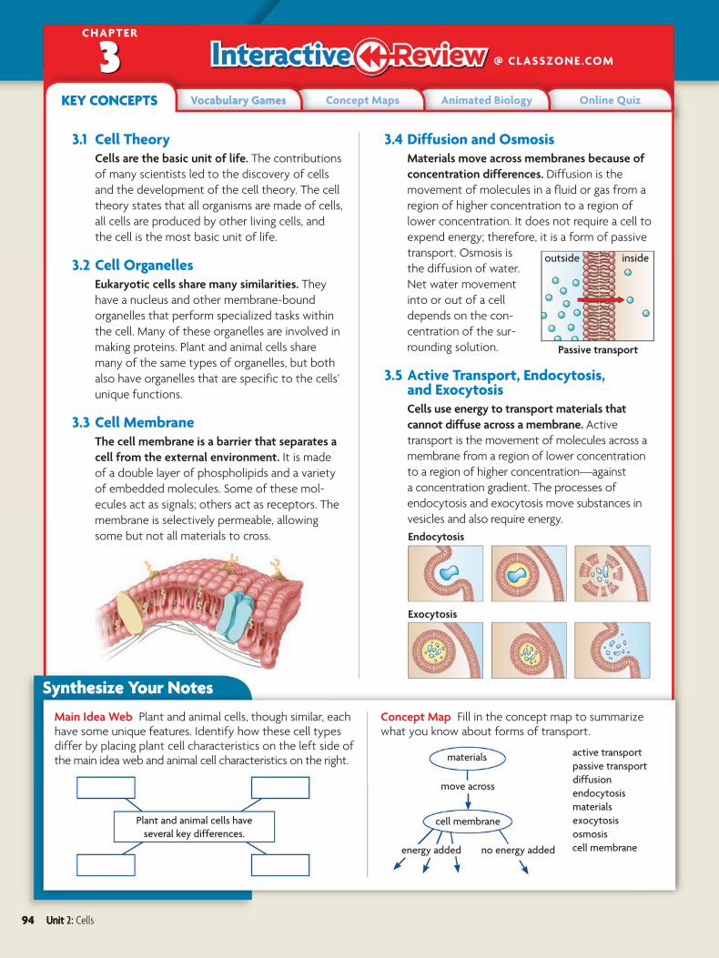
3.4 Diffusion and OsmosisMaterials move across membranes because of
concentration differences. Diffusion is the
movement of molecules in a fluid or gas from a
region of higher concentration to a region of
lower concentration. It does not require a cell to
expend energy; therefore, it is a form of passive
transport. Osmosis is
the diffusion of water.
Net water movement
into or out of a cell
depends on the con-
centration of the sur-
rounding solution.
3.5 Active Transport, Endocytosis, and ExocytosisCells use energy to transport materials that
cannot diffuse across a membrane. Active
transport is the movement of molecules across a
membrane from a region of lower concentration
to a region of higher concentration—against
a concentration gradient. The processes of
endocytosis and exocytosis move substances in
vesicles and also require energy.
Main Idea Web Plant and animal cells, though similar, each have some unique features. Identify how these cell types differ by placing plant cell characteristics on the left side of the main idea web and animal cell characteristics on the right.
Concept Map Fill in the concept map to summarize what you know about forms of transport.
materials
exocytosis
diffusion
endocytosis
passive transport
osmosis
cell membrane
Plant and animal cells have
several key differences.
active transport
materials
move across
cell membrane
no energy addedenergy added
Synthesize Your Notes
outside inside
Passive transport
Endocytosis
Exocytosis
94 Unit 2: Cells
Chapter Assessment
3.1 cell theory, p. 71 cytoplasm, p. 72 organelle, p. 72 prokaryotic cell, p. 72 eukaryotic cell, p. 72
3.2 cytoskeleton, p. 73 nucleus, p. 75 endoplasmic reticulum, p. 76 ribosome, p. 76 Golgi apparatus, p. 76 vesicle, p. 77
mitochondrion, p. 77 vacuole, p. 77 lysosome, p. 78 centriole, p. 78 cell wall, p. 79 chloroplast, p. 79
3.3 cell membrane, p. 81 phospholipid, p. 81 fluid mosaic model, p. 82 selective permeability, p. 83 receptor, p. 84
3.4 passive transport, p. 85 diffusion, p. 85 concentration gradient, p. 85 osmosis, p. 86 isotonic, p. 86 hypertonic, p. 86 hypotonic, p. 87 facilitated diffusion, p. 87
3.5 active transport, p. 89 endocytosis, p. 90 phagocytosis, p. 90 exocytosis, p. 91
Chapter Vocabulary
Reviewing Vocabulary
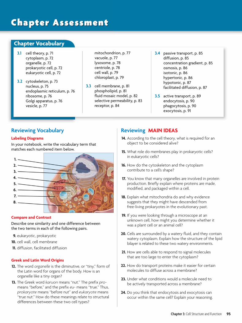
Labeling Diagrams
In your notebook, write the vocabulary term that matches each numbered item below.
Compare and Contrast
Describe one similarity and one difference between the two terms in each of the following pairs.
9. eukaryotic, prokaryotic
10. cell wall, cell membrane
11. diffusion, facilitated diffusion
Greek and Latin Word Origins
12. The word organelle is the diminutive, or “tiny,” form of the Latin word for organs of the body. How is an organelle like a tiny organ?
13. The Greek word karuon means “nut.” The prefix pro- means “before,” and the prefix eu- means “true.” Thus, prokaryote means “before nut” and eukaryote means “true nut.” How do these meanings relate to structural differences between these two cell types?
Reviewing MAIN IDEAS
14. According to the cell theory, what is required for an object to be considered alive?
15. What role do membranes play in prokaryotic cells? in eukaryotic cells?
16. How do the cytoskeleton and the cytoplasm contribute to a cell’s shape?
17. You know that many organelles are involved in protein production. Briefly explain where proteins are made, modified, and packaged within a cell.
18. Explain what mitochondria do and why evidence suggests that they might have descended from free-living prokaryotes in the evolutionary past.
19. If you were looking through a microscope at an unknown cell, how might you determine whether it was a plant cell or an animal cell?
20. Cells are surrounded by a watery fluid, and they contain watery cytoplasm. Explain how the structure of the lipid bilayer is related to these two watery environments.
21. How are cells able to respond to signal molecules that are too large to enter the cytoplasm?
22. How do transport proteins make it easier for certain molecules to diffuse across a membrane?
23. Under what conditions would a molecule need to be actively transported across a membrane?
24. Do you think that endocytosis and exocytosis can occur within the same cell? Explain your reasoning.
1.
2.
3.
4.
5.
6.
7.
8.
Chapter 3: Cell Structure and Function 95
Critical Thinking
25. Summarize How was the development of cell theory closely tied to advancements in technology?
26. Analyze What structural differences suggest that eukaryotic cells evolved from prokaryotic cells?
27. Synthesize If vesicles are almost constantly pinching off from the ER to carry proteins to the Golgi apparatus, why does the ER not shrink and finally disappear?
28. Compare and Contrast You know that both vesicles and vacuoles are hollow compartments used for storage. How do they differ in function?
29. Infer When cells release ligands, they are sent through the blood stream to every area of the body. Why do you think that only certain types of cells will respond to a particular ligand?
30. Provide Examples What are two ways in which exocytosis might help a cell maintain homeostasis?
31. Compare How is facilitated diffusion similar to both passive transport and active transport?
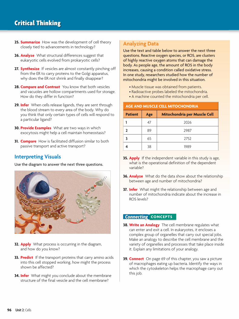
Interpreting Visuals
Use the diagram to answer the next three questions.
32. Apply What process is occurring in the diagram, and how do you know?
33. Predict If the transport proteins that carry amino acids into this cell stopped working, how might the process shown be affected?
34. Infer What might you conclude about the membrane structure of the final vesicle and the cell membrane?
Analyzing DataUse the text and table below to answer the next three questions. Reactive oxygen species, or ROS, are clusters of highly reactive oxygen atoms that can damage the body. As people age, the amount of ROS in the body increases, causing a condition called oxidative stress. In one study, researchers studied how the number of mitochondria might be involved in this situation.
• Muscle tissue was obtained from patients.• Radioactive probes labeled the mitochondria.• A machine counted the mitochondria per cell.
AGE AND MUSCLE CELL MITOCHONDRIA
Patient Age Mitochondria per Muscle Cell
1 47 2026
2 89 2987
3 65 2752
4 38 1989
35. Apply If the independent variable in this study is age, what is the operational definition of the dependent variable?
36. Analyze What do the data show about the relationship between age and number of mitochondria?
37. Infer What might the relationship between age and number of mitochondria indicate about the increase in ROS levels?
Connecting CONCEPTS
38. Write an Analogy The cell membrane regulates what can enter and exit a cell. In eukaryotes, it encloses a complex group of organelles that carry out special jobs. Make an analogy to describe the cell membrane and the variety of organelles and processes that take place inside it. Explain any limitations of your analogy.
39. Connect On page 69 of this chapter, you saw a picture of macrophages eating up bacteria. Identify the ways in which the cytoskeleton helps the macrophage carry out this job.
96 Unit 2: Cells
For more test practice,go to ClassZone.com.
Chapter 3: Cell Structure and Function 97
1. The cell theory states that the cell is the mostbasic unit of life, all organisms are made of cells,and all cells come from cells. What makes thecell theory a scientific theory?
A It is based on a scientific publication that isread by scientists worldwide.
B It is based on the work of many scientists andleads to accurate predictions.
C It is based on ideas that have been proven trueand that are not subject to revision.
D It is based on preliminary evidence but stillneeds to be confirmed with experiments.
2.Molecule Concentration Outside and Inside a Cell
Mol
ecu
les
ente
rin
g ce
ll
Outsideconcentration
LOW HIGH
HIGH
This graph shows that as the concentration ofmolecules increases outside of a cell, more andmore molecules enter the cell. These moleculesare able to enter the cell because the
A molecules are polar.
B cytoplasm is warm.
C cell membrane is semipermeable.
D nucleus is regulating movement.
Which answer choice explains why molecules can enter a cell?
THINK THROUGH THE QUESTION
3. Which of the following pairs incorrectlymatches a cell structure with its function?
A cell membrane: protein synthesis
B nucleus: information (DNA) storage
C vacuole: storage
D chloroplast: energy conversion
4. Some viruses attack cells by inserting their ownDNA into the host cells’ DNA. Why might it besimpler for these viruses to attack prokaryoticcells than eukaryotic cells?
A Prokaryotic cells have less DNA than doeukaryotic cells.
B Unlike eukaryotic cells, prokaryotic cells donot have a nucleus.
C The cell wall in prokaryotic cells is a lesseffective barrier.
D The rapid growth of prokaryotic cells generatesmore virus.
5. Which of the following is a function of theGolgi apparatus?
A storing genetic information
B recycling waste products of the cell
C providing a protective barrier
D processing and storing proteins
6.
Mit
och
ond
ria
per
cel
l
Number of Mitochondria in Different Cell Types
Skeletalmuscle
Skin Liver0
1000
2000
3000
4000
The graph shows the number of mitochondriaper cell for different cell types. Which statementbest explains these data?
A The number of mitochondria in the skin willincrease if the number in the liver decreases.
B The liver requires the greatest amount ofenergy per cell.
C The skeletal muscle was taken from a veryactive athlete.
D The skin requires the least amount of energyper cell.