center of excellence in environmental...
TRANSCRIPT

CENTER OF EXCELLENCEIN ENVIRONMENTAL TOXICOLOGY
Friday, May 22, 2015
Smilow Center for Translational ResearchPerelman School of Medicine at the University of Pennsylvania
Tenth Anniversary Symposium
Celebrating Environmental Health Science at Penn
Center of Excellence in Environmental Toxicology (CEET)
TEnTh AnnivErsAry symposium
Environmental Health Science at Penn
Arthur h. rubenstein Auditoriumsmilow Center for Translational research
perelman school of medicine at the university of pennsylvania
may 22, 2015
schedule 1 – 2
Keynote speaker 3
mission and vision statement 4
Reflections from the Director 5
members of CEET 6 – 7
poster Abstracts
Lung and Airway Disease 8 – 9
oxidative stress and oxidative stress injury 10 – 12
Reproduction, Endocrinology, and Development 13 – 18
Gene-Environment Interactions 19 – 20
CEET informatics Core 20
Translational Biomarker Core 21 – 23
integrated health sciences Facility Core 24
Community Outreach and Engagement Core 25 – 28
Superfund Research Program 29 – 32
Acknowledgements 34
Contents

1
7:30 – 8:30 A.m. COntInEntAL BREAkfASt AnD REGIStRAtIOn
Morning – PaSt and CurrEnt
8:30 – 8:35 A.m. Welcome trevor M. Penning, PhD The Thelma Brown and henry Charles molinoff professor of pharmacology; professor, Biochemistry & Biophysics and OB/GYn; Director, Center of Excellence in Environmental toxicology
8:35 – 8:45 A.m. introduction to EHSCC Program Claudia L. thompson, Ph.D. Branch Chief, Susceptibility and Population Health Branch; Program Director, EHSCC, nIEHS
8:45 – 9:15 A.m. Successes in Meeting the Environmental Health Challenges in Pa and Beyond trevor M. Penning, PhD
CuttInG EDGE CEEt RESEARCH - tOPIC 1 Moderator: Ian Blair, PhD
9:15 – 9:35 A.m. toxicant Exposures: Mechanisms of airway Hyperresponsiveness Rey Panettieri, MD professor, pulmonary, Allergy & Critical Care, psom; Co-Director of CEEt and Co-Director of the CEEt Integrative Health Sciences facility Core
9:35 – 9:55 A.m. oxidative Stress and the Pathogenesis of alzheimer’s disease Paul H. Axelsen, MD professor of pharmacology, Biochemistry and Biophysics, and medicine, perelman school of medicine
9:55 – 10:15 A.m. Children and Lead: a Sleepy Story Rebecca Simmons, MD Hallam Hurt Professor in neonatology, Department of Pediatrics
10:15 – 10:35 A.m. BaP1 gene and Environmental Susceptibility to asbestos Joseph R. testa, PhD professor, Carol & Kenneth E. Weg Chair in human Genetics; Chair, mesothelioma Working Group, Fox Chase Cancer Center
10:35 – 10:55 A.m. BrEAK
COMMunItY-OutREACH AnD EnGAGEMEnt-RESEARCH In tRAnSLAtIOn moderator: richard pepino, ms
10:55 – 11:15 A.m. Strategy, Communication, understanding: Keys to Community translation ted Emmett, MD, MS Professor, Director of Academic Programs, Occupational Medicine, Perelman School of Medicine
11:15 – 11:35 A.m. Living in a Superfund Site: Voices from ambler frances k. Barg, PhD, MEd Associate professor of Family medicine and Community health, hospital of the university of pennsylvania
11:35 – 11:55 A.m. improving Environmental Health through Multi-directional Community Engagement Marilyn Howarth, MD Adjunct Associate professor of Emergency medicine; Director, CEEt Community Outreach and Engagement Core
12:00 – 1:15 p.m. LunCh
Tenth Anniversary CEET symposiumCelebrating ten Years of Environmental Health Science
at the university of PennsylvaniaFriday, may 22, 2015
smilow Center for Translational research, perelman school of medicine

2
aftErnoon – futurE dirECtionS
pipELinE oF nEW GEnErATion oF EnvironmEnTAL hEALTh sCiEnTisTs Moderator: Rebecca Simmons, MD
1:15 – 1:30 p.m. Meeting Community Health Challenges in urban West Philadelphia richard pepino, ms Director, Academically-Based Community Service Courses; Deputy Director, CEEt Community Outreach and Engagement Core
1:30 – 1:45 p.m. LC-MS Profiling of Rotenone-induced Mitochondrial Dysfunction Andrew Worth, PhD, T32 trainee in Environmental health sciences
1:45 – 2:00 p.m. In situ Liquid-cell Confocal Microscopy of Chrysotile fiber diffusion, aggregation and growth dynamics Lei Wu, PhD, postdoctoral srp-Trainee
2:00 – 2:15 p.m. Microengineered Physiological Biomimicry: Human organs-on-chips Dan Huh, PhD Wilf Family Term Assistant professor of Bioengineering, school of Engineering and Applied science
2:15 – 2:30 p.m. developmental Programming of offspring Exposed to Bisphenol a during Human Pregnancy: investigations into Epigenetic and Metabolic Mechanisms Sara Pinney, MD, MS Associate Professor of Pediatrics, Division of Endocrinology and Diabetes, The Children’s hospital of philadelphia
2:30 – 3:30 p.m. POStER SESSIOn AnD BREAk
mEETinG ThE ChALLEnGE oF ThE niEhs sTrATEGiC pLAn Moderator: Rey Panettieri, MD
3:30 – 3:50 p.m. airborne Magnetite Particles and their interaction with Human Lung Cells Reto Gieré, PhD professor, Chair of Earth and Environmental sciences, school of Arts and sciences
3:50 – 4:10 p.m. Exposure and Biological response Biomarkers for Environmental Chemicals Ian Blair, PhD A.n. richards professor of pharmacology; Director, CEEt translational Biomarker Core, Perelman School of Medicine
4:10 – 4:30 p.m. Multigenerational Effects of BPa Exposure in a Mouse Model Marisa Bartolomei, PhD Professor, Cell and Developmental Biology, Perelman School of Medicine
4:30 – 4:50 p.m. global impact of genetic and Environmental Variation on Metabolic and anthropometric traits in africa Sarah tishkoff, PhD David and Lyn Silfen university Professor, Departments of Genetics and Biology, perelman school of medicine
5:00 – 6:00 p.m. Keynote Introduction: trevor Penning, PhD dioxins, Clocks and oxygen: Prototype Signals of an Environmental Sensor Superfamily Chris Bradfield, PhD professor of oncology, mcArdle Laboratory for Cancer research university of Wisconsin-madison, school of medicine & public health
6:00 – 7:00 p.m. rECEpTion
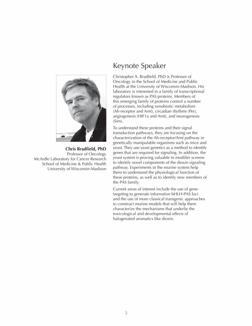
3
Keynote speakerChristopher A. Bradfield, PhD is Professor of oncology in the school of medicine and public health at the university of Wisconsin-madison. his laboratory is interested in a family of transcriptional regulators known as pAs proteins. members of this emerging family of proteins control a number of processes, including xenobiotic metabolism (Ah-receptor and Arnt), circadian rhythms (per), angiogenesis (hiF1a and Arnt), and neurogenesis (sim).
To understand these proteins and their signal transduction pathways, they are focusing on the characterization of the Ah-receptor/Arnt pathway in genetically manipulable organisms such as mice and yeast. They use yeast genetics as a method to identify genes that are required for signaling. in addition, the yeast system is proving valuable in modifier screens to identify novel components of the dioxin signaling pathway. Experiments in the murine system help them to understand the physiological function of these proteins, as well as to identify new members of the pAs family.
Current areas of interest include the use of gene-targeting to generate informative bhLh-pAs loci and the use of more classical transgenic approaches to construct murine models that will help them characterize the mechanisms that underlie the toxicological and developmental effects of halogenated aromatics like dioxin.
Chris Bradfield, PhDprofessor of oncology
mcArdle Laboratory for Cancer research school of medicine & public health
university of Wisconsin-madison

4
Mission and Vision Statement
The Center of Excellence in Environmental Toxicology (CEET) was launched in 2005 and receives grant support from the national institute of Environmental health sciences (niEhs). it is one of only twenty designated Envi-ronmental health sciences Core Centers in the nation and the only one in the Commonwealth of pennsylvania
the CEEt elucidates the mechanistic links between environmental exposures and human disease and trans-lates its findings into action to improve the health of vulnerable individuals, and local, national and global communities.
The CEET marries its relevant research excellence to tackle the environmental challenges that may represent an assault on our public health. many of these challenges have their origins in community-based concerns. Examples include the hazard presented by petrogenic polycyclic aromatic hydrocarbons from the Deepwater horizon oil-spill in the Gulf of mexico; the fate, transport, remediation and adverse health effects of asbestos exposure in the Ambler Community in sE. pennsylvania (which is home to one of the largest superfund As-bestos hazardous waste sites in the country); and natural gas drilling operations in the marcellus shale, where citizens are concerned about the effects of air-pollution and water contamination on their health. These com-munity public health concerns are often identified by our Community Outreach and Engagement Core which has a history in conducting community-based participatory research, wherein research findings are translated back to the affected community using a “community-first communication model”.
The CEET has research excellence in themes that are related to environmental health that exist in our imme-diate area. Its Affinity Group in Lung and Airway Disease examines the relationship between poor air quality and air pollution in our region (ozone, fine particulate matter, allergens, SO2, no2 and Co emissions) and disease (asthma, lung cancer, mesothelioma and COPD); Its Affinity Group in Reproduction, Endocrinology, and Development examines the relationship between exposures in windows of susceptibility and resultant health outcomes. investigators explore the association between in utero exposures, epigenetic imprinting, and the developmental basis of adult disease. these organ-based themes are linked to our Affinity Groups in disease mechanism, which include oxidative stress and oxidative stress injury and Gene-Environment interactions.
The CEET enables its investigators to conduct exposure science using its Translational Biomarker Core which uses sophisticated liquid chromatography mass spectrometry methods to identify and develop assays of bio-markers of exposure and effect, and measure changes in targeted and unbiased metabolomes following re-sponse to exposures, disease onset and progression. The CEET maintains a bioinformatics core so that large siloed data bases in genomics, proteomics, and metabolomics can be merged as reporter of system wide re-sponses and adaptation to environmental exposure that can explain the presenting phenotype. The integrated health sciences Facility Core (ihsFC) of the CEET provides assistance with a broad range of transdisciplinary services including study design, enrollment of research subjects, population and community exposures, access to biospecimens via a CEET biorepository and data management, and genetic and non-genetic biostatistical analyses.
The CEET engages six communities in s.E. pennsylvania to empower them with new knowledge so that they are better informed to tackle issues of environmental health threats, health disparities, and environmental justice. To improve the environmental health of these and similar affected communities, the CEET is actively involved in the education of health care professionals (residency program in occupational and Environmental health, nursing concentration in occupational and Environmental health, and masters of public health programs).
the CEEt also disseminates findings to all stakeholders including community organizations, local, state and federal officials and agencies (Pennsylvania Department of Health, Pennsylvania Department of Environmental protection, u.s. Environmental protection Agency) to affect change in environmental health and public health policies.
the CEEt is a flexible entity that marshals excellence in basic, translational, patient-oriented and popula-tion-based research in the school of medicine and Children’s hospital of philadelphia. Although primarily housed in the School of Medicine, the 62 CEEt Investigators belong to 16 departments and five schools at the university of pennsylvania.

5
Reflections from the Director
it is with enormous pride that i welcome you to the Tenth Anniversary sym-posium of the Center of Excellence in Environmental Toxicology (CEET), the university of pennsylvania Environmental health sciences Core Center.
The planning of the CEET began in 2003 when small meetings of vision-ary faculty felt that significant environmental health challenges exist in the aging infra-structure of our urban environment. one of those faculty mem-bers was Dr. Edward (ted) Emmett who had a long history of working with rev. horace strand on the cumulative exposure of environmental hazards on the health of residents in Chester City, pA (an environmental justice community). This led us to examine disease registries of pennsylvania to identify diseases of environmental etiology that may affect our urban region disproportionately. these data identified lung and airway disease as a major urban health problem and brought Drs. Rey Panettieri (ozone exacerbated asthma) and steven Albelda (thoracic malignancies with a focus on meso-thelioma) into the CEEt. Health registry data also identified pre-term birth, low birth weight and development defects as affecting our region dispro-portionately which attracted Drs. Jerome Strauss and Jeanne Manson into
becoming Center members. this nucleus of faculty led to our first Environmental Health Sciences Core Center (EHSCC) application in 2004. At that time, Dr. Emmett was Deputy Director and Director of the Community Outreach and Education Core (COEC). Dr. Emmett was working on exposure to perflurooctanoate, a known endocrine disruptor, in Little hocking, oh where the water supply was contaminated with this byproduct of teflon manufacturing. ted felt that the community had a right to know about these exposures, their source, and their health implications which led to the “community-first” communication model to empower the commu-nity to affect immediate change. Our EHSCC application to nIEHS was not successful at first submission, as is the case for many large Center grants. in the following year we resubmitted the application and were funded in 2006 as a new EhsCC with an almost perfect score.
from these small beginnings we can reflect back on the fact that when the CEEt was first funded, it was the Ambler community that came to us to seek help with their asbestos exposure problem. This blossomed into our Superfund Research and training Program: “Asbestos, fate, exposure, remediation and adverse health effects” (1P42 ES023720) led by Dr. Ian A. Blair, which was funded in 2014.
The CEET also established a pipeline to train environmental health scientists of the future through its summer programs for high school students (tREES) and undergraduates (StEER) led by Dr. Jeffrey field (R25 ES021649). this led to an establishment of a Certificate Program in Environmental Health Sciences for graduate education and the eventual award of a T32 institutional Training program: “Translational research Training program in Environmental Health Sciences” (1t32 ES019851). Remarkably, before the existence of the CEEt, there was no formalized training in environmental health sciences at the university of pennsylvania.
The CEET now provides an intellectual home to tackle large environmental health problems by building trans-lational research teams so that findings can be translated back to the community using the model provided by Dr. Emmett. this translation of research findings back to the community now utilizes the skills of Dr. Marilyn Howarth (current COEC Director) and Mr. Richard Pepino (Deputy Director). this evolution of the CEEt has occurred due to the imagination and dedication of its affinity group leaders and faculty who are CEEt mem-bers; institutional support from Deans Rubenstein and Jameson, Perelman School of Medicine and successive Vice Provosts of Research – Drs. Steven fluharty and Dawn Bonnell; and generous continued funding from niEhs.
– trevor Penning

6
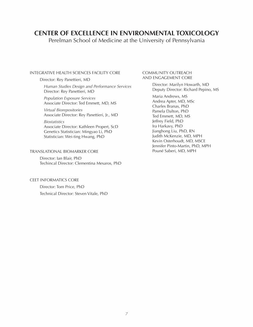
CEntEr of EXCELLEnCE in EnVironMEntaL toXiCoLogYperelman school of medicine at the university of pennsylvania
ADMInIStRAtIVE COREDirector: trevor Penning, Ph.D.
Deputy Director: Reynold Panettieri, M.D.
Affinity Group ILunG AnD AIRWAY DISEASE
Director: Michael Beers, M.DSteve Albelda, MDAndrea Apter, MD, MScJason Christie, MD, MSCEMelpo Christofidou-Solomidou, PhDPamela Dalton, PhD Peter DeCarlo, PhDW. Michael foster, PhDReto Gieré, PhDDan Huh, PhDHoward kipen, MD, MPHVera krymskaya, PhDRey Panettieri, MDtrevor Penning, PhDAnil Vachani, MD
Affinity Group IIOXIDAtIVE StRESS AnD OXIDAtIVE StRESS InJuRY
Director: Ian Blair, PhDPaul Axelsen, MDJoseph Baur, PhDMichael Beers, MDBrenda Casper, PhDJeffrey field, PhD Aron fisher, MD Garret fitzGerald, MDReto Gieré, PhDDan Huh, PhDHarry Ischiropoulos, PhD Douglas Jerolmack, PhDkelly Jordan-Sciutto, PhD Vladimir Muzykantov, MD, PhDtrevor Penning, PhDRebecca Simmons, MD Andrew Strasser, PhD Jane Willenbring, PhD
Affinity Group IIIREPRODuCtIOn, EnDOCRInOLOGY, AnD DEVELOPMEnt (READ)
Director: George Gerton, PhDMarisa Bartolomei, PhDShelley Berger, PhDSamantha Butts, MD, MSCECristos Coutifaris, MD, MPHted Emmett, MD, MSStruan Grant, PhDBrett kaufman, PhDkaren knudsen, PhDMichael Levine, MDJianghong Liu, PhD/RnSarah Millar, PhDkatherine nathanson, MDSam Parry, MDtrevor Penning, PhDSara Pinney, MD, MSRebecca Simmons, MD Virginia Stallings, MDJeremy Wang, MD/PhD
Affinity Group IVGEnE-EnvironmEnT inTErACTions
Director: Marisa Bartolomei,PhDShelley Berger, PhDIan Blair, PhDJinbo Chen, PhDYouhai Chen, MD/PhDJason Christie, MD, MSCEStruan Grant, PhDHakon Hakonarson, MD/PhDJohn Hogenesch, PhDHongzhe Li, PhDSarah Millar, PhDJason H. Moore, PhDkatherine nathanson, MDJennifer Pinto-Martin, PhD/MPHtrevor Penning, PhDtimothy Rebbeck, PhDVirginia Stallings, MDSarah tishkoff, PhDAalim Weljie, PhDSteve Whitehead, PhD
7
CEntEr of EXCELLEnCE in EnVironMEntaL toXiCoLogYperelman school of medicine at the university of pennsylvania
inTEGrATivE hEALTh sCiEnCEs FACiLiTy CorE
Director: Rey Panettieri, MD
Human Studies Design and Performance Services Director: Rey Panettieri, MD
Population exposure Services Associate Director: ted Emmett, MD, MS
Virtual Biorepositories Associate Director: Rey Panettieri, Jr., MD
Biostatistics Associate Director: kathleen Propert, ScD Genetics Statistician: Mingyao Li, PhD Statistician: Wei-ting Hwang, PhD
TrAnsLATionAL BiomArKEr CorE
Director: Ian Blair, PhD techincal Director: Clementina Mesaros, PhD
CEET inFormATiCs CorE
Director: tom Price, PhD
technical Director: Steven Vitale, PhD
CommuniTy ouTrEACh AnD EnGAGEMEnt CORE
Director: Marilyn Howarth, MD Deputy Director: Richard Pepino, MS
maria Andrews, ms Andrea Apter, MD, MSc Charles Branas, PhD Pamela Dalton, PhD ted Emmett, MD, MS Jeffrey field, PhD Ira Harkavy, PhD Jianghong Liu, PhD, Rn Judith Mckenzie, MD, MPH kevin Osterhoudt, MD, MSCE Jennifer Pinto-Martin, PhD, MPH Pouné Saberi, MD, MPH

8
Lung and Airway Disease
posTEr ABsTrACTs
L1 Metabolic activation of 3-nitrobenzanthrone by aldo-keto reductases (aKr1C1-aKr1C4)
Jessica Murray1, Meng Huang1, Tianzhu Zang1, Volker Arlt2, Trevor M. Penning1
1Center of Excellence in Environmental Toxicology, Department of Systems Pharmacology and Translational Therapeutics, Perelman School of Medicine at the University of Pennsylvania, Philadelphia, PA, United States. 2Analytical and Environmental Sciences Division, MRCPHE Centre for Environment and Health, King’s College London, London, United Kingdom Email: [email protected]
In 2012, the International Agency for Research on Cancer (IARC) classified diesel exhaust as a Group 1 carcinogen due to sufficient evidence that exposure is associated with increased risk for lung cancer in humans. However, only a subset of individuals exposed to diesel exhaust develops cancer, indicating the need to identify the genes involved in metabolic activation of these compounds and their genetic variants. Nitro-polycyclic aromatic hydrocarbons (NO2-PAH) are a major component of diesel exhaust and require metabolic activation to exert their carcinogenic activity. A representative NO2-PAH, 3-nitro-benzanthrone (3-NBA), is metabolically activated to 3-aminobenzanthrone (3-ABA) via a 6-electron nit-roreduction catalyzed by NQO1 and POR. The reaction leads to the formation of 3-aminobenzanthrone (3-ABA) derived DNA adducts which promote G to T transversions. Building upon previous data that shows human aldo-keto reductase 1C3 (AKR1C3) contains nitroreductase activity towards chemother-apeutic agents (Guise, C.P., Abbattista, M.R., et al., Cancer Res, 70(4), 2010), we chose to examine the nitroreductase activity of AKR1C1–AKR1C4 towards NO2-PAH. We have demonstrated here for the first time that AKR1C enzymes catalyze the nitroreduction of 3-NBA to 3-ABA. We monitored reac-tions with reverse phase HPLC coupled to in-line photo-diode-array detection (PDA) and fluorescence detection (FLD) to quantify 3-NBA and 3-ABA levels. Fluorescence and UV spectroscopy were used to validate the identity of the compounds. This method was adapted for discontinuous enzymatic assays to measure steady state kinetic parameters for the nitoreductase activity of AKR1C1-AKR1C4 and NQO1. In addition, high-resolution Orbitrap mass spectrometry was used to identify 3-NBA, the nitroso- and hydroxylamino-in termediates, and 3-ABA providing evidence for the 6-electron reduction mechanism catalyzed by AKRs. Results indicate that the NQO1 catalyzed reduction of 3-NBA has a higher specific activity, but the combined specific activities of AKR1C catalyzed reduction may play a more significant role in the overall production of 3-ABA. These results suggest that the relative expression of NQO1 and AKR1C enzymes will determine their respective contribution to 3-NBA reduction, especially since all the aforementioned enzymes are inducible by the Nrf2-Keap1 system.
This work is supported by P30E513508 and RO1 CA39504 to TMP.

9
Lung and Airway Disease
posTEr ABsTrACTs
L2 Cigarette Smoke (CS) Enhances Carbachol-induced airway narrowing in Hu-man Precision-cut Human Lung Slices (PCLS)
Joseph Jude1,2, Christie Ojiaku1,2, Cynthia Koziol-White1, William Jester1, Gaoyuan Cao1, Rey Panettieri1,2
1Airways Biology Initiative, University of Pennsylvania; 2Center of Excellence in Environmental Toxicology, Department of Systems Pharmacology and Translational Therapeutics, Perelman School of Medicine at the University of Pennsylvania, Philadelphia, PA Email: [email protected]
Background: Tobacco smoke is associated with a variety of human diseases, including COPD and asth-ma. Tobacco smoke exposure exacerbates clinical symptoms in asthmatics and COPD patients. Cigarette smoke (CS) delivers ~4000 different chemical compounds to the smoker’s lung and modulates functions of both structural and transient cells of airways to induce lung inflammation and injury. The impact of CS on airway smooth muscle (ASM) function remains unknown. We hypothesize that cigarette smoke enhances the agonist-induced contractile response of human ASM. Methods: Precision-cut human lung slices (PCLS) were prepared from non-asthmatic human donors. PCLS were exposed to CS (Kentucky research cigarette, 3R4F) in Vitrocell VC1 smoking machine using the International Organization of Standardization (ISO)-defined parameters. Clean air (83% relative humidity, 25°C) was used as the control exposure. PCLS were incubated overnight (21 h) following clean air or cigarette smoke exposure and carbachol (Cch) dose-response was determined for airway narrowing. Maximal response (Emax), Log concentration for 25%-maximal response (Log EC25) and area under the curve (AUC) were determined from the carbachol dose-response curve. In some experiments, slices were pre-contracted with carbachol and dose-response was determined for formoterol-induced bronchodilation. The culture supernatants from the PCLS were used to determine IL-6 and IL-8 levels by ELISA. Human ASM (HASM) cells were exposed to CS or clean air and agonist-induced [Ca2+]i was determined 21 h post-exposure. Results: CS increased the Emax and AUC of the Cch dose-response curve in PCLS. The Log EC25 of the curve was reduced in CS-treated slices compared to that of clean air-treated slices (n=5 donors/group), although the difference was not statistically significant. Formoterol-induced bronchodilation was marginally attenu-ated in CS-treated slices compared to that of clean air-treated slices (n=2 donors/group). There was little effect on IL-6 or IL-8 levels in the supernatant of slices following CS or clean air exposures. In HASM cells, histamine or bradykinin-induced [Ca2+]i was unaltered by CS exposure. Conclusion: CS induces airway hyperresponsiveness by enhancing the contractile response of ASM to agonists, with little effect on inflammatory mediator release in lung slices or [Ca2+]i in HASM cells. The findings predict that CS-induced hypercontractility may involve Ca2+ sensitization mechanisms in ASM cells.
Supported by T32 ES019851

10
oxidative stress and oxidative stress injury
posTEr ABsTrACTs
os1 the role of nadPH oxidase isoforms and reactive oxygen Species in Human airway Smooth Muscle Cell function
Christie Ojiaku, Joseph A. Jude, Reynold A. Panettieri
Center of Excellence in Environmental Toxicology, Department of Systems Pharmacology and Translational Therapeutics, Perelman School of Medicine at the University of Pennsylvania, Philadelphia, PA Email: [email protected]
Background: Reactive oxygen species (ROS) serve as signaling molecules and evoke pathogen destruction through inflammatory cell-mediated respiratory burst. However, dysregulated ROS production via NA-DPH Oxidase (NOX) mediates the pathogenesis of many diseases, including asthma. Studies in human airway smooth muscle cells (HASMCs) suggest that the NOX4 and NOX2 isoforms contribute to the intrinsic airway hyperresponsiveness and inflammation characteristic of asthma. However, the predom-inant NOX isoform expressed in HASMCs and its contributions to the asthmatic phenotype remain unknown. We hypothesize that altered expression of NOX isoforms modulate intracellular ROS levels and airway hyperresponsiveness in HASMCs.
Methods: HASMCs were isolated and cultured from the airways of non-asthmatic (n=5) and asthmatic (n=3) lung donors. Total RNA was isolated and cDNA was synthesized from 72-hour serum-starved non-asthma and asthma-derived HASMCs. Basal expression of NOX isoforms 1-5, Dual oxidase 1 (DUOX1) and DUOX2 was investigated using real-time PCR. NOX4 expression was induced by TGF- 1 (50ng/ml, 48 h or 1ng/ml, 20hr). Changes in NOX4 gene expression and intracellular ROS production were assessed. Additionally, basal ROS production was determined in non-asthma (n=3) and asthma-de-rived (n=3) HASMCs.
Results: Our findings show that NOX4, NOX2, and NOX5 are the dominant NOX isoforms expressed in HASMCs. Basal NOX4 and NOX5 mRNA expression trended towards higher levels in asthma-de-rived versus that of non-asthma-derived HASMCs. TGF-b1 treatment (50ng/ml) significantly induced a 55-fold and 87-fold (±34.45) increase in NOX4 mRNA expression in non-asthma and asthma-derived HASMCs, respectively, with larger-fold increases occurring in HASMCs exhibiting low basal NOX4 expression. We also observed little change in basal intracellular ROS production between non-asthma and asthma-derived HASMCS. TGF-b1 (50 ng/ml) treatment showed a 1.35-fold increase in ROS pro-duction in non-asthma-derived HASMCs, and TGF-b1 (1 ng/ml) treatment induced a 1.34-fold higher increase in ROS production in asthma versus non-asthma-derived HASMCs.
Conclusions: Our findings suggest that basal upregulation of NOX4 and NOX5 in HASMCs may play a role in altered human airway smooth muscle function and asthma pathogenesis. The induction of NOX4 expression depends on the basal expression level of NOX4, which may be a contributing factor to the heterogeneity of disease pathogenesis in asthma. The augmented increase in ROS production in asthma versus non-asthma-derived HASMCs following TGF-b1 treatment suggests that NOX or other ROS-producing enzymes may be more sensitive to upregulation in asthma-derived HASMCs. Together, these data suggest a potential role for NOX expression and ROS production to serve as a biomarker for asthma.
Supported by T32 GM008076
posTEr ABsTrACTs

11
oxidative stress and oxidative stress injury
posTEr ABsTrACTs
os2 Acute Ozone Exposure Induces Neuroinflammation and Upregulation of the alzheimer’s-associated Protein Beta-secretase-1
Michelle Erickson1,2, Hengjiang Zhao3, Joseph Jude2,3, Gabriel van de Walle6, Amy Lee5, Ngan Nguyen4, William Jester3, Reynold Panettier, Jr.2,3, Kelly Jordan-Sciutto1, 2
1School of Dental Medicine, University of Pennsylvania; 2 Center of Excellence in Environmental Toxicology, Department of Systems Pharmacology and Translational Therapeutics, Perelman School of Medicine at the University of Pennsylvania, Philadelphia, PA; 3 Airways Biology Initiative, University of Pennsylvania, Philadelphia, PA; 4 Drexel University, Philadelphia, PA; 5 School of Engineering, University of Pennsylvania, Philadelphia, PA; 6 School of Arts and Sciences, University of Pennsylvania Email: [email protected]
Ozone is a widespread toxicant in air pollution that has adverse effects on many organ systems, includ-ing the central nervous system (CNS). Both acute and chronic exposures to ozone are associated with elevated oxidative damage in the CNS, deficits in learning and memory, and increased risk of clinical di-agnosis of Alzheimer’s disease (AD). The mechanisms by which ozone exposure could contribute to AD, however, have not been well-addressed. Systemic and neuroinflammation can promote accumulation of the AD-plaque protein amyloid beta (Ab) in the brain. Therefore, we hypothesized that ozone-induced inflammation would be associated with CNS changes favoring Ab accumulation. To test this, we uti-lized a well-characterized mouse model of ozone-induced pulmonary inflammation. Balb/c mice were exposed to forced air or ozone (3 ppm) for 2 h and markers of systemic, pulmonary and CNS inflam-mation were determined at 6 or 24 h post-exposure. We found that ozone induced inflammation not only in the lungs, but also in the serum and CNS. In serum, the acute phase protein serum amyloid A (SAA), but not C-reactive protein was increased at 6 and 24 hours post-ozone exposure. In the cerebral cortex, nuclear localization of the p65 subunit of NF-kB was increased at 6 hours post-ozone exposure, but returned to baseline by 24 hours. Ozone also affected the expression of the Ab-producing enzyme beta-secretase 1 (BACE1). In ozone-treated mice, BACE1 protein levels were significantly decreased at 6 hours post-exposure, and significantly increased at 24 hours post-exposure. These findings suggest that inflammation-induced BACE1 upregulation is a mechanism by which ozone exposure could contribute to AD onset and progression.
Supported by T32 ES019851
12
oxidative stress and oxidative stress injury
posTEr ABsTrACTs
os3 Quantification of Serum Apolipoprotein A1 by LC-MRM/MS with a SILAC Labeled Protein internal Standard reveals reduced Levels in Smokers
Qingqing Wang, Hong Su, Lili Guo, Christine Busch, Ian Blair
Center of Excellence in Environmental Toxicology and Penn SRP Center, Department of Systems Pharmacology and Translational Therapeutics, Perelman School of Medicine at the University of Pennsylvania, Philadelphia, PA Email: [email protected]
Stable isotope dilution liquid chromatography (LC) coupled with multiple reaction monitoring/mass spectrometry (MRM/MS) has become a valuable alternative to immunoassay for the absolute quantifica-tion of protein biomarkers. We report the development of an absolute quantification approach by using stable isotope labeling by amino acids in cell culture (SILAC) for the quantification of biomarker ApoA1 in human serum samples. Recombinant ApoA1 labeled with [13C6
15N2]-lysine and [13C915N1]-tyrosine
expressed in HEK293 cells, was used as a protein internal standard by addition to serum samples at the beginning of the sample preparation procedure. The method was fully validated using nine signature pep-tides generated from native ApoA1 and SILAC-labeled ApoA1. The influence of methionine oxidation on ApoA1 quantification was evaluated by monitoring oxidized and non-oxidized methionine. Satisfac-tory dilutional linearity, method accuracy and precision were obtained from 15.6 mg/dL to 500.0 mg/dL. Using this validated method, the endogenous ApoA1 levels in human serum samples from 50 non-smok-ers and 50 smokers were analyzed and compared. The mean concentration of ApoA1 in non-smokers was 169.4 mg/dL and an 18.4% reduction to 138.2 mg/dL in smokers was found. All nine peptides showed the same trends in the two groups, and significant differences were observed for eight peptides by smoking status. Peptide correlation results suggested that there were unidentified post-translational modifications present on peptide LAEYHAK, ATEHLSTLSEK and WQEEMELYR. Poor correlations were found between LC-MRM/MS and an ELISA-based method. The rigorously validated LC-MRM/MS approach using a stable isotope labeled protein internal standard will provide an additional paradigm for evaluating the effects of smoking in human populations.
Supported by P30ES013508
13
r1 the Effect of Early Life Methyl donor Supplementation on obesity development
Sarah E. McKee1, Teresa M. Reyes2
1Center of Excellence in Environmental Toxicology, Department of Systems Pharmacology and Translational Therapeutics, Perelman School of Medicine at the University of Pennsylvania, Philadelphia, PA; 2University of Pennsylvania, University of Cincinnati Email: [email protected]
Excessive maternal weight gain during pregnancy contributes to an increased risk for obesity in the off-spring. In a mouse model of excessive maternal weight gain, we find that offspring have increased prefer-ence for sucrose and fat, increased expression of genes that underlie reward-related behaviors, and both global and gene specific DNA hypomethylation. These changes in reward-related neural circuitry may contribute to the increased risk for the development of obesity in the offspring by altering the animal’s re-sponse to highly palatable, energy dense foods. Methyl donor supplementation (MDS) during pregnancy can reverse some of these phenotypes, yet it is unknown whether postnatal MDS can reverse these phe-notypes. To determine this, offspring from dams fed either a high fat diet (HFD) or control diet during gestation/lactation were fed a methyl donor supplemented diet during early life (age 3-6 weeks). We find that postnatal MDS significantly decreased body weight in both male and female adult HFD offspring, and does not alter body weights of control diet offspring. Further, postnatal MDS can normalize adult male fat preference and contributes to regional specific normalization of DNA hypomethylation.
Supported by T32 ES019851
r2 assessing the transmission of an altered Epigenotype and Phenotype follow-ing Exposure to Endocrine disrupting Compounds (EdCs)
Frances Xin1, Martha Susiarjo1, Amita Bansal 2, Martha Stefaniak1, Changhong Li3, Rebecca Simmons4, Marisa Bartolomei1
1Department of Cell and Developmental Biology, University of Pennsylvania; Center of Excellence in Environmental Toxicology, Perelman School of Medicine, University Pennsylvania, Philadelphia, PA; 2Division of Neonatology, University of Pennsylvania; 3Division of Endocrinology and Metabolism, The Children’s Hospital of Philadelphia; 4Division of Neonatology, University of Pennsylvania; Center of Excellence in Environmental Toxicology, Perelman School of Medicine, University Pennsylvania, Philadelphia, PA Email: [email protected]
Fetal exposure to endocrine disrupting compounds (EDCs) results in aberrant developmental outcomes and increased disease susceptibility in adult life. Not only does exposure to EDCs in utero affect the developing fetus, but these effects can also be transmitted across multiple generations. Although the precise mechanisms by which these compounds act remain to be elucidated, it has been proposed that epigenetic pathways mediate their effects. Exposure to the EDCs bisphenol A (BPA) and di(2-ethylhex-yl)phthalate (DEHP) have been shown to alter DNA methylation, an epigenetic regulatory mechanism critical for proper development. DNA methylation is also a well-established mechanism of imprinted gene regulation. In our mouse model, fetal exposure to BPA results in aberrant regulation of imprinted genes in a gene- and tissue-specific manner, which corresponds with altered DNA methylation at regu-latory elements of imprinted genes. In adulthood, BPA-exposed male offspring exhibit increased body fat, impaired glucose homeostasis, and altered glucose-stimulated insulin secretion. Interestingly, these phenotypic changes are transmitted to the next (F2) generation. At the molecular level, misregulation of Igf2, a growth-promoting imprinted gene, is associated with the observed phenotype. Total expression
Reproduction, Endocrinology, and Development (READ)
posTEr ABsTrACTs

14
Reproduction, Endocrinology, and Development (READ)
posTEr ABsTrACTs
of the Igf2 gene and methylation at its corresponding Differentially Methylated Region (DMR) 1 are altered in the F1 (exposed as a fetus) and F2 (exposed as the fetal germ cells) offspring. Because humans are rarely exposed to a single EDC, it is critical to assess the synergistic and/or antagonistic effects of com-binatorial exposures. Our preliminary data demonstrate that fetal exposure to the combination of BPA and DEHP produces a greater perturbation in the allelic expression of imprinted genes in the F1 placenta as compared to single compounds exposures, suggesting an additive or synergistic effect. Identifying the detrimental effects of early-life EDC exposure on fetal and postnatal development across multiple gen-erations and determining their mode of action will ultimately improve human health risk assessments of these compounds.
Supported by T32 ES019851
r3 investigating Estrogen and androgen Metabolism in in vitro Models of aro-matase inhibitor therapy and resistance
Lisa N. Bottalico, Qingqing Wang, Clementina Mesaros, Ian Blair
NIEHS Superfund Research Program / Centers of Excellence in Environmental Toxicology and Cancer Pharmacology, Department of Systems Pharmacology and Translational Therapeutics, University of Pennsylvania Perelman School of Medicine, Philadelphia, PA Email: [email protected]
Aromatase inhibitors (AI) are highly effective as a first-line of therapy against estrogen receptor (ER) positive breast cancers. However, approximately 15-19% of women relapse within 10 years on AI-main-tenance therapy after breast cancer. Mechanisms of resistance to aromatase inhibitors are varied and not well understood. The hallmark of acquired resistance to AI is considered to be activation of growth signaling pathways that can drive proliferation independent of ERa. However, changes in expression of estrogen metabolizing enzymes with AI treatment have also been seen clinically. This study aims to investigate whether estrogen deprivation can incite metabolic alterations which allow the breast tumor to produce ERa agonists locally and via routes in addition to the aromatase pathway. Cell models employed to investigate this question include MCF-7, an ER-positive hormone-responsive breast cancer cell line; MCF-7Aro, an MCF-7 cell line in which Aromatase (CYP19A1) is overexpressed and which is a model for aromatase-overexpressing breast cancer; and LTED-Aro, a derivative of the MCF-7Aro cell line which has adapted to growth in the absence of estradiol and is a model of late-stage endocrine therapy resistance. In vitro investigations include real-time PCR analysis of expression of relevant steroid-metabolizing en-zymes, cell-based proliferation assays to assess cellular growth response to isolated steroid precursors and aromatase inhibitors, and LC-MS quantification of a panel of estrogen and androgen precursors with AI treatment. Initial studies in the MCF-7Aro cell line demonstrate increased expression of aromatase and AKR1C3 with serum deprivation, a robust proliferative response to a physiological range of testosterone and a dose-dependent decrease in proliferation with AI treatment. Current studies are aimed at correlat-ing proliferative responses with LC-MS quantification of a panel of estrogen metabolites and androgen precursors with AI treatment.
Supported by T32 GM008076

15
Reproduction, Endocrinology, and Development (READ)
posTEr ABsTrACTs
r4 Development of Stable Isotope Dilution LC-ESI-MS/MS Method for the Quantitation of Human Hydroxy-androgens
Tianzhu Zang1, Daniel Tamae1, Clementina Mesaros2, Ian A. Blair3, Trevor M. Penning3
1Center of Excellence in Environmental Toxicology, Department of Systems Pharmacology and Translational Therapeutics, Perelman School of Medicine, University of Pennsylvania; 2Centers for Cancer Pharmacolo-gy and Excellence in Environmental Toxicology, Perelman School of Medicine, University of Pennsylvania; 3Center for Excellence in Environmental Toxicology, Center for Cancer Pharmacology, Department of Systems Pharmacology and Translational Therapeutics, Perelman School of Medicine, University of Pennsylvania Email: [email protected]
Prostate cancer (CaP) is the most commonly diagnosed cancer and the second leading cause of cancer death among North American men. The development of CaP is androgen-dependent and advanced localized disease can be treated by surgical or chemical castration. However, the recurrence of prostate cancer, also called castration resistant prostate cancer (CRPC), occurs despite castrate levels of circulating testosterone (T) and dihydrotesterone (DHT) and has the potential to become more metastatic. CRPC remains hormonally driven and new drugs such as abiraterone acetate (a P450c 17 inhibitor) or en-zalutamide (an AR antagonist) have been approved by FDA for the treatment of CRPC by targeting the androgen signaling axis, but resistance to both drugs eventually occurs. In order to better understand the response to hormonal therapy and mechanisms of drug resistance, a comprehensive investigation of how androgen levels change in serum and prostate cancer tumors is imperative. So far, traditional immunoas-says cannot give an accurate quantitation of circulating or tumor androgens because of the lack of spec-ificity. Our group has developed a stable isotope dilution liquid chromatography electrospray ionization tandem mass spectrometric (SID-LC-ESI-MS/MS) method to quantify human keto-androgens with the requisite specificity and sensitivity. So far, we have also expanded our method to quantify hydroxy-andro-gens, using picolinic acid derivatization so that the entire androgen metabolome involved in the canon-ical, alternative and backdoor synthetic pathways can be displayed. Nine derivatized hydroxy-androgens including T and DHT can be separated and simultaneously detected by LC-ESI-MS/MS. The LLOQ of six derivatized hydroxy-androgens with a mono-hydroxyl group is between 1.56 pg and 0.78 pg. The intra- and inter-assay precision and accuracy match with FDA guidelines for bioanalytical method vali-dation. We also used enzymatic methods to synthesize stable isotopically labeled hydroxy-androgens as internal standards, so dihydroxy androgens e.g. 5-androstene-3b, 17b-diol, 5a-androstane-3b, 17b-diol and 5a-androstane-3a, 17b-diol can also be quantified as bis-picolinates. The new method when com-bined with keto-androgen quantitation would permit further interrogation of mechanisms of drug resis-tance to hormone ablative therapy.
Supported by 1P01 CA163227-01A1 and a Prostate Cancer Foundation Challenge Grant awarded to Trevor M. Penning

16
Reproduction, Endocrinology, and Development (READ)
posTEr ABsTrACTs
r5 In vitro fertilization induces altered Morphology as Well as Epigenetic defects at imprinted and non-imprinted genes in term Mouse Placentae
Eric de Waal1,4, Lisa Vrooman1, Terri Ord2, Richard M. Schultz3, Marisa Bartolomei1,4
1Department of Cell and Developmental Biology, Perelman School of Medicine, University of Pennsylvania, Philadelphia, PA; 2Reproductive Endocrinology and Infertility, Departments of Obstetrics and Gynecology, University of Pennsylvania, Philadelphia, PA; 3Department of Biology, University of Pennsylvania, Philadelphia, PA; 4 Center of Excellence in Environmental Toxicology, Department of Systems Pharmacology and Translational Therapeutics, Perelman School of Medicine at the University of Pennsylvania, Philadelphia, PA Email: [email protected]
It has been estimated that over 5 million babies have been born worldwide through the use of assisted reproductive technologies (ART). While most ART-conceived children appear to be healthy, epidemi-ologic data show that a subset of ART-conceived children have an increased risk for birth defects, low birth weight and metabolic abnormalities. Of particular concern is that the ex vivo manipulations utilized during ART can induce a suboptimal intrauterine environment that will predispose the developing fetus to adult-onset diseases. The developmental origins of health and disease hypothesis states that environ-mental exposures during critical stages of development will lead to adaptive changes in the gestating fetus, which may result in medical consequences later in life, such as diabetes and cardiovascular disease. The molecular mechanisms responsible for adverse health outcomes in ART-conceived children are un-known, but may entail impaired placental function, altered epigenetic marks in somatic tissues, or a combination of these factors. We have previously demonstrated that epigenetic marks in mid-gestation placentae are highly sensitive to ART compared to embryonic tissues. In the current study, we analyzed epigenetic profiles in fetal tissues and term placentae from embryonic day (E) 18.5 fetuses produced by in vitro fertilization (IVF) or natural conception. We also generated fetuses using embryo transfer alone or superovulation and embryo transfer to determine if we can detect phenotypic abnormalities without the use of in vitro fertilization and embryo culture. Placental morphology and function were analyzed in all groups of mice, and bisulfite pyrosequencing was utilized to measure DNA methylation at several imprinted genes in both fetal and placental tissues from E18.5 conceptuses. Histological analyses were performed to assess placental morphology, and placental to fetal (P:F) weight ratios as well as expression profiles of key transporter genes were used to evaluate placental function. Notably, full-term placentae from E18.5 IVF-derived fetuses exhibited a substantial increase in the incidence of epigenetic defects at imprinted genes compared to fetal tissues. IVF-derived placental tissues also had higher P:F weight ratios, an expanded junctional zone and aberrant expression of genes that play important roles in the transport of glucose and amino acids to the fetus.
In addition, we found that aberrant expression of glucose and amino acid transporter genes were associ-ated with abnormal methylation at cis-regulatory elements indicating that epigenetic profiles of non-im-printed genes are also influenced by ART. Interestingly, the use of embryo transfer or superovulation and embryo transfer induced morphological abnormalities in the placental tissues, but did not influence DNA methylation profiles of imprinted genes or expression levels of transporter genes. Collectively, these results demonstrate that the use of IVF can alter placental morphology and influence DNA methylation levels at imprinted and non-imprinted loci. Moreover, while the use of superovulation and embryo trans-fer contributes to the abnormal placental phenotype observed in IVF offspring, epigenetic defects are likely caused by prolonged embryo culture. Given that normal placental function is critical for providing an optimal intrauterine environment for the developing fetus, it is imperative that we continue to opti-mize all of the manipulations that are utilized during ART to protect the placenta from developmental and epigenetic abnormalities that are commonly observed in IVF-derived placental tissues.
Supported by T32 ES019851

17
Reproduction, Endocrinology, and Development (READ)
posTEr ABsTrACTs
r6 Multigenerational Sex-specific Effects of Maternal BPA Exposure on Glucose and KiC Stimulated insulin Secretion and islet gene Expression in Mice
Amita Bansal1,2,3, Tom van der Meer1,4, Changhong Li5, Cetewayo Rashid1,2,3, Martha Susiarjo2,6, Marisa S. Bartolomei2,6, Rebecca A. Simmons1,2,3
1Center for Research on Reproduction and Women’s Health, Perelman School of Medicine, University of Pennsylvania, Philadelphia, PA; 2Center of Excellence in Environmental Toxicology, University of Pennsylvania Perelman School of Medicine, Philadelphia, PA; 3Division of Neonatology, Department of Pediatrics, Perelman School of Medicine, University of Pennsylvania, Philadelphia, PA; 4Department of Pediatrics, University of Groningen, Groningen, Netherlands; 5Division of Endocrinology and Metabolism, The Children’s Hospital of Philadelphia, 802B Abramson Research Center, Philadelphia, PA, and 6Department of Cell and Developmental Biology, Perelman School of Medicine, Smilow Center for Translational Research, University of Pennsylvania, Philadelphia, PA Email: [email protected]
Background: Bisphenol A (BPA), an endocrine disruptor, is associated with type 2 diabetes and obesity in humans and animals. We demonstrated that maternal BPA exposure from 2 weeks prior to mating until weaning in C57BL/6 mice is associated with higher body fat and impaired glucose tolerance in F1 and F2 male but not female offspring. The underlying mechanisms are unknown. We determined multigen-erational effects of maternal BPA exposure on glucose (GSIS) and a-ketoisocaproate stimulated insulin secretion (KICSIS), and islet gene expression in mice. Methods: Islets were isolated from adult F1 and F2 offspring (n=4-6 per sex per group) of F0 mothers exposed to 10 μg/kg/day (LowerB), 10 mg/kg/day (UpperB) BPA and 7% corn oil (Control) diets. GSIS and KICSIS were determined by perifusion ramp studies; data were analysed by 2-way ANOVA. mRNA levels were determined by qPCR. P<0.05 was con-sidered significant. Results: LowerB F1 and F2 males, but not females, had reduced GSIS and increased igf2 and ucp2 mRNA expression than Controls; F2 males also had increased ogdh mRNA expression. UpperB F1 and F2 males had reduced KICSIS and increased igf1 mRNA expression than Controls; F1 males also had increased basal insulin secretion, pdx1, igf2 and hnf1a mRNA expression. Conclusion: Early life lower and upper dose BPA exposure at representative human exposure levels leads to islet mito-chondrial defects, and altered insulin secretion and islet gene expression across two generations in male mice.
Supported by RO1 ES 562150
18
Reproduction, Endocrinology, and Development (READ)
posTEr ABsTrACTs
r7 Paternal Exposure to ubiquitous Contaminant Bisphenol a does not impair glucose tolerance in Mice
Cetewayo Rashid1,2,3, Amita Bansal1,2,3, Marisa S. Bartolomei1,2,4 , Rebecca A. Simmons1,2,3
1Center for Research on Reproduction and Women’s Health, Perelman School of Medicine, University of Pennsylvania, Philadelphia, PA; 2Center of Excellence in Environmental Toxicology, Department of Systems Pharmacology and Translational Therapeutics, Perelman School of Medicine at the University of Pennsylvania, Philadelphia, PA; 3Division of Neonatology, Department of Pediatrics, Perelman School of Medicine, University of Pennsylvania, Philadelphia, PA; 4Department of Cell and Developmental Biology, Perelman School of Medicine, Smilow Center for Translational Research, University of Pennsylvania, Philadelphia, PA Email: [email protected]
Greater than 90% of the US population is environmentally exposed to the endocrine disrupting chem-ical bisphenol A (BPA). Maternal exposure to BPA has been shown to restrict intrauterine growth and impair glucose tolerance in offspring, however, glucose homeostasis in paternally exposed offspring has been rarely investigated. To explore the consequences of paternal BPA exposure on offspring growth and glucose tolerance, we employed a mouse model of dietary exposure with BPA concentrations of 0 (Control), 10μg (Low) and 10mg/kg/day (High). These BPA doses are comparable to the NOAEL and LOAEL, respectively, and similar to human exposure levels. Sires remained on their respective diets 12 weeks prior to mating. Dams and their offspring were maintained on standard breeder chow. Paternal BPA exposure did not affect birth weights compared to controls. Low BPA exposed male and female off-spring were, however, growth restricted after weaning while offspring of the High BPA group was similar to controls. Interestingly, male offspring born to High BPA-exposed sires were more glucose tolerant than other experimental groups. This difference in glucose disposal was not sequelae of obesity evidenced by comparable body composition between groups as analyzed by DEXA, and insulin tolerance tests proved similar insulin-dependent glucose excursion. Enhanced efficiency of glucose tolerance was sex-specific, with female offspring showing no significant difference in blood glucose levels upon glucose or insulin administration. DEXA scanning did reveal decreased lean mass in the female offspring of the Low BPA group compared to controls. In conclusion, paternal BPA exposure attenuated body weight gain and displayed sex and dose-specific improvements in glucose homeostasis.
Supported by RO1 ES 56210

19
posTEr ABsTrACTs
Gene-Environment interactions
G1 anthropometric and Cardiovascular trait Variation in diverse rural african Populations: impact of genetic ancestry and diet
Matthew E.B. Hansen1,2, Joseph Lachance1, Sameer Soi1, Laura Scheinfeldt1, Alessia Ranciaro1, Simon Thompson1, Jibril Hirbo1, and Sarah A. Tishkoff1,2
1Departments of Biology and Genetics, University of Pennsylvania, Philadelphia, PA; 2Center of Excellence in Environmental Toxicology, Department of Systems Pharmacology and Translational Therapeutics, Perelman School of Medicine at the University of Pennsylvania, Philadelphia, PA Email: [email protected]
The African continent is home to a diverse range of indigenous peoples that have adapted to a wide range of ecological environments and subsistence lifestyles. Many complex traits are expected to display variation between populations due to demographic history and/or natural selection to these diverse envi-ronments. In an effort to survey phenotypic variation in Africa and begin to understand the genetic and environmental factors that contribute to this variation, we have collected trait measurements on height (N=5,125), BMI (N=5,098), grip strength (N=1,968), systolic and diastolic blood pressure (N=2,002), and pulse (N=2,008) from agricultural, pastoral, and hunter-gatherer communities across eastern and western sub-Saharan Africa. We present the observed variation in these traits between genders, across populations, and across subsistence practices. We find significant differences in trait values among these categories. A subset of 697 individuals were genotyped on the Illumina 1M-Duo SNP array. We per-formed a GWAS using a linear mixed model approach that controls for relatedness, and find that only height broadly replicated GWAS top hits from non-African cohorts (p-value enrichment). To assess the impact of genetic ancestry and subsistence on trait variation, we performed STRUCTURE analysis to determine ancestral cluster proportions, and used both linear regression and mixture model analyses to infer the trait dependence on these factors. The fraction of variance explained by the models is discussed, as well as the implications for future genotype/phenotype analysis within sub-Saharan Africa for these and related traits.
Supported by T32 ES019851
G2 Estrogen receptor Mediated PaH Metabolite translocation to the nucleus Trevor Penning, Isabelle Lee
Center of Excellence in Environmental Toxicology, Department of Systems Pharmacology and Translational Therapeutics, Perelman School of Medicine at the University of Pennsylvania, Philadelphia, PA Email: [email protected]
Polycyclic aromatic hydrocarbons (PAHs) are a diverse class of environmental toxicants with two or more fused benzene rings; these compounds are common byproducts of incomplete combustion of fossil fuels, and they are suspect human carcinogens. Even though they have natural sources such as forest fires and volcanoes, most PAHs in ambient air are anthropogenic. This class of compounds can be found in diesel exhaust, smoked or barbecued foods and cigarette smoke. Cigarette smoke and tobacco products account for 90% of human lung cancers in the United States. PAHs have to be metabolically activated into reactive genotoxins in order to cause their mutagenic and carcinogenic effects. One of the pathways of PAH activation involves the formation of PAH o-quinones by aldo-keto reductases, and these o-qui-nones are ligands for the aryl hydrocarbon receptor (AhR). Previous work in our laboratory has provided evidence that the AhR acts as a carrier involved in the shuttling and concentration of the representative PAH o-quinone, benzo[a]pyrene-7,8-dione (B[a]P-7,8-dione); to the nucleus (Park et al, 2009). This process enhances PAH o-quinone-mediated oxidative DNA damage in the form of DNA strand breaks and generation of mutagenic 8-oxo-7,8-dihydro-2-deoxyguanosin (8-oxo-dG) lesions. Given the simi-

20
Gene-Environment interactions
posTEr ABsTrACTs
larity between the planar B[a]P-7,8-dione to the estrogen quinones and evidence by Wang et al that the estrogen receptor acts as a “Trojan Horse” to shuttle these genotoxic estrogen metabolites to the nucleus to promote oxidative DNA damage, we hypothesize that PAH o-quinones can be translocated into the nucleus by the estrogen receptor in a similar fashion, to enhance oxidative DNA damage. We have used Ishikawa cells, a human endometrial adenocarcinoma cell-line, and alkaline phosphatase activity as the read-out for estrogen receptor activation to determine whether B[a]P-7,8-dione activates the estrogen receptor. Thus far, we have demonstrated that B[a]P-7,8-dione is a ligand for the estrogen receptor and seek to determine the cross talk between the estrogen receptor and AhR in mediating the genotoxic effects of PAH in human lung cells.
Supported by 5R01 CA39504 and P30 ES13508
CEET informatics Core (CiC)
CEEt informatics Core Steven Vitale
Perelman School of Medicine, University of Pennsylvania, Philadelphia, PA Email: [email protected]
The CEET Informatics Core is building tools to explore the influence of exposure on health. The prima-ry focus is on characterizing gene activity in relation to disease mediated by one or more toxicants.
Secondarily, we hope to advance models on variation and susceptibility. To accomplish this, open source tools are used and written which rely heavily on public gene databases. Analysis software is being ex-panded to include combined genotypic/phenotypic and exposure data. Updated software will allow for collaborative annotation of datasets and real-time shared complex queries.
Current and planned projects also involve specimen management, instrumentation and QC software maintenance as required for the streamlined analysis of incoming samples. Primary datasets, core utilities and software libraries are stored on a server that we recently built and housed within a Tier-2 datacenter on campus. Expanded compute power is also available for software deployed to a campus high perfor-mance cluster.
Software to support these projects is in the public domain and available at: https://github.com/CEETBioin
Supported by P30 ES013508
CiC1

21
Translational Biomarker Core (TBC)
posTEr ABsTrACTs
translational Biomarker Core Services Clementina Mesaros and Ian A. Blair
Center of Excellence in Environmental Toxicology; Penn SRP Center; Department of Systems Pharmacology and Translational Therapeutics, Perelman School of Medicine, University of Pennsylvania, Philadelphia, PA Email: [email protected]
The Core is a unique resource for Center of Excellence in Environmental Toxicology (CEET) investiga-tors because the CEET is the only stakeholder. This makes it possible to offer a wide range of innovative liquid chromatography/mass spectrometry (LC-MS) assays at a very modest cost. Sophisticated analyt-ical methodologies based on LC-MS are used to identify and quantify biomarkers of diseases/disorders that have environmental etiology such as lung and airway disease, cardiovascular disease, endocrine and reproduction disruption, and neurodegenerative disease. Major translational biomarkers that are ana-lyzed include: amino acid metabolism, cardiovascular disease, cellular oxidative stress, drug metabolism, dysregulated lipid metabolism, Friedreich’s ataxia, in vivo oxidative stress, inflammation, metabolism, mitochondrial oxidative stress, mitochondrial dysfunction, and psychosocial stress. The overall goals of the Translational Biomarker Core are to provide bioanalytical services primarily based on LC-MS and high resolution LC-MS methodology. The Core also has substantial expertise in sensitive and specific analysis of serum proteins that can be brought to bear on particular projects. New assays that are devel-oped for individual CEET investigators will eventually become a Core service. More recently the Core has developed novel approaches to the analysis of metabolomic biomarkers using high-resolution mass spectrometry instrumentation. Cutting edge and diverse mass spectrometry instrumentation is available to CEET investigators through the Translational Biomarker Core including: Thermo TSQ Quantum Ultra Triple Quadrupole, Thermo TSQ Vantage Triple Quadrupole, Applied Biosystems 4000 Triple Quadrupole, Agilent 6460A Triple Quadrupole, Thermo LTQ ion trap, and high-resolution Thermo LTQ-XL Orbitrap and Thermo Q-Exactive HF.
Supported by P30 ES13508
resistance to P450c17 inhibitors in Castration resistant Prostate Cancer May result from the dHEa-S depot that remains and Can Be used by aKr1C3 for intratumoral androgen Biosynthesis
Daniel Tamae1, Ling Duan1, Elahe A. Mostaghel2, Mary E. Taplin3, Ian A. Blair1, Trevor M. Penning1
1Centers for Cancer Pharmacology and Excellence in Environmental Toxicology, Department of Systems Pharmacology and Translational Therapeutics, Perelman School of Medicine, University of Pennsylvania, Philadelphia, PA; 2Fred Hutchinson Cancer Center, University of Washington, Seattle, WA; 3Dana Farber Cancer Institute, Harvard Medical School, Boston, MA Email: [email protected]
Localized intermediate and high-risk prostate cancer can be treated with androgen deprivation therapy (ADT). Initially, patients undergo remission but inevitably relapse, due to the emergence of castration resistant prostate cancer (CRPC). Newer agents such as the P450c17 inhibitor, abiraterone acetate (AA) have gained approval for the treatment of CRPC and increased median survival by 4 months. We have developed a novel and validated stable isotope dilution liquid chromatography electrospray ionization selected reaction monitoring mass spectrometry (SID-LC/ESI/SRM/MS) method for the quantification of conjugated and unconjugated keto-androgens in human serum (JSBMB (2013) 138:281). This meth-od was applied to human serum from patients enrolled in a neoadjuvant AA clinical trial (J Clin Oncol (2014) 32:3705). We found that testosterone (T) and 5alpha-dihydrotestosterone (DHT) did not always decrease in tandem, which suggests that pathways that bypass T may lead to DHT. These may include the alternative pathway (delta-4-androstene-3,17-dione ➝ 5alpha-androstane-3,17-dione ➝ DHT) or the backdoor pathway (androsterone ➝ 5alpha-androstane-3alpha,17beta-diol ➝ DHT). Second, despite
TBC1
TBC2

22
Translational Biomarker Core (TBC)
posTEr ABsTrACTs
achieving >90% inhibition of P450c17, the level of DHEA-S that remains (20,000 ng/dL) is 4,000-fold higher than castrate levels of T achieved in the trial. We hypothesize that this depot of DHEA-S that re-mains is sufficient to feed intratumoral androgen biosynthesis and this could account for the clinical fail-ure of P450c17 inhibitors in CRPC. Adaptive intratumoral androgen biosynthesis can be facilitated by the up-regulation of AKR1C3 (type 5 17beta-hydroxysteroid dehydrogenase). To test our hypothesis, we have developed a panel of isogenic prostate cancer cell lines that either express AKR1C3 or are AKR1C3 null. These cell lines are then challenged with post-AA levels of circulating androgens (DHEA-S, DHEA and delta-4-androstene-3,17-dione) to determine whether they can still make sufficient androgens to activate the androgen receptor, where androgen metabolism is measured using SID-LC/ESI/SRM/MS. Using LNCaP and LNCaP-AKR1C3 cells, we find that the level of T detected as T-17beta-glucuronide in the AKR1C3 expressing cells could be sufficient to activate the AR. These results would suggest that AKR1C3 inhibitors in combination with AA could benefit patients who might otherwise fail AA treat-ment.
Supported by grants R25-CA101871, P30-ES-13508, P01-CA-163337
Metabolism of representative alkylated and oxygenated Petrogenic Polycyclic aromatic Hydrocarbons in Human Hepatoma (Hepg2) Cells
Huang Meng, Zhang Li, Clementina Mesaros, Ian Blair, Trevor M. Penning
Centers of Excellence in Environmental Toxicology and Cancer Pharmacology, Department of Systems Pharmacology and Translational Therapeutics, Perelman School of Medicine at the University of Pennsylvania, Philadelphia, PA Email: [email protected]
Exposure to petrogenic polycyclic aromatic hydrocarbons (PAHs) in the food-chain is the major human health hazard associated with the Deepwater Horizon gulf-oil spill. Risk assessment is based on the as-sumption that petrogenic and pyrogenic PAHs have similar toxicological profiles yet petrogenic PAHs are either alkylated or oxygenated and information on their metabolism is lacking. We report the metabolic fate of 6 representative alkylated petrogenic PAHs in the Macondo oil, and 3 representative oxygenated petrogenic PAHs that result from weathering in human HepG2 cells. The structures of the metabolites were identified by HPLC-UV-fluorescence detection, ion trap LC-MS/MS and Orbitrap LC-HRMS/MS. Alkylated petrogenic PAHs show no evidence of metabolism on the alkyl side chain. Metabolism is ring-based and involves formation of phenols and tetraols (P450 derived), o-quinones and catechols (AKR derived). Pretreatment with oil extracts inhibits P450 mediated metabolism indicating that pa-rental alkylated PAHs may persist after absorption. Oxygenated PAHs show evidence for catechol and catechol conjugates (detoxication) and quinone metabolites that can still redox-cycle. These pathways show evidence for both detoxication and metabolic activation. Sulfated and glucuronidated catechols are observed as metabolites of both alkylated and oxygenated PAHs, and can be used as biomarkers of human exposure.
Supported by U19ES020676-03
TBC3

23
Translational Biomarker Core (TBC)
posTEr ABsTrACTs
analysis of Polar Metabolites in Biological Samples by ion-Paring Liquid Chromatography-Mass Spectrometry
Lili Guo, Andrew J. Worth, Clementina Mesaros, Ian Blair
Penn SRP Center, Center of Excellence in Environmental Toxicology, and Department of Systems Pharmacology and Translational Therapeutics, Perelman School of Medicine, University of Pennsylvania, Philadelphia, PA Email: [email protected]
Many metabolites within central energy metabolism are polar molecules containing carboxylic acids, phosphate groups or nucleotides. Most of these metabolites carry a negative charge and as such are difficult to analyze by conventional reverse-phase liquid chromatography-mass spectrometry (LC-MS). Although separations can be achieved by LC with ion-pairing reagents, many ion-pairing reagents are not sufficiently volatile and thus are not optimal for MS analysis using conventional negative ion elec-trospray ionization (ESI). Previous analyses of oligonucleotides were greatly enhanced by the addition of 1,1,1,3,3,3-hexafluoro-2-propanol (HFIP) to the mobile phase with diisopropylethylamine (DIPEA) as the ion-pairing reagent. Here we have expanded the utilization of HFIP as an additive to aid in the analysis of the hydrophilic molecules that make up central energy metabolism. LC-MS analysis was performed on an Agilent 1200 series HPLC system coupled to an Agilent 6460 Triple Quadrupole mass spectrometer with an ESI source operating in negative mode. Analytes were separated by chroma-tography using a Luna C18(2) 150mm × 2mm, 3μm particles column (Phenomenex) and DIPEA as the ion-pairing reagent. A two-solvent system was used, with solvent A as HFIP and DIPEA in water and Solvent B as HFIP and DIPEA in methanol. The optimal concentration of DIPEA and HFIP and the linear gradient conditions were investigated. Preliminary results showed that HFIP can significant-ly affect the MS intensity and LC separation of carboxylic acids, sugar phosphates and phospho-car-boxylic acids. 10 mM DIPEA and 400 mM HFIP provided the best chromatographic performance. The method successfully separated and detected twenty-nine commercially available compounds. These compounds include glucose-6-phosphate, fructose-6-phosphate, fructose-1,6-bisphosphate, glyceralde hyde-3-phosphate, dihydroxyacetone-phosphate, 2-phosphoglycerate, phosphoenolpyruvate, 2-hydrox-yglutarate, 6-phosphogluconate, ribulose-5-phosphate, ribose-5-phosphate, sedoheptulose-7-phosphate, erythrose-4-phosphate, flavinadenine dinucleotide (FAD), nicotinamide adenine dinucleotide (NAD), NADH, nicotinamide adenine dinucleotide phosphate (NADP), NADPH, pyruvate, citrate, isocitrate, succinate, fumarate, malate, lactate, a-ketoglutarate, oxaloacetate, glutamate, and aspartate. This method was used to identify affected metabolic pathways within lymphoma cells treated with mTOR inhibitors by both absolute quantification and stable isotope glucose labeling.
Supported by P30 ES013508, P30 CA016520, T32 ES019851
TBC4

24
integrative health sciences Facility Core
posTEr ABsTrACTs
integrative Health Sciences facility Core Reynold Panettieri
Center of Excellence in Environmental Toxicology, Department of Systems Pharmacology and Translational Therapeutics, Perelman School of Medicine at the University of Pennsylvania, Philadelphia, PA; Airway Biology Initiative, University of Pennsylvania Email: [email protected]
The Integrative Health Sciences Facility Core (IHSFC) supports all human research in the CEET. It is the entity that permits the CEET to attain its Strategic Vision to perform translational environmen-tal health research that will impact patients, communities, and the public. It provides highly focused transdisciplinary services including study design, enrollment of subjects, data management, access to biological samples, biostatistical analyses, interpretation of results and dissemination of results to the public through the Community Outreach and Engagement Core (COEC). Over the past four years, the IHSFC has uniquely focused on delivery of five specific services that include: human studies design and performance services, human exposure laboratories (ozone, PM2.5, in vitro environmental tobacco smoke), human population exposure services, CEET virtual biorepositories, and biostatistics group. Each service provides CEET investigators with unique tools to translate fundamental research into improve-ments in individual human and population-based cohorts to advance PREcision Environmental Med-icine (PREEM). In a bi-directional manner, the IHSFC communicates with the COEC to formulate community-based questions and concerns into specific hypotheses for investigators with expertise in population-based studies. An important goal of the IHSFC is to promote inter-Environmental Health Science Core Center (EHS CC) collaborative projects in human subjects. Over the past four years, five interactive projects involving inter-EHS CC pilot projects or R01 equivalents have been initiated. The interaction of the IHSFC with other centers greatly extends the expertise to a network of EHS CCs to address inter-center human research. As described in the Strategic Vision, the IHSFC supports integrat-ed research themes in humans that transcend affinity groups and the formation of translational research teams that involve CEET investigators and COEC members to address community-based questions.
Supported by P30 ES13508
ihsFC1

25
Community outreach and Engagement Core
posTEr ABsTrACTs
the Effects of oil and gas activity on Water Quality and Human Health in Colorado
Kyra Reumann-Moore1, Richard Pepino2, Jessica Rogers3, Joseph Ryan3, Stephen Osborn4
1School of Arts and Sciences, University of Pennsylvania, Philadelphia, PA; 2Department of Earth and Environmental Science, University of Pennsylvania and Center of Excellence in Environmental Toxicology, Department of Systems Pharmacology and Translational Therapeutics, Perelman School of Medicine at the University of Pennsylvania, Philadelphia, PA; 3University of Colorado Boulder, Boulder, CO; 4California State Polytechnic University, Pomona, CA Contact email: [email protected]
Colorado is home to several geologic basins that are productive for oil and gas. There are currently about 50,000 oil and natural gas wells in the state as a whole (COGCC, 2013c; Hunt, 2013). Many of these wells are stimulated by hydraulic fracturing, and the number of wells continues to grow. Many people are concerned about the potential environmental and health effects of this oil and gas activity, especially in terms of drinking water contamination. This has led to a need for the gathering and analysis of rele-vant data. The Colorado Oil and Gas Conservation Commission (COGCC) has an online Geographic Information System (GIS) database that contains the locations of oil and gas wells and water wells where samples have been taken. These samples measure many water quality parameters, out of which this study will focus on organic compounds.
This study compiles Colorado water quality data – from the COGCC database and collected through additional field sampling – in a GIS format to determine if there are any general or spatial trends of ele-vated concentrations with regards to the BTEX (benzene, toluene, ethylbenzene, and total xylenes) com-pounds. Since it is difficult to know baseline concentrations due to variation in local geology, we looked for elevated concentrations and health risks above EPA guidelines. We also conducted a drinking water health risk assessment that looked at cancer and non-cancer health risks from BTEX compounds. Some sample sites violated drinking water standards for one or more parameters. Many parts of the spatial anal-ysis and health risk assessment were found to be inconclusive or without clear correlations. However, a significant cancer risk above the EPA target Risk Screening Level was found in the health risk assessment. This indicates a need for further research into the relationship between compromised water quality and the proximity of oil and natural gas extraction wells in Colorado and elsewhere.
Colorado oil and gas industry: is Water Quality Protected? Adriana Lucia Garcia
Earth and Environmental Science Senior Thesis (Mentor: Richard V. Pepino) Email: [email protected]
Hydraulic fracturing is used in unconventional oil and gas extraction to recover natural gas that was previously considered inaccessible. However, with any large-scale anthropogenic interference in a natural system comes potential risks to both public welfare and public health. One problem that has attracted media attention is that of water quality, in particular the potential for contamination of drinking wa-ter wells due to hydraulic fracturing. Organic compounds used as chemical additives in the hydraulic fracturing fluids may migrate into private water wells through pathways created by well casing failures, abandoned wells, or existing faults and fractures in geological formations. Relatively little peer-reviewed research has been conducted to understand the risks to water quality, and baseline data is often unavail-able, making it difficult to identify any changes in water quality due to recent oil and gas operations.
The objective of this work was to understand the occurrence of eight organic contaminants (BTEX com-pounds, which include benzene, toluene, ethylbenzene, and total xylenes, as well as isopropanol, ethylene
CoEC1
CoEC2

26
Community outreach and Engagement Core
posTEr ABsTrACTs
glycol, glutaraldehyde, and 2-butoxyethanol) in groundwater and whether or not their occurrence may be correlated to oil and gas activity in Colorado. I focused on samples containing concentrations of ben-zene above the EPA’s Maximum Contaminant Level (MCL) for drinking water using Colorado Oil and Gas Conservation Commission (COGCC) reports. The majority of the benzene incidents could not be definitively linked to oil and gas activity. The number of samples containing benzene concentrations over the MCL was relatively constant with time and with basin. The values of the benzene concentrations over the MCL also did not vary with time or with basin. Additionally, despite flaws in the national Energy Policy Act of 2005, Colorado’s state laws are generally effective at responding to homeowners’ complaints and holding industry accountable for remediation when necessary to ensure safe drinking water.
the three Phases of Matters from Hydraulic fracturing: gas, Liquid (Waste), and Solid (Waste)
George L Gerton1,2, Jeffrey Field2,3, Douglas Wiebe2,4
1Center for Research on Reproduction and Women’s Health and Department of Obstetrics and Gynecology; 2Center of Excellence in Environmental Toxicology, 3Department of Systems Pharmacology and Translational Therapeutics; 4Department: Biostatistics and Epidemiology; Perelman School of Medicine at the University of Pennsylvania, Philadelphia, PA
In Pennsylvania’s Marcellus Shale region, hydraulic fracturing (“fracking”) is an unconventional gas (UG) extraction process that releases natural gas from deep shale that exists below approximately 75% of the Commonwealth’s surface. Since 2007, the rapid growth of the hydraulic fracturing industry has posi-tioned Pennsylvania as one of the major energy producers in the Unites States. The contributions to the business economy of the state are easily understood by acknowledging the profits accrued from the sale of large quantities of energy. However, what is not fully appreciated at present is the full cost to society with regards to population health, agricultural economy, lifestyles, and environment. To begin address-ing these issues, we set out to analyze public databases of unconventional gas production and the solid and liquid waste generated by this industry to gain an understanding of the range of production-related activities that are underway and identify the locations where they occur. This information is necessary to provide a foundation for future studies on public health. Using geographical information systems (GIS approaches), we mapped the UG wells by location, start date, and annual gas production volume. We also examined the quantities of liquid and solid wastes reported for the wells per county per year. As part of this analysis, we determined the locations where the liquid and solid wastes were transported and how they were handled in the waste disposal stream. These studies provide a lens from which to view the changing dynamics of the industrial processes used in UG well drilling and hydraulic fracturing and em-phasize that studies of health consequences must account for the evolution of the ways that gas is extract-ed and waste is disposed. Furthermore, these studies illustrate that the consequences of UG well drilling and hydraulic fracturing in the Marcellus Shale are not restricted to the Commonwealth of Pennsylvania but impact other states that receive exported liquid and solid wastes for treatment and disposal.
Supported in part by a pilot grant P30 E513508
CoEC3
27
CoEC4 Community outreach: the teen research and Education in Environmental Science (trEES) Summer Program for High School Students
Jeffrey Field
Center of Excellence in Environmental Toxicology, Department of Systems Pharmacology and Translational Therapeutics, Perelman School of Medicine at the University of Pennsylvania Email: [email protected]
In 2007, the Center of Excellence in Environmental Toxicology launched a community outreach ed-ucation program for High School students called the Teen Research and Education in Environmental Science (TREES) summer program. The TREES program is a unique hands-on research experience for high school students to introduce them to laboratory science. Each year about eight students are recruited from local high schools for the eight week program. They are taught by graduate student mentors, re-turning high school student mentors and faculty members, all of whom volunteer their time to guide the students one-on-one or in the small group. There is a daily lecture on an environmental issue and a mini-course in toxicology. TREES students also watch and discuss movies about environmental issues (e.g. An Inconvenient Truth, Thank You for Smoking). Other classes discuss “survival” skills such as laboratory safety, library and internet research, responsible conduct of research, scientific writing and presentation skills. There is a college admissions workshop, a library tour and a formal campus tour. Students prepare reports on a natural product that originated from an environmentally sensitive region of the world. To-gether with STEER undergraduates, TREES students attend a weekly lecture from the faculty and go on a field trip. TREES begins with two weeks of structured laboratory exercises to teach basic lab techniques such as pipetting, weighing, sterile technique, and several spectrophotometer level assays. This sets the basic training for what is the most unique aspect of the program: an individually guided research project on a topic chosen by the student. The projects are developed in consultation with the mentors and faculty and then executed by the student. At the end of the program students present their results in a report, a poster, and an oral symposium. Students are encouraged to present their projects in their community, back at school, and in local science fairs. Over the years, a diverse set of projects addressed environmental issues, including air, water, and food safety. TREES scholars have been highly successful in science fairs with most winning local science fairs, a number winning national honors, and several publishing their work. About 80% of the students major in STEM fields in college with about 25% majoring in environ-mental science, far above the national average.
Supported by R25 ES021649
Community outreach: Short term Educational Experiences for research (StEEr grant) or Penn undergraduate Environmental Health Scholars Program, Summer Program for undergraduate Students
Jeffrey Field
Center of Excellence in Environmental Toxicology, Department of Systems Pharmacology and Translational Therapeutics, Perelman School of Medicine at the University of Pennsylvania Email: [email protected]
Each summer, the Center of Excellence in Environmental Toxicology runs a community outreach edu-cation program for undergraduate students interested in environmental health science called the Penn Undergraduate Environmental Health Scholars Program through the Short Term Educational Experi-ences for Research (STEER) grant. STEER is funded by the National Institute of Health, and a $3500 stipend is provided to each student. Approximately 8 students are accepted each year into this 10-week program. The heart of the program is working one-on-one with a faculty member on an original research
Community outreach and Engagement Core
posTEr ABsTrACTs
CoEC5

28
project. Projects address environmental health problems, but the specific topics vary greatly. The research can be laboratory research, field research, public health research, or a combination of these. Each student is matched up with a faculty mentor, most of whom are part of CEET, based on stated interests and ca-reer goals. The student-mentor relationship usually endures long after the program ends, with research often continuing into the school year. The program also involves a weekly “Cutting Edge” lecture. Most lectures are academic and are delivered by faculty mentors or other environmental health experts. Past lecture topics have included endocrine disruptors, environmental justice, integrated pest management, natural toxins, and tobacco exposure and PAHs. Additionally, a career discussion panel introduces stu-dents to future career opportunities and offers guidance, and a “responsible conduct of research” session emphasizes the importance of ethics in today’s research. STEER also includes a weekly field trip to envi-ronmental sites. Past locations have included superfund sites, environmental justice communities, waste-water treatment facilities, wildlife refuges, and local gardens. Field trips and lectures take place with the TREES high school students (STEER scholars serves as role models for them). At the end of the program, the STEER scholars present their work in an oral symposium.
Supported by R25 ES021649
the ‘Community Exposome’: a Cumulative impacts assessment Model for the Environmentally overburdoned Community of Eastwick
Marilyn Howarth, Richard Pepino, Janelle Bronaugh
Center of Excellence in Environmental Toxicology, Pharmacology and Translational Therapeutics, Perelman School of Medicine at the University of Pennsylvania, Philadelphia, PA Email: [email protected]
Some communities are overburdened by environmental exposure. Regardless of the many reasons that may contribute to this circumstance, residents bear the health impact of cumulative environmental exposure. The Community Outreach and Engagement Core of the Center of Excellence in Environ-mental Toxicology of the Perelman School of Medicine at the University of Pennsylvania (CEET: P30-ES013508) has been working with the community of Eastwick in Philadelphia. This community, located in the southwest corner of Philadelphia, borders a major highway (I-95), the Philadelphia Airport, a re-finery and a superfund site contaminated with polycyclic aromatic hydrocarbons and heavy metals. The community has asked for our assistance in evaluating their increased asthma rates and their perception of increased cancer among residents. Research methods for cumulative impacts assessment are not well established. The COEC has embarked on this assessment by considering a ‘community exposome’. This method of cumulative risk assessment aims to identify as many environmental exposures impacting a community as possible. We will describe the apparent exposures to residents by incorporating multiple sources of data. In addition we will assess key indicators of environmental exposure effects such as asthma and cancer rates. The mixed methods approach includes involvement of community members, the EPA, City of Philadelphia regulators, and the Delaware Valley Regional Transportation Authority. The aim of the community exposome analysis is to identify the environmental exposures that pose the greatest risk and develop hypotheses that CEET researchers can address. In addition, we hope to identify exposure mitigation strategies that can be communicated to the community and engage regulatory agencies in being more proactive in their consideration of cumulative impacts in their work. We will outline our ac-tivities, partnerships, and events, as well as discuss our future plans for engagement to help the Eastwick community answer important questions around cumulative environmental impacts.
Supported by P30 ES13508
CoEC6
Community outreach and Engagement Core
posTEr ABsTrACTs

29
superfund research program
posTEr ABsTrACTs
s1 An Historical Cohort Study of the Influence of Occupational and non-occupational Exposure to asbestos
Atu Agawu1, Douglas J. Wiebe1, Luke A. Basta1, Shabnam Elahi1, Anil Vachani2, Frances K. Barg2
1Perelman School of Medicine, University of Pennsylvania, Philadelphia, PA; 2Center of Excellence in Environmental Toxicology, Department of Systems Pharmacology and Translational Therapeutics, Perelman School of Medicine at the University of Pennsylvania, Philadelphia, PA Email: [email protected]
Context: Asbestos exposure is most widely studied as an occupational phenomenon. However, there is emerging evidence of asbestos-related diseases among non-occupationally exposed individuals. Using Ambler, Pennsylvania (PA), a community with substantial occupational and community exposure to asbestos, we aimed to characterize non-occupational exposure to asbestos and its resultant mortality.
Methods: We used publicly available census records to identify individuals living in Ambler, PA in 1930. We extracted names, address, gender, race, occupation and industry. Occupational exposure was defined on the basis of an individual’s occupation and listed industry. Para-occupational exposure was defined as having the same address as an individual with occupational exposure. We calculated summary statistics of demographic variables, tabulated exposures, and used chi-square tests to describe associations among exposure variables and race/gender.
Results: 4,524 individuals were identified with a median age of 32 years and an interquartile range of 37. Half were male (50.6%), with individuals being predominantly white (87.6%) and a small Black population (12.4%). Only 9.6% of the population had occupational exposure, whereas approximately one third had paraoccupational exposure (36.2%). A smaller proportion of women had occupational exposure compared to males (2.5% vs. 18.3%, p<0001), although the trend was reversed for paraoccu-pational exposure (38.9% for females, 33.5% for males, p<0.001) A higher proportion of Blacks had occupational (15.7%) and paraoccupational (57.3%) exposure compared to Whites (9.7% occupational, 33.2% paraoccupational) and the differences were statistically significant for both (p<0.001).
Conclusions: In this large cohort of individuals living near a large asbestos manufacturing plant we found significant paraoccupational exposure to asbestos. Additionally, Blacks had significantly higher occupa-tional and paraoccupational exposure to asbestos. Future efforts will focus on characterizing mortality in the cohort as function of occupational and paraoccupational exposure.
Supported by 3P42ES023720-01S1
s2 Siderophore-mediated iron dissolution from asbestos Sanjay Mohanty1, Cedric Gonneau2, Jane K. Willenbring 1,3, Brenda B. Casper 2,3, Douglas Jerolmack1,3
1Department of Earth and Environmental Science, University of Pennsylvania; 2Department of Biology, University of Pennsylvania; 3Center of Excellence in Environmental Toxicology, Department of Systems Pharmacology and Translational Therapeutics, Perelman School of Medicine at the University of Pennsylvania, Philadelphia, PA Email: [email protected]
Asbestos waste poses serious health risks because the exposure to asbestos fibers can cause asbestosis and mesothelioma. Iron, a structural component of most asbestos, contributes to asbestos toxicity, partly be-cause iron promotes generation of free radicals, which can damage DNA and cell membranes. Iron is also an essential element for soil microorganisms and fungi, which often excrete siderophores, iron-chelating compounds, to scavenge iron from soil minerals. This opens up a new possibility for bioremediation of asbestos-contaminated soil. We examined the dissolution of iron from chrysotile, the most commonly
30
superfund research program
posTEr ABsTrACTs
used asbestos mineral, by microbial and fungal siderophores. We conducted a series of experiments to better understand the mechanism of iron dissolution from asbestos in the presence of siderophores and the factors that affect the dissolution. We documented the changes in surface properties and morphology of asbestos fibers after iron dissolution. Future work will explore the effects of mineral coating and reduc-ing conditions on siderophore-mediated iron dissolution from the asbestos mineral.
Supported by 3P42ES023720-01S1
s3 animal Models of asbestos-induced Mesothelioma Craig W. Menges, Mitchell Cheung, Yuwaraj Kadariya, Jacqueline Talarchek, Joseph Testa
Fox Chase Cancer Center, Philadelphia, PA Email: [email protected]
Malignant mesothelioma (MM) is a highly aggressive, notoriously treatment-resistant cancer usually caused by exposure to asbestos fibers. With estimates of >20 million individuals at risk worldwide, new approaches in disease management and prevention are badly needed. In Ambler, Pennsylvania, there is an elevated incidence of MM linked to decades of asbestos manufacturing, and the presence of an asbestos-contaminated waste site continues to jeopardize the health of residents living in the vicinity. The genetic basis for MM has historically focused on somatic mutations of the tumor suppressor genes CDKN2A (encoding p16INK4A and p14ARF, components of the Rb and p53 pathways, respectively) and NF2 as key alterations influencing initiation and progression. Very recently, the BAP1 ubiquitin carboxy-terminal hydrolase has been strongly implicated as a major player in MM based on genetic anal-yses. Testa et al. discovered germline BAP1 mutations in two families with a high incidence of MM and other cancers, the first demonstration that genetics influences risk of MM. Moreover, BAP1 mutations are common in sporadic (non-familial) cases of MM, as well. Whether germline (hereditary) mutations of BAP1 are sufficient to predispose to the development of spontaneous MMs is currently unknown. Previous in vivo carcinogenicity studies with heterozygous (+/mut) Nf2 and Cdkn2a knockout mice have revealed that induction of MM by crocidolite is accelerated compared to that of wild-type mice. Addi-tionally we have demonstrated that mice with germline mutations to Bap1 also exhibit accelerated MM development when chronically exposed to asbestos. These data suggest that tumor suppressor gene loss is a hallmark of MM pathogenesis and predisposition to asbestos-induced cancers. However, whether spe-cific epigenetic alterations are also required for M M development has not been formally tested to date. Moreover, whether Nf2+/mut;Cdkn2a+/mut and Bap1 germline mutant mice are similarly vulnerable to other forms of asbestos, such as low dose crocidolite or chrysotile are currently unknown. Additionally, whether biological remediation of asbestos or inhibition of reactive oxygen species (ROS) generation by asbestos deposition in tissue abolishes its carcinogenicity in vivo has not been previously addressed.
These studies represent a comprehensive approach bringing together tumor biology/genetics with epigen-etic regulation, which will yield basic insights into mechanisms/interactions that drive MM development and progression, with translational implications for understanding tumor susceptibility and prevention.
Supported by P42 ES023720
31
superfund research program
posTEr ABsTrACTs
s4 flaxseed and its Lignan Component Protect from asbestos-induced Inflammation in Mice
Ralph A Pietrofesa1, Craig Menges2, Joseph R. Testa2,3, Steven M. Albelda1,3, Melpo Christofidou-Solomidou1,3
1Department of Medicine, University of Pennsylvania, Philadelphia, PA; 2The Fox Chase Cancer Center, Philadelphia, PA; 3Center of Excellence in Environmental Toxicology, Department of Systems Pharmacology and Translational Therapeutics, Perelman School of Medicine at the University of Pennsylvania, Philadelphia, PA Email: [email protected]
Background: Malignant mesothelioma (MM) is a highly lethal form of thoracic cancer with high mor-tality and poor treatment options. Development of MM has been linked directly to exposure to asbestos fibers, but with a long latency period. Recent studies have indicated that the pathogenesis of asbestos-in-duced cancers is due, in part, to chronic inflammation and oxidative tissue damage caused by persistent asbestos fibers. Whole grain flaxseed (FS) has known antioxidant, anti-inflammatory and cancer chemo-preventive properties. Rationale: As a prelude to future chemoprevention studies, we tested the ability of oral FS and its lignan component (FLC) which is enriched in the lignan secoisolariciresinol diglucoside (SDG), to prevent acute asbestos-induced inflammation and inflammatory cytokine release in MM-prone Nf2+/mut; Cdkn2a+/mut mice. Methods: Mice were given a single intraperitoneal bolus of 400 μg of crocidolite asbestos. They were then placed on 10% FS or 10% FLC supplemented diets 1 day prior (-1) to or after (+1) asbestos instillation. All mice were evaluated 3 days after injection of asbestos for abdominal inflammation and proinflammatory cytokine release. The Nf2+/mut; Cdkn2a+/mut model was selected as it develops an accelerated form of MM when exposed to asbestos. Results: Using liquid chromatography and tandem mass spectrometry (LC/MS/MS), we showed that systemic levels (plasma) of flaxseed lignan metabolites (such as the mammalian lignans enterolactone (EL) and enterodiol (ED)) were comparable to those in other mouse models where FS was shown to be an effective chemopreventive agent. The numbers of macrophages and neutrophils in peritoneal lavage fluid indicated that both FS and FLC blunted acute abdominal inflammation induced by asbestos. In addition, the levels of pro-inflam-matory cytokines TNF alpha and IL-1 beta in lavage fluid were also decreased by the dietary agents.
Conclusions: Our findings suggest that the known chemopreventive properties of FS and its lignan component appear to reduce short-term asbestos-induced inflammation and may thus prove to be a promising dietary agent in the chemoprevention of MM.
Supported by P42 ES023720 Penn Superfund Research Program Center Grant and 1R03CA180548-02
s5 Metabolomics of asbestos Exposure Liwei Weng1, Clementina Mesaros1, Andrew J. Worth1, Nathaniel W. Snyder2, Ian Blair1
1Centers of Excellence in Environmental Toxicology and Cancer Pharmacology, Department of Systems Pharmacology and Translational Therapeutics, Perelman School of Medicine at the University of Pennsylvania, Philadelphia, PA; 2Drexel University Email: [email protected]
Industrial use of asbestos has resulted in a wide range of exposures in human populations, which is known to cause lung cancer. Moreover, the health and economic impacts of asbestos exposure are well document-ed and being felt by many today. The exceptionally long latency periods of most asbestos related diseases has hampered preventative and precautionary steps thus far. Therefore there remains a large unmet need for the evaluation and quantification of asbestos exposure on an individual basis. Accurate evaluation of exposure levels would aid in identifying at risk individuals as well as determining the effectiveness of

32
superfund research program
posTEr ABsTrACTs
remediation strategies. A small number of proteins are currently available to serve as the biomarkers of asbestos exposure. However, identification and implementation of small molecules biomarkers are likely to contribute to evaluating asbestos exposure across human populations. Herein, we conducted the study to identify small-molecule biomarkers of asbestos exposure through untargeted metabolomics analysis. Samples were obtained from three groups of people: healthy controls (C) (n=20), asbestos exposed (A) (n=20), and mesothelioma (M) (n=20). Following serum process using a modified Folch extraction, the organic phase was concentrated and analyzed by high-resolution nanospray LC-HRMS (liquid chro-matography-high resolution mass spectrometry) under both positive and negative modes. The software platform SIEVE 2.0 (Thermo Scientific) was used to analyze all data.
Supported by P42 ES023720
s6 development of Lipid Biomarker Panels for Mesothelioma via Penalized regression
Rengyi Xu1, Clementina Mesaros2, Ian A. Blair2, Wei-ting Hwang1
1Department of Biostatistics and Epidemiology, University of Pennsylvania; 2Centers for Cancer Pharmacology and Excellence in Environmental Toxicology, Perelman School of Medicine, University of Pennsylvania, Philadelphia, PA Email: [email protected]
Objective: To determine which serum lipid or combination of serum lipids can be useful to distinguish patients with mesothelioma from control subjects.
Methods: Serum samples from patients diagnosed with mesothelioma (n=40) and control (n=40) were analyzed by the XCMC differential analysis and SIEVE software (Thermo Scientific). Forty-two serum lipids were identified for further statistical analysis. A least absolute shrinkage and selection operator (LASSO) logistic regression model was used for selection of discriminatory biomarkers. This approach minimizes the usual sum of squared errors similar to ordinary least square (OLS) method but uses a penalty term on the sum of the absolute values of the regression coefficient. This penalty term will shrink some coefficients and sets others to 0 so it can reduce the issue of collinearity among biomarkers while increase the prediction accuracy. Discrimination of a model was evaluated using internally cross-valuated area under the ROC curve (cvAUC). The findings from the penalized regression were compared with results from (1) univariate model with a single serum lipid and (2) multi variate model contains all lipids with a false discovery rate (FDR) corrected p-value0.9), the statistical algorithm for fitting a multivari-ate model failed. A stepwise variable selection procedure based on Akaike information criterion (AIC) selected lipid with m/z ratio of 367.34 and 377.27 with cvAUC of 1. When a LASSO-based penalized regression was used to address the issue of collinearity, we had identified 6 lipids with m/z ratio of 372.31, 735.63, 782.57, 494.32 and 538.16 that had generated the smallest AIC without breaking down the estimation procedure. We did not include the next lipid (m/z ratio of 829.68) identified by this model because with it is highly correlated with the first lipid with m/z ratio of 372.31. The cvAUC was 0.997.
Conclusions: Because many of the lipids biomarkers evaluated in this investigation are highly correlated, different statistical models had selected different but equivalent set of lipids depending on different search algorithms. Useful serum lipid biomarkers for mesothelioma can be identified but it is important to apply approaches to address the issue of collinearity first before any multivariate regression analyses. Results of the current investigation will warrant additional validation using independent samples.
Supported by P42 ES023720

33

34
The Center of Excellence in Environmental Toxicology would like to thank all those who made this Symposium possible including: Perelman School of Medicine at the University of Pennsylvania, the National Institute of Environmental Health Sciences, and the invited speaker.
ACknOWLEDGEMEntS
http://ceet.upenn.edu
Artwork and graphic design: Mary A. Leonard Biomedical Art & Design, Perelman School of Medicine at the University of Pennsylvania