cerebral cortex 1 - university of...
TRANSCRIPT


Want to meet?
• Coffee hour
• 10-11am Tuesday 11/27
• Surdyk’s

Overview and organization of the cerebral cortex
• What is the cerebral cortex?
• Where is each lobe, and what are its functions?
• How are areas of the cerebral cortex determined?
• What is the typical cellular organization of the cortex?
• What types of cells are in the cortex?
• What is the white matter through which the cortex communicates?
• Brain size & intelligence

What is the cerebral cortex? Where is it?
• The cerebral cortex is the most prominent part of the mammalian brain and consists of the cellular layers on the outer surface of the cerebral hemispheres.• divided into two halves: right and left
• joined by two bundles of axons called the corpus callosum and the anterior commissure.
• more highly developed in humans than other species.
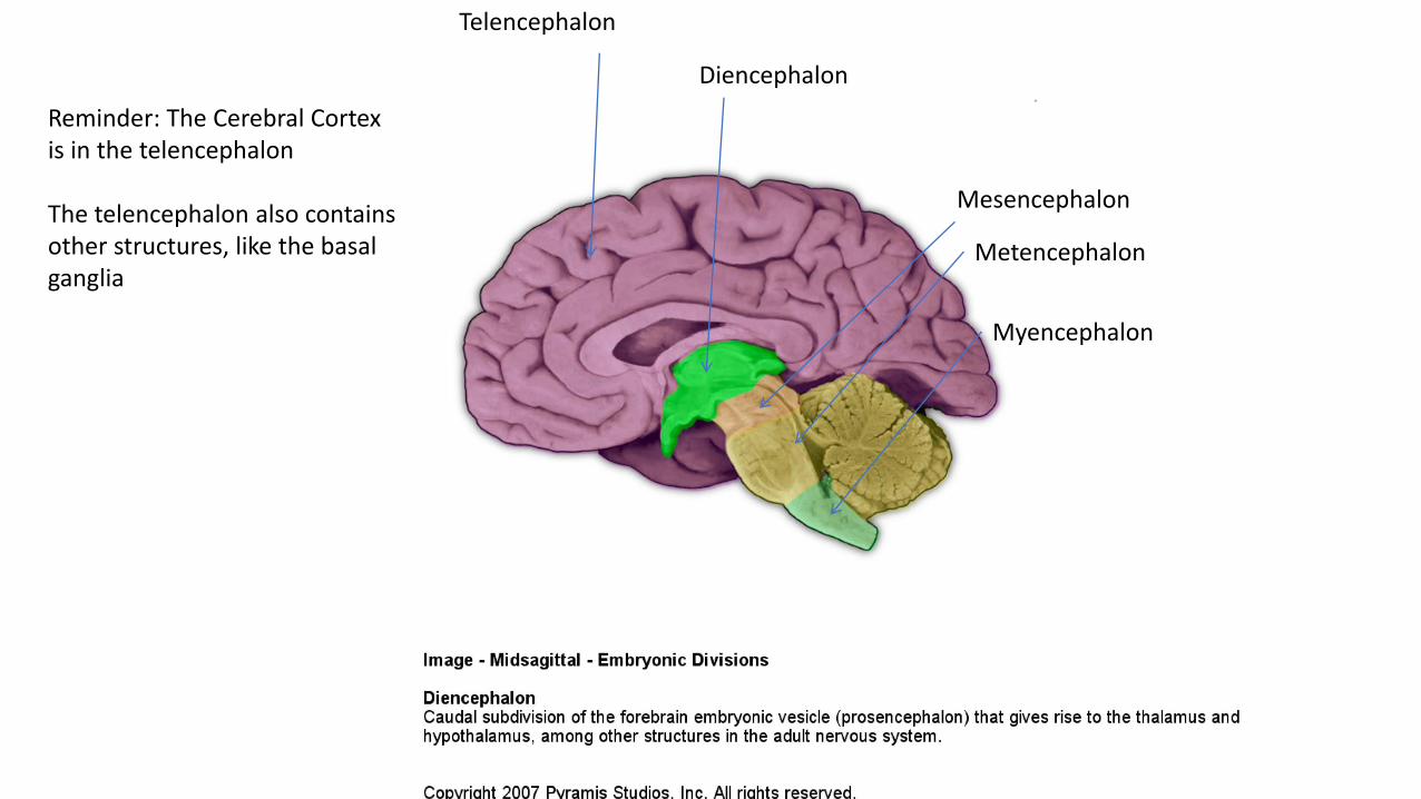
Diencephalon
Mesencephalon
Metencephalon
Myencephalon
Telencephalon
Reminder: The Cerebral Cortex is in the telencephalon
The telencephalon also contains other structures, like the basal ganglia
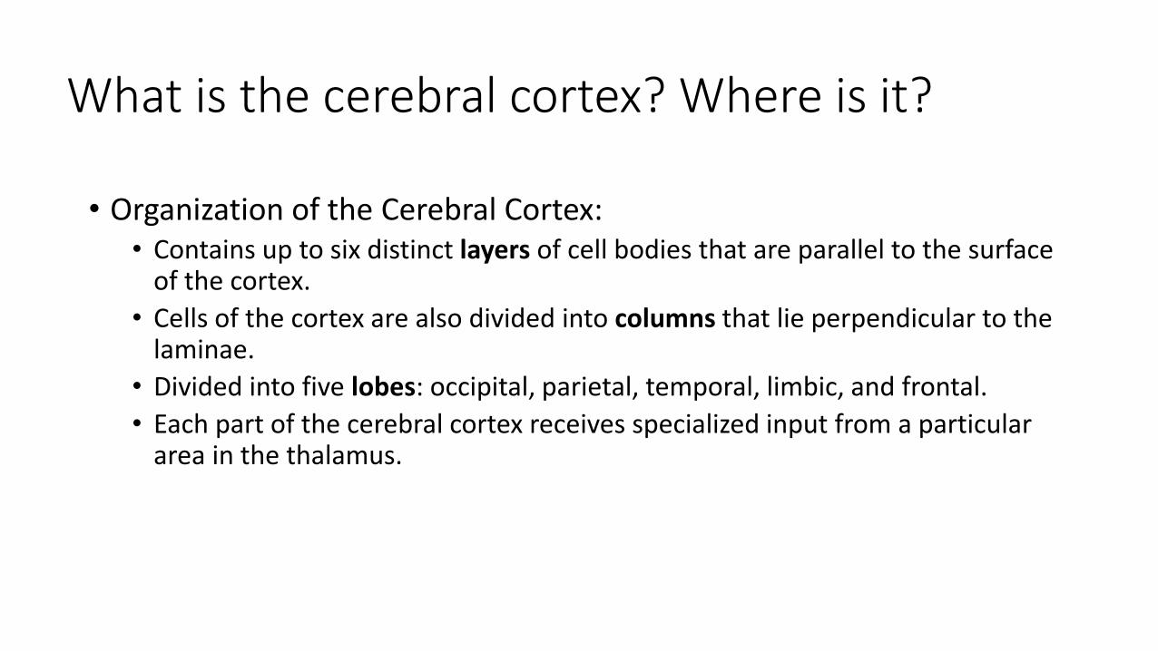
• Organization of the Cerebral Cortex:• Contains up to six distinct layers of cell bodies that are parallel to the surface
of the cortex.
• Cells of the cortex are also divided into columns that lie perpendicular to the laminae.
• Divided into five lobes: occipital, parietal, temporal, limbic, and frontal.
• Each part of the cerebral cortex receives specialized input from a particular area in the thalamus.
What is the cerebral cortex? Where is it?

• Neocortex has six layers. Most of the human cerebral cortex is neocortex
• Allocortex has less than six layers• Piriform cortex, olfactory tubercle, anterior olfactory nucleus, hippocampus,
olfactory bulb, some others
• More layers are thought to reflect more complex processing
Types of cortex

5 lobes of the cerebral cortex

Occipital Lobe
• Bordered by:• (imaginary) parieto-occipital line
• Includes:• Primary visual area
• Visual association areas
Parieto-occipital line
• Thalamic input from:• Lateral geniculate nucleus

Parietal Lobe
• Bordered by:• Lateral fissure
• Central sulcus
• (imaginary) parieto-occipital line
• Includes:• Primary somatosensory area
• Somatosensory association areas
Central sulcus
Lateral fissure
Parieto-occipital line
• Thalamic input from:• Ventral posterior nucleus

Temporal Lobe
• Bordered by:• Lateral fissure
• (imaginary) parieto-occipital line
• Includes:• Primary auditory area
• Auditory association areas
Lateral fissure
Parieto-occipital line
• Thalamic input from:• Medial geniculate nucleus
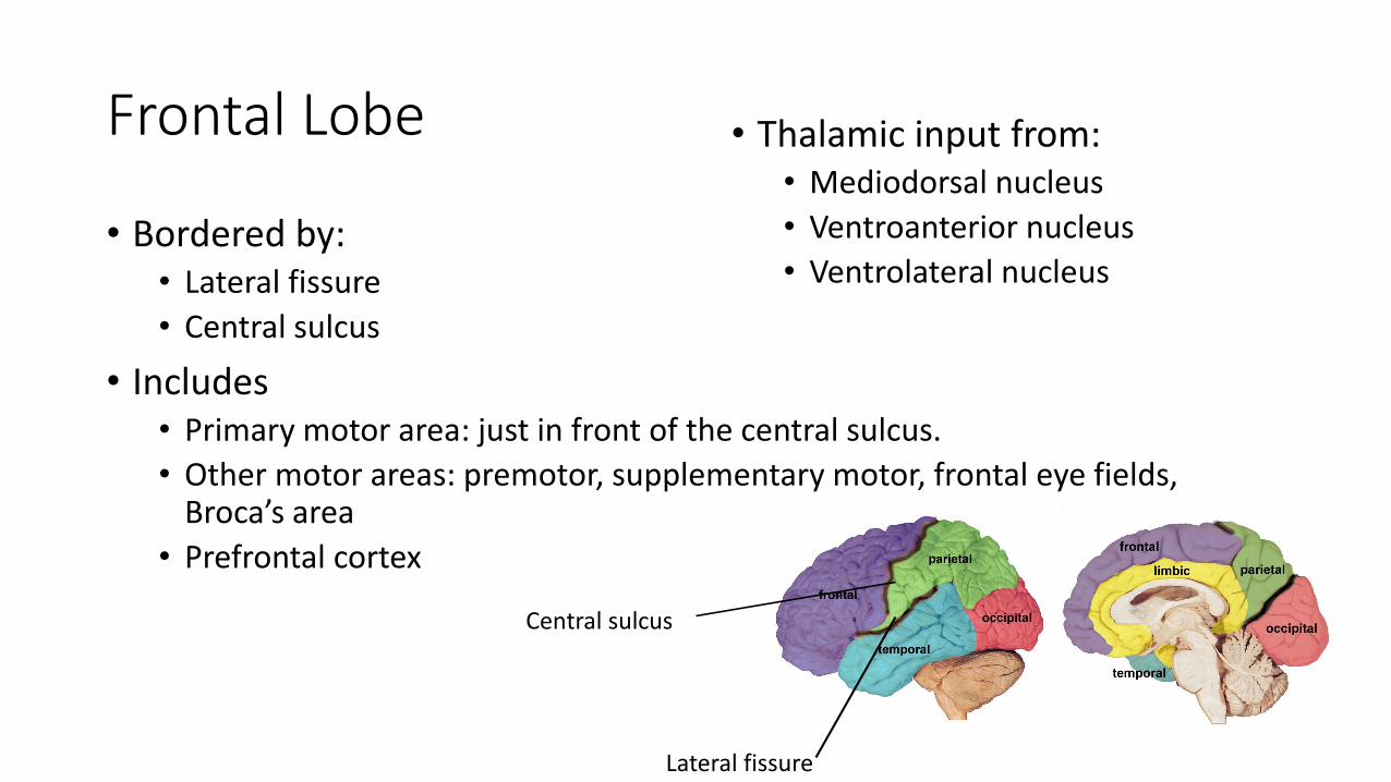
Frontal Lobe
• Bordered by:• Lateral fissure
• Central sulcus
• Includes• Primary motor area: just in front of the central sulcus.
• Other motor areas: premotor, supplementary motor, frontal eye fields, Broca’s area
• Prefrontal cortex
Lateral fissure
Central sulcus
• Thalamic input from:• Mediodorsal nucleus
• Ventroanterior nucleus
• Ventrolateral nucleus

Limbic Lobe
• Located on the medial surface of the cerebral cortex
• Includes:• Hippocampus
• Amygdala
• Parahippocampal gyrus
• Cingulate gyrus
• Thalamic input from:• Anterior thalamic nuclei
Microscopic anatomy of the cortex
• Radially organized into layers
• Each layer contains a specific subset of neurons that have a unique pattern of input and output projections
• The density and arrangement of neurons in each layer (this results in the microscopic appearance called cytoarchitecture) differ between areas
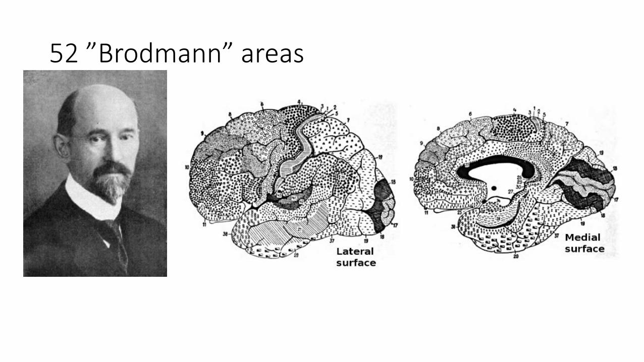
52 ”Brodmann” areas
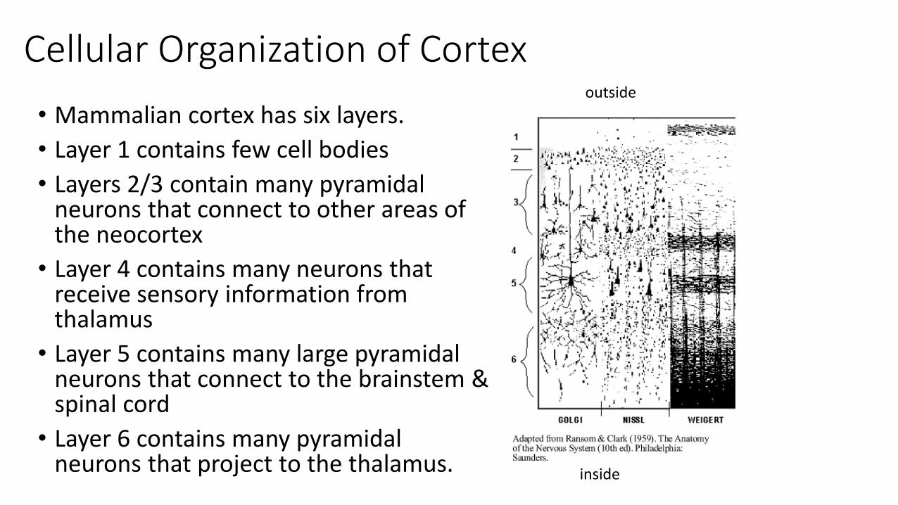
Cellular Organization of Cortex
• Mammalian cortex has six layers.
• Layer 1 contains few cell bodies
• Layers 2/3 contain many pyramidal neurons that connect to other areas of the neocortex
• Layer 4 contains many neurons that receive sensory information from thalamus
• Layer 5 contains many large pyramidal neurons that connect to the brainstem & spinal cord
• Layer 6 contains many pyramidal neurons that project to the thalamus.
outside
inside
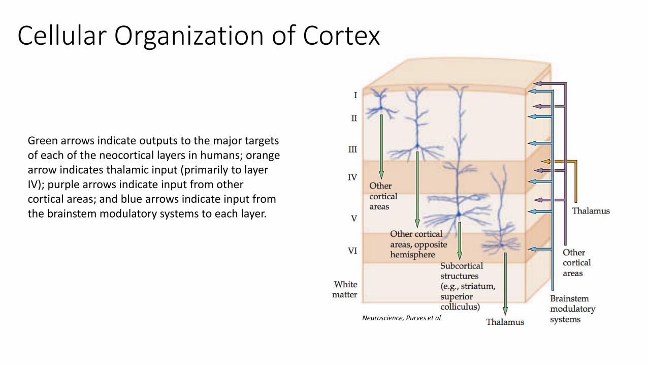
Green arrows indicate outputs to the major targets of each of the neocortical layers in humans; orange arrow indicates thalamic input (primarily to layer IV); purple arrows indicate input from other cortical areas; and blue arrows indicate input from the brainstem modulatory systems to each layer.
Cellular Organization of Cortex
Neuroscience, Purves et al

Main cell types in the cerebral cortex
• Pyramidal neurons• Large
• Long dendrites
• Primary source of axons that leave the cortex
• Spiny stellate neurons• Small and star-shaped
• No apical dendrites
• Exclusively located in layer 4 of primary sensory areas of the cortex
• Local circuit interneurons

White matter of the cerebral cortex
• Corpus callosum and anterior commissure are the primary white matter bundles connecting the two hemispheres
• Uncinate fasciculus connects the frontal and temporal lobes
• Cingulum bundle and longitudinal fasciculi run rostral-caudal and connect the frontal, parietal, and occipital lobes
• Internal capsule connects cortical regions with thalamus, subthalamic nucleus, and brainstem

The cerebral cortex and intelligence across species• Does size of the brain, or size of the cerebral
cortex more specifically, correlate with general intelligence across species?
• Lots of problems with this:• Cerebral cortex size tends to scale with body size
• Measuring intelligence across species is hard!
Neuroscience, Purves et al
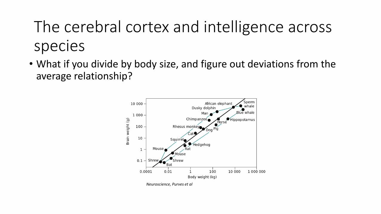
• What if you divide by body size, and figure out deviations from the average relationship?
The cerebral cortex and intelligence across species
Neuroscience, Purves et al

• What if you divide by body size, and figure out deviations from the average relationship?
• Fast selection on body size can throw these measurements off: “Chihuahua effect”
The cerebral cortex and intelligence across species

• Specialization of brain areas means each species may have enlarged areas according to evolutionary niches
• For example, songbirds have enlarged song production nuclei. The number of songs correlates with size of the nucleus across species
The cerebral cortex and intelligence across species
Overview and organization of the cerebral cortex
• What is the cerebral cortex?
• Where is each lobe, and what are its functions?
• How are areas of the cerebral cortex determined?
• What is the typical cellular organization of the cortex, and how are they connected?
• What types of cells are in the cortex?
• What is the white matter through which the cortex communicates?
• Brain size & intelligence


Methods for investigating the cerebral cortex
• Structural imaging• CT
• MRI
• Functional imaging• PET
• fMRI
• Advantages/disadvantages to each method

Brain imaging
• How can we understand the biological basis of cognitive and emotional functions?
• Use lab animals for invasive experiments (electrophysiology, tract-tracing, etc)
• Clinical studies of patients with cognitive disorders
• Structural imaging
• Functional imaging• Noninvasive methods used to observe areas of the human brain

Structural imaging
Detailed 3D (but static) anatomical images of the brain in a living human. Can examine the relationship between structural variance and function, including lesions.
• CT (X-ray computerized tomography)
• MRI (Magnetic resonance imaging)
• DTI (Diffusion tensor imaging)
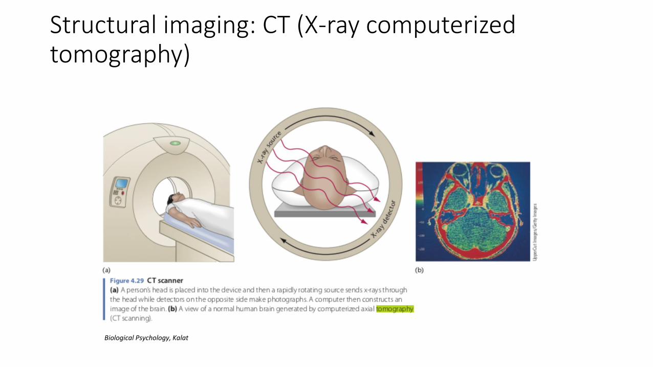
Structural imaging: CT (X-ray computerized tomography)
Biological Psychology, Kalat

Structural imaging: MRI (magnetic resonance imaging)
• Uses very strong magnetic fields to generate images
• Can see fine details of sulci and gyri in the cortex
Visualization and Query Tool for a Human Brain Database LARS FORSBERG

Structural imaging: DTI (diffusion tensor imaging)• Technically a particular type of MRI, in which specific sequences are
used that take advantage of diffusion of water molecules along white matter in the brain
• Specifically used to look at white matter.

Structural imaging: comparison across methods• MRI offers much higher resolution than CT
• CT has less distortion (important for guiding neurosurgeries)
• Tissue variation is typically clearer on an MRI
• In reality, CT scans are more likely to be used medically, whereas MRIs are more common in research settings

Functional imaging
Detailed 3D images of the brain in a living human, with the added benefit of being able to examine which brain regions are more active in response to particular conditions.
• PET
• fMRI
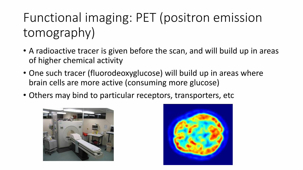
Functional imaging: PET (positron emission tomography)• A radioactive tracer is given before the scan, and will build up in areas
of higher chemical activity
• One such tracer (fluorodeoxyglucose) will build up in areas where brain cells are more active (consuming more glucose)
• Others may bind to particular receptors, transporters, etc

Functional imaging: fMRI functional magnetic resonance imaging • Takes advantage of the fact that changes in blood flow are associated
with neural activity
• Measures the BOLD (blood-oxygenation level dependent) signal, technically does not measure neural activity, but BOLD correlates highly with neural activity.
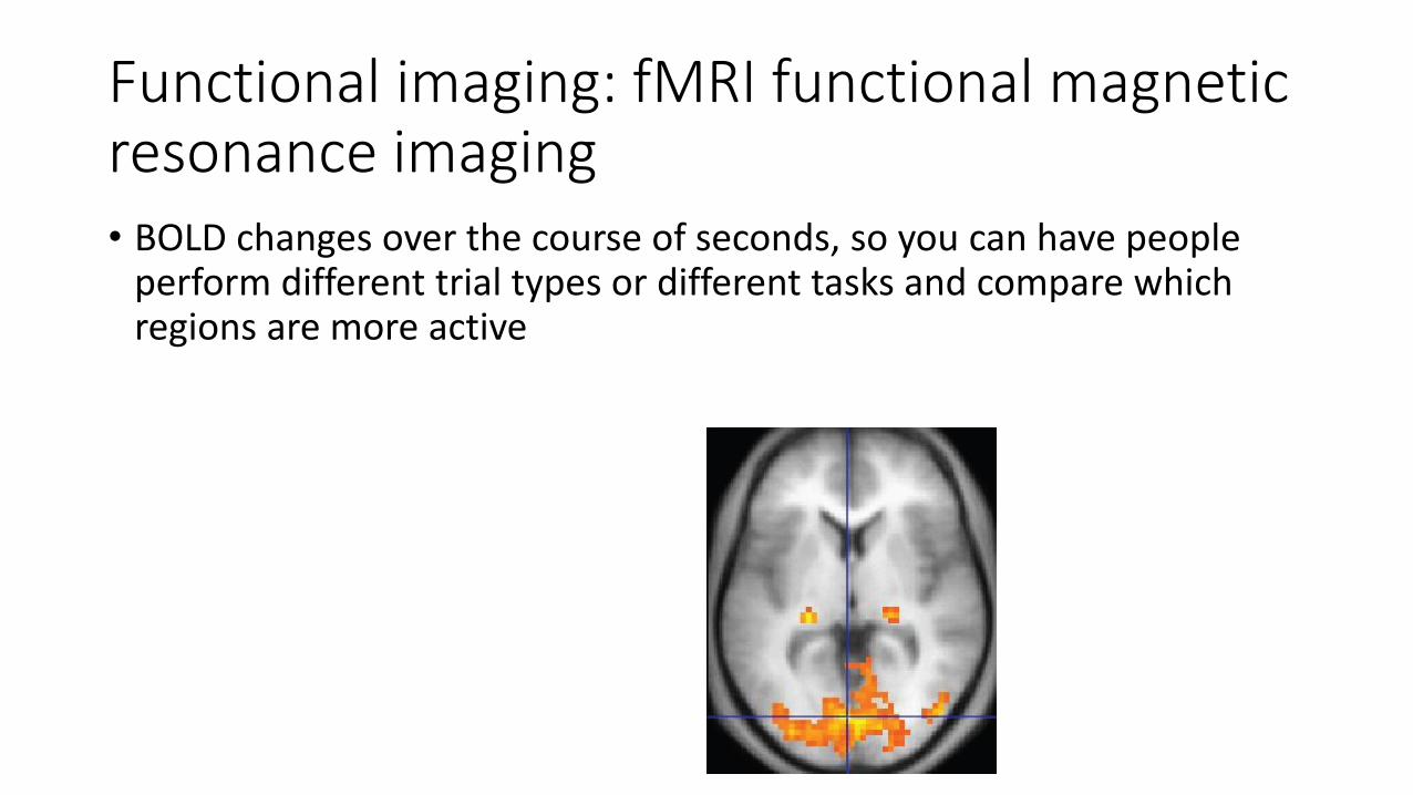
Functional imaging: fMRI functional magnetic resonance imaging • BOLD changes over the course of seconds, so you can have people
perform different trial types or different tasks and compare which regions are more active

Structural imaging: comparison across methods• fMRI offers a much faster temporal resolution than PET. PET takes a
“snapshot” of what occurred following the injection of the radiotracer. fMRI measures the BOLD signal as it fluctuates continuously during task performance.
• PET also requires an injection of a radiotracer.
• Because of this, fMRI is now far more commonly used in research settings.

• Structural imaging• CT
• MRI
• Functional imaging• PET
• fMRI
• Advantages/disadvantages to each method
Methods for investigating the cerebral cortex

Functions of the Cerebral Cortex:Sensorimotor cortices
• Maps in primary motor cortex
• Maps in primary visual cortex
• Maps in primary auditory cortex

• …of the body
musculature
in the primary
motor cortex
Neuroscience, 3rd EditionMaps in the cerebral cortex
Neuroscience, Purves et al
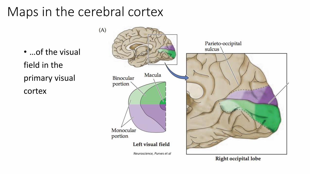
• …of the visual
field in the
primary visual
cortex
Maps in the cerebral cortex
Neuroscience, Purves et al

• …of orientation
in the
primary visual
cortex
Maps in the cerebral cortex
Neuroscience, Purves et al

• …of sound
frequency in the
primary auditory
cortex
Maps in the cerebral cortex
Neuroscience, Purves et al

Functions of the Cerebral Cortex:Sensorimotor cortices
• Maps in primary motor cortex
• Maps in primary visual cortex
• Maps in primary auditory cortex

Functions of the Cerebral Cortex:Association cortices
• What are association areas?
• What is modularity?
• Parietal association cortex
• Temporal association cortex
• Frontal association cortex

What are association areas?
• Parts of the cortex that neither receive direct sensory information through the major sensory pathways or motor thalamic nuclei
• Unimodal: Located adjacent to or near their primary sensorimotor cortical areas, processes one type of sensorimotor information
• Multimodal: can process many types of information, in complex ways
Neuroscience, Purves et al

• Association cortices often have extensive connections with sensory, motor, and other association regions. Also connect to subcortical structures
• Integrate information and perform higher mental function
• Occupy a much larger fraction of the total brain in humans vs other animals
What are association areas?
Van Essen

Modularity
• The concept of “modularity” in neuroscience/psychology posits that particular brain areas are highly specialized for specific functions
• This is obviously true of primary sensorimotor cortical areas, but what about higher cognitive functions in association areas?
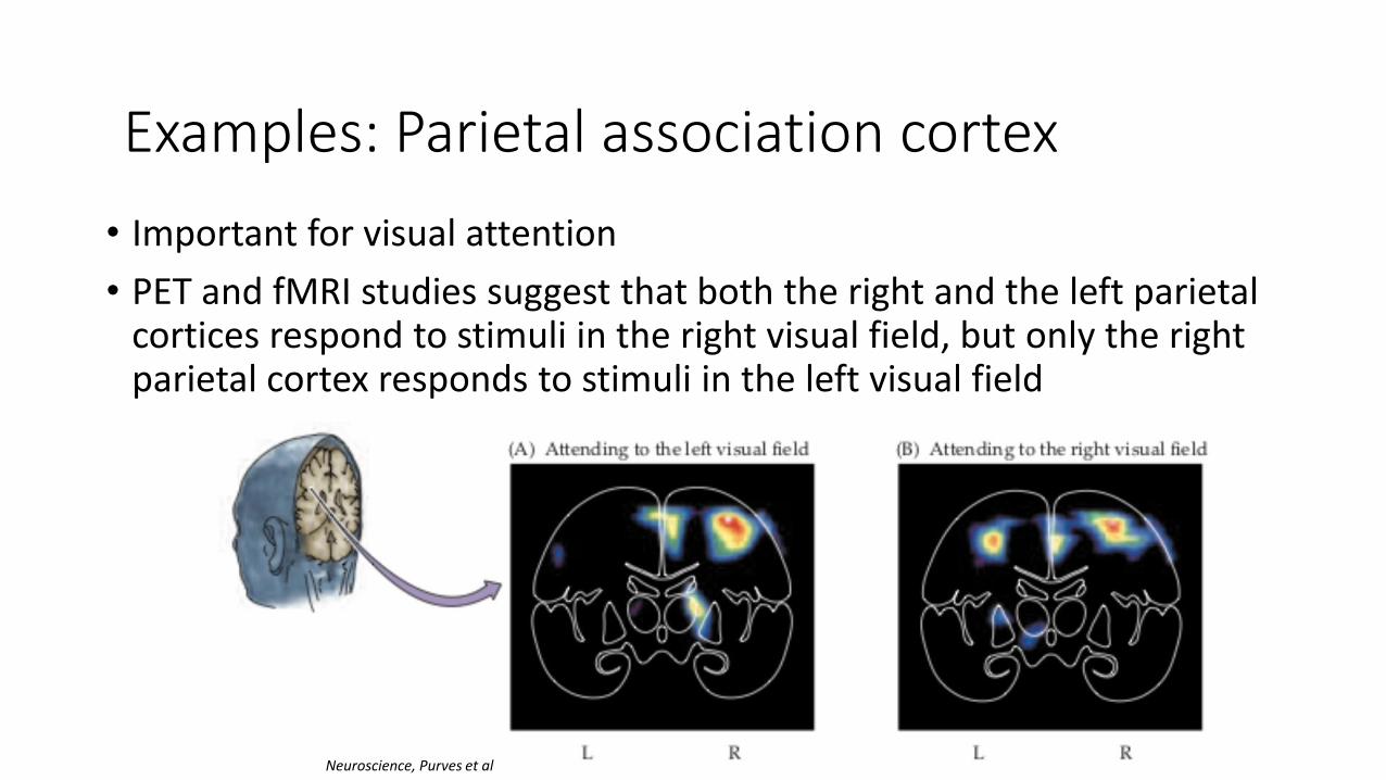
• Important for visual attention
• PET and fMRI studies suggest that both the right and the left parietal cortices respond to stimuli in the right visual field, but only the right parietal cortex responds to stimuli in the left visual field
Examples: Parietal association cortex
Neuroscience, Purves et al

Examples: Parietal association cortex• This means that damage to the right parietal cortex will be
particularly impactful, since there’s no ”extra” representation of the left visual field
Patients with damage to the right parietal cortex have trouble attending to the left visual field
This phenomenon is known as parietal neglect
• Video: https://www.youtube.com/watch?v=d4FhZs-m7hA
Examples: Parietal association cortex
Examples: Temporal association cortex
• Temporal association cortex processes high-level features of objects, faces, and places
• Lesions cause agnosias: difficulty recognizing and/or naming things

Examples: Temporal association cortex
• The fusiform face area is one of the strongest examples of modularity in the association cortex.
A particular part of the brain is much more active when someone views faces than when they view almost anything else

Examples: Frontal association cortex
• Damage to the frontal lobe is often interpreted as a problem with the patient’s “character”
• Diverse functions found in different areas of the frontal lobe, including planning, decision-making, abstract though, representation of self, and so on
• Phineas Gage is the classic example
Functions of the Cerebral Cortex:Association cortices
• What are association areas?
• What is modularity?
• Parietal association cortex
• Temporal association cortex
• Frontal association cortex