cervical kinesio taping and postural correction exercises
TRANSCRIPT
The 19th
International Scientific Conference Faculty of Physical Therapy Cairo, 22-23 March, 2018
1
Cervical Kinesio Taping and Postural Correction Exercises Influence
Clinical and Electromyographic Characteristics in Mechanical Neck
Dysfunction Patients:
A Randomized Clinical Trial
Aliaa M El-abd, Msc,PT* , Abeer R Ibrahim, PhD, PT
**,Haytham M Elhafez, PhD,
PT**
*Department of basic science, faculty of physical therapy, Pharos University,
Alexandria, Egypt.
**Department of basic science, faculty of physical therapy, Cairo University, Egypt.
Abstract Background: Mechanical neck dysfunction (MND) with impaired axioscapular
muscles functions is a major health burden. While postural correction exercises
(PCEs) are common treatment option, efficacy of kinesio tape (KT) has received
considerable attention.Purpose:to investigate efficacy of KT with or without PCEs on
pain, disability and upper trapezius (UT) kinesiological electromyography (EMG) in
MND patients. Subjects and Methods: Ninety chronic MND patientsaged 18–40
years were randomly and equally assigned into 1 of 3 groups received 4 weeks
treatment;group A received KT, B (control) received PCEs and group C received both
modalities. Neck pain, disability, UT root mean square (RMS) and median frequency
(MDF) were measured pre and post-treatmentby visual analogue scale, neck disability
index and EMG respectively. Between groups comparisons were performed using 2
ways MANOVA while intra-group comparisons were performed using paired t test.
Results: MANOVA indicate a statistically significant group-by-time interaction
(P=0.00). There were statistical significant pain reducion in group C more than B (p =
0.025), disability was reduced in groups A and C more than B (p < 0.01 and 0.034).
While RMS was reduced in group C when compared to B (p= 0.037), MDF was
increased in group C when compared to groups A and B (P =.001 0.00). Paired t tests
were significant for all outcomes in all groups (p= 0<01).Conclusion: Although KT is
promising for MND treatment, its integration with PCEs would have more beneficial
outcome related to pain, disability and upper trapezius functions.
Key words: Cervical pain, exercises therapy, athletic tape, electromyography.

The 19th
International Scientific Conference Faculty of Physical Therapy Cairo, 22-23 March, 2018
2
Introduction
In spite of medical developments
and growing knowledge pertaining to
spinal diseases, mechanical neck pain
(MNP) remains one of the most
prevalent and costly health problems
worldwide.1for which, numerous
treatments, including manual therapies,
passive physical modalities, and
acupuncture, are commonly used.
However, few interventions have been
demonstrated to be effective and most
are associated with short-term benefits.2
Among complementary and
alternative treatments, exercise therapy
may be considered as the most widely
used conservative treatment.3 recently,
there has been a focus on postural
correction exercises involving repeated
cervical and scapular retractions that
prove to be effective and may be
superior to general exercises for
management of MNP. The approach is
aimed at improving the neuromuscular
control, strength, and endurance of the
active subsystem stabilizing the
spine.4,5
However, recent reviews
revealed that there is still controversy
regarding the evidence about the
effectiveness of postural correction
exercises for neck pain.6,7
From our
point of view, the challenge that
clinicians face may result from focusing
on pathoanatomy as an etiological
factor of MNP, ignoring the significant
role of dysfunction.
Occupational tasks involving
sustained posture may be associated
with mechanical neck dysfunction
(MND) due to impaired axioscapular
muscles functions. 10
Altered
coordination oftheir function influence
mechanical loading of
cervicalstructures leading to pain
provocation.9,10
In contrast, pain may
cause alteration in axioscapularmuscles
activities.11
Amplitude of the
myoelectricsignal may provide some
insight into the pain-spasm-paintheory
of musculoskeletal dysfunction.12
Whatever the cause andeffect direction,
axioscapular muscles activities should
be considered in the management of
MND.
Because the upper trapezius
muscle is suitable for surface EMG
detection due to its size and superficial
location, It can be used to give rise to
axioscapular muscles behavior and
several studies have reported that

Aliaa M El-abdet al.,
3
sustained trapezius muscle activity
correlates with the presence of neck
pain.13- 15
.This concept is based on the
so-called Cinderella hypothesis which
states that during a prolonged low-level
activity the same muscle fibers are
always active.16
Moreover, decrease in
median frequency in the EMG during
tasks might be an indicator of fatigue of
muscle fibers.17
Kinesio tape (KT) is a thin elastic
tape which can be stretched up to 130-
140% of its original length thereby
providing constant shear force on the
skin. According to its creators, KT has
beneficial effects and possible useful
mechanisms to suppress pain, relax
muscles, support joints, and improve
circulation.18
Pain relief by KT has been
reported in a number of previous
studies involving different conditions
such as myofascial pain syndrome 19,20
,
shoulder impingement syndrome 21
,
acute whiplash22
, and chronic low back
pain23
. The interaction of KT on
muscle’s function has also been
reported.24
Determining the most appropriate
intervention for individuals with MND
remains a priority for researchers.
While conflicting results emerged from
studies examining the effect of KT for
MND, further research is recommended
focusing on the development of new
predictions about its efficacy and/or
combination of interventions.25-27
Wehypothesizedthatkinesiotaping
of cervical paraspinal muscles of
mechanical neck dysfunction (MND)
patients combinedwith postural
correction exercisescouldinterferewith
muscles functions, therebyinfluencing
MND. On the basis of thishypothesis,
the aim of ourstudywasto investigate
the efficacy of kinesio taping with or
without postural correction exercises on
pain intensity, neck disability and
cervical muscles electromyographic
(EMG) activities in form of upper
trapezius normalized root mean square
(RMS) as an indication of activation
amplitude and median frequency
(MDF) as an indication of muscular
fatigue.
Subjects, Instrumentations and
Methods
Subjects:
The study was conducted in
accordance with the 1964 helsinki
declaration and its later amendments,
approved by the research ethics
committee of physical therapy college,

The 19th
International Scientific Conference Faculty of Physical Therapy Cairo, 22-23 March, 2018
4
Cairo University and reported with
respect to CONSORT guidelines
provided by EQUATOR Network.
Participation was voluntary and
Informed consent was obtained from
each patient before participation in the
study.
Design of the study:
Randomized controlled clinical
trial with 3 parallel groups.
Sample-Size Determination:
Sample-size calculations were
performed for neck pain as a primary
outcome measure using G power 3.1
soft ware. The calculations were based
on .343 effect size (partial eta squared
measured in our pilot study = 0.105), an
alpha level of .05, a desired power of
80%, numerator degree of freedom of 2
and 3 experimental groups. The
estimated desired total sample size for
the study was 86 patients. To
accommodate the expected dropouts
before the study’s completion, a total of
90 participants were included in the
study
Participants:
Participants in this study were
ninety patients of both sexes (47 males
and 43 females) diagnosed by
orthopedist with MND, their age ranged
from 20 to 40 years and BMI ranged
from 25.1 to 32. They had a history of
neck pain that last for more than three
months duration and necessitate them to
visit an orthopedist. They were
recruited during the period from
October 2015 to October 2016 from the
students and employee of faculty of
Physical Therapy Cairo University and
also from patients referred to its out-
patient clinic, where the data was
collected. Other inclusion criteria were
score above 15 in the neck disability
index (NDI); which indicate the
presence of at least a mild neck
disorder.33
while exclusion criteria
were: any defined muculo-skeletal,
neuro-muscular, inflammatory or
traumatic diseases, and any tape allergy.
All patients were examined for
taping allergy before allocation as
follow; a small portion of tape was
applied on inner part of patient's arm
and kept for a day. Next day the tape
was removed and if there was reaction,
the patient was excluded. Patients were
instructed to avoid anti-inflammatory
drugs for 72 hours before the study.
They received a standardized physical
examination by an assessor blinded to

Aliaa M El-abdet al.,
5
patients' allocation. They provided
demographic and clinical information
and completed self-report measures at
baseline.
Concealed Allocation:
After the baseline examination,
patients with eligibility criteria were
assigned with simple randomization to
receive Kinesio taping (group A),
postural correction exercises (group B,
control one) or both (group C). A
researcher not involved in either
recruitmentor treatment of the patients
used a computer-generated randomized
table of numbers created prior to the
start of data collection for Concealed
allocation. Sequentially, individually
numbered index cardscontaining the
randomly assigned interventiongroup
were folded and placedin opaque,
sealed envelopes. The envelope was
openedby a secondtherapist blinded to
baseline examinationfindings .the
treatment was preceded accordingto the
group assignment on the day of
theinitial examination.
Outcome measures:
The primary outcome measure
was neck pain intensity, with disability
and UT muscle EMG parameters
(normalized RMS for muscular activity
and MDF for muscular fatigue) as
secondary outcomes.
Instrumentation and assessment
procedures:
(1) The visual analogue scale(VAS)
was used to measure pain
intensity (a 10 cm horizontal line
anchored by ‘‘no pain’’ on the left
and ‘‘worst imaginable pain’’ on
the right). Patients indicated their
pain intensity by marking on the
point at the line that reflects their
pain. Then the score was
determined by measuring from the
left end of the line to the point that
the patient marked. It is a simple
and efficient measuring tool with
established reliability and validity.
28,29
(2) Neck disability index (NDI) was
usedto measure Functional neck
disability. It includes 10 questions
of which 7 examine functional
activities, 2 ask about symptoms
and a question considers
concentration. Each Patient
circled one of the six options
describing the severity of each
item (0–5).the marks were
counted and divided by 50 or 45 if
one section was missingwith total
score ranging from 0 (no
disability) to 50 (complete

The 19th
International Scientific Conference Faculty of Physical Therapy Cairo, 22-23 March, 2018
6
disability). NDI has established
validity and reliability.30,31
(3) EMG MyoSystem 1400A,
delesyInc, Scottsdale, USA was
used for measuring upper
trapezius normalized RMS and
MDF. The sites of the electrodes
placement had been shaved and
cleaned by a piece of cotton and
alcohol to reduce skin impedance,
Electrodes sites were located on
each subject’s dominant side as
follows: Active electrode was
located 2 cm lateral to the
midpoint of a line drawn between
C7spinous process and the
posterolateral acromion while the
reference one was located over
the C7spinous process.4Raw EMG
was amplified (bandwidth = 20-
450 Hz, common mode rejection
ratio>80 db at 60 Hz, input
impedance = 10 GΩ) and collected
with a ±2.5 V rang. EMG signals
had systemic bias were removed,
and were full wave rectified prior
to being filtered. The resulting
linear envelope signals were then
normalized to maximal voluntary
isometric contractions
(MVIC).Assessment of the MVIC of
upper trapezius(UT): was
performed as described by
Mclean4; the subject performed
isometric shoulder abduction
with the arm at 900 abduction and
neutral rotation. Each contraction
was maintained for 7 seconds and
repeated three times against
manual resistance with 30
seconds rest between repetitions.
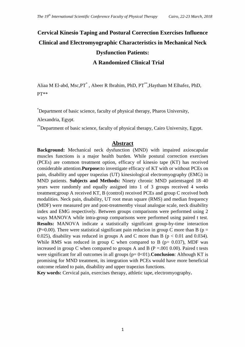
After assessment of theMVIC,
participants were asked to write for 15
minute; this task was chosen because it
is the most common daily task for
participants and it involve semi static
load which aggravate their symptoms.
During the examination, the patient
remained seated in a chair with back
completely supported, feet flat and
supported on the floor and hips and
knees flexed 900. Positioning of head,
neck, shoulder and the spine had been
standardized to avoid their effect on the
activities of UT.32
(Figure
1)Normalized RMS was calculated as
follow:Normalized RMS %= EMG
amplitude during writing task / (average
of the 3 trials of MVIC)*100.16
the
median frequency was calculated from
the raw EMG signals.
Aliaa M El-abdet al.,
7
Figure 1:Assessment of
UT MVIC and the writing
task
All outcomes were collected at
baseline and 4 weeks after the
intervention by an assessor blinded to
the patients treatment allocation.
Patients were blinded to their allocation
and uninformed of what intervention
the other group would receive.
Treatment procedures:
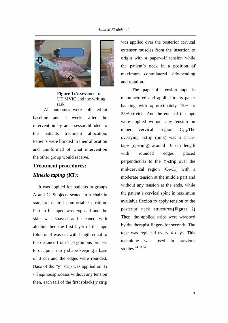
Kinesio taping (KT):
It was applied for patients in groups
A and C. Subjects seated in a chair in
standard neutral comfortable position.
Part to be taped was exposed and the
skin was shaved and cleaned with
alcohol then the first layer of the tape
(blue one) was cut with length equal to
the distance from T1-T2spinous process
to occiput in to y shape keeping a base
of 3 cm and the edges were rounded.
Base of the “y” strip was applied on T1
- T2spinousprocesss without any tension
then, each tail of the first (black) y strip
was applied over the posterior cervical
extensor muscles from the insertion to
origin with a paper-off tension while
the patient’s neck in a position of
maximum contralateral side-bending
and rotation.
The paper-off tension tape is
manufactured and applied to its paper
backing with approximately 15% to
25% stretch. And the ends of the tape
were applied without any tension on
upper cervical region C1-2.The
overlying I-strip (pink) was a space-
tape (opening) around 10 cm length
with rounded edges placed
perpendicular to the Y-strip over the
mid-cervical region (C3-C6) with a
moderate tension at the middle part and
without any tension at the ends, while
the patient’s cervical spine in maximum
available flexion to apply tension to the
posterior neck structures.(Figure 2)
Then, the applied strips were wrapped
by the therapist fingers for seconds. The
tape was replaced every 4 days. This
technique was used in previous
studies.24,33,34

The 19th
International Scientific Conference Faculty of Physical Therapy Cairo, 22-23 March, 2018
8
Figure 2: Application of
kinesio tapeon neck
extensors
Postural correction exercises
(PCE):
PCE were applied for patients in
groups B and C. This program was
conducted according to the protocols of
Pearson and Walmsley.35
Each exercise
was performed as 3 sets of 10
repetitions each for 2 times/ week for 4
weeks. The patients were instructed to
continue the exercises as a daily home
program to influence the self correction
kinesthetic awareness.Exercises were
performed while the patients in a
neutral sitting posture obtained as
recommended by Falla, et al36
;
Participants were asked to sit on a chair
where their feet were flat on the floor
and their buttocks were fully supported.
The chair's height was set so that the
hips were approximately in 1000 of
flexion. In this position hypomobility in
some patients posterior hip structures
would not prevent them from
comfortably anteriorly rotating the
pelvis to achieve a neutral lumbar spine
posture.
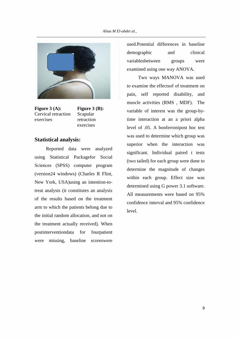
A) Cervical retraction exercises:
The patient was asked to pull
the head and neck into a position in
which the head is aligned more
directly over the thorax (chin in) while
the head and eyes remain level (as if
hiding behind the wall) for 10 seconds.
(Figure 3A)
B) Scapular retraction exercise:
While sitting, patient was
instructed to take deep inspiration and
expand the chest. Then, move his or her
shoulders backward bringing the
scapulae together for 10 seconds.
(Figure B)
C) Instructions for daily activities:
Patients were given home
instructions regarding proper sitting,
computer and telephone using, lifting
and reading.
Aliaa M El-abdet al.,
9
Statistical analysis:
Reported data were analyzed
using Statistical Packagefor Social
Sciences (SPSS) computer program
(version24 windows) (Charles R Flint,
New York, USA)using an intention-to-
treat analysis (it constitutes an analysis
of the results based on the treatment
arm to which the patients belong due to
the initial random allocation, and not on
the treatment actually received). When
postinterventiondata for fourpatient
were missing, baseline scoreswere
used.Potential differences in baseline
demographic and clinical
variablesbetween groups were
examined using one way ANOVA.
Two ways MANOVA was used
to examine the effectsof of treatment on
pain, self reported disability, and
muscle activities (RMS , MDF). The
variable of interest was the group-by-
time interaction at an a priori alpha
level of .05. A bonferronipost hoc test
was used to determine which group was
superior when the interaction was
significant. Individual paired t tests
(two tailed) for each group were done to
determine the magnitude of changes
within each group. Effect size was
determined using G power 3.1 software.
All measurements were based on 95%
confidence interval and 95% confidence
level.
Figure 3 (A): Cervical retraction
exercises
Figure 3 (B): Scapular
retraction
exercises

The 19th
International Scientific Conference Faculty of Physical Therapy Cairo, 22-23 March, 2018
10
RESULTS
One hundred and eight consecutive patients were screened for eligibility criteria.
Ninety patients (mean ± SD age, 27.49 ± 4.513 years; BMI, 28.21 ± 3.24; 47 males)
satisfied the eligibility criteria, agreed to participate, and were randomized to group A:
Kinesio Tape (n = 30) (age, 27.3 ± 4.46 years; BMI, 28.05 ±3.34; 17 males) , group B
(n = 30): posture correction (age, 27.633 ± 3.96 years; BMI, 28.85 ± 2.99; 14 males) ,
and group C (n = 30): both modalities (age, 27.53 ± 5.18 years; BMI, 27.731 ± 3.38;
16 males). The reasons for ineligibility are found in a flow diagram of patient
recruitment and retention(Figure 4). There was no significant difference between
groups for both demographic (age, BMI, sex) and measured variables at base line
(Table 1).
Figure 4: A flow diagram of patient recruitment and retention
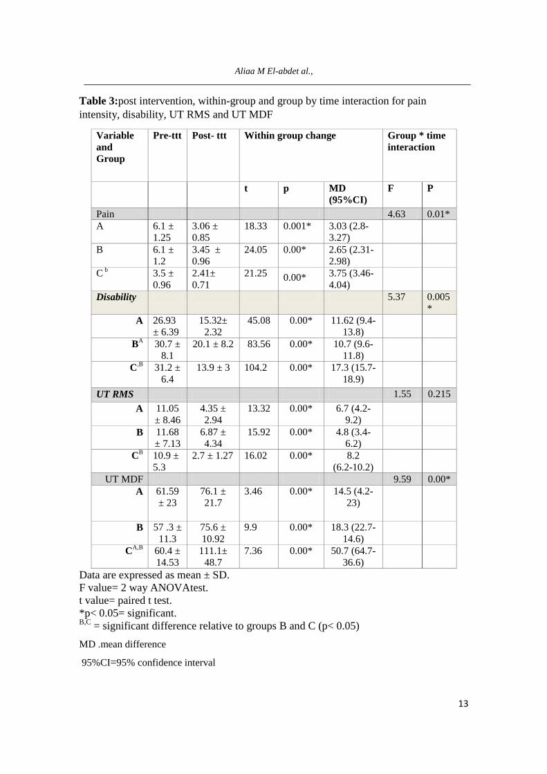
Multivariate tests for outcome measures indicate a statisticallysignificant group
by time interaction (F= 3.61,P=0.00). Theunivariate group- by- time interaction was

Aliaa M El-abdet al.,
11
statistically significant for VAS (F= 4.63, p =0.011), NDI (F= 5.37, P = 0.005) and
MDF (F = 9.594, P= 0.00) but there was no statistical significant group by time
interaction for RMS (F = 1.55, P =.215).
Post hoc tests revealed that mean values of VAS was significantly reduced in
patients received both modalities (groups C) when compared to paients in group B who
received posture correction exercises (p= 0.025), Regarding disability, mean values of
NDI was significantly reduced in groups A and C when compared to group B (p < 0.01
and p = 0.034). While mean values of RMS was significantly reduced in group C when
compared to group B (p= 0.037), MDF was significantly increased in group C when
compared to either group A (P =.001) or B (P = 0.00).
Paired t test revealed that there was a statistical significant decrease in the mean
values of VAS, NDI, RMS and significant increase in mean values of MDF in all
groups (p= 0<01).
Table 1:Demographic and base line features of the three studied groups
Data are expressed as mean ± SD.
p> 0.05= not significant.
UT: Upper trapezius
Group A
(n= 30)
Group B (n= 30) Group C (n= 30) P
value
Sex 17 males
and 13 females
14 males and 16
females
16 males and 14
females
.59
Age (yrs.) 27.3 ± 4.46 27.63 ± 3.96 27.53 ± 5.18 .959
BMI
(Kg/m2)
28.05 ± 3.34 28.85 ± 2.99 27.73 ± 3.38 .39
Pain
intensity 6.17 ± 1.15 6.2 ± 1.06 6.2 ± 1.08 .974
Neck
disability 27.43 ± 6.31 30.74 ± 8.2 30.71 ± 6.09 .110
UT RMS 10.22 ± 3.68 9.84 ± 4.79
9.9043 ± 4.65 .938
UT MDF 59.43 ± 21.21 57.27 ± 11.46
59.15 ± 11.68 .842

The 19th
International Scientific Conference Faculty of Physical Therapy Cairo, 22-23 March, 2018
12
RMS: root mean square.
MDF: median frequency.
Table 2: Multivariate Analysis of Variance (MANOVA) for all dependent variables at
different measuring periods between studied groups.
Source of Variation F-value P-value
Groups 5.19 0.000*
Measuring periods 127.78 0.000*
Interaction (group*time) 3.6 0.000*
*Significant at alpha level <0.05.
Aliaa M El-abdet al.,
13
Table 3:post intervention, within-group and group by time interaction for pain
intensity, disability, UT RMS and UT MDF
Group * time
interaction
Within group change Post- ttt Pre-ttt Variable
and
Group
P F MD
(95%CI)
p t
0.01* 4.63 Pain
3.03 (2.8-
3.27)
0.001* 18.33 3.06 ±
0.85
6.1 ±
1.25
A
2.65 (2.31-
2.98)
0.00* 24.05 3.45 ±
0.96
6.1 ±
1.2
B
3.75 (3.46-
4.04) 0.00* 21.25 2.41±
0.71
3.5 ±
0.96
C b
0.005
*
5.37 Disability
11.62 (9.4-
13.8)
0.00* 45.08 15.32±
2.32
26.93
± 6.39 A
10.7 (9.6-
11.8)
0.00* 83.56 20.1 ± 8.2 30.7 ±
8.1
BA
17.3 (15.7-
18.9)
0.00* 104.2 13.9 ± 3 31.2 ±
6.4
C,B
0.215 1.55 UT RMS
6.7 (4.2-
9.2)
0.00* 13.32 4.35 ±
2.94
11.05
± 8.46 A
4.8 (3.4-
6.2)
0.00* 15.92 6.87 ±
4.34
11.68
± 7.13 B
8.2
(6.2-10.2)
0.00* 16.02 2.7 ± 1.27 10.9 ±
5.3
CB
0.00* 9.59 UT MDF
14.5 (4.2-
23)
0.00* 3.46 76.1 ±
21.7
61.59
± 23 A
18.3 (22.7-
14.6)
0.00* 9.9 75.6 ±
10.92
57 .3 ±
11.3 B
50.7 (64.7-
36.6)
0.00* 7.36 111.1±
48.7
60.4 ±
14.53
CA,B
Data are expressed as mean ± SD.
F value= 2 way ANOVAtest.
t value= paired t test.
*p< 0.05= significant. B,C
= significant difference relative to groups B and C (p< 0.05)
MD .mean difference
95%CI=95% confidence interval

The 19th
International Scientific Conference Faculty of Physical Therapy Cairo, 22-23 March, 2018
14
DISCUSION
This study was conducted to
examine the efficacy of kinesio tape
with and without postural correction
exercises on pain, disability, UT muscle
activity and fatigue in patients with
MND.Our results suggested that KT
with or without postural correction
exercises might be an alternative
treatmentoption in the treatment of
MND. However, we recommend
treatment of MND by both modalities
combined because it has better effects
to decrease pain, disability and also to
normalize cervical muscle activities
than application of either intervention
alone.
There were a number of
explanations for these findings. Neck
pain is commonly associated with
protectivespasm in the surrounding
muscles producing pressurewithin the
muscle, thus developing ischemia,more
pain, and abnormal neck posture. This
viciouscycle that can occur in reverse,
may be broken byrelieving the pain, by
reducing the muscle spasm, or
bycorrecting the abnormal neck
posture.1,3
The effect of KT:
The cutaneous stretch
stimulation provided by KT may
interfere with the transmission of
mechanical and painful stimuli. KT
may provide afferent impulses
inhibiting pain through gait control
theory.Furthermore, KT increase
lymphatic and vascular flow, and aid in
the correction of possible articular
malalignments.18,23,37
thus, it improves
functional abilities of patients.
While MND was found to be
associated with altered muscular
activities11
, KT may normalize muscle
function through two main mechanisms.
The first is mechanical; taping influence
the length of muscle fibres, inducing a
shift of the length-tension curve of
those muscles changing the relative
position of subsequent joints or directly
by influencing the direction of muscle
fibers. The second mechanism, called
“proprioceptive”, considers the
amplification of kinesthetic information
reaching the central nervous system
through taping-induced cutaneous
stimulation.38
Aliaa M El-abdet al.,
15
This agrees with the results of
Lin, et al39
who suggested effect of KT
on shoulder muscles
EMG.Paoloni23
stated that, KT alter
lumber muscles activities and affect low
back pain. Also, Tayloret al40
suggest
KT for neck and shoulder pain. Our
results agree with Mariana, et al41
showed that both massage and kinesio
tape decrease pain and increase range of
motion in MND.
On the other hand, the results of
the systemic review conducted by
Parreira et al 42
did not support the use of
KT in clinical practice. Fu, et
al43
showed that Kinesio taping on the
anterior thigh has no effete on muscle
strength in healthy non-injured young
athletes. In the previous studies the
possible explanation that they got
different results may be due to applying
kinesio tape on healthy subjects in
many studies. Furthermore, the methods
of assessment they used were different
from that we used.
The effect of postural correction
exercises:
Frequent correction to an upright
neutral posture serves two functions.
First, It provides a regular reduction of
adverse loads on the cervical joints
induced by poor cervical and scapular
postures. Second, it trains the deep
postural stabilizing muscles of the spine
in their supporting role. Patients are
encouraged to perform these exercises
repeatedly throughout the day, with the
emphasis being on a change in postural
habits.4,44
We suggest the effect due to
neutral postural awareness that relieve
the tension causing pain.
Our results agree with the results
of Katherineet al45
. Abd El-wahab, et al
46 concluded that, Neck retractions
appeared to alter H reflex amplitude so;
it may be used for C7 radiculopathy. In
contrast, Willford, et al47
did not
support correlation between head
posture and neck pain. The discrepancy
and conflict found in the results
obtained by the previous study cannot
be directly compared with the current
study. It was a correlational study and
not true experimental study which look
for a degree of association between
variables without the ability to ascribe
cause and effect.
The effect of both modalities
combined:
Taping may act as continuous
analgesic stimulus on neck muscles as
our patients were taped continuously for

The 19th
International Scientific Conference Faculty of Physical Therapy Cairo, 22-23 March, 2018
16
four weeks.The neural feedback
provided
to the patients can facilitate their ability
to move the cervical spine with a
reduced mechanical irritation on the
soft tissues thus improving the
efficiency of postural correction
exercises. Continuous sensory feedback
of the KT allows the tape to correct
postural imbalance.23,24
There is lack in the literature
regarding studies that combine kinesio
tape with postural correction exercises.
However, Greenstein et al 25
recommended application of kinesio
taping immediately following cervical
mobilization. Also,Added et al48
suggested adding KT to physical
therapy program for mechanical low
back pain.
Limitation of the study:
The duration of the interventions
was 4 weeks to find the short term
effects. No follow up was done to know
the long lasting effect and recurrence of
symptoms.Another important limitation
may be heterogeneity related to the
etiology of MND.
CONCLUSION:
Although KT is promising for
MND treatment, Application of both
KT and postural correction exercises
program combined lead to greater
reduction in pain severity and disability
and to better muscle function
restoration than application of either KT
or exercises program separately.
Funding support
This research received no specific
grant from any funding agency in the
public, commercial, or medical for-
profit sectors.
Conflict of Interest:
The Authors declare that there is
no conflict of interest.
Aliaa M El-abdet al.,
17
REFERENCES
1- Genebra CV, Maciela NM, Bento
TP, Simeão P, Vitta AD. Prevalence
and factors associated with neck
pain: a population-based study. Braz
J PhysTher.2017;21(4):274-80.doi:
10.1016/j.bjpt.2017.05.005
2- Hurwitz EL, Carragee EJ, van der
Velde G, Carroll LJ, Nordin M,
Guzman J, et al. Treatment of neck
pain: noninvasive interventions:
results of the Bone and Joint Decade
2000-2010 task force on neck pain
and its associated disorders. Spine.
2008; 33(4): S123–52. doi: 10.1097/
BRS.0b013e3181644b1d.
3- Cohen SP, Hooten WM. Advances
in the diagnosis and management of
neck pain. BMJ. 2017;358:j3221.
doi:10.1136/bmj.j3221.
4- Mclean L. The effect of postural
correction on muscle activation
amplitudes recorded from the
cervicobrachial region.J.
Electromyogr. Kinesiol. 2005; 15
(6): 527-35.http:
//dx.doi.org/10.1016/j.jelekin.2005.0
6.003.
5- Javanshir K, Amiri M,
MohseniBandpei MA, De lasPenas
CF, Rezasoltani A. The effect of
different exercise programs on
cervical flexor muscles dimensions
in patients with chronic neck pain. J
Back MusculoskeletRehabil. 2015;
28(4):833–40. DOI 10.3233/BMR-
150593.
6- Gross A, Kay TM, Paquin JP,
Blanchette S, Lalonde P, Christie T,
et al. Exercises for mechanical neck
disorders. Cochrance Database Syst
Rev. 2015; 28;1:CD004250. doi:
10.1002/14651858.CD004250.pub5.
7- Gross AR, Paquin JP, Dupont DG,
Blanchette S, Lalonde P, Cristie T, et
al. Exercises for mechanical neck
disorders. A Cochrane review
update. Man Ther. 2016; 24: 25-45.
http://dx.doi.org/10.1016/j.math.201
6.04.005.
8- Eltayeb S, Staal JB, Hassan A, de
Bie RA. Work related risk factors for
neck, shoulder and arms complaints:
a cohort study among Dutch
computer office workers. J. Occup.
Rehabil. 2009;19 (4): 315-
22.doi: 10.1007/s10926-009-9196-x.
9- Kumar S, Prasad N. Cervical EMG
profile differences between patients

The 19th
International Scientific Conference Faculty of Physical Therapy Cairo, 22-23 March, 2018
18
of neck pain and Control.
DisabilRehabil. 2010;32(25):2078-
87.doi:
10.3109/09638288.2010.481029.
10- Zakharova-Luneva E, Jull G,
Johnston V, O’Leary S. Altered
trapezius muscle behavior in
individuals with neck pain and
clinical signs of scapular
dysfunction. J Manipulative
PhysiolTher. 2012;35 (5):346-53.
DOI:
http://dx.doi.org/10.1016/j.jmpt.201
2.04.011
11- Hodges PW, Tucker K. Moving
differently in pain: A new theory
toexplain the adaptation to pain.
Pain. 2011;152(3 Suppl):S90-S98.
doi:10.1016/j.pain.2010.10.02
12- Tsang SM, Szeto GP, Lee RY.
Altered spinal kinematics and
muscle recruitment pattern of the
cervical and thoracic spine in people
with chronic neck pain during
functional task. J
ElectromyogrKinesiol.
2014;24(1):104-13.
13- Westgaard RH, Vasseljen O,
Holte KA. Trapezius muscle activity
as a risk indicator for shoulder and
neck pain in female service workers
with low biomechanical exposure.
Ergonomics. 2001; 44(3): 339–53.
doi: 10.1080/00140130119649.
14- Zakharova-Luneva E, Jull G,
Johnston V, O'Leary S. Altered
Trapezius Muscle Behavior in
Individuals with Neck Pain and
Clinical Signs of Scapular
Dysfunction. J Manipulative
PhysiolTher. 2012;35(5):346-53.
doi:10.1016/j.jmpt.2012.04.011.
15- Hanvold TN, Wærsted M,
Mengshoel AM, Bjertness E, Stigum
H, Twisk J, et al.The effect of work-
related sustained trapezius muscle
activity on the development of neck
and shoulder pain among young
adults. Scand J Work Environ
Health.201339(4):390-400.
doi:10.5271/sjweh.3357.
16- Nicoletti C, Spengler C, Läubli T.
Physical workload, trapezius muscle
activity, and neck pain in
nurses’night and day shifts: A
physiological evaluation.
ApplErgon. 2014; 45(3):741-6..
http://dx.doi.org/10.1016/j.apergo.2
013.09.016.
17- Gerdle B, Fugel-Meyer AR. Is the
mean frequency shift of the EMG a
selective indicator of fatigue of the
Aliaa M El-abdet al.,
19
fast twitch motor units? A
ctaPhysiol Scand. 1992;145(2):129-
38.
18- Kase K, Wallis J, Kase T. (2003)
Clinical Therapeutic Applications of
the Kinesio Taping Method. Ken
Ikai Co Ltd, Tokyo.
19- Hashemirad F, Karimi N,
Keshavarz R. The effect of Kinesio
taping technique on trigger points of
the piriformis muscle. J
BodywMovTher. 2016;20(4):807-14.
doi: 10.1016/j.jbmt.2016.02.002.
20- Wu WT, Hong CZ, Chou LW.The
kinesio taping method for
myofascial pain control. Evid Based
Complement Alternat Med.2015;
Article ID 950519, 9 pages.
http://dx.doi.org/10.1155/2015/9505
19.
21- Kaya E, Zinnuroglu M, Tugeu I.
Kinesio taping comparing to
physical therapy modalities for the
treatment of the shoulder
impingement syndrome.
ClinRheumatol. 2011; 30(2): 201–7.
doi: 10.1007/s10067-010-1475-6.
22- González-Iglesias J, Fernández-de-
las-Peñas C, Cleland JA,Huijbregts
P, del Rosario Gutiérrez-Vega M.
Short-term effects of cervical
kinesio taping on pain and, cervical
range of motion in patients with
acute whiplash injury: a randomized
clinical trial. J Orthop Sports
PhysTher. 2009;39(7): 515–20. doi:
10.2519/jospt.2009.3072.
23- Paoloni M, Bernetti A, Fratocchi G.
Kinesio taping applied to lumber
muscles influences clinical and
electromyographic characteristics in
chronic low back pain patients. Eur
J PhysRehabil Med. 2011; 47(2):
237–44
24- Saavedra-Hernandez M, Castro-
Sanchez A, Arroyo-Morales MA,
Cleland JC, Lara-Palomo I,
Fernandez-De-Las-Penas C. Short-
term effects of kinesio taping versus
cervical thrust manipulation in
patients with mechanical neck pain:
a randomized clinical trial. J Orthop
Sports PhysTher. 2012;42(8): 724–
30. doi:10.2519/jospt.2012.4086.
25- Greenstein J, McNamara T, Bishop
B, Etnoyer-Slaski J, Topp R. The
Effect of Kinesiology Tape on Pain
and Neck Range of Motion After
Cervical Manipulation. JPHR.
2017; 1(1): 18–25.DOI:
10.25036/jphr.2017.1.1.
26- Ay S, Konak HE, Evcik D, Kibar S.
The effectiveness of Kinesio Taping

The 19th
International Scientific Conference Faculty of Physical Therapy Cairo, 22-23 March, 2018
20
on pain and disability in cervical
myofascial pain syndrome. Rev
Bras Reumatol.2016 .pii: S0482-
5004(16)00042-5. doi:
10.1016/j.rbr.2015.12.004.
27- Takasaki H, Delbridge B, Johnston
V (2015) Taping across the upper
trapezius muscle reduces activity
during a standardized typing task –
An assessor-blinded randomized
cross-over study. J
ElectromyogrKinesiol25(1): 115-20.
doi: 10.1016/j. jelekin.2014.10.004.
28- Van Roo J, Lazio MP, Pesce C,
Malik S, Courtney DM. Visual
Analog Scale (VAS) for
Assessment of Acute Mountain
Sickness (AMS) on Aconcagua.
WildernessEnviron Med.
2011;22(1):7–14.doi:
10.1016/j.wem.2010.10.002.
29- Marqui M, Duarte LR, Marin C,
Lauquec D, Lauque D, Sorum P.
How patients and physicians rate
patients’ pain in a French
emergency department using a
verbally administered numerical
rating scale and a visual analog
scale. Acute Pain. 2008;10(1):31-7.
doi:10.1016/j.acpain.2008.01.003.
30- En MC, Clair DA, Edmondston SJ.
Validity of the Neck Disability
Index and Neck Pain and Disability
Scale for measuring disability
associated with chronic, non-
traumatic neck pain. Man Ther.
2009;14(4):433–8.
doi:10.1016/j.math.2008.07.005.
31- Swanenburg j, Humphreys k,
Langenfeld A, Brunner F, Wirth B.
Validity and reliability of a German
version of the Neck Disability Index
(NDI-G). Man Ther. 2014;19(1):52-
8.
http://dx.doi.org/10.1016/j.math.201
3.07.004.
32- Szeto G, Straker L, O’Sullivan P.
Examining the low, high and range
measures of muscle activity
amplitudes in symptomatic and
asymptomatic computer users
performing typing and mousing
tasks. Eur J Appl Physiol.
2009;106(2):243–51. DOI
10.1007/s00421-009-1019-4.
33- González-Iglesias J, Fernández-de-
Las-Peñas C, Cleland JA, Huijbregts P,
Del Rosario Gutiérrez-Vega M. Short-
term effects of cervical kinesio taping
on pain and, cervical range of motion
in patients with acute whiplash

Aliaa M El-abdet al.,
21
injury: A Randomized Clinical Trial. J
Orthop Sports PhysTher. 2009;
39(7):515-20. doi:
10.2519/jospt.2009.3072.
34- Dawood R, Kattabei O, Nasef S,
Battarjee K, Abdelraouf O.
Effectiveness of kinesio taping
versus cervical traction on
mechanical neck dysfunction.
IJTRR. 2013;2(2):1-5. DOI:
10.5455/ijtrr.00000019.
35- Pearson ND, Walmsley RP. Trial
into the effects of repeated neck
retractions in normal subjects.
Spine. 1995;02(11):1245-50.
36- Fallaa D, O’Learya S, Fagana A,
Jull G. Recruitment of the deep
cervical flexor muscles during a
postural-correction exercise
performed in sitting. Man Thera.
2007; 12(2):139–43.
doi:10.1016/j.math.2006.06.003.
37- Garcıa-Muro F, Rodrıguez-
Fernandez A, Herrero-de-Lucas A.
Treatment of myofascial pain in the
shoulder with Kinesio Taping. A
case report. Man Ther. 2010;15(3):
292–5.
doi:10.1016/j.math.2009.09.002.\
38- Gusella A, Bettuolo M, Contiero F,
Volpe G. Kinesiologic taping and
muscular activity: a myofascial
hypothesis and a randomised,
blinded trial on healthy individuals.
J Body MovTher. 2014;18(3):405-
11.
http://dx.doi.org/10.1016/j.jbmt.201
3.11.007.
39- Lin JJ, Hung CJ, Yang PL. The
effects of scapular taping on
electromyographic muscle activity
and proprioception feedback in
healthy shoulders. J Orthop Res.
2011; 29(1):53-7.doi:
10.1002/jor.21146.
40- Taylor RL , O’Brien L, Brown T. A
scoping review of the use of elastic
therapeutic tape for neck or upper
extremity conditions. J Hand Ther.
2014;27(3):235-45.
http://dx.doi.org/10.1016/j.jht.2014.
03.004.
41- Mariana C, Carmen-Oana T.
Massage versus kinesio taping.
possibilities to enhance the kinetic
program in mechanically triggered
neck pain. ProcediaSocBehav Sci.
2014;117:639 –
45.https://doi.org/10.1016/j.sbspro.2
014.02.275
42- ParreiraPdo C, Costa Lda C,
Hespanhol LC Jr, Lopes AD, Costa

The 19th
International Scientific Conference Faculty of Physical Therapy Cairo, 22-23 March, 2018
22
LO. Current evidence does not
support the use of Kinesio Taping in
clinical practice: a systematic
review. J Physiother .
2014;60(1):31-
9http://dx.doi.org/10.1016/j.jphys.2
013.12.008.
43- Fu T, Wong A, Pei Y, Wu K, Chou
S, Lin Y. Effect of Kinesio taping
on muscle strength in athletes–A
pilot study. J Sci Med Sport.2008;
11(2): 198-201.
doi:10.1016/j.jsams.2007.02.011.
44- Morningstar M. Cervical curve
restoration and forward head
posture reduction for the treatment
of mechanical thoracic pain using
the pettibon corrective and
rehabilitative procedures. J Chiropr
Med. 2002;1(3):113–15. doi:
10.1016/S0899-3467(07)60013-5.
45- Katherine H, Kozey C, Butler H.
Effectiveness of an Exercise
Program to Improve Forward Head
Posture in Normal Adults: A
Randomized, Controlled 10-Week
Trial. J. Man. Manipulative
Ther.2005;13:163-76.
46- Abdulwahab S, Sabbahi M. Neck
retractions, cervical root
decompression, and radicular pain.
J Orthop Sports PhysTher.
2000;30(1):4-9. DOI: 10.2519
/jospt.2000.30.1.4.
47- Willford CH, Kisner C, Glenn TM,
Sachs L. The interaction of wearing
multifocal lenses with head posture
and pain. J Orthop Sports Phys The.
1996;23(3):194–9.
doi:10.2519/jospt.1996.23.3.194.
48- Added MA, Costa LO, Fukuda TY,
de Freitas DG, Salomão EC,
Monteiro RL,. Efficacy of Adding
the Kinesio Taping Method to
Guideline-Endorsed Conventional
Physiotherapy in Patients with
Chronic Nonspecific Low Back
Pain: a Randomised Controlled
Trial. BMC MusculoskeletDisord.
2013; 24;14:301. doi:
10.1186/1471-2474-14-301.
Aliaa M El-abdet al.,
23
المستخلص
يعتبر خلل الرقبة الوظيفي الميكانيكي ومايصاحبه من اعتلال وظائف عضلات لوح الكتف المحورية : ةالخلفي
فقد اثارت فاعلية لاصقة نهج علاجي شائع لهذا الاختلال تمرينات تصحيح القوامبينما تعد.مشكلة صحية كبيره
تحديد فاعلية لاصقة الكينيسيو مع او بدون تمرينات تصحيح القوام علي درجة :الغرض.الكينيسو اهتماما كبيرا
عجز الرقبة الوظيفي وانشطة العضلة شبه المنحرفة العلوية في مرضي الخلل الوظيفي الميكانيكي - الالم
قد شارك تسعون ممن يعانون من الخلل الوظيفي الميكانيكي للرقبه تتراوح اعمارهم : منهج البحث 0للرقبة
سنه حيث قسموا عشوائيا الي ثلاثة مجموعات متساويه تلقي كل منهم جلستي علاج اسبوعيا لمده 40-18من
تمرينات تصحيح القوام بينما تلقت : لاصقة الكينيسيو وتلقت مجموعة ب: اربعة اسابيع حيث تلقت مجموعه أ
عجز الرقبة الوظيفيوايضا الانشطة العضليه للعضلة شبه - مدي الالم : كليهما وتم قياس كل من: مجموعة ج
استبيان مؤشر العجز الوظيفي للرقبة وجهاز - المنحرفة العلوية باستخدام كل من مقياس التناظر البصري
وقد تم تحليل النتائج احصائيا واجراء المقارنات بين .قياس النشاط العضلي الكهربي قبل وبعد العلاج
المجموعات باستخدام تحليل التناين الثنائي المتعدد بينما استخدم اختبار ت للعينات المرتبطه لايضاح الفروق
اظهرت النتائج تغير ذو دلالة احصائية حيث قل معدل الالم في المجموعة الثالثة : النتائج.داخل كل مجموعة
وقل عجز الرقبة الوظيفي في المجموعت الاولي والثالثة عن الثانية وقد حظيت المجموعة الثالثة –عن الثانية
باقصي معدل لاعتدال وظائف العضلة شبه المنحرفة العلوية وبالنسبة للمقارنات داخل كل مجموعة فقد كانت
توصي النتائج باستخدام كل من لاصقة : الخلاصة .كل التغيرات ذات دلال احصائية في كل المجموعات
الكينيسو وتمرينات تصحيح القوام مجتمعين لعلاج الخلل الوظيفي الميكانيكي للرقبه حيث يؤدي الي نتائج
.افضل من استخدام ايا منهما منفردا