ch 6 a tour of the cell - region 14 - pride through excellence · a tour of the cell. 2 ... b....
TRANSCRIPT

1
Ch 6 A Tour of the Cell
2
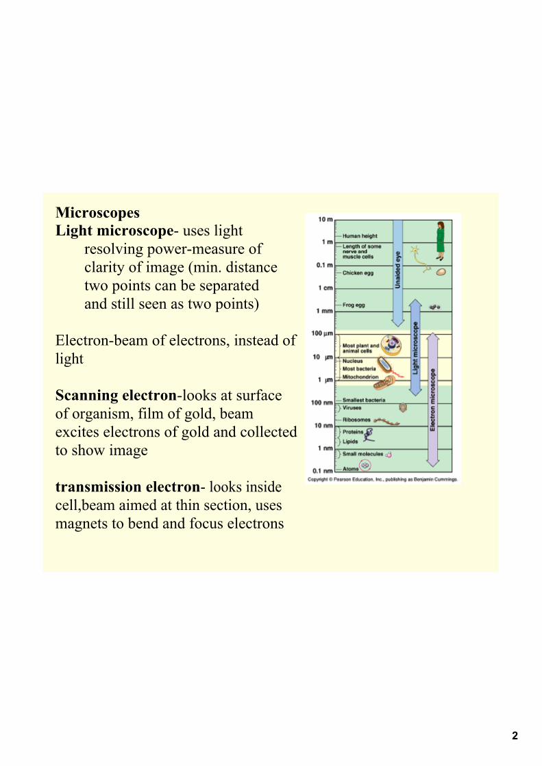
MicroscopesLight microscope uses light
resolving powermeasure of clarity of image (min. distancetwo points can be separatedand still seen as two points)
Electronbeam of electrons, instead of light
Scanning electronlooks at surface of organism, film of gold, beam excites electrons of gold and collectedto show image
transmission electron looks inside cell,beam aimed at thin section, uses magnets to bend and focus electrons
3
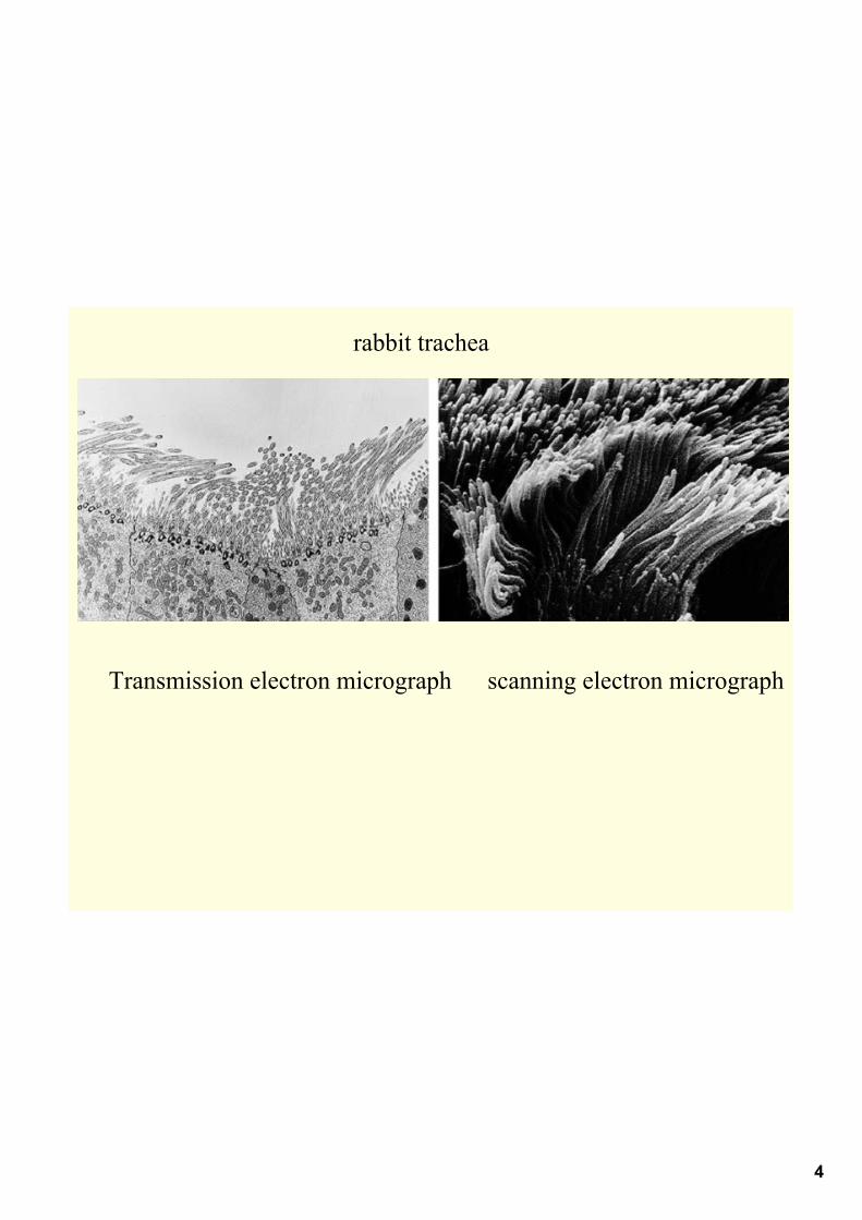
4
Transmission electron micrograph scanning electron micrograph
rabbit trachea

5
How do you think scientists study the components of cells?

6
Cell Fractionation takes cells apartuses ultracentrifuge 130,000 rpm
7
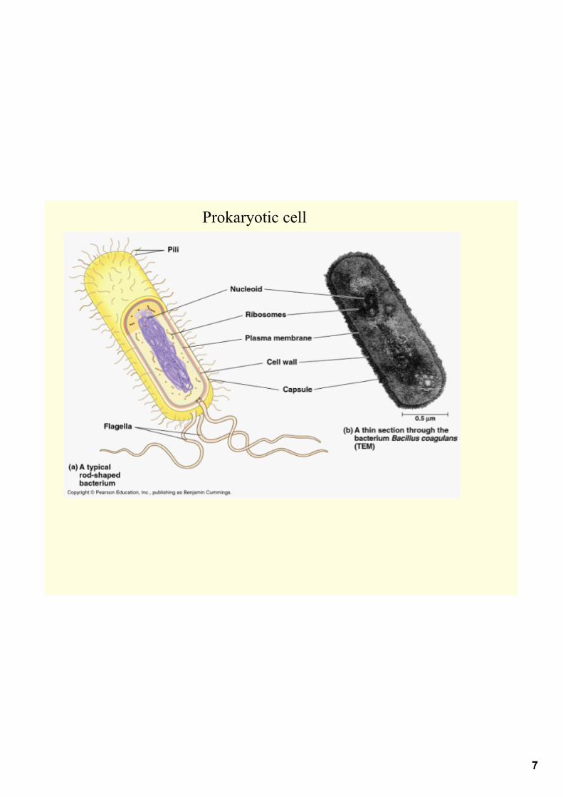
Prokaryotic cell
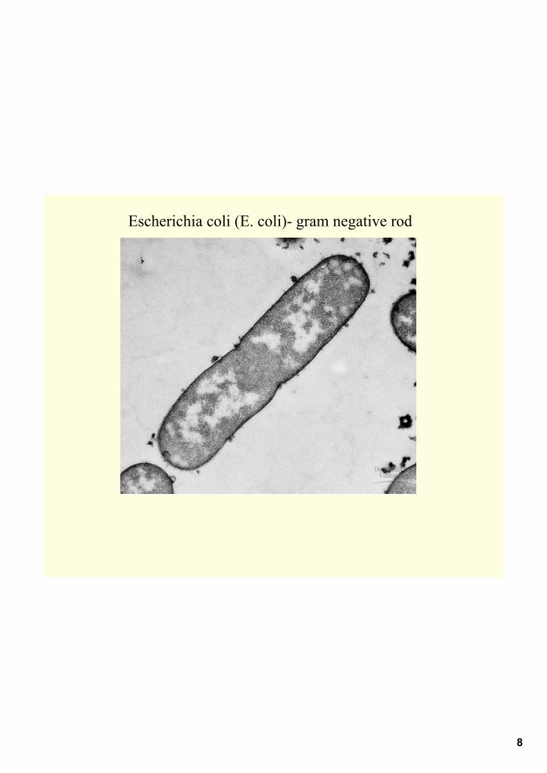
8
Escherichia coli (E. coli) gram negative rod

9
At a minimum, what structures or components must a cell contain to be considered alive?
Out of the cells living today, which types of cells have the above components?
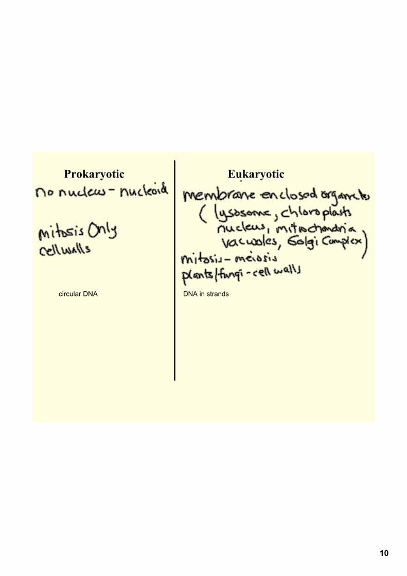
10
Prokaryotic Eukaryotic
circular DNA DNA in strands

11
Surface Area to volume Ratio
higher the ratio, the easier it is for cells to exchange materials
12
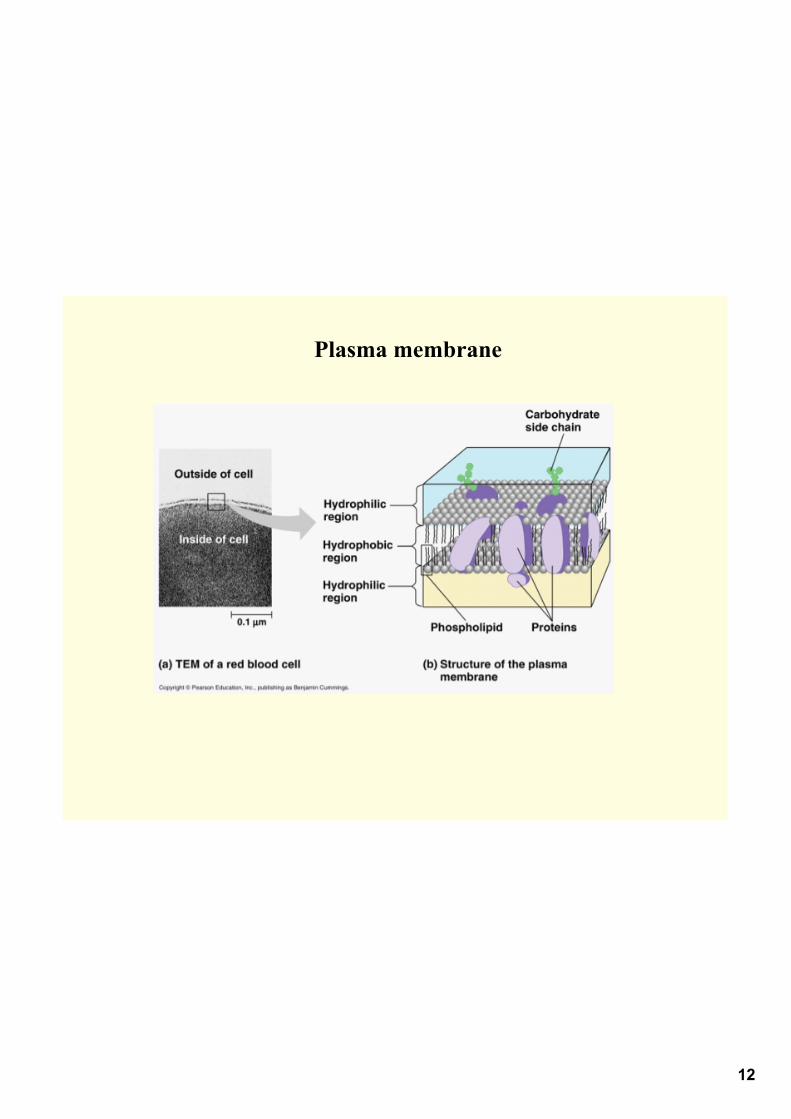
Plasma membrane

13
Animal Cell

14
Plant Cell

15
Importance of membranes
• partition cell into compartments
• help in cell metabolismhave enzymes in membranesspecific metabolic reactions happen here
• allows for many reactions to happen at once in cell
• each membrane has unique makeup of lipids, proteins depending on the function
ex. mitochondria for cell respiration
16
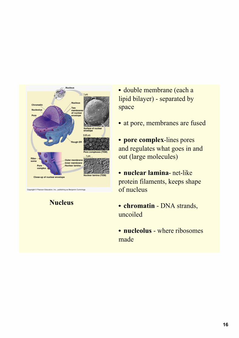
Nucleus
• double membrane (each a lipid bilayer) separated by space
• at pore, membranes are fused
• pore complexlines pores and regulates what goes in and out (large molecules)
• nuclear lamina netlike protein filaments, keeps shape of nucleus
• chromatin DNA strands, uncoiled
• nucleolus where ribosomes made

17
Ribosomes ribosomal RNA and protein
18
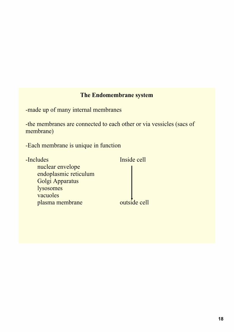
The Endomembrane system
made up of many internal membranes
the membranes are connected to each other or via vessicles (sacs of membrane)
Each membrane is unique in function
Includes Inside cellnuclear envelopeendoplasmic reticulumGolgi Apparatuslysosomesvacuolesplasma membrane outside cell

19
Endoplasmic reticulum
is half of all membranes in eukaryotic cell
includes tubules and cisternae (fluid filled spaces)
continuous with nuclear envelope
cisternae is continuous with nuclear envelope space

20
Smooth ER
no ribosomes attached
lots of enzymes located here synthesize lipids (oils, phospholipids, steroids)
catalyzes a key step in moving glucose from stored glycogen in liver
allows glucose to leave cell
other enzymes of liver detoxify drugs/poisons
muscles cells rely on these enzymes to move calcium for a contraction
21
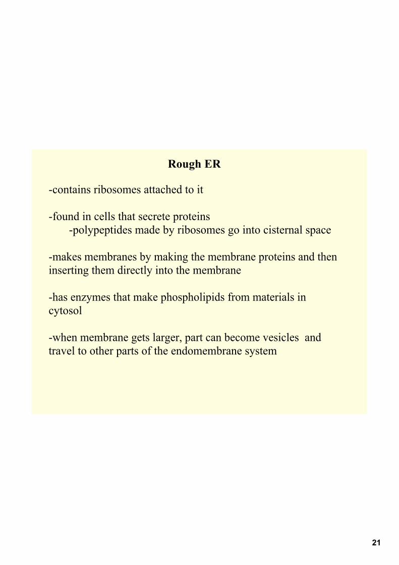
Rough ER
contains ribosomes attached to it
found in cells that secrete proteinspolypeptides made by ribosomes go into cisternal space
makes membranes by making the membrane proteins and then inserting them directly into the membrane
has enzymes that make phospholipids from materials in cytosol
when membrane gets larger, part can become vesicles and travel to other parts of the endomembrane system
22
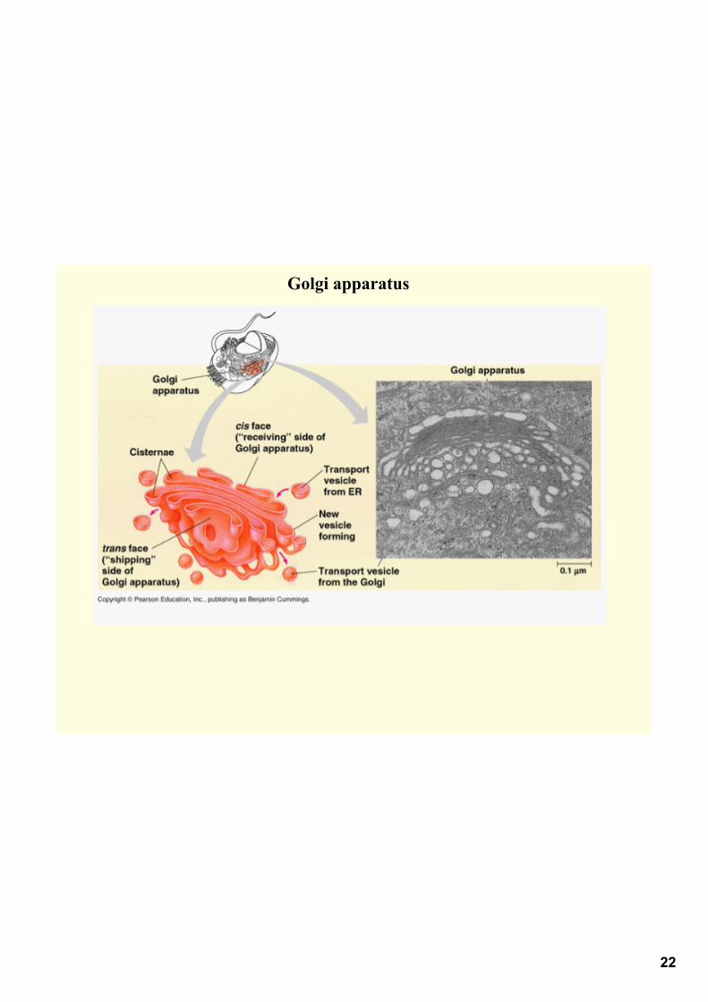
Golgi apparatus

23
"packaging and shipping part of a factory"
flattened membranous cisternaecis side = receives material (fusing of vesicle)trans side = giving off side, vesicles leave from this side
going from cis to trans sides products get modified due to enzymes
manufactures pectin, and other noncellulose polysaccharides
packages materials into vesicles to go to other places

24
Lysosomes
membrane bound sac filled with digestive enzymes
hydrolyzes all organic compounds
enzymes work at pH 5
25
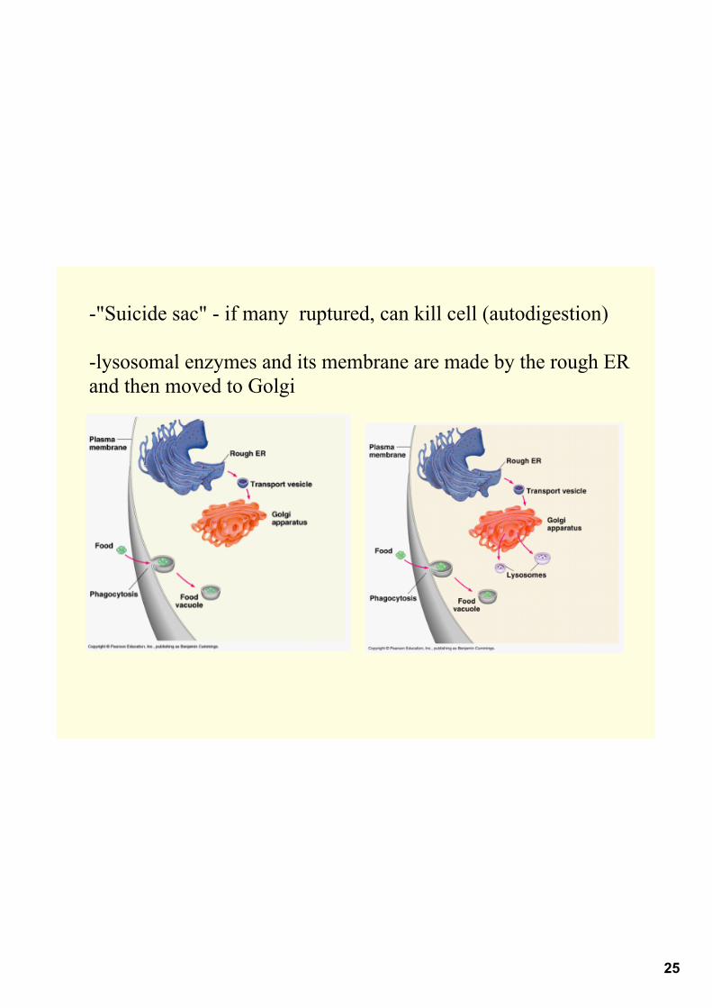
"Suicide sac" if many ruptured, can kill cell (autodigestion)
lysosomal enzymes and its membrane are made by the rough ER and then moved to Golgi
26
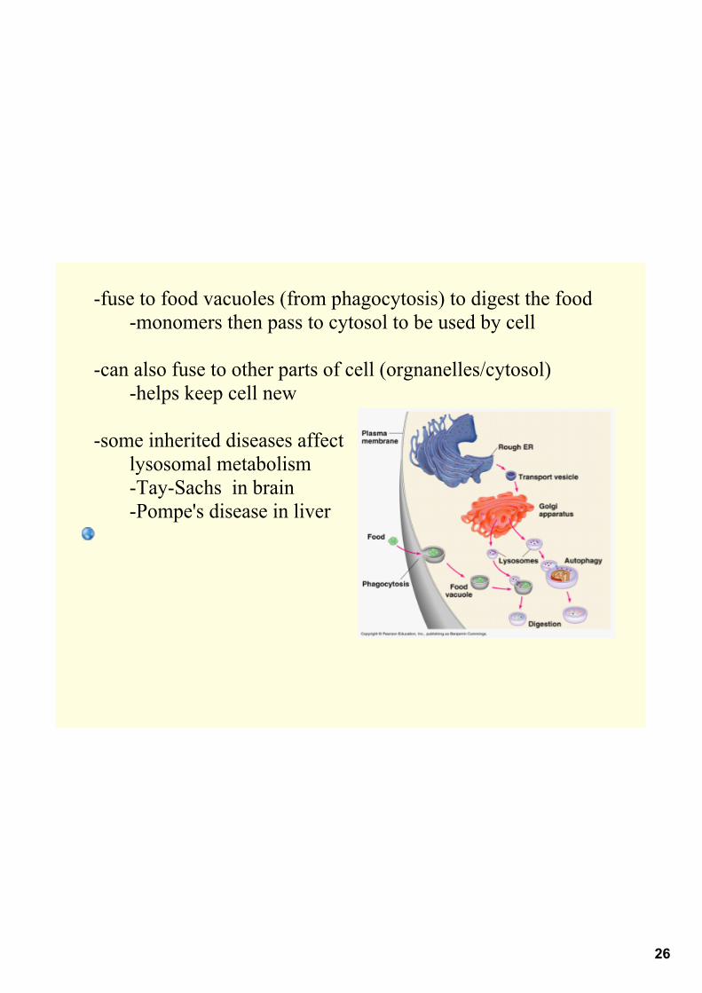
fuse to food vacuoles (from phagocytosis) to digest the foodmonomers then pass to cytosol to be used by cell
can also fuse to other parts of cell (orgnanelles/cytosol)helps keep cell new
some inherited diseases affect lysosomal metabolismTaySachs in brainPompe's disease in liver
27
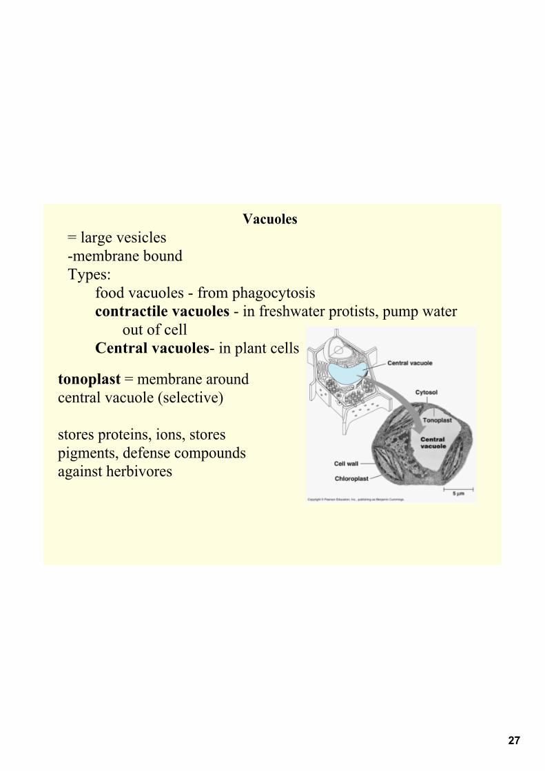
Vacuoles= large vesiclesmembrane boundTypes:
food vacuoles from phagocytosiscontractile vacuoles in freshwater protists, pump water
out of cellCentral vacuoles in plant cells
tonoplast = membrane around central vacuole (selective)
stores proteins, ions, stores pigments, defense compounds against herbivores
28
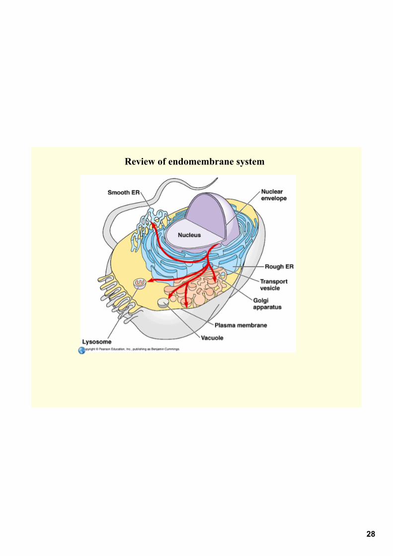
Review of endomembrane system

29
Mitochondria
• sites of cellular respiration, make ATP from sugars, fats, and other fuels
• not part of the endomembrane system their proteins come from free ribosomes and their own ribosomes
• contain a small amount of DNA (circular like prokaryotes)
• can grow and reproduce by themselves
• eukaryotic cells may have one large mitochondria or many small mitochondria
• move around in cell on tracks of cytoskeleton

30
• have smooth outer membrane and a folded inner membrane (cristae)• cristae increase surface area for cell respiration to happen• fluid is between the membranes• inside inner membrane = mitochondrial matrix contains DNA, ribosomes, enzymes, fluid
31
Chloroplasts
• found in plants and eukaryotic algae
• sites of photosynthesis
• not part of endomembrane system
• has own DNA (circular), ribosomes
• its protein comes from free ribosomes and from their own ribosomes

32
• type of plastid amyloplasts store starch in roots and tuberschromoplasts store pigments for fruits and flowerschloroplasts contain green pigment chlorophyll produce sugar
via photosynthesis
• has two membranesinside inner membrane fluid = stroma (Contains DNA, ribosomes &
enzymes)also contains membranous sacs = thylakoids (stacked as grana where
light reactions happen)

33

34
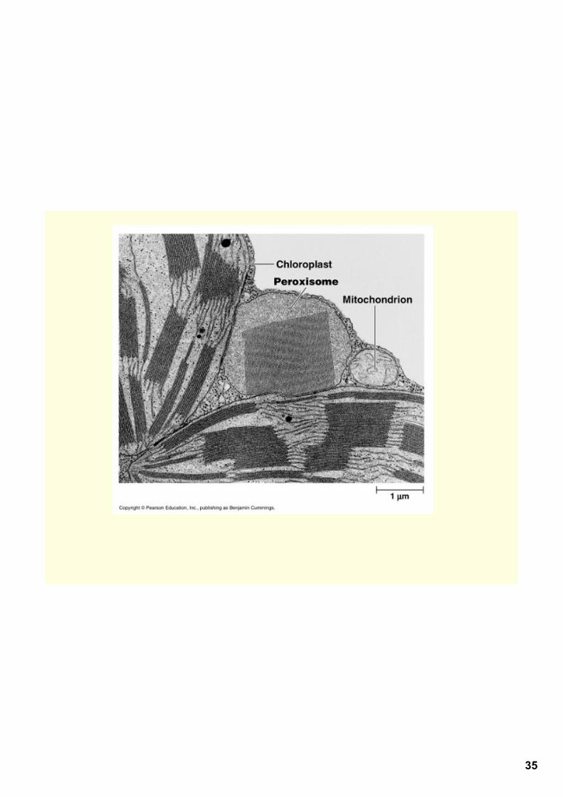
Peroxisomes• contain enzymes to transfer hydrogen from substrates to oxygen
intermediate = hydrogen peroxide (poisonous)has another enzyme that converts hydrogen peroxide to water
and oxygen gas
• functions:breakdown fatty acids used in mitochondria for fueldetoxify alcohol and other harmful compoundsglyoxysomes convert fatty acid in seeds to sugars
• have a single membrane
• not from endomembrane system
• divide when get to a certain size
35

36
Cytoskeleton
= network of fibers throughout cytoplasm
function organizes structures and activities of cell

37
Cytoskeleton Support• provides anchorage for organelles and cytosolic enzymes• can come apart in one part of cell and reassemble in another part
helps to change shape of cell
Cell motility• interacts with motor proteins
cilia/flagella motor proteins pull cytoskeleton past each other
happens in muscle cells too• motor molecules also act like "monorails" carrying vesicles or
organelles• cytoskeleton and motor proteins move materials via streaming
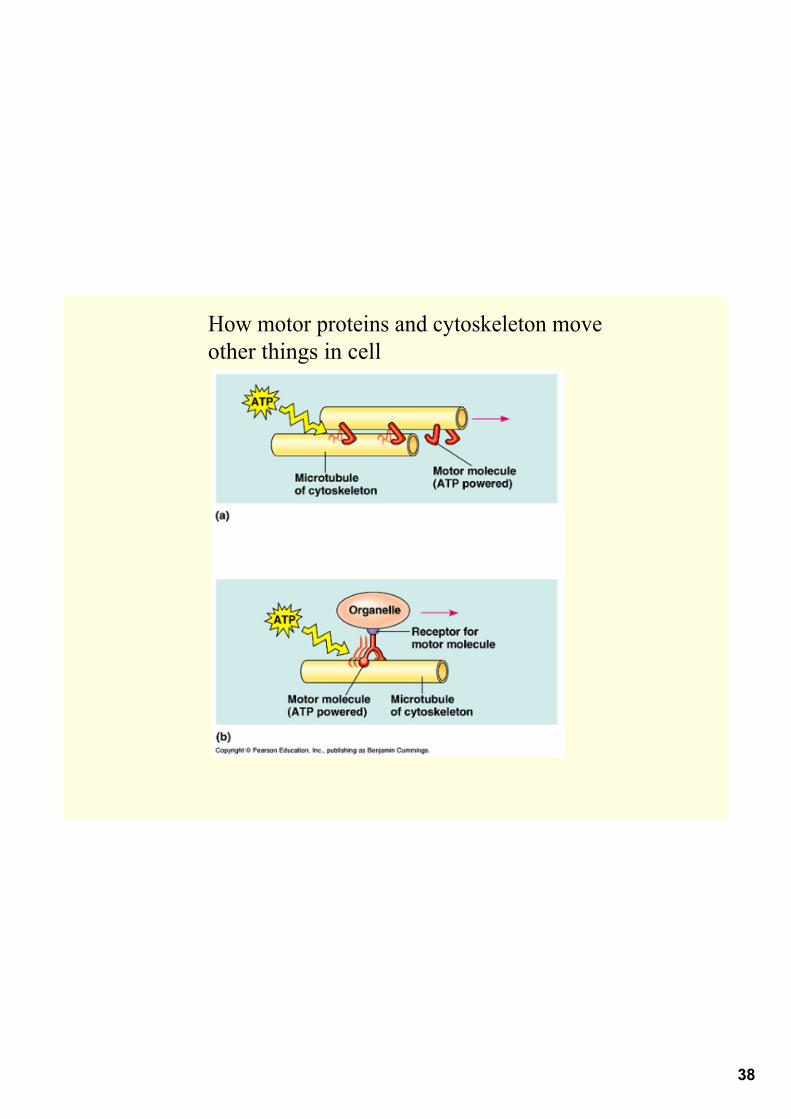
38
How motor proteins and cytoskeleton move other things in cell
39
Three types of fibers of cytoskeleton
1. microtubulesthickest fibers, hollow rodsmade of globular protein tubulin can grow or shrink based on # or tubulin moleculesfunction:
a. move chromosomes during cell divisionb. guide motor proteins that carry organelles to different places in cellc. structural support for cilia and flagella
cilia hairlike structures, move like oars, manyflagella one or few, long, whiplike structure

40
grow out of a centrosome near nucleus
in animals cells has pair of centrioles with 9 triplets of microtubules in a ring
during cell division centrioles replicate

41
Flagella vs. cilia

42
Both cilia and flagella have same structurecore of microtubules covered in plasma membranenine doublets of microtubules around a pair at center
= "9 + 2"patternanchored by basal body structure is like centriole

43
cilia and flagella are driven by motor protein called dynein
dynein grabs, moves and releases the outer microtubules

44
II. Microfilaments• =thinnest of cytoskeletal fibers• made of solid rods of actin
• designed to resist tension
• form a network just inside membrane
• actin is also found in muscle tissue with myosin, myosin walks along actin so contraction can happen
• actin/myosin divides cytoplasm of animal cells in cell division• also causes amoeboid movement (pseudopodia)

45
Actin and myosin in plants cause cytoplasmic streaming=circular flow of cytoplasm, helps move materials

46

47
III. Intermediate filaments
function: bear tension
made from keratins
reinforce cell shape and fix organelle position
48
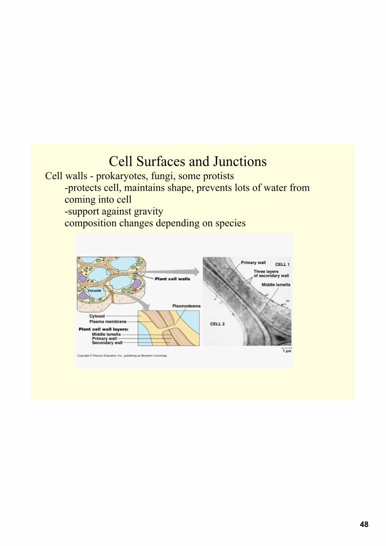
Cell Surfaces and JunctionsCell walls prokaryotes, fungi, some protists
protects cell, maintains shape, prevents lots of water from coming into cellsupport against gravitycomposition changes depending on species
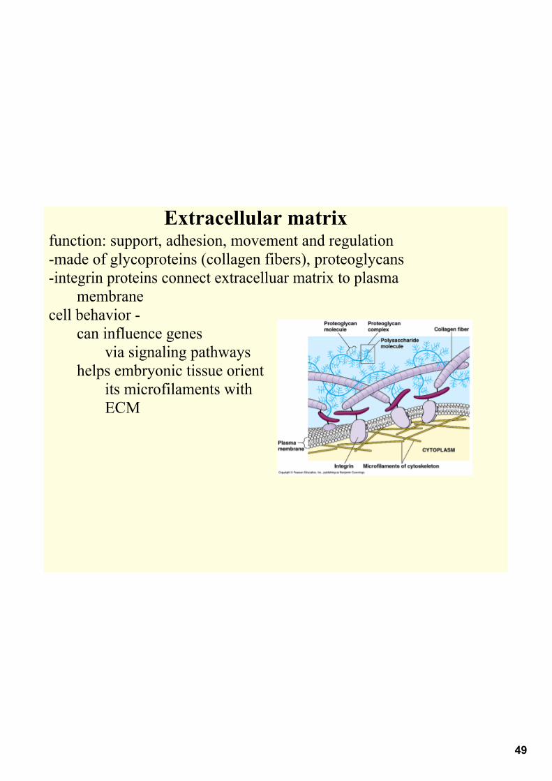
49
Extracellular matrixfunction: support, adhesion, movement and regulationmade of glycoproteins (collagen fibers), proteoglycansintegrin proteins connect extracelluar matrix to plasma
membranecell behavior
can influence genes via signaling pathways
helps embryonic tissue orient its microfilaments with ECM

50
Types of Cell Junctions
Plasmodesmatachannels between adjacent plant cellscytosol goes through here
In animalstight junctions membranes of adjacent cells are fused
form "belts'prevent leakage of extracellular fluid
Desmosomes anchoring junctions, fasten cells togetherkeratin reinforces desmosomes
Gap Junctions communicating junctionsmembrane proteins surround poressalt ions, sugar, amino acids, small molecules pass through here in embryos communication for development here

51
Animal junctions

52