chain class switching hhs public access 1 baff/blys and
TRANSCRIPT

EBV-Encoded Latent Membrane Protein 1 Cooperates with BAFF/BLyS and APRIL to Induce T Cell-Independent Ig Heavy Chain Class Switching1
Bing He*, Nancy Raab-Traub†, Paolo Casali‡, and Andrea Cerutti2,*
*Department of Pathology and Laboratory Medicine, Weill Medical College of Cornell University, New York, NY 10021
†Department of Microbiology and Immunology and Lineberger Comprehensive Cancer Center, University of North Carolina, Chapel Hill, NC 27599
‡Center for Immunology, University of California, Irvine, CA 92697
Abstract
By substituting the H chain C region of IgM with that of IgG, IgA, or IgE, class switching enables
Abs to acquire new effector functions that are crucial for the neutralization of invading pathogens.
Class switching occurs through class switch DNA recombination (CSR) and usually requires
engagement of CD40 on B cells by CD40 ligand on Ag-activated CD4+ T cells. CSR must be
tightly regulated because abnormal IgG and IgA production favors the onset of autoimmunity,
whereas increased switching to IgE leads to atopy. These inflammatory disorders can be triggered
or exacerbated by EBV infection. In this study, we show that EBV induces CD40-independent
CSR from Cµ to multiple downstream Cγ, Cα, and C∊ genes through latent membrane protein 1
(LMP1), a CD40-like viral protein that signals in a ligand-independent fashion. LMP1-induced
CSR is associated with transcriptional activation of germline Cγ, Cα, and C∊ genes and triggers the
up-regulation of activation-induced cytidine deaminase, a crucial component of the CSR
machinery. In addition, LMP1 induces B cells to express B cell-activating factor of the TNF
family and a proliferation-inducing ligand, two molecules that mediate B cell survival and T cell-
independent Ab production. B cell-activating factor of the TNF family and a proliferation-
inducing ligand cooperate with LMP1 to induce Ig class switching because their neutralization by
appropriate soluble decoy receptors attenuates CSR in LMP1-expressing B cells. By showing that
LMP1 triggers T cell-independent CSR, our findings suggest that EBV could play an important
role in the pathogenesis of disorders with aberrant IgG, IgA, and/or IgE production.
Immunoglobulin H chain class switching diversifies the Ab effector functions by
substituting the H chain C region (CH) of IgM with that of IgG, IgA, or IgE (1). B cells
undergo IgH chain class switching through class switch DNA recombination (CSR),3 an
intriguing process that involves the recombination of the switch µ (Sµ) region 5′ of the Cµ
1This work was supported by National Institutes of Health Grants AR47872 (to A.C.) and AR40908 (to P.C.) and by grants from the Leukemia Research Foundation and the Systemic Lupus Erythematosus Foundation/New York Chapter (to A.C.)2Address correspondence and reprint requests to Dr. Andrea Cerutti, Department of Pathology and Laboratory Medicine, Weill Medical College of Cornell University, 1300 York Avenue, New York, NY 10021. [email protected].
HHS Public AccessAuthor manuscriptJ Immunol. Author manuscript; available in PMC 2015 October 29.
Published in final edited form as:J Immunol. 2003 November 15; 171(10): 5215–5224.
Author M
anuscriptA
uthor Manuscript
Author M
anuscriptA
uthor Manuscript
gene with an analogous Sγ , Sα, or S∊ region 5′ of Cγ , Cα, and C∊, respectively (2). Most
Ags, including complex viral and bacterial proteins, induce CSR by up-regulating CD40
ligand (CD40L) on CD4+ T cells (3). Engagement of CD40 on IgD+ naive B cells by
CD40L triggers NF-κB-dependent transcriptional activation of IH gene promoters that are
located 5′ of each S region and encompass a noncoding IH exon (2). The resulting germline
IH-CH transcription increases the accessibility of the targeted S region to the CSR
machinery. This enzymatic complex includes activation-induced cytidine deaminase (AID),
a B cell-specific and CD40-inducible enzyme that induces CSR through an as-yet-elusive
mechanism (2, 4).
T cell-dependent (TD) Ags trigger CD40-dependent CSR in B cells located within the
germinal center (GC) of secondary lymphoid follicles (5, 6). These GC B cells subsequently
differentiate to long-lived IgD− memory B cells or Ab-secreting plasma cells (7, 8). T cell-
independent (TI) Ags, such as viral glycoproteins and bacterial polysaccharides, elicit
CD40-independent CSR and Ab production in extrafollicular marginal zone and intestinal B
cells (9–11). This process requires B cell-activating factor of the TNF family (BAFF; also
known as B lymphocyte stimulator) and a proliferation-inducing ligand (APRIL) (12–16),
two CD40L-related molecules produced by myeloid cells (17–19). BAFF binds to three
receptors specifically expressed on B cells, including transmembrane activator and calcium
modulator and cyclophylin ligand interactor (TACI), B cell maturation Ag (BCMA), and
BAFF-R (also known as BR3) (20–22). In addition to favoring Ab production, BAFF-R
delivers survival signals that are crucial for the conservation of the peripheral B cell
repertoire (23,24). Unlike BAFF, APRIL binds to TACI and BCMA, but not BAFF-R (25,
26). Similarly to CD40 (3), TACI, BCMA, and BAFF-R signal by recruiting TNFR-
associated factors (TRAFs) to their cytoplasmic tails (24). By activating IκB kinase, TRAFs
induce phosphorylation-dependent degradation of IκB, a cytoplasmic inhibitor of NF-κB
(27, 28). The subsequent nuclear translocation of NF-κB transcriptionally activates genes
involved in B cell proliferation, differentiation, and survival (29).
Dysregulated switching to IgG and IgA is central to the pathogenesis of autoimmune
disorders such as systemic lupus erythematosus (SLE) (30), whereas aberrant switching to
IgE underlies the pathogenesis of atopic disorders such as allergic asthma and atopic
dermatitis (31). Both autoimmunity and atopy can be triggered or exacerbated by viral
infections, including EBV infection (32–34). EBV is a B lymphotropic herpes virus that
infects >90% of the human population during the first years of life (35). EBV infection is
usually asymptomatic, because most EBV-containing B cells are eliminated by CD8+ CTLs
(36). However, a few latently infected B cells persist for the lifetime (36). In some
predisposed subjects, latent EBV infection would favor production of IgG and IgA
autoantibodies (37, 38). Abnormal switching to IgG, IgA, and IgE can be also observed in
adolescents with infectious mononucleosis, a self-limiting lymphoproliferative disorder
3Abbreviations used in this paper: CSR, class switch DNA recombination; S, IgH switch region; CD40L, CD40 ligand; AID, activation-induced cytidine deaminase; TD, T cell dependent; TI, T cell independent; GC, germinal center; BAFF, B cell-activating factor of the TNF family; APRIL, a proliferation-inducing ligand; TACI, transmembrane activator and calcium modulator and cyclophylin ligand interactor; BCMA, B cell maturation Ag; TRAF, TNFR-associated factor; SLE, systemic lupus erythematosus; LMP, latent membrane protein; EBNA, EBV-encoded nuclear Ag; mBAFF, mouse BAFF; SC, switch circle; LUC, luciferase reporter plasmid; wt, wild type; CT, circle transcript; PB, peripheral blood; LCL, lymphoblastoid B cell line; BL, Burkitt’s lymphoma; tet, tetracycline; CTAR, C-terminal activation region; BCR, B cell Ag receptor.
He et al. Page 2
J Immunol. Author manuscript; available in PMC 2015 October 29.
Author M
anuscriptA
uthor Manuscript
Author M
anuscriptA
uthor Manuscript
secondary to acute EBV infection (34), as well as in immunocompromised subjects with
EBV-associated B cell lymphoproliferative disorders (39–41). It is unclear how EBV
dysregulates the Ab response.
In the initial phase of the infection, EBV drives tonsillar IgD+ naive B cells to undergo
extrafollicular activation and proliferation through three latent membrane proteins (LMP1,
−2A, and −2B) and six EBV-encoded nuclear Ags (EBNA1 to −6) (42, 43). This growth
program, also known as latency III, allows the expansion of the viral episome in the B cell
compartment until a strong antiviral T cell response is established (44). Later on, EBV
induces infected IgD+ blasts to switch to a default program, also known as latency II, which
entails only EBNA-1, LMP1, and LMP2A, and allows infected B cells to differentiate to
class-switched IgD− memory B cells (42, 43, 45). By further down-regulating LMP1 and
LMP2A, memory B cells acquire a latency program, also known as latency I, which includes
only EBNA1 and allows the persistence of EBV in a transcriptionally quiescent state (43,
46). Periodic reactivation of LMP1 and LMP2A in the tonsillar microenvironment would
generate growth and survival signals that enable latently infected memory B cells to persist
for the lifetime (36). The mechanisms by which EBV-infected IgD+ blasts differentiate to
class-switched IgD− memory B cells remain elusive.
Among EBV-encoded proteins, LMP1 is essential to induce B cell activation, proliferation,
survival (35,47), as well as in vitro B cell transformation (48). The LMP1 cytoplasmic tail
has extensive functional homology with CD40 and, like CD40, induces IκBα degradation
and NF-κB nuclear translocation by recruiting TRAFs and IκB kinase (49–51). Unlike
CD40, which delivers transient signals upon engagement by CD40L (3), LMP1
constitutively signals in a ligand-independent fashion (52). This observation prompted us to
hypothesize that EBV might dysregulate IgG, IgA, and IgE production by delivering CD40-
like signals to B cells.
In this study, we show that B cell infection by EBV actively induces CSR from Cµ to
multiple Cγ, Cα, and C∊ genes through LMP1. This viral protein further dysregulates CSR
by triggering aberrant BAFF and APRIL expression in B cells. Our findings suggest that
neutralization of BAFF and APRIL by soluble TACI and BCMA decoy receptors may
attenuate dysregulated IgG, IgA, and IgE production in certain patients with latent or active
EBV infection.
Materials and Methods
Cells and reagents
IARC, BL16, Bjab, and HL-60 cell lines (from American Type Culture Collection
(Manassas, VA) and R. Dalla-Favera (Columbia University, New York, NY)) were cultured
in RPMI 1640 medium (Invitrogen, Carslbad, CA). IgD+ B cells and monocytes were
obtained from PBMCs as described (53). IgD+ B cells were incubated with EBV (B95-8
strain) for 2 h at 37°C After virus removal, B cells were incubated for 3 wk at a density of
106 cells/ml. All cultures were conducted in RPMI 1640 medium supplemented with 10%
FCS, antibiotics, and glutamine. Ramos subclones expressing EBV proteins (from R. Harris
and M. Neuberger (Medical Research Council Laboratory of Molecular Biology,
He et al. Page 3
J Immunol. Author manuscript; available in PMC 2015 October 29.
Author M
anuscriptA
uthor Manuscript
Author M
anuscriptA
uthor Manuscript
Cambridge, U.K.)) were cultured in medium supplemented with 1 µg/ml puromycine
(Sigma-Aldrich, St. Louis, MO). tet-LMP1 Bjab cells (from N. Lam and B. Sugden
(University of Wisconsin-Madison, Madison, WI)) were cultured with medium
supplemented with 1 µg/ml puromycine and 200 µg/ml geneticin (Invitrogen), and with or
without 1 ng/ml doxycycline (Sigma-Aldrich). Control MOPC-21 (Sigma-Aldrich), TACI-
Ig), BCMA-Ig (Alexis Biochemicals, San Diego, CA), and CD40-Ig (Ancell, Bayport, MN)
were used at 30 µg/ml.
Flow cytometry
CD3, CD14, CD19, CD23 (BD PharMingen, San Diego, CA), IgM, IgG, and IgA (Southern
Biotechnologies Associates, Birmingham, AL) were detected with PE- or FITC-conjugated
Abs. Mouse BAFF (mBAFF) was labeled with a mouse Ab to BAFF (Alexis Biochemicals)
and a PE-conjugated anti-mouse Ab (BD PharMingen). BAFF-Rs were labeled with a CD8-
BAFF fusion protein (Ancell) and a PE-conjugated Ab to CD8 (BD PharMingen). Cells
were acquired using a FACSCalibur analyzer (BD Immunocytometry Systems, San Jose,
CA).
Genomic PCRs and RT-PCRs
DNA and RNA extractions were preceded by removal of dead B cells through Ficoll.
Genomic DNA was extracted from 10 × 106 viable B cells by using the QIAmp DNA mini
kit (Qiagen, Valencia, CA). Switch circles (SCs) were amplified from 500 ng of genomic
DNA (13). Total RNA was extracted from 5 × 10⁶ viable B cells by using the RNeasy total
RNA kit (Qiagen). cDNA was reverse transcribed from 3 µg of total RNA (13). PCRs were
made semiquantitative by varying the number of cycles and performing dilutional analysis
so that there was a linear relationship between the amount of cDNA used and the intensity of
the PCR product. Germline IH-CH transcripts, mature VDJ-CH transcripts, AID, BAFF,
APRIL, and β-actin were amplified as described (13). TACI, BCMA, BAFF-R, and LMP1
were amplified by using the following primer pairs: TACI, forward, 5′-
AAGAAGAGGGGGGATCCCTGC-3′, and reverse, 5′-TTATGCACCTGGGCCCCC-3′;
BCMA, forward, 5′-CTAAGGAA GATAAACTCTGAACCA-3′, and reverse, 5′-
TTACCTAGCAGAAATT GATTTCTC-3′; BAFF-R, forward, 5′-
GTGAGCTGGAGGCGGCGACAG-3′, and reverse, 5′-
CTATTGTGCTCAGGGCCGGC-3′; and LMP1, forward, 5′-
CTTCAGAAGAGACCTTCTCT-3′, and reverse, 5′-ACAATGCCTGT CCGTGCAAA-3′.
The conditions were as follows: denaturation for 1 min at 94°C, annealing for 1 min at 60°C,
and extension for 1 min at 72°C.
Southern blots
PCR products were fractionated onto agarose gels, transferred overnight to nylon
membranes, and hybridized with radiolabeled probes as described (13). SCs were hybridized
with a probe recognizing the recombined Sµ region; circle transcripts (CTs) and mature
VDJ-Cµ transcripts were hybridized with a probe encompassing nt 1–250 of the first Cµ
exon; and mature VDJ-Cγ transcripts were hybridized with a 5′-CAGGGGGAA
GACCGATGG-3′ oligoprobe recognizing a consensus Cγ sequence.
He et al. Page 4
J Immunol. Author manuscript; available in PMC 2015 October 29.
Author M
anuscriptA
uthor Manuscript
Author M
anuscriptA
uthor Manuscript
Vectors
−449/+265 and −291/+131 genomic DNA fragments encompassing the Iγ3 and I∊ promoters
were inserted into a promoterless pGL3-Basic vector (Promega, Madison, WI) containing a
luciferase (LUC) reporter gene (54, 55). Wild-type (wt) LMP1 and mutant LMP1, including
DEL 187–351 LMP1, 187-STOP LMP1, and 231-STOP LMP1, were cloned into a pcDNA3
expression vector (Invitrogen) (56). The κB(2X)-LUC reporter vector is driven by an NF-κB-
responsive minimal promoter with two NF-κB-binding κB sites. κB(2X)-LUC and the IκBα-
pcDNA3 expression vector were provided by E. Cesarman (Weill Medical College of
Cornell University, New York, NY). The −839/+232 genomic DNA segment encompassing
the BAFF promoter was PCR amplified from placental genomic DNA by using sense 5′-
CACAGGTCCACCAAGTCAACAA CAGA-3′ and antisense 5′-
ATCACTACTTGAACTTTGAAGGTTGG-3′ primers with 5′ overhangs containing KpnI
(sense) and XhoI (antisense) restriction sites. The resulting DNA segment was sequenced
and then cloned into the pGL3-Basic reporter vector. The MatInspector software
(Genomatix Software, Munchen, Germany) was used to identify putative NF-κB-binding κB
motifs.
Primer extension gene analysis
A Primer Extension System-Avian Myeloblastosis Virus Reverse Transcriptase (Promega)
was used to identify the BAFF gene transcription initiation site. Briefly, total RNA from
HL-60 or BL16 cells was reversed transcribed with an avian myeloblastosis virus reverse
transcriptase and an end-labeled 5′-CAGTAGGTTTGCTGGCATTTACCCTC-3′ antisense
primer recognizing a sequence immediately upstream of the first BAFF exon. The resulting
cDNA was analyzed on a denaturing polyacrylamide gel to identify the number of bases
between the labeled oligonucleotide and the 5′ end of the targeted RNA.
Luciferase reporter assays
Cells (20 × 106/ml) were transfected with 40 µl of plasmid DNA-Tris-EDTA solution
containing 20 µg of pGL3-Basic vector and 200 ng of pRL-TK control vector (Promega).
pGL3 or κB(2X)-LUC reporter vectors were cotransfected with 2 µg/ml pcDNA3.1
expression vectors containing wt or mutated LMP1. Electroporation was performed at 625
V/cm (HL-60 and BL16) or 525 V/cm (Bjab) and 950 µF using a Gene Pulser II apparatus
(Bio-Rad Laboratories, Hercules, CA). Transfected cells (1 × 10⁶/ml) were cultured for 48
h. The luciferase activity was measured with the Dual-Luciferase Assay System (Promega).
Immunoblots
Total proteins were fractionated onto a 10% SDS-PAGE and transferred to polyvinylidene
difluoride membranes (Bio-Rad). After blocking, membranes were probed with Abs to
BAFF (Upstate Biotechnology, Lake Placid, NY), APRIL, TACI, BCMA, actin (Santa Cruz
Biotechnology, Santa Cruz, CA), and LMP1 (DAKO, Carpinteria, CA). Proteins were
detected with an ECL detection system (Amersham, Little Chalfont, U.K.).
He et al. Page 5
J Immunol. Author manuscript; available in PMC 2015 October 29.
Author M
anuscriptA
uthor Manuscript
Author M
anuscriptA
uthor Manuscript
EMSAs
Cells (5 × 10⁶) were lysed to extract nuclear proteins as reported (54). A double-stranded
oligonucleotide probe overlapping the κB3 site (residues −236 to −216,
GTGTCTGGACTCCCCCTCGCC) of the Iγ3 promoter (54) was end labeled with
[γ-32P]ATP by T4 kinase and used at ∼30,000 cpm in each EMSA reaction. Reaction
mixtures (20 µl) contained 1 ng of DNA probe, 4 µg of nuclear proteins, 2 µg of poly(dI-dC)
(Sigma-Aldrich), and 2 µl of binding buffer (12.5 mM HEPES (pH 7.9), 5 mM KCl, 0.1 mM
EDTA, 1 mM dithiothreitol, and 0.05% Nonidet P-40). Reaction samples were incubated at
room temperature for 15 min and then electrophoresed through a 6% nondenaturing
polyacrylamide gel in 0.25 × Tris-borate EDTA buffer at 150 V.
Results
B cells up-regulate AID and undergo CSR from Cµ to Cγ Cα, and C∊ upon infection by EBV
CSR from Cµ to a downstream Cγ3, Cγ1, Cα1, Cγ2, Cγ4, Cα2, or C∊ gene is preceded by the
transcription of that gene in the form of a noncoding germline IH-CH transcript that includes
the IH exon 5′ of the targeted S region and CH (1). CSR also requires the up-regulation of
the B cell-specific enzyme AID (2). By inducing looping-out deletion of the IgH DNA
between Sµ and the targeted downstream S region, CSR generates a single-copy
extrachromosomal reciprocal DNA recombination product, also known as SC (57). Because
CSR does not target a consensus sequence within the S region (58), actively class-switching
B cells generate multiple SCs with different sizes. After excision from the IgH locus, SCs
transcribe short-lived chimeric I-Cµ CTs that include the promoter upstream of the targeted
IH exon, the IH exon, and Cµ (58). These CTs often undergo posttranscriptional remodeling,
thereby generating more than one band after PCR amplification (58). Together with AID
and germline IH-CH transcripts, SCs and CTs constitute specific markers of ongoing CSR
and, in healthy individuals, are usually detected only in IgD− GC B cells (59). Ongoing CSR
was analyzed in noninfected (EBV−) normal peripheral blood (PB) IgD+ naive B cells,
which display S regions in an unrearranged configuration (13, 59) as well as in monoclonal
IARC549 and IARC100 lymphoblastoid cell lines (LCLs) obtained by transforming normal
polyclonal PB B cells with EBV in vitro. CSR was also studied in monoclonal BL16 cells, a
neoplastic Burkitt’s lymphoma (BL) B cell line that, like IARC549 and IARC100, expresses
surface IgD on most of its elements and harbors a type-III EBV gene latency program.
Noninfected IgD+ B cells from healthy subjects lacked total Sγ-Sµ and Sα-Sµ SCs (Fig. 1A),
expressed no or low germline Iγ1-Cγ1, Iγ3-Cγ3, and Iα1-Cα1 transcripts, and lacked Iγ1/2-
Cµ, Iγ3-Cµ and Iα1/2-Cµ CTs as well as AID transcripts (B). In contrast, lymphoblastoid and
EBV+ BL16 B cells contained total Sγ-Sµ and Sα-Sµ SCs, expressed large amounts of
germline Iγ1-Cγ1, Iγ3-Cγ3, and Iα1-Cα1 transcripts, and contained Iγ1/2-Cµ, Iγ3-Cµ, and
Iα1/2-Cµ CTs, and AID transcripts. In both lymphoblastoid and EBV+ BL16 cells, 5–15% of
the clonal elements expressed surface IgG or IgA, but not surface IgD and IgM (not shown),
further suggesting ongoing CSR. Additional experiments were performed to verify whether
infection of normal IgD+ B cells by EBV induces CSR. Compared with noninfected IgD+ B
cells (Fig. 1A), EBV-infected IgD+ B cells up-regulated AID transcripts and contained
extrachromosomal Sγ1/2-Sµ, Sγ 3-Sµ, Sγ4-Sµ, Sα1/ 2-Sµ and S∊-Sµ SCs (C), which reflect
He et al. Page 6
J Immunol. Author manuscript; available in PMC 2015 October 29.
Author M
anuscriptA
uthor Manuscript
Author M
anuscriptA
uthor Manuscript
ongoing CSR from Cµ to Cγ1/Cγ2, Cγ3, Cγ4, Cα1/Cα2, and C∊ respectively. These findings
indicate that B cells undergo CD40-independent CSR from Cµ to multiple downstream Cγ,
Cα, and C∊ genes upon infection by EBV.
EBV up-regulates germline IH-CH transcription, AID expression, and CSR through LMP1
To evaluate the mechanism by which EBV induces CSR, we took advantage of IgM+
subclones established from the EBV− BL cell line Ramos and stably expressing LMP1,
LMP2A, EBNA1, EBNA2, or EBNA-LP expression vectors. Compared with control
subclones transfected with empty expression vectors, B cell sub-clones expressing LMP1
contained Iγ1/2-Cµ and Iγ3-Cµ CTs as well as mature VHDJH-Cγ1 and VHDJH-Cγ3
transcripts (Fig. 2), the end product of CSR from Cµ to Cγ1 and Cγ3. B cell subclones
expressing LMP2A contained lower amounts of Iγ3-Cµ CTs and VHDJH-Cγ3 transcripts, but
lacked Iγ1/2-Cµ and VHDJH-Cγ1 transcripts. In contrast, B cell subclones expressing
EBNA1, EBNA2, or EBNA-LP lacked Iγ1/2-Cµ and Iγ3-Cµ CTs as well as mature VHDJH-
Cγ1 and VHDJH-Cγ3 transcripts. Finally, all B cell sub-clones expressed β-actin transcripts
as well as mature VHDJH-Cµ transcripts. These findings suggest that LMP1 and, to a lesser
extent, LMP2A trigger CD40-independent CSR.
The CSR-inducing activity of LMP1 was further evaluated in an EBV− BL cell line, Bjab,
stably expressing a tetracycline (tet)-inducible LMP1 expression vector. Like Ramos B cells,
Bjab B cells express IgM but not IgG, IgA, or IgE on the surface. Bjab-tet-LMP1 B cells up-
regulated LMP1 (shown below) and activated luciferase reporter vectors containing the Iγ3
gene promoter (Iγ3-LUC), the I∊ gene promoter (I∊-LUC), or κB(2X)-LUC upon incubation
with doxycycline for 2 days (Fig. 3A). Overexpression of the NF-κB inhibitor IκBα
inhibited doxycycline-induced activation of Iγ3-LUC, I∊-LUC, and κB(2X)-LUC. In addition
to activating the Iγ3 and I∊ promoters, doxycycline up-regulated the expression of germline
Iγ1-Cγ1,Iγ3-Cγ3, Iα1-Cα1, and I∊-C∊ transcripts (Fig. 3B). Furthermore, Bjab-tet-LMP1 B
cells exposed to doxycycline for 4 days up-regulated AID transcripts and induced Iγ1/ 2-Cµ
Iγ3-Cµ Iα1/2-Cµ and I∊-Cµ CTs (Fig. 3C), which reflect ongoing CSR from Cµ to Cγ1/Cγ2,
Cγ3, Cα1/Cα2, and C∊, respectively. The induction of CSR by doxycycline was associated
with up-regulation of surface IgG and IgA and down-regulation of surface IgM (Fig. 3D).
These findings indicate that B cells undergo NF-κB-dependent germline IH-CH transcription
and CSR upon activation by LMP1.
LMP1 induces BAFF and APRIL expression in B cells
We have recently found that dendritic cells up-regulate BAFF and APRIL, two inducers of
TI CSR, upon engagement of CD40 by CD40L (13). Given its ability to mimic CD40
signaling, LMP1 might up-regulate BAFF and APRIL in B cells as CD40 does in dendritic
cells. Compared with control subclones, Ramos B cell subclones expressing LMP1 or, to a
lesser extent, LMP2A contained more BAFF and APRIL transcripts and proteins (Fig. 4A).
In contrast, Ramos B cell subclones expressing EBNA1, EBNA2, or EBNA-LP contained
BAFF and APRIL transcripts and proteins in amounts comparable with those detected in
control subclones. Additional experiments evaluated the expression of BAFF and APRIL in
Bjab-tet-LMP1 B cells. When exposed to tet, Bjab-tet-LMP1 B cells up-regulated LMP1 as
well as BAFF and APRIL transcripts and proteins (Fig. 4B). tet also up-regulated surface
He et al. Page 7
J Immunol. Author manuscript; available in PMC 2015 October 29.
Author M
anuscriptA
uthor Manuscript
Author M
anuscriptA
uthor Manuscript
CD23, a canonical LMP1-inducible B cell-activation protein, BAFF, as well as the total
BAFF-binding activity (Fig. 4C), which reflects the surface density of TACI, BCMA, and
BAFF-R receptors. In contrast, tet did not up-regulate CD3, a T cell-restricted component of
the TCR complex, or CD19, a component of the B cell receptor complex expressed by all
mature B cells. These findings indicate that LMP1 up-regulates BAFF and APRIL in B
cells.
LMP1 activates the BAFF gene promoter through NF-κB
NF-κB is crucial for the activation of B cells by LMP1 (35, 51). We took advantage of Bjab-
tet-LMP1 B cells to verify whether LMP1 up-regulates BAFF through NF-κB. DNA
sequence analysis showed that the BAFF gene promoter contains at least six NF-κB-binding
κB sites (Fig. 5A). Bjab-tet-LMP1 B cells activated a luciferase reporter vector carrying the
BAFF gene promoter (BAFF-LUC) as well as κB(2X)-LUC upon incubation with
doxycycline for 2 days. Overexpression of IκBα inhibited the induction of BAFF-LUC and
κB(2X)-LUC by doxycycline (Fig. 5B), suggesting that NF-κB is critical to up-regulate
BAFF. Because LMP1 recruits TRAFs and activates NF-κB through C-terminal activation
region (CTAR)-1 and CTAR-2 (49, 56), we verified whether disruption of one or both
CTARs affects the up-regulation of BAFF by LMP1. wt Bjab B cells activated BAFF-LUC
as well as κB(2X)-LUC upon transfection with wt LMP1, which contains both CTAR-1 and
CTAR-2. In contrast, 187-STOP LMP1, which lacks both CTAR-1 and CTAR-2, failed to
activate BAFF-LUC as well as control κB(2X)-LUC. Furthermore, both BAFF-LUC and
κB(2X)-LUC were activated by DEL 187–351 LMP1, which lacks only CTAR-1, or 231-
STOP LMP1, which lacks only CTAR-2. These findings suggest that the up-regulation of
BAFF by LMP1 requires the integrity of at least one CTAR domain. Finally, transfection of
Bjab B cells with graded amounts of IκBα progressively inhibited the activation of BAFF-
LUC and κB(2X)-LUC by wt LMP1, further indicating that NF-κB is crucial for the up-
regulation of BAFF by LMP1.
B cells express BAFF and APRIL upon infection by EBV
Additional experiments were performed to verify whether purified B cells up-regulate BAFF
and APRIL upon EBV infection. Purified noninfected IgD+ B cells lacked BAFF and
APRIL transcripts (Fig. 6A) as well as BAFF and APRIL proteins (B). Similar normal B
cells expressed CD19, but most of them lacked the EBV (LMP1)-inducible Ag CD23 as
well as mBAFF and the myeloid Ag CD14 (Fig. 6C). In contrast, purified EBV-infected
IgD+ B cells contained BAFF and APRIL transcripts and proteins and coexpressed CD19,
CD23, and mBAFF on the surface. Similar EBV-infected normal B cells lacked CD14,
indicating a lack of contaminating monocytes and macrophages. The expression of BAFF
and APRIL was also measured in B cell lines harboring a type-III EBV latency gene
program. IARC504 lymphoblastoid B cells and neoplastic EBV+ BL16 B cells expressed
BAFF and APRIL transcripts and proteins in amounts comparable with those expressed in
myeloid cells, including HL60 AML cells. Moreover, lymphoblastoid and malignant BL16
B cells, which contain LMP1 (Fig. 6, A and B), expressed more BAFF and APRIL
transcripts than Akata and Mutu I (not shown), two EBV+ BL B cell lines that, unlike LCLs
and BL16, express a type-I EBV latency gene program and therefore lack LMP1. Finally, all
B cell types under study but not HL60 AML cells expressed TACI, BCMA, and BAFF-R
He et al. Page 8
J Immunol. Author manuscript; available in PMC 2015 October 29.
Author M
anuscriptA
uthor Manuscript
Author M
anuscriptA
uthor Manuscript

transcripts (Fig. 6A) and proteins (B). These findings indicate that B cells express TACI,
BCMA, and BAFF-R, and aberrantly up-regulate BAFF and APRIL upon infection by EBV.
BAFF and APRIL up-regulate AID and enhance CSR in EBV-infected B cells
The above findings prompted us to hypothesize that engagement of TACI, BCMA, and/or
BAFF-R by autocrine BAFF and APRIL enhances LMP1-induced CSR in EBV-infected B
cells. To verify this, we took advantage of soluble TACI-Ig and BCMA-Ig decoy receptors,
which prevent the binding of BAFF and APRIL to cell-bound TACI, BCMA, and BAFF-R.
Lymphoblastoid IARC549 B cells down-regulated total Sγ-Sµ and Sα-Sµ SCs (Fig. 7A),
Iγ1/2-Cµ, Iγ3-Cµ, and Iα1/2-Cµ CTs, as well as AID transcripts upon exposure to soluble
TACI-Ig and BCMA-Ig decoy receptors for 2 days (B). This down-regulation was specific,
as similar lymphoblastoid B cells did not attenuate CSR upon exposure to a control Ig or
CD40-Ig, which blocks CD40L-CD40 interaction. These findings suggest that EBV triggers
CD40-independent CSR not only through LMP1 but also through endogenous (LMP1-
induced) BAFF and APRIL.
BAFF and APRIL enhance NF-κB activation and germline IH-CH transcription in EBV-infected B cells
Additional experiments were set up to assess whether BAFF and APRIL released by EBV-
infected LMP1-expressing B cells activate NF-κB. Exposure of lymphoblastoid IARC549 B
cells to BCMA-Ig but not control Ig or CD40-Ig down-regulated the binding of nuclear NF-
κB to an oligonucleotide encompassing a DNA sequence crucial for the activation of the
human Iγ3 promoter by CD40 (Fig. 7C). As expected, this effect was associated with down-
regulated expression of germline Iγ3-Cγ3 transcripts (Fig. 7D). In contrast, the expression of
germline Iγ3-Cγ3 transcripts was not affected by control Ig or CD40-Ig. These results
indicate that engagement of TACI, BCMA, and/or BAFF-R by autocrine BAFF and APRIL
activates NF-κB and enhances the expression of germline IH-CH transcripts in EBV-infected
LMP1-expressing B cells.
Discussion
We have shown that EBV-encoded LMP1 induces CD40-independent CSR from Cµ to
multiple CγCα, and C∊ genes in B cells. This induction is associated with NF-κB-dependent
activation of downstream CH gene promoters and up-regulation of germline IH-CH
transcripts and AID transcripts. LMP1 up-regulates also BAFF and APRIL through an NF-
κB-dependent mechanism that requires at least one CTAR domain. By engaging TACI,
BCMA, and BAFF-R on B cells, BAFF and APRIL activate NF-κB and further enhance
CSR. These findings suggest that EBV could play an important role in the pathogenesis of
disorders associated with aberrant IgG, IgA, and/or IgE production.
EBV is thought to initially infect naive IgD+ B cells in the mantle zone of lymphoid follicles
located beneath the tonsillar epithelium (44). By expressing a full set of EBNA and LMP
proteins, EBV induces IgD+ B cells to become blasts, which proliferate outside the GC (43).
Subsequent down-regulation of most EBV proteins but EBNA1, LMP1, and LMP2A would
enable infected IgD+ blasts to enter the GC and differentiate to class-switched IgD− memory
He et al. Page 9
J Immunol. Author manuscript; available in PMC 2015 October 29.
Author M
anuscriptA
uthor Manuscript
Author M
anuscriptA
uthor Manuscript
B cells (43, 45). After further down-regulation of LMP1 and, to a lesser extent, LMP2A,
latently infected memory B cells would leave the tonsil and enter the circulation (43,46). It
remains unclear whether IgD+ blasts undergo IgH class switching upon stimulation by viral
proteins or as a result of CD40-dependent progression through the GC in response to a TD
Ag. By showing that EBV induces CD40-independent CSR, our data suggest that at least
some infected IgD+ B cells rapidly undergo TI class switching to IgG, IgA, or IgE outside
the GC.
Viruses induce CD40-independent IgH class switching through mechanisms that remain
largely unknown (60–62). Our data indicate that EBV transcriptionally activates
downstream germline CH gene promoters, including Cγ3 and C∊, through LMP1. This
CD40-like viral protein would transactivate Cγ3 and C∊ genes through an NF-κB-dependent
mechanism. Consistent with this, overexpression of the NF-κB inhibitor IκBα or (not
shown) disruption of both NF-κB-activating CTAR domains within the LMP1 cytoplasmic
tail impairs the induction of germline IH-CH transcription by LMP1. In LMP1-expressing B
cells, germline IH-CH transcription is associated with up-regulation of AID transcripts and
induction of CSR from Cµ to multiple downstream Cγ , Cα, and C∊ genes. These findings
provide a mechanistic explanation for previous studies showing that enforced LMP1
expression restores TD IgG, IgA, and IgE production in CD40-deficient mice (63), which
otherwise show severely impaired TD IgH class switching (64). Whereas CD40 transmits
transient ligand-dependent signals (3), LMP1 continuously signals in a ligand-independent
fashion (52). This implies that LMP1-induced CSR is subject to less regulatory constraints
than CD40-induced CSR. By showing that EBV-infected LMP1-expressing IgD+ B cells
constitutively express high levels of IH-CH transcripts and AID and continuously undergo
CSR, our data extend recent findings indicating that artificial Sµ and Sγ3 DNA substrates
undergo spontaneous recombination in lymphoblastoid B cells (65). By triggering
unrestrained CSR, LMP1 may play a key role in the pathogenesis of dysregulated IgG, IgA,
and IgE production occurring in certain EBV-infected individuals, including
immunocompromised HIV-infected subjects and transplant recipients (39–41).
In normal B cells, switching from IgM to IgG, IgA, or IgE requires two signals, one
delivered by CD40L and the other delivered by a cytokine (1, 2). Among cytokines, IL-4
induces switching to IgG and IgE (66–69), IL-10 to IgG and IgA (13, 66, 70, 71), and TGF-
β to IgA (71–73). In EBV-infected LMP1-expressing B cells, CSR to Cγ and Cα occurs in
the absence of exogenous cytokines. This does not imply that cytokines do not play any role
in EBV-induced CSR, because EBV induces B cells to produce large amounts of autocrine
IL-10 through LMP1 (74) as well as small nonpolyadenylated viral RNAs, also referred to
as EBER1 and EBER2 (75). Consistent with this, neutralization of IL-10 by a specific
blocking Ab partially inhibits switching to IgG and IgA in LMP1-expressing B cells (not
shown). In addition to up-regulating endogenous IL-10, EBV produces an IL-10-like protein
through a viral gene known as BCRF1 (76). Furthermore, EBV-infected B cells produce
IL-13 (77), which, like IL-4, activates switching from IgM to IgG4 and IgE (78, 79). This
might explain our finding that EBV-infected LMP1-expressing B cells actively switch to
Cγ4 and C∊ in the absence of external IL-4.
He et al. Page 10
J Immunol. Author manuscript; available in PMC 2015 October 29.
Author M
anuscriptA
uthor Manuscript
Author M
anuscriptA
uthor Manuscript

Unlike CD40, which mediates CSR in GC B cells (3, 5), LMP1 elicits IgG and IgA
production outside the GC of secondary lymphoid follicles (63). This extrafollicular pattern
is also associated with B cell responses to TI Ags with repetitive structure, including
envelope glycoproteins from viruses and capsular polysaccharides from bacteria (10, 60,
80). By showing that LMP1 up-regulates BAFF and APRIL, two mediators of TI Ab
production (13–16), our data suggest that EBV exploits an otherwise physiological TI
pathway to maximize CSR in infected IgD+ B cells. Engagement of TACI, BCMA, and
BAFF-R by LMP1-induced BAFF and APRIL would enhance CSR from Cµ to a targeted
downstream CH gene by activating the germline transcription of that gene through NF-κB.
Consistent with this, neutralization of autocrine BAFF and APRIL by soluble TACI-Ig and
BCMA-Ig decoy receptors attenuates NF-κB activation and down-regulates the expression
of downstream germline IH-CH transcripts in LMP1-expressing B cells. In similar cells,
TACI-Ig and BCMA-Ig decoy receptors down-regulate AID and impair CSR. Thus,
autocrine BAFF and APRIL would cooperate with LMP1 to trigger NF-κB-dependent
germline IH-CH transcription and CSR.
In addition to transactivating downstream CH genes, NF-κB would play an important role in
the LMP1-mediated up-regulation of BAFF. Consistent with this, overexpression of the NF-
κB inhibitor IκBα interferes with the transcriptional activation of the BAFF gene promoter
in LMP1-activated B cells. Furthermore, disruption of the two NF-κB-activating CTARs
within the LMP1 cytoplasmic tail severely impairs the activation of the BAFF gene
promoter by LMP1. Although playing a key role, NF-κB may not be the only transcription
factor involved in LMP1-mediated up-regulation of BAFF. Consistent with this, the BAFF
gene promoter includes several putative STAT-binding γ-IFN-activated sequences (not
shown), which could be activated by STAT proteins induced by LMP1 (81). STAT proteins
could be also induced by IL-10 (82), an LMP1-inducible cytokine that up-regulates BAFF in
myeloid cells (74, 83). Thus, LMP1-induced NF-κB and STAT transcription factors might
synergistically activate the BAFF gene promoter upon binding to cooperative κB and γ-IFN-
activated sequence sites.
LMP1 is not the only CSR-inducing viral protein, because LMP2A triggers CSR from Cµ to
Cγ3 and up-regulates BAFF and APRIL, although to a lesser extent than LMP1. Unlike
LMP1, LMP2A activates B cells by mimicking signaling through the B cell Ag receptor
(BCR) (36). Consistent with this, the cytoplasmic tail of LMP2A encompasses
immunoreceptor tyrosine-based activation motifs similar to those found in the Igα and Igβ
signal-transducing subunits of the BCR complex (36). In addition to modulating B cell
proliferation and survival (84), signals emanating from BCR modulate IgH class switching.
For instance, BCR engagement by certain TI Ags induces CD40-independent switching to
IgG3 both in vivo and in vitro (80, 85). In addition, BCR engagement cooperates with BAFF
and APRIL to induce CD40-independent IgG production (13,19,25). Thus, it is conceivable
that LMP2A triggers TI CSR to Cγ3 through a BCR-like pathway. This pathway would
cooperate with LMP1 as well as endogenous BAFF and APRIL to optimize IgH class
switching in EBV-infected B cells.
Our findings raise the possibility that IgH class switching confers a specific functional
advantage to EBV. When engaged by Ag, surface IgM and IgD (i.e., BCR) deliver
He et al. Page 11
J Immunol. Author manuscript; available in PMC 2015 October 29.
Author M
anuscriptA
uthor Manuscript
Author M
anuscriptA
uthor Manuscript

proliferation and survival signals through the Igα-Igβ heterodimer (84). These signals are
negatively regulated by CD22, an inhibitory coreceptor that contains typical
immunoreceptor tyrosine-based inhibitory motifs (86). Recent studies indicate that B cells
become resistant to CD22-mediated inhibitory signals upon switching from IgM and IgD to
IgG (87). In this fashion, IgG+ (IgD−) memory B cells become more sensitive to BCR-
driven proliferation and survival signals upon exposure to Ag. It is tempting to speculate that
EBV triggers TI CSR in extrafollicular IgD+ B cells to rapidly generate a pool of IgD− B
cells expressing protective BCRs, such as IgG. Due to their lower sensitivity to CD22-
mediated inhibitory signals, these de novo class-switched B cells would facilitate the initial
expansion of the viral episome.
In addition to inducing TI CSR, LMP1 and LMP2A deliver signals that are essential for the
survival of infected B cells (36). Our findings imply that these signals might be greatly
amplified by autocrine BAFF, a powerful inducer of B cell survival (24). BAFF exerts most
of its prosurvival activity through BAFF-R (21, 23), which is expressed in large amounts by
both EBV-infected and noninfected B cells. By engaging BAFF-R on bystander self-reactive
B cells, BAFF expressed on and released by latently infected tonsillar B cells might
facilitate the onset of autoimmune disorders, including SLE (33, 34, 37). Consistent with
this, SLE patients display increased levels of circulating soluble BAFF (24), and mice
overexpressing BAFF develop an SLE-like syndrome with kidney deposition of IgG and
IgA autoantibodies (22). Finally, BAFF and APRIL might also be implicated in the
pathogenesis of autoimmune and IgE-mediated atopic disorders arising in certain HIV-
infected subjects and transplant recipients with EBV-associated B cell lymphoproliferative
disorders (35, 40, 41, 88). In these immune-compromised individuals, neutralization of
BAFF and APRIL by soluble decoy receptors or blocking Abs might attenuate production of
self-reactive IgG and IgA, dysregulated switching to IgE, and aberrant B cell accumulation.
Acknowledgments
We thank Drs. Reuben S. Harris and Michael S. Neuberger (Medical Research Council Laboratory of Molecular Biology, Cambridge, U.K.) for Ramos subclones, Drs. Ngam Lam and Bill Sugden (University of Wisconsin-Madison, Madison, WI) for tet-LMP1-Bjab cells, Dr. Riccardo Dalla-Favera (Columbia University, New York, NY) for IARC and BL16 cell lines, and Dr. Ethel Cesarman (Weill Medical College of Cornell University, New York, NY) for κB(2X)-LUC and IκBα-pcDNA3.
References
1. Stavnezer J. Antibody class switching. Adv. Immunol. 1996; 61:79. [PubMed: 8834495]
2. Manis JP, Tian M, Alt FW. Mechanism and control of class-switch recombination. Trends Immunol. 2002; 23:31. [PubMed: 11801452]
3. Banchereau JF, Bazan F, Blanchard F, Briere F, Galizzi JP, van Kooten C, Liu YJ, Rousset F, Saeland S. The CD40 antigen and its ligand. Annu. Rev. Immunol. 1994; 12:881. [PubMed: 7516669]
4. Okazaki I, Kinoshita K, Muramatsu M, Yoshikawa K, Honjo T. The AID enzyme induces class switch recombination in fibroblasts. Nature. 2002; 416:340. [PubMed: 11875397]
5. MacLennan IC. Germinal centers. Annu. Rev. Immunol. 1994; 12:117. [PubMed: 8011279]
6. Liu YJ, Malisan F, de Bouteiller O, Guret C, Lebecque S, Banchereau J, Mills FC, Max EE, Martinez-Valdez H. Within germinal centers, isotype switching of immunoglobulin genes occurs after the onset of somatic mutation. Immunity. 1996; 4:241. [PubMed: 8624814]
He et al. Page 12
J Immunol. Author manuscript; available in PMC 2015 October 29.
Author M
anuscriptA
uthor Manuscript
Author M
anuscriptA
uthor Manuscript

7. Calame KL. Plasma cells: finding new light at the end of B cell development. Nat. Immunol. 2001; 2:1103. [PubMed: 11725300]
8. McHeyzer-Williams MG, Ahmed R. B cell memory and the long-lived plasma cell. Curr. Opin. Immunol. 1999; 11:172. [PubMed: 10322151]
9. Martin F, Kearney JF. Marginal-zone B cells. Nat. Rev. Immunol. 2002; 2:323. [PubMed: 12033738]
10. Fagarasan S, Honjo T. T-Independent immune response: new aspects of B cell biology. Science. 2000; 290:89. [PubMed: 11021805]
11. Fagarasan S, Honjo T. Intestinal IgA synthesis: regulation of frontline body defences. Nat. Rev. Immunol. 2003; 3:63. [PubMed: 12511876]
12. MacLennan I, Vinuesa C. Dendritic cells, BAFF, and APRIL: innate players in adaptive antibody responses. Immunity. 2002; 17:235. [PubMed: 12354377]
13. Litinskiy MB, Nardelli B, Hilbert DM, He B, Schaffer A, Casali P, Cerutti A. DCs induce CD40-independent immunoglobulin class switching through BLyS and APRIL. Nat. Immunol. 2002; 3:822. [PubMed: 12154359]
14. Balazs M, Martin F, Zhou T, Kearney J. Blood dendritic cells interact with splenic marginal zone B cells to initiate T-independent immune responses. Immunity. 2002; 17:341. [PubMed: 12354386]
15. Stein JV, Lopez-Fraga M, Elustondo FA, Carvalho-Pinto CE, Rodriguez D, Gomez-Caro R, De Jong J, Martinez AC, Medema JP, Hahne M. APRIL modulates B and T cell immunity. J. Clin. Invest. 2002; 109:1587. [PubMed: 12070306]
16. von Bulow GU, van Deursen JM, Bram RJ. Regulation of the T-independent humoral response by TACI. Immunity. 2001; 14:573. [PubMed: 11371359]
17. Moore PA, Belvedere O, Orr A, Pieri K, LaFleur DW, Feng P, Soppet D, Charters M, Gentz R, Parmelee D, et al. BLyS: member of the tumor necrosis factor family and B lymphocyte stimulator. Science. 1999; 285:260. [PubMed: 10398604]
18. Hahne M, Kataoka T, Schroter M, Hofmann K, Irmler M, Bodmer JL, Schneider P, Bornand T, Holler N, French LE, et al. APRIL, a new ligand of the tumor necrosis factor family, stimulates tumor cell growth. J. Exp. Med. 1998; 188:1185. [PubMed: 9743536]
19. Schneider P, MacKay F, Steiner V, Hofmann K, Bodmer JL, Holler N, Ambrose C, Lawton P, Bixler S, Acha-Orbea H, et al. BAFF, a novel ligand of the tumor necrosis factor family, stimulates B cell growth. J. Exp. Med. 1999; 189:1747. [PubMed: 10359578]
20. Yan M, Marsters SA, Grewal IS, Wang H, Ashkenazi A, Dixit VM. Identification of a receptor for BLyS demonstrates a crucial role in humoral immunity. Nat. Immunol. 2000; 1:37. [PubMed: 10881172]
21. Thompson JS, Bixler SA, Qian F, Vora K, Scott ML, Cachero TG, Hession C, Schneider P, Sizing ID, Mullen C, et al. BAFF-R, a newly identified TNF receptor that specifically interacts with BAFF. Science. 2001; 293:2108. [PubMed: 11509692]
22. Gross JA, Johnston J, Mudri S, Enselman R, Dillon SR, Madden K, Xu W, Parrish-Novak J, Foster D, Lofton-Day C, et al. TACI and BCMA are receptors for a TNF homologue implicated in B-cell autoimmune disease. Nature. 2000; 404:995. [PubMed: 10801128]
23. Schiemann B, Gommerman JL, Vora K, Cachero TG, Shulga-Morskaya S, Dobles M, Frew E, Scott ML. An essential role for BAFF in the normal development of B cells through a BCMA-independent pathway. Science. 2001; 293:2111. [PubMed: 11509691]
24. Mackay F, Browning JL. BAFF: a fundamental survival factor for B cells. Nat. Rev. Immunol. 2002; 2:465. [PubMed: 12094221]
25. Yu G, Boone T, Delaney J, Hawkins N, Kelley M, Ramakrishnan M, McCabe S, Qiu WR, Kornuc M, Xia XZ, et al. APRIL and TALL-I and receptors BCMA and TACI: system for regulating humoral immunity. Nat. Immunol. 2000; 1:252. [PubMed: 10973284]
26. Rennert P, Schneider P, Cachero TG, Thompson J, Trabach L, Hertig S, Holler N, Qian F, Mullen C, Strauch K, et al. A soluble form of B cell maturation antigen, a receptor for the tumor necrosis factor family member APRIL, inhibits tumor cell growth. J. Exp. Med. 2000; 192:1677. [PubMed: 11104810]
He et al. Page 13
J Immunol. Author manuscript; available in PMC 2015 October 29.
Author M
anuscriptA
uthor Manuscript
Author M
anuscriptA
uthor Manuscript
27. Reigner CH, Song HY, Gao X, Goeddel DV, Cao Z, Rothe M. Identification and characterization of an IκB kinase. Cell. 1997; 90:373. [PubMed: 9244310]
28. Zandi E, Rothwarf DM, Delhase M, Hayakawa M, Karin M. The IκB kinase complex (IKK) contains two kinase subunits, IKKα and IKKβ, necessary for IκB phosphorylation and NF-κB activation. Cell. 1997; 91:243. [PubMed: 9346241]
29. Gugasyan R, Grumont R, Grossmann M, Nakamura Y, Pohl T, Nesic D, Gerondakis S. Rel/NF-κB transcription factors: key mediators of B-cell activation. Immunol. Rev. 2000; 176:134. [PubMed: 11043773]
30. Plotz PH. The autoantibody repertoire: searching for order. Nat. Rev. Immunol. 2003; 3:73. [PubMed: 12511877]
31. Geha RS, Jabara HH, Brodeur SR. The regulation of immuno-globulin E class-switch recombination. Nat. Rev. Immunol. 2003; 3:721. [PubMed: 12949496]
32. Okudaira H, Shuto H, Shuto C, Chiba T, Akiyama H, Ohta I, Matsuzaki G. A shadow of Epstein-Barr virus in the pathogenesis of atopic diseases. Clin. Exp. Allergy. 2001; 31:18. [PubMed: 11167946]
33. James JA, Kaufman KM, Farris AD, Taylor-Albert E, Lehman TJ, Harley JB. An increased prevalence of Epstein-Barr virus infection in young patients suggests a possible etiology for systemic lupus erythematosus. J. Clin. Invest. 1997; 100:3019. [PubMed: 9399948]
34. Vaughan JH. The Epstein-Barr virus and systemic lupus erythematosus. J. Clin. Invest. 1997; 100:2939. [PubMed: 9399937]
35. Rickinson, AB.; Kieff, E. Epstein-Barr virus. In: Fields, BN.; Knipe, DM.; Howley, P., editors. Virology. Vol. 2. Philadelphia: Lippincott-Raven; 1996. p. 2397
36. Thorley-Lawson DA. Epstein-Barr virus: exploiting the immune system. Nat. Rev. Immunol. 2001; 1:75. [PubMed: 11905817]
37. Vaughan JH. The Epstein-Barr virus in autoimmunity. Springer Semin. Immunopathol. 1995; 17:203. [PubMed: 8571169]
38. Vaughan JH, Nguyen MD, Valbracht JR, Patrick K, Rhodes GH. Epstein-Barr virus-induced autoimmune responses. II. Immunoglobulin G autoantibodies to mimicking and nonmimicking epitopes: presence in autoimmune disease. J. Clin. Invest. 1995; 95:1316. [PubMed: 7533789]
39. Morris L, Binley JM, Clas BA, Bonhoeffer S, Astill TP, Kost R, Hurley A, Cao Y, Markowitz M, Ho DD, Moore JP. HIV-1 antigen-specific and -nonspecific B cell responses are sensitive to combination antiretroviral therapy. J. Exp. Med. 1998; 188:233. [PubMed: 9670036]
40. Del Prete G, Maggi E, Pizzolo G, Romagnani S. CD30, Th2 cy-tokines and HIV infection: a complex and fascinating link. Immunol. Today. 1995; 16:76. [PubMed: 7888070]
41. Faro A. Interferon-α and its effects on post-transplant lymphoproliferative disorders. Springer Semin. Immunopathol. 1998; 20:425. [PubMed: 9870255]
42. Kurth J, Spieker T, Wustrow J, Strickler GJ, Hansmann LM, Rajewsky K, Kuppers R. EBV-infected B cells in infectious mononucleosis: viral strategies for spreading in the B cell compartment and establishing latency. Immunity. 2000; 13:485. [PubMed: 11070167]
43. Babcock GJ, Hochberg D, Thorley-Lawson AD. The expression pattern of Epstein-Barr virus latent genes in vivo is dependent upon the differentiation stage of the infected B cell. Immunity. 2000; 13:497. [PubMed: 11070168]
44. Joseph AM, Babcock GJ, Thorley-Lawson DA. Cells expressing the Epstein-Barr virus growth program are present in and restricted to the naive B-cell subset of healthy tonsils. J. Virol. 2000; 74:9964. [PubMed: 11024124]
45. Babcock GJ, Thorley-Lawson DA. Tonsillar memory B cells, latently infected with Epstein-Barr virus, express the restricted pattern of latent genes previously found only in Epstein-Barr virus-associated tumors. Proc. Natl. Acad. Sci. USA. 2000; 97:12250. [PubMed: 11035774]
46. Chen F, Zou JZ, di Renzo L, Winberg G, Hu LF, Klein E, Klein G, Ernberg I. A subpopulation of normal B cells latently infected with Epstein-Barr virus resembles Burkitt lymphoma cells in expressing EBNA-1 but not EBNA-2 or LMP1. J. Virol. 1995; 69:3752. [PubMed: 7745723]
47. Kilger E, Kieser A, Baumann M, Hammerschmidt W. Epstein-Barr virus-mediated B-cell proliferation is dependent upon latent membrane protein 1, which simulates an activated CD40 receptor. EMBO J. 1998; 17:1700. [PubMed: 9501091]
He et al. Page 14
J Immunol. Author manuscript; available in PMC 2015 October 29.
Author M
anuscriptA
uthor Manuscript
Author M
anuscriptA
uthor Manuscript
48. Kaye KM, Izumi KM, Kieff E. Epstein-Barr virus latent membrane protein 1 is essential for B-lymphocyte growth transformation. Proc. Natl. Acad. Sci. USA. 1993; 90:9150. [PubMed: 8415670]
49. Devergne O, Hatzivassiliou E, Izumi KM, Kaye KM, Kleijnen MF, Kieff E, Mosialos G. Association of TRAF1, TRAF2, and TRAF3 with an Epstein-Barr virus LMP1 domain important for B-lymphocyte transformation: role in NF-κB activation. Mol. Cell. Biol. 1996; 16:7098. [PubMed: 8943365]
50. Mosialos G, Birkenbach M, Yalamanchili R, VanArsdale T, Ware C, Kieff E. The Epstein-Barr virus transforming protein LMP1 engages signaling proteins for the tumor necrosis factor receptor family. Cell. 1995; 80:389. [PubMed: 7859281]
51. Sylla BS, Hung SC, Davidson DM, Hatzivassiliou E, Malinin NL, Wallach D, Gilmore TD, Kieff E, Mosialos G. Epstein-Barr virus-transforming protein latent infection membrane protein 1 activates transcription factor NF-κB through a pathway that includes the NF-κB-inducing kinase and the IκB kinases IKKα and IKKβ. Proc. Natl. Acad Sci. USA. 1998; 95:10106. [PubMed: 9707608]
52. Gires O, Zimber-Strobl U, Gonnella R, Ueffing M, Marschall G, Zeidler R, Pich D, Hammerschmidt W. Latent membrane protein 1 of Epstein-Barr virus mimics a constitutively active receptor molecule. EMBO J. 1997; 16:6131. [PubMed: 9359753]
53. Cerutti A, Schaffer A, Zan H, Liou H-C, Goodwin RG, Casali P. CD30 is a CD40-inducible molecule that negatively regulates CD40-mediated immunoglobulin class switching in non-antigen-selected human B cells. Immunity. 1998; 9:247. [PubMed: 9729045]
54. Schaffer A, Cerutti A, Zan H, Casali P. The evolutionary conserved sequence upstream of the human Sγ3 region is a functional promoter: synergistic activation by CD40 ligand and IL-4 via cooperative NF-κB and STAT-6 binding sites. J. Immunol. 1999; 162:5327. [PubMed: 10228008]
55. Thienes CP, De Monte L, Monticelli S, Busslinger M, Gould HJ, Vercelli D. The transcription factor B cell-specific activator protein (BSAP) enhances both IL-4- and CD40-mediated activation of the human ∊ germ-line promoter. J. Immunol. 1997; 158:5874. [PubMed: 9190940]
56. Miller WE, Mosialos G, Kieff E, Raab-Traub N. Epstein-Barr virus LMP1 induction of the epidermal growth factor receptor is mediated through a TRAF signaling pathway distinct from NF-κB activation. J. Virol. 1997; 71:586. [PubMed: 8985387]
57. von Schwedler U, Jack HM, Wabl M. Circular DNA is a product of the immunoglobulin class switch rearrangement. Nature. 1990; 345:452. [PubMed: 2111465]
58. Kinoshita K, Harigai M, Fagarasan S, Muramatsu M, Honjo T. A hallmark of active class switch recombination: transcripts directed by I promoters on looped-out circular DNAs. Proc. Natl. Acad Sci. USA. 2001; 98:12620. [PubMed: 11606740]
59. Cerutti A, Zan H, Kim EC, Shah S, Schattner EJ, Schaffer A, Casali P. Ongoing in vivo immunoglobulin class switch DNA recombination in chronic lymphocytic leukemia B cells. J. Immunol. 2002; 169:6594. [PubMed: 12444172]
60. Szomolanyi-Tsuda E, Welsh RM. T-cell-independent antiviral antibody responses. Curr. Opin. Immunol. 1998; 10:431. [PubMed: 9722919]
61. Rager KJ, Langland JO, Jacobs BL, Proud D, Marsh DG, Imani F. Activation of antiviral protein kinase leads to immunoglobulin E class switching in human B cells. J. Virol. 1998; 72:1171. [PubMed: 9445015]
62. Yu P, Morawetz RA, Chattopadhyay S, Makino M, Kishimoto T, Kikutani H. CD40-deficient mice infected with the defective murine leukemia virus LP-BM5def do not develop murine AIDS but produce IgE and IgG1 in vivo. Eur. J. Immunol. 1999; 29:615. [PubMed: 10064078]
63. Uchida J, Yasui T, Takaoka-Shichijo Y, Muraoka M, Kulwichit W, Raab-Traub N, Kikutani H. Mimicry of CD40 signals by Epstein-Barr virus LMP1 in B lymphocyte responses. Science. 1999; 286:300. [PubMed: 10514374]
64. Xu J, Foy TM, Laman JD, Elliott EA, Dunn JJ, Waldschmidt TJ, Elsemore J, Noelle RJ, Flavell RA. Mice deficient for the CD40 ligand. Immunity. 1994; 5:423. [PubMed: 7882172]
65. Li MJ, Maizels N. Activation and targeting of immunoglobulin switch recombination by activities induced by EBV infection. J. Immunol. 1999; 163:6659. [PubMed: 10586061]
He et al. Page 15
J Immunol. Author manuscript; available in PMC 2015 October 29.
Author M
anuscriptA
uthor Manuscript
Author M
anuscriptA
uthor Manuscript
66. Cerutti A, Zan H, Schaffer A, Bergsagel L, Harindranath N, Max EE, Casali P. CD40 ligand and appropriate cytokines induce switching to IgG, IgA, and IgE and coordinated germinal center-like phenotype differentiation in a human monoclonal IgM+IgD+ B cell line. J. Immunol. 1998; 160:2145. [PubMed: 9498752]
67. Pene J, Rousset F, Briere F, Chretien I, Bonnefoy JY, Spits H, Yokota T, Arai N, Arai K, Banchereau J, et al. IgE production by normal human lymphocytes is induced by interleukin 4 and suppressed by interferons γ and α and prostaglandin E2. Proc. Natl. Acad. Sci. USA. 1988; 85:6880. [PubMed: 2970644]
68. Mao CS, Stavnezer J. Differential regulation of mouse germline Ig γ1 and ∊ promoters by IL-4 and CD40. J. Immunol. 2001; 167:1522. [PubMed: 11466373]
69. Berton MT, Uhr JW, Vitetta ES. Synthesis of germ-line γ1 immunoglobulin heavy-chain transcripts in resting B cells: induction by interleukin 4 and inhibition by interferon γ. Proc. Natl. Acad. Sci. USA. 1989; 86:2829. [PubMed: 2495537]
70. Malisan F, Briere F, Bridon J-M, Harindranath N, Mills FC, Max EE, Banchereau J, Martinez-Valdez H. Interleukin-10 induces immunoglobulin G isotype switch recombination in human CD40-activated naive B lymphocytes. J. Exp. Med. 1996; 183:937. [PubMed: 8642297]
71. Defrance T, Vanbervliet B, Briere F, Durand I, Rousset F, Banchereau J. Interleukin 10 and transforming growth factor β cooperate to induce anti-CD40-activated naive human B cells to secrete immunoglobulin A. J. Exp. Med. 1992; 175:671. [PubMed: 1371300]
72. Zan H, Cerutti A, Schaffer A, Dramitinos P, Casali P. CD40 engagement triggers switching to IgA1 and IgA2 in human B cells through induction of endogenous TGF-β: evidence for TGF-β-dependent but not IL-10-depen-dent direct Sµ→Sα and sequential Sµ→Sγ, Sγ→Sα DNA recombination. J. Immunol. 1998; 161:5217. [PubMed: 9820493]
73. Shockett P, Stavnezer J. Effect of cytokines on switching to IgA and α germline transcripts in the B lymphoma I.29 µ: transforming growth factor-β activates transcription of the unrearranged Cα gene. J. Immunol. 1991; 147:4374. [PubMed: 1753105]
74. Vockerodt M, Haier B, Buttgereit P, Tesch H, Kube D. The Ep-stein-Barr virus latent membrane protein 1 induces interleukin-10 in Burkitt’s lymphoma cells but not in Hodgkin’s cells involving the p38/SAPK2 pathway. Virology. 2001; 280:183. [PubMed: 11162833]
75. Kitagawa N, Goto M, Kurozumi K, Maruo S, Fukayama M, Naoe T, Yasukawa M, Hino K, Suzuki T, Todo S, Takada K. Epstein-Barr virus-encoded poly(A) RNA supports Burkitt’s lymphoma growth through interleukin-10 induction. EMBO J. 2000; 19:6742. [PubMed: 11118209]
76. Hsu DH, de Waal Malefyt R, Fiorentino DF, Dang MN, Vieira P, de Vries J, Spits H, Mosmann TR, Moore KW. Expression of interleukin-10 activity by Epstein-Barr virus protein BCRF1. Science. 1990; 250:830. [PubMed: 2173142]
77. de Waal Malefyt R, Abrams JS, Zurawski SM, Lecron JC, Mohan-Peterson S, Sanjanwala B, Bennett B, Silver J, de Vries JE, Yssel H. Differential regulation of IL-13 and IL-4 production by human CD8+ and CD4+ Th0, Th1 and Th2 T cell clones and EBV-transformed B cells. Int. Immunol. 1995; 7:1405. [PubMed: 7495748]
78. McKenzie AN, Culpepper JA, de Waal Malefyt R, Briere F, Punnonen J, Aversa G, Sato A, Dang W, Cocks BG, Menon S, et al. Interleukin 13, a T-cell-derived cytokine that regulates human monocyte and B-cell function. Proc. Natl. Acad. Sci. USA. 1993; 90:3735. [PubMed: 8097324]
79. Punnonen J, Aversa GG, Cocks BG, McKenzie ANJ, Menon S, Zurawski J, de Waal Malefyt R, de Vries JE. Interleukin-13 induces interleukin-4-dependent IgG4 and IgE synthesis and CD23 expression by human B cells. Proc. Natl. Acad. Sci. USA. 1993; 90:3730. [PubMed: 8097323]
80. Mond JJ, Lees A, Snapper CM. T cell-independent antigens type 2. Annu. Rev. Immunol. 1995; 13:655. [PubMed: 7612238]
81. Gires O, Kohlhuber F, Kilger E, Baumann M, Kieser A, Kaiser C, Zeidler R, Scheffer B, Ueffing M, Hammerschmidt W. Latent membrane protein 1 of Epstein-Barr virus interacts with JAK3 and activates STAT proteins. EMBO J. 1999; 18:3064. [PubMed: 10357818]
82. Larner AC, David M, Feldman GM, Igarashi K, Hackett RH, Webb DS, Sweitzer SM, Petricoin III EF, Finbloom DS. Tyrosine phos-phorylation of DNA binding proteins by multiple cytokines. Science. 1993; 261:1730. [PubMed: 8378773]
He et al. Page 16
J Immunol. Author manuscript; available in PMC 2015 October 29.
Author M
anuscriptA
uthor Manuscript
Author M
anuscriptA
uthor Manuscript
83. Nardelli B, Belvedere O, Roschke V, Moore PA, Olsen HS, Migone TS, Sosnovtseva S, Carrell JA, Feng P, Giri JG, Hilbert DM. Synthesis and release of B-lymphocyte stimulator from myeloid cells. Blood. 2001; 97:198. [PubMed: 11133761]
84. Niiro H, Clark EA. Regulation of B-cell fate by antigen-receptor signals. Nat. Rev. Immunol. 2002; 2:945. [PubMed: 12461567]
85. de Vinuesa CG, Cook MC, Ball J, Drew M, Sunners Y, Cascalho M, Wabl M, Klaus GG, MacLennan IC. Germinal centers without T cells. J. Exp. Med. 2000; 191:485. [PubMed: 10662794]
86. O’Rourke L, Tooze R, Fearon DT. Co-receptors of B lymphocytes. Curr. Opin. Immunol. 1997; 9:324. [PubMed: 9203413]
87. Wakabayashi C, Adachi T, Wienands J, Tsubata T. A distinct signaling pathway used by the IgG-containing B cell antigen receptor. Science. 2002; 298:2392. [PubMed: 12493916]
88. Babcock GJ, Decker LL, Freeman RB, Thorley-Lawson DA. Epstein-Barr virus-infected resting memory B cells, not proliferating lympho-blasts, accumulate in the peripheral blood of immunosuppressed patients. J. Exp. Med. 1999; 190:567. [PubMed: 10449527]
He et al. Page 17
J Immunol. Author manuscript; available in PMC 2015 October 29.
Author M
anuscriptA
uthor Manuscript
Author M
anuscriptA
uthor Manuscript

FIGURE 1. B cells undergo CSR from Cµ to Cγ,Cα, and C∊ upon infection by EBV. A, Total
extrachromosomal Sγ-Sµ and Sα-Sµ SCs and genomic β-actin DNA in normal IgD+ B cells,
IARC549 and IARC100 LCLs, and EBV+ BL16 cells. B, Germline Iγ1-Cγ1, Iγ3-Cγ3, and
Iα1-Cα1 transcripts, Iγ1/2-Cµ, Iγ3-Cµ, and Iα1/2-Cµ CTs, AID transcripts, and β-actin
transcripts in normal IgD+ B cells, IARC549 and IARC100 LCLs, and EBV+ BL16 cells. C,
AID transcripts, β-actin transcripts, genomic β-actin DNA, and extrachromosomal Sγ1/2-Sµ,
Sγ3-Sµ, Sγ4-Sµ, Sα1/2-Sµ, and S∊-Sµ SCs in EBV-infected IgD+ B cells from three healthy
subjects. Data depicted in A-C represent one of three similar experiments. IgD+ B cells were
from three healthy donors.
He et al. Page 18
J Immunol. Author manuscript; available in PMC 2015 October 29.
Author M
anuscriptA
uthor Manuscript
Author M
anuscriptA
uthor Manuscript

FIGURE 2. EBV-encoded LMP1 and LMP2A proteins induce CSR in B cells. Iγ1/2-Cµ and Iγ3-Cµ CTs,
and VDJ-Cγ1, VDJ-Cγ3, and VDJ-Cµ transcripts in Ramos B cell subclones stably
transfected with pRH132 (control 1), pRH132-pSG5 (control 2), pRH132-LMP1, pRH132-
LMP2A, pRH132-EBNA1, pRH132-EBNA2, or pRH132-pSG5-EBNA-LP expression
vectors. Data represent one of three similar experiments.
He et al. Page 19
J Immunol. Author manuscript; available in PMC 2015 October 29.
Author M
anuscriptA
uthor Manuscript
Author M
anuscriptA
uthor Manuscript

FIGURE 3. LMP1 induces NF-κB-dependent germline IH-CH transcription and CSR in B cells. A, Bjab-
tet-LMP1 B cells transfected with Iγ3-LUC, I∊-LUC, or κB(2X)-LUC were incubated
without (tet off) or with (tet on) doxycycline. Luciferase activity was measured after 2 days.
B, Germline Iγ1-Cγ1, Iγ3-Cγ3, Iα1-Cα1, and I∊-C∊ transcripts in Bjab-tet-LMP1 B cells
incubated without (tet off) or with (tet on) doxycycline for 2 days. C, Iγ1/2-Cµ, Iγ3-Cµ,
Iα1/2-Cµ, and I∊-Cµ CTs, AID transcripts, and β-actin transcripts in Bjab-tet-LMP1 B cells
incubated without (tet off) or with (tet on) doxycycline for 4 days. D, Surface IgM, IgG, and
He et al. Page 20
J Immunol. Author manuscript; available in PMC 2015 October 29.
Author M
anuscriptA
uthor Manuscript
Author M
anuscriptA
uthor Manuscript
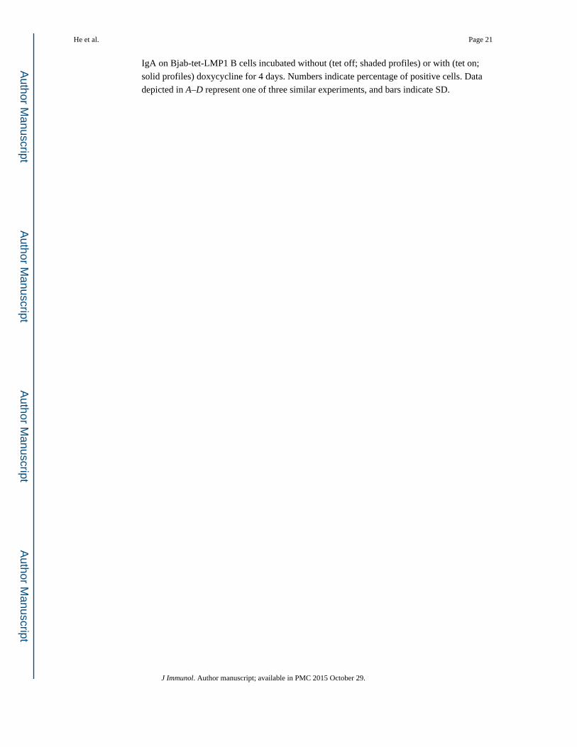
IgA on Bjab-tet-LMP1 B cells incubated without (tet off; shaded profiles) or with (tet on;
solid profiles) doxycycline for 4 days. Numbers indicate percentage of positive cells. Data
depicted in A–D represent one of three similar experiments, and bars indicate SD.
He et al. Page 21
J Immunol. Author manuscript; available in PMC 2015 October 29.
Author M
anuscriptA
uthor Manuscript
Author M
anuscriptA
uthor Manuscript

FIGURE 4. LMP1 up-regulates BAFF and APRIL. A, BAFF, APRIL, LMP1, and actin transcripts and
proteins in EBV− Ramos B cell subclones stably transfected with pRH132 (control 1),
pRH132-pSG5 (control 2), pRH132-LMP1, pRH132-LMP2A, pRH132-EBNA1, pRH132-
EBNA2, or pRH132-pSG5-EBNA-LP expression vectors. B, BAFF, APRIL, LMP1, and
actin transcripts and proteins in Bjab-tet-LMP1 B cells incubated without (d 0) or with (tet
on) doxycycline. C, CD3, CD23, CD19, mBAFF, and BAFF-binding activity on Bjab-tet-
LMP1 B cells incubated without (tet off) or with (tet on) doxycycline for 2 days. Numbers
indicate percentage of positive cells. Data depicted in A—C represent one of three similar
experiments.
He et al. Page 22
J Immunol. Author manuscript; available in PMC 2015 October 29.
Author M
anuscriptA
uthor Manuscript
Author M
anuscriptA
uthor Manuscript
FIGURE 5. LMP1 elicits NF-κB-dependent up-regulation of BAFF and APRIL in B cells. A, DNA
sequence of the BAFF promoter (GenBank accession no. AY129225). +232 indicates the 3′
end of the promoter, a turned arrow indicates the major initiation site (+1), and boxes depict
putative κB motifs. B, Left, Bjab-tet-LMP1 B cells transfected with BAFF-LUC ( ) or
κB(2X)-LUC (■) in the presence or absence of IκBα-pcDNA3.1 were incubated without (tet
off) or with (tet on) doxycycline. Right, wt Bjab B cells were cotransfected with BAFF-LUC
( ) or κB(2)-LUC (■) and wt LMP1, 187-STOP LMP1, 231-STOP LMP1, ΔEL 187–351
He et al. Page 23
J Immunol. Author manuscript; available in PMC 2015 October 29.
Author M
anuscriptA
uthor Manuscript
Author M
anuscriptA
uthor Manuscript
LMP1, or 20, 10, and 2 µg of IκBα-pcDNA3.1. The luciferase activity was measured after 2
days. Data represent one of three similar experiments, and bars indicate SD.
He et al. Page 24
J Immunol. Author manuscript; available in PMC 2015 October 29.
Author M
anuscriptA
uthor Manuscript
Author M
anuscriptA
uthor Manuscript
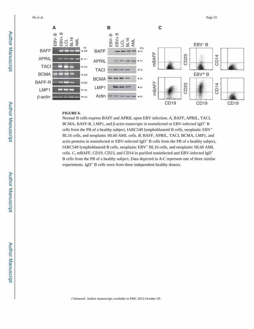
FIGURE 6. Normal B cells express BAFF and APRIL upon EBV infection. A, BAFF, APRIL, TACI,
BCMA, BAFF-R, LMP1, and β-actin transcripts in noninfected or EBV-infected IgD+ B
cells from the PB of a healthy subject, IARC549 lymphoblastoid B cells, neoplastic EBV+
BL16 cells, and neoplastic HL60 AML cells. B, BAFF, APRIL, TACI, BCMA, LMP1, and
actin proteins in noninfected or EBV-infected IgD+ B cells from the PB of a healthy subject,
IARC549 lymphoblastoid B cells, neoplastic EBV+ BL16 cells, and neoplastic HL60 AML
cells. C, mBAFF, CD19, CD23, and CD14 in purified noninfected and EBV-infected IgD+
B cells from the PB of a healthy subject. Data depicted in A-C represent one of three similar
experiments. IgD+ B cells were from three independent healthy donors.
He et al. Page 25
J Immunol. Author manuscript; available in PMC 2015 October 29.
Author M
anuscriptA
uthor Manuscript
Author M
anuscriptA
uthor Manuscript
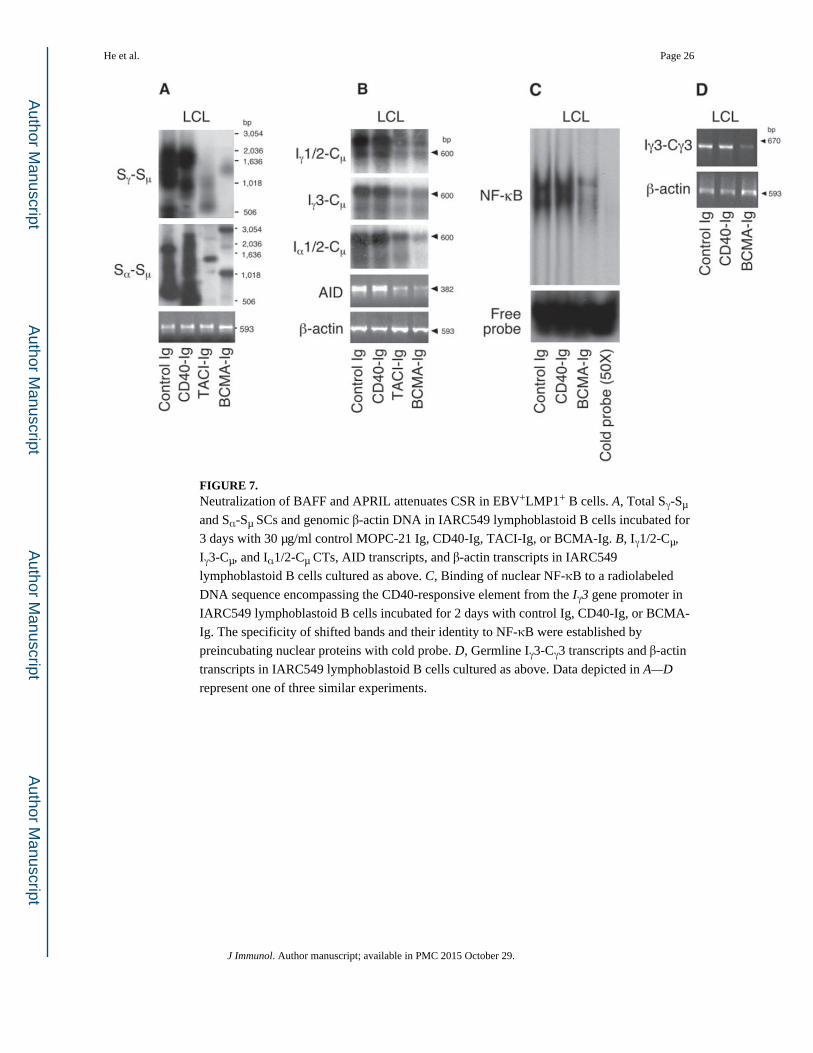
FIGURE 7. Neutralization of BAFF and APRIL attenuates CSR in EBV+LMP1+ B cells. A, Total Sγ-Sµ
and Sα-Sµ SCs and genomic β-actin DNA in IARC549 lymphoblastoid B cells incubated for
3 days with 30 µg/ml control MOPC-21 Ig, CD40-Ig, TACI-Ig, or BCMA-Ig. B, Iγ1/2-Cµ,
Iγ3-Cµ, and Iα1/2-Cµ CTs, AID transcripts, and β-actin transcripts in IARC549
lymphoblastoid B cells cultured as above. C, Binding of nuclear NF-κB to a radiolabeled
DNA sequence encompassing the CD40-responsive element from the Iγ3 gene promoter in
IARC549 lymphoblastoid B cells incubated for 2 days with control Ig, CD40-Ig, or BCMA-
Ig. The specificity of shifted bands and their identity to NF-κB were established by
preincubating nuclear proteins with cold probe. D, Germline Iγ3-Cγ3 transcripts and β-actin
transcripts in IARC549 lymphoblastoid B cells cultured as above. Data depicted in A—D
represent one of three similar experiments.
He et al. Page 26
J Immunol. Author manuscript; available in PMC 2015 October 29.
Author M
anuscriptA
uthor Manuscript
Author M
anuscriptA
uthor Manuscript