chalmers publication librarypublications.lib.chalmers.se/records/fulltext/185383/... ·...
TRANSCRIPT

Chalmers Publication Library
Bone Response to Surface-Modified Titanium Implants: Studies on the Early TissueResponse to Implants with Different Surface Characteristics
This document has been downloaded from Chalmers Publication Library (CPL). It is the author´s
version of a work that was accepted for publication in:
International Journal of Biomaterials (ISSN: 1687-8787)
Citation for the published paper:Larsson Wexell, C. ; Thomsen, P. ; Aronsson, B. (2013) "Bone Response to Surface-Modified Titanium Implants: Studies on the Early Tissue Response to Implants withDifferent Surface Characteristics". International Journal of Biomaterials, vol. 2013 pp.Article ID 412482.
http://dx.doi.org/10.1155/2013/412482
Downloaded from: http://publications.lib.chalmers.se/publication/185383
Notice: Changes introduced as a result of publishing processes such as copy-editing and
formatting may not be reflected in this document. For a definitive version of this work, please refer
to the published source. Please note that access to the published version might require a
subscription.
Chalmers Publication Library (CPL) offers the possibility of retrieving research publications produced at ChalmersUniversity of Technology. It covers all types of publications: articles, dissertations, licentiate theses, masters theses,conference papers, reports etc. Since 2006 it is the official tool for Chalmers official publication statistics. To ensure thatChalmers research results are disseminated as widely as possible, an Open Access Policy has been adopted.The CPL service is administrated and maintained by Chalmers Library.
(article starts on next page)

Hindawi Publishing CorporationInternational Journal of BiomaterialsVolume 2013, Article ID 412482, 10 pageshttp://dx.doi.org/10.1155/2013/412482
Research ArticleBone Response to Surface-Modified Titanium Implants:Studies on the Early Tissue Response to Implants with DifferentSurface Characteristics
C. Larsson Wexell,1,2,3 P. Thomsen,1,3,4 B.-O. Aronsson,5,6 P. Tengvall,3,4,7 M. Rodahl,5
J. Lausmaa,3,4,8 B. Kasemo,5 and L. E. Ericson1
1 Institute of Anatomy and Cell Biology, University of Goteborg, Goteborg, Sweden2Department of Oral and Maxillofacial Surgery, SAS, 501 82 Boras, Sweden3Department of Biomaterials, Sahlgrenska Academy at University of Gothenburg, Goteborg, Sweden4BIOMATCELL VINN Excellence Center of Biomaterials and Cell Therapy, Goteborg, Sweden5Department of Applied Physics, Chalmers University of Technology, Goteborg, Sweden6Nano Bridging Molecules SA, 1196 Gland, Switzerland7Department of Physics and Measurement Technology, Linkoping University, Linkoping, Sweden8Department of Chemistry, Materials and Surfaces, SP Technical Research Institute of Sweden,501 15 Boras, Sweden
Correspondence should be addressed to C. Larsson Wexell; [email protected]
Received 9 April 2013; Revised 7 August 2013; Accepted 7 August 2013
Academic Editor: Bikramjit Basu
Copyright © 2013 C. Larsson Wexell et al. This is an open access article distributed under the Creative Commons AttributionLicense, which permits unrestricted use, distribution, and reproduction in any medium, provided the original work is properlycited.
In a series of experimental studies, the bone formation around systematically modified titanium implants is analyzed. In thepresent study, three different surface modifications were prepared and evaluated. Glow-discharge cleaning and oxidizing resultedin a highly stoichiometric TiO
2surface, while a glow-discharge treatment in nitrogen gas resulted in implants with essentially a
surface of titanium nitride, covered with a very thin titanium oxide. Finally, hydrogen peroxide treatment of implants resulted inan almost stoichiometric TiO
2, rich in hydroxyl groups on the surface. Machined commercially pure titanium implants served
as controls. Scanning Auger Electron Spectroscopy, Scanning Electron Microscopy, and Atomic Force Microscopy revealed nosignificant differences in oxide thickness or surface roughness parameters, but differences in the surface chemical composition andapparent topography were observed. After surface preparation, the implants were inserted in cortical bone of rabbits and evaluatedafter 1, 3, and 6 weeks. Light microscopic evaluation of the tissue response showed that all implants were in contact with bone andhad a large proportion of newly formed bone within the threads after 6 weeks. There were no morphological differences betweenthe four groups. Our study shows that a high degree of bone contact and bone formation can be achieved with titanium implantsof different surface composition and topography.
1. Introduction
This study is part of a multidisciplinary approach wherethe long-term objective is to understand the role of specificsurface properties when bone and marrow are exposed toan implant. The objective and rationale for the approachare presented in an earlier report [1]. In short, the sur-faces of machined, threaded titanium implants are modified
and characterized in different ways and the bone responseand bone-implant interface are investigated in vivo [1–3].Whereas earlier studies addressed the role of roughness andsurface oxide thickness, modified by electrochemical meth-ods, in this study we inquire further into smaller chemicalchanges on implant surfaces. The hypothesis is that surfacechemical composition does influence the tissue responseincluding the bone response to titanium implant. We chose

2 International Journal of Biomaterials
machined titanium implants as the control because this wasthe starting material for three different surface modificationsthat were studied.
In previous studies, we examined the response of bonearound threaded titanium implants with different surfacemodifications (machined, electropolished, machined, andelectropolished with different oxide thicknesses obtained byanodic oxidation).The formation of bone was evaluatedmor-phometrically after 1, 3, 6, 7, 12, and 52 weeks. At the earliertime periods, the formation of bone was less around espe-cially the very smooth electropolished implants. The resultssuggested that the type of surface modification performedmainly influenced the early phase of bone regenerationaround the implants. In the present study, we have examinedthe biological response of cortical bone around titaniumimplants modified with respect to both surface structure andchemical composition by glow-discharge plasma techniquesand by hydrogen peroxide treatment, 1, 3, and 6 weeks afterimplantation.
At first, the implants were electropolished sincemorphol-ogy andmicrostructure of the surface oxides depend stronglyon themicrostructure of the underlyingmetal over which theoxide grows. Machined and mechanically polished surfacesconsist of a plastically deformed, amorphous layer whichmay extend several microns into the bulk of the material.Electropolishing or other etching treatments remove thisamorphous surface layer, resulting in surfaces that havea polycrystalline termination in which the grain structureof the material is visible. The thinnest thermal oxides ontitanium have a very homogenous and essentially featurelessmorphology.
Glow-discharge techniques are widely used for cleaning,sterilizing, and modification of biomaterial surfaces [1, 4–9]. These methods offer great advantages with respect to thepossible range of modifications, as well as to process control(purity).With appropriate plasma parameters, argon plasmasremove all chemical traces at the surface from former treat-ments, such as adsorbed contaminants, impurities, and nativeoxide layers [4]. On such a “cleaned” metallic surface, newsurface layers can thereafter be built up under well-controlledconditions in the vacuum chamber of the plasma equipment.It is further possible to perform in situ characterization of theresulting surface properties.
There are different methods to grow surface oxides onmetals in a controlled way, electrochemically or by glowdischarge plasmas. Other techniques used are, for example,physical vapor deposition (PVD) techniques and thermaloxidation. The anodic oxidation and discharge (DC) plasmaoxidation techniques enhance the diffusion rate of oxygenand titanium atoms andmake it possible to grow thick oxideson metals [4]. With glow-discharge plasmas it is possibleto both prepare and then characterize the obtained surfacein ultrahigh vacuum equipment, which gives a better con-trol over the preparation procedure. Glow-discharge plasmatreatment has early on been proposed as a cleaning andsterilization method for metal instruments [10–12]. Glow-discharge treatment generally results in high surface energy[13–15] immediately after the treatment, due to the removalof material and creation of unsaturated surface bonds. This
in turn makes the surface reactive and prone to rapid oxida-tion and recontamination due to reaction with oxygen andadsorption of airborne contaminants such as hydrocarbons.The samples in this study were exposed to pure oxygen orpure nitrogen prior to air exposure, thereby reducing theamount of contaminants from the air. Substituting the pureoxygen with nitrogen makes it possible to form TiN in acontrolled way. Dion and coworkers [16, 17] have shown exvivo and with a physical vapor deposition (PVD) techniqueon Ti-alloy that compared with silicon, TiN surfaces donot adsorb as much albumin/fibrinogen as silicon surfacesdo. However, the protein layer on TiN (fibril meshwork)entrapped mainly red cells and no leukocytes compared withsilicon adsorbing platelets retaining red blood cells and a largeamount of leukocytes. N-ion implantation is another surfacetreatment technique used with the purpose of increasingthe protection against ion release (corrosion) and eventuallyincreases biocompatibility [18, 19].
The third surface modification method used is basedon previous studies [20–23] on the interaction betweenhydrogen peroxide, that is formed during inflammatoryconditions and titanium. Hydrogen peroxide, H
2O2, is a
strong oxidizer and is also used in cleaning solutions [24]. Invitro studies of the interaction betweenmetallic titanium andmore concentrated H
2O2have shown that titanium peroxide
(TiO2
2-) and titanium superoxide (TiO2-) were formed [21–
23].In the present study, we have used glow-discharge plasma
procedures, developed in our laboratory [4] for preparingclean oxide surfaces and nitride layers and a process ofhydrogen peroxide treatment of titanium implants.
Reported results show that neither of these, relativelydramatic, surfacemodifications lead to significant differencesin early bone healing around the titanium implants in thismodel. In the present study, we have examined the biologicalresponse in cortical bone 1, 3, and 6 weeks after implantationof titanium implants modified by changing the surfacecharacteristics (Table 1).
2. Materials and Methods
2.1. Implant Preparation. Ninety-six threaded implants(03.75mm, length 4.0mm)weremanufactured bymachiningfrom a commercially pure (99.7%) titanium rod. Theywere divided into four different groups. The preparationprocedures are briefly summarized in Table 1 together withthe resulting surface characteristics.
After machining and before any further treatment, allsamples (including controls) were ultrasonically cleaned insuccessive baths of trichloroethylene, acetone, and ethanol ofanalysis grade purity for ∼15 minutes in each.
Two groups (24 + 24 samples) of machined implantswere modified using a glow-discharge procedure, carriedout in different gases and resulting in the formation of twodifferent surface compositions, titanium oxide and titaniumnitride layers, respectively. The glow discharge method hasbeen described in detail elsewhere, and it will only be brieflydescribed here [5]. The implants were treated in a plasma
International Journal of Biomaterials 3
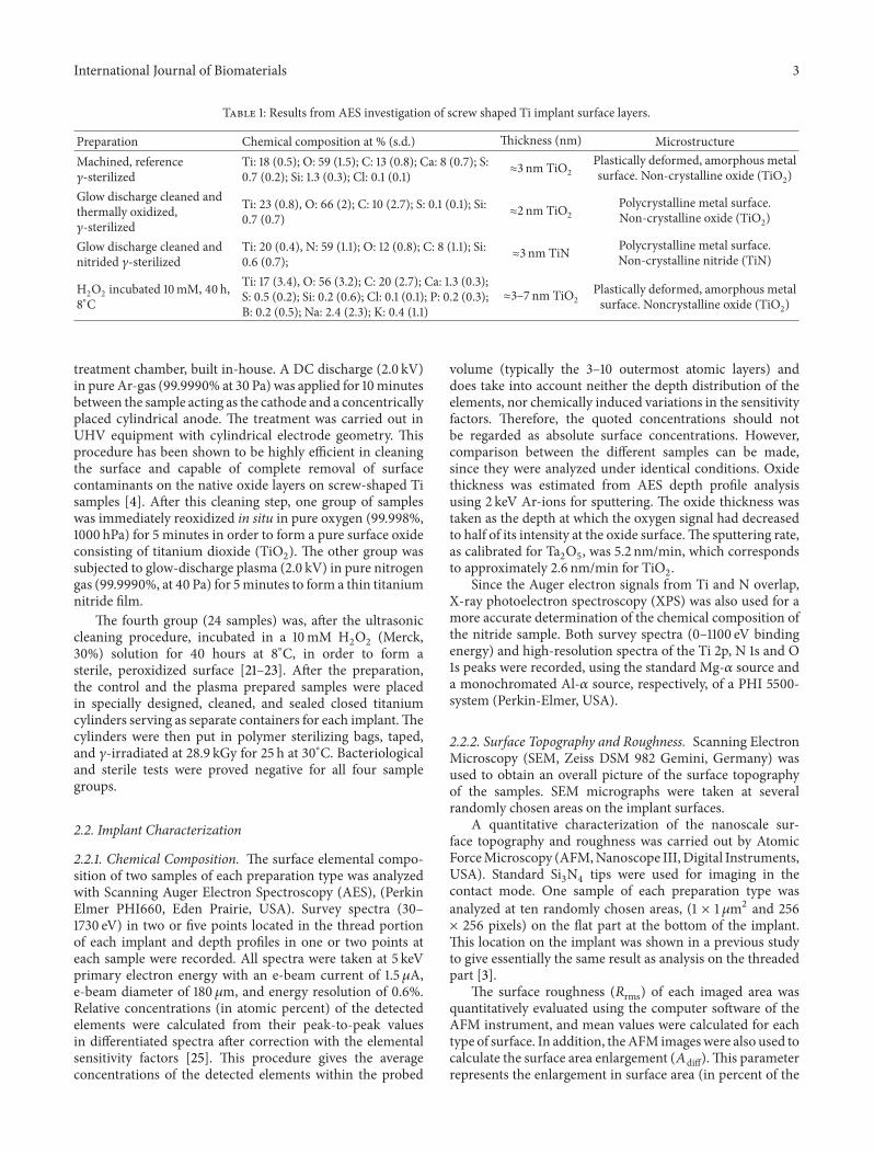
Table 1: Results from AES investigation of screw shaped Ti implant surface layers.
Preparation Chemical composition at % (s.d.) Thickness (nm) MicrostructureMachined, reference𝛾-sterilized
Ti: 18 (0.5); O: 59 (1.5); C: 13 (0.8); Ca: 8 (0.7); S:0.7 (0.2); Si: 1.3 (0.3); Cl: 0.1 (0.1) ≈3 nm TiO2
Plastically deformed, amorphous metalsurface. Non-crystalline oxide (TiO2)
Glow discharge cleaned andthermally oxidized,𝛾-sterilized
Ti: 23 (0.8), O: 66 (2); C: 10 (2.7); S: 0.1 (0.1); Si:0.7 (0.7) ≈2 nm TiO2
Polycrystalline metal surface.Non-crystalline oxide (TiO2)
Glow discharge cleaned andnitrided 𝛾-sterilized
Ti: 20 (0.4), N: 59 (1.1); O: 12 (0.8); C: 8 (1.1); Si:0.6 (0.7); ≈3 nm TiN Polycrystalline metal surface.
Non-crystalline nitride (TiN)
H2O2 incubated 10mM, 40 h,8∘C
Ti: 17 (3.4), O: 56 (3.2); C: 20 (2.7); Ca: 1.3 (0.3);S: 0.5 (0.2); Si: 0.2 (0.6); Cl: 0.1 (0.1); P: 0.2 (0.3);B: 0.2 (0.5); Na: 2.4 (2.3); K: 0.4 (1.1)
≈3–7 nm TiO2Plastically deformed, amorphous metalsurface. Noncrystalline oxide (TiO2)
treatment chamber, built in-house. A DC discharge (2.0 kV)in pure Ar-gas (99.9990% at 30 Pa) was applied for 10minutesbetween the sample acting as the cathode and a concentricallyplaced cylindrical anode. The treatment was carried out inUHV equipment with cylindrical electrode geometry. Thisprocedure has been shown to be highly efficient in cleaningthe surface and capable of complete removal of surfacecontaminants on the native oxide layers on screw-shaped Tisamples [4]. After this cleaning step, one group of sampleswas immediately reoxidized in situ in pure oxygen (99.998%,1000 hPa) for 5 minutes in order to form a pure surface oxideconsisting of titanium dioxide (TiO
2). The other group was
subjected to glow-discharge plasma (2.0 kV) in pure nitrogengas (99.9990%, at 40 Pa) for 5minutes to form a thin titaniumnitride film.
The fourth group (24 samples) was, after the ultrasoniccleaning procedure, incubated in a 10mM H
2O2(Merck,
30%) solution for 40 hours at 8∘C, in order to form asterile, peroxidized surface [21–23]. After the preparation,the control and the plasma prepared samples were placedin specially designed, cleaned, and sealed closed titaniumcylinders serving as separate containers for each implant.Thecylinders were then put in polymer sterilizing bags, taped,and 𝛾-irradiated at 28.9 kGy for 25 h at 30∘C. Bacteriologicaland sterile tests were proved negative for all four samplegroups.
2.2. Implant Characterization
2.2.1. Chemical Composition. The surface elemental compo-sition of two samples of each preparation type was analyzedwith Scanning Auger Electron Spectroscopy (AES), (PerkinElmer PHI660, Eden Prairie, USA). Survey spectra (30–1730 eV) in two or five points located in the thread portionof each implant and depth profiles in one or two points ateach sample were recorded. All spectra were taken at 5 keVprimary electron energy with an e-beam current of 1.5𝜇A,e-beam diameter of 180 𝜇m, and energy resolution of 0.6%.Relative concentrations (in atomic percent) of the detectedelements were calculated from their peak-to-peak valuesin differentiated spectra after correction with the elementalsensitivity factors [25]. This procedure gives the averageconcentrations of the detected elements within the probed
volume (typically the 3–10 outermost atomic layers) anddoes take into account neither the depth distribution of theelements, nor chemically induced variations in the sensitivityfactors. Therefore, the quoted concentrations should notbe regarded as absolute surface concentrations. However,comparison between the different samples can be made,since they were analyzed under identical conditions. Oxidethickness was estimated from AES depth profile analysisusing 2 keV Ar-ions for sputtering. The oxide thickness wastaken as the depth at which the oxygen signal had decreasedto half of its intensity at the oxide surface.The sputtering rate,as calibrated for Ta
2O5, was 5.2 nm/min, which corresponds
to approximately 2.6 nm/min for TiO2.
Since the Auger electron signals from Ti and N overlap,X-ray photoelectron spectroscopy (XPS) was also used for amore accurate determination of the chemical composition ofthe nitride sample. Both survey spectra (0–1100 eV bindingenergy) and high-resolution spectra of the Ti 2p, N 1s and O1s peaks were recorded, using the standard Mg-𝛼 source anda monochromated Al-𝛼 source, respectively, of a PHI 5500-system (Perkin-Elmer, USA).
2.2.2. Surface Topography and Roughness. Scanning ElectronMicroscopy (SEM, Zeiss DSM 982 Gemini, Germany) wasused to obtain an overall picture of the surface topographyof the samples. SEM micrographs were taken at severalrandomly chosen areas on the implant surfaces.
A quantitative characterization of the nanoscale sur-face topography and roughness was carried out by AtomicForceMicroscopy (AFM,Nanoscope III, Digital Instruments,USA). Standard Si
3N4tips were used for imaging in the
contact mode. One sample of each preparation type wasanalyzed at ten randomly chosen areas, (1 × 1𝜇m2 and 256× 256 pixels) on the flat part at the bottom of the implant.This location on the implant was shown in a previous studyto give essentially the same result as analysis on the threadedpart [3].
The surface roughness (𝑅rms) of each imaged area wasquantitatively evaluated using the computer software of theAFM instrument, and mean values were calculated for eachtype of surface. In addition, theAFM imageswere also used tocalculate the surface area enlargement (𝐴diff).This parameterrepresents the enlargement in surface area (in percent of the
4 International Journal of Biomaterials
projected area) caused by surface roughness in the range froma few nm (resolution of the images) up to 1𝜇m (size of theimaged area). The surface area enlargement was estimatedfrom the sum of the area of all triangles formed by threeadjacent pixels divided by the projected image area [26].Additional topographical characterization on the micro-scale (𝑅
𝑎-value) was obtained by an optical profilometer for
three-dimensional measurements, TopScan3D (HeidelbergInstruments GmbH, Germany) [27].
2.3. Animals and Surgery. Twenty-four adult New Zealandwhite female rabbits, weighing 3-4 kg, were used. The exper-iments were approved by the Local Ethics Committee. Theanimals were allowed to run free in a specially designedroom with food and water ad libitum. The procedures forsurgery and implant insertion are described in detail inprevious reports [1, 3]. In summary, a standard procedurefor implant installation was carried out with careful surgicaltechnique, generous irrigation with saline, and low-speeddrill (2000 rpm). After prethreading, two implants wereinserted 10mm apart in each proximal tibial metaphysisin a pre-determined order; thus, each animal received oneimplant of each type.
The animals were sacrificed with an overdose of bar-biturates intravenously and fixed by perfusion with 2.5%glutaraldehyde in 0.05M sodium cacodylate buffer, pH 7.2.The implants and surrounding tissue were removed en bloc,further immersed in glutaraldehyde overnight and thenpostfixed in osmium tetroxide for two hours. After dehydra-tion, the undecalcified specimens were embedded in plasticresin, L R White (The London Resin Co. Ltd., Hampshire,England).
2.4. Morphology and Morphometry. Ground sections of 10–15 𝜇m thickness were prepared [28] and examined, usingLeitzMicrovid equipment connected to a personal computer.Measurements were performed directly in the microscope.The contact ratio between the implant surface and bone tissuewas calculated. Similarly, the proportion of bone tissuewithinthe threads along the implant was calculated. The data aregiven as percentage bone-implant contact (referred to as bonecontact) and percentage of the total area within the threadscontaining mineralized bone (referred to as bone area). Allfive consecutive threads (with number 1 and 2 located in thecortex) were evaluated.Themean of the left and right sides ofthe section and mean values for each thread in the differentgroups were calculated.
2.5. Statistics. The Fisher exact test for paired samples wasused.
3. Results
3.1. Implant Surface Characterization
3.1.1. Surface Composition and Oxide/Nitride Thickness. Therelative concentrations (in atomic%) of the detected elementspresent on the sample surfaces, as measured by AES, are
Table 2: Results from AFM (surface roughness (𝑅rms), surface areaenlargement (𝐴diff)), and optical profilometer (𝑅
𝑎)measurements.
Preparation 𝑅rms (nm),mean (s.d.)
𝐴diff %,mean (s.d.)
𝑅𝑎value,(𝜇m)
Machined, reference,𝛾-sterilized 26.3 (17.6) 13.1 (8.96) 0.6
Glow discharge cleaned andthermally oxidized,𝛾-sterilized
10.2 (4.45) 0.78 (0.49) 0.4
Glow discharge cleaned andnitrided, 𝛾-sterilized 25.2 (11.1) 8.63 (6.88) 0.6
H2O2 treated, 10mM, 40 h,8∘C 25.6 (11.2) 20.5 (5.39) 0.7
presented in Table 1. On all samples the dominant peaks werefrom Ti, O or N/O, and C. All samples, showed carbon levelsof 10–15 at %, which is low in comparison with other studies(typically 30% or more) [29].The shapes of the TiLMV peaksindicated that the oxides on the control, the glow dischargeoxidized, and the H
2O2incubated samples, respectively, were
nearly stoichiometric titanium dioxide. The depth profilesalso showed a similar oxide thickness (2-3 nm) for these threegroups.
Glow-discharge plasma treatment in pure nitrogenresulted in 3 nm thick stoichiometric titanium nitride films,as judged from the depth profiles for the TiLMM + NKVVand TiLMV peaks, respectively [30–33]. The presence oftitanium nitride was also evident in the XPS spectra, whichshowed N 1s and Ti 2p peak positions and shapes consistentwith Ti nitride. The oxygen detected on the nitride sampleswas shown to be present only on the outermost surface. Thebinding energy of the XPS O 1s signal indicated that most ifnot all of the oxygen was bound to carbon, that is, in organicmolecules adsorbed on the very surface from air exposure.However, the formation of a small amount of titanium oxide(or oxygen dissolved in the nitride) cannot be excluded [34–36]. The presence of large amounts of nitrogen in the formof a titanium nitride constitutes a markedly different surfacechemistry compared to the other groups.
Further, when comparing the preparations made in ourearlier studies [1–3], it can be concluded that overall cleanerimplant surfaces were obtained when using well-controlledglow-discharge plasma treatments and 𝛾-irradiation ratherthan wet chemical procedures and autoclaving.
3.1.2. Surface Topography and Roughness. Figures 1 and 2show representative SEM images of plasma treated and H
2O2
incubated samples. The quantitative AFM results are givenin Table 2. The control sample (SEM and AFM image notshown) had the typical topography of machined samplescharacterized by machining grooves (on the scale up to10 𝜇m) which are oriented in the cutting direction. Thesurface roughness parameters, 𝑅rms, as measured by AFMandoptical profilometrywere 26 nmand 0.6𝜇m, respectively,and in agreement with previous work. The seemingly large

International Journal of Biomaterials 5
Figure 1: SEM images showing the surface topography of the samples (a) glow discharge cleaned and thermally oxidized sample. Bar = 5 𝜇m;(b) glow discharge cleaned and nitrided sample. Bar = 5 𝜇m; (c) hydrogen peroxide treated sample. Bar = 2𝜇m.
Figure 2: SEM images of the glow-discharge nitrided sample; (a) the machining grooves are smoothened and grain structures are visible.Bar = 10𝜇m; (b) the surface topography of the individual grains is relatively smooth. Bar = 5 𝜇m.
difference of these results stems from the difference in mea-sured topographical features between the twomethods. AFMgives information on a nanometer scale (𝑧-range: 1 nm–6𝜇m;lateral range: 1 nm–100𝜇m), while the optical technique givesinformation on a micrometer scale (𝑧-range: 6 nm–108𝜇m;lateral range: 1 𝜇m–2mm).
The two plasma treated samples (Figures 1(a), 1(b), and 2)have qualitative similar surface topographies which, however,are distinctly different from the other two sample groups.The surfaces have a relatively smooth appearance in the SEM,with clearly visible grains and grain boundaries. While themajority of the grains have smooth surfaces, some of themshow a corrugated topography on the submicron level, bothof which are characteristics of sputter-etched surfaces. TheAFM analysis revealed a wavy structure with amplitude ofabout 50 nm and a period of 10 nm at the nitrided sample. Incontrast to the oxidized samples which were only reoxidizedin pure oxygen after the argon plasma cleaning step, thenitrided group was subjected to a second plasma treatmentin N2, which presumably leads to the observed differences in
topography. Quantitatively, the two plasma treated surfacesdiffer somewhat: the oxide sample has a lower roughness,while for the nitride it is similar to the control sample. Atthe submicron level, the plasma oxidized samples had a lower𝑅rms value than the other implant surfaces. The 𝑅
𝑎-values
were within the range of 0.4–0.7 𝜇m. The nitrided sample,however, has a larger surface enlargement than the oxidizedsurface (Table 2).
The H2O2treated sample (Figure 1(c)) shows clear traces
from the machining. In addition, an irregular roughness on
the submicron level from etching in H2O2is superimposed
on this topography.The roughness of this sample is similar tothe control samples, but with a larger surface area (Table 2).This topography reflects the etching action of the peroxidetreatment.
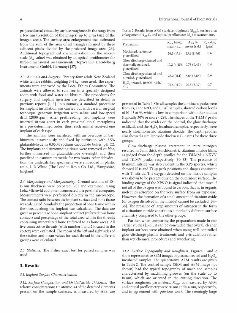
3.2. Bone Morphology and Morphometry. The results of themorphometric evaluation of the relative bone area and bone-implant contact for the entire implant are shown in Figures3(a) and 3(b). No significant differences were observedbetween the mean values of the different groups.
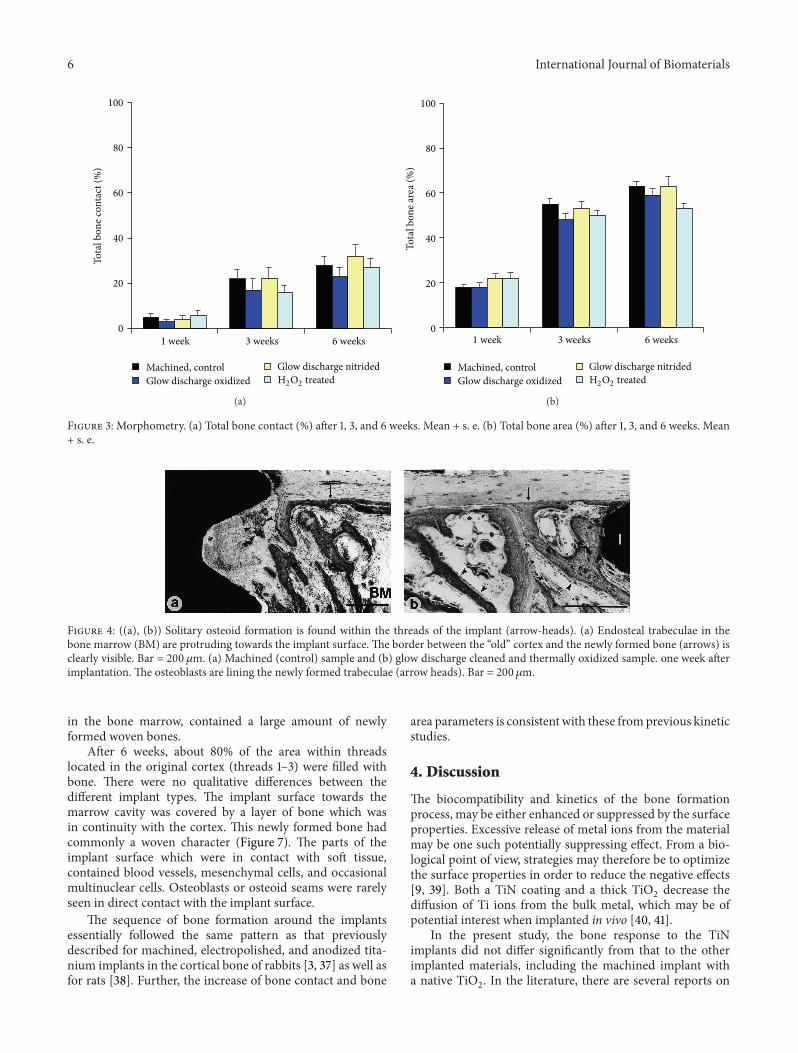
One week after implantation, formation of new bone wasobserved as trabecular woven bone covered with osteoblastseams at the endosteal surface, beginning 1–1.5mm fromthe implant surface. At this time period, solitary osteoidformations were detected within the threads of the implant.In addition, long bone trabeculae reached down from theendosteal surface in the bone marrow towards the implant(Figures 4(a) and 4(b)). No bone formation was seen at thecut edge (drilling hole) of the cortex. A large amount ofwovenbone filled the threads located in the original cortex and thebone was to a large extent in contact with the surface of theimplant (Figure 5). Only about 5% of the implant surface wasin direct contact with mineralized bone after 1 week, andno quantitative differences were found between the differentgroups (Figures 3(a) and 3(b)).
Three weeks after implantation, resorption was clearlyobserved on the cortical surface close to the implant surface(Figures 6(a)–6(c)). All threads, including the threads located
6 International Journal of Biomaterials
0
20
40
60
80
100
1 week 3 weeks 6 weeks
Tota
l bon
e con
tact
(%)
Machined, controlGlow discharge oxidized
Glow discharge nitridedH2O2 treated
(a)
1 week 3 weeks 6 weeks
Tota
l bon
e are
a (%
)
0
20
40
60
80
100
Machined, controlGlow discharge oxidized
Glow discharge nitridedH2O2 treated
(b)
Figure 3: Morphometry. (a) Total bone contact (%) after 1, 3, and 6 weeks. Mean + s. e. (b) Total bone area (%) after 1, 3, and 6 weeks. Mean+ s. e.
Figure 4: ((a), (b)) Solitary osteoid formation is found within the threads of the implant (arrow-heads). (a) Endosteal trabeculae in thebone marrow (BM) are protruding towards the implant surface. The border between the “old” cortex and the newly formed bone (arrows) isclearly visible. Bar = 200 𝜇m. (a) Machined (control) sample and (b) glow discharge cleaned and thermally oxidized sample. one week afterimplantation. The osteoblasts are lining the newly formed trabeculae (arrow heads). Bar = 200𝜇m.
in the bone marrow, contained a large amount of newlyformed woven bones.
After 6 weeks, about 80% of the area within threadslocated in the original cortex (threads 1–3) were filled withbone. There were no qualitative differences between thedifferent implant types. The implant surface towards themarrow cavity was covered by a layer of bone which wasin continuity with the cortex. This newly formed bone hadcommonly a woven character (Figure 7). The parts of theimplant surface which were in contact with soft tissue,contained blood vessels, mesenchymal cells, and occasionalmultinuclear cells. Osteoblasts or osteoid seams were rarelyseen in direct contact with the implant surface.
The sequence of bone formation around the implantsessentially followed the same pattern as that previouslydescribed for machined, electropolished, and anodized tita-nium implants in the cortical bone of rabbits [3, 37] as well asfor rats [38]. Further, the increase of bone contact and bone
area parameters is consistent with these fromprevious kineticstudies.
4. Discussion
The biocompatibility and kinetics of the bone formationprocess, may be either enhanced or suppressed by the surfaceproperties. Excessive release of metal ions from the materialmay be one such potentially suppressing effect. From a bio-logical point of view, strategies may therefore be to optimizethe surface properties in order to reduce the negative effects[9, 39]. Both a TiN coating and a thick TiO
2decrease the
diffusion of Ti ions from the bulk metal, which may be ofpotential interest when implanted in vivo [40, 41].
In the present study, the bone response to the TiNimplants did not differ significantly from that to the otherimplanted materials, including the machined implant witha native TiO
2. In the literature, there are several reports on
International Journal of Biomaterials 7
Figure 5: H2O2treated implant after 1 week. An intense remodeling
activity (arrow-heads) is observed immediately located beneath thecorticomedullary border (arrows). Bar = 200𝜇m.
the biological reactions at TiN surfaces, mainly prepared byPVD and CVD techniques, including the responses of bone[42–44], soft tissues [42, 45] blood [16, 17, 46, 47], platelets[48], human mesenchymal stem cells [49], and osteoblasts[50, 51]. Several of those studies indicate that TiN surfaceshave beneficial or comparable properties in comparison withother currently and frequently used materials.
Another strategy is to chemically modify the titaniumand titanium oxide surfaces by incorporation of, for exam-ple, cations such as lanthanum [52]. At physiological pHthe hydrated TiO
2has a net negative surface charge, thus
attracting cations like calcium, [53]. Chemical treatment ofTiO2powder (anatase) by adsorption of lanthanum cations
resulted in an increased adsorption of albumin and serumproteins in comparison with controls [52]. Furthermore, 2–10 weeks after implantation in rats and rabbits of lanthanumtreated titanium implants, a fibrous encapsulation and lowerpush-out values than controls were recorded. On the otherhand, pretreatment with fluoride ions was shown to increasethe push-out values [54] and bonemorphometric values [55].
These and other observations indicate that the surfacecharge influences the bone tissue response, possibly by influ-encing the types and amount of proteins that are adsorbedto the surface. The interactions between proteins, cells andimplant surfaces may be influenced not only by the chemicalproperties of the surface but also the surface roughnesswhich in turn may influence the wettability (hydrophilicity)which play an important role. [56]. Therefore, optimizationof both surface chemical and topographical properties needto be considered when new materials are designed [57]. Therole of implant surface energy and cleanliness for interfacialevents, molecular adsorption, and cellular adhesion has beenaddressed by several authors [7, 14, 15, 57–62].
In vitro studies on osteoblast-like cells [63, 64] and osteo-clasts have been performed on glow-discharge titanium [65]and titanium alloy plates [66]. Increased platelet adhesion
and activity [66], as well as increased protein adsorption [63,65] are factors that are demonstrated to influence the cellularresponse on surfaces with highwettability. In addition, resultsfrom recent in vivo studies indicate enhanced bone responseon titanium surfaces with higher surface energy [60, 67, 68]under experimental conditions.
The surfaces subjected to the present preparation tech-niques followed by sterilization using 𝛾-irradiation and sub-sequent air exposure had a relatively low amount of surfacecontaminants. Previous studies have shown that machinedand electropolished titanium implants with and withoutthick (180–220 nm) oxides have hydrophilic surfaces (watercontact angles 15–33 degrees) with the highest contact anglesobserved for electropolished and machined surfaces [69]. Inthe latter study, oxide thickness and carbon contaminationhad no clear influence on protein adsorption and activationof blood coagulation [69]. In the present study, the amountof carbon contamination was lower than that detected onour previous samples [1, 3]. However, comparisons betweensamples analyzed at different occasions should be madewith caution. Interestingly, in comparison with the earlierbone morphometry data, the present degree of bone-implantcontact and amount of bone within threads were higher.These observations indicate that surface contamination maybe one of several important factors influencing the biologicalresponse. This has also been observed by Aita et al. [70]who investigated the effect of UV irradiation on the bio-logical response to titanium surfaces. In that study, severalbeneficial effects of the UV treatment on the bone healingwere observed, which the authors ascribed partly to thedecreased carbon contamination levels after treatment. Acomparison between autoclaving and 𝛾-irradiation indicatesthat the latter technique has a major advantage: identicallyprepared and cleaned, but autoclaved, machined titaniumimplants had 34 at % C contamination [3] whereas in thepresent study the 𝛾-irradiated samples had only 13 at % C.The presently used methods of direct current glow dischargeplasma treatment, followed by plasma oxidation or plasmanitriding, and subsequent sterilizationwith 𝛾-irradiationmaytherefore be of interest for controlled preparation, cleaning,and sterilization of medical implants.
Acknowledgments
The financial support from the King Gustaf V 80-year Fund,the Medical Faculty and the Faculty of Odontology, Univer-sity of Gothenburg, the Swedish National Association againstRheumatism, the Hjalmar Svensson Research Foundation,the Loo and Hans Osterman Fund, the Swedish NationalBoard for Technical Development (VINNOVA), and theSwedish Medical Research Council (9495) are gratefullyacknowledged. This work was in part carried out within theNUTEK/NFR-funded Biomaterial Consortium. A financialsupport for the process development was obtained fromNobel Biocare AB, Sweden. C. Larsson Wexell received theAstra Meditec Nordic Grant for research in Oral Implantol-ogy and Biomaterials. They are grateful for the skillful tech-nical assistance by Lena Emanuelsson, AnnKristin Blomgren,

8 International Journal of Biomaterials
Figure 6:Three weeks after implantation.The cortical bone surface facing the implant surface is characterized by marked signs of resorption(large arrow heads) and the occurrence of new bone formation (small arrow heads). The border between the lamellar cortical bone and thenewly formed bone is visible (arrows); (a) Machined (control) sample. Bar = 200 𝜇m; (b) glow discharge cleaned and nitrided sample. Bar =200 𝜇m; (c) glow discharge cleaned and thermally oxidized sample. Bar = 200 𝜇m.
Figure 7: Six weeks after implantation.The implant surface towardsthe marrow cavity was in general in continuity with the corticalbone. Close bone-implant contact was seen for all implant types,and the threads were filled with woven bone. H
2O2treated implant.
Bar = 200𝜇m.
and Anna Johansson. They wish to thank Ann Wennerbergfor the generous help with topographical characterizationusing the optical profilometer (TopScan 3D).
References
[1] C. Larsson, P. Thomsen, J. Lausmaa, M. Rodahl, B. Kasemo,and L. E. Ericson, “Bone response to surface modified titaniumimplants: studies on electropolished implants with differentoxide thicknesses andmorphology,” Biomaterials, vol. 15, no. 13,pp. 1062–1074, 1994.
[2] C. Larsson, L. Emanuelsson, P. Thomsen et al., “Bone responseto surface modified titanium implants. Studies on the tissueresponse after one year to machined and electropolishedimplants with different oxide thicknesses,” Journal of MaterialsScience, vol. 8, no. 12, pp. 721–729, 1997.
[3] C. Larsson, P. Thomsen, B.-O. Aronsson et al., “Bone responseto surface-modified titanium implants: studies on the earlytissue response to machined and electropolished implants withdifferent oxide thicknesses,” Biomaterials, vol. 17, no. 6, pp. 605–616, 1996.
[4] B.-O. Aronsson, Preparation and Chacterization of GlowDischarge Modified Titanium Surfaces, Goteborg University,Goteborg, Sweden, 1995.
[5] B.-O. Aronsson, J. Lausmaa, and B. Kasemo, “Glow dischargeplasma treatment for surface cleaning and modification ofmetallic biomaterials,” Journal of BiomedicalMaterials Research,vol. 35, no. 1, pp. 49–73, 1997.
[6] W. Gombotz and A. Hoffman, “Gas-discharge tecniques forbiomaterial modification,” Critical Reviews in Biocompatibility,vol. 4, pp. 1–42, 1987.
[7] B. Kasemo and J. Lausmaa, “Biomaterial and implant surfaces:on the role of cleanliness, contamination, and preparationprocedures,” Journal of Biomedical Materials Research, vol. 22,no. 2, pp. 145–158, 1988.
[8] D. C. Smith, R. M. Pilliar, J. B. Metson, and N. S. McIn-tyre, “Dental implant materials. II. Preparative procedures andsurface spectroscopic studies,” Journal of Biomedical MaterialsResearch, vol. 25, no. 9, pp. 1069–1084, 1991.
[9] A. Zhecheva, W. Sha, S. Malinov, and A. Long, “Enhancingthe microstructure and properties of titanium alloys throughnitriding and other surface engineering methods,” Surface andCoatings Technology, vol. 200, no. 7, pp. 2192–2207, 2005.
[10] J. W. McGowan and M. J. Malachowski, “Soft x-ray replicationof biological material—x-ray microscopy and microchemicalanalysis of cells,” Annals of the New York Academy of Sciences,vol. 342, pp. 288–303, 1980.

International Journal of Biomaterials 9
[11] M. Moisana, J. Barbeaub, S. Moreauc, J. Pelletierd, M.Tabrizianc, and L. H. Yahiac, “Low-temperature sterilizationusing gas plasmas: a review of the experiments and an analysisof the inactivation mechanisms,” International Journal of Phar-maceutics, vol. 226, no. 1-2, pp. 1–21, 2001.
[12] H. Rauscher, O. Kylian, J. Benedikt, A. von Keudell, and F.Rossi, “Elimination of biological contaminations from surfacesby plasma discharges: chemical sputtering,” ChemPhysChem,vol. 11, no. 7, pp. 1382–1389, 2010.
[13] R. E. Baier and A. E. Meyer, “Implant surface preparation,”TheInternational Journal of Oral &Maxillofacial Implants, vol. 3, no.1, pp. 9–20, 1988.
[14] R. Baier, A. Meyer, and J. Natiella, “Implant surface physicsand chemistry: improvements and impediments to bioadhe-sion,” in Tissue Integration in Oral, Orthopedic & MaxillofacialReconstruction, W. Laney and D. Tolman, Eds., pp. 240–249,Quintessence, Chicago, Ill, USA, 1992.
[15] K. Duske, I. Koban, E. Kindel et al., “Atmospheric plasmaenhances wettability and cell spreading on dental implantmetals,” Journal of Clinical Periodontology, vol. 39, no. 4, pp.400–407, 2012.
[16] I. Dion, C. Baquey, P. Havlik, and J. R. Monties, “A new modelto test platelet adhesion under dynamic conditions. Applicationto the evaluation of a titanium nitride coating,” InternationalJournal of Artificial Organs, vol. 16, no. 7, pp. 545–550, 1993.
[17] I. Dion, X. Roques, N. More et al., “Ex vivo leucocyte adhesionand protein adsorption on TiN,” Biomaterials, vol. 14, no. 9, pp.712–719, 1993.
[18] L. Thair, U. K. Mudali, N. Bhuvaneswaran, K. G. M. Nair, R.Asokamani, and B. Raj, “Nitrogen ion implantation and invitro corrosion behavior of as-cast Ti-6Al-7Nb alloy,” CorrosionScience, vol. 44, no. 11, pp. 2439–2457, 2002.
[19] I. Braceras, J. I. Alava, J. I. Oate et al., “Improved osseointegra-tion in ion implantation-treated dental implants,” Surface andCoatings Technology, vol. 158-159, pp. 28–32, 2002.
[20] P. Tengvall, Titanium-Hydrogen Peroxide Interaction With Ref-erence To Biomaterial Applications, University of Linkoping,Linkoping, Sweden, 1990.
[21] P. Tengvall, H. Elwing, and I. Lundstrom, “Titanium gel madefrom metallic titanium and hydrogen peroxide,” Journal ofColloid And Interface Science, vol. 130, no. 2, pp. 405–413, 1989.
[22] P. Tengvall, H. Elwing, L. Sjoqvist, I. Lundstrom, and L. M.Bjursten, “Interaction between hydrogen peroxide and tita-nium: a possible role in the biocompatibility of titanium,”Biomaterials, vol. 10, no. 2, pp. 118–120, 1989.
[23] P. Tengvall, I. Lundstrom, L. Sjoqvist, H. Elwing, and L.M. Bjursten, “Titanium-hydrogen peroxide interaction: modelstudies of the influence of the inflammatory response ontitanium implants,”Biomaterials, vol. 10, no. 3, pp. 166–175, 1989.
[24] B. Walivaara, In Vitro Studies of Selected Blood Proteins on SolidSurfaces, Linkoping University, Linkoping, Sweden, 1996.
[25] G. Davis, M. Natan, and K. A. Anderson, “Study of titaniumoxides using Auger line shapes,” Applications of Surface Science,vol. 15, no. 1–4, pp. 321–333, 1983.
[26] J. Griffith, D. Grigg, M. Vasile, P. Russell, and E. Fitzgerald,“Scanning probe metrology,” Journal of Vacuum Science Tech-nology A, vol. 10, no. 4, pp. 674–679, 1992.
[27] A.Wennerberg,On a surface Roughness and Implant Incorpora-tion, Goteborg University, Goteborg, Sweden, 1996.
[28] K. Donath and G. Breuner, “A method for the study ofundecalcified bones and teeth with attached soft tissues. The
Sage-Schliff (sawing and grinding) technique,” Journal of OralPathology, vol. 11, no. 4, pp. 318–326, 1982.
[29] J. Lausmaa, “Surface spectroscopic characterization of titaniumimplant materials,” Journal of Electron Spectroscopy and RelatedPhenomena, vol. 81, no. 3, pp. 343–361, 1996.
[30] I. Bertoti, M. Mohai, J. L. Sullivan, and S. O. Saied, “Surfacecharacterisation of plasma-nitrided titanium: an XPS study,”Applied Surface Science, vol. 84, no. 4, pp. 357–371, 1995.
[31] P. Dawson and K. Tzatzov, “Quantitative auger electron analysisof titanium nitrides,” Surface Science, vol. 149, no. 1, pp. 105–118,1985.
[32] J. Lausmaa, T. Rostlund, and H. McKellop, “Surface spectro-scopic study of nitrogen ion-implanted Ti and Ti-6Al-4V wearagainst UHMWPE,” Surface and Interface Analysis, vol. 15, no.5, pp. 328–336, 1990.
[33] E. Rolinski, “Mechanism of high-temperature plasma nitridingof titanium,” Materials Science and Engineering C, vol. 100, pp.193–199, 1988.
[34] H. Tompkins, “Oxidation of titanium nitride in room air and indryO
2,” Journal of Applied Physics, vol. 70, no. 7, pp. 3876–3880,
1991.[35] H. Tompkins, “The initial stages of the oxidation of titanium
nitride,” Journal of Applied Physics, vol. 71, no. 2, pp. 980–983,1992.
[36] M. Vasile, A. Emerson, and F. Baiocchi, “The characterizationof titanium nitride by x-ray photoelectron spectroscopy andRutherford backscattering,” Journal of Vacuum Science Technol-ogy A, vol. 8, no. 1, pp. 99–105, 1990.
[37] L. Sennerby, P. Thomsen, and L. E. Ericson, “Early tissueresponse to titanium implants inserted in rabbit cortical bone.Part I. Light microscopic observations,” Journal of MaterialsScience, vol. 4, no. 3, pp. 240–250, 1993.
[38] R. Branemark, A Biomechanical Study of OsseointegRation. InVivo Measurements in Rat, Rabbit, Dog and Man, GoteborgUniversity, Goteborg, Sweden, 1996.
[39] P. G. Coelho, J. M. Granjeiro, G. E. Romanos et al., “Basicresearch methods and current trends of dental implant sur-faces,” Journal of Biomedical Materials Research B, vol. 88, no.2, pp. 579–596, 2009.
[40] K. Healey and P. Ducheyne, “Themechanism of passive dissolu-tion of titanium in a model physological environment,” Journalof Biomedical Materials Research, vol. 26, no. 3, pp. 319–338,1992.
[41] K. Healy and P. Ducheyne, “Oxidation kinetics of titanium thinfilms in model physiologic environments,” Journal of ColloidAnd Interface Science, vol. 150, no. 2, pp. 404–417, 1992.
[42] Y. Tamura, A. Yokoyama, F. Watari, and T. Kawasaki, “Surfaceproperties and biocompatibility of nitrided titanium for abra-sion resistant implant materials,” Dental Materials Journal, vol.21, no. 4, pp. 355–372, 2002.
[43] A. Scarano, M. Piattelli, G. Vrespa, G. Petrone, G. Iezzi, and A.Piattelli, “Bone healing around titanium and titanium nitride-coated dental implants with three surfaces: an experimentalstudy in rats,” Clinical Implant Dentistry and Related Research,vol. 5, no. 2, pp. 103–111, 2003.
[44] S. Durual, P. Rieder, G. Garavaglia, A. Filieri, M. Cattani-Lorente, S. S. Scherrer et al., “TiNOx coatings on roughenedtitanium and CoCr alloy accelerate early osseointegration ofdental implants in minipigs,” Bone, vol. 52, no. 1, pp. 230–237,2013.

10 International Journal of Biomaterials
[45] M. Therin, A. Meunier, and P. Christel, “A histomorphometriccomparison of the muscular tissue reaction to stainless steel,pure titanium and titanium alloy implant materials,” Journal ofMaterials Science, vol. 2, no. 1, pp. 1–8, 1991.
[46] I. Dion, C. Baquey, B. Candelon, and J. R. Monties, “Hemocom-patibility of titanium nitride,” International Journal of ArtificialOrgans, vol. 15, no. 10, pp. 617–621, 1992.
[47] Y. Yang, S. F. Franzen, and C. L. Olin, “In vivo comparisonof hemocompatibility of materials used in mechanical heartvalves,” Journal of Heart Valve Disease, vol. 5, no. 5, pp. 532–537,1996.
[48] V. Karagkiozaki, S. Logothetidis, N. Kalfagiannis, S. Lousinian,and G. Giannoglou, “Atomic force microscopy probing plateletactivation behavior on titanium nitride nanocoatings forbiomedical applications,”Nanomedicine, vol. 5, no. 1, pp. 64–72,2009.
[49] M. Annunziata, A. Oliva, M. A. Basile et al., “The effectsof titanium nitride-coating on the topographic and biologicalfeatures of TPS implant surfaces,” Journal of Dentistry, vol. 39,no. 11, pp. 720–728, 2011.
[50] R. P. van Hove, P. A. Nolte, C. M. Semeins, and J. Klein-Nulend,“Differences in proliferation, differentiation, and cytokineproduction by bone cells seeded on titanium-nitride andcobalt-chromium-molybdenum surfaces,” Journal of Biomate-rials Applications, vol. 28, no. 2, pp. 278–287, 2013.
[51] P. Rieder, S. Scherrer, A. Filieri, H. W. Wiskott, and S. Durual,“TiNOx coatings increase human primary osteoblasts prolifera-tion independently of the substrate: a short report,” Bio-MedicalMaterials and Engineering, vol. 22, no. 5, pp. 277–281, 2012.
[52] J. Ellingsen and E. Pinholt, “Pretreatment of titanium implantswith lanthanum ions alters the bone reaction,” Journal ofMaterials Science, vol. 6, no. 3, pp. 125–129, 1995.
[53] M. Abe, “Oxides and hydrous oxides of multivalent metals asinorganic ion exchangers,” in Inorganic Ion Exchange Materials,A. Clearfield, Ed., pp. 179–185, CRC Press, Boca Raton, Fla,USA, 1982.
[54] J. Ellingsen, “Pre-treatment of titanium implants with fluorideimproves their retention in bone,” Journal of Materials Science,vol. 6, no. 12, pp. 749–753, 1995.
[55] C. Johansson, A. Wennerberg, A. Holmen, and J.-E. Ellingsen,“Enhanced fixation of bone to fluoride-modified implants,” inProceedings of the 6th World Biomatterials Congress, p. 601,Society for Biomaterials, Kamuela, Hawaii, USA, 2000.
[56] D. Kaelble, Physical Chemistry of Adhesion, Wiley Interscience,New York, NY, USA, 1971.
[57] G. Zhao, Z. Schwartz, M. Wieland et al., “High surface energyenhances cell response to titanium substrate microstructure,”Journal of Biomedical Materials Research A, vol. 74, no. 1, pp.49–58, 2005.
[58] G. Zhao, A. L. Raines, M. Wieland, Z. Schwartz, and B. D.Boyan, “Requirement for both micron- and submicron scalestructure for synergistic responses of osteoblasts to substratesurface energy and topography,” Biomaterials, vol. 28, no. 18, pp.2821–2829, 2007.
[59] K. Navaneetha Pandiyaraj, V. Selvarajan, Y. H. Rhee, H.W. Kim,and M. Pavese, “Effect of dc glow discharge plasma treatmenton PET/TiO
2thin film surfaces for enhancement of bioactivity,”
Colloids and Surfaces B, vol. 79, no. 1, pp. 53–60, 2010.[60] D. K. Pattanayak, S. Yamaguchi, T. Matsushita, and T. Kokubo,
“Effect of heat treatments on apatite-forming ability of NaOH-and HCl-treated titanium metal,” Journal of Materials Science,vol. 22, no. 2, pp. 273–278, 2011.
[61] J. H. Park, R. Olivares-Navarrete, R. E. Baier et al., “Effect ofcleaning and sterilization on titanium implant surface proper-ties and cellular response,” Acta Biomaterialia, vol. 8, no. 5, pp.1966–1975, 2012.
[62] R. A. Gittens, R. Olivares-Navarrete, A. Cheng et al., “The rolesof titanium surface micro/nanotopography and wettability onthe differential response of human osteoblast lineage cells,”ActaBiomaterialia, vol. 9, no. 4, pp. 6268–6277, 2013.
[63] Y. Shibata, M. Hosaka, H. Kawai, and T. Miyazaki, “Glowdischarge plasma treatment of titanium plates enhances adhe-sion of osteoblast-like cells to the plates through the integrin-mediated mechanism,” International Journal of Oral and Max-illofacial Implants, vol. 17, no. 6, pp. 771–777, 2002.
[64] T. Youngblood and J. L. Ong, “Effect of plasma-glow dischargeas a sterilization of titanium surfaces,” Implant Dentistry, vol. 12,no. 1, pp. 54–60, 2003.
[65] H. Kawai, Y. Shibata, and T. Miyazaki, “Glow discharge plasmapretreatment enhances osteoclast differentiation and survivalon titanium plates,” Biomaterials, vol. 25, no. 10, pp. 1805–1811,2004.
[66] E. Czarnowska, J. Morgiel, M. Ossowski, R. Major, A. Sowinska,and T. Wierzchon, “Microstructure and biocompatibility oftitanium oxides produced on nitrided surface layer under glowdischarge conditions,” Journal of Nanoscience and Nanotechnol-ogy, vol. 11, no. 10, pp. 8917–8923, 2011.
[67] G. Giro, N. Tovar, L. Witek et al., “Osseointegration assess-ment of chairside argon-based nonthermal plasma-treated Ca-P coated dental implants,” Journal of Biomedical MaterialsResearch A, vol. 101, no. 1, pp. 98–103, 2013.
[68] F. P. Guastaldi, D. Yoo, C. Marin et al., “Plasma treatmentmaintains surface energy of the implant surface and enhancesosseointegration,” International Journal of Biomaterials, vol.2013, Article ID 354125, 6 pages, 2013.
[69] B. Walivaara, B.-O. Aronsson, M. Rodahl, J. Lausmaa, and P.Tengvall, “Titanium with different oxides: in vitro studies ofprotein adsorption and contact activation,” Biomaterials, vol. 15,no. 10, pp. 827–834, 1994.
[70] H. Aita, N. Hori, M. Takeuchi et al., “The effect of ultravioletfunctionalization of titanium on integration with bone,” Bioma-terials, vol. 30, no. 6, pp. 1015–1025, 2009.

Submit your manuscripts athttp://www.hindawi.com
ScientificaHindawi Publishing Corporationhttp://www.hindawi.com Volume 2013
CorrosionInternational Journal of
Hindawi Publishing Corporationhttp://www.hindawi.com Volume 2013
Hindawi Publishing Corporationhttp://www.hindawi.com Volume 2013
Polymer ScienceInternational Journal of
ISRN Corrosion
Hindawi Publishing Corporationhttp://www.hindawi.com Volume 2013
Hindawi Publishing Corporationhttp://www.hindawi.com Volume 2013
CompositesJournal of
Advances in
Materials Science and EngineeringHindawi Publishing Corporationhttp://www.hindawi.com Volume 2013
International Journal of
BiomaterialsHindawi Publishing Corporationhttp://www.hindawi.com Volume 2013
ISRN Ceramics
Hindawi Publishing Corporationhttp://www.hindawi.com Volume 2013
Hindawi Publishing Corporationhttp://www.hindawi.com
Volume 2013
MaterialsJournal of
NanotechnologyHindawi Publishing Corporationhttp://www.hindawi.com Volume 2013
Journal of
ISRN Materials Science
Hindawi Publishing Corporationhttp://www.hindawi.com Volume 2013
Hindawi Publishing Corporation http://www.hindawi.com Volume 2013Hindawi Publishing Corporation http://www.hindawi.com Volume 2013
The Scientific World Journal
ISRN Nanotechnology
Hindawi Publishing Corporationhttp://www.hindawi.com Volume 2013
NanoparticlesJournal of
Hindawi Publishing Corporationhttp://www.hindawi.com Volume 2013
Smart Materials Research
Hindawi Publishing Corporationhttp://www.hindawi.com Volume 2013
Hindawi Publishing Corporationhttp://www.hindawi.com Volume 2013
MetallurgyJournal of
BioMed Research International
Hindawi Publishing Corporationhttp://www.hindawi.com Volume 2013
ISRN Polymer Science
Hindawi Publishing Corporationhttp://www.hindawi.com Volume 2013
Na
nom
ate
ria
ls
Hindawi Publishing Corporationhttp://www.hindawi.com Volume 2013
Journal ofNanomaterials