change in the muscle tension of the shoulder girdle
TRANSCRIPT
Journal of Sports Science 8 (2020) 47-55 doi: 10.17265/2332-7839/2020.02.003
Change in the Muscle Tension of the Shoulder Girdle
Muscles in Patients with Spinal Pain, Using the Tone
Control® Technique
Umberto Motta1, 2 PT, M.Sc. and Ester da Pos1 PT
1. ASP Pio Albergo Trivulzio, Milano, Italy
2. School of Physiotherapy, University of Milan, Milan, Italy
Abstract: The aim of the study was to verify the efficacy of the Tone-Control method in inducing a reduction in the tension of the muscles of the shoulder girdle, and therefore a normalisation of posture in the segment. The authors analysed the change in posture, which was related to the muscle tension of the pectoralis major muscle and the trapezius muscle, resulting from the administration of a programme of encoded exercises known as the Tone Control method. The study was conducted on 70 patients with postural back pain, aged between 25 and 81 years and with a mean age of 61.9 years, 11 male patients and 59 female patients, who attended group rehabilitation for a minimum of 10 and a maximum of 15 sessions. Acute phase patients, patients on anti-inflammatory pharmacological treatment and patients with hernias or bulging causing thecal sac impingement were excluded from the study. Measurements were taken of the angles of the joints in the scapulohumeral segment during the first and last sessions. The NRS (numeric rating scale) pain scale was administered at the start and end of the cycle of sessions. Conclusions: Patients in the study group experienced improvements in the angle measurements that were proportionally greater than those of the control group, together with a considerable reduction in perceived pain, with an overall improvement in posture and shoulder girdle function. Key words: Tone control, muscle tone change, improvement in muscle contraction, shoulder girdle rehabilitation.
1. Introduction
The purpose of the study was to test the efficacy of
the Tone Control technique.
This technique was developed when, in an attempt
in routine practice to obtain a postural improvement in
and harmonisation of muscle tone within the
individual psychosomatic unit, the application of the
principles of eutonia [1] was combined with
biomechanical articular facilitations [2, 3]. On a
practical level, it was seen that providing a fulcrum
outside the joints considered and simultaneously
increasing the proprioceptive inputs [4] in that
segment produced positive responses. Moreover, if the
perceptive input was provided by an elastic surface,
Corresponding author: Umberto Motta, PT, M.Sc,
Professor at School of Physiotherapy - University of Milan and Ester da Pos, PT, Covatech Pilates Teacher.
the gain in terms of tone and postural readaptation
appeared to be greater than anticipated.
It was therefore decided to encode procedures that
could be versatile enough to be adapted to the
necessary individualisation of treatment to meet the
specific needs of each patient, but that at the same
time were reproducible with uniformity of technique,
making them suitable for verification.
Attempting to adapt muscle tone [5] is one of the
key aspects of a therapist’s work, when dealing with
posture and its alterations. One useful, albeit not
exhaustive definition of posture, is chosen by F.
Scoppa [6]. “Posture can be taken to mean the
position of the body in space and the spatial
relationship between the skeletal segments, the
purpose of which is to maintain balance (antigravity
function), in both static and dynamic conditions, to
which a contribution is made by the
D DAVID PUBLISHING
Change in the Muscle Tension of the Shoulder Girdle Muscles in Patients with Spinal Pain, Using the Tone Control® Technique
48
neurophysiological, biomechanical, psychoemotive
and interpersonal factors related to the evolution of the
species”. Scoppa’s words allow us to highlight certain
aspects that are fundamental to Tone Control, i.e. the
spatiality and the concurrence of a number of factors.
To verify the efficacy of the approach with the
Tone Control treatment, in this study it was therefore
decided to evaluate the changes obtained when
applying the technique to the rehabilitation of the
shoulder girdle, thereby placing attention on the
postural changes relating to different states of muscle
tone in certain muscles in the girdle and in their main
actions.
More specifically, the correlation between the tone
of the anterior thoracic muscles (pectoralis major
muscle) and the anteposed attitude of the shoulder was
chosen together with the correlation between the tone
of the trapezius muscle (upper and middle) + the
levator scapulae muscle and the elevation of the entire
girdle on the frontal plane. Shoulder flexion on the
sagittal plane was chosen to investigate the possibility
of joint movement, in the interests of a practical
approach. As a subjective parameter, the authors also
took into account the subject’s assessment of pain.
The neurophysiological and biomechanical bases
referred to are: proprioceptive stimulation [4], the
stretch reflex and reciprocal inhibition principle [3].
The concept of fixation and facilitation studied by Dr.
Irvin Korr in 1975 was also considered [2, 4].
The afferent discharge of the gamma motor neurons
from the neuromuscular spindle produces a contractile
reflex and blocks articular mobility in a given position.
By controlling the contraction of the intrafusal muscle
fibres by means of gamma motor neuron stimulation,
the central nervous system is able to reset the degree
of muscle length, the tone and the sensitivity of the
spindles to stretching [7]. This mechanism generates a
pre-alert state in the muscle, which is therefore able to
respond even to small changes in lengthening.
Moreover, the greater the gamma stimulation, the
greater the muscle spindles’ sensitivity to stretching
[4]. Muscle stretching with high gamma stimulation
produces a more intense discharge by the muscle
spindles and, consequently, a greater muscle
contraction reflex. In dysfunctional tissue [4], with an
alteration of muscle tone, the muscle spindles
continue to discharge [6]. Tone control exercises aim
to reduce muscle contraction by reducing the aberrant
discharge at the origin of the spindle and, therefore,
muscle tone [8].
The measurements were taken as follows [9]:
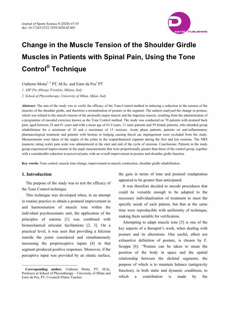
(1) shoulder anteposition: measured in cm from the
frontal plane by putting the patient in a seated position,
back against a wall, by measuring the perpendicular
distance between the acromial process and the wall the
patient is resting against. (Fig. 1.1)
(Fig. 1.1) Measuring shoulder anteposition.
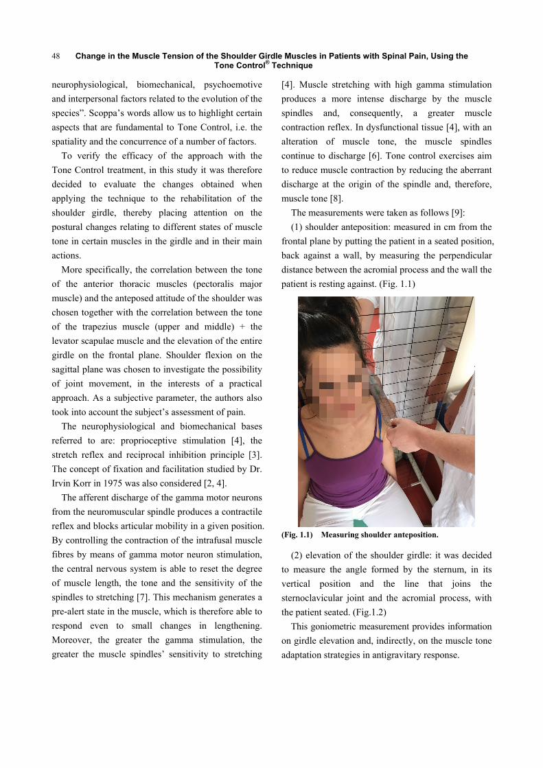
(2) elevation of the shoulder girdle: it was decided
to measure the angle formed by the sternum, in its
vertical position and the line that joins the
sternoclavicular joint and the acromial process, with
the patient seated. (Fig.1.2)
This goniometric measurement provides information
on girdle elevation and, indirectly, on the muscle tone
adaptation strategies in antigravitary response.
Change in the Muscle Tension of the Shoulder Girdle Muscles in Patients with Spinal Pain, Using the Tone Control® Technique
49
(Fig. 1.2) elevation of the shoulder girdle.
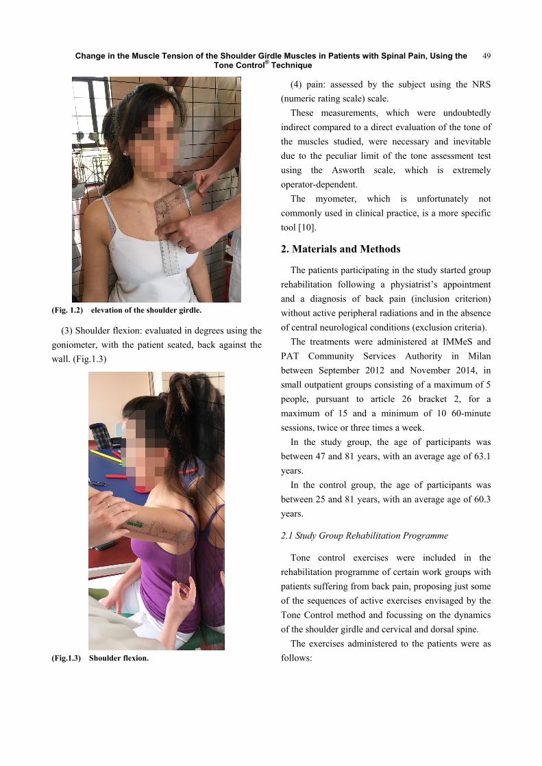
(3) Shoulder flexion: evaluated in degrees using the
goniometer, with the patient seated, back against the
wall. (Fig.1.3)
(Fig.1.3) Shoulder flexion.
(4) pain: assessed by the subject using the NRS
(numeric rating scale) scale.
These measurements, which were undoubtedly
indirect compared to a direct evaluation of the tone of
the muscles studied, were necessary and inevitable
due to the peculiar limit of the tone assessment test
using the Asworth scale, which is extremely
operator-dependent.
The myometer, which is unfortunately not
commonly used in clinical practice, is a more specific
tool [10].
2. Materials and Methods
The patients participating in the study started group
rehabilitation following a physiatrist’s appointment
and a diagnosis of back pain (inclusion criterion)
without active peripheral radiations and in the absence
of central neurological conditions (exclusion criteria).
The treatments were administered at IMMeS and
PAT Community Services Authority in Milan
between September 2012 and November 2014, in
small outpatient groups consisting of a maximum of 5
people, pursuant to article 26 bracket 2, for a
maximum of 15 and a minimum of 10 60-minute
sessions, twice or three times a week.
In the study group, the age of participants was
between 47 and 81 years, with an average age of 63.1
years.
In the control group, the age of participants was
between 25 and 81 years, with an average age of 60.3
years.
2.1 Study Group Rehabilitation Programme
Tone control exercises were included in the
rehabilitation programme of certain work groups with
patients suffering from back pain, proposing just some
of the sequences of active exercises envisaged by the
Tone Control method and focussing on the dynamics
of the shoulder girdle and cervical and dorsal spine.
The exercises administered to the patients were as
follows:
Change in the Muscle Tension of the Shoulder Girdle Muscles in Patients with Spinal Pain, Using the Tone Control® Technique
50
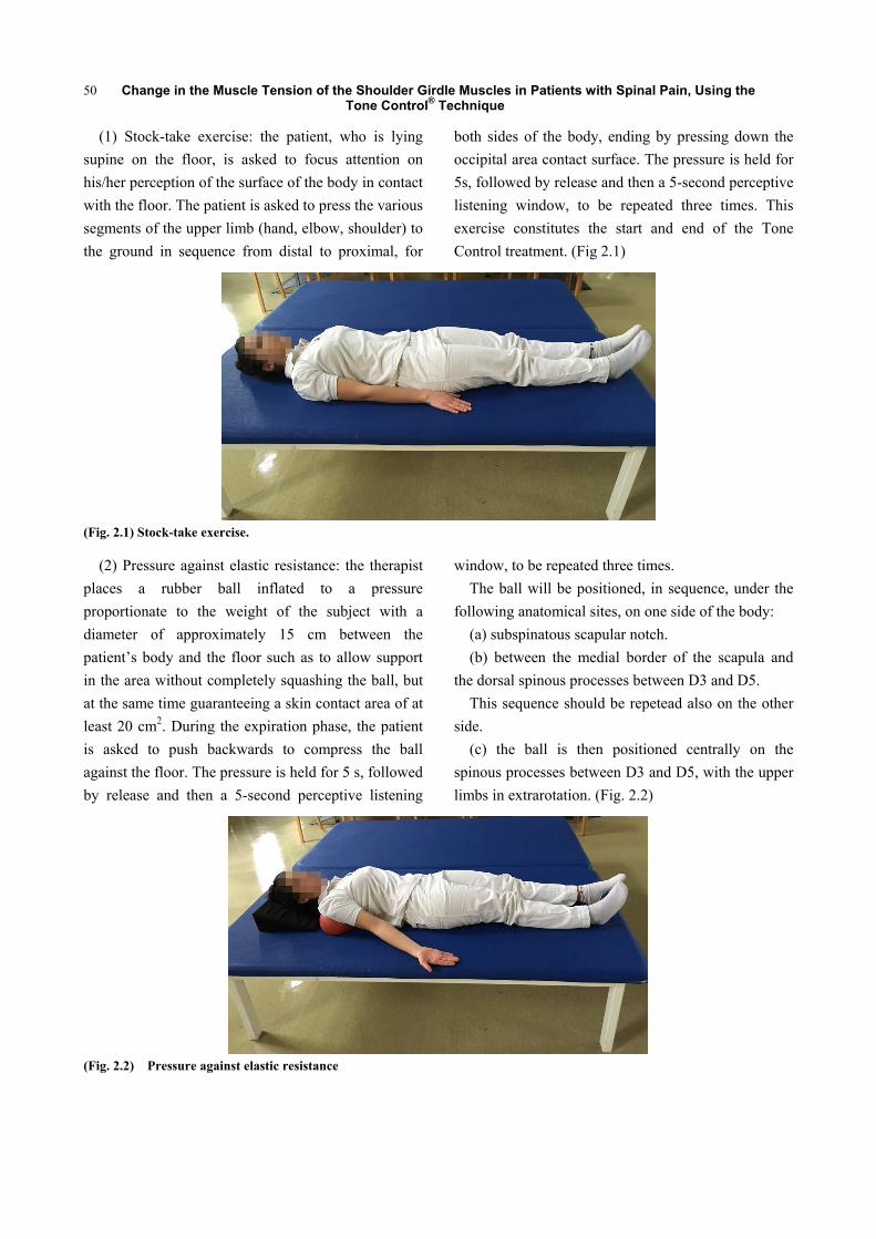
(1) Stock-take exercise: the patient, who is lying
supine on the floor, is asked to focus attention on
his/her perception of the surface of the body in contact
with the floor. The patient is asked to press the various
segments of the upper limb (hand, elbow, shoulder) to
the ground in sequence from distal to proximal, for
both sides of the body, ending by pressing down the
occipital area contact surface. The pressure is held for
5s, followed by release and then a 5-second perceptive
listening window, to be repeated three times. This
exercise constitutes the start and end of the Tone
Control treatment. (Fig 2.1)
(Fig. 2.1) Stock-take exercise.
(2) Pressure against elastic resistance: the therapist
places a rubber ball inflated to a pressure
proportionate to the weight of the subject with a
diameter of approximately 15 cm between the
patient’s body and the floor such as to allow support
in the area without completely squashing the ball, but
at the same time guaranteeing a skin contact area of at
least 20 cm2. During the expiration phase, the patient
is asked to push backwards to compress the ball
against the floor. The pressure is held for 5 s, followed
by release and then a 5-second perceptive listening
window, to be repeated three times.
The ball will be positioned, in sequence, under the
following anatomical sites, on one side of the body:
(a) subspinatous scapular notch.
(b) between the medial border of the scapula and
the dorsal spinous processes between D3 and D5.
This sequence should be repetead also on the other
side.
(c) the ball is then positioned centrally on the
spinous processes between D3 and D5, with the upper
limbs in extrarotation. (Fig. 2.2)
(Fig. 2.2) Pressure against elastic resistance
Change in the Muscle Tension of the Shoulder Girdle Muscles in Patients with Spinal Pain, Using the Tone Control® Technique
51
(d) The ball is placed beneath the external occipital
protuberance. From the occipital contact point, the
patient is asked to perform a twisting motion of the
head with a lateral inclination to reach a point of
contact limited by the temporal mastoid process. From
this position, the patient is asked to perform pressing
motions with the same parameters as previously on
both sides.
(3) The sequence of Tone Control exercises ends by
repeating the stock-take exercise, in order to allow the
patient to perceive any differences between the body
image before and after treatment.
These sequences were included in a programme
involving a sequence of active mobilisation exercises
for the lumbar spine and the lower limbs, to improve
the segmental strength of the abdominal muscles,
quadriceps muscles and of the stabilisers of the pelvis,
the stretching of the posterior chain of the lower limbs
and of proprioceptive stimulation during loading with
both static and dynamic balance exercises.
2.2 Control Group Rehabilitation Programme
The exercises for the mobilisation and proprioceptive
stimulation of the shoulder girdle administered to
patients in the control group were as follows:
(a) Hand-guided passive anterior flexion of the head,
from a supine position on the floor. The position is
held for 5 seconds and repeated three times.
(b) Hand-guided passive twisting motion with
lateral inclination of the head. The position is held for
5 s and repeated three times.
(c) Scapulothoracic sliding, with arms together and
pushed forward during the expiratory phase. The
position is held for 5 s and repeated three times.
(d) Stretching of the muscles of the forearm,
pectoral muscle and of the entire upper limb chain,
holding the positions for 30 s and repeating them
twice.
(e) Active downward pulling of extrarotated upper
limbs, placed on the floor. The position is held for 5 s
and repeated three times.
As for the study group patients, the exercises
described were included in the same sequence of
active mobilisation exercises for the lumbar spine and
the lower limbs, to improve the segmental strength of
the abdominal muscles, quadriceps muscles and
stabilisers of the pelvis, stretching of the posterior
chain of the lower limbs and of proprioceptive
stimulation during loading with both static and
dynamic balance exercises.
During their initial physiatrist’s appointment,
following the prescription of small-group
physiotherapy, the doctor explained the purpose of the
rehabilitation pathway, by informing the patient that
he/she would have to do exercises to improve global
joint movement, spinal and global function, to control
pain and for postural training. The patient was
informed that a study was being conducted on the
efficacy of the Tone Control technique, but was not
informed whether he/she was part of the study group
or control group.
3. Statistical Data Processing
3.1 Pain
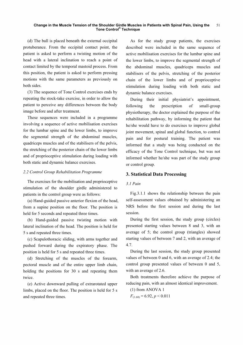
Fig.3.1.1 shows the relationship between the pain
self-assessment values obtained by administering an
NRS before the first session and during the last
session.
During the first session, the study group (circles)
presented starting values between 8 and 3, with an
average of 5; the control group (triangles) showed
starting values of between 7 and 2, with an average of
4.7.
During the last session, the study group presented
values of between 0 and 6, with an average of 2.4; the
control group presented values of between 0 and 5,
with an average of 2.6.
Both treatments therefore achieve the purpose of
reducing pain, with an almost identical improvement.
(1) from ANOVA 1
F(1.68) = 6.92, p < 0.011
Change in the Muscle Tension of the Shoulder Girdle Muscles in Patients with Spinal Pain, Using the Tone Control® Technique
52
Fig. 3.1.1 Subjective evaluation of pain before and after treatment, in the special (circles) and in the standard (triangles) treatment groups.
The two groups show significantly different overall
results, therefore the two treatments give significantly
different results (better in the study group).
(2) from ANOVA 2
Wilks’ lambda = 0.294, partial = 0.71 (Partial age
squared), F(1.68) = 163.5, p < 0.0000.
The main effect is significant: both the treatments
achieve the purpose of significantly altering the pain
assessment, reducing it.
The variance of error of the dependent variable is
the same for both groups.
Levene’s test: F(1.68) = 0.858, p < 0.358 (from
ANOVA 5: on the differences between before and
after).
The difference in the gain between the two groups
is not significant F(1.68) = 1.96, p < 0.166, partial =
0.028 (partial age-squared, ANOVA 5).
Conclusion: both treatments therefore achieve the
purpose of reducing pain, with an almost identical
improvement, although the gain of the special
treatment group is 1.25 times that of the standard
group.
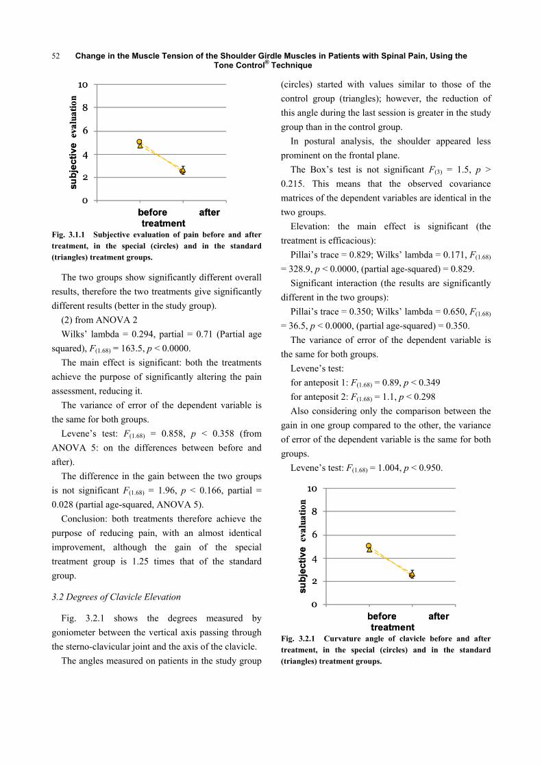
3.2 Degrees of Clavicle Elevation
Fig. 3.2.1 shows the degrees measured by
goniometer between the vertical axis passing through
the sterno-clavicular joint and the axis of the clavicle.
The angles measured on patients in the study group
(circles) started with values similar to those of the
control group (triangles); however, the reduction of
this angle during the last session is greater in the study
group than in the control group.
In postural analysis, the shoulder appeared less
prominent on the frontal plane.
The Box’s test is not significant F(3) = 1.5, p >
0.215. This means that the observed covariance
matrices of the dependent variables are identical in the
two groups.
Elevation: the main effect is significant (the
treatment is efficacious):
Pillai’s trace = 0.829; Wilks’ lambda = 0.171, F(1.68)
= 328.9, p < 0.0000, (partial age-squared) = 0.829.
Significant interaction (the results are significantly
different in the two groups):
Pillai’s trace = 0.350; Wilks’ lambda = 0.650, F(1.68)
= 36.5, p < 0.0000, (partial age-squared) = 0.350.
The variance of error of the dependent variable is
the same for both groups.
Levene’s test:
for anteposit 1: F(1.68) = 0.89, p < 0.349
for anteposit 2: F(1.68) = 1.1, p < 0.298
Also considering only the comparison between the
gain in one group compared to the other, the variance
of error of the dependent variable is the same for both
groups.
Levene’s test: F(1.68) = 1.004, p < 0.950.
Fig. 3.2.1 Curvature angle of clavicle before and after treatment, in the special (circles) and in the standard (triangles) treatment groups.

Change in the Muscle Tension of the Shoulder Girdle Muscles in Patients with Spinal Pain, Using the Tone Control® Technique
53
The effect of the treatment, considered from the point
of view of the result of the comparison between before
and after, is significant in both groups (both groups
receive a benefit, gain, that is significantly different
from superior to zero, when considered separately):
Pillai’s trace = 0.834; Wilks’ lambda = 0.176, F(1.68)
= 341.1, p < 0.0000 (partial age-squared) = 0.834.
Pillai’s trace = 0.485; Wilks’ lambda = 0.515, F(1.68)
= 63.96, p < 0.0000 (partial age-squared) = 0.485.
From ANOVA 5: analysis based on the difference
between two groups:
elevation//F(1.68) = 36.55, p < 0.0000 (partial
age-squared) = 0.350.
The gain of the special group is double (twice) that
of the standard group.
Conclusion: the two groups start on the same level
and whilst the standard treatment group reaches a
good level, the special treatment group reaches a
significantly better level.
3.3 Shoulder Flexion
Fig 3.3.1 shows the modification in shoulder
flexion before and after the treatment. Shoulder
flexion was measured using a goniometer con
orthostatic patients resting against a wall. This is a
significant value because the possibility of raising the
upper limb is representative of the functional
capacities of the shoulder and shoulder girdle. Study
group patients started from lower values (circles) than
those of the control patients (triangles) and at the end
of treatment they reached values that were objectively
lower than those reached in control group patients,
although gain was greater for the study group.
Flexion: the main effect is significant (the treatment
is efficacious):
Pillai’s trace = 0.728; Wilks’ lambda = 0.272, F(1.68)
= 182.3, p < 0.0000, (partial age-squared) = 0.728.
Significant interaction [the results are significantly
different in the two groups]:
Pillai’s trace = 0.063; Wilks’ lambda = 0.937, F(1.68)
= 4.6, p < 0.036, (partial age-squared) = 0.063.
The variance of error of the dependent variable is
the same for both groups.
Levene’s test:
for flexion 1: F(1.68) = 0.016, p < 0.9
for flexion 2: F(1.68) = 3.05, p < 0.085
Also considering only the comparison between the
gain in one group compared to the other, the variance
of error of the dependent variable is the same for both
groups.
Levene’s test: F(1.68) = 0.321, p < 0.573.
The effect of treatment is significant in both groups:
Pillai’s trace = 0.677; Wilks’ lambda = 0.323, F(1.68)
= 142.7, p < 0.0000, (partial age-squared) = 0.677.
Pillai’s trace = 0.454; Wilks’ lambda = 0.546, F(1.68)
= 56.5, p < 0.0000, (partial age-squared) = 0.454.
From ANOVA 5: Comparing the gains between the
two flexion groups: F(1.68) = 32.06, p < 0.0000, (partial
age-squared) = 0.320.
The gain of the special group is 1.38 times that of
the standard group.
Conclusion: the two groups start on different levels
(special treatment group worse level) and whilst they
reach the same level, the improvement is greater in the
special treatment group.
3.4 Shoulder Anteposition
Fig. 3.4.1 shows the shoulder anteposition evaluated
in cm of distance from the considered surface, with
Fig. 3.3.1 Shoulder flexion before and after treatment, in the special (circles) and in the standard (triangles) group.

Change in the Muscle Tension of the Shoulder Girdle Muscles in Patients with Spinal Pain, Using the Tone Control® Technique
54
Fig . 3.4.1 Shoulder anteposition.
the patient orthostatic and the dorsal segment resting
against a wall. In the postural analysis, this value was
related directly with the anterior closure of the
shoulder girdle and the scapula’s outward sliding on
the dorsal contact surface.
The values for the patients in the study group
started off worse (circles) than the values of the
control group (triangles), but by the last session, a
considerable improvement had been seen with an
exceeding of the values obtained—again in the last
session—by the control group.
Main effect significant (the treatment is
efficacious):
Pillai’s trace = 0.645; Wilks’ lambda = 0.355, F(1.68)
= 123.5, p < 0.0000, (partial age-squared) = 0.645.
Significant interaction [the results are significantly
different in the two groups]:
Pillai’s trace = 0.328, Wilks’ lambda = 0.672, F(1.68)
= 33.1, p < 0.0000, (partial age-squared) = 0.328.
The variance of error of the dependent variable is
the same for both groups.
Levene’s test:
for shoulder 1: F(1.68) = 4.8, p < 0.031
for shoulder 2: F(1.68) = 6.6, p < 0.012
The effect is significant in both groups:
Pillai’s trace = 0.709, Wilks’ lambda = 0.291, F(1.68)
= 165.98, p < 0.0000, (partial age-squared) = 0.709.
Pillai’s trace = 0.156, Wilks’ lambda = 0.844, F(1.68)
= 12.55, p < 0.001, (partial age-squared) = 0.156.
As regards the comparison between the two gains,
the variance of error of the dependent variable is the
same for both groups.
Levene’s test: F(1.68) = 2.123, p < 0.150
The difference in the gain between the two groups
is significant.
F(1.68) = 33.14, p < 0.0000, (partial age-squared) =
0.328.
The entity of the difference is considerable: the gain
of the special group is 3.15 that of the standard group.
The repeated measurements on the results of the
ANOVA 5 differences show the interaction between
the two groups to be significant:
F(3.204) = 26.1, p < 0.0000, partial age-squared =
0.277.
Levene (OK) for all four measurements/gains:
F(1.68) = 0.858, p < 0.358
F(1.68) = 0.004, p < 0.950
F(1.68) = 0.321, p < 0.573
F(1.68) = 0.858, p < 0.358
The variance of error of the dependent variable is
the same for both groups.
Univariate test: ANOVA5
Interaction of measurements for each group (gain ×
groups) is significant:
F(3.204) = 24.15, p < 0.0000, (partial age-squared) =
0.262.
1 pain: F(1.68) = 1.961, p < 0.166, (partial
age-squared) = 0.028
2 antep: F(1.68) = 36.550, p < 0.000, (partial
age-squared) = 0.350
3 flex: F(1.68) = 32.065, p < 0.000, (partial age
squared) = 0.320
4 shoulder: F(1.68) = 33.140, p < 0.000, (partial
age-squared) = 0.328
The differences in gain are significant for the three
“objective” values.
4. The Results
Overall, the results described show a significant
improvement in the parameters chosen at the start of
the study and make it possible to establish a
Change in the Muscle Tension of the Shoulder Girdle Muscles in Patients with Spinal Pain, Using the Tone Control® Technique
55
relationship with an effective postural improvement in
the patients treated. Indeed, the general situation is
characterised by an adaptation of patients’ posture,
with a reduction in shoulder girdle elevation, reduction
in shoulder anteposition and an improvement in upper
limb function in the physiological flexion motion.
It can therefore be concluded that the Tone Control
method is as efficacious as conventional active
mobilisation, whereas it was seen to be more
efficacious in the postural improvement due to a more
efficacious normalisation of muscle tone in the
segments examined.
The study conducted made it possible to obtain the
first data regarding the Tone-control option in the
group rehabilitation setting, requiring completely
active patient participation. It would be interesting to
compare the results that can be obtained with patients
of different types, such as, for example, young athletes,
in order to obtain more useful information on patient
compliance.
It would also be very interesting to further test the
method in an individual rehabilitation situation, so as
to also exploit operator-dependent stimulation
techniques, which require direct contact between the
therapist and the patient and make it possible to
combine active work with the listening of a passive
movement induced by the therapist.
References
[1] Gerda, A. 1991. Eutonia, Paidos.
[2] Korr, I. M. 1970. “The Segmental Nervuos System as
Mediator and Organizer of Disease Process.” In The
Physiological Basis of Osteopathic Medicine.
[3] Kandel, S. J. 2003. “Principi di neuroscienze.”
Ambrosiana 36: 702-12.
[4] Korr, I. M. 1975. “Proprioceptors and Somatic
Dysfunction.” J AM Osteopath Assoc 74: 638-50.
[5] 1958. “Physiopathology & Clinical Manifestations of
Skeletal Muscle Tone.” Arch Phys Ther (Leipz) 10 (4):
276-86.
[6] Posturologia, S. F., and Corporeo, S. 2001. “Attualità in
Terapia Manuale e Riabilitazione.” 4: 5-16.
[7] Radovanovic, D., Peikert, K., Lindstrom, M. and
Domellof, F. P. 2015. “Sympathetic Innervation of
Human Muscle Spindles.” J. Anat. 226: 542-8.
[8] 1986. “Controversial Aspects of Skeletal Muscle Tone.”
Hník P.Biomed Biochim Acta 45 (1-2): S139-43.
[9] 1962. “On the Problem of the Technic of Measurement of
Skeletal Muscle Tone.” PANFILOV BK, Klin Med (Mosk)
40 65-9.
[10] Ianieri, G., Saggini, R., Marvulli, R., Tondi, G., Aprile,
A., Ranieri, M., Altini, S., Goffredo, L., Megna, M., and
Megna, G. 2008. “Tono, elasticità e stiffness: valutazione
miometrica delle proprietà muscolari.” Eur. Med. Phys.
44(Suppl. 1 to No. 3).