chapter 1. general introduction - universiteit utrecht · chapter 1. general introduction ... 11...
TRANSCRIPT

7
Chapter 1. General introduction
1.1 The membrane is an omnipresent, important and highly complex biological structure
All living cells, from the unicellular organisms to the cells in our own body, are surrounded by membranes. The
cytoplasmic membrane determines the boundaries of the cell, defining in and out and preventing the vital cell
ingredients to dissipate in the surrounding. In addition to this membrane, a number of membranes are present within
the eukaryotic cells, determining the boundaries of cell organelles [1].
The established model for the structure of the biological membrane describes it as a lipid bilayer with which
proteins are associated in a number of different ways. Proteins can be either adsorbed on the surface of the membrane
(peripheral proteins), or could traverse the lipid bilayer (integral proteins). Some of the membrane proteins could be
modified with acyl chain, which anchors the protein to the membrane (for representation of some of the types of
membrane proteins see figure 1.) The lipid bilayer hypothesis was proposed in 1925, when Gorter and Grendel
performed their fundamental experiment, in which they determined the limiting area per molecule for lipid molecules,
extracted from red blood cells [2]. Comparing the obtained limiting area, multiplied with the number of lipid
molecules per cell, with the available surface area of the erythrocyte, they found a 2:1 ratio and therefore postulated
that the membrane consist of two layers of lipid molecules. Later on it was argued that this result is achieved through
a fortunate compensation of two errors, acting in opposite directions, but nevertheless, the correct concept of the
biological membrane as a lipid bilayer was born.
Figure 1. Schematic representation of the organization of the biological membrane. For simplicity, some classes of
lipids (glycomodified lipids) and proteins are not presented.

8
However, the membrane is much more than a lipid bilayer which defines the cell boundaries and provides
compartmentization within the cell. A number of vital cell processes are performed by the membrane proteins. They
are encoded by ~ ¼ of the open reading frames and are complex molecules, sometimes assembling in multimolecular
protein complexes. Membrane proteins serve as an active interface between the cell and the environment, providing
mass transport between the cell and surrounding, signal transduction and specific recognition. Since these functions
are crucial for the cell survival, the life of the cell depends on the proper functioning of the membrane proteins.
The importance of the membrane as a vital part of the cell attracted the attention of researchers and the biological
membrane became a subject of rather extensive scientific investigations. As a result of this, nowadays it is established
that the membrane is a highly complex and sophisticated structure. The lipid bilayer comprises a multitude of lipid
species and serves as a matrix, providing an appropriate environment for the wide variety of membrane proteins
(Figure 1). Moreover, lipids and proteins are not homogeneously distributed throughout the membrane and could form
assemblies with a specific composition, such as lipid domains, and this further increases the level of complexity of the
membrane. In the next sections we will provide a brief description of the lipids and proteins, their functions and the
structures they form within the membrane.
1.2 Lipids. More than 500 lipid species are identified in biological membranes in different cells and organisms, and
the composition of specialized membranes within the cells varies significantly [3]. Each lipid species possesses certain
physical and chemical properties, and consequently, its presence in the membrane regulates in a certain manner the
properties of the membrane. Some of the common membrane lipids in eukaryotic organisms, such as
phosphatidylcholine, sphingomyelin and cholesterol, are presented in figure 2.
Figure 2. Structure of some of the membrane lipids.

9
One important feature of a lipid molecule is its amphipatic character, meaning that it has both a hydrophobic part,
containing one or more acyl chains, and a hydrophilic part - a polar head group. This dual character is crucial for the
behaviour of the lipid molecules in an aqueous environment and determines their self-assembly in different structures,
including a lipid bilayer. Importantly, the ratio between the sizes of the hydrophobic and hydrophilic parts is crucial in
determining whether a certain lipid forms a bilayer, when dispersed in an aqueous environment (Figure 3) [1]. If this
ratio is close to 1, a stable bilayer is formed.
Figure 3. The sizes of the hydrophobic and hydrophilic parts of the lipid molecule are important in determining the
structures, which they form in aqueous environment.
The predominant state in biological membranes is the so-called liquid crystalline state (Lα) (figure 4). In this state
the acyl chains of the lipid molecules are disordered, the bilayer has fluid-like behaviour and molecules within the
bilayer are highly mobile (thus the other term for this phase – “liquid disordered, ld”). When the temperature is below
a certain value, the lipid bilayer is in a gel state - Lβ. In this state the acyl chains in the lipid molecules are straightened
and the molecule occupies a minimal area. The lateral mobility of the molecules within this highly ordered state is
strongly suppressed. The state of the bilayer depends on the length and degree of unsaturation of the acyl chains of the
lipid molecules. Generally, the longer or the more saturated the acyl chains are, the higher is the respective transition
temperature (the temperature, at which the bilayer changes its state from gel to liquid-crystalline). The state of the
bilayer is crucially important for the overall physical properties of the bilayer – fluidity and elasticity of the bilayer
itself, as well as for the mobility and functionality of the membrane incorporated molecules.
The acyl chains – the hydrophobic part of the lipid molecule - can be connected through a glycerol (glycerolipids)
or sphingosine (sphingolipids) backbone to a hydrophilic head group, which also varies in its chemical composition.
The polar head group of the lipid molecule varies in charge and size and these characteristic features of the lipid head
group are important in certain membrane processes. One abundant class of glycerolipids in animal cell membranes is
phosphatidylcholine (PC). PC lipids have two acyl chains and are so-called bilayer-forming lipids, because they
spontaneously self-assemble in an aqueous environment into bilayers. Another important feature of the PC lipids is
their zwitterionic character. The choline group is positively charged, which compensates the negative charge of the
phosphate. As a result the PC molecule bears zero net charge.
Another important group of lipids, encountered in the plasma membrane of many mammalian cells, are
sphingolipids. A typical representative of sphingolipids is sphingomyelin (SM) which has a same polar head group as
PC glycerolipids (figure 2). Despite having the same head group, these two groups of lipid species differ significantly

10
in their properties [4]. Sphingolipids are characterized by long and largely saturated acyl chains. Due to this acyl chain
composition they have a relatively high transition temperature. Perhaps even more important from a biological point
of view is the higher affinity of sphingomyelin for sterols - another major class of membrane lipids.
Figure 4. Some of the states of the lipid bilayer, encountered in biological membranes.
A representative of the sterols is cholesterol, which is a non-charged lipid molecule of crucial importance in
eukaryotes and which in some animal cells accounts for up to 50 % of the lipid content of the plasma membrane.
Cholesterol has a small hydrophilic head group, consisting of a hydroxyl group, and a hydrophobic part, which
contains a rather rigid structure of four fused rings. The precise mechanisms of the interaction between cholesterol and
phospholipids are still under intensive investigations and a point of debates. However, it is generally accepted that the

11
ring structure of cholesterol interacts with the acyl chains of surrounding phospholipids, optimizing the hydrophobic
interactions. A strong interaction between the hydroxyl group of cholesterol and the PC polar head group was also
proposed. Cholesterol also interacts strongly with glycolipids. The demonstrated strong affinity between cholesterol
and sphingolipids was explained by a proposed formation of a hydrogen bond between the hydroxyl group of the
cholesterol and the sphingosine backbone of sphingomyelin.
Due to these specific interactions, cholesterol significantly modifies the physical properties of the lipid bilayer.
When present in a membrane above a certain threshold level, and when saturated lipids are present, cholesterol can
induce the formation of a lipid bilayer in a specific state, called the liquid ordered (lo) state. In this state the bilayer is
characterized by a high lateral mobility of its components, as in the case of the liquid disordered state, but the ordering
of the acyl chains is as in the gel phase bilayers. In the lo phase the physical properties of the bilayer are significantly
different from the properties of both Lα and Lβ phases. For instance, the thickness of the bilayer is modified in the
presence of cholesterol, and quite significantly from a biological point of view, the bending and compression moduli
of the bilayer are substantially increased [5].
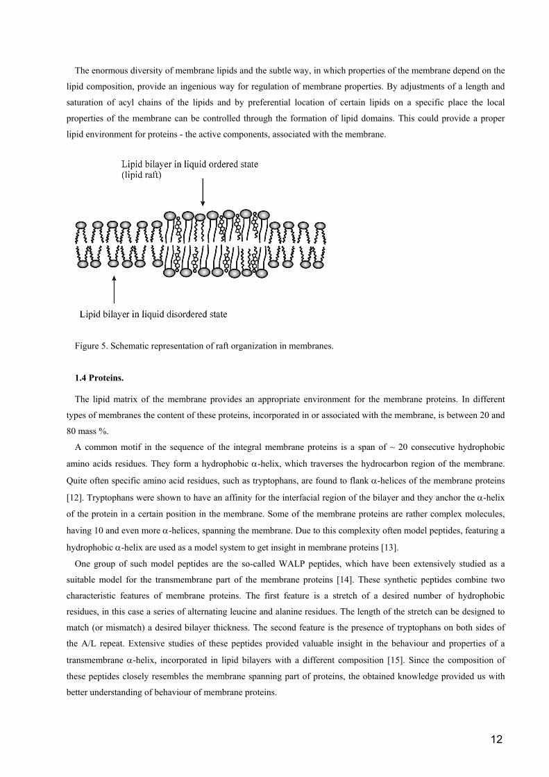
1.3 Phase separation in membranes and lipid bilayers
The complexity of the membrane is determined not only by the huge diversity and complexity of its components,
but also by the fact that they are not uniformly distributed. This asymmetry is both between leaflets, causing
enrichment of each leaflet in certain lipid species, and lateral, leading to formation of domains with defined lipid
composition within the membrane. The existence of such domains was postulated after the discovery that lipid
fractions, enriched in SM and cholesterol, can be extracted from biological membranes upon treatment with the
surfactant Triton X-100 at low temperatures (~5o C). Supposedly these domains are involved in important cell
processes, such as signaling and protein sorting [6]. The isolated membrane fractions are enriched in certain proteins,
while depleted in others. A necessity of certain proteins to be present in such domains (also known as rafts) in order to
function properly was also proposed [7]. The phase separation, leading to domain formation, is a complex process,
influenced both by the head group and the order of the acyl chains of the lipid. A crucial role plays the already
described effect of cholesterol on properties of saturated lipids, leading to the formation of a liquid ordered phase
(figure 5).
The proposed important role of the lipid rafts generated a great interest, and many studies were concentrated on
characterizing their composition, sizes and physical properties [8]. Since the possibilities for studying the processes of
domain formation and the properties of the formed domains in real membranes are rather limited, model systems were
predominantly studied. Monolayers and bilayers with a lipid composition, resembling the lipid composition of
biological membranes, were extensively investigated. In these systems a process of spontaneous domain formation is
visualized. Using different techniques, such as fluorescence microscopy and Atomic Force Microscopy, insight in the
organization and sizes of lipid domains within model membranes was obtained. Numerous studies demonstrated the
subtle manner in which the precise chemistry of lipids influences the process of phase separation within the bilayers
[9]. Quite significant is the observation of detergent resistant domains in model bilayers [10]. Protein/lipid interactions
also can be studied and ordering of model transmembrane peptides within lipid bilayers was demonstrated [11]. This
suggests that investigating the behaviour of lipid bilayers with different composition and the effect of incorporated
model peptides on the bilayer will give us insight in the organization of a biological membrane.
12
The enormous diversity of membrane lipids and the subtle way, in which properties of the membrane depend on the
lipid composition, provide an ingenious way for regulation of membrane properties. By adjustments of a length and
saturation of acyl chains of the lipids and by preferential location of certain lipids on a specific place the local
properties of the membrane can be controlled through the formation of lipid domains. This could provide a proper
lipid environment for proteins - the active components, associated with the membrane.
Figure 5. Schematic representation of raft organization in membranes.
1.4 Proteins.
The lipid matrix of the membrane provides an appropriate environment for the membrane proteins. In different
types of membranes the content of these proteins, incorporated in or associated with the membrane, is between 20 and
80 mass %.
A common motif in the sequence of the integral membrane proteins is a span of ~ 20 consecutive hydrophobic
amino acids residues. They form a hydrophobic α-helix, which traverses the hydrocarbon region of the membrane.
Quite often specific amino acid residues, such as tryptophans, are found to flank α-helices of the membrane proteins
[12]. Tryptophans were shown to have an affinity for the interfacial region of the bilayer and they anchor the α-helix
of the protein in a certain position in the membrane. Some of the membrane proteins are rather complex molecules,
having 10 and even more α-helices, spanning the membrane. Due to this complexity often model peptides, featuring a
hydrophobic α-helix are used as a model system to get insight in membrane proteins [13].
One group of such model peptides are the so-called WALP peptides, which have been extensively studied as a
suitable model for the transmembrane part of the membrane proteins [14]. These synthetic peptides combine two
characteristic features of membrane proteins. The first feature is a stretch of a desired number of hydrophobic
residues, in this case a series of alternating leucine and alanine residues. The length of the stretch can be designed to
match (or mismatch) a desired bilayer thickness. The second feature is the presence of tryptophans on both sides of
the A/L repeat. Extensive studies of these peptides provided valuable insight in the behaviour and properties of a
transmembrane α-helix, incorporated in lipid bilayers with a different composition [15]. Since the composition of
these peptides closely resembles the membrane spanning part of proteins, the obtained knowledge provided us with
better understanding of behaviour of membrane proteins.

13
1.5 Lipid – Protein interactions
Membrane proteins are amazing molecular machines. Under normal circumstances they perform their functions
with high efficiency. The complex architecture of these proteins determines their functionality. However, the
membrane lipids are not only the passive matrix, which surrounds the proteins. The process of protein insertion and
assembly within the membrane, as well as proper functioning of the proteins is modulated by the membrane lipids. In
these processes both protein-protein (P/P) and lipid-protein (L/P) interactions are operative. The dependence of
protein targeting and sorting on P/P and P/L interactions was also recognized [16]. The membrane/peptide interactions
of some extracellular peptides, such as prion proteins and antimicrobial peptides are also dependent on P/L
interactions between these molecules and the target membrane. In each of these processes a number of interactions are
operative which differ in nature. For instance, hydrophobic interactions are dominant in the process of insertion of
transmembrane proteins [17] and electrostatic interactions mediate the recognition process of antimicrobial peptides
with the target membranes [18].
This dependence of the protein localization, conformation and functionality on the L/P and P/P interactions is quite
significant and knowledge in this area will substantially increase our understanding on the behaviour of membrane
proteins and of the processes, performed by them. Some progress has been made in characterizing these interactions.
The preferential location of each amino acid in a certain position with respect to the bilayer has been described. For
instance, hydrophobic residues with a typical representative leucine prefer the hydrophobic core of the bilayer, while
tryptophans prefer the interfacial region of the bilayer and charged residues such as lysine are more likely to be found
in the parts of the protein, residing in the aqueous environment [19].
However, still many obscure points remain and on a molecular level our understanding is still limited. Nonetheless,
information for the interaction between single protein and lipid molecules within a bilayer will be needed in order to
understand in detail the way in which membrane proteins insert, assemble and perform their functions. The problem
with the complexity of the proteins is also encountered here and this suggests that model peptides, such as the already
described WALP peptides would be a convenient system to study P/L interactions in more detail.
1.6 Membrane as a target for antimicrobial peptides.
The cell membrane is important not only as a structure, defining the boundary of the cell and performing vital cell
functions through the incorporated proteins. It is well established that the membrane also is a target for certain
molecules, which attack the cell. From a practical point of view rather important is the group of substances, which
selectively attack the bacterial membrane and provide us with weapons in the war against bacteria. This group of
bactericides consists of small proteinaceous molecules, commonly referred to as antimicrobial peptides, which are
synthesized by different organisms as a weapon against target cells. These molecules target the bacterial membrane
and through different mechanisms kill the bacteria. They vary in their precise chemical structure, but the large part of
them are ~ 15 - 45 amino acid residues long peptides [20]. They are amphipatic molecules, quite often they have a
non-defined secondary structure in an aqueous environment and adopt an α-helical conformation upon binding to the
membrane surface. Their predominantly cationic character suggests that the anionic lipids, commonly located in the
outer leaflet of the bacterial membranes are important in the membrane recognition and penetration of these peptides.
The action of the antimicrobial peptides can be non-specific, when the binding to the membrane does not require a
specific target molecule (see figure 6). Typical examples of such peptides are the antimicrobial peptides from the
families of the magainins and clavanins. Through binding to the membrane this type of antimicrobial peptides

14
accumulate on its surface and upon reaching a certain threshold level they disrupt the barrier properties of the
membrane. Various models for the process of membrane disruption are proposed. One of the discussed mechanisms is
pore formation, where the antimicrobial peptide forms defined pores within the membrane, and pores of different
topologies were proposed [21]. Another mechanism is carpet-like membrane destabilization [22]. In both proposed
mechanisms the barrier properties of the membrane are lost and this causes cell ingredients to flow out in the
environment. This eventually kills the targeted microbial cell.
Figure 6. Two different mechanisms are operative in the action of membrane-interacting antimicrobial antibiotics.
The targeting of the cell membrane by antibacterial peptides can be non-specific, as in the case of clavanins, when no
presence of a specific molecule is required for the antimicrobial action (A), or mediated by a specific target molecule,
as in the case of nisin, which forms pore complexes with lipid II (B).
Another group of antimicrobial peptides interacts specifically with target molecules within the membrane in the
process of their antimicrobial action. Such a target molecule is Lipid II (L II), (Figure 7), a complex lipid molecule,
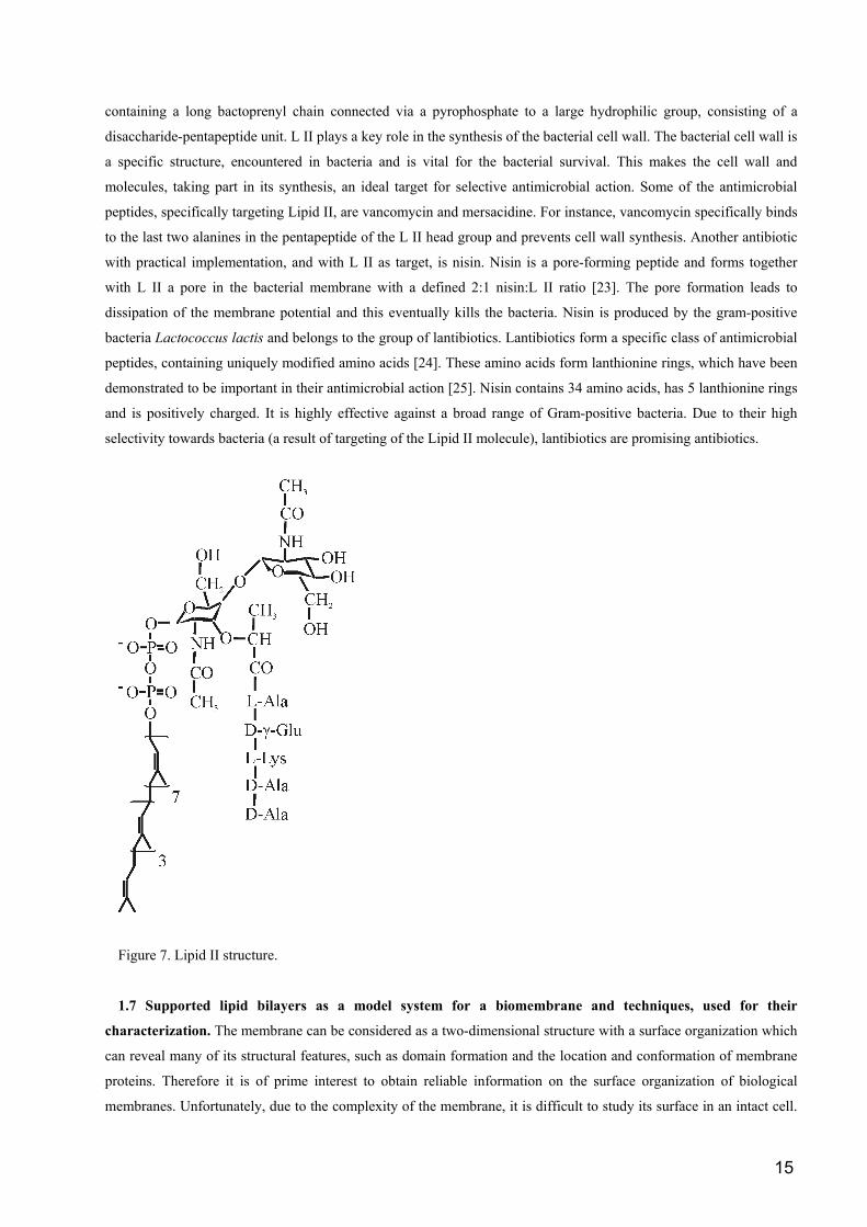
15
containing a long bactoprenyl chain connected via a pyrophosphate to a large hydrophilic group, consisting of a
disaccharide-pentapeptide unit. L II plays a key role in the synthesis of the bacterial cell wall. The bacterial cell wall is
a specific structure, encountered in bacteria and is vital for the bacterial survival. This makes the cell wall and
molecules, taking part in its synthesis, an ideal target for selective antimicrobial action. Some of the antimicrobial
peptides, specifically targeting Lipid II, are vancomycin and mersacidine. For instance, vancomycin specifically binds
to the last two alanines in the pentapeptide of the L II head group and prevents cell wall synthesis. Another antibiotic
with practical implementation, and with L II as target, is nisin. Nisin is a pore-forming peptide and forms together
with L II a pore in the bacterial membrane with a defined 2:1 nisin:L II ratio [23]. The pore formation leads to
dissipation of the membrane potential and this eventually kills the bacteria. Nisin is produced by the gram-positive
bacteria Lactococcus lactis and belongs to the group of lantibiotics. Lantibiotics form a specific class of antimicrobial
peptides, containing uniquely modified amino acids [24]. These amino acids form lanthionine rings, which have been
demonstrated to be important in their antimicrobial action [25]. Nisin contains 34 amino acids, has 5 lanthionine rings
and is positively charged. It is highly effective against a broad range of Gram-positive bacteria. Due to their high
selectivity towards bacteria (a result of targeting of the Lipid II molecule), lantibiotics are promising antibiotics.
Figure 7. Lipid II structure.
1.7 Supported lipid bilayers as a model system for a biomembrane and techniques, used for their
characterization. The membrane can be considered as a two-dimensional structure with a surface organization which
can reveal many of its structural features, such as domain formation and the location and conformation of membrane
proteins. Therefore it is of prime interest to obtain reliable information on the surface organization of biological
membranes. Unfortunately, due to the complexity of the membrane, it is difficult to study its surface in an intact cell.

16
Therefore, an appropriate model system which is suitable for investigations of its surface organization and properties
is needed. We should be able to control the composition of the studied model system, in order to closely reproduce the
composition of a biological membrane of interest. Acquired results for a chosen model system, if obtained at
physiological conditions, will give us insight into the organization and properties of biological membranes.
One model system, which fulfills these requirements, is a supported lipid bilayer, formed on an (atomically) flat
support (mica, silica or other hydrophilic surface). The supported lipid bilayer closely resembles the biological
membrane. A water layer, app. 1 nm thick [26] is situated between the bilayer and the supporting surface and isolates
the bilayer from the support, ensuring mobility of the bilayer species similar to the mobility of the components in a
membrane [27]. Using established protocols, we have the possibility to prepare supported bilayers with desired
composition and to investigate them at controlled conditions. Moreover, with supported bilayers one can follow
adsorption processes of different membrane-interacting molecules, such as antimicrobial peptides, which gives the
possibility to study membrane/drug interactions [28]. The possibility to follow in-time interactions of supported
bilayers with molecules of interests also suggest that supported lipid bilayer can be used as biosensors.
Supported bilayers can be formed by the Langmuir-Blodgett technique [29], by the vesicle fusion method [30] or by
spin-coating [31]. The first technique allows to deposit and study bilayers at predetermined surface pressures and with
different compositions of each monolayer, while vesicle fusion allows formation of a bilayer at surface pressures,
presumably resembling surface pressure in a cell membrane. Moreover, with the vesicle fusion method one could
incorporate a (part of) a protein in the bilayer. Unfortunately, the process of formation of the bilayer from vesicle
suspensions is not yet completely understood. However, some insight in this area has been gained, following the early
stages of adsorption of single vesicles, their interaction and spreading over the substrate [32]. The latter technique,
spin coating was recently developed and allows formation of multilamellar bilayer stacks. Controlling the deposition
parameters, it is possible to deposit large-scale ordered samples, containing from 2 up to 30 bilayers. Supported
bilayers are also a convenient system to study lipid phase separation and peptide aggregation within lipid bilayers.
Lipid and lipid/peptide domains with a specific composition were observed and the dependence of their morphology
on the exact chemistry of lipids and peptides was described [33].
The diversity of processes that can be studied using supported bilayers and the fact that they closely resemble
biological membranes makes them a suitable model system to study properties of membranes. Therefore, currently
they are receiving increasing attention. A number of techniques can be used to get insight in the structure and
properties of supported bilayers. Here we will mention only a few techniques as an example for the possibilities to
investigate and characterize supported lipid bilayers. The NMR technique is used to probe the ordering of the acyl
chains in multilamellar bilayer stacks [34]. Fourier Transformed InfraRed (FTIR) spectroscopy provides information
on the chemical structure of the supported bilayers, conformation of incorporated proteins [35] and lipid/protein
interactions [36]. Adsorption and desorption processes on lipid bilayers can be followed by Surface Plasmon
Resonance (SPR) technique [37] or quartz crystal microbalance [38].
Surface imaging techniques can further provide information on specific features on the surface of the investigated
samples, and they are extensively used on supported bilayers. One of these techniques is fluorescence microscopy. It
allows visualization of formed lipid domains and localization of protein molecules in supported bilayers. However, its
resolution is limited to several hundred nanometers, and since many features in membranes and membrane-mimicking
systems are smaller than this limit, a more powerful surface imaging technique is needed. Such technique, proved to
be rather fruitful for characterization of supported bilayers is Atomic Force Microscopy (AFM).

17
The first article, in which the AFM technique was described, was published in 1986 [39]. Since than this technique
provided a wealth of data, concerning biological systems, such as bilayers [40], proteins [41], DNA molecules [42]
and cells [43]. These studies demonstrated the possibility of the AFM method to visualize with nanometer resolution
the topography of the investigated supported sample. Imaging can be performed at physiological conditions - in an
aqueous environment and at room temperature, and without the necessity chemically to modify the investigated
molecules. Moreover, we could mechanically manipulate molecules of interest, which makes AFM rather unique
amongst the other biophysical techniques. In the following part we will discuss the principle of operation of AFM, its
capabilities and limitations, and we will give some of the more significant achievements, obtained by the AFM
technique.
1.8 AFM – principle of operation, possibilities and limitations.
AFM “sees” the investigated sample via interaction of a sharp tip with the underlying sample. This technique allows
visualization of the sample with nanometer (and in some cases, with sub-nanometer [44]) resolution, in contrast with
optical microscopy, where light is used to create an optical image of the investigated specimen and the resolution is
limited by the diffraction limit of the used light (~ 500-600 nm). A sharp, nanometer sized AFM tip is mounted on the
end of a long flexible cantilever, connected to a holder (Figure 8).
Figure 8. AFM technique – principle of operation

18
The backside of the cantilever is highly reflective and a beam from a laser diode is projected onto the end of the
cantilever and reflected on the position sensitive diode. The slightest movement of the tip in the z direction will reflect
the laser beam on a different spot on the position sensitive diode, which creates a signal, used by the AFM to maintain
the z-position of the tip. The studied sample is adsorbed on an atomically flat support (mica for instance) and the tip is
brought in contact with the sample, until some preset value of the force between tip and the sample is reached. Either
the tip assembly, or the support with the sample, is mounted on a three-dimensional piezoscanner, which can move the
tip (or alternatively, the sample) in the x, y and z directions. For clarity, further we will assume that the tip is fixed and
the sample is moved by the piezoscanner. The sample is moved by the piezoscanner in the x and y directions, thus
being raster-scanned by the tip. The electronics of the AFM keeps, by moving the piezoscanner in z direction, the
force between the tip and the sample constant at each point, scanned in the x and y directions, which allows
simultaneous information on the height of the sample at the respective x-y coordinate to be registered. In this way we
collect the data for the location of each point on the surface of the sample in three-dimensional space, which allows a
3D image of the surface of the sample to be reconstructed. In the created image the lower areas in the sample are
presented as darker, and the higher areas as lighter.
The use of the laser diode and position sensitive diode create a so-called optical lever with a light-path of a few
centimeters, which allows detection of the z-position of the tip with a precision of 0.1 nm. This determines the vertical
resolution of the AFM and this high sensitivity allows discrimination between areas with very small height
differences, such as phase-separated lipid bilayers [45]. The resolution in the x and y direction is directly determined
by the radius of the tip, which can be as low as several nanometers. This allows nanometer and sub-nanometer
resolution to be achieved, but in the same time could be one of the drawbacks of this technique. Even if we use a
sharp tip, it could be polluted with material from the sample during scanning, thus deteriorating the resolution [46].
This is especially a potential threat in imaging of soft biological samples. With such samples another problem could
arise due to their softness. Since a certain force, exerted on the tip is needed for visualization, the tip could deform the
sample and even penetrate through it, which results in images, which do not represent correctly the surface of the
sample. The basic principle of operation of the AFM - imaging the sample through surface forces, could cause still
another problem. If the surface properties of the sample, and as a consequence – tip/sample interaction – are
substantially different in different sample points, than the obtained image would not represent correctly the true
surface profile of the sample, and the image would be influenced by the surface inhomogenities [47]. These
considerations come to show that, as every other technique, the AFM is not a perfect tool. However, being aware of
the potential pitfalls, one can (almost) always construct an experiment and interpret the data in such a way that reliable
information for the sample can be achieved.
1.8.1 AFM is a technique, allowing imaging of biological samples at physiological conditions.
Since no vacuum or low temperatures are required for the AFM to operate, this technique is ideally suited to
investigate biological samples at physiological conditions – in aqueous environment and at room temperature. This
feature places it in a rather unique position amongst other microscopic techniques, being the only one which allows
visualization at such conditions with a nanometer and sub-nanometer resolution. Moreover, the composition of the
aqueous media can be exchanged during the experiment, which allows additional flexibility. In this way in-time
adsorption of different molecules of interest can be investigated [48], as well as the reaction of the sample to the
change in the experimental conditions. This suitability of the technique to image biological samples at relevant

19
conditions quickly established AFM as a valuable tool in biophysical laboratories. A constantly growing number of
AFM studies revealed a wealth of data on a wide variety of biological systems (for a recent review, see [49]). Next we
will discuss some of the more significant achievements in this field.
One of the systems, most intensively studied by AFM, are supported lipid bilayers of different composition [50].
The thickness of bilayers is the most direct parameter, which can be derived [51]. It was shown that depending on the
state and composition of the bilayer, the thickness of the supported bilayer is between 4 and 6 nm. This value includes
a water layer, approximately one nanometer thick, lying between the support and the bilayer and is in good agreement
with the data from other techniques. The water layer isolates the bilayer from the support and provides a mobility of
the bilayer components, comparable to the mobility in non-supported bilayers [52]. When more than one lipid species
is present in the bilayer, a phase separation may be observed [53], clearly demonstrating the possibility of the AFM to
resolve gel and liquid crystalline domains within the bilayer. Further elaborations of the system are also possible,
allowing observation of ternary lipid mixtures. Quite significant is the observation of detergent resistant domains in
cholesterol-containing ternary mixtures, which system is a direct model for lipid rafts in biomembranes [54].
Other highly informative experiments are the observations of ordered arrays of membrane proteins. Classical
examples are the imaging of the purple membrane in Halobacterium Salinarium [55], and the Hexagonally Packed
Intermediate layer in Deinococcus radiodurans [56]. On these samples, as well as on the number of other membrane
proteins [57], a sub-nanometer resolution was achieved, allowing identification of individual α-helices of these
membrane proteins. Since the α-helix is a predominant motif of membrane proteins, some studies concentrated on the
behaviour of model peptides with a single α-helix, incorporated within a lipid bilayer. Intriguing self-assembling
ordered domains were observed in such systems, using so-called WALP peptides [58]. Observed domains consist of
regularly spaced dark (lower) lines with defined repeat distance, separated by lighter (higher) areas. The domains are
typically connected by cracks in the bilayer. The observed ordering phenomenon is similar to the already observed
ordering of gramicidin A in a lipid bilayer [59], which suggest that the ordering within a bilayer could be a more
general property of transmembrane peptides.
Another intensively studied area in membrane research is the interaction between membrane interacting
antimicrobial peptides and lipid bilayers of different composition. Understanding the process of action of these
peptides is an important step in developing new weapons in the war against bacteria. The process of interaction
between membrane interacting substances and lipid bilayers could be conveniently followed in time, using the
imaging capabilities of the AFM [60]. The damage, caused by antimicrobial peptides to the membrane, also can be
visualized [61].
AFM can visualize not only membrane mimicking systems and their interactions with biologically relevant
molecules, but also individual biological molecules, such as DNA [62] and single protein molecules [63]. In these
experiment the characteristic helical pattern of the DNA molecule was resolved, as well as the specific shape of
investigated proteins. Problems in such systems could arise from the requirements for proper adsorption of the
investigated molecule to the solid support. The protein or DNA should be attached strongly enough to allow a stable
scanning and in the same time not to be deformed by exceedingly strong interaction forces with the substrate. This
requires from the researcher to fine-tune the chemical composition of the imaging buffer and/or the substrate [64].

20
The possibility of the AFM method to follow in-time processes on a single molecular level recently was employed
to study the aggregation and fibrilization of amyloid-forming peptides. These peptides are present in the human body
usually as monomers, but under some circumstances they start to aggregate, forming fibrils, which could damage the
cell. Since this process leads to some life threatening diseases, a profound understanding of the process of fibrilization
is of vital importance. A number of emerging AFM studies, providing information on processes of fibrilization [65]
and the fibril/membrane interactions [66] demonstrated the possibility of the AFM to be implemented in this field.
The possibility to change the environment during an experiment will allow the researcher to study the fibrilization
process at different conditions.
1.8.2 AFM can mechanically manipulate the investigated samples, increasing the possibilities for
characterization.
Soon after the development of AFM, its potential not only to image, but also to manipulate mechanically the sample
on a single molecular level and to measure inter- and intra-molecular forces was realized. The first description of the
force measurement approach was published in 1992 [67] and since then the area of force measurements became an
extensively studied research field (for recent review, see [68]). The main idea behind force manipulation is that the
AFM tip can in a controlled way either push or pull the target molecules within the investigated sample. Since in each
moment the z-position of the tip is known, and the spring constant of the cantilever can be determined, the force,
which the tip exerts on the sample, is known with a good precision.
When AFM is used as a force measuring device, the tip is positioned over a point of interest on the surface of the
sample and the movement of the sample is restricted in the x and y directions. Then the sample performs a cycle,
approaching the tip, making a contact with it, and after reaching the predetermined position the sample is retracted to
its initial position. At each moment the position of the tip and the cantilever is known and from this information the
so-called force-distance curve is created. According to Hook’s law the force, exerted by the tip on the sample (in the
case we want to push the sample) or by which the tip is held in contact with the sample (in case of measurement of the
inter- and intra-molecular forces) is:
F=d.k,
where d is the displacement of the tip and k is the spring constant of the cantilever (Figure 9).
When we exert the force on the sample, we can use the tip as a tool to probe local mechanical properties of the
sample, such as elasticity, or the force, needed to penetrate through the investigated specimen. In this way the force,
needed to penetrate through a lipid bilayer can be measured. The dependence of this penetration force on the lipid
composition was investigated [69]. This possibility renders AFM as an alternative, or complementary technique, to the
surface force apparatus (SFA). While AFM offers the possibility to image the sample, SFA provides much better
characterization of the shape and size of the surfaces with a possibility for following a more rigorous theoretical
interpretation.
Another fruitful application of AFM as a force sensor is to determine inter- and intra-molecular forces with a
picoNewton precision. This possibility, combined with the imaging capability of AFM, lacking in any other force
measuring technique (such as laser tweezers [70] or biomembrane force probe [71]) makes a unique combination,
which provided some amazing results during the last few years. A more detailed description of the force measurement
procedure and some of the most prominent examples will be discussed further.

21
Figure 9. AFM tip can be implemented as a tool to mechanically manipulate the sample. In (A) the cantilever is in
equilibrium and no mechanical force is applied on the tip. In (B) the positive displacement of the tip from its
equilibrium position causes the underlying sample to be mechanically pressed and possibly deformed with a defined
force. In (C) the negative displacement of the tip applies a pulling force on the formed molecular complex, thus
probing the strength of the assembly.
1.9 AFM as a tool to measure forces of interaction between biomolecular complexes and mechanical
properties of single molecules.
The AFM technique allows detection of the smallest deflections in the position of the cantilever. Transformed in
forces, according to the Hook’s law, the resolution limit of one Ångstrom in the z-direction theoretically gives a force
sensitivity of ~ 1-2 pN for the softest cantilevers! However, due to thermal motions of the cantilever, caused by the
Brownian motion of the molecules in the aqueous medium, the sensitivity is limited to app. 10-20 pN.
First we will discuss measurements of the internal mechanical properties of molecules. The basic idea is rather
straightforward. The molecules of interests are immobilized on the support, possibly visualized, and afterwards the tip
is positioned on a point of interest on the sample. Then a force-distance curve is recorded and as the tip is brought in
contact with the sample, a part of the investigated molecule is attached to the tip, either by the applied force
(physisorption), or by chemical interaction between the modified tip and the investigated molecule (chemisorption). In
this way a molecular bridge between the tip and sample is created. Only one molecule should be captured between the
tip and the sample in order for a reliable data interpretation to be possible. This can be achieved by varying the
concentration of the molecules under investigation.

22
When a stable attachment of the molecule to both the substrate and the tip is achieved, upon retraction of the sample
the molecule will be stretched. As a result of this the cantilever will bend and will start to apply gradually increasing
force to the bridging molecule. As an example let us consider a modular protein molecule, such as a muscle protein
titin, having a number of folded domains (Figure 10). This molecule is fixed on one end to the substrate and on its
other end to the tip. Upon separation, at a certain moment the elastic force of the cantilever, exerted on the molecule
becomes larger than the unfolding strength of its weakest domain and this domain unfolds, adding its unfolded length
to the bridge between the tip and sample, thus releasing (part of) the elastic force. The continuing separation between
the tip and the sample loads again the remaining folded domains of the molecule, until a critical unfolding force for
the next domain is reached and this domain yields and unfolds. This sequence continues until all domains in the
protein are unfolded, and/or the attachment to the tip or substrate is broken.
Figure 10. AFM force apparatus, used as a set-up to measure intramolecular forces, such as unfolding strength of a
modular protein. The recorded force-distance curves demonstrate the unfolding of each individual protein domain.
The peaks in the curve (B-C, C-D, etc.) indicate for the strength of the unfolded domain.
During the process of the molecule pulling, the position of the tip is registered at each moment. From the recorded
force-distance curve we can directly determine the force, applied by the cantilever through the tip on the sample. In
23
this way the value of the force, at which a certain domain within the studied molecule unfolds, can be precisely
determined from the height of the respective peaks. In order to have a reliable statistics, many force curves should be
recorded and analyzed (usually > 100, depending on the system). Differences in the mechanical strength of different
domains in the studied molecule can be reliably detected, as demonstrated in [72]. The shape of the curves, preceding
each peak in the force curve usually is fitted with a theoretical curve according to two proposed models, Worm-Like
Chain (WLC) and Freely Jointed Chain (FJC). The parameters, obtained from both models are the persistence length
(usually interpreted as the smallest non-deformable part in the studied molecule) and the contour length of the
molecule (its fully extended length). Moreover, using an appropriate theory (described in part 1.10 of the present
thesis) other parameters of the molecule, such as the natural rate of unfolding of domains can be estimated. In a study
on polysaccharides it was demonstrated that even the chair-boat transition in the sugar monomer could be detected by
AFM [73]. In all these and many other studies it was demonstrated that by mechanical manipulation of a target
molecule, one can get information about some of its fundamental properties.
The AFM as a force-measuring device is used not only to determine the properties of single molecules. A rather
fruitful field during the last years was the determination of the strength of binding of different biomolecular
complexes [74]. In this way the interaction strength of many ligand/receptor and antibody/antigen couples were
determined and interpreted [75]. The procedure in this case is as follows (Figure 11). One of the molecules in the
couple is immobilized on the solid support, and the tip is incubated with the other molecule. Sometimes a special
chemical treatment of the tip and/or the support is needed in order to have the molecules properly attached. Often this
treatment includes introduction of a so-called spacer, a long polymer molecule between the immobilized molecule and
the support or the tip, which allows conformational freedom of the attached molecule. Since the precise geometry of
contact in the antigen/antibody complex is crucial for a proper recognition, sometimes this spacer molecule is
essential for successful experiments in these systems. The preferential experimental condition is when only a single
complex is formed between the tip and the sample. This is achieved by controlling the density of the coverage of the
tip and the substrate with the investigated molecules [76]. The length of the spacer molecule also can be varied,
changing the effective radius, in which the molecule, connected to the spacer could probe for the other molecule in the
complex. However, controlling these factors is not always possible and sometimes more than one couple is formed
and at the retract cycle of the tip more than one bond is broken in succession or even almost simultaneously. If
multiple rupture events occur, the obtained force distribution should be scrutinized and correlation analysis to be
preformed in order strength of the single complex to be determined. As in the case of force measurements within a
single molecule, in the case of intermolecular force determination, hundred and sometimes even thousands of force
events are collected and analyzed.
As a classical example for force measurements we could mention the determination of strength of integration of a
bacteriorhodopsin (BR) molecule in a purple membrane [77]. This protein has seven transmembrane α–helices and
assembles as a trimer in a so-called purple membrane. After visualization of the sample, the tip was brought in contact
with the protein molecule and a part of the protein was attached to the tip. Upon withdrawal of the tip a characteristic
saw-tooth pattern was observed with up to four separate peaks. The intricate pattern of observed force peaks were
analyzed in detail and to each peak withdrawal and unfolding of a specific helix/helices was ascribed [78].
Remarkably, after each recorded force curve, a vacancy in the 2D ordered protein assembly of the purple membrane
was observed, which is an indication for the absence of a BR molecule, removed during the force manipulation.
Another interesting example of intermolecular force measurements is the determination of force between

24
complementary strands of DNA molecules. In these experiments a value for the strength of the bond between the
complementary DNA bases were determined [79]. In another AFM study [80] on complementary DNA strands some
thermodynamic properties and the range of the interaction between DNA strands were determined, using an earlier
developed theory [81], which describes the dissociation of the biological bonds as a thermally activated process. The
next part presents the key points in this theory.
Figure 11. AFM as a tool to measure intermolecular forces between molecular complexes, such as complementary
DNA strands, ligand/receptor couples, etc.
1.10 The strength of a biomolecular bond depends on the way we load it upon probing its strength.
So far we discussed the force measurements in biological systems as if each biological bond has a defined value,
independent of the experimental conditions. In fact, the first studies (up to 1997) treated the subject in this way. Then
in 1997 a theory was proposed by Evans [82], based on the previous work of Bell [83]. This theory placed the force
measurements on a completely new, quantitative basis.
1.10.1 The natural lifetime of each bond is the fundamental property, which determines the behaviour of the
bond upon loading.

25
The key idea in the proposed theory, which already is established as an indispensable tool in the interpretation of
force measurements, is that each bond is characterized not by some inherent strength, but rather by a defined lifetime.
This lifetime in the case of biological non-covalent bonds could be from a few milliseconds up to days or even longer.
In a period longer than the lifetime of the bond, it has zero strength due to spontaneous dissociation of the complex.
The natural lifetime of the bond is determined by the height and position of the energy barrier preventing the thermal
dissociation of this bond. The applied force deforms the energy landscape of the interaction and lowers the energy
barrier which prevents the spontaneous dissociation, thus decreasing the lifetime of the bond (Figure 12).
Figure 12. Energy landscape of a bond between biomolecules in the absence of an applied external force (A) and in
the presence of external force, which deforms the landscape and lowers the energy barrier (B).

26
In the AFM force measurement we do not load the bond instantaneously, but the force increases gradually with
time, as the tip retracts from the support. From the experimental point of view this means that if we load the
investigated bond slowly enough, we will not detect any bond rupture due to the spontaneous dissociation of the bond
during the experiment! However, at a certain loading rate the accumulated force within the lifetime of the bond will
lower sufficiently the energy barrier and dissociation will be registered at a certain applied force. In this way a defined
rupture strength, different from zero is registered. These considerations show that the measured strength of the bond
depends on the rate, at which we load it.
Once realizing that no definitive value could be ascribed to the studied bond, but that its behaviour depends on its
natural lifetime, determined by the energy barriers involved, it is easy to follow the theory in more detail. After the
decrease in the energy for dissociation ΔE0 by an applied external force F, postulated by Bell, the theory was further
developed for the case of gradual loading, as in AFM experiments. Evans [84] derived a simple relation of the
registered bond strength as a function of loading rate, predicting the dependency of the registered most probable force
F* on the loading rate:
F*= (kbT/ Xb)ln (r/((kbT/Xb).Koff))
Here Xb is the distance between the ground state (the potential well, where the molecular complex resides) and the
transition state (the position of the energy barrier), r is the loading rate, kbT is the thermal energy and Koff is the
natural off-rate of the complex (1/lifetime). An important assumption in this equation is that the energy barrier is
sharp, which means that it does not change its position under influence of the applied external force. From this
equation one sees that the registered force will increase linearly with the natural logarithm of the loading rate.
Moreover, probing the strength of a biological complex over several orders of magnitude of loading rates will give us
information about the energy landscape of the interaction. From the slope (kbT/ Xb) and the x-intercept of each linear
regime we can get information on the position Xb of each energy barrier along the dissociation pathway. The intercept
with the x - axis gives the off - rate at zero applied force Koff through the dependency r(F=0) = (kbT/ Xb).Koff.
The potential of the proposed theory was quickly appreciated and in almost all recent force measurement studies a
force/loading rate dependency was investigated and the data were interpreted according to this theory. Some
ligand/receptor and antigen/antibody couples were characterized and details in their energetic landscapes were
described [85, 86]. Moreover, in some cases the natural off-rate, determined by the AFM force measurement is
compared with the data, obtained by bulk techniques, giving additional information for the energy landscape of the
studied couple [87]. The interpretation of the obtained parameters – distances bound/transition state and natural off-
rates - in terms of the macroscopic functions, performed by the studied molecular complexes, is also attempted [88]. A
number of important biomolecular couples (or molecular assemblies), which interactions are not characterized yet,
promise the appearance of new interesting studies.
Scope of the thesis
In the present thesis AFM was used as an image and force measuring device on a variety of membrane mimicking
systems. In Chapter 2 the behaviour of the model WALP peptide, which resembles the transmembrane part of
integral membrane proteins, and its ability to form a specific ordered domains in lipid bilayers of different
compositions is studied. In Chapter 3 we studied further the behaviour of WALP in lipid bilayers and investigated the

27
strength of integration of this transmembrane peptide, using the force measuring possibilities of the AFM. Data
analysis according to the described theory gives insight in the factors, governing the stability of integration of
transmembrane proteins. Another example for possibilities of the AFM as a manipulating tool is presented in Chapter
4, where Lipid II - an important biological molecule, is studied and size and orientation of its head group, when the
molecule is incorporated in a bilayer, was obtained. The thesis finishes with a summary and perspectives in Chapter
5.

28
[1] Gennis, R.B., Biomembranes: Molecular structure and function Springer-Verlag, London, 1989
[2] Gorter, E. and Grendel, F. 1925. On bimolecular layers of lipoids on the chromocytes of the blood. J. Exp. Med.
41:439-443.
[3] Veerkamp JH. Lipids of the cell plasma membrane. Biomembranes. 1972; 3:159-79
[4] Niemela P, Hyvonen MT, Vattulainen I.Structure and dynamics of sphingomyelin bilayer: insight gained
through systematic comparison to phosphatidylcholine. Biophys J. 2004; 87(5):2976-89.
[5] Tierney KJ, Block DE, Longo ML.Elasticity and phase behavior of DPPC membrane modulated by cholesterol,
ergosterol, and ethanol. Biophys J. 2005; 89(4):2481-93.
[6] Simons K, Toomre D. Lipid rafts and signal transduction. Nat Rev Mol Cell Biol. 2000; 1(1):31-9.
[7] Lafont F, Simons K. Raft-partitioning of the ubiquitin ligases Cbl and Nedd4 upon IgE-triggered cell signaling.
Proc Natl Acad Sci U S A. 2001; 98(6):3180-4.
[8] Rajendran L, Simons K. Lipid rafts and membrane dynamics. J Cell Sci. 2005;118(Pt 6):1099-102
[9] de Almeida RF, Loura LM, Fedorov A, Prieto M. Lipid rafts have different sizes depending on membrane
composition: a time-resolved fluorescence resonance energy transfer study. J Mol Biol. 2005; 4;346(4):1109-20.
[10] Visualizing detergent resistant domains in model membranes with atomic force microscopy. FEBS Lett. 2001;
13;501(1):92-6.
[11] Rinia HA, Boots JW, Rijkers DT, Kik RA, Snel MM, Demel RA, Killian JA, van der Eerden JP, de Kruijff B.
Domain formation in phosphatidylcholine bilayers containing transmembrane peptides: specific effects of flanking
residues. Biochemistry. 2002; 41(8):2814-24.
[12] Killian JA, von Heijne G. How proteins adapt to a membrane-water interface. Trends Biochem Sci. 2000;
25(9):429-34
[13] de Kruijff B, Killian JA, Ganchev DN, Rinia HA, Sparr E. Striated domains: self-organizing ordered
assemblies of transmembrane alpha-helical peptides and lipids in bilayers. Biol Chem. 2006 Mar;387(3):235-41
[14] Killian JA. Synthetic peptides as models for intrinsic membrane proteins. FEBS Lett. 2003; 555(1):134-8
[15] Rinia HA, Kik RA, Demel RA, Snel MM, Killian JA, van Der Eerden JP, de Kruijff B. Visualization of highly
ordered striated domains induced by transmembrane peptides in supported phosphatidylcholine bilayers.
Biochemistry. 2000; 39(19):5852-8.
[16] van Meer G, Sprong H.Membrane lipids and vesicular traffic. Curr Opin Cell Biol. 200; 16(4):373-8.
[17] Hessa T, Kim H, Bihlmaier K, Lundin C, Boekel J, Andersson H, Nilsson I, White SH, von Heijne G.
Recognition of transmembrane helices by the endoplasmic reticulum translocon. Nature. 2005; 433(7024):377-81
[18] Matsuzaki K. Why and how are peptide-lipid interactions utilized for self-defense? Magainins and tachyplesins
as archetypes. Biochim Biophys Acta. 1999;1462(1-2):1-10
[19] Killian JA, von Heijne G. How proteins adapt to a membrane-water interface. Trends Biochem Sci. 2000;
25(9):429-34
[20] Powers JP, Hancock RE. The relationship between peptide structure and antibacterial activity. Peptides. 2003;
24(11):1681-91
[21] Shai Y. Mechanism of the binding, insertion and destabilization of phospholipid bilayer membranes by alpha-
helical antimicrobial and cell non-selective membrane-lytic peptides. Biochim Biophys Acta. 1999; 1462(1-2):55-70
[22] carpet-like membrane destabilization

29
[23] Hasper HE, de Kruijff B, Breukink E.Assembly and stability of nisin-lipid II pores. Biochemistry. 2004;
43(36):11567-75.
[24] Sahl HG, Bierbaum G. Lantibiotics: biosynthesis and biological activities of uniquely modified peptides from
gram-positive bacteria. Annu Rev Microbiol. 1998; 52:41-79.
[25] Hsu ST, Breukink E, Tischenko E, Lutters MA, de Kruijff B, Kaptein R, Bonvin AM, van Nuland NA. The
nisin-lipid II complex reveals a pyrophosphate cage that provides a blueprint for novel antibiotics. Nat Struct Mol
Biol. 2004; 11(10):963-7.
[26] Marra J, Israelachvili J Direct measurements of forces between phosphatidylcholine and
phosphatidylethanolamine bilayers in aqueous electrolyte solutions. Biochemistry. 1985; 24(17):4608-18
[27] Hughes T, Strongin B, Gao FP, Vijayvergiya V, Busath DD, Davis RC. AFM visualization of mobile influenza
A M2 molecules in planar bilayers. Biophys J. 2004; 87(1):311-22.
[28] Rigby-Singleton SM, Davies MC, Harris H, O'Shea P, Allen S. Visualizing the solubilization of supported
lipid bilayers by an amphiphilic peptide. Langmuir. 2006; 22(14):6273-9
[29] Zasadzinski JA, Viswanathan R, Madsen L, Garnaes J, Schwartz DK. Langmuir-Blodgett films. Science. 1994;
263(5154):1726-33
[30] Brian, A. A., and H. M. McConnell. 1984. Allogenic stimulation of cytotoxic T cells by supported planar
membranes. Proc. Natl. Acad. Sci. USA. 81:6159–6163
[31] Pompeo G, Girasole M, Cricenti A, Cattaruzza F, Flamini A, Prosperi T, Generosi J, Castellano AC. AFM
characterization of solid-supported lipid multilayers prepared by spin-coating. Biochim Biophys Acta. 2005;
1712(1):29-36.
[32] Reviakine, I., and A. Brisson. 2000. Formation of supported phospholipid bilayers from unilamellar vesicles
investigated by atomic force microscopy. Langmuir. 16:1806–1815
[33] Rinia HA, de Kruijff B. Imaging domains in model membranes with atomic force microscopy. FEBS Lett.
2001; 504(3):194-9
[34] Guo W, Kurze V, Huber T, Afdhal NH, Beyer K, Hamilton JA. A solid-state NMR study of phospholipid-
cholesterol interactions: sphingomyelin-cholesterol binary systems. Biophys J. 2002; 83(3):1465-78
[35] Haris PI, Chapman D. The conformational analysis of peptides using Fourier transform IR spectroscopy.
Biopolymers. 1995; 37(4):251-63
[36] Frias MA, Diaz SB, Ale NM, Ben Altabef A, Disalvo EA. FTIR analysis of the interaction of arbutin with
dimyristoyl phosphatidylcholine in anhydrous and hydrated states. Biochim Biophys Acta. Jul 20, 2006
[37] Rossi C, Homand J, Bauche C, Hamdi H, Ladant D, Chopineau J. Differential mechanisms for calcium-
dependent protein/membrane association as evidenced from SPR-binding studies on supported biomimetic
membranes. Biochemistry. 2003 Dec 30;42(51):
[38] Janshoff A, Steinem C. Label-free detection of protein-ligand interactions by the quartz crystal microbalance.
Methods Mol Biol. 2005;305:47-64
[39] Binnig G, Quate CF, Gerber C. Atomic force microscope. Phys Rev Lett. 1986; 56(9):930-933.
[40] Dufrene YF, Lee GU. Advances in the characterization of supported lipid films with the atomic force
microscope. Biochim Biophys Acta. 2000; 1509(1-2):14-41
[41] Muller DJ, Janovjak H, Lehto T, Kuerschner L, Anderson K. Observing structure, function and assembly of
single proteins by AFM. Prog Biophys Mol Biol. 2002; 79(1-3):1-43.
30
[42] Hansma HG. Surface biology of DNA by atomic force microscopy. Annu Rev Phys Chem. 2001; 52:71-92
[43] You HX, Yu L. Atomic force microscopy imaging of living cells: progress, problems and prospects. Methods
Cell Sci. 1999; 21(1):1-17
[44] Muller DJ, Heymann JB, Oesterhelt F, Moller C, Gaub H, Buldt G, Engel A. Atomic force microscopy of
native purple membrane. Biochim Biophys Acta. 2000; 1460(1):27-38
[45] Giocondi MC, Le Grimellec C. Temperature dependence of the surface topography in
dimyristoylphosphatidylcholine/distearoylphosphatidylcholine multibilayers. Biophys J. 2004; 86(4):2218-30.
[46] Taatjes DJ, Quinn AS, Lewis MR, Bovill EG. Quality assessment of atomic force microscopy probes by
scanning electron microscopy: correlation of tip structure with rendered images. Microsc Res Tech. 199; 44(5):312-
26.
[47] Schneider J, Dufrene YF, Barger WR Jr, Lee GU Atomic force microscope image contrast mechanisms on
supported lipid bilayers. Biophys J. 2000; 79(2):1107-18
[48] Tang J, Jiang J, Song Y, Peng Z, Wu Z, Dong S, Wang E. Conformation change of horseradish peroxidase in
lipid membrane.Chem Phys Lipids. 2002; 120(1-2):119-29
[49] Fotiadis D, Scheuring S, Muller SA, Engel A, Muller DJ.Imaging and manipulation of biological structures
with the AFM. Micron. 2002; 33(4):385-97.
[50] Hui SW, Viswanathan R, Zasadzinski JA, Israelachvili JN. The structure and stability of phospholipid bilayers
by atomic force microscopy. Biophys J. 1995; 68(1):171-8.
[51] Dufrene YF, Boland T, Schneider JW, Barger WR, Lee GU.Characterization of the physical properties of
model biomembranes at the nanometer scale with the atomic force microscope. Faraday Discuss. 1998;(111):79-94
[52] Lateral mobility of lipid analogues and GPI-anchored proteins in supported bilayers determined by fluorescent
bead tracking. J Membr Biol. 1993; 135(1):83-92.
[53] Giocondi MC, Pacheco L, Milhiet PE, Le Grimellec C. Temperature dependence of the topology of supported
dimirystoyl-distearoyl phosphatidylcholine bilayers. Ultramicroscopy. 2001; 86(1-2):151-7
[54] Rinia HA, Snel MM, van der Eerden JP, de Kruijff B. Visualizing detergent resistant domains in model
membranes with atomic force microscopy. FEBS Lett. 2001; 501(1):92-6.
[55] Muller DJ, Heymann JB, Oesterhelt F, Moller C, Gaub H, Buldt G, Engel A. Atomic force microscopy of
native purple membrane. Biochim Biophys Acta. 2000; 1460(1):27-38
[56] Muller DJ, Baumeister W, Engel A. Conformational change of the hexagonally packed intermediate layer of
Deinococcus radiodurans monitored by atomic force microscopy. J Bacteriol. 1996; 178(11):3025-30
[57] Muller DJ, Fotiadis D, Scheuring S, Muller SA, Engel A. Electrostatically balanced subnanometer imaging of
biological specimens by atomic force microscope. Biophys J. 1999; 76(2):1101-11.
[58] de Kruijff B, Killian JA, Ganchev DN, Rinia HA, Sparr E. Striated domains: self-organizing ordered
assemblies of transmembrane alpha-helical peptides and lipids in bilayers. Biol Chem. 2006; 387(3):235-41.
[59] Mou J, Czajkowsky DM, Shao Z. Gramicidin A aggregation in supported gel state phosphatidylcholine
bilayers.Biochemistry. 1996; 35(10):3222-6
[60] Berquand A, Mingeot-Leclercq MP, Dufrene YF.Real-time imaging of drug-membrane interactions by atomic
force microscopy. Biochim Biophys Acta. 2004; 1664(2):198-205
[61] van Kan EJ, Ganchev DN, Snel MM, Chupin V, van der Bent A, de Kruijff B.The peptide antibiotic clavanin
A interacts strongly and specifically with lipid bilayers. Biochemistry. 2003; 42(38):11366-72.

31
[62] Mou J, Czajkowsky DM, Zhang Y, Shao Z. High-resolution atomic-force microscopy of DNA: the pitch of the
double helix. FEBS Lett. 1995; 371(3):279-82
[63] Silva LP.Imaging proteins with atomic force microscopy: an overview. Curr Protein Pept Sci. 2005
Aug;6(4):387-95.
[64] Hansma HG, Bezanilla M, Zenhausern F, Adrian M, Sinsheimer RL. Atomic force microscopy of DNA in
aqueous solutions. Nucleic Acids Res. 1993; 21(3):505-12.
[65] Green JD, Goldsbury C, Kistler J, Cooper GJ, Aebi U.Human amylin oligomer growth and fibril elongation
define two distinct phases in amyloid formation. J Biol Chem. 2004; 279(13):12206-12
[66] Green JD, Kreplak L, Goldsbury C, Li Blatter X, Stolz M, Cooper GS, Seelig A, Kistler J, Aebi U. Atomic
force microscopy reveals defects within mica supported lipid bilayers induced by the amyloidogenic human amylin
peptide. J Mol Biol. 2004; 342(3):877-87.
[67] Tao NJ, Lindsay SM, Lees S. Measuring the microelastic properties of biological material. Biophys J. 1992;
63(4):1165-9.
[68] Kienberger F, Ebner A, Gruber HJ, Hinterdorfer P.Molecular recognition imaging and force spectroscopy of
single biomolecules.Acc Chem Res. 2006; 39(1):29-36
[69] Schneider J, Dufrene YF, Barger WR Jr, Lee GU. Atomic force microscope image contrast mechanisms on
supported lipid bilayers. Biophys J. 2000; 79(2):1107-18
[70] Cui Y, Bustamante C.Pulling a single chromatin fiber reveals the forces that maintain its higher-order structure.
Proc Natl Acad Sci U S A. 2000; 97(1):127-32.
[71] Merkel R, Nassoy P, Leung A, Ritchie K, Evans E.Energy landscapes of receptor-ligand bonds explored with
dynamic force spectroscopy. Nature. 1999; 397(6714):50-3.
[72] Oberhauser AF, Badilla-Fernandez C, Carrion-Vazquez M, Fernandez JM.The mechanical hierarchies of
fibronectin observed with single-molecule AFM. J Mol Biol. 2002; 319(2):433-47
[73] Marszalek, P. E., Oberhauser, A. F., Pang, Y. P. & Fernandez, J. M. Polysaccharide elasticity governed by
chair-boat transitions of the glucopyranose ring. Nature 1998; 396, 661-664
[74] Berquand A, Xia N, Castner DG, Clare BH, Abbott NL, Dupres V, Adriaensen Y, Dufrene YF. Antigen
binding forces of single antilysozyme Fv fragments explored by atomic force microscopy. Langmuir. 2005;
21(12):5517-23
[75] Dammer U, Hegner M, Anselmetti D, Wagner P, Dreier M, Huber W, Guntherodt HJ. Specific
antigen/antibody interactions measured by force microscopy. Biophys J. 1996; 70(5):2437-41
[76] Allen S, Davies J, Davies MC, Dawkes AC, Roberts CJ, Tendler SJ, Williams PM. The influence of epitope
availability on atomic-force microscope studies of antigen-antibody interactions. Biochem J. 1999; 341
[77] Oesterhelt F, Oesterhelt D, Pfeiffer M, Engel A, Gaub HE, Muller DJ. Unfolding pathways of individual
bacteriorhodopsins. Science. 2000; 288(5463):143-6
[78] Muller DJ, Kessler M, Oesterhelt F, Moller C, Oesterhelt D, Gaub H. Stability of bacteriorhodopsin alpha-
helices and loops analyzed by single-molecule force spectroscopy. Biophys J. 2002; 83(6):3578-88
[79] Lee GU, Chrisey LA, Colton RJ. Direct measurement of the forces between complementary strands of DNA.
Science. 1994; 266(5186):771-3.
[80] Schumakovitch I, Grange W, Strunz T, Bertoncini P, Guntherodt HJ, Hegner M. Temperature dependence of
unbinding forces between complementary DNA strands. Biophys J. 2002; 82(1 Pt 1):517-21

32
[81] Merkel R, Nassoy P, Leung A, Ritchie K, Evans E. Energy landscapes of receptor-ligand bonds explored with
dynamic force spectroscopy. Nature 1999; 397(6714):50-3.
[82] Evans E., Ritchie K., Dynamic strength of molecular adhesion bonds. Biophys J. 1997; 72(4):1541-55.
[83] Bell GI. Models for the specific adhesion of cells to cells. Science. 1978; 200(4342):618–627
[84] Evans E., Energy landscapes of biomolecular adhesion and receptor anchoring at interfaces explored with
dynamic force spectroscopy. Faraday Discuss. 1998;(111):1-16.
[85] Strunz T, Oroszlan K,Schumakovitch I, Guntherodt H, Hegner M Model energy landscapes and the force-
induced dissociation of ligand-receptor bonds. Biophys J. 2000; 79(3):1206-12
[86] Yuan C, Chen A, Kolb, P. and Moy, V.T. Energy Landscape of Streptavidin-Biotin Complexes Measured by
Atomic Force Microscopy Biochemistry; 2000; 39(33) pp 10219 - 10223;
[87] Schwesinger, F., Ros, R., Struntz, T., Anselmetti, D., Güntherodt, H-J., Honegger, A., Jermutus, L.,
Tiefenauer, L. and Pluckthun, A. Unbinding forces of single antibody-antigen complexes correlate with their thermal
dissociation rates, Proc. Natl. Acad. Sci. USA 2000; 97, 9972-9977
[88] Zhang X, Wojcikiewicz E, Moy VT. Force spectroscopy of the leukocyte function-associated antigen-
1/intercellular adhesion molecule-1 interaction. Biophys J. 2002; 83(4):2270-9