chapter 11 - amazon s3s3.amazonaws.com/prealliance_oneclass_sample/vozkbnxwgb.pdfchapter 11...
TRANSCRIPT

Chapter 11
FUNCTIONS OF BLOOD1. Transportation
> Oxygen, nutrients, wastes, carbon dioxide, hormones and more
2. Defense
> Against invasion by pathogens
3. Regulatory functions
> Body temperature, water-salt balance and body pH
COMPOSITIONS OF BLOODPlasma
• 92% of water and 8% of salts and organic molecules
• Straw-coloured liquid that makes up about 55% of blood
• Plasma protein: dissolved substance in the blood
> Help balance water flow between blood and the cells
> Albumins
- Most abundant and important for plasma’s osmotic pressure as well as the transport of other molecules
> Globulins
- Transports lipids and fat-soluble vitamins
- Some are antibodies
> Fibrinogen
- Important for the formation of blood clots
the process for the blood stem cells differentiating into these descendants is called hematopoiesis
Formed elements
• Red blood cells / erythrocytes
• white blood cell/ leukocytes
• Formed by the red bone marrow
• The sponge like framework supports fat cells but also supports the undifferentiated cells called blood stem cells
Platelets
• Also called thrombocytes, essential for blood clotting
• Made from fragments of large cells called megakaryocytes made in the red bone marrow
• Makes 200 billion/day
• Blood proteins named thrombin and fibrinogen are important for blood clotting by generating fibrin threads that catch red blood cells
Disorders involving platelets
• Thrombocytopenia
> A disorder in which the # of platelets is too low due to not enough born in the bone marrow or the ↑ breakdown outside the marrow
• Thromboembolism
> When a clot forms(thrombus) and breaks off from its site of origin (embolus) and plugs another vessel
• Hemophilia
> A genetic disorder that results in a deficiency of a clotting factor so that when a person damages a blood vessel they are unable to properly clot their blood both internally and externally

BLOOD CLOTTING4 main things
• Platelets
• Clotting factors (special proteins)
• Fibrin (a mesh of special kinds of clotting factors)
• Other cells – red and white
Steps
1. When a blood vessel is broken, the nearby platelets becomes sticky and stick to each other and the hole of the blood vessel
2. Clotting factors that float by the tear reinforces the platelet plug
> Converts inactive blood protein to prothrombin activator, which then coverts prothrombin (plasma protein made by liver) to an active form, thrombin
> Thrombin causes changes in other plasma proteins produced by the liver called fibrinogen, then it forms long strand of fibrins
3. Fibrins, a web acts like glue and forms a blood clot
4. Other cells float by and sticks to the clot
Red blood cell• Also called erythrocytes
• Picks up oxygen from the lungs and ferry it to all the cells of the body
Red blood cells and hemoglobin
• The shape of the cell, dented on both sides maximizes the surface area for gas exchange

• Each red blood cell is packed with hemoglobin, the oxygen-binding pigment that is responsible for the cells’ red colour
• Contains about 280 million hemoglobin molecules and each hemoglobin molecules binds 4 O2
• It lacks a nucleus and few organelles
• The hem group contains an iron ion that actually binds to the oxygen and is then called oxyhemoglobin
Life cycle of red blood cells
• Take about 6 days to make red blood cell
1. Very immature cells become a factory for hemoglobin molecules, and after the cell is packed with hemoglobin, the nucleus is pushed out
2. A structural metamorphosis occurs culminating in a mature red blood cell with a typical biconcave shape
3. Once this occurs, the cell leaves the bone marrow and enter the blood stream
• It lives for about 120 days, but travels a lot
• It lives so short because of its lack of nucleus
> Without a nucleus, protein synthesis needed to replace key enzymes can’t take place, causing the cell to be rigid and fragile
• Liver and spleen are the “graveyard ” where worn out red blood cells are removed from circulation
Production of red blood cell
• Controlled by negative feedback
• When there is a loss of blood, it triggers a homeostatic mechanism that speeds up the rate of red blood cell production
> Initiated by a ↓ in the oxygen supply to the body’s cells
• Certain cells in kidney sense the ↓ in oxygen, so they respond by producing the hormone erythropoietin (EPO)
> Excreted by the kidney
• The EPO travels to the red marrow where it steps up both the division rate of the stem cells and the maturation rate of immature red blood cells

Disorders of red blood cells
• Anemia: blood’s ability to carry oxygen is ↓ which can result from too little hemoglobin, too few red blood cells or both
> Anemic person’s heart beat faster to compensate for the blood’s ↓ ability to carry oxygen
• Iron deficiency anemia: insufficiency of iron in the body, which leads to inadequate hemoglobin production
• Sickle-cell anemia: genetic disease that causes red blood cells to be sickle shaped that tend to rupture when the blood’s oxygen is too low
• Pernicious anemia: red blood cell # drop when the production of red blood cells are halted or impaired
> Depend on supply of vitamin B12 which is absorbed by the small intestine but produced by the stomach lining
• Hemolytic disease of the newborn: a condition with incompatible blood types that leads to rupturing blood cells in a baby before and continuing after birth
WHITE BOLD CELLS• Also called leukocytes removes wastes, toxins, and damaged or abnormal cells
• Serve as a warrior in the body’s fight against disease
• Produced in red bone marrow
• Production is regulated by colony-stimulating factor (CSF)
• Marge blood cells that have nucleus
• Can leave the circulatory system and move to sites of infection around by squeezing in between neighbouring cells
• Some live days and other live months and years unlike red blood cells
Granulocytes
• Contain noticeable granules, lobed nuclei
• Sacs containing chemicals that are used as weapons to destroy invading pathogens, bacteria
1. Neutrophil
> Not stained by dyes
> About 50-70% of WBC

> Multi lobed nucleus
> Most abundant of all white blood cells, and blood cell soldiers on the front line
> Arriving at the infection site 1st, it begins to engulf microbes by phagocytosis, thus curbing the spread of infection
> After eating 12 bacteria, it dies, but while dying, it releases chemicals that attract more neutrophils
> After it dies, it becomes pus
2. Eosinophil
> Stained by eosin and turns red
> Small % of WBC
> Contain a bilobed nucleus
> Contains substances that are important in the body’s defense against tape work and hookworm
> Lessen the severity of allergies by eating antibody-antigen complexes and inactivating inflammatory chemicals
3. Basophil
> Stained by basic dye and turns blue
> Small % of WBC
> U-shaped or lobed nucleus
> Release histamine, a chemical that attracts other white blood cells to the site of infection and cause blood vessel to dilate to ↑blood flow to the infection area
Arganulocytes
• Lack cytoplasmic granules
• Nonlobed nuclei
• Classified as monocytes and lymphocytes
1. Monocytes
> Largest of all formed elements
> WBC with horseshoe shaped nucleus
> Leaves the bloodstream and enter various tissues and then develops into macrophages

- Phagocytic cells that eat invading microbes, dead cells and cellular debris
2. Lymphocytes
> About 25-35% of all WBC
> Large nucleus that takes up most of the cytoplasm
> B lymphocytes
- Give rise to plasma cells that produces antibodies which is called antigens that on the surface of invading microbes or other foreign cells
> T lymphocytes
- Specialized WBC that play role in the body’s defence mechanism
Disorders of white blood cells
• Infectious mononucleosis
> Also known as kissing disease
> The infection causes an ↑ in lymphocytes that have atypical appearance
> Spread by person to person contact or by using the same utensils
> Causes fatigue, sore throat and swollen lymph nodes
• Leukemia
> A group of cancers that affect WBC in which the cells divide uncontrollably
> Abnormal cell that remains immature and are therefore unable to defend the body against infectious organisms
> They “take over” bone marrow, preventing the development of normal blood cells, like RBC, WBC and platelets
• Severe combined immunodeficiency disease (SCID)
> An inherited disease in which stem cells of WBC’s lack of enzymes that allow them to fight any infection
BLOOD TYPING• Antigen: a foreign substance, often a polysaccharide or a protein that stimulates an
immune response
• Antibody: proteins made in response to an antigen in the body and binds to that antigen and help to eliminate it from the body
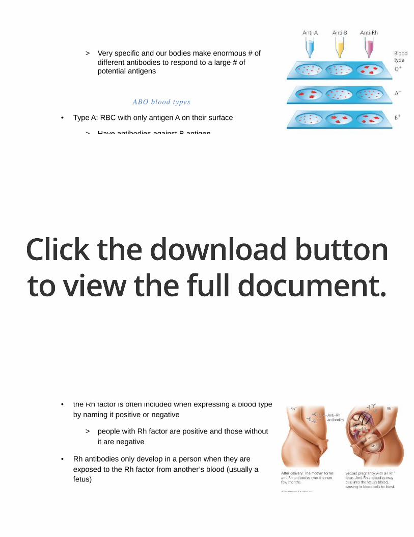
> Very specific and our bodies make enormous # of different antibodies to respond to a large # of potential antigens
ABO blood types
• Type A: RBC with only antigen A on their surface
> Have antibodies against B antigen
• Type B: RBC with only antigen B on their surface
> Have antibodies against A antigen
• Type AB: RBC with antigen A&B on their surface
> Don’t have antigen A & B
• Type O: RBC with no antigen A or B on their surface
> Have antibodies against A and B antigens
• If 2 different blood types are together, it will agglutinate (clump together)
> clumped cells can get stuck in small blood vessels and block blood flow to body cells
> can also break open and release hemoglobin which then clogs the filtering system in the kidney, causing death
Rh factor
• the Rh factor is often included when expressing a blood type
by naming it positive or negative
> people with Rh factor are positive and those without
it are negative
• Rh antibodies only develop in a person when they are
exposed to the Rh factor from another’s blood (usually a fetus)

• Hemolytic disease of new-born can happen during pregnancy under these condition:
- Mon : Rh-
- Dad: Rh +
- Baby: Rh +
How to prevent hemolytic disease
• Rh- women are giving an injection of anti-Rh antibodies no later than 72 hours after birth
of an Rh+ baby
• These antibodies attack fetal RBC in the mother before the mother’s immune system
can bake antibodies
• This will have to be repeated if an Rh- mother has another Rh+ baby