chapter 18 the endocrine system -...
TRANSCRIPT

1/26/14
1
1
Collin College
BIOL 2402 Anatomy/Physiology 2
Chapter 18 The Endocrine System
2
The Pituitary Gland
• Also called hypophysis • Lies within sella turcica • Hangs inferior to hypothalamus and between optic
chiasma and mamillary bodies • Connected to the hypothalamus by infundibulum
Pituitary Gland or Hypophysis

1/26/14
2
3
Pituitary Gland has a two part embryological origin
Pituitary Gland or Hypophysis
The stomodeum a depression between the brain and peri-cardium and is the precursor to mouth and anterior lobe of the Pituitary Gland.
(image : 3 week embryo (Gray’s Anatomy)
• One part develops from floor of the diencephalon
• Other part comes from the roof of the stomodeum
4
Around 4th week, an epithelial pouch grows upwards from the stomodeum towards the developing brain (= Rathke’s Pouch), where it meets up with a down growth (= Infundibulum) from the brain region.
Pituitary Gland or Hypophysis

1/26/14
3
5
Around 6th week, the connection between Rathke’s pouch (RP) and stomodeum become degenerated (see C) Cells of RP proliferate to form the Pars distalis, and upwards the infundibulum to form the Pars tuberalis, while the infundibulum expands inferiorly.
Pituitary Gland or Hypophysis
6
Pituitary Gland or Hypophysis
In later stages, the sphenoid bone isolates the pituitary gland from the roof region , protecting it within the sella tursica.

1/26/14
4
7
Pituitary Gland or Hypophysis
Optic chiasm
Infundibulum
Pars intermedia
Pars distalis
Pars tuberalis
Anterior lobe
Sphenoid (sella turcica)
Posterior pituitary
lobe
HYPOTHALAMUS
Mamillary body Median
eminence
Third ventricle
The adult form is thus made from a posterior lobe and an anterior lobe (containing Pars tuberalis, Pars distalis and Pars intermedia)
8
ANTERIOR LOBE
• Anterior lobe of the PG is also called the adeno-hypophysis (or anterior pituitary gland)
• Has 3 distinct regions • Pars distalis • Pars tuberalis • Pars intermedia
• It is derived from epithelial tissue, differentiated into several groups with important glandular functions.
• There is no neural connection with the HT

1/26/14
5
9
Anterior pituitary gland
• This requires an intricate network of blood vessels and capillaries to direct the hypothalamic hormones to APG cells
• The secretion of these hormones is controlled by specific releasing and inhibiting hormones produced in the Hypothalamus ( tropic hormones)
• 6 distinct hormones are produced in the adenohypophysis
10
This system is called the hypophyseal portal system and is fed by the superior hypophyseal artery.
Hypophyseal Portal System
MEDIAN
EMINENCE
Optic chiasm
Capillary beds
ANTERIOR LOBE OF PITUITARY GLAND
Mamillary body
Superior hypophyseal artery
Infundibulum
Portal vessels
Inferior hypophyseal artery
POSTERIOR LOBE OF PITUITARY GLAND
Endocrine cells
Hypophyseal veins
There are thus two capillary beds connected via portal vessels !

1/26/14
6
11
Posterior pituitary gland or PPG
• Contains mostly nerves • The cell bodies of the nerves
are located in the hypothalamus
• Axons extend into the PPG • Hormones are directed to the
axon terminals and secreted by nerve stimuli. Thus, other names for the PPG are
• an inferior hypophyseal artery and vein supply and drain the posterior lobe
Neuro-hypophysis
Pars Nervosa
HYPOTHALAMUS
Hormones of the posterior pituitary gland
Only two hormones are released by the PPG
• Oxytocin • Anti-diuretic Hormone (ADH)
The hormones are produced by neuronal cell bodies, located in the Hypothalamus and directed via neurons of the hypothalamic-hypophyseal tract to the capillary bed of the PPG.
(why is it called a tract ? )

1/26/14
7
Those neuronal cell bodies in the HT are :
• Supraoptic nuclei • these produce ADH
• Paraventricular nuclei • these produce Oxytocin
Optic chiasm
Hormones of the posterior pituitary gland
HYPOTHALAMUS
Supraoptic nuclei
Paraventricular nuclei
14
AntiDiuretic Hormone or ADH (Vasopressin )
• Physiological effect is to decrease the urine output and urine production
• ADH causes the kidneys to remove water from the urine that is being formed in the nephrons of the kidneys and re-direct it into the bloodstream.
• Since ADH promotes water return to the blood, it regulates blood volume and the state of dehydration of our body
• ADH production is thus of homeostatic importance and regulation is by means of negative feedback

1/26/14
8
15
Hypothalamic nuclei have osmo-receptors that detect changes in blood osmolarity
Increased state of Dehydration means less fluid and results in increased "particles" per volume unit = osmolarity
This increased blood osmolarity is sensed by the osmo-receptors in hypothalamus and results in the release of ADH from the PPG into the blood
ADH travels via the blood stream to the target organ (the kidney ), where it promotes water retention.
less urine production
Feedback Regulation of Kidneys
16
Dehydration
In Kidneys

1/26/14
9
17
Feedback Regulation of Kidneys
Release of ADH is directly correlated to the degree of dehydration ( blood osmolarity)
• normal blood osmolarity = 285 mOsm) • above that = indicates dehydration
18
Factors that release ADH
• Dehydration
• Low blood pressure • blood loss
• excessive sweating • not enough water intake • diarrhea
• Osmolarity below 280 Osm • over-hydration
Factors that inhibit ADH release
• Alcohol • inhibits ADH secretion
Factors that affect ADH Release

1/26/14
10
19
Diabetes insipidus
• Results from hypo-secretion of ADH or hypo-responsiveness • damage to PPG or nonfunctional ADH receptors
• Symptoms : excretion of large amounts of urine, dehydration, thirst
• Normal urine volume ~ 1 to 1.5 L / day
• With no ADH present : urine volume ~ 24 L / day
Clinical Correlation
20
Oxytocin
Major function in the female, less important in the male
• Enhances smooth muscle contraction in the uterus during labor
• It is one of the few positive feedback mechanisms
• stretching of cervix during delivery stimulates stretch receptors in the uterine wall
• receptors signal HT to release more oxytocin • results in more contractions and movement of
baby through the birth canal, additional stretching • Additional “help” is provided by release of local
prostaglandins.

1/26/14
11
21
Oxytocin
22
Oxytocin also Enhances milk ejection by stimulation of smooth muscle in lacteal ducts of the mammary glands after childbirth
ProLactin, an APG hormone, is responsible for milk production and secretion. (see later)
Oxytocin
1/26/14
12
23
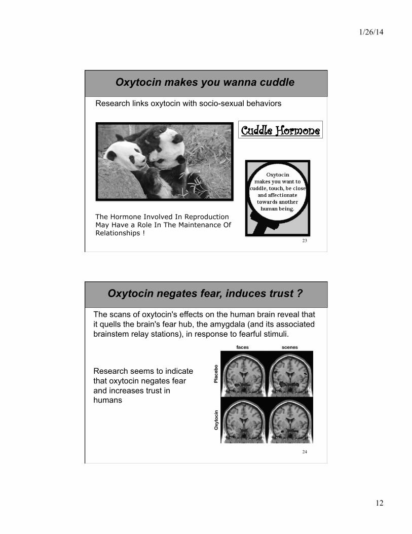
Research links oxytocin with socio-sexual behaviors
The Hormone Involved In Reproduction May Have a Role In The Maintenance Of Relationships !
Cuddle Hormone
Oxytocin makes you wanna cuddle
24
Oxytocin negates fear, induces trust ?
The scans of oxytocin's effects on the human brain reveal that it quells the brain's fear hub, the amygdala (and its associated brainstem relay stations), in response to fearful stimuli.
Research seems to indicate that oxytocin negates fear and increases trust in humans
1/26/14
13
25
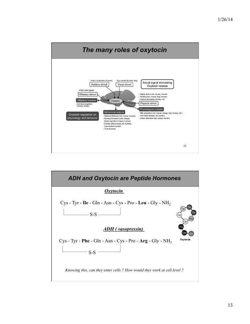
The many roles of oxytocin
Oxytocin
Cys - Tyr - Ile - Gln - Asn - Cys - Pro - Leu - Gly - NH2
S-S
ADH ( vasopressin)
Cys - Tyr - Phe - Gln - Asn - Cys - Pro - Arg - Gly - NH2
S-S
ADH and Oxytocin are Peptide Hormones
Knowing this, can they enter cells ? How would they work at cell level ?

1/26/14
14
27
Oxytocin for sale !