chapter 19 blood vessels
TRANSCRIPT

Copyright © 2004 Pearson Education, Inc., publishing as Benjamin Cummings
Human Anatomy & Physiology, Sixth Edition
Elaine N. Marieb
PowerPoint® Lecture Slides prepared by Vince Austin, University of Kentucky
19The Cardiovascular System: Blood Vessels

Copyright © 2004 Pearson Education, Inc., publishing as Benjamin Cummings
Blood Vessels
Blood is carried in a closed system of vessels that begins and ends at the heart
The three major types of vessels are arteries, capillaries, and veins
Arteries carry blood away from the heart
Veins carry blood toward the heart
Capillaries contact tissue cells and directly serve cellular needs

Copyright © 2004 Pearson Education, Inc., publishing as Benjamin Cummings
Generalized Structure of Blood Vessels
Arteries and veins are composed of three tunics – tunica interna, tunica media, and tunica externa
Lumen – central blood-containing space surrounded by tunics
Capillaries are composed of endothelium with sparse basal lamina

Copyright © 2004 Pearson Education, Inc., publishing as Benjamin Cummings
Generalized Structure of Blood Vessels
Figure 19.1b

Copyright © 2004 Pearson Education, Inc., publishing as Benjamin Cummings
Tunics
Tunica interna (tunica intima)
Endothelial layer that lines the lumen of all vessels
In vessels larger than 1 mm, a subendothelial connective tissue basement membrane is present
Tunica media
Smooth muscle and elastic fiber layer, regulated by sympathetic nervous system
Controls vasoconstriction/vasodilation of vessels

Copyright © 2004 Pearson Education, Inc., publishing as Benjamin Cummings
Tunics
Tunica externa (tunica adventitia)
Collagen fibers that protect and reinforce vessels
Larger vessels contain vasa vasorum
Copyright © 2004 Pearson Education, Inc., publishing as Benjamin Cummings
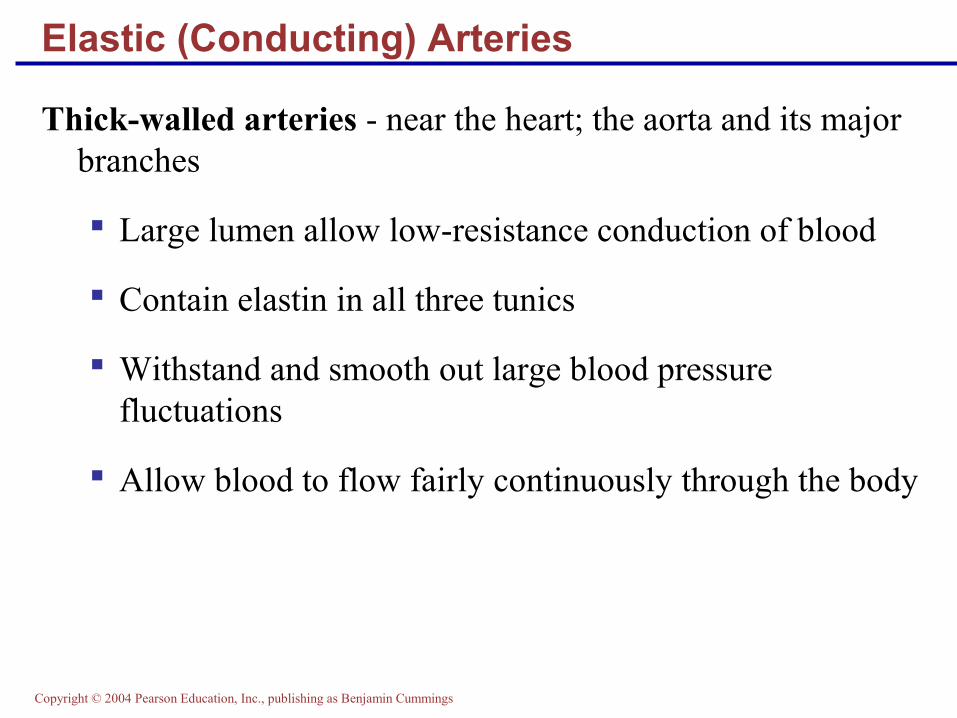
Elastic (Conducting) Arteries
Thick-walled arteries - near the heart; the aorta and its major branches
Large lumen allow low-resistance conduction of blood
Contain elastin in all three tunics
Withstand and smooth out large blood pressure fluctuations
Allow blood to flow fairly continuously through the body

Copyright © 2004 Pearson Education, Inc., publishing as Benjamin Cummings
Muscular (Distributing) Arteries and Arterioles
Muscular arteries – distal to elastic arteries; deliver blood to body organs
Have thick tunica media with more smooth muscle and less elastic tissue
Active in vasoconstriction
Arterioles – smallest arteries; lead to capillary beds
Control flow into capillary beds via vasodilation and constriction

Copyright © 2004 Pearson Education, Inc., publishing as Benjamin Cummings
Capillaries
Capillaries - the smallest blood vessels
Walls consisting of a thin tunica interna, one cell thick
Allow only a single RBC to pass at a time
There are three structural types of capillaries: continuous, fenestrated, and sinusoids

Copyright © 2004 Pearson Education, Inc., publishing as Benjamin Cummings
Capillary Beds
Figure 19.4a

Copyright © 2004 Pearson Education, Inc., publishing as Benjamin Cummings
Capillary Beds
Figure 19.4b

Copyright © 2004 Pearson Education, Inc., publishing as Benjamin Cummings
Blood Flow Through Capillary Beds
Precapillary sphincter
Cuff of smooth muscle that surrounds each true capillary
Regulates blood flow into the capillary

Copyright © 2004 Pearson Education, Inc., publishing as Benjamin Cummings
Venous System: Venules
Venules
Are formed when capillary beds unite
Allow fluids and WBCs to pass from the bloodstream to tissues
Large venules have one or two layers of smooth muscle (tunica media)

Copyright © 2004 Pearson Education, Inc., publishing as Benjamin Cummings
Venous System: Veins
Veins
Formed when venules converge
Composed of three tunics, with a thin tunica media and a thick tunica externa consisting of collagen fibers and elastic networks

Copyright © 2004 Pearson Education, Inc., publishing as Benjamin Cummings
Venous System: Veins
Veins have much lower blood pressure and thinner walls than arteries
To return blood to the heart, veins have special adaptations
Large-diameter lumens, which offer little resistance to flow
Valves (resembling semilunar heart valves), which prevent backflow of blood
Venous sinuses – specialized, flattened veins with extremely thin walls (e.g., coronary sinus of the heart and dural sinuses of the brain)

Copyright © 2004 Pearson Education, Inc., publishing as Benjamin Cummings
Systemic Blood Pressure
Figure 19.5

Copyright © 2004 Pearson Education, Inc., publishing as Benjamin Cummings
Factors Aiding Venous Return
Figure 19.6

Copyright © 2004 Pearson Education, Inc., publishing as Benjamin Cummings
The vascular system has two distinct circulations:
Pulmonary circulation – short loop that runs from the heart to the lungs and back to the heart
Systemic circulation – routes blood through a long loop to all parts of the body and returns to the heart
Circulatory Pathways

Copyright © 2004 Pearson Education, Inc., publishing as Benjamin Cummings
Differences Between Arteries and Veins
Arteries Veins
DeliveryBlood pumped into single systemic artery – the aorta
Blood returns via superior and interior venae cavae and the coronary sinus
LocationDeep, and protected by tissue Both deep and superficial
Pathways Fair, clear, and defined Convergent interconnections
Supply/drainage Predictable supplyDural sinuses and hepatic portal circulation

Copyright © 2004 Pearson Education, Inc., publishing as Benjamin Cummings
Figure 19.17a
Pulmonary Circulation

Copyright © 2004 Pearson Education, Inc., publishing as Benjamin Cummings
Figure 19.18
Systemic Circulation

Copyright © 2004 Pearson Education, Inc., publishing as Benjamin Cummings
Know these arteries!

Copyright © 2004 Pearson Education, Inc., publishing as Benjamin Cummings

Copyright © 2004 Pearson Education, Inc., publishing as Benjamin Cummings Figure 19.20b
Arteries of the Head and Neck

Copyright © 2004 Pearson Education, Inc., publishing as Benjamin Cummings
Copyright © 2004 Pearson Education, Inc., publishing as Benjamin Cummings
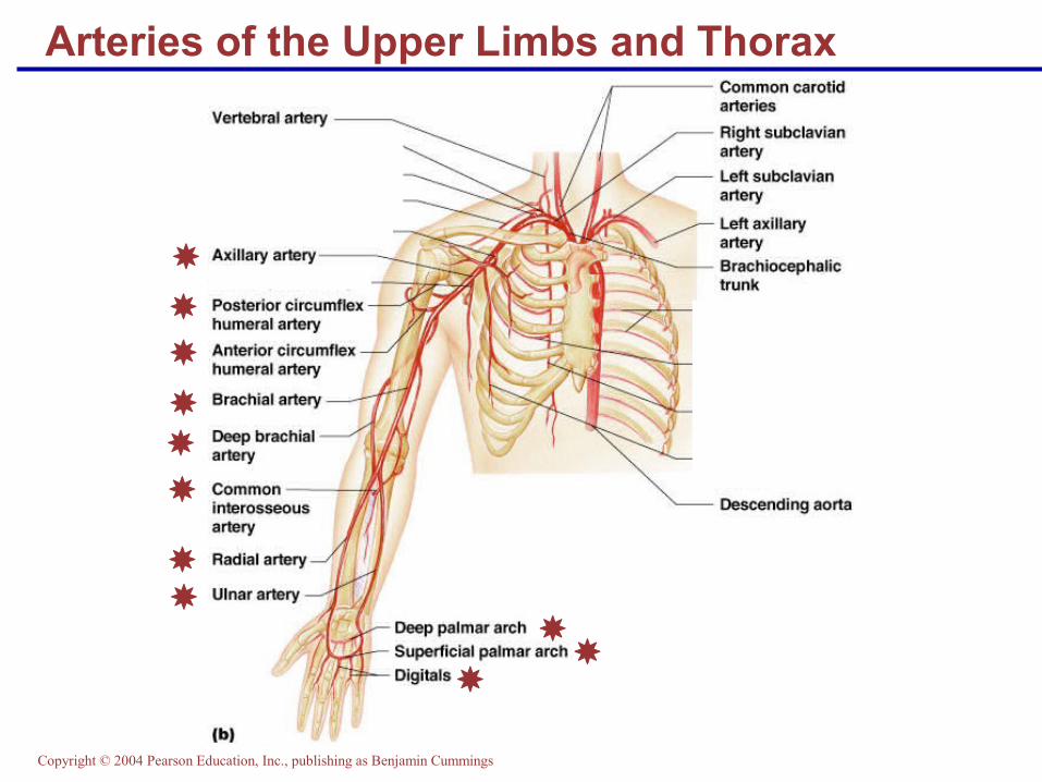
Arteries of the Upper Limbs and Thorax

Copyright © 2004 Pearson Education, Inc., publishing as Benjamin Cummings
1
2
3
4
5
6
7

Copyright © 2004 Pearson Education, Inc., publishing as Benjamin Cummings
Figure 19.22c
Arteries of the Abdomen
1
2
3
4
5
6
7

Copyright © 2004 Pearson Education, Inc., publishing as Benjamin Cummings
Figure 19.22d
Arteries of the Abdomen

Copyright © 2004 Pearson Education, Inc., publishing as Benjamin Cummings

Copyright © 2004 Pearson Education, Inc., publishing as Benjamin Cummings
Arteries of the Lower Limbs
Figure 19.23b, c

Copyright © 2004 Pearson Education, Inc., publishing as Benjamin Cummings
Figure 19.24b
Know these veins!

Copyright © 2004 Pearson Education, Inc., publishing as Benjamin Cummings
Figure 19.25b
Veins of the Head and Neck

Copyright © 2004 Pearson Education, Inc., publishing as Benjamin Cummings
Figure 19.26b
Veins of the Upper Limbs and Thorax

Copyright © 2004 Pearson Education, Inc., publishing as Benjamin Cummings
Figure 19.27b
Veins of the Abdomen

Copyright © 2004 Pearson Education, Inc., publishing as Benjamin Cummings
Figure 19.27c
Veins of the Abdomen

Copyright © 2004 Pearson Education, Inc., publishing as Benjamin Cummings
Figure 19.28b, c
Veins of the Pelvis and Lower Limbs