chapter 2. listeria biofilm formation on abiotic...
TRANSCRIPT
Page | 21
Chapter 2. Listeria Biofilm formation on abiotic surfaces
related to food processing environments
[2.1]. Introduction
In the recent years behavior of bacteria in complex communities or consortia, which are naturally
more diffuse in natural environments than freely planktonic single cells, has been increasingly studied.
Main reason for this interest is due to the “methodological holistic approach”, which changed object of
study from single individuals to interaction between bacterial species and within multispecies complex
communities (Kuramitsu, et al. 2007).
The technical advances in microscopic techniques and the molecular methods of investigation has
revealed that more than 90% bacteria in natural environments exist as biofilms adhered to different
substrates, instead of freely floating cells, even if this phenomenon was already noted in first half of
twentieth century (Hall-Stoodley, et al. 2004). Earlier biofilms were defined as passive cellular
aggregates simply adhered on surfaces.
Currently there is a strong conviction that biofilms represent dynamic biological systems, that are
highly structurated. This kind of association is also possible to be observed in human body, where it is
correlate to medical devices (lenses, catheters or prostheses) leading, as final result, to chronic infections.
Similar structures can be found also in food processing environments, where, if composed of pollutant or
pathogen species, biofilms can cause food spoilage and foodborne diseases (Hall-Stoodley, et al. 2004;
Ilyina, et al. 2004; McLandsborough, et al. 2006; Palmer and Stoodley, 2007).
[2.2]. Defining biofilms and understanding their architectural complexity
The most complete definition of biofilms is “spatially and metabolically structured microbial
communities embedded in an extracellular polymer matrix and located at a phase interface”. Currently
there are four different known types of interphase confines, where biofilms can develop (Nikolaev and
Plakunov, 2007):

Page | 22
Liquid (or any aqueous medium)-solid surface
Liquid-air
Between two immiscible liquid systems
Solid surface-air
Many experiments focused on exploiting bacterial biofilms and their characteristics reported in
literature have focused on slid surface immersed in liquid systems, but within this category several forms
are included:
1) Monolayer: single layer of cells of one or more microbial species, within which it is
not possible to distinguish any morphological differentiation apart form an internal
and an external part;
2) Mats: they are multispecies communities, in which different bacteria establish
association between each other leading to stratificated structures. The thickness of
mats can reach even ten centimeters and is regulated on gradient of key factors
involved (e.g. light, nutrients or oxidation potential);
3) Plaques: dental biofilms are bacterial communities extensively studied and described,
where hundreds of bacteria can coexist simultaneously;
4) Films in turbulent flow with band-like appearance due to flow tensile strength;
5) Fungi: in this three dimensional structure bacterial create an articulate and complex
architecture rich in channels, voids and pores. Many researchers have also studied role
of quorum sensing process in similar phenomenon;
6) Benthic and river sediments (Nikolaev and Plakunov, 2007).
From above mentioned categories it is possible to conclude that biofilm spatial architecture and
organization of biofilm can be strongly related to environmental conditions to which cells are exposed
and thus biofilms can be thought as best survival strategies adopted by microorganisms: if cells grow in
turbulent aqueous flows, it is more probable that adopted form could be filamentous streamers, while, in
case of backwater-like conditions, tower-like appendices are likely to be formed. Environment does not
select just for more appropriate forms and/or architectures, because its influence is more wide-ranged:

Page | 23
Environment includes also chemical gradients of molecules which can affect gene
expression patterns resulting in a modified cellular phenotype more suitable to survive
in special conditions;
Chemical and physical attributes of natural locations can also be thought as selective
agents for the detection of more suitable and competitive microbes in given
conditions;
Natural factors can also modify tolerance against bactericidal agents and improve
environmental adaption of microbes (Stewart and Franklin, 2008).
[2.3]. Stages of biofilm formation
Biofilm development results from several steps of a dynamic process (Fig. 2.1): solid surfaces,
once submerged in liquids, change their superficial attributes because they are covered by different
materials (conditioning films). In this phase superficial properties of materials are radically modified in
order to allow the attachment of living microorganisms. An example comes from dental plaque, where
proteins of saliva play an important role in the attachment of different bacteria: Rosan and Lamont
(2000) considered the layer of salivary pellicle onto teeth to be a “biologically active substrate to which
the pioneer organism may attach”. This conclusion was confirmed by Rudney (2000): this author
reported that, because they possess negatively charged domains, salivary proteins formed a thin pellicle
(or enamel), which act as a conditining layer for bacterial attachment on oral cavity surfaces. The
importance of the enamel in the bacterial attachment onto oral cavity surfaces was confirmed also by
Marsh (2004) and by Jenkinson and Lamont (2005).

Page | 24
Fig. 2.1: Biofilm formation in turbulent flow with relevant stages of the process. [from
http://wvlc.uwaterloo.ca/biology447/Biofilms/biofilmsoverview.htm, last acces 12/10/2010]
Next step is reversible adhesion, in which nonspecific interaction forces and cell superficial
elements are involved and bacteria are not strongly linked to the surface material. After this, cells lose
their intrinsic motility and begin the excretion of extracellular polymeric substances (EPS), which form
organic amorphous matrix in which cells are embedded. At this point microcolonies are formed on
surfaces (Irreversible adhesion). The secondary colonizers starts attaching on surfaces of the primary
colonizers leading to thickning of the biofilm and to the formation of specific architectural elements (like
channel, cavities and tower-like appendices) in the biofilm.
These structural elements play an important role in nutrient movements and waste removal
strengthening the idea of spatially organized community in which form is perfectly linked with the
assumed function (deBeer, et al. 1994). Mature biofilm can increase its thickness and complexity for
long period of time, after which structural degradation and cell dispersal take place. This last step has
been seen in biofilm formation in turbulent condition, where, once reached its maximum of growth, cells
and aggregates of organic matter detach from the biofilm and colonize new environments.
[2.4]. Adherence ability and biofilm formation of Listeria
The bacterial adherence onto surfaces, as well as growth and main growth-related parameters of
different strains (Lianou, et al. 2006), is differently expressed within Listeria, even from strains isolated
in the same environmental niche: Kuswahara and Muriana (2009) examined Listeria spp. isolates from
three different meat processing plants observing different prevalences. Within isolates from the same
plant authors observe large variation in terms of the capacity of Listeria to adhere on available surfaces,
even if two of the three examined plants involve only weak and moderate adhesive strains. The third

Page | 25
plant, which was also the oldest one, included also very firmly attached L.monocytogenes probably
related to its age of construction and level of sanitation practices adopted .
The origin of Listeria strains was investigated for its influence on bacterial adhesion to surfaces
and consequent biofilm formation. Tresse et al. (2007) found weak but significative variations among
101 examined strains in function of colonized surface and origin of the strain. Clinical isolates showed
better adhesive ability on polystyrene than stainless steel, while the industrial strains were more adherent
than clinical isolates. Unfortunately no possible relation with belonging to a specific genetic lineage was
observed, even if 4b strains are reported as less adhered than 1/2a and 1/2b. This observation was
confirmed also by Moltz and Martin (2005), Lianou et al. (2006) and Bonaventura et al. (2008).
Despite the large amount of studies regarding Listeria spp. biofilms, there are still many questions
and unclear aspects. The present study aims at studying the development mechanisms of Listeria spp.
biofilms on different materials. Moreover, adhesion under static conditions will be compared with
adhesion in turbulent conditions; factors influencing surface attachment (e.g.: temperature, nutrients) will
also be investigated.
[2.5]. Materials and methods
I. Test materials
Stainless steel coupons were used to test Listeria adhesion in static conditions of incubation. 0,1
mm thickness wire of stainless steel (SS), polyethylene terephtalate (PET), and Copper (C), which were
acquired from local supermarkets, were used as surface test materials in dynamic regime tests, covering
a large range of possible materials present in food transforming locations.
II. Bacterial strains
Listeria spp. strains were reported in Tab. 2.1. Each strain was grown on ALOA (Agar Listeria
Ottaviani & Agosti, Biolife Italiana Srl) plate and in Blood Agar (LABM, United Kingdom) at 37°C for
48 and 24 hours, respectively. These two media were used for species identification and β-hemolysis

Page | 26
assay, respectively. Single colonies from the plates were transferred into Brain Heart Infusion (BHI;
LABM, United Kingdom) and incubated for 16-18 h at 37°C and 200 rpm in a shaking bath. Stock
cultures were kept at -70°C.
III. Static biofilm on stainless steel
Stainless steel coupons (Fig. 2.2) were used as test surface: each coupon has ten squares (1 cm x
1.cm x 0.1 cm), organized in two rows, for an overall superficial area of 1 cm2. Selected strains reported
in Tab. 2.1 were subcultured in BHI broth for 18 h at 200 rpm at 37°C. Each strain was harvested at 4000
xg for 5 min and washed twice with sterile peptone water. Cell numbers were determined by serial
dilutions and plating on TSA (Tryptone Soy Agar, LABM, United Kingdom) plates, after incubation for
at 37°C 24 h. In parallel to plate counting, optical density at 600 nm (OD600) was measured on the same
dilutions, in order to have a rapid evaluation of bacterial density. Aliquots were taken from aqueous cell
suspensions and inoculated into 25 ml BHI Broth. The volume of aliquots to be added into 25 ml BHI
broth, was calculated in order to provide a density of 105-10
6 CFU/ml. Inoculated medium was added
into a Petri plate containing a previously washed and autoclaved steel coupon. After 24 h incubation at
37°C, the coupon was transferred into a new Petri plate, into which 25 ml of 10% diluted BHI (dBHI).
The so treated coupon was incubated for 7 days at 37°C. At defined periods (0, 3 and 7 days) coupons
were scraped with wet cotton swabs. Swabs were serially diluted into sterile peptone water and plated
onto TSA plates, which were incubated as above. Three replicates were performed for each strain.

Page | 27
Fig. 2.2: Stainless steel surface used for monitoring bacterial adhesion.
Strains Serotype Β-hemolysis Description UC 8410 - - L. innocua
cheese brine isolate UC 8409 - - L. innocua
cheese brine isolate EELA Oulu ba 2392/4 1/2a - L.monocytogenes
Fetal bovine brain EELA Hki L211 1/2a - L.monocytogenes
Cold smoked rainbow trout foodborne out break Food isolate
EELA Hki L627 3a - L.monocytogenes Butter foodborne outbreak Food isolate
KTL IHD 42526 1/2a - L.monocytogenes Cold smoked rainbow trout foodborne out break Clinical isolate
KTL IHD 42573 3a - L.monocytogenes Butter foodborne out break Clinical isolate
Tab. 2.1: Listeria spp. strains with relevant properties of interest and their origin of isolation.
IV. Biofilm formation test under dynamic conditions
The apparatus (Fig. 2.3) was composed by 5 glass tubes (10 cm long, internal diameter 0. 4 mm),
into which the wire test material was introduced, linked with teflon junctions to a solution dispenser (30
cm long, i.d. 20 mm). The apparatus was chemically disinfected with an succession of disinfection

Page | 28
solutions (NaOH 0.5 mol and 50% EtOH) and washed with sterile physiological solution. An over-night
culture of L. innocua strains in skimmed milk was introduced into the system at 1 ml/min flow rate and,
when the pipeline was full, the flow was stopped and the whole system was incubated at 37°C for 1h. A
washing step with physiological solution was performed to remove only slightly adhered cells. After this
step, the nutrient solution was flown inside the appatatus at a flow rate for 1 ml/min. At defined time
points (0, 3, 6, 18, and 24 h), the flow was stopped and aliquots of both tested materials and eluate were
taken and subjected to serial dilutions and plating on BHI agar, to count both sessile and plancktonic
cells, respectively. All the experiments were performed in triplicate.
Fig. 2.3: Schematic representation of employed system with enlargement of glass tube (Xn) in which the wire of testing
material was inserteed. F1: nutrient solution (skimmed milk) input, T1: exhausted nutrient solution, Vn is referred to valve to
open and close the flow of milk, while Ln represent the tubes to allow flow moving.
V. Sample preparation for SEM observation
Each sample was subjected to 8 h dehydration steps with increasing concentration of ethanol (70,
85, 95 and 100 %). The samples were then put into a Critical Point Dryer (Bal Tec CPD 030, Pubish),
where several washings with liquid CO2 (99,5%, Sapio) were performed until the CO2 critical point
(40°C and 7 atm was reached. The so treated sample is completely dehydrated and stored under vacuum.

Page | 29
Further steps were metallization by doration through Top Autocoater SC20 (Pubish) and storage in
dessicator. Each sample was examined by Scanning Electron Microscope (SEM) FEI ESEM XL30.
VI. Microbial adhesion to solvents (MATS)
This assay was performed as described by Brianet et al. (1999): old liquid culture was harvested at
2500 xg for 5 min and washed twice with 0.85 % NaCl in water. Cells suspension was divided into 2.4
aliquots, to which 0.4 ml of hexadecane (Sigma-Aldrich) was added. The mixture was vortexed for 1
min and then incubate at room temperature for 15 min, to allow complete separation of the two phases
composing the mixture. 1 ml was taken from the mixture and optical density at 400 nm was measured.
Results were collected from three replicates obtaining by using two indipendent subcultures and were
expressed as percentage of affinity with solvent by using the equation:
% affinity with the solvent = 100 x [1 –(A/A0)]
VII. Statistical analysis and treatment of data
Data were statistically analysed by SPSS 14.0, to One-way ANOVA, t test and post-hoc analysis
in order to detect significant parameters and quantitative definition of influence of selected parameter
(i.e. strain, temperature, nutrients, surface material) .
[2.6]. Results
I. Adhesion under static conditions
Different strains of Listeria from 7 up to 8 Log were examined for their capacity to adhere on
stainless steel and their ability to survive on this surface with limited amount of nutrients. Compared to
TSB, BHI supports better microbial growth and thus supposely helps to distinguish more clearly any
eventual difference between tested strains. dBHI was chosen to assess survival and biofilm formation of
examined strains under limited presence of nutrients. Dilution factor of growth medium was, instead of
0.5 or 0.2x, 0.1x, to simulate nutrient depletion. Obtained results are reported in Fig. 2.4. L. innocua UC

Page | 30
8410 (isolated from cheese brine) was compared with two L. monocytogenes isolates from European
cases of foodborne listeriosis. L. monocytogenes EELA HKi L211 was isolated from a sample of the
cold smoked rainbow trout involved in the listeriosis outbreak occurred in 1994-1995 in Sweden, while
L. monocytogenes KTL IHD 42526 was isolated from a hospitalized patient of same episode. L.
monocytogenes EELA Hki L627 and L. monocytogenes KTL IHD 42573 were food and clinical isolates,
respectively, from butter-related listeriosis outbreak. L. monocytogenes Oulu ba 2392 is referred to
clinical animal case.
After 24 h ours of incubation Listeria spp. were present at high levels (more than 107 CFU/cm
2),
even if the extent of adhesion was slightly different among tested isolates. EELA Hki L211 was revealed
the least adherent strain, while Oulu ba 2392/4 and EELA Hki L627 exhibited the highest adhesion on
stainless steel, reaching levels greater than 108 CFU/cm
2. Strong similarity in surface adhesion was
observed in the both clinical isolates (KTL IHD 42573 and 42526), while corresponding food isolates
presented different extent of adhesive properties. Despite the quantitatively different extent of adhesion,
investigated strains did not significantly differ from each other, demonstrating that both the species and
origin of isolation cannot completely account for diversified bacterial attachment on surface.
When the strains were grown under nutrient depletion, a geneally common trend of behavior was
observed: incubation in these conditions led to a 3-fold stronger overall reduction at 168 h (1.5 Log
reduction factor) than 72 h (0,4 LOG). Nevertheless, bacteria were not completely killed by the
extended incubation, and the counts of the most susceptible strain, UC 8410, never decreased at levels
lower than 103 CFU/cm
2 after 168 h). Neither foodborne outbreak-related clinical isolates were strongly
affected by prolonged limited source of nourishment (0.5 LOG reduction factor for both the isolates)
unlike the food isolates. The animal clinical isolate Oulu ba 1283/2 had shown to be notably susceptible
to nutrient depletion. Even if it showed higher values, EELA Hki L627 was 1-fold more susceptible than
EELA Hki L211 within 168 h (0,25 LOG reduction compared with 1.5 LOG, repsectively). Statistical
analysis did not detect any significant differences between the strains of different origins.

Page | 31
UC 8410
24 h 72 h 168 h 0
1
2
3
4
5
6
7
8
9
Time
LO
G(C
FU
)
EELA Oulu ba 2392/4
24 h 72 h 168 h 0
1
2
3
4
5
6
7
8
9
Time
LO
G(C
FU
)EELA Hki L211
24 h 72 h 168 h 0
1
2
3
4
5
6
7
8
9
Time
LO
G(C
FU
)
KTL IHD 42526
24 h 72 h 168 h 0
1
2
3
4
5
6
7
8
9
Time
LO
G(C
FU
)
EELA Hki L627
24 h 72 h 168 h 0
1
2
3
4
5
6
7
8
9
Time
LO
G(C
FU
)
KTL IHD 42573
24 h 72 h 168 h 0
1
2
3
4
5
6
7
8
9
Time
LO
G(C
FU
)
Fig. 2.4: Adhesion assay of Listeria spp. strains for 7 days at 37°C. Bars represent LOG(CFU) referred to an overall
superficial area of 1 cm2. Data are expressed as mean of three replicates ± standard error mean.

Page | 32
II. Bacterial adhesion in turbulent flows
In the scientific literature static apparatus has been commnonly usedto test the biofilm forming
ability of strains. There are instead few reports about biofilm formation under moving flows. For this
purpose a suitable apparatus was designed (Fig. 2.3). Among the studied strains L. innocua UC 8410 was
chosen for biofilm formation assay under dynamic regimen together with L. innocua strain UC 8409
(isolated from same source), in order to verify if strains isolated from the same environmental niche
could possess the same adhesion ability. These strains were also selected as a model of the pathogenic
species L.monocytogenes.
The first experiment was conducted with stainless steel wire (20 cm long, 1 mm i.d): stainless
steel was chosen because of its common use in food manufacturing plants. Milk was employed as
nutrient solution as a model of food, which supports the surface adhesion and functions as a nourishment
source for bacteria. Experiment was performed with skimmed milk to avoid any effect attributable to fats
and lipids. The apparatus used allowed the analysis of biofilm under nutrient flow on different materials.
A wire of tested materials (in this case SS) was introduced into a glass pipe and then exposed, after
initial inoculum (i.e. static incubation of the wire for 1 h in presence of an over-night bacterial culture)
within the incubation cell itself, to skimmed milk flow for 24 h. At defined periods of time, the flow was
temporarily stopped, in order to allow sampling of aliquots of tested surfaces and nutrient solution,
which were used to monitor sessile and planktonic cells, respectively (Fig. 2.5).
After 1 h incubation, UC 8410 showed a gradually increasing attachment to the SS wire, of which
the maximum reached level was 108 CFU/cm
2 after 6 h of monitoring in presence of a milk flow. Despite
the high density of cells freely suspended in growth medium, UC 8409 was demonstrated not to be able
to adhere on tested surfaces in the same experimental conditions. From these results, it was observed that
UC 8410 possessed a higher adhesive capacity than UC 8409 and thus was able to form biofilms under
dynamic regimen.

Page | 33
UC 8409
0 3 6 180
2
4
6
8
10
Time (hours)
LO
G(C
FU
)
UC 8410
0 3 6 180
2
4
6
8
10
Time (hours)
LO
G(C
FU
)
Fig. 2.5: Surface attachment of L. innocua strains on stainless steel wire at 37°C. Sessile cells (white bars)are expressed as
LOG(CFU/cm2) superficial area, while planktonic cells (grey bars) are expressed as LOG(CFU/ml). 0 h is referred to 1 h
incubation of over-night colture in skimmed milk, while value obtained until 18 h demonstrate adhesion capacity of the two
employed strains during 18 h testing period under dynamic conditions. 1 LOG(CFU) is to be intended as less than 10 CFU.
III. Cell superficial hydrophobicity
Based on the data obtained using SS in the presence of a milk flow, cell hydrophobicity of both L.
innocua UC 8409 and UC 8410 was measured, in order to obtain an explanation to the differing adhesive
capacity of the analysed strains. The test was conducted using protocol described by Briandet et al.
(1999). Microbial adhesion to solvents assay (MATS) was performed in triplicate for the two strains and
the results are shown in Fig. 2.6.
UC 8410 UC 84090
5
10
15
20
25
Aff
init
y w
ith
hexad
ecan
e (
%)
Fig. 2.6: Percentage of affinity of Listeria innocua strains with hexadecane. Bars represent the average of three replicates ±
standard error of mean.

Page | 34
UC 8410 exhibited low affinity with hexadecane, demonstrating strong hydrophilic properties of
overall cell surface, while UC 8409 showed a better comnpatibilty with the employed solvent. As
reported by Bellon-Fontaine et al. (1996), Briandet et al. (1999) and Takahashi et al. (2010), L. innocua
UC 8410 was demonstrated not to possess relevant electron-donating and Lewis acid properties, which
were instead much more relevantly shown in UC 8409. The great quantitative difference among the two
isolates suggested that the adhesive capacity of a strain could partially be related to intrinsic superficial
properties of the cell itself, altough it cannot be excluded the contribution of other factors involved in
this phenomenon.
IV. Surface materials
The second aim was to study if the test surface could affect L. innocua UC 8410 adhesion and thus
biofilm formation in the presence of shear forces due to flowing solution. Three different materials were
tested (SS, PET, C). The analysis of bacterial counts conducted on all tested material (shown in Fig. 2.7)
indicated that UC 8410 exhibited relevant adhesive capacity, whose reached levels depended on
employed materials.
L. innocua UC 8410, in presence of SS, showed gradually increasing denisties on wire until to
levels almost comparable with the corresponding ones observed for the planktonic cells. When PET was
employed as testing surface, constant cell recoveries, which were lower than those obtained with SS,
were observed with regard to sessile cells, altough planktonic cell density was almost 109 CFU/ml in
both these samples. C was suggested not able to support the adhesion of milk protein or Listeria spp.,
because absence of colonies have been observed even at the lowest dilution since 3 h sampling (Fig. 2.7).
Statistical analyses on the data indicate that the tested materials affected significantly the surface
bacterial attachment (p < 0.01). Post hoc tests further revealed that both SS and PET showed strong
similarities (p > 0.01), but moved away significantly from C (p < 0.01).

Page | 35
Stainless steel (SS)
0 3 6 18 240
2
4
6
8
10
12
Time (hours)
LO
G(C
FU
)
Polyethylene terephthalate (PET)
0 3 6 18 240
2
4
6
8
10
12
Time (hours)
LO
G(C
FU
)
Copper (C)
0 3 6 18 240
2
4
6
8
10
12
Time (hours)
LO
G(C
FU
)
Fig. 2.7: Adhesion on different materials of L. innocua under dynamic conditions. Data are expressed as mean of three
replicates ± standard error mean (SEM). White bars: sessile cells, gray bars: planktonic cells. Sessile cells are expressed as
LOG(CFU/cm2), while planktonic cells are expressed as LOG (CFU/ml). 1 LOG(CFU) is to be intended as less than 10 CFU.

Page | 36
V. SEM examination of inoculated wires
All tested materials were examined with SEM, in order to observe growth of biofilm on tested
wires during the 24 h testing period (Fig. 2.8 to Fig. 2.12). SS showed a slightly rough surface, whose
irregularities could be used as contact point for cells to establish sessile consortia (Fig. 2.8). Deposition
of organic matter was observed in samples after 1 h of incubation with Listeria in milk suspension,
which lead to a complete coverage of the whole available surface (Fig. 2.9). 3 h samples showed the
presence of mushroom- like structures connected to each other (Fig. 2.10). After 6 h it was possible to
observe a multi-layered architecture of relevant thickness along the whole sample surface. The complex
matrix of biofilm covered the whole surface and seemedto hide bacterial cells, which were thus not
observed with SEM.
Fig. 2.8: Images of stainles steel coupon after chemical disinfection steps obtained at magnitudo of 150 (leftt image).
Fig. 2.9: SS sampled 0 h after initial inoculum. Magnitudo was 1900.000 and 4500.0000 for left and right image, respectively.
Page | 37
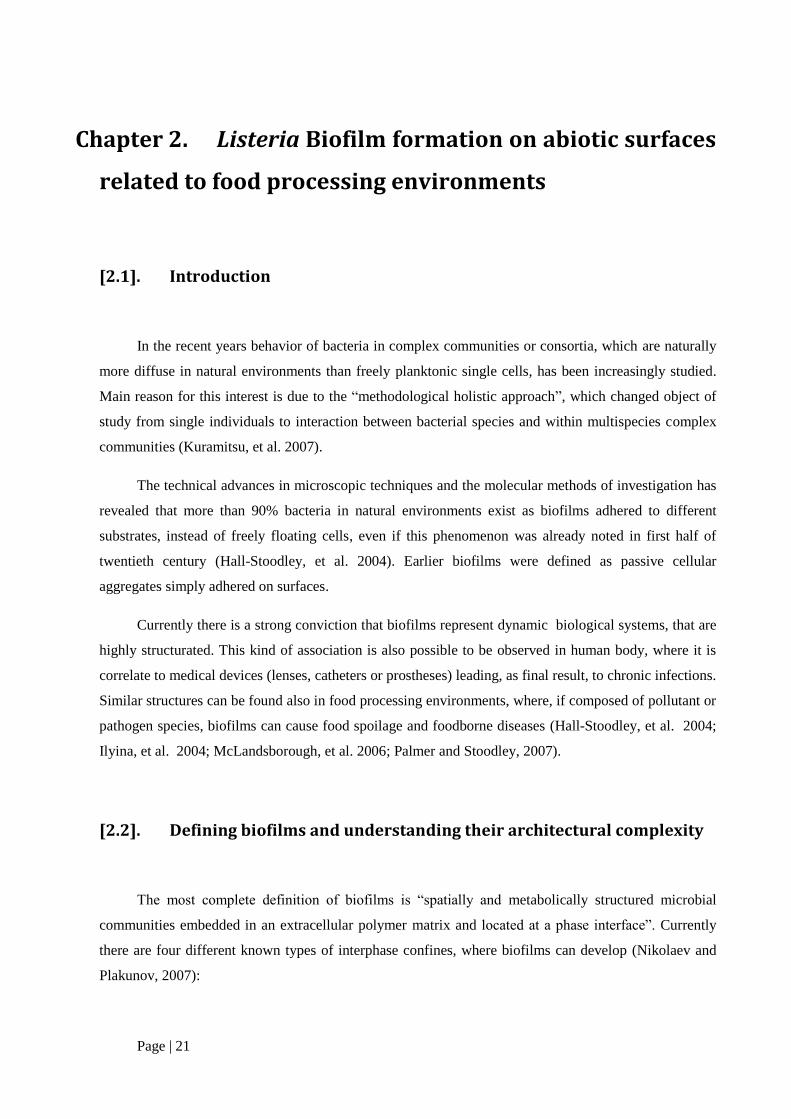
Fig. 2.10 SS specimen after 3 h from test beginning [magnitudo of 100 (left image) and 3052.311 (right image)].
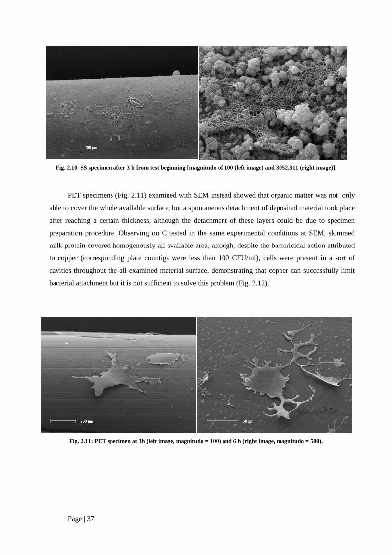
PET specimens (Fig. 2.11) examined with SEM instead showed that organic matter was not only
able to cover the whole available surface, but a spontaneous detachment of deposited material took place
after reaching a certain thickness, although the detachment of these layers could be due to specimen
preparation procedure. Observing on C tested in the same experimental conditions at SEM, skimmed
milk protein covered homogenously all available area, altough, despite the bactericidal action attributed
to copper (corresponding plate countigs were less than 100 CFU/ml), cells were present in a sort of
cavities throughout the all examined material surface, demonstrating that copper can successfully limit
bacterial attachment but it is not sufficient to solve this problem (Fig. 2.12).
Fig. 2.11: PET specimen at 3h (left image, magnitudo = 100) and 6 h (right image, magnitudo = 500).

Page | 38
Fig. 2.12: C specimen at 3 (left image, magnitudo = 15000) and 6 h (right image, magnitudo = 10000).
VI. Temperature
In order to assess the role of temperature on Listeria biofilm formation, further experiments were
performed. SS was choosen as a test surface, because, based on already obtained results, it gave
quantitative results concerning bacterial adhesion under the test conditons. The studied temperatures (20,
30 and 37°) allowed the comparison with results from former studies, in which static apparatus were
employed.
Both adhered and planktonic cells were evaluated with the same experimental procedure discussed
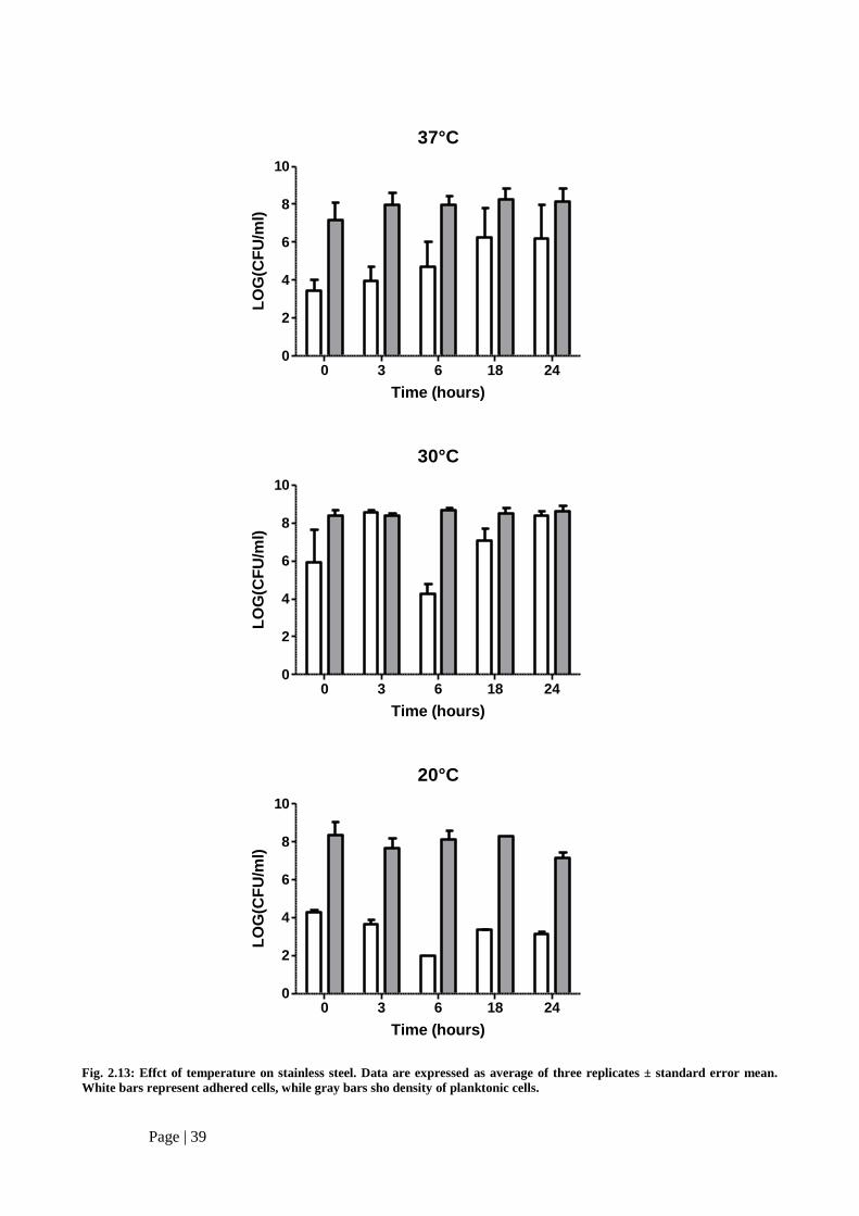
in par. III, sect. 2.5. Results are shown in Fig. 2.7. At 37°C the cell present in the flow of nutrient ranged
between 107-10
9 CFU/ml. Shifting the temperature from 37°C to 30°C did not result in a significant
amount of planktonic cells, which were kept almost constant (109 CFU/ml), while slight decrease was
observed in the recoveries of planktonic cells at 20°C both after 3 and 24 h.
Regarding the adhered cells, increasing recoveries were observed at 37°C up to 24 h, when the
number of recovered cells reached the maximum level (ranging between 106-10
8 CFU/cm
2). When the
dynamic apparatus was incubated at 30°C, the adhesion of the strain was notably modified: cells adhered
to SS wire were 106-10
8 CFU/cm
2 after initial inoculum and then raised up to 10
9 CFU/cm
2 at 3 h. A
sharp reduction until 104 CFU/cm
2 was observed at 6 h. Elapsing the time, cells present on taken
specimen of wire showed higher levels until to restore the density observed at 3 h (108-10
9 CFU/ cm
2 at
24 h), which slightly diverged from the value corresponding to planktonic cells at the same time period.
A similar trend was observed when temperature was reduced when temperature was lowered at
20°C, even if cell recoveries were significantly lower (2-fold lower than the corresponding one at 30°C
or 37°C). These reduced values could be attributed to a not efficient cellular metabolism when bacteria
were grown at non- optimal temperatures.
Page | 39
37°C
0 3 6 18 240
2
4
6
8
10
Time (hours)
LO
G(C
FU
/ml)
30°C
0 3 6 18 240
2
4
6
8
10
Time (hours)
LO
G(C
FU
/ml)
20°C
0 3 6 18 240
2
4
6
8
10
Time (hours)
LO
G(C
FU
/ml)
Fig. 2.13: Effct of temperature on stainless steel. Data are expressed as average of three replicates ± standard error mean.
White bars represent adhered cells, while gray bars sho density of planktonic cells.

Page | 40
VII. Influence of the medium on biofilm formation
In order to obtain a more detailed prospective of establishment of sessile communities in the
presence of turbulent flows, adhesion monitoring of diverse substrates was performed. Most of the
earlier studies have used growth medium such as BHI or TSB broth (diluted or not) to simulate adhesion
to food processing surfaces (Cheok and Schraft 2000,Kalmokoff et al. 2001Moltz and Martin 2005,
Tresse et al. 2007, Harvey et al. 2007) BHI was also included in the present study in order to compare
the collected results with previous findings. In parallel the same experimental conditions were applied
using skimmed milk, which reproduces realistical bacterial contamination of plants processing or
handling liquid foodstuffs. Beside comparison of these two mentioned media, role of microbial activity
in the formation of above mentioned multilayered architectures was investigated: in this case assessment
was conducted with skimmed milk supplemented with 10 μg/ml.
SS was employed as a test material due to its previously demonstrated ability to support bacterial
adhesion in the presence of shear forces, while inoculum was obtained through static incubation of the
wire in presence of an over-night culture of L. innocua UC 8410 (this treatment was not performed with
chloramphenicol supplemented milk). All these media were used into apparatus used in par. III for 24 h
at flow rate of 1 ml/min and, at defined time point, flow was interrupted to allow sampling of aliquots of
tested wire These specimen were examined through observations at SEM (Fig. 2.15).
In the presence of BHI (image A), no deposition of organic matter took place and no colonies
were detected even at lowest dilution, suggesting the incapacity of BHI to support bacterial attachment
on surfaces under dynamic conditions. When skimmed milk (image B) was used as a nutrient solution, it
was possible to observe tower-like appendices, which were then substituted by very thick multi-layered
structures, within which bacterial cells were probably embedded (although they were not found in any
sample examined at SEM). Even if there was a relevant deposition of organic materials on wire surfaces,
10 µg/ml chloramphenicol-supplemented milk did not achieve the same matrix architecture (also in
terms of number of observed structures typical of bacterial biofilms and strucutral complexity) observed
when L. innocua UC 8410 was grown with a flow of sterile milk. These architectural elements could
therefore be attributed to bacterial metabolic activity, instead of being considered as mere artefacts due
to milk protein deposition on SS wire.

Page | 41
Skimmed milk
0 3 6 18 240
2
4
6
8
10
12
Time (hours)
LO
G(C
FU
)
BHI broth
0 3 6 18 240
2
4
6
8
10
12
Time (hours)
LO
G(C
FU
)
Fig. 2.14: Adhesion of L. innocua UC 8410 on SS wire in presence of different substrates. White bars: sessile cells, gray bars:
planktonic cells. Sessile cells are expressed as LOG(CFU/cm2), while planktonic cells are expressed as LOG (CFU/ml). 1
LOG(CFU) is to be intended as less than 10 CFU. Error bars are omitted for clarity.

Page | 42
Fig. 2.15: SEM images of SS under flow of skimmed milk after 3h. A: SS wire in BHI broth (magnitudo = 2000, B: SS sample
inoculated with L. innocua UC8410 (magnitudo = 3052.311), C: SS wire exposed to 1 h contact with milk supplemented with
10 μg/ml chloramphenicol (magnitudo = 6250).
A
B
C

Page | 43
[2.7]. Discussion
Seven Listeria spp. strains (six L. monocytogenes and one isolate of L. innocua) were studied for
their capacity to attach and survive on stainless steel. To mimic the starvation conditions, the medium
was diluted up to 0.1% of the usual concentration, to assess its influence of strictly stringent depletion of
nutrients on survival capacity and surface adhesivity was one of investigated parameters. Limited
presence of nutrients resulted more notably significantly at 7 days than 3 days, altough differences
among employed strains were observed. Clinical isolates were much more tolerant to limited amounts of
nutrients than the food isolates, and L. monocytogenes proved to be much more robust than L. innocua,
although both the species were grown in non-optimal conditions. Possible association between the source
of isolation, species identity and bacterial adhesion were investigated, but no clear conclusions could be
drawn. Apparently neither the species or the origin can account for the differences detected. This finding
is in agreement with the results of Lianou et al. (2007) and Tresse et al. (2007).
Bacterial adhesion onto surface is a multifactorial phenomenon, in which several aspects were
investigated in the present research. The strain of the same species do not possess similar adhesivity.
This property is differently expressed among sporadic or persistent strains (Harvey et al. 2007,
Kushwaha and Muriana 2009). This data is in accord with previous results of Borucki et al. (2003),
which confirms the presence of interstrain variability within Listeria monocytogenes in biofilm
formation, which is not attributable to a certain serotype or phylogenetic division. Similar conclusion
were drawn by Moltz and Martin (2005), Bonaventura et al. (2008) and Lianou et al. (2006).
Most of the previous studies on biofilms have employed static apparatus (like microtiter plates or
stainless steel coupon), which can give useful information about strain adhesion, but, as underlined by
Perni et al. (2007), cells deposition is more likely to take place, instead of biofilm formation, in similar
conditions. Moreover flow cells repeat better the conditions of food processes. For this purpose a
suitable laboratory-sized system was constructed in order to monitor bacterial attachment onto the
surfaces in presence of a flowing solution, simulating bacterial contamination within a plant processing
liquid foodstuffs like milk.
Examination of hydrophobicity revealed major differences between the two L. innocua strains
used in the first trials of biofilm development under dynamic conditions. This fact could at least partially
be explained by the differences of adhesivity observed on stainless steel wire. This result is in agreement
with conclusions drawn by Briandet et al. (1999): the authors suggested that both electrical and Lewis
acid-base interactions play a role in the adhesion of L. monocytogenes Scott A on stainless steel coupon.
Analyzing their results, Takahashi et al. (2010) hypothesized that hydrophobicity and thus properties of

Page | 44
cellular surface are mainly involved in surface adhesion, while Lewis acid-base characteristics seem not
to be so markedly important , but not enough to be completely excluded.
In the present investigation attention was given on few selected parameters, in order to test the
compliance of the used apparatus with the devices used in literature by other authors: obtained results
seem to be in agreement with most of former reports not only in terms of final quantitative data obtained,
but also about significance of the monitored factors (Borucki, et al. 2003, Lianou et al. 2006, Mai and
Conner 2007, Kushwaha and Muriana 2009). Tested materials were chosen to reflect the variety of
possible equipment and surfaces present in food processing environments, ranging from stainless steel to
plastics (such as PET) and showing different capacities in supporting adhesion: it was interesting that
PET is closer to SS, altough this latter one was considered markedly different from C. Furthermore,
copper has been known to be bactericidal, but in our experiments it did not demonstrate to be able to
eradicate completely microbial contamination: SEM images show, when copper is used, very few cells
were enveloped within a very thin layer of organic matter superficially attached, even if counts are less
than 160 CFU/cm2.
Varying typology of nutrient, a different trend in surface colonization is shown: while plate counts
using BHI as growth substrate showed ineffective attachment to tested materials, skimmed milk
supported better the cells in the establishment of sessile communities. This was probably due to the
presence of amphoter proteins, which represent both a source of nourishment and physical support for
colonization. Parallel experiments in the same analytical conditions with BHI as a nutrient solution did
not produce the multi-layered structure. Trials with skimmed milk supplemented with 10 μg/ml
chloramphenicol did not bring about the same superficial tower-like architecture, in terms of both
amount and structural complexity, confirming the role of microbial activity in the formation of similar
complexes.
In literature it has been reported that milk and its components compete with Listeria cells for the
attachment on surfaces. Wong (1998) showed that milk and milk proteins significantly reduced cell
recoveries from stainless steel surfaces, even if milk could support the growth of bacteria on not
adequately cleaned surfaces. Similar conclusion were reported by Barnes et al. (1999): the authors
hypothesized that milk protein can inhibit bacterial attachment by surface characteristic modification
and by the establishment of a conditioning layer on tested material. Our results suggest instead an
opposite effect: milk did not only support the attachment and growth of Listeria in turbulent regime, but
also acted as a building material for multilayered structures, which could reduce significantly cellular
recoveries if a suitable disruptive method for such matrices is not applied. A similar phenomenon has
been observed with dental plaque (Rosan and Lamont, 2000; Marsh, 2004; Jenkinson and Lamont, 2005;
Filoche et al., 2010,)

Page | 45
Among the investigated parameters, temperature significantly affected the bacterial attachment, in
agreement with the findings of Mai and Conner (2007). The range of temperature used was chosen in
order not to create too strongly limiting condition (lowest temperature used was 20°C). When 37°C was
used as incubation temperature, an increasing adhesion to SS within 24 h was observed, while a stronger
variability was observed with lower temperatures. Both 30 and 20°C showed similar trends: after initial
attachment adhesion was reduced at 6 h and then increased after 12 h, even if obtained results are
quantitatively different (maximal difference observed was 4 LOG). Statistiscal analysis revealed that
37°C and 20°C did not significantly differ between each other and could be assumed as top and bottom
temperature concerning bacterial attachment under dynamic regimen. 30°C significantly move away
from other two investigated values despite sharing similar kinetic of attachment on SS.
Even if an effect attributable to the shift from optimal conditions for growth cannot be excluded,
temperature modified sensibly the entity of adhered cells maybe because of thermo-dependent regulation
of superficial attributes such as flagella. It has been suggested that flagellum-mediated motility is
required for initial cell attachment of Listeria during biofilm formation to overcome any repulsive
interfacial force. Flagella have also reported to act as adhesive support facilitating early attachment of
Listeria on stainless steel, even if Lemon et al. (2007) reported that for L.monocytogenes the real critical
factor for adhesion to and biofilm formation on abiotic surfaces is flagellum-mediated motility. In
agreement with findings by Djordjevic et al. (2002), Bonaventura et al. (2008) found negative correlation
between biofilm formation on stainless steel and swimming motility, suggesting that motility and related
characteristics are not required for the biofilm formation of L.monocytogenes and that other changes in
superficial structures apart of flagella contribute to attachment of the bacterium to solid surfaces.
Microbial cell surface charge and hydrophobicity play an important role in the initial steps of
microbial adhesion (van Loosdrecht et al., 1990). Bonaventura et al. (Bonaventura et al., 2008) suggested
that growth temperature can positively modulate the hydrophobicity of L.monocytogenes. Despite th
quantitative differences experimentally observed among employed strains in this investigation, no
significant difference was observed. Similar conclusions could be found in Takahashi et al. (2010),
where a positive correlation was found (even if less than 0.90). All these observations could suggest that
bacterial surface properties could be involveld in the attachment of Listeria on surfaces along with other
potential parameters, which should be further investigated.

Page | 46
[2.8]. Bibliografia
[1]. Barnes, L.-M., M. F. Lo, M. R. Adams, and A. H. L. Chamberlain. (1999) "Effect of milk
proteins on adhesion of bacteria to stainless steel surfaces." Appl. Eniviron. Microbiol. 65,
4543-4548.
[2]. Bellon-Fontaine, M.-N., J. Rault, and C. J. van Oss. (1996) "Microbial adhesion to solvents: a
novel method to determine the electron-donor/electron acceptor or Lewis acid-base properties
of microbial cells." Colloid. Surface B 7, 47-53.
[3]. Belval, S. C., L. Gal, S. Margiewes, D. Garmyn, P. Piveteau, and J. Guzzo. (2006)
"Assessment of the roles of LuxS, S-ribosyl Homocysteine, and autoinducer 2 in cell
attachment during biofilm formation by Listeria monocytogenes EGD-e." Appl. Environ.
Microbiol. 72, 2644-2650.
[4]. Bonaventura, G., R. Piccolomini, D. Paludi, V. D. Orio, A. Vergara, and M. Conter. (2008)
"Influence of temperature on biofilm formation by Listeria monocytogenes on various food-
contact surfaces: relationship with motility and cell surface hydrophobicity." J. Appl.
Microbiol. 104, 1552-1561.
[5]. Borucki, M. K., J. D.. Peppin, D. White, F. Loge, and D. R. Call. (2003) "Variation in biofilm
formation among strains of Listeria monocytogenes." Appl. Environ.Microbiol. 69, 7336-
7342.
[6]. Briandet, R. T., C. Meylheuc, C. Maher, and M. N. Bellon-Fontaine. (1999) "Listeria
monocytogenes Scott A: cell surface charge, hydrophobicity, and electron donor and acceptor
characteristics under different environmental growth conditions." Appl. Environ. Microbiol.
65, 5328-5333.
[7]. deBeer, D., P. Stoodley, F. Roe, and Z. Lewandowski. (1994) "Effects of biofilms structures
on oxygen distribution and mass transport." Biotechnol. Bioeng. 43, 1131-1138.
[8]. Djordjevic, D., M. Wiedmann, and L. A. Mclandsborough. (2002) "Microtiter plate assay for
assessment of Listeria monocytogenes biofilm formation." Appl. Environ. Microbiol. 68,
2950-2958.
[9]. Filoche, S., L. Wong, and C. H. Sissons. (2010) "Oral Biofilms: Emerging Concepts in
Microbial Ecology." J. Dent. Res. 89, 8-18.
[10]. Hall-Stoodley, L., J. W. Costerton, and P. Stoodley. (2004) "Bacterial biofilms: from the
natrual environment to infectious diseases." Nat. Rev. Microbiol. 2, 95-108.

Page | 47
[11]. Hood, S. K., and E. A. Zottola. (1995) "Biofilms in food processing." Food Control 6, 9-18.
[12]. Ilyina, T.S., Y. M. Romanova, and A. L. Gintsburg (2004) "Biofilms as a mode of existence
of bacteria in external environmental and host body: the phenomenon, genetic control, and
regulation systems of development." Russ. J. Genet. 40, 1189-1198.
[13]. Jenkinson, H. F., and R. J. Lamont. (2005) "Oral microbial communities inb sickness and in
health." Trends Nicrobiol. 13, 589-595.
[14]. Kalmokoff, M. L., J. W. Austin, X.-D. Wan, G. Sanders, S. Banerjee, and J. M. Farber.
(2001) "Adsorption, attachment and biofilm formation among isolates of Listeria
monocytogenes using model conditions." J. Appl. Microbiol. 91, 725-734.
[15]. Kuramitsu, H. K., X. He, R. Lux, and M. H. Anderson. (2007) "Interspecies interactions
within oral microbial communities." Microbiol. Mol. Biol. Rev. 71, 653-670.
[16]. Kushwara, K., and P. M. Muriana. (2009) "Adherence characteristics of Listeria strains
isolated from three ready-to-eat meat processing plants." J. Food Protect. 72, 2125-213.
[17]. Lemon, K. P., D. E. Higgins, and R. Kolter. (2007) "Flagellar motility is critical for Listeria
monocytogenes biofilm formation." J. Bacteriol. 189, 4418-4124.
[18]. Lendenmann, U., J. Grogan, and F. G. Oppenheim. (2000) "Saliva and dental plaque - A
review." Adv. Dent. Res. 14, 22-28.
[19]. Lianou, A., J. D. Stopforth, Y. Yoon, M. Wiedmann, and J. N. Sofos. (2006) "Growth and
stress resistance variation in culture both among Listeria monocytogenes strains of various
serotypes and origins." J. Food Prot. 69, 2640-2647.
[20]. Mai, T. L., and D. E. Conner. (2007) "Effect of temperature and growth media on the
attachment of Listeria monocytogenes to stainless steel." Int. J. Food Microbiol. 120, 282-286.
[21]. Mantel, S. "Die Empfindlichkeit von Listeria monocytogenes aus unterschiedlichen Quellen
gegenüber verschiedenen Bioziden." Berlino, Germania, 2010.
[22]. Marsh, P. D. (2004) "Dental plaque as a microbial biofilm." Caries Res. 38, 204-211.
[23]. McLandsborough, L., A. Rodriguez, D. Pérez-Conesa, and J. Weiss. (2006) "Biofilms: at the
interface between biophysics and microbiology." FOBI 1, 94-114.
[24]. Moltz, A. G., and S. E. Martin. (2005) "Formation of biofilms by Listeria monocytogenes
under various growth conditions." J. Food Prot. 68, 92-97.

Page | 48
[25]. Nikolaev, Y. A., and V. K. Plakunov. (2007) "Biofilm - "City of microbes" or an analogue of
multicellularorganisms?" Microbiology 76, 125-138.
[26]. Oulahl, N., W. Brice, A. Martial, and P. Degraeve. (2008) "Quantitative analysis of
Staphylococcus aureus or Listeria innocua on two types of surfaces: polyproylene and
stainless steel in contact with three different dairy products." Food Control 19, 178-185.
[27]. Palmer, R. J. Jr., and P. Stoodley. (2007) "Biofilms 2007: broadened horizons and new
emphasis." J. Bacteriol. 189, 7948-7960.
[28]. Perni, S., et al. (2007) "The resistance detachment of dairy strains of Listeria monocytogenes
from stainless steel by shear stress is related to the fluid dynamic characteristics of the location
of isolation." Int. J. Food Microbiol. 16, 384-390.
[29]. Perni, S., S. J. Jordan, P. W. Andrew, and G. Shamaa. (2006) "Biofilm development by
Listeria innocua in turbulent flow regimes." Food Control 17, 875-883.
[30]. Poulsen, L. V. (1999) "Microbial biofilm in food processing." Lebens.-Wissen. Technol. 32,
321-326.
[31]. Romanova, N. A., V. G. Purushottam, Y. B. Lubov, and W. G. Mansel. (2007) "Rapid
methods to assess sanitizing efficacy of benzalkonium chloride to Listeria monocytogenes
biofilms." J. Microbiol. Meth. 714, 231-237.
[32]. Rosan, B., and R. J. Lamont. (2000) "Dental plaque formation." Microbes Infect. 2, 1599-
1607.
[33]. Rudney, J. D. (2000) "Saliva and dental plaque." Adv. Dent. Res. 14, 29-39.
[34]. Sela, S., S. Frank, E. Belausov, and R. Pinto. (2006) "A mutation in the luxS gene influences
Listeria monocytogenes biofilm formation." Appl. Environ. Microbiol. 72, 5653-5658.
[35]. Silva, S., P. Teixeira, R. Oliveira, and J: Azeredo. (2008) "Adhesion to and viability of
Listeria monocytogenes on Food contact surfaces." J: Food Protect. 71, 1379-1385.
[36]. Somers, E. B., and A. C. L. Wong. (2004) "Efficacy of two cleaning and sanitizing
combinations on Listeria monocytogenes biofilms formed at low temperature on a variety of
materials in the presence of ready-to-eat residue." J. Food Protect. 67, 2218-2229.
[37]. Stepanović, S., I. Ćirković, L. Ranin, and M. Švabić-Vlahović. (2004) "Biofilm formation by
Salmonella spp. and Listeria monocytogenes on plastic surface." Lett. Appl. Microbiol. 38,
428-432.

Page | 49
[38]. Stepanović, S., I. Cirković, L. Ranin, and M. Svabić-Vlahoviić. (2004) "Biofilm formation
by Salmonella spp. and Listeria monocytogenes on plastic surface." Lett. Appl. Microbiol. 38,
428-432.
[39]. Stewart, P. S., and M. J. Franklin. (2008) "Physiological heterogenity in biofilms." Nat. Rev.
Microbiol. 6, 199-210.
[40]. Takahashi, H., T. Suda, Y. Tanaka, and B. Kimura. (2010) "Cellular hydrophobicity of
Listeria monocytoegenes involves initial attachment and biofilm formation on the surface of
polyvinyl chloride." Lett. Appl. Microbiol. 50, 618-625.
[41]. Trachoo, N. (2003) "Biiofilms and the food industry." Songklanakarin J. Sci. Technol. 25,
807-815.
[42]. Tresse, O., K. Shannon, A. Pinon, P. Malle, M. Vialette, and G. Midelet Bourdin. (2007)
"Variable adhesion of Listeria monocytogenes isolates from food-processing facilities and
clinical cases to inert surfaces." J. Food Protect. 70, 1569-1578.
[43]. Wong, A. C. L. (1998) "Biofilms in food processing environments." J. Dairy Sci. 81, 2765-
2770.