chapter 28 the origin of eukaryotic diversity. figure 28.1 unicellular and colonial eukaryotes in a...
TRANSCRIPT

Chapter 28 The Origin of Eukaryotic Diversity

Figure 28.1 Unicellular and colonial eukaryotes in a drop of pond water
(LM)
50 m

• One trend was the evolution of multicellular prokaryotes, where cells specialized for different functions.
• A second trend was the evolution of complex communities of prokaryotes, with species benefiting from the metabolic specialties of others.
• A third trend was the compartmentalization of different functions within single cells, an evolutionary solution that contributed to the origins of eukaryotes.
Copyright © 2002 Pearson Education, Inc., publishing as Benjamin Cummings

• Under one evolutionary scenario, the endomembrane system of eukaryotes (nuclear envelope, endoplasmic reticulum, Golgi apparatus, and related structures) may have evolved from infoldings of plasma membrane.
• Another process, called endosymbiosis, probably led to mitochondria, plastids, and perhaps other eukaryotic features.
Fig. 28.4

• The evidence is now overwhelming that the eukaryotic cell originated from a symbiotic coalition of multiple prokaryotic ancestors.
• A mechanism for this was originated by a Russian biologist C. Mereschkovsky and developed extensively by Lynn Margulis of the University of Massachusetts.
2. Mitochondria and plastids evolved from endosymbiotic
bacteria
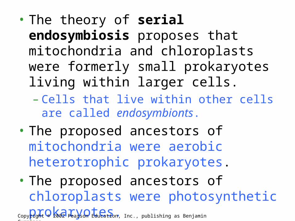
• The theory of serial endosymbiosis proposes that mitochondria and chloroplasts were formerly small prokaryotes living within larger cells.– Cells that live within other cells are called
endosymbionts.
• The proposed ancestors of mitochondria were aerobic heterotrophic prokaryotes.
• The proposed ancestors of chloroplasts were photosynthetic prokaryotes.
• Other organelles: cilia, flagella, basal bodies and centrioles.
Copyright © 2002 Pearson Education, Inc., publishing as Benjamin Cummings

• Several lines of evidence support a close similarity between bacteria and the chloroplasts and mitochondria of eukaryotes.– These organelles and bacteria are similar is size.– Enzymes and transport systems in the inner
membranes of chloroplasts and mitochondria resemble those in the plasma membrane of modern prokaryotes.
– Replication by mitochondria and chloroplasts resembles binary fission in bacteria.
Copyright © 2002 Pearson Education, Inc., publishing as Benjamin Cummings

– The single circular DNA in chloroplasts and mitochondria lack histones and other proteins, as in most prokaryotes.
– Both organelles have transfer RNAs, ribosomes, and other molecules for transcription of their DNA and translation of mRNA into proteins.
– The ribosomes of both chloroplasts and mitochondria are more similar to those of prokaryotes than to those in the eukaryotic cytoplasm that translate nuclear genes.
Copyright © 2002 Pearson Education, Inc., publishing as Benjamin Cummings

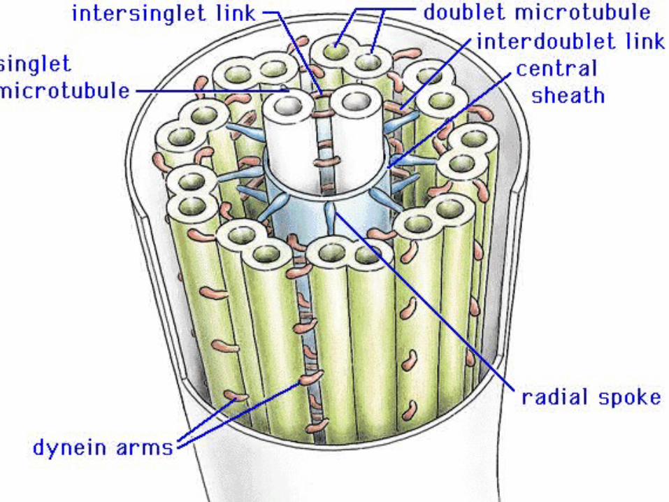
• A comprehensive theory for the origin of the eukaryotic cell must also account for the evolution of the cytoskeleton and the 9 + 2 microtubule apparatus of the eukaryotic cilia and flagella.– Some researchers have proposed that cilia and
flagella evolved from symbiotic bacteria (especially spirochetes).
– However, the evidence for this proposal is weak.
Copyright © 2002 Pearson Education, Inc., publishing as Benjamin Cummings

A Spirochete living symbiotically with a protist may have evolved into the organelles flagella and cilia

• The chimera of Greek mythology was part goat, part lion, and part serpent.
• Similarly, the eukaryotic cell is a chimera of prokaryotic parts:– mitochondria from one bacteria– plastids from another– nuclear genome from the host cell (the prokayotic
symbiont passes some of its genetic control to the host’s nucleus- thus losing its independence)
3. The eukaryotic cell is a chimera of prokaryotic ancestors
Copyright © 2002 Pearson Education, Inc., publishing as Benjamin Cummings

Example of cell with symbiotic bacteriaExample of cell with symbiotic bacteria
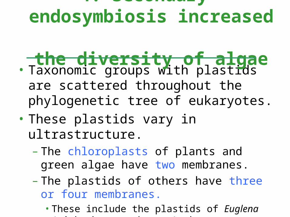
• Taxonomic groups with plastids are scattered throughout the phylogenetic tree of eukaryotes.
• These plastids vary in ultrastructure.– The chloroplasts of plants and green algae have
two membranes.– The plastids of others have three or four
membranes.• These include the plastids of Euglena (with three
membranes) that are most closely related to heterotrophic species.
4. Secondary endosymbiosis increased
the diversity of algae

• The best current explanation for this diversity of plastids is that plastids were acquired independently several times during the early evolution of eukaryotes.– Those algal groups with more than two membranes
were acquired by secondary endosymbiosis.– It was by primary endosymbiosis that certain
eukaryotes first acquired the ancestors of plastids by engulfing cyanobacteria.
– Secondary endosymbiosis occurred when a heterotrophic protist engulfed an algae containing plastids.
Copyright © 2002 Pearson Education, Inc., publishing as Benjamin Cummings
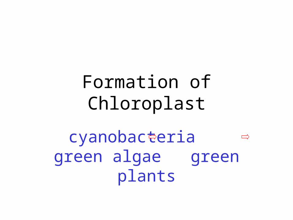
Formation of Chloroplast
cyanobacteria green algae green plants

• Each endosymbiotic event adds a membrane derived from the vacuole membrane of the host cell that engulfed the endosymbiont.
Fig. 28.52
3

Figure 28.3 Diversity of plastids produced by secondary endosymbiosis
Cyanobacterium
Heterotrophiceukaryote
Primaryendosymbiosis
Red algae
Green algae
Secondaryendosymbiosis
Secondaryendosymbiosis
Plastid
Dinoflagellates
Apicomplexans
Ciliates
Stramenopiles
Euglenids
Chlorarachniophytes
Plastid
Alv
eola
tes
Green Plants
23

Horizontal Gene Transfer
• Horizontal gene transfer (HGT), also Lateral gene transfer (LGT), is any process in which an organism transfers genetic material to another cell that is not its offspring
• Genes from endosymbiotic bacteria may have undergone HGT to its host chromosomes eg. genes for ATP synthase.

• The first great adaptive radiation, the metabolic diversification of the prokaryotes, set the stage for the second.
• The second wave of diversification was catalyzed by the greater structural diversity of the eukaryotic cell.
• The third wave of diversification followed the origin of multicellular bodies in several eukaryotic lineages.
5. The origin of eukaryotes catalyzed a second great wave of
diversification

• The kingdom Protista formed a paraphyletic group, with some members more closely related to animals, plants, or fungi than to other protists.
• Systematists have split the former kingdom Protista into as many as 20 separate kingdoms.
• Still,“protist” is used as an informal term for this great diversity of eukaryotic kingdoms.
Copyright © 2002 Pearson Education, Inc., publishing as Benjamin Cummings
Fig. 28.2

polyphyletic
monophyletic

Dip
lom
on
ad
s
Pa
rab
asa
lids
Kin
eto
pla
stid
s
Eu
gle
nid
s
Din
ofla
ge
llate
s
Ap
ico
mp
lexa
ns
Cili
ate
s
Oo
myc
ete
s
Dia
tom
s
Go
lde
n a
lga
e
Bro
wn
alg
ae
Ch
lora
rach
nio
ph
yte
s
Fo
ram
inife
ran
s
Ra
dio
laria
ns
Gym
na
mo
eb
as
En
tam
oe
ba
s
Pla
smo
dia
l slim
e m
old
s
Ce
llula
r sl
ime
mo
lds
Fu
ng
i
Ch
oa
no
flag
ella
tes
Me
tazo
an
s
Re
d a
lga
e
Ch
loro
ph
yte
s
Ch
aro
ph
yce
an
s
Pla
nts
Ancestral eukaryote
Ch
loro
ph
yta
Pla
nta
e
Rh
od
op
hy
ta
An
ima
lia
Fu
ng
i
(Opisthokonta) (Viridiplantae)Dip
lom
on
ad
ida
Pa
rab
as
ala
Eu
gle
no
zoa
Alveolata Stramenopila Ce
rco
zoa
Ra
dio
lari
a
Amoebozoa
Flagella (>2), 2 nuclei, no mitochondria, no plastids, simple cytoskeleton, EX:
Giardia

Figure 28.5 Diplomonads
and parabasalids
5 µm(a) Giardia intestinalis, a diplomonad (colorized SEM)
(b) Trichomonas vaginalis, a parabasalid (colorized SEM)
Flagella
Undulating membrane 5 µm

Dip
lom
on
ad
s
Pa
rab
asa
lids
Kin
eto
pla
stid
s
Eu
gle
nid
s
Din
ofla
ge
llate
s
Ap
ico
mp
lexa
ns
Cili
ate
s
Oo
myc
ete
s
Dia
tom
s
Go
lde
n a
lga
e
Bro
wn
alg
ae
Ch
lora
rach
nio
ph
yte
s
Fo
ram
inife
ran
s
Ra
dio
laria
ns
Gym
na
mo
eb
as
En
tam
oe
ba
s
Pla
smo
dia
l slim
e m
old
s
Ce
llula
r sl
ime
mo
lds
Fu
ng
i
Ch
oa
no
flag
ella
tes
Me
tazo
an
s
Re
d a
lga
e
Ch
loro
ph
yte
s
Ch
aro
ph
yce
an
s
Pla
nts
Ancestral eukaryote
Ch
loro
ph
yta
Pla
nta
e
Rh
od
op
hy
ta
An
ima
lia
Fu
ng
i
(Opisthokonta) (Viridiplantae)Dip
lom
on
ad
ida
Pa
rab
as
ala
Eu
gle
no
zoa
Alveolata Stramenopila Ce
rco
zoa
Ra
dio
lari
a
Amoebozoa
Anterior pocket where 1-2 flagella emerge, paramylum, a
glucose polymer used for storage, many are autotrophic or
heterotrophic

Figure 28.7 Trypanosoma, the kinetoplastid that causes sleeping
sickness
9 m
Trypanosoma is a kinetoplastid from the Kingdom Euglenozoa
it is the cause of African sleeping sickness which is spread by the bite of the Tsetse fly
Kinetoplast- an organelle that houses extracellular DNA w/ associated mitochondria
Trypanosoma is a kinetoplastid from the Kingdom Euglenozoa
it is the cause of African sleeping sickness which is spread by the bite of the Tsetse fly
Kinetoplast- an organelle that houses extracellular DNA w/ associated mitochondria

Euglena – Kingdom Euglenozoa
Paramylum-glucose polymer
This Kingdom has changed classification several times because it is unicellular, photosynthetic and heterotrophic
MIXOTROPH
Anterior pocket where 1-2 flagella emerge, paramylum, a
glucose polymer used for storage, many are autotrophic
or heterotrophic

Dip
lom
on
ad
s
Pa
rab
asa
lids
Kin
eto
pla
stid
s
Eu
gle
nid
s
Din
ofla
ge
llate
s
Ap
ico
mp
lexa
ns
Cili
ate
s
Oo
myc
ete
s
Dia
tom
s
Go
lde
n a
lga
e
Bro
wn
alg
ae
Ch
lora
rach
nio
ph
yte
s
Fo
ram
inife
ran
s
Ra
dio
laria
ns
Gym
na
mo
eb
as
En
tam
oe
ba
s
Pla
smo
dia
l slim
e m
old
s
Ce
llula
r sl
ime
mo
lds
Fu
ng
i
Ch
oa
no
flag
ella
tes
Me
tazo
an
s
Re
d a
lga
e
Ch
loro
ph
yte
s
Ch
aro
ph
yce
an
s
Pla
nts
Ancestral eukaryote
Ch
loro
ph
yta
Pla
nta
e
Rh
od
op
hy
ta
An
ima
lia
Fu
ng
i
(Opisthokonta) (Viridiplantae)Dip
lom
on
ad
ida
Pa
rab
as
ala
Eu
gle
no
zoa
Alveolata Stramenopila Ce
rco
zoa
Ra
dio
lari
a
Amoebozoa

Figure 28.9 AlveoliFlagellum Alveoli0.2 µm

Ceratium is a dinoflagellate from the Kingdom Aveolota
Ceratium is a dinoflagellate from the Kingdom Aveolota

Figure 28.10 Pfiesteria shumwayae, a dinoflagellate
3 µm
Flagella

Pfiesteria piscicida is another dinoflagellate that can produce toxins that can result in red tides -Caretenoids pigments give it its characteristic color
Pfiesteria piscicida is another dinoflagellate that can produce toxins that can result in red tides -Caretenoids pigments give it its characteristic color
Their characteristic shape is reinforced by internal plates of cellulose – they have chloroplasts
they have two unequal flagella set in perpendicular grooves which result in its characteristic spinning motion
Their characteristic shape is reinforced by internal plates of cellulose – they have chloroplasts
they have two unequal flagella set in perpendicular grooves which result in its characteristic spinning motion

28.17 Dino Flagellate

Some dinoflagellates act as mutualistic symbionts in corals

Figure 28.12 Structure and Function in the Ciliate Paramecium caudatum
50 µmThousands of cilia cover
the surface of Paramecium.
The undigested contents of food vacuoles are released when the vacuoles fuse with a specialized region of the plasma membrane that functions as an anal pore.
Paramecium, like other freshwater protists, constantly takes in water
by osmosis from the hypotonic environment. Bladderlike contractile vacuoles accumulate
excess water from radial canals and periodically expel it through the plasma membrane.
Food vacuoles combine with lysosomes. As the food is digested, the vacuoles follow a looping path through the cell.
Paramecium feeds mainly on bacteria. Rows of cilia along a funnel-shaped oral groove move food into the cell mouth, where the food is engulfed into food vacuoles by phagocytosis.
Oral groove
Cell mouth
Micronucleus
Macronucleus
FEEDING, WASTE REMOVAL, AND WATER BALANCE
Contractile vacuole

Just the micronucleus undergo meiosis and syngamy which increases genetic diversity
Just the micronucleus undergo meiosis and syngamy which increases genetic diversity

The cilia on a parameciumThe cilia on a paramecium
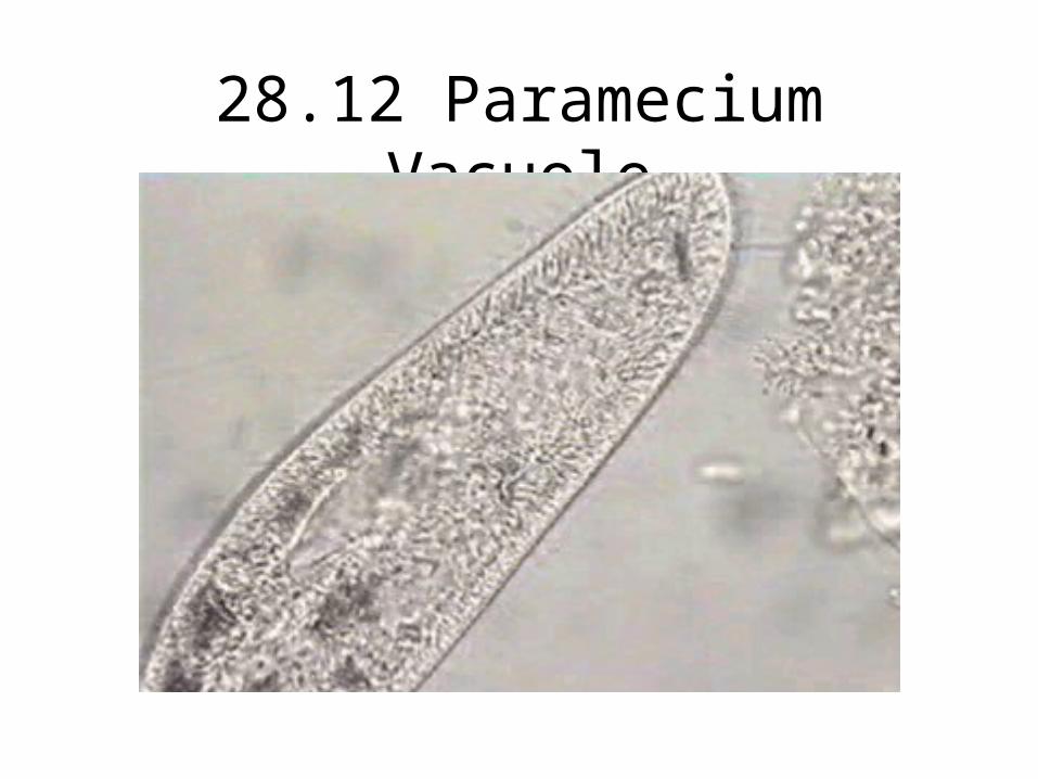
28.12 Paramecium Vacuole

• All apicomplexans are parasites of animals and some cause serious human diseases.– The parasites disseminate as tiny infectious cells
(sporozoites) with a complex of organelles specialized for penetrating host cells and tissues at the apex of the sporozoite cell.
– Most apicomplexans have intricate life cycles with both sexual and asexual stages and often require two or more different host species for completion.
Copyright © 2002 Pearson Education, Inc., publishing as Benjamin Cummings

The apical complex is its characteristic structure
The apical complex is its characteristic structure

Figure 28.11 The two-host life cycle of Plasmodium, the apicomplexan that causes malaria
Inside mosquito Inside human
Sporozoites(n)
Oocyst
MEIOSIS
Liver
Liver cell
Merozoite(n)
Red bloodcells
Gametocytes(n)
FERTILIZATION
Gametes
Zygote(2n)
Key
Haploid (n)Diploid (2n)
Merozoite
Red blood cell
Apex
0.5 µm
An infected Anopheles mosquito bites a person, injecting Plasmodium sporozoites in its saliva.
The sporozoites enter the person’s liver cells. After several days, the sporozoites undergo multiple divisions and become merozoites, which use their apical complex to penetrate red blood cells (see TEM below).
The merozoites divide asexually inside the red blood cells. At intervals of 48 or 72 hours (depending on the species), large numbers of merozoites break out of the blood cells, causing periodic chills and fever. Some of the merozoites infect new red blood cells.
Some merozoites form gametocytes.
Gametes form from gametocytes.Fertilization occurs in the mosquito’sdigestive tract, and a zygote forms.The zygote is the only diploid stagein the life cycle.
Another Anopheles mosquitobites the infected person and picksup Plasmodium gametocytes alongwith blood.
An oocyst developsfrom the zygote in the wall of the mosquito’s gut. Theoocyst releases thousands
of sporozoites, whichmigrate to the mosquito’s
salivary gland.
1 2
3
4
56
7

28.12 Vorticella Habitat

Dip
lom
on
ad
s
Pa
rab
asa
lids
Kin
eto
pla
stid
s
Eu
gle
nid
s
Din
ofla
ge
llate
s
Ap
ico
mp
lexa
ns
Cili
ate
s
Oo
myc
ete
s
Dia
tom
s
Go
lde
n a
lga
e
Bro
wn
alg
ae
Ch
lora
rach
nio
ph
yte
s
Fo
ram
inife
ran
s
Ra
dio
laria
ns
Gym
na
mo
eb
as
En
tam
oe
ba
s
Pla
smo
dia
l slim
e m
old
s
Ce
llula
r sl
ime
mo
lds
Fu
ng
i
Ch
oa
no
flag
ella
tes
Me
tazo
an
s
Re
d a
lga
e
Ch
loro
ph
yte
s
Ch
aro
ph
yce
an
s
Pla
nts
Ancestral eukaryote
Ch
loro
ph
yta
Pla
nta
e
Rh
od
op
hy
ta
An
ima
lia
Fu
ng
i
(Opisthokonta) (Viridiplantae)Dip
lom
on
ad
ida
Pa
rab
as
ala
Eu
gle
no
zoa
Alveolata Stramenopila Ce
rco
zoa
Ra
dio
lari
a
Amoebozoa

Stramenopila
• Water Molds and their relatives (Oomycota)– lack chloroplasts– unicellular or have coenocytic hyphae– cause of the Irish potato famine
• Diatoms
• Golden Algae – have yellow and brown carotenoid and
xanthophyll accessory pigments
• Brown Algae

StramenopilaStramenopilaChloroplasts of stramenopiles have two additional membranes outside of the usual chloroplast
Chloroplasts of stramenopiles have two additional membranes outside of the usual chloroplast
This is evidence that the chloroplasts did not evolve directly from cyanobacteria - it probably came from a eukaryotic source - probably and endosymbiotic red algae
This is evidence that the chloroplasts did not evolve directly from cyanobacteria - it probably came from a eukaryotic source - probably and endosymbiotic red algae

Figure 28.13 Stramenopile flagella
Smoothflagellum
Hairyflagellum
5 µm

Figure 28.14 The life cycle of a water mold (layer 1)
MEIOSIS
Egg nucleus(n)
Key
Haploid (n)
Diploid (2n)
Oogonium
Antheridialhypha withsperm nuclei(n)
Oomycetes causes molds, mildew, rusts, blights

Figure 28.14 The life cycle of a water mold (layer 2)
Zoosporangium(2n)
Zygotegermination
FERTILIZATIONSEXUALREPRODUCTION
Zygotes(oospores)(2n)
Key
MEIOSIS
Egg nucleus(n) Antheridial
hypha withsperm nuclei(n)
Key
Haploid (n)
Diploid (2n)
Oogonium
Cell walls made of cellulose, dominant diploid causes potato late blight (Irish famine)

Figure 28.14 The life cycle of a water mold (layer 3)
Cyst
Zoospore(2n)
ASEXUALREPRODUCTION
Zoosporangium(2n)
Germ tube
Zoosporangium(2n)
Zygotegermination
FERTILIZATIONSEXUALREPRODUCTION
Zygotes(oospores)(2n)
Key
MEIOSIS
Egg nucleus(n)
Key
Haploid (n)
Diploid (2n)
Oogonium
Antheridialhypha withsperm nuclei(n)

28.14 Water Mold Oogonium
Cell walls of cellulose, dominant diploid causes potato blight (Irish famine)

• Diatoms (Bacillariophyta) have unique glasslike walls composed of hydrated silica embedded in an organic matrix. Autotrophic– The wall is divided into two parts that overlap like a shoe box and
lid.
Copyright © 2002 Pearson Education, Inc., publishing as Benjamin Cummings
Fig. 28.17

The glass like shell of a diatom (bacillariophyta)
The glass like shell of a diatom (bacillariophyta)

Fossilized diatoms make up the majority of diatomaceous earthFossilized diatoms make up the majority of diatomaceous earth

28.16 Diatoms Moving

• Brown algae (Phaeophyta) are the largest and most complex algae.– Most brown algae are multicellular.– Most species are marine.
• Brown algae are especially common along temperate coasts in areas of cool water and adequate nutrients.
• They owe their characteristic brown or olive color to accessory pigments like fucoxanthin in the plastids.
Copyright © 2002 Pearson Education, Inc., publishing as Benjamin Cummings
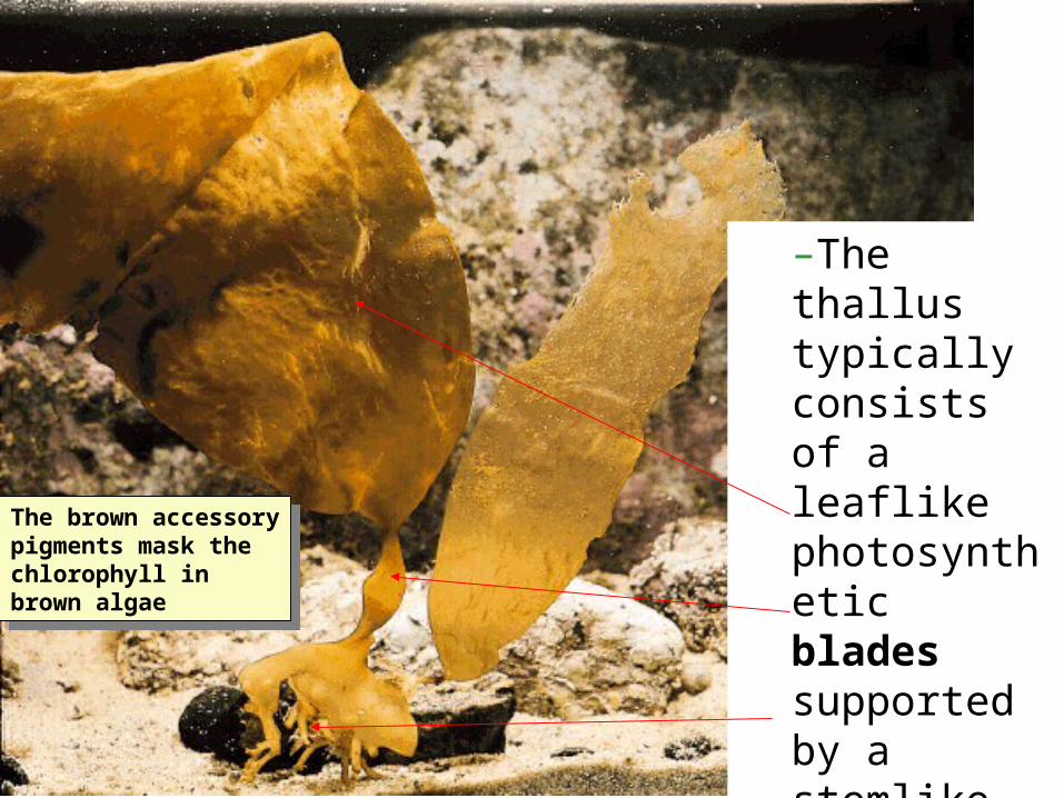
The brown accessory pigments mask the chlorophyll in brown algae
The brown accessory pigments mask the chlorophyll in brown algae
–The thallus typically consists of a leaflike photosynthetic blades supported by a stemlike stipe, and a rootlike holdfast.

Fucus a brown algae
Fucus a brown algae

Giant kelpGiant kelp
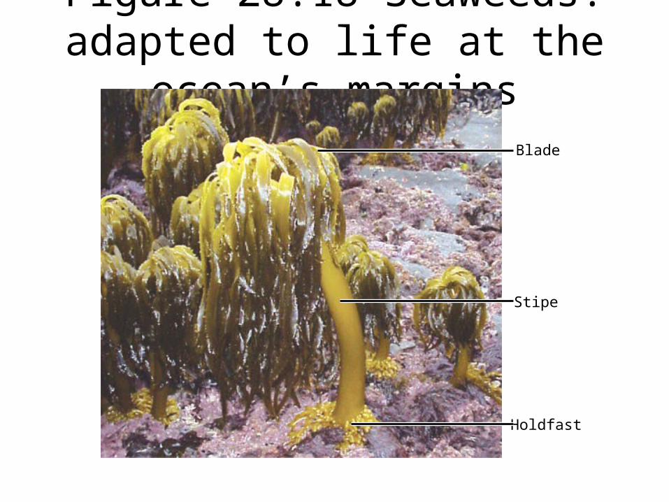
Figure 28.18 Seaweeds: adapted to life at the ocean’s margins
Blade
Stipe
Holdfast

Figure 28.19 A kelp forest

(a) The seaweed is grown on nets in shallow coastal waters.
(b) A worker spreadsthe harvested sea-weed on bambooscreens to dry.
(c) Paper-thin, glossy sheetsof nori make a mineral-rich wrap for rice, seafood, and vegetables in sushi.
Figure 28.20 Edible seaweed
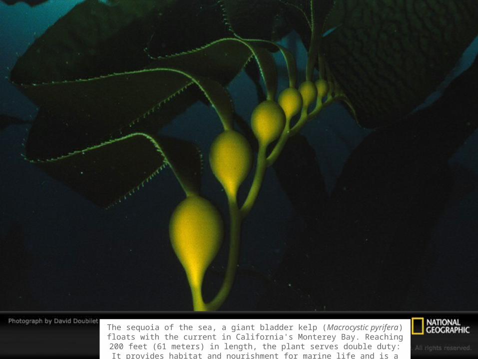
The sequoia of the sea, a giant bladder kelp (Macrocystic pyrifera) floats with the current in California's Monterey Bay. Reaching 200 feet (61 meters) in length, the plant serves double
duty: It provides habitat and nourishment for marine life and is a source of algin, a stabilizing, thickening, gelling, and suspending agent used in human food preparation.

• The life cycle of the brown alga Laminaria is an example of alternation of generations.
• heteromorphic• The diploid
individual, the sporophyte, produces haploid spores (zoospores) by meiosis.
• The haploid individual, the gametophyte, produces gametes by mitosis that fuse to form a diploid zygote.
Fig. 28.21
Dip
lom
on
ad
s
Pa
rab
asa
lids
Kin
eto
pla
stid
s
Eu
gle
nid
s
Din
ofla
ge
llate
s
Ap
ico
mp
lexa
ns
Cili
ate
s
Oo
myc
ete
s
Dia
tom
s
Go
lde
n a
lga
e
Bro
wn
alg
ae
Ch
lora
rach
nio
ph
yte
s
Fo
ram
inife
ran
s
Ra
dio
laria
ns
Gym
na
mo
eb
as
En
tam
oe
ba
s
Pla
smo
dia
l slim
e m
old
s
Ce
llula
r sl
ime
mo
lds
Fu
ng
i
Ch
oa
no
flag
ella
tes
Me
tazo
an
s
Re
d a
lga
e
Ch
loro
ph
yte
s
Ch
aro
ph
yce
an
s
Pla
nts
Ancestral eukaryote
Ch
loro
ph
yta
Pla
nta
e
Rh
od
op
hy
ta
An
ima
lia
Fu
ng
i
(Opisthokonta) (Viridiplantae)Dip
lom
on
ad
ida
Pa
rab
as
ala
Eu
gle
no
zoa
Alveolata Stramenopila Ce
rco
zoa
Ra
dio
lari
a
Amoebozoa
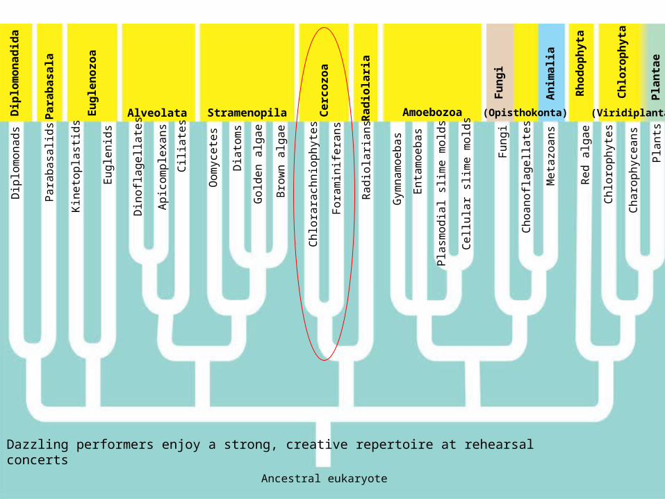
Dazzling performers enjoy a strong, creative repertoire at rehearsal concerts
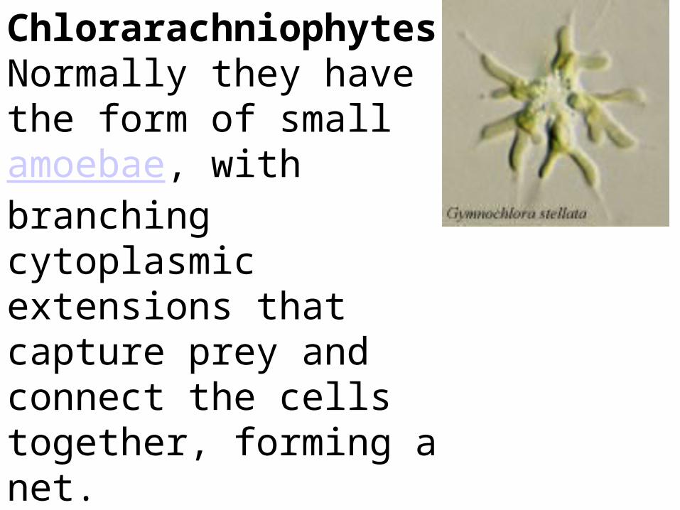
Chlorarachniophytes Normally they have the form of small amoebae, with branching cytoplasmic extensions that capture prey and connect the cells together, forming a net.


• Foraminiferans, or forams, are almost all marine.– Most live in sand or attach to rocks or algae.– Some are abundant in the plankton.– Forams have snail-like, coiled, multichambered,
porous shells, consisting of organic materials hardened with calcium carbonate.
Copyright © 2002 Pearson Education, Inc., publishing as Benjamin Cummings
Fig. 28.28

Foraminiferans - pore bearing shellsmany have symbiotic algae in which they derive benefit from photosynthesis
Foram fossil are excellent markers for dating marine sediment and sedimentary rock
Foraminiferans - pore bearing shellsmany have symbiotic algae in which they derive benefit from photosynthesis
Foram fossil are excellent markers for dating marine sediment and sedimentary rock

Radiolarians have skeletons made of silica
most of the marine ooze found on the ocean floor is composed of their skeletons
Radiolarians have skeletons made of silica
most of the marine ooze found on the ocean floor is composed of their skeletons

• Actinopod (heliozoans and radiolarians), “ray foot,” refers to slender pseudopodia (axopodia) that radiate from the body.– Each axopodium is reinforced by a bundle of
microtubules covered by a thin layer of cytoplasm.
Copyright © 2002 Pearson Education, Inc., publishing as Benjamin Cummings
Fig. 28.27

Figure 28.4 A tentative phylogeny of eukaryotesD
iplo
mo
na
ds
Pa
rab
asa
lids
Kin
eto
pla
stid
s
Eu
gle
nid
s
Din
ofla
ge
llate
s
Ap
ico
mp
lexa
ns
Cili
ate
s
Oo
myc
ete
s
Dia
tom
s
Go
lde
n a
lga
e
Bro
wn
alg
ae
Ch
lora
rach
nio
ph
yte
s
Fo
ram
inife
ran
s
Ra
dio
laria
ns
Gym
na
mo
eb
as
En
tam
oe
ba
s
Pla
smo
dia
l slim
e m
old
s
Ce
llula
r sl
ime
mo
lds
Fu
ng
i
Ch
oa
no
flag
ella
tes
Me
tazo
an
s
Re
d a
lga
e
Ch
loro
ph
yte
s
Ch
aro
ph
yce
an
s
Pla
nts
Ancestral eukaryote
Ch
loro
ph
yta
Pla
nta
e
Rh
od
op
hy
ta
An
ima
lia
Fu
ng
i
(Opisthokonta) (Viridiplantae)Dip
lom
on
ad
ida
Pa
rab
as
ala
Eu
gle
no
zoa
Alveolata Stramenopila Ce
rco
zoa
Ra
dio
lari
a
Amoebozoa

Amoebozoans
• Lobe shaped rather than thread like pseudopodia
the microtubules and microfilaments of the cytoskeleton help in amoeboid movement VIDEO
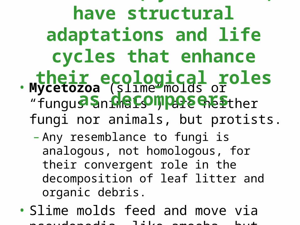
• Mycetozoa (slime molds or “fungus animals”) are neither fungi nor animals, but protists.– Any resemblance to fungi is analogous, not
homologous, for their convergent role in the decomposition of leaf litter and organic debris.
• Slime molds feed and move via pseudopodia, like amoeba, but comparisons of protein sequences place slime molds relatively close to the fungi and animals.
Slime molds (mycetozoans) have structural adaptations and life
cycles that enhance their ecological roles as decomposers

Figure 28.4 A tentative phylogeny of eukaryotesD
iplo
mo
na
ds
Pa
rab
asa
lids
Kin
eto
pla
stid
s
Eu
gle
nid
s
Din
ofla
ge
llate
s
Ap
ico
mp
lexa
ns
Cili
ate
s
Oo
myc
ete
s
Dia
tom
s
Go
lde
n a
lga
e
Bro
wn
alg
ae
Ch
lora
rach
nio
ph
yte
s
Fo
ram
inife
ran
s
Ra
dio
laria
ns
Gym
na
mo
eb
as
En
tam
oe
ba
s
Pla
smo
dia
l slim
e m
old
s
Ce
llula
r sl
ime
mo
lds
Fu
ng
i
Ch
oa
no
flag
ella
tes
Me
tazo
an
s
Re
d a
lga
e
Ch
loro
ph
yte
s
Ch
aro
ph
yce
an
s
Pla
nts
Ancestral eukaryote
Ch
loro
ph
yta
Pla
nta
e
Rh
od
op
hy
ta
An
ima
lia
Fu
ng
i
(Opisthokonta) (Viridiplantae)Dip
lom
on
ad
ida
Pa
rab
as
ala
Eu
gle
no
zoa
Alveolata Stramenopila Ce
rco
zoa
Ra
dio
lari
a
Amoebozoa

• The plasmodial slime molds (Myxogastrida) are brightly pigmented, heterotrophic organisms.
• The feeding stage is an amoeboid mass, the plasmodium, that may be several centimeters in diameter.– The plasmodium is
not multicellular, but a single mass of cytoplasm with multiple nuclei.
Fig. 28.29

Plasmodial Slime Mold belongs to Mycetozoa/ Myxogastrida
is composed of ONE multinucleated cell or amoeboid mass called a plasmodium
most species are diploid
Plasmodial Slime Mold belongs to Mycetozoa/ Myxogastrida
is composed of ONE multinucleated cell or amoeboid mass called a plasmodium
most species are diploid

• The cellular slime molds (Dictyostelida) straddle the line between individuality and multicellularity.– The feeding stage consists of solitary cells.– When food is scarce, the cells form an aggregate
(“slug”) that functions as a unit.• Each cell retains its identity in the aggregate.
Copyright © 2002 Pearson Education, Inc., publishing as Benjamin Cummings

Copyright © 2002 Pearson Education, Inc., publishing as Benjamin Cummings
Fig. 28.30

Cellular Slime Molds are multicellular
haploid
have fruiting bodies
Cellular Slime Molds are multicellular
haploid
have fruiting bodies

Figure 28.4 A tentative phylogeny of eukaryotesD
iplo
mo
na
ds
Pa
rab
asa
lids
Kin
eto
pla
stid
s
Eu
gle
nid
s
Din
ofla
ge
llate
s
Ap
ico
mp
lexa
ns
Cili
ate
s
Oo
myc
ete
s
Dia
tom
s
Go
lde
n a
lga
e
Bro
wn
alg
ae
Ch
lora
rach
nio
ph
yte
s
Fo
ram
inife
ran
s
Ra
dio
laria
ns
Gym
na
mo
eb
as
En
tam
oe
ba
s
Pla
smo
dia
l slim
e m
old
s
Ce
llula
r sl
ime
mo
lds
Fu
ng
i
Ch
oa
no
flag
ella
tes
Me
tazo
an
s
Re
d a
lga
e
Ch
loro
ph
yte
s
Ch
aro
ph
yce
an
s
Pla
nts
Ancestral eukaryote
Ch
loro
ph
yta
Pla
nta
e
Rh
od
op
hy
ta
An
ima
lia
Fu
ng
i
(Opisthokonta) (Viridiplantae)Dip
lom
on
ad
ida
Pa
rab
as
ala
Eu
gle
no
zoa
Alveolata Stramenopila Ce
rco
zoa
Ra
dio
lari
a
Amoebozoa

Besides chlorophyll a, red algae has an accessory pigment phycoerythin that give it its red color by masking the chlorphyll
They are multicellular and live in deep water
Besides chlorophyll a, red algae has an accessory pigment phycoerythin that give it its red color by masking the chlorphyll
They are multicellular and live in deep water

Figure 28.28 Red algae
Bonnemaisonia hamifera. This red alga has a filamentous form.
(a)
Dulse (Palmaria palmata). This edible species has a “leafy” form.
(b)
A coralline alga. The cell walls ofcoralline algae are hardened by calcium carbonate. Some coralline algae aremembers of the biological communities around coral reefs.
(c)

The plastids of red algae evolved from cyanobacteria by primary endosymbiosis

Figure 28.4 A tentative phylogeny of eukaryotesD
iplo
mo
na
ds
Pa
rab
asa
lids
Kin
eto
pla
stid
s
Eu
gle
nid
s
Din
ofla
ge
llate
s
Ap
ico
mp
lexa
ns
Cili
ate
s
Oo
myc
ete
s
Dia
tom
s
Go
lde
n a
lga
e
Bro
wn
alg
ae
Ch
lora
rach
nio
ph
yte
s
Fo
ram
inife
ran
s
Ra
dio
laria
ns
Gym
na
mo
eb
as
En
tam
oe
ba
s
Pla
smo
dia
l slim
e m
old
s
Ce
llula
r sl
ime
mo
lds
Fu
ng
i
Ch
oa
no
flag
ella
tes
Me
tazo
an
s
Re
d a
lga
e
Ch
loro
ph
yte
s
Ch
aro
ph
yce
an
s
Pla
nts
Ancestral eukaryote
Ch
loro
ph
yta
Pla
nta
e
Rh
od
op
hy
ta
An
ima
lia
Fu
ng
i
(Opisthokonta) (Viridiplantae)Dip
lom
on
ad
ida
Pa
rab
as
ala
Eu
gle
no
zoa
Alveolata Stramenopila Ce
rco
zoa
Ra
dio
lari
a
Amoebozoa

Figure 28.30 Colonial and multicellular chlorophytesVolvox, a colonial freshwater chlorophyte. The colony is a hollowball whose wall is composed of hundreds or thousands of biflagellated cells (see inset LM) embedded in a gelatinous matrix. The cells are usually connected by strands of cytoplasm;if isolated, these cells cannot reproduce. The large colonies seen here will eventually release the small “daughter” colonies within them (LM).
(a)
Caulerpa, an inter-tidal chlorophyte.The branched fila-ments lack cross-walls and thus are multi-nucleate. In effect,the thallus is onehuge “supercell.”
(b)
Ulva, or sea lettuce. This edible seaweed has a multicellular thallus differentiated into leaflike blades and a rootlike holdfast that anchors the alga against turbulent waves and tides.
(c)
20 µm50 µm

Volvox is a colonial Green Alga
-most are unicellular
-closely related to plants
Volvox is a colonial Green Alga
-most are unicellular
-closely related to plants
Chlorophyta Green Algae are photosynthetic autotrophs that arose by an endosymbiotic association between a flagellated, heterotrophic eukaryote and a cyanobacterium
Chlorophyta Green Algae are photosynthetic autotrophs that arose by an endosymbiotic association between a flagellated, heterotrophic eukaryote and a cyanobacterium

28.30 Volvox Colony

28.30 Volvox Flagella

In spirogyra there is one or more large spiral chloroplast
Sexual reproduction occurs between conjugation tubules.
In spirogyra there is one or more large spiral chloroplast
Sexual reproduction occurs between conjugation tubules.

• Most green algae have both sexual and asexual reproductive stages.– Most sexual species have biflagellated gametes
with cup-shaped chloroplasts.
Copyright © 2002 Pearson Education, Inc., publishing as Benjamin CummingsFig. 28.24
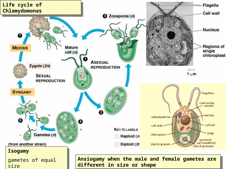
Isogamy
gametes of equal size
Isogamy
gametes of equal size
Life cycle of ChlamydomonasLife cycle of Chlamydomonas
Ansiogamy when the male and female gametes are different in size or shape
Ansiogamy when the male and female gametes are different in size or shape

• Photosynthetic protists have evolved in several clades that also have heterotrophic members.
• Different episodes of secondary endosymbiosis account for the diversity of protists with plastids.
Copyright © 2002 Pearson Education, Inc., publishing as Benjamin Cummings
Fig. 28.25

Ulva is a multicellular green alga with specialized cells that organized into tissues
Ulva is a multicellular green alga with specialized cells that organized into tissues

Isomorphic alternation of generations in Ulva
Isomorphic alternation of generations in Ulva

The choanoflagellate is a colonial protist that is believed to be the ancestor of animals
The choanoflagellate is a colonial protist that is believed to be the ancestor of animals

Figure 28.4 A tentative phylogeny of eukaryotesD
iplo
mo
na
ds
Pa
rab
asa
lids
Kin
eto
pla
stid
s
Eu
gle
nid
s
Din
ofla
ge
llate
s
Ap
ico
mp
lexa
ns
Cili
ate
s
Oo
myc
ete
s
Dia
tom
s
Go
lde
n a
lga
e
Bro
wn
alg
ae
Ch
lora
rach
nio
ph
yte
s
Fo
ram
inife
ran
s
Ra
dio
laria
ns
Gym
na
mo
eb
as
En
tam
oe
ba
s
Pla
smo
dia
l slim
e m
old
s
Ce
llula
r sl
ime
mo
lds
Fu
ng
i
Ch
oa
no
flag
ella
tes
Me
tazo
an
s
Re
d a
lga
e
Ch
loro
ph
yte
s
Ch
aro
ph
yce
an
s
Pla
nts
Ancestral eukaryote
Ch
loro
ph
yta
Pla
nta
e
Rh
od
op
hy
ta
An
ima
lia
Fu
ng
i
(Opisthokonta) (Viridiplantae)Dip
lom
on
ad
ida
Pa
rab
as
ala
Eu
gle
no
zoa
Alveolata Stramenopila Ce
rco
zoa
Ra
dio
lari
a
Amoebozoa