chapter 41 animal nutrition - weeblycontractions of muscles in the wall of the canal •valves...
TRANSCRIPT
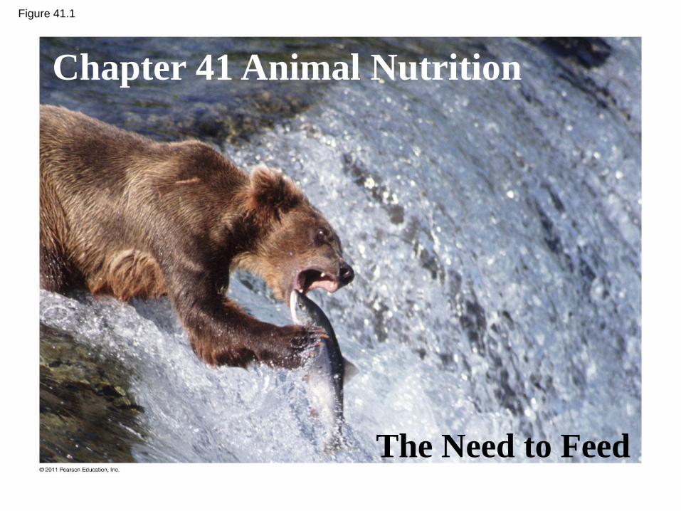
Figure 41.1
Chapter 41 Animal Nutrition
The Need to Feed
http://birdlovers.tumblr.com/page/2
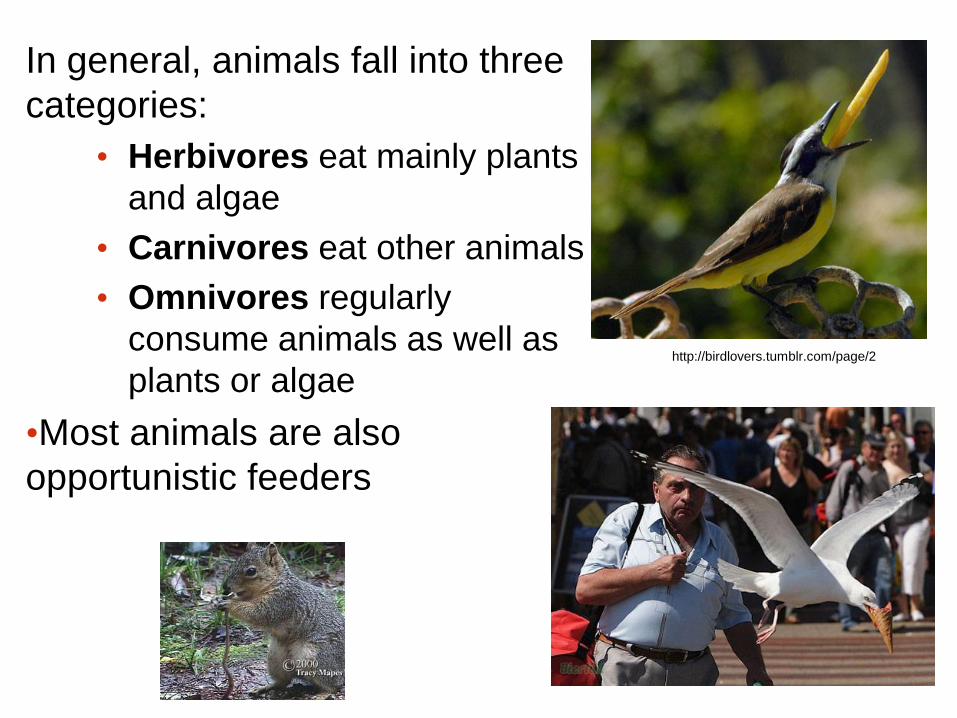
In general, animals fall into three
categories:
• Herbivores eat mainly plants
and algae
• Carnivores eat other animals
• Omnivores regularly
consume animals as well as
plants or algae
•Most animals are also
opportunistic feeders
An animal’s diet must supply chemical
energy, organic molecules, and essential
nutrients
• An animal’s diet provides:
– Chemical energy, which is converted into ATP to
power cellular processes
– Organic building blocks, such as organic carbon
and organic nitrogen, to synthesize a variety of
organic molecules
– Essential nutrients, which are required by cells
and must be obtained from dietary sources
© 2011 Pearson Education, Inc.

Essential Nutrients
• There are four classes of essential nutrients:
– Essential amino acids
– Essential fatty acids
– Vitamins
– Minerals
© 2011 Pearson Education, Inc.

• Animals require 20 amino acids and can
synthesize about half from molecules in their diet
• The remaining amino acids, the essential amino
acids, must be obtained from food in
preassembled form
• Meat, eggs, and cheese provide all the essential
amino acids and are thus “complete” proteins
© 2011 Pearson Education, Inc.
Essential Amino Acids
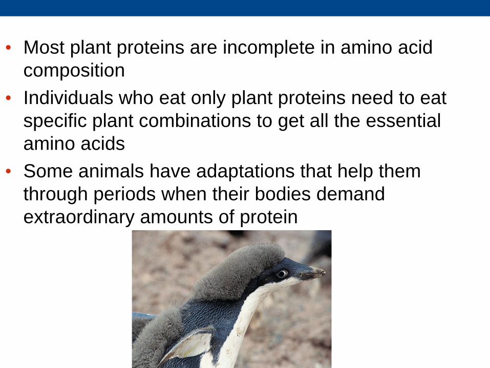
• Most plant proteins are incomplete in amino acid
composition
• Individuals who eat only plant proteins need to eat
specific plant combinations to get all the essential
amino acids
• Some animals have adaptations that help them
through periods when their bodies demand
extraordinary amounts of protein

Vitamins • Vitamins are organic molecules required in the
diet in small amounts
• Thirteen vitamins are essential for humans
• Vitamins are grouped into two categories: fat-
soluble and water-soluble
• Minerals are simple inorganic nutrients, usually
required in small amounts
• Ingesting large amounts of some minerals can
upset homeostatic balance
Minerals

Deficiencies in Essential Nutrients
• Deficiencies in essential nutrients can cause
deformities, disease, and death
• Results when a diet does
not provide enough chemical
energy
Obtaining essential nutrients.
Undernutrition

Figure 41.5
Mechanical
digestion Chemical digestion (enzymatic hydrolysis)
Nutrient molecules
enter body cells
Undigested
material
Elimination Absorption Digestion Ingestion 1 2 3 4
Ingestion is the act of eating

Bulk Feeders
Suspension Feeders and Filter Feeders
Fluid Feeders
Baleen
Feces
Caterpillar
Substrate Feeders
Figure 41.6 Some types of feeding
strategies
• Digestion is the process of breaking food down
into molecules small enough to absorb
• Mechanical digestion, including chewing,
increases the surface area of food
• Chemical digestion splits food into small
molecules that can pass through membranes;
these are used to build larger molecules
• In chemical digestion, the process of enzymatic
hydrolysis splits bonds in molecules with the
addition of water
© 2011 Pearson Education, Inc.

• Absorption is uptake of nutrients by body cells
• Elimination is the passage of undigested
material out of the digestive system
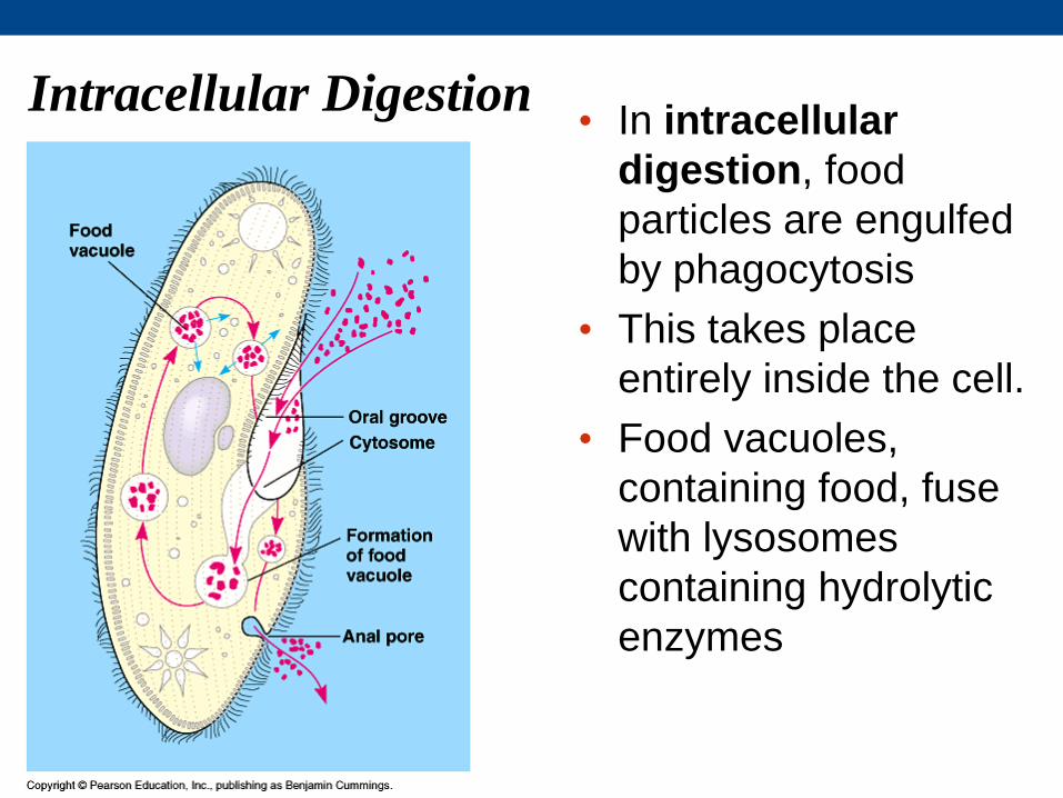
Intracellular Digestion • In intracellular
digestion, food
particles are engulfed
by phagocytosis
• This takes place
entirely inside the cell.
• Food vacuoles,
containing food, fuse
with lysosomes
containing hydrolytic
enzymes
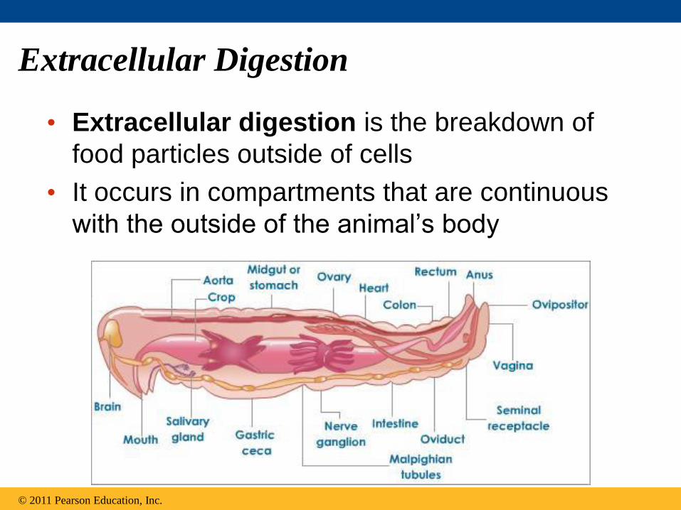
Extracellular Digestion
• Extracellular digestion is the breakdown of
food particles outside of cells
• It occurs in compartments that are continuous
with the outside of the animal’s body
© 2011 Pearson Education, Inc.

Figure 41.7
Mouth
Tentacles
Food
Epidermis Gastrodermis
Food particles engulfed and digested
Food particles broken down
Digestive enzymes released 1
2
3
Simple body plans have a
gastrovascular cavity that
functions in BOTH digestion
and distribution of nutrients

• More complex animals have a digestive tube with
two openings, a mouth and an anus
• WHO? Or Rather who have we studied that
doesn’t?
• This digestive tube is called a complete
digestive tract or an alimentary canal
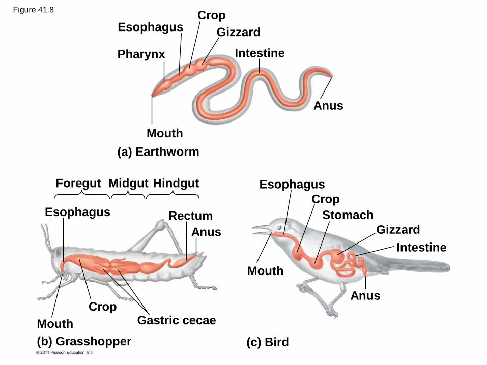
Crop
Gizzard
Intestine
Anus
Esophagus
Pharynx
Mouth
(a) Earthworm
Midgut Esophagus
Crop
Mouth
Stomach
Gizzard
Intestine
Anus
Anus
Rectum Esophagus
Crop
Hindgut Foregut
Mouth Gastric cecae
(b) Grasshopper (c) Bird
Figure 41.8

Specialized organs in the mammalian
digestive system
• The mammalian digestive system consists of an
alimentary canal and accessory glands that
secrete digestive juices through ducts
• Mammalian accessory glands are the salivary
glands, the pancreas, the liver, and the
gallbladder

• Food is pushed along by peristalsis, rhythmic
contractions of muscles in the wall of the canal
• Valves called sphincters regulate the movement
of material between compartments
What type of muscles function
to create the peristaltic
movement?
(Smooth, rough or cardiac?)
peristalsis in the lower part
of the stomach, the antrum
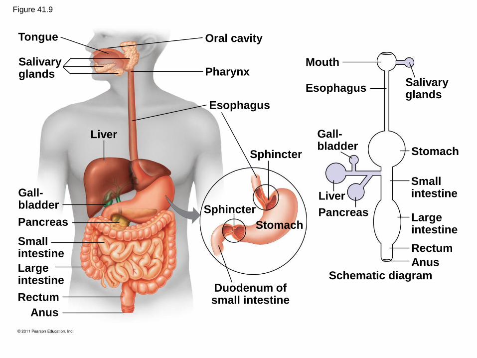
Figure 41.9
Liver
Salivary glands
Gall- bladder
Esophagus
Pharynx
Oral cavity
Sphincter
Mouth
Stomach
Esophagus
Tongue
Pancreas
Small intestine
Large intestine
Rectum
Anus
Sphincter
Stomach
Duodenum of small intestine
Pancreas
Schematic diagram
Anus
Rectum
Large intestine
Small intestine Liver
Salivary glands
Gall- bladder

The Oral Cavity, Pharynx, and Esophagus
• The first stage of digestion is mechanical and
takes place in the oral cavity
• Salivary glands deliver saliva to lubricate food
• Teeth chew food into smaller particles that are
exposed to salivary amylase, initiating
breakdown of glucose polymers
• Saliva also contains mucus, a viscous mixture of
water, salts, cells, and glycoproteins
© 2011 Pearson Education, Inc.

• The tongue shapes food into a bolus and
provides help with swallowing
• The throat, or pharynx, is the junction that opens
to both the esophagus and the trachea
• The esophagus connects to the stomach
• The trachea (windpipe) leads to the lungs
(unfortunately these are close together and this
can cause what problems?
video of human swallowing

Tongue
Pharynx
Glottis
Larynx
Bolus of
food
Epiglottis
up
Esophageal
sphincter
contracted
Esophagus
To lungs To stomach
Relaxed
muscles
Contracted
muscles
Sphincter
relaxed
Stomach
Trachea
Figure 41.10-3
The esophagus conducts food from the
pharynx down to the stomach by
peristalsis
Swallowing causes the epiglottis to
block entry to the trachea, and the bolus
is guided by the larynx, the upper part of
the respiratory tract

Digestion in the Stomach
• The stomach stores food and secretes gastric
juice, which converts a meal to acid chyme
• digestion movie
© 2011 Pearson Education, Inc.

Chemical Digestion in the Stomach
• Gastric juice has a low pH of about 2, which kills
bacteria and denatures proteins
• Gastric juice is made up of hydrochloric acid
(HCl) and pepsin
• Pepsin is a protease, or protein-digesting
enzyme, that cleaves proteins into smaller
peptides
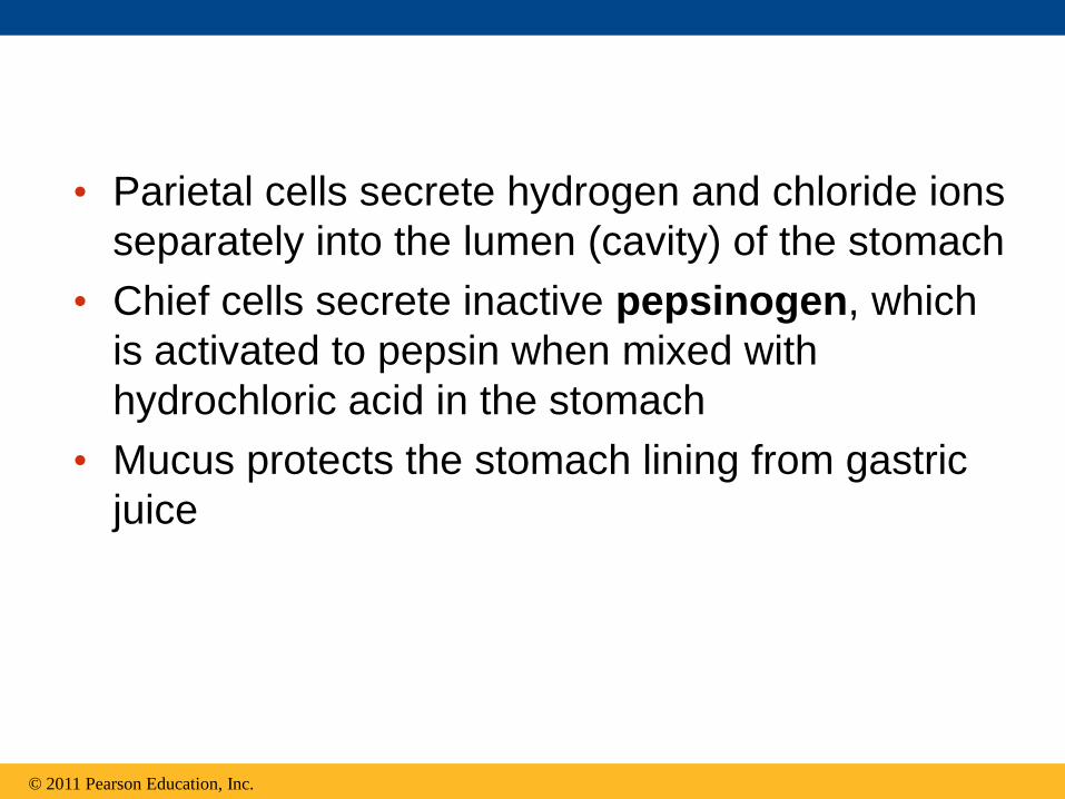
• Parietal cells secrete hydrogen and chloride ions
separately into the lumen (cavity) of the stomach
• Chief cells secrete inactive pepsinogen, which
is activated to pepsin when mixed with
hydrochloric acid in the stomach
• Mucus protects the stomach lining from gastric
juice
© 2011 Pearson Education, Inc.
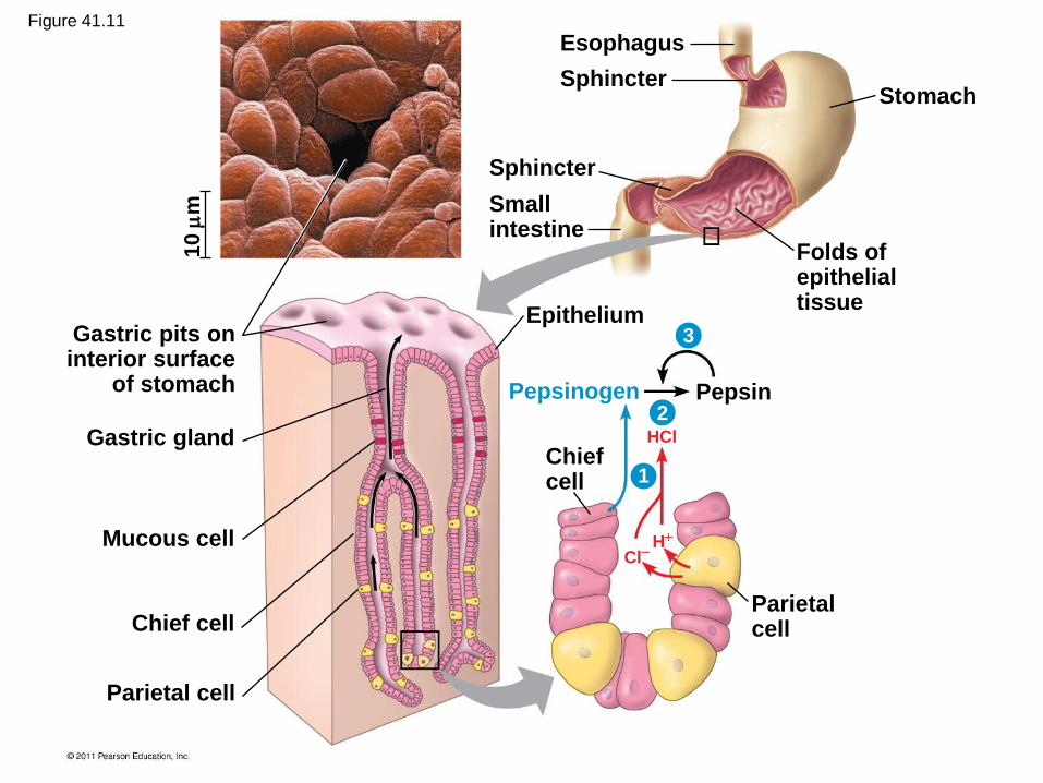
Gastric gland
Gastric pits on interior surface
of stomach
Sphincter
Small intestine
Epithelium
Mucous cell
Chief cell
Parietal cell
Chief cell
Pepsinogen
Parietal cell
Pepsin
Folds of epithelial tissue
Sphincter
Esophagus
Stomach
3
2
1
10
m
HCl
H Cl
Figure 41.11
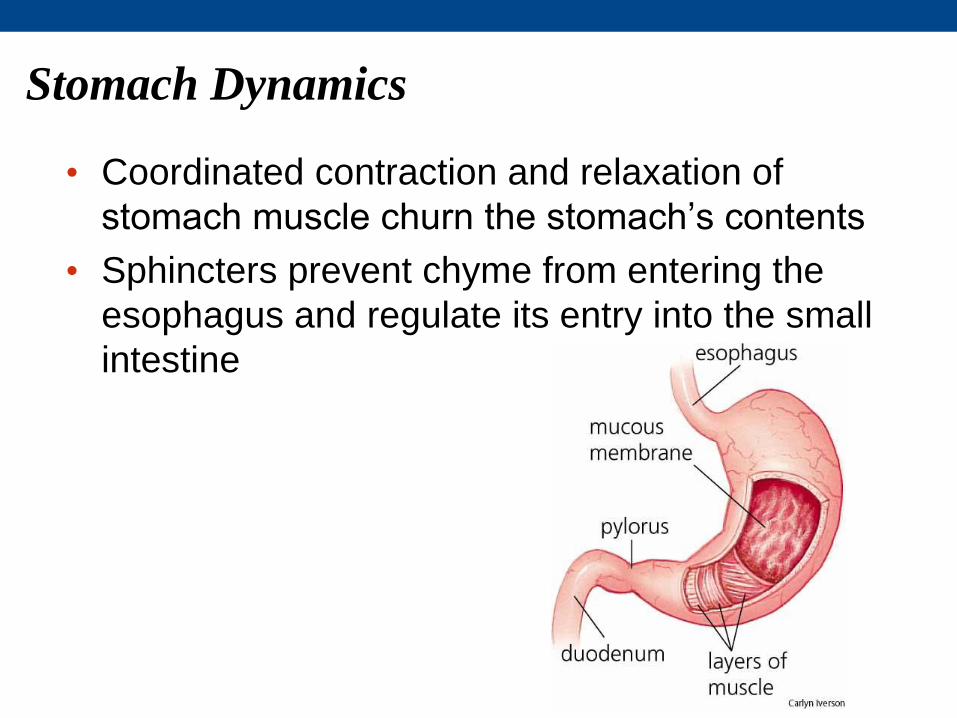
Stomach Dynamics
• Coordinated contraction and relaxation of
stomach muscle churn the stomach’s contents
• Sphincters prevent chyme from entering the
esophagus and regulate its entry into the small
intestine

Digestion in the Small Intestine
• The small intestine is the longest section of the
alimentary canal
• It is the major organ of digestion and absorption
© 2011 Pearson Education, Inc.
Flashback to chapter 40 Loose Connective tissue holds the organs (this is a close-up of a frog…)

Fat digestion Nucleic acid digestion
Protein digestion
Fat (triglycerides) DNA, RNA
Nucleotides
Pancreatic nucleases
Pancreatic lipase
Glycerol, fatty acids, monoglycerides
Nucleotidases
Nucleosides
Nucleosidases and phosphatases
Nitrogenous bases,
sugars, phosphates Amino acids
Dipeptidases, carboxy- peptidase, and aminopeptidase
Small peptides
Pancreatic carboxypeptidase
Smaller polypeptides
Pancreatic trypsin and chymotrypsin
Small polypeptides
Proteins
Pepsin
Carbohydrate digestion
Polysaccharides Disaccharides
Salivary amylase
Smaller
polysaccharides Maltose
Pancreatic amylases
Disaccharides
Disaccharidases
Monosaccharides
Small intestine (enzymes from epithelium)
Small intestine (enzymes from pancreas)
Stomach
Oral cavity, pharynx, esophagus
Figure 41.12-4
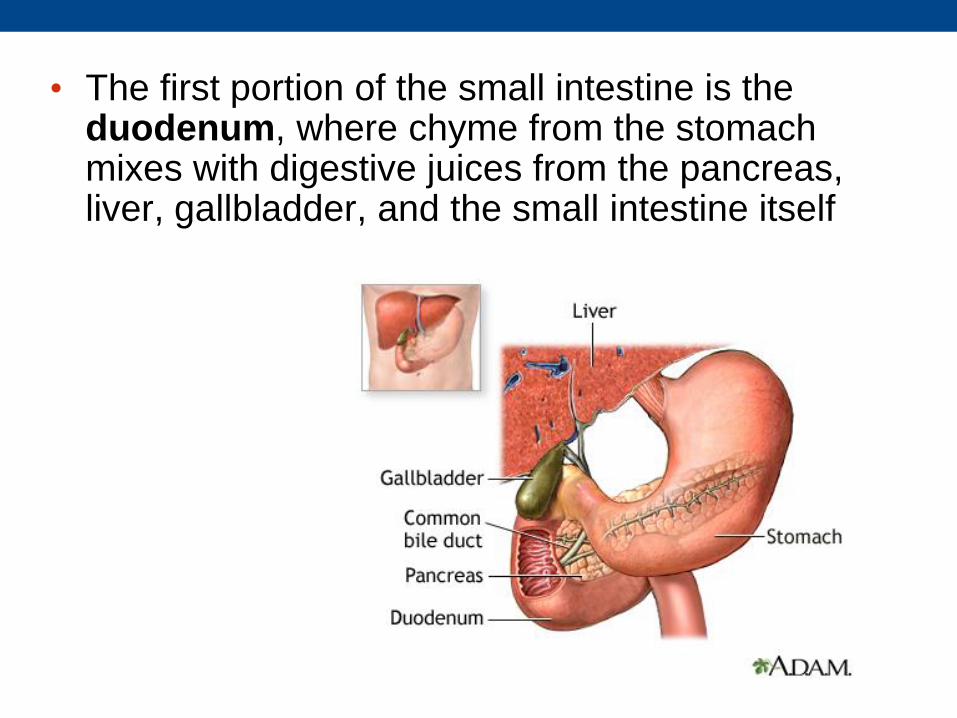
• The first portion of the small intestine is the duodenum, where chyme from the stomach mixes with digestive juices from the pancreas, liver, gallbladder, and the small intestine itself

Pancreatic Secretions
• The pancreas produces the digestive enzymes
proteases trypsin and chymotrypsin that are
activated in the lumen of the duodenum in its
Pancreatic acini
• Its solution is alkaline and neutralizes the acidic
chyme
• We will revisit the pancreas again since it also
helps in hormone production (endocrine system)
in the area of the pancreas called the tropical
sounding Islets of Langerhans
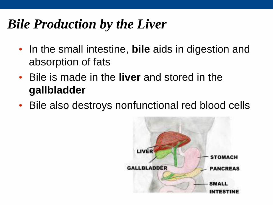
Bile Production by the Liver
• In the small intestine, bile aids in digestion and
absorption of fats
• Bile is made in the liver and stored in the
gallbladder
• Bile also destroys nonfunctional red blood cells
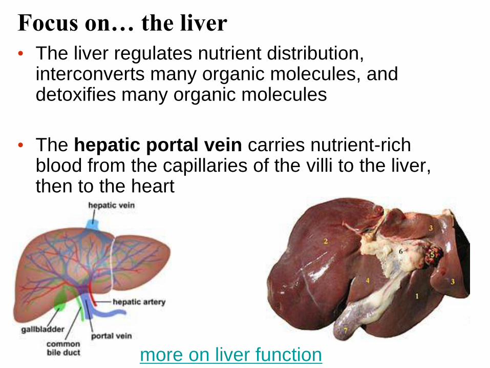
• The liver regulates nutrient distribution, interconverts many organic molecules, and detoxifies many organic molecules
• The hepatic portal vein carries nutrient-rich blood from the capillaries of the villi to the liver, then to the heart
more on liver function
Focus on… the liver

Secretions of the Small Intestine
• The epithelial lining of the duodenum produces
several digestive enzymes
• Enzymatic digestion is completed as peristalsis
moves the chyme and digestive juices along the
small intestine
• Most digestion occurs in the duodenum; the
jejunum and ileum function mainly in absorption
of nutrients and water
© 2011 Pearson Education, Inc.

Absorption in the Small Intestine
• The small intestine has a huge surface area, due to villi and microvilli that are exposed to the intestinal lumen
• The enormous microvillar surface creates a brush border that greatly increases the rate of nutrient absorption

Figure 41.13
Vein carrying blood to liver
Muscle layers
Blood capillaries
Villi
Intestinal wall
Epithelial cells
Large circular folds
Key
Nutrient absorption
Villi
Microvilli (brush border) at apical (lumenal) surface
Epithelial cells
Lumen
Basal surface
Lacteal
Lymph vessel
Transport across the epithelial cells can be passive or active depending on the nutrient

• Epithelial cells absorb fatty acids and monoglycerides and recombine them into triglycerides
• These fats are coated with phospholipids, cholesterol, and proteins to form water-soluble chylomicrons
• Chylomicrons are transported into a lacteal, a lymphatic vessel in each villus
• Lymphatic vessels deliver chylomicron-containing lymph to large veins that return blood to the heart
© 2011 Pearson Education, Inc.

Figure 41.14 LUMEN OF SMALL INTESTINE
Triglycerides
Epithelial cell Fatty acids
Mono- glycerides
Triglycerides
Chylomicron
Phospho- lipids,
cholesterol, and proteins
Lacteal

Figure 41.15
Ascending portion of colon
Small intestine
Appendix Cecum
• The colon of the large
intestine is connected to
the small intestine
• The cecum aids in the
fermentation of plant
material and connects
where the small and large
intestines meet
• The human cecum has an
extension called the
appendix, which plays a
very minor role in
immunity
Absorption in the Large Intestine

• A major function of the colon is to recover water that has entered the alimentary canal
• The colon houses bacteria (e.g., Escherichia coli) which live on unabsorbed organic material; some produce vitamins
• Feces, including undigested material and bacteria, become more solid as they move through the colon

• Feces are stored in the rectum until they can be
eliminated through the anus
• Two sphincters between the rectum and anus
control bowel movements
© 2011 Pearson Education, Inc.
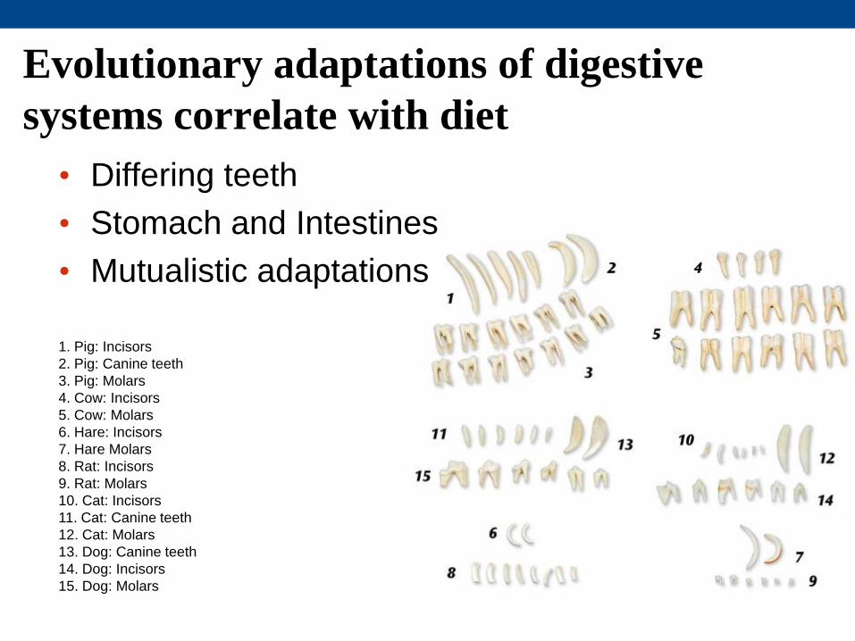
Evolutionary adaptations of digestive
systems correlate with diet
• Differing teeth
• Stomach and Intestines
• Mutualistic adaptations
1. Pig: Incisors
2. Pig: Canine teeth
3. Pig: Molars
4. Cow: Incisors
5. Cow: Molars
6. Hare: Incisors
7. Hare Molars
8. Rat: Incisors
9. Rat: Molars
10. Cat: Incisors
11. Cat: Canine teeth
12. Cat: Molars
13. Dog: Canine teeth
14. Dog: Incisors
15. Dog: Molars

Dental Adaptations
• Teeth vary depending on diet
• The success of mammals is due in part to their
dentition (teeth structure)
• Other vertebrates have less specialized teeth,
though exceptions exist
– For example, the teeth of poisonous snakes are
modified as fangs for
injecting venom

Figure 41.16
Carnivore
Herbivore Omnivore
Molars Premolars Canines Incisors Key

Stomach and Intestinal Adaptations
• Many carnivores have large, expandable
stomachs
• Herbivores and omnivores generally have longer
alimentary canals than carnivores, reflecting the
longer time needed to digest vegetation
© 2011 Pearson Education, Inc.

Small intestine Stomach
Cecum
Carnivore
Colon (large intestine)
Small intestine
Herbivore
The alimentary canals of a carnivore and herbivore

Reticulum
Esophagus
Omasum Abomasum
Intestine
Rumen 1 2
3 4
Mutualistic Adaptations
The most elaborate adaptations for an herbivorous diet have evolved in the animals called ruminants

Satiety center
Ghrelin
Insulin
Leptin
PYY
• Overnourishment causes
obesity, which results from
excessive intake of food
energy with the excess stored
as fat
• Obesity contributes to
diabetes (type 2), cancer of
the colon and breasts, heart
attacks, and strokes
Regulation of Appetite and
Consumption
Figure 41.21

• Hormones regulate long-term and short-term
appetite by affecting a “satiety center” in the brain
• Leptin, the product of fat cells, signals satiety, or
fullness.
• Studies on mice revealed that the hormone leptin
plays an important role in regulating obesity, and
depression.
In one study higher
leptin levels were
linked to decreased
symptoms of anxiety
and depression
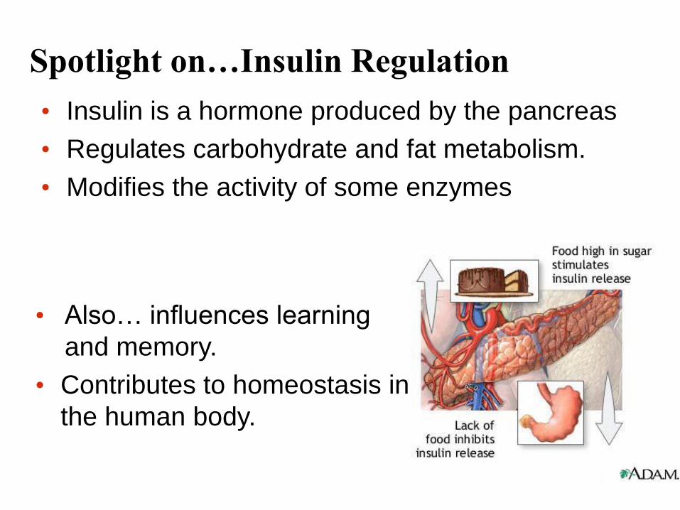
Spotlight on…Insulin Regulation
• Insulin is a hormone produced by the pancreas
• Regulates carbohydrate and fat metabolism.
• Modifies the activity of some enzymes
• Also… influences learning
and memory.
• Contributes to homeostasis in
the human body.
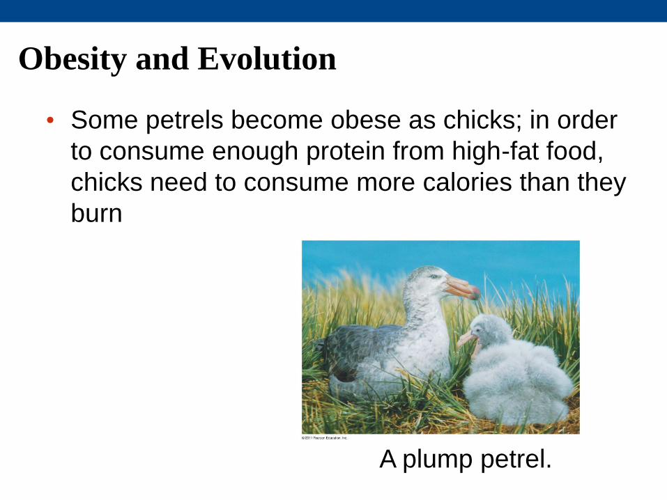
Obesity and Evolution
• Some petrels become obese as chicks; in order
to consume enough protein from high-fat food,
chicks need to consume more calories than they
burn
A plump petrel.

Red Knots feed on enormous amounts of food in
order to store up enough energy for their flight to
the nesting grounds.
William Dalton (c) (all rights reserved) from flickr
http://ottgallerymv.com/lannymcdowellavianart/index.php
/global-conservation-alliance-2009-season/
More on the Red Knot
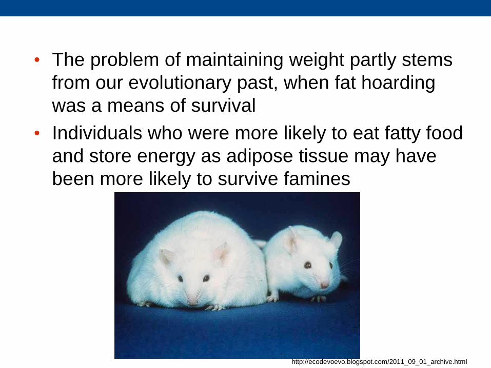
• The problem of maintaining weight partly stems
from our evolutionary past, when fat hoarding
was a means of survival
• Individuals who were more likely to eat fatty food
and store energy as adipose tissue may have
been more likely to survive famines
http://ecodevoevo.blogspot.com/2011_09_01_archive.html