characteristics and survival of malignant cardiac tumors...
TRANSCRIPT

DOI: 10.1161/CIRCULATIONAHA.115.016418
1
Characteristics and Survival of Malignant Cardiac Tumors:
A 40-Year Analysis of Over 500 Patients
Running title: Oliveira et al.; Malignant cardiac tumors
Guilherme H. Oliveira, MD1,2*; Sadeer G. Al-Kindi, MD1*; Christopher Hoimes, DO2;
Soon J. Park, MD3
1Onco-Cardiology Program and Advanced Heart Failure Center, Harrington Heart and Vascular
Institute, University Hospitals Case Medical Center, Cleveland, OH; 2Seidman Cancer Center
and Case Western Reserve University, Cleveland, OH; 3Dept of Cardiac Surgery, Harrington
Heart and Vascular Institute, University Hospitals Case Medical Center, Cleveland, OH
*contributed equally; joint first authors
Address for Correspondence:
Guilherme H. Oliveira, MD
Harrington Heart & Vascular Institute
Case Western Reserve University School of Medicine
University Hospitals Case Medical Center
11100 Euclid Avenue
Cleveland, OH 44106
Tel: 216-844-8242
Fax: 216-844-8318
E-mail: [email protected]
Journal Subject Term: Epidemiology
1Onco-Cardiology Program and Advanced Heart Failure Center, Harrington Hearttt aaand dd VaVaVascscsculululararar
Institute, University Hospitals Case Medical Center, Cleveland, OH; 2Seidman Cancer Center
ananand d d CaCaCasesese WWWessteteterrnr Reserve University, Clevelalaandndnd, OH; 3Dept of f f Cardddiaiaiac Surgery, Harrington
Heart and d VaVaVassccululularrr IIInsnsnstiitutututetete,, UnUnUniviviveersiiitytyty HoHH sppittalsss CaCaCasesese MMMedededicicicalala CCCennter,r,r, CCClelelevevevelall ndndnd, OHOHOH
***contrribi uuuteed equququallyy; jjjoinnnttt fififirsrsst authhhooors
Address for Correspondence:

DOI: 10.1161/CIRCULATIONAHA.115.016418
2
Abstract
Background—To investigate the incidence, histopathology, demographics and survival
associated with primary malignant cardiac tumors (PMCTs).
Methods and Results—We queried the Surveillance, Epidemiology and End-Results (SEER) 18-
registry from National Cancer Institute for all PMCTs diagnosed from 1973 to 2011. We
describe PMCT histopathology and incidence, comparing characteristics and survival of these
patients, to those with extra-cardiac malignancies of similar histopathology. From a total of
7,384,580 cases of cancer registered in SEER, we identified 551 (0.008%) PMCTs. The
incidence of PMCT diagnosis is 34 cases per 100 million persons and has increased over time:
25.1 (1973-1989), 30.2 (1990-1999) and 46.6 (2000-2011). Most patients are female (54.1%),
white (78.6%), with median age at diagnosis of 50 years. The most common PMCTs are
sarcomas (n=357, 64.8%), followed by lymphomas (n=150, 27%), and mesotheliomas (n=44,
8%). Most patients are diagnosed with tissue sample (96.8%). Although use of chemotherapy is
not documented in SEER, 19% of patients received radiation and 44% had surgery. After a
median follow-up of 80 months, 413 patients had died. The 1, 3, and 5- year survival was 46%,
22%, and 17% and has improved over the eras with (1-, 3-, 5-year) of 32%, 17%, 14% (1973-
1989) to 50%, 24%, 19% (2000-2011) (p=0.009). Cardiac sarcomas and mesotheliomas are the
most lethal PMCTs, with 1-, 3-, 5- year survival of 47%, 16%, and 11%, and 51%, 26%, and
23%, respectively, compared with lymphoma 59%, 41%, and 34%, respectively (log rank test
p<0.001). Patients with cardiac lymphomas and sarcomas are younger and have worse survival
than patients with extra-cardiac disease of similar histopathology (p<0.001).
Conclusions—PMCTs are extremely rare and continue to be associated with poor prognosis.
Over the past five decades, the incidence and survival of patients diagnosed with PMCT appears
to have increased. Compared to those with extra-cardiac cancers of similar histopathology,
patients with PMCTs are often younger and have worse survival.
Key words: tumor; neoplasia; epidemiology
white (78.6%), with median age at diagnosis of 50 years. The most common PMCTs s are
arcomas (n=357, 64.8%), followed by lymphomas (n=150, 27%), and mesotheliiomomomasss (((n=n=n=444444, ,,
8%). Most patients are diagnosed with tissue sample (96.8%). Although use of chemotherapy is
not documented in n SEER, 19% of patients received d radiation and 44% hahad surgery. After a
mememedididian folloloowww-upp of 80 months, 413 patients had diiied. The 1, 3, ,, ananand 5- yyear survival was 46%,
2222 %%%, and 17% aandnnd haas iiimpm rororovvved ovovover thhe errrasss witthhh (1-,-,-, 3-,,, 5---yeaaar))) of 3332%%, 111777%, 141414% %% (((199973-3--
1999898989) ) ) to 50%0%%, 2444%%,% 19%9%9% (2000000000-20101011)) (pp=p=0.0.0.00009). CCCardddiaaac sssarcrcrcomommaaas aaanddd mmesesesotototheliiiomomomass aaare thehhe
most lethal PMCTs,,, with 1-, ,, 3-, ,, 5- yyyear survival of 47%,,, 16%, ,, and 11%,,, and 51%,,, 26%,,, and

DOI: 10.1161/CIRCULATIONAHA.115.016418
3
Introduction
Primary malignant cardiac tumors (PMCTs) are extremely rare neoplasms of varying
histopathology that originate within cardiac structures and display biologically aggressive
behavior1-3. Because most practitioners may only see a handful of such cases in their lifetime,
the accumulated experience on this subject has been collated and summarized in multiple
extensive literature reviews1, 4. Nevertheless, the core knowledge of PMCTs has continued to
come from single center studies, consisting of surgical case series and autopsy reports2, 4-7.
Because of the relatively small numbers and significant referral bias of these reports, the
incidence of PMCTs remains unclear, their histology incompletely defined, treatment ineffectual
and prognosis thought to be universally poor.
We therefore sought to better understand PMCTs by utilizing the largest cancer registry
in the United States.
Methods
We conducted a retrospective analysis of all PMCTs in the Surveillance, Epidemiology, and End
Results (SEER) Program (www.seer.cancer.gov) from 1973-2011. We used the 18-Registry
Research Data with Hurricane Katrina Impacted Louisiana Cases, Nov 2013 Submission (1973-
2011 varying) from the National Cancer Institute, Division of Cancer Control and Population
Sciences (DCCPS), Surveillance Research Program, Surveillance Systems Branch, released
April 2013, based on the November 2013 submission. SEER 18 captures cancer data from 18
cancer registries in the US: Atlanta, Connecticut, Detroit, Hawaii, Iowa, New Mexico, San
Francisco-Oakland, Seattle-Puget Sound, Utah, Los Angeles and San Jose-Monterey, Rural
Georgia, the Alaska Native, Greater California, Kentucky, Louisiana, New Jersey, and Greater
and prognosis thought to be universally poor.
We therefore sought to better understand PMCTs by utilizing the largest cancer registry
n thehehe UUUnininiteteted dd Statatateees.
MeMeMethththods
WeWeWe cccononondudductctctededed aaa rrretetetrororospspspececectititiveeve aaanananalllyssysisisis ooofff alalallll PMPMPMCTCTCTsss ininin ttthehehe SSSurrurveieieillllllananancecece, EpEpEpidididemememioioiololologyggy, anananddd EnEnEndddd

DOI: 10.1161/CIRCULATIONAHA.115.016418
4
Georgia. The data collection and reporting for the SEER are described elsewhere8.
All data were extracted from the registry using SEER*STAT v8.2.1 from the surveillance
research program of the division of cancer control and population sciences, National Cancer
Institute (Calverton, MD) on March 1, 2014. We used the following selection criteria: Case
selection (Site and Morphology. Primary Site – labeled) = 'C38.0-Heart'. We only included
patients with known age, who were actively followed, and had tumors with malignant behavior.
Our search was limited to cases within the research database. We excluded patients with either
death certificate only or autopsy report only (however, no patients were excluded based on these
criteria). The study cut-off date is defaulted to December 2010.
We used Code II, IX, XII (a.5) for lymphomas, sarcomas and mesotheliomas,
respectively. We performed subgroup analyses based on age groups (pediatrics: 18 years vs.
adults: >18 years), histologic type (viz. angiosarcoma, selected with ICCC code IX (d.8)), and by
era of diagnosis year (1973-1989, 1990-1999, 2000-2011). Data from SEER*STAT were
imported into IBM SPSS v19 (2010) for statistical analyses. All categorical variables were
presented as frequencies and percentages. Where appropriate, mean (standard deviation, SD) and
median (25th, 75th percentiles) were presented for continuous data variables. Survival curves
were formulated using Kaplan-Meier methods. All tumors were selected using the ICCC site
recode (ICD-0-3/WHO 2008). Per SEER guidelines, histopathologic data are entered based on
the most recent available diagnosis, and the registry does not contain information on the method
used for histologic sampling- whether biopsy, excision, or autopsy.
Incidence data was calculated using rate sessions within SEER*STAT program. For the
incidence calculations, we used the SEER 9 (1973-2011) based on November 2013 submission.
This registry pulls data from following cancer registries: Atlanta, Connecticut, Detroit, Hawaii,
We used Code II, IX, XII (a.5) for lymphomas, sarcomas and mesotheliomamamas,s,s,
espectively. We performed subgroup analyses based on age groups (pediatrics: 18 years vs.
adulltststs::: >1>1>188 8 yeyey ars)s)s),,, histologic type (viz. angiosarcrcrcomomoma, selected with ICCCCCCCCC code IX (d.8)), and by
ererera of diagnosisss yyyeear rr (((19797973-3-3-191919898989, , 191919909090-19999999,9, 200000-20101011)1)1). DDDatatat frororommm SESSEERR*S*S*STATATATTT wewewererr
mmmpopoportr ed intntto o o IBBMM M SPPPSS v1999 (((2022 1000))) fofoforr r statatatisssticaal aaanaaalyysysesss. AlAlAll caaateeegogog rricccall vvvaaariabllleseses wwerrre
prprpresesesenenenteteteddd asasas fffrererequqquenenencicicieseses aaandndnd pppererercececentntntagagageseses. WhWhWhererereee apapapprprpropopopriririatatateee, mmmeaeaeannn (s(s(statatandndndarararddd dededeviiviatatatioioionnn, SSSD)D)D) aaandndndd

DOI: 10.1161/CIRCULATIONAHA.115.016418
5
Iowa, New Mexico, San Francisco-Oakland, Seattle-Puget Sound, and Utah. Age-adjusted
incidence was standardized to the U.S.2000 standard-million population (19 age groups). Age-
adjusted rates for incidence were calculated by summing the products of the age-specific rate (for
each 5-year age group [0–4, 5–9, etc]), multiplied by the fraction of the 2000 U.S. population in
each age range. We calculated the incidence by era of diagnosis and by histology9.
Chi-square was used to compare categorical data. Independent t-test was used to compare
means when normally distributed and non-parametric test (Mann-Whitney) were used if data
were not normally distributed. Kaplan-Meier method was used to present survival, and log-rank
test was used for all survival differences throughout the manuscript. Median survival (25th, 75th
percentile) is presented taking into account censoring. We compared characteristics and survival
of cardiac tumors with non-cardiac tumors of similar histopathologic type (based on ICCC
classifications). In all tests, p<0.05 was considered statistically significant.
Results
Epidemiology
Of 7,384,580 cases of cancer with known age registered in SEER, we identified 551 (0.008%)
PMCTs. The majority were females (298, 54.1%), with median age (25th, 75th percentiles) at
diagnosis of 50 (35, 67) years. Most patients were white (433, 78.6%) followed by blacks (57,
10.3%), see table 1. A histogram of age at diagnosis is shown in figure 1. The majority of
tumors were sarcomas (n=357, 64.8%), followed by lymphomas (150, 27.0%), and
mesotheliomas (44, 8.0%), table 2. There were 27 (4.9%) pediatric patients: 19 with sarcoma, 7
with lymphoma, and 1 with mesothelioma.
The calculated age-adjusted incidence was 34 cases per 100 million persons. Since 1973,
percentile) is presented taking into account censoring. We compared characteristiiicscscs aaandndnd sssurururvivivivvval d
of cardiac tumors with non-cardiac tumors of similar histopathologic type (based on ICCC
classsisisififificacacatititiononons). InInIn all tests, p<0.05 was considerrrededed statistically significcananant.
ReReResuuults
EpEpEpidididemememioioiololologyggy

DOI: 10.1161/CIRCULATIONAHA.115.016418
6
the incidence has increased over 3 eras (per 100 million persons): 25.1 (1973-1989), 30.2 (1990-
1999), 46.6 (2000-2011) and was higher in males than in females (38.2 vs. 30.0 per 100 million
persons). Cause-specific analysis shows that the incidence of both lymphomas (2.8, 10.3, 15.8)
and sarcomas (16.8, 17.1, 29.2) has increased while that of mesothelioma has decreased (5.5, 2.8,
1.5), figure 2.
Most patients were diagnosed by tissue samples (96.8%). Although chemotherapy data is
not available in SEER, 19.1% of patients received radiation, and 43.6% of patients had surgery.
Overall, 10.2% underwent both surgery and radiation as a part of their treatment.
Overall Survival
Survival data were available for 516 (93.6%) patients. At a median follow-up of 80 months (33,
119), 413 patients had died. Median survival (25th, 75th percentiles) was 10 months (1, 29) with
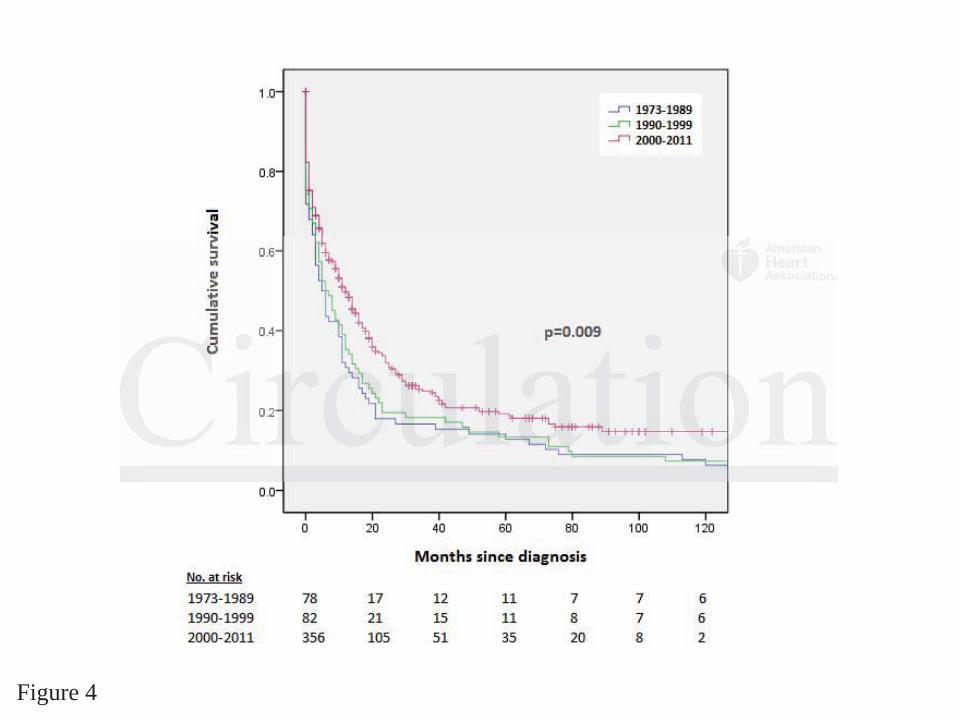
1-, 3- and 5- year survival of 46%, 22%, and 17% (Figure 3). Survival has improved over the
study period from (1-, 3-, 5-year) of 32%, 17%, 14% (1973-1989) to 50%, 24%, 19% (2000-
2011), p=0.009 (figure 4). Pediatric patients had better survival than adults with 1, 3, 5- year
survival rate of 71%, 47%, 47% vs. 44%, 21%, 16% (p<0.001), Supplemental Figure 1.
Cardiac sarcomas have worse survival with 1-, 3-, 5- year survival of 47%, 16%, and 11%
compared with lymphoma survival of 59%, 41%, and 34%, respectively (log rank test p<0.001).
Survival analysis reveals that more than 80% of the patients die within 20 months of diagnosis.
Specific Histologic Types
Sarcomas
Cardiac sarcomas represented 0.3% of all sarcomas with median age (25th, 75th percentiles) at
diagnosis of 45 (32, 59) years. The most common histopathologic type was angiosarcoma
(43.4%), followed by leiomyosarcoma (6.4%) and rhabdomyosarcoma (4.5%). Cardiac
Survival data were available for 516 (93.6%) patients. At a median follow-up of 8880 0 0 momomontntnthshshs (((333333,
119), 413 patients had died. Median survival (25th, 75th percentiles) was 10 months (1, 29) with
1-, 3-3-3- aaandndnd 555- yeyy ararar sssurvival of 46%, 22%, and 17% % % (((FiFF gure 3). Survival l hahahas improved over the
tttudddy period ffrooom mm (11-,-,, 333-,--, 555-y-y-yeaeaea )r)r) of f f 323232%%, 171717%%%, 14%4%% ((191919737373-11198889)99 tttoo 505050%%, 224%4%4%, 191919% %% (2(2(20000000-0-0-
200011111),),) p=0.0000900 (((fiiigurrre 4). PePePedidid atriririccc pppattitiennttts hhhad beeetteeer surrrviivivalala thhahan nn adduultsss wwwiti h 111, 333, 55- yyyearrr
uurvrrviviivalalal rrratatateee ofofof 7771%1%1%, 474747%%%, 4447%7%7% vsss. 4444%4%4%, 212121%%%, 1116%6%6% (((p<p<p<000.0000001)1)1), SuSSupppppplelelememementntntalalal FFFigigigurrureee 111.

DOI: 10.1161/CIRCULATIONAHA.115.016418
7
angiosarcomas had slightly worse survival than other types of cardiac sarcomas with 1-, 3- and
5-year survival of 39%, 9% and 8% vs. 47%, 21% and 14% (p=0.045). There were no
statistically significant differences in cardiac sarcoma survival over the 3 eras with 1-, 3-, and 5-
year survival of 9%, 11%, 6% (1973-1989); 24%, 10%, 6% (1990-1999) and 31%, 11%, 8%
(2000-2011), p=0.173.
Compared with those with extra-cardiac sarcomas, patients with cardiac sarcomas were
younger (46.1 vs. 52.8 years, p=0.001), more likely to be female (47.1% vs. 40.8%, p=0.02), less
likely to have previous history of malignancy (7.6% vs. 12.7%, p=0.005), more likely to have
surgical resection (60.4% vs. 26.2%, p<0.001), table 3. Median survival (25th, 75th percentiles)
was 9.0 months (1.2, 21). Patients with cardiac sarcomas had worse survival than those with
extra-cardiac sarcomas (log rank p<0.001), figure 5A.
Lymphomas
Cardiac lymphomas accounted for 0.03% of all lymphomas with median age (25th, 75th
percentiles) at diagnosis of 67 (50, 79) years. The most common histopathologic type was diffuse
B-cell lymphoma (60.6%). There was a trend towards improved survival for cardiac lymphomas
over the 3 eras with 1-, 3-, and 5- year survival of 63%, 38%, 38% (1973-1989); 38%, 23%, 19%
(1990-1999) and 62%, 46%, 38% (2000-2011), p=0.087.
Compared with extra-cardiac lymphomas, those with cardiac disease were less likely to
be black (5.0% vs. 9.6%, p=0.002), but had no difference in age, gender, or history of previous
malignancy. There was a non-significant trend towards fewer surgeries in patients with cardiac
lymphomas (16.5% vs. 24.9%, p=0.06); but no difference in utilization of radiation therapy,
table 3. Median survival (25th, 75th percentiles) for cardiac lymphomas was 23 months (5, 120).
Compared with extra-cardiac lymphomas, cardiac lymphomas had worse survival (log rank
was 9.0 months (1.2, 21). Patients with cardiac sarcomas had worse survival than n n thththosososeee wiwiwiththth
extra-cardiac sarcomas (log rank p<0.001), figure 5A.
Lympmpmphohohomamamasss
CCCarrdrdiac lymphooomamam s acacaccococounununteeed d d fofofor rr 0.0.0.030303%% ofofof aaalll lyyymmmphohoomamamas ss wiww ththth mmmededediaiaian ageee (2(2(25t5t5th,h,h, 775t5t5thhh
peeercrcrcenee tiles))) aaatt t diiiagggnosssisss of 6667 (5(5(50,0,0, 7779))) yyyeae rsrss. TTThe mmmossst cccommmmmmononon hhhisi totot pppattholllogogogic typypypee wwasss diffffuuse
BBB-cececellllll lllymmymphphphomomomaaa (6(6(6000.6%6%6%))). TTThehehererere wasasas aaa tttrererendndnd tttowoowaaardrdrdsss imimimprprprovoovededed sssurrurviivivaavalll fofoforrr cacacardrdrdiaiaiaccc lyllympmpmphohohomamamasss
DOI: 10.1161/CIRCULATIONAHA.115.016418
8
p<0.001), figure 5B.
Mesotheliomas
Pericardial mesotheliomas represented 0.3% of all mesotheliomas with median age (25th, 75th
percentiles) at diagnosis of 53 (40, 70) years. Median survival (25th, 75th percentiles) of patients
diagnosed with pericardial mesothelioma was 2 months (0, 12), with 1-, 3- and 5- year survival
of 26%, 14%, and 9%. There was no statistically significant difference in survival for cardiac
sarcoma over the 3 eras with 1- year survival of 25% (1973-1989), 20% (1990-1999) and 7%
(2000-2011), log rank p=0.338.
Compared with extra-cardiac mesotheliomas, patients with pericardial mesotheliomas
were younger (mean 54.1 years vs. 70.2 years, p<0.001), less likely to be males (45.5% vs.
77.6%, p<0.001), more likely to be blacks and other minorities (15.9% vs. 4.9% and 6.8% vs.
3.3%, respectively; p=0.001). Patients with cardiac mesotheliomas were more likely to have
surgery (31.8% vs. 24.0%, p<0.001), but had similar use of radiation therapy, table 3. Cardiac or
extra-cardiac location of mesothelioma did not appear to impact prognosis (p=0.06), figure 5C.
Discussion
This study reports the characteristics of PMCTs utilizing data amassed over five decades from a
large-scale national registry. In it we confirm the rarity and lethality of PMCTs, and also offer
insight into their epidemiology, histopathology, demographics, and outcomes. Because we have
studied numbers twenty-fold larger than existing reports, we have debunked previous
misconceptions and shed light on unknown aspects of PMCTs.
The pre-mortem diagnosis of PMCTs is much more uncommon than previously reported.
In unselected autopsy reports, benign and malignant tumors are found in 0.021% of deaths10. Of
were younger (mean 54.1 years vs. 70.2 years, p<0.001), less likely to be males (4(445.5.5.5%5%5% vvvs.s.s
77.6%, p<0.001), more likely to be blacks and other minorities (15.9% vs. 4.9% and 6.8% vs.
3.3%%%, rererespspspececectitt veeelylyly; p=0.001). Patients with cardddiaiaiaccc mesotheliomas werrreee momm re likely to have
uuurgggery (31.8%%% vvvs. 222444.0%0%0%,,, p<p<p<0.0.0.0000001)1)1), bubbutt hahah d dd sis miilaaar usususeee ofofof rrradadadiaiaiatiiiononon ttthhherrapyyy, tatatablblble ee 33. CaCaCardrdrdiaiaiac c or
exxxtrtrt a-a-a-cardiaaac c c loccac ttit on offf mesessototothehh liiiomomo aaa ddid d nononot appppeeearr tooo immmpapaactctc prrrogngg ooosiis (((p=p=p=0.0666),),), rrr figgug rrre 5C.

DOI: 10.1161/CIRCULATIONAHA.115.016418
9
these, malignant cardiac tumors were even less common, representing 5.1% to 28.7% of all heart
tumors in small series2. Our study shows that clinically apparent PMCTs have an estimated
prevalence of 34 cases per 100 million persons; over 100 times lower than previous estimates.
This discrepancy may be partially explained by the possibility that many of the autopsy-
discovered tumors may have been incidentalomas rather than clinically significant tumors.
Indeed, in a Spanish series, one quarter of all cardiac tumors were incidental findings11. In
addition, SEER only includes patients diagnosed with cancer prior to death and not post-mortem
findings. Although PMCTs commonly present with dyspnea, chest pain, palpitations, and
edema11-13, they can also remain clinically silent until causing ventricular arrhythmias13 and
sudden cardiac death14, thus escaping inclusion in SEER. Nevertheless, more in line with our
findings, a recent study in Grosseto’s county in Italy (1998-2011) estimated the incidence of
PMCTs at around 130 per 100 million persons15.
Over the study period, the incidence of PMCTs appears to have increased, driven by
higher frequency of lymphomas and sarcomas. This increment may reflect better pre-mortem
diagnostic capabilities brought about by developments in cardiac imaging, such as
echocardiography, computed tomography and magnetic resonance imaging, not widely available
in the first decades of the study period. The incidence of cardiac lymphomas mirrors that of non-
Hodgkin lymphoma in the general population which peaked in the 1980’s and 1990’s but has
remained stable since 200016 because of improvements in human immunodeficiency virus
management. Conversely, there has been a steady decrease in the incidence of pericardial
mesotheliomas as a result of less common asbestos exposure17.
We confirm that PMCTs can present at any age, with a peak incidence in the fifth decade
of life, affects predominantly whites and have slight female predilection, consistent with
udden cardiac death14, thus escaping inclusion in SEER. Nevertheless, more in liliinenene wwwititith h h ououour r r
findings, a recent study in Grosseto’s county in Italy (1998-2011) estimated the incidence of
PMCTCTCTsss atatat aaarororounnnddd 130 per 100 million persons15. .
Over thehehe sttudydydy ppperererioioiod,d,d, ttthehehe iiinnnciiidededencee offf PMCMCCTsss aaappppppeaeaearss ttto hhahaveveve inncreeeasasasededed,, drdrdrivvvenene bbbyyy
hiiighghgheree freququuenenencyyy ooof lyyymmpm homamamas ss annnd dd saaarcccommmasass. Thhisss innncrrremmmenntnt mmmayayay rrrefeflllecct bbbetetttett r prprpreee-moorrrtemm
dididiagagagnononostststicicic cccapapapabababilililitititieieiesss brbrbrouooughghghttt aaabobobouttut bbby dededeveevelololopmpmpmenenentststs iiinnn cacacardrdrdiaiaiaccc imimimagagaginininggg, sssuccuchhh asasas ttt

DOI: 10.1161/CIRCULATIONAHA.115.016418
10
previous reports11, 12, 18-21. The reasons for age distribution, racial preference and slight female
preponderance cannot be gleaned from this study. We can speculate, however, that women
receive more chest radiation for breast cancer22, 23 and that blacks have less access to medical
care than whites24, however there may also be genetic and environmental factors that cannot be
inferred from this study.
We also report and shed light on histopathologic sub-types of PMCTs and their
frequencies. For example, whereas we confirm that sarcomas are indeed the most common
PMCT, we demonstrate that lymphomas affect the heart ten times more frequently than
previously thought. For example, a previous single-center surgical series reported only 10 (6.9%)
lymphomas of 143 malignant cardiac tumors2, likely a result of referral bias, since lymphomas
typically are chemo-responsive and not treated surgically. Therefore, whereas lymphomas were
previously believed to represent 1.3%-2% of all cardiac tumors2, 4, in our series they accounted
for 27% of all PMCTs. Also, although a systematic literature analysis of 197 cardiac lymphomas
in 201025 reported a male: female ratio of 1.94, we show more balanced gender distribution, not
different than what is seen in extra-cardiac lymphomas. While non-Hodgkin lymphoma has a
strong tendency to involve the myocardium, with up to 20% of patients with NHL having
evidence of myocardial involvement at autopsy26, immunosuppressed patients (transplant
recipients, HIV, etc) typically present with primary cardiac lymphoma without extracardiac
involvement2. In fact, 41% of all patients with primary cardiac lymphomas are
immunocompromised, and have universally poor survival25. Previous reports show that diffuse
large B-cell lymphomas (DLBCL) have a predilection for the right side of the heart (92% had
involvement of right atrium or right ventricle), and usually presents with dyspnea, constitutional
symptoms, pain and arrhythmias25, 27-29. About 90% of those patients receive anthracycline-based
ymphomas of 143 malignant cardiac tumors2, likely a result of referral bias, sinccee e lylylympmpmphohohomamamasss
ypically are chemo-responsive and not treated surgically. Therefore, whereas lymphomas were
prevvvioioiousususlylyly bbbelee ieveveved dd to represent 1.3%-2% of alll cccararardiac tumors2, 4, in ooururur series they accounted
fofofor 272 % of allll PPPMCMMCTsTsTs. AAAlslslso,o,o, aaaltltlthohohouguu h hh aa syystss eeematticcc liteteterararatututureee aanann lylylysisis ss ofoof 19777 cccararrdididiacaca lllymymymphphphomomomas
nnn 22200101025 repepepooorteeed a mmmaala e: fffemememalaa e rararatiooo oofo 111.9.94,4, wwe shhhowowow mmmorrre ee bbballlancnn eeed gennndededer diistststrrribuutiiion, noot
dididifffffferererenenenttt thththananan whahahattt isisis ssseeeeeennn ininin eeexttxtrarara cc-cararardddiaiaiaccc lyllympmpmphohohomamamasss. WWWhihihilelele nnnononon HH-Hodododgkgkgkininin lllymmymphphphomomomaaa hahahasss aaa

DOI: 10.1161/CIRCULATIONAHA.115.016418
11
regimen, with high treatment-related mortality25. Historically, about 28% of patients are treated
with surgery and 20% with radiation, slightly higher than what we found, at 16.5% and 15.1%,
respectively25.
We also investigated incidence of different subtypes of sarcomas. In the largest previous
series of 143 cases of malignant cardiac tumors, angiosarcomas were most common at 23.1%,
followed by leiomyosarcoma 20.3% and rhabdomyosarcoma 4.2%2. Our data shows similar
angiosarcoma (25.8%) and rhabdomyosarcoma (2.6%) distribution, but much lower prevalence
of leiomyosarcoma (3.7%). The predominance of angiosarcoma was also reported previously by
researchers in Italy (28.6%)18, Mayo Clinic (41%)19 and the British Columbia Registry20. In
contradistinction, a single study from Germany reported the predominance of undifferentiated
sarcoma20, which may suggest either regional variances in histological distribution, or
differences in histological classifications across the eras. Whereas we found no gender
predilection for cardiac sarcomas, we note low utilization of surgery (43.6%), and radiation
(19.1%). This may suggest that these patients have advanced disease at presentation and may not
be surgical or radiation candidates, or alternatively, are predominantly treated with
chemotherapy, not captured in the SEER database. Also, sarcomas have been reported to present
at later stages of life and are difficult to diagnose4. In contrast, here we show that patients with
cardiac sarcomas present at a younger age than those with extracardiac disease.
For the first time we investigated demographic differences between patients with cardiac
and extra-cardiac disease of similar histopathology. We found that patients with cardiac
sarcomas and pericardial mesotheliomas are significantly younger than those with extracardiac
disease of similar histology. Whereas the reason for this is unclear, it may be related to lead-time
bias with earlier clinical presentation because of cardiac-related symptoms, or pre-existing risk
contradistinction, a single study from Germany reported the predominance of undididifffffferererenenentititiatatatededed
arcoma20, which may suggest either regional variances in histological distribution, or
diffferererenenencececesss ininin hissstototological classifications across ttthehehe eras. Whereas we fofofouuund no gender
pppredddilection fforrr cccaardididiaaca sssarararcococomamam s,s,, wwwe nnonote lllowowow utiiliizzattioioionnn ofofof sssururgegegeryryry (((44343.6%)%)%), ananand d d rararadidiiaatatiooon n n
1119.9.9 1%1%1%). Thihihisss maamayy y suggggggest thththatatat thesesesee pppattit ennnttts havve advavavancn eeded dddisiseaaaseee attt pprp esssenenentatt tiononon andd mmmayy nnot
bebebe sssurrurgigigicacacalll ororor rrradadadiaiaiatititiononon cccananandidididadadatetetesss, ooorrr aaaltltltererernananatititiveevelylly, ararareee prprpredededomomominininananantltltly trtrtreaeaeateteteddd wiiwiththth

DOI: 10.1161/CIRCULATIONAHA.115.016418
12
factors for early development of these cardiac malignancies. For example, because radiation has
been implicated in some cases of sarcomas30 and other cancers31, it is possible that survivors of
childhood cancers who received radiation to the chest are at higher risk of developing cardiac
sarcomas. Another possibility is that cardiac sarcomas are associated with gene mutations32, 33
that predispose patients to develop these cancers at an earlier age. Interestingly, we also found
ethnic differences between cardiac and extracardiac diseases across all histopathology groups.
Cardiac lymphomas and sarcomas are more prevalent in minority groups, while mesotheliomas
more common in blacks. The reasons for this observation remain speculative and could be
related to genetic predisposition34, risk factors35 or environmental exposures36.
Lastly, we performed extensive survival analyses among multiple subtypes of PMCTs as
wells as among those with cardiac versus extra-cardiac disease. We found that, despite overall
poor prognosis of PMCTs across all histopathology types, survival appears to have slightly
improved over the past five decades. Because during this period, lymphoma treatment and rates
of cure have improved dramatically37, the overall increased survival of PMCTs may be
attributable to that alone. However, it may also be possible that survival has improved because of
earlier diagnosis of PMCTs due to more common utilization of cardiac imaging. Incidental
detection of these tumors when echocardiography is performed for other reasons, might lead to
earlier treatment with better outcomes than in the past when diagnosis relied predominantly on
the presence of symptoms. . In contrast, the paucity of advances in the treatment of sarcomas and
mesotheliomas as well as the low utilization of surgery and radiation, likely explain their worse
survival. Because most of these patients are treated at large academic centers where radiation and
surgical expertise is adequate19, 21, the underutilization of these options probably reflects poor
patient candidacy.
Lastly, we performed extensive survival analyses among multiple subtypeseses ooof ff PMPMPMCTCTCTss s as
wells as among those with cardiac versus extra-cardiac disease. We found that, despite overall
poorrr ppprororogngngnosososis ooofff PMPP CTs across all histopathololoogygygy types, survival appppeaeaears to have slightly
mmmpprp oved over thththe papaassst fffivivivee e dededecccadededesss. BBBecauusses durrinnng thhhisisis ppperererioii d,d,d, lymymymphphphooma trtrtreeeatmtmtmenene t tt anaa d d d rararatetetes ss
offf cururure haveee iiimppmproooveddd ddrd ammmatata icicicallylyly3733 , thhhe ovovveererall innncreaeaeases dd d suuurvrvr ivivivaal oof PMMCTCTCTs ss maaay y y beb
atatattrtrtribibibuttutababablelele tttooo thththatatat aaalololonenene. HoHoHoweeweveeverrr, iiittt mamamay alalalsososo bebebe pppososossisisiblblbleee thththatatat sssurrurviivivaavalll hahahasss imimimprprprovoovededed bbbecececauaausesese oooffff

DOI: 10.1161/CIRCULATIONAHA.115.016418
13
Overall, less than 50% of patients with PMCTs are alive by the end of the first year, with
a sharp decrease in survival for sarcoma and mesothelioma patients. As can be expected we
found that overall survival from a “real world” registry is slightly worse than at high volume
tertiary centers. For example, the median overall survival was 12 months in 32 patients with
PMCTs at Mayo Clinic (1975-2007)19, compared to 10 months in our series. Yet, our reported
survival of sarcomas is much better than previously published reports (1 year survival of 47% vs.
20%11). Most probably, the modest improvement in observed overall survival of patients with
PMCTs is driven by better treatment outcomes of lymphoma and sarcoma patients.
Another unique aspect of this study is that we provide survival comparisons between
cardiac and extra-cardiac malignancies stratified by histopathology. We show that cardiac
sarcomas and lymphomas have significantly worse survival compared with similar cancers of
extra-cardiac origin, suggesting that any cardiac involvement, whether primary or metastatic
carries worse prognosis. It further implies that patients with extra-cardiac malignancies of
histopathology types that affect the heart more commonly, such as angiosarcomas and diffuse
large B-cell lymphoma, may need to be screened for cardiac involvement with
echocardiography38, cardiac MRI39, or cardiac PET40 at diagnosis. This likely does not apply to
mesotheliomas, since they have similarly poor survival regardless of location.
In summary, we confirm that PMCTs are rare and currently have limited treatment
options leading to poor patient survival. There may be opportunities to better understand these
tumors and their survival differences in the context of cancer genomics. Minimally invasive
diagnostic techniques or circulating tumor assays may be necessary for early diagnosis and
eventually inform treatment decisions. Diagnostic and therapeutic clinical trials as well as
locally directed approaches should be incorporated into future treatment considerations.
cardiac and extra-cardiac malignancies stratified by histopathology. We show thatatat cccararardididiacacac
arcomas and lymphomas have significantly worse survival compared with similar cancers of
extrra-a-a-cacacardrdrdiaiaiac cc origigigininin, suggesting that any cardiacc iiinvnvnvolvement, whether prprprimary or metastatic
cccarrrries worse prprprogogo nooossisis..s. IIItt t fufufurtrtrtheheherrr imimimplpp iees thththattt pattieeents s s wiwiwiththth eeextttrarara-t cacacardrdrdiiaiacc maaalililigngngnananancicc eseses of f f
hiiistss opopopatholooogygg typypypes ttthaaat afffefefectctct theee hhheaaarttt moooreee commmm onononllyly,, sususuchhh aaass s ananngiiiossarcococommmas ananand ddiffffuseee tt
aaargrgrgeee BBB-cececellllll lllymmymphphphomomomaaa, mmmayaay nnneeeeeeddd tototo bbbeee scscscrerereenenenededed fffororor cccararardididiacacac iiinvnnvolololveevememementntnt wititithhh

DOI: 10.1161/CIRCULATIONAHA.115.016418
14
Limitations
Despite being the largest of its kind, this study has multiple important limitations. It is based on a
national registry, which, though extensive, lacks fundamental information that severely limits our
results and conclusions. Also, although SEER is frequently used as a research tool, the quality
and accuracy of its data collection cannot be ascertained and is prone to human error and
inaccuracy. Further, because the data in this study was collected over 5 decades and analyzed
retrospectively, there are confounding factors that cannot be avoided despite adjustments. For
example, histopathologic classifications, as well as diagnostic and treatment modalities most
likely do not reflect modern practices3 and can therefore confound estimates of survival and
incidence of PMCT types. Specifically, determination of histopathologic type is confounded by
the many reclassifications of PMCTs that have occurred since the 1970’s, making inferences
about PMCT sub-type incidence less reliable. Therefore, it is possible that the increase in
PMCTs and different subtypes that we report, more accurately reflects an increased incidence in
the diagnosis of PMCTs than that of the actual disease. Unfortunately, because the SEER registry
does not include data on chemotherapy and other treatment modalities, we have no information
on the role of chemotherapy on specific histopathologic types. Similarly, granular clinical
information cannot be gleaned by this study, such as method of diagnosis, clinical presentation,
location of cardiac tumors, most common method of histologic sampling (whether biopsy,
excision or autopsy) etc. In addition, we can also offer no insight on details of surgery or
radiation therapy. Lastly, the lack of information on mode of death also limits our understanding
of the natural history of PMCTs and potentially confounds survival analyses. Whereas no other
data source will likely be able to provide such high numbers of PMCT patients, small case series
will remain the only source of more granular information on this subject.
ncidence of PMCT types. Specifically, determination of histopathologic type is ccconononfofofoununundededed d d bybyby
he many reclassifications of PMCTs that have occurred since the 1970’s, making inferences
abouuuttt PMPMPMCTCTCT subbb-t-t-typy e incidence less reliable. Theheherererefore, it is possible ttthahahat the increase in
PMPMPMCTs and did fffffferere enennt tt sususubtbtbtypypypeseses ttthahahat tt wewewe reepororrt, morreee acccccuururatatatelelely rerereflllececectstss ann incrcrcreeaeaseseseddd ininincicici edeencncnce ee inii
hhhee e dididiagnosiiis s s of PPPMMCM TsTsTs thaannn thththat ooof ff thhhe actutuuaalal disseaaaseee. UUnUnffforrtrtunununatttellly,yy bbbeecauuuseee theee SSSEEEEER R regigiisttryt
dododoeseses nnnototot iiincncnclulludedede dddatatataaa ononon ccchehehemomomothththerererapapapy anananddd otototheheherrr trtrtreaeaeatmtmtmenenenttt momomodadadalililitititieseses, weewe hhhavaaveee nonono iiinfnfnfororormamamatititiononon

DOI: 10.1161/CIRCULATIONAHA.115.016418
15
Conclusions
Cardiac sarcomas, lymphomas and mesotheliomas are the most common PMCTs but remain
extremely rare and associated with dismal prognosis. Over the past five decades, the incidence
and survival of patients diagnosed with PMCT appears to have increased. Compared with those
with extra-cardiac cancers of similar histopathology, patients with PMCTs are often younger and
have worse survival.
Acknowledgments: Role of contributors: GHO: analyzed and interpreted data and drafted the
manuscript. SGA: obtained data, performed statistical analyses, analyzed and interpreted data
and drafted the manuscript. CH: analyzed data, and revised the manuscript. SJP: participated in
discussions and revised the manuscript. All authors approved the final version.
Conflict of Interest Disclosures: None.
References:
1. Sheppard MN, Mohiaddin R. Tumors of the heart. Future Cardiol. 2010;6:181-193. 2. Burazor I, Aviel-Ronen S, Imazio M, Markel G, Grossman Y, Yosepovich A, Adler Y. Primary malignancies of the heart and pericardium. Clin Cardiol. 2014;37:582-588. 3. Travis WD. Who classification of tumours of the lung, pleura, thymus and heart. IARC; 2015.\
4. Marialuisa Valente SR, Ornella Leone, Cristina Basso. Primary malignant tumors of the heart. In: Cristina Basso MV, Gaetano Thiene, ed. Cardiac tumor pathology. New York: Springer; 2013.
5. Tazelaar HD, Locke TJ, McGregor CG. Pathology of surgically excised primary cardiac tumors. Mayo Clin Proc. 1992;67:957-965.
6. Bear PA, Moodie DS. Malignant primary cardiac tumors. The cleveland clinic experience, 1956 to 1986. Chest. 1987;92:860-862.
7. Leja MJ, Shah DJ, Reardon MJ. Primary cardiac tumors. Tex Heart Inst J. 2011;38:261-262.
discussions and revised the manuscript. All authors approved the final version.
Conflict of Interest Disclosures: None.
ReRR ffef rences:
1. SShehehepppppparararddd MNNN, MoMoMohihhiadadaddin R.RR TTTuumorrsss ofofof ttthehehe hhheaearttrt. FuFuFutututureree CCCardididiololol. 222011010;00 6:1818181-1-1 11193.

DOI: 10.1161/CIRCULATIONAHA.115.016418
16
8. Hankey BF, Ries LA, Edwards BK. The surveillance, epidemiology, and end results program: A national resource. Cancer Epidemiol Biomarkers Prev. 1999;8:1117-1121. 9. Surveillance E, and End-Results Program. Seer*stat tutorials: Calculating age-adjusted rates. http://seer.cancer.gov/seerstat/tutorials/aarates/definition.html. 10. Reynen K. Frequency of primary tumors of the heart. Am J Cardiol. 1996;77:107. 11. Barreiro M, Renilla A, Jimenez JM, Martin M, Al Musa T, Garcia L, Barriales V. Primary cardiac tumors: 32 years of experience from a spanish tertiary surgical center. Cardiovasc Pathol. 2013;22:424-427. 12. Yu L, Gu T, Shi E, Xiu Z, Fang Q, Wang C, Wang X, Cheng Y. Primary malignant cardiac tumors. J Cancer Res Clin Oncol. 2014;140:1047-1055. 13. Miyake CY, Del Nido PJ, Alexander ME, Cecchin F, Berul CI, Triedman JK, Geva T, Walsh EP. Cardiac tumors and associated arrhythmias in pediatric patients, with observations on surgical therapy for ventricular tachycardia. J Am Coll Cardiol. 2011;58:1903-1909. 14. Cronin B, Lynch MJ, Parsons S. Cardiac fibroma presenting as sudden unexpected death in an adolescent. Forensic Sci Med Pathol. 2014;10:647-650. 15. Cresti A, Chiavarelli M, Glauber M, Tanganelli P, Scalese M, Cesareo F, Guerrini F, Capati E, Focardi M, Severi S. Incidence rate of primary cardiac tumors: A 14-year population study. J Cardiovasc Med (Hagerstown). 2014 Jul 11. [Epub ahead of print]. 16. Shiels MS, Engels EA, Linet MS, Clarke CA, Li J, Hall HI, Hartge P, Morton LM. The epidemic of non–hodgkin lymphoma in the united states: Disentangling the effect of hiv, 1992–2009. Cancer Epidemiol Biomarkers Prev. 2013;22:1069-1078. 17. Weill H, Hughes JM, Churg AM. Changing trends in us mesothelioma incidence. OccupEnviron Med. 2004;61:438-441. 18. Pacini D, Careddu L, Pantaleo A, Parolari A, Leone O, Daprati A, Gargiulo GD, Di Bartolomeo R. Primary malignant tumors of the heart: Outcomes of the surgical treatment. AsianCardiovasc Thorac Ann. 2015;23:645-651. 19. Simpson L, Kumar SK, Okuno SH, Schaff HV, Porrata LF, Buckner JC, Moynihan TJ. Malignant primary cardiac tumors: Review of a single institution experience. Cancer. 2008;112:2440-2446. 20. Truong PT, Jones SO, Martens B, Alexander C, Paquette M, Joe H, Hart J, Allan SJ. Treatment and outcomes in adult patients with primary cardiac sarcoma: The british columbia cancer agency experience. Ann Surg Oncol. 2009;16:3358-3365. 21. Randhawa JS, Budd GT, Randhawa M, Ahluwalia M, Jia X, Daw H, Spiro T, Haddad A.
urgical therapy for ventricular tachycardia. J Am Coll Cardiol. 2011;58:1903 1909.
14. Cronin B, Lynch MJ, Parsons S. Cardiac fibroma presenting as sudden unexpecececteteteddd dededeatatath h h ininin an adolescent. Forensic Sci Med Pathol. 2014;10:647-650.
15. CrCrCresesestititi AAA, Chiaiaiavvav relli M, Glauber M, Tangannnelelellilili P, Scalese M, Cesaaarerereo F, Guerrini F, CapatiE,E,, FFFooocardi MMM, , Severi S. Incidence rate of primary caaardiac tumors:s:s: A 14-yey ar poppulation study.yf J CCCarrrdiovasc Mededed ((HaHaHaggegersrsrstototownwnwn))). 202020141414 JJJuul 1111.11 [[[Epubub aheheeadadad ooof ff prprininint]]].
1666. . SShShiels MMMS,SS EEEngggelsss EEAE , LLLinenenet MSMSMS, ClClClarkekeke CCCA, LLLi JJ,,, HHHalllll HHHI,II HHHaaartgtgtge P,, MMMororrtot n LMLMLM. ThThheepidddemememicicic ooofff non–nn hohohodgdgdgkiikinn n lympmpphohohomam in nn thththeee unununiiitededed sstttata eseses::: DiDiDisesesentanannglglglinnngg thhthe e effffefefectctct oof hihihivv, 11199999922–202020090909. CaCCancer EEEpiipideddemiimiollol BBBioiiomarkkrkers PrPPrev. 202020131313;2;2;22:2:2:101010696969 11-1070707888.

DOI: 10.1161/CIRCULATIONAHA.115.016418
17
Primary cardiac sarcoma: 25-year cleveland clinic experience. Am J Clin Oncol. 2014 Jul 17. [Epub ahead of print]. 22. Neragi-Miandoab S, Gangadharan SP, Sugarbaker DJ. Cardiac sarcoma 14 years after treatment for pleural mesothelioma. N Eng J Med. 2005;352:1929-1930. 23. Ramalho J, Nunes S, Marques I, Marques F. Primary cardiac sarcoma after breast cancer. BMJ Case Rep. 2013;2013:bcr2013008947. 24. Weinick RM, Zuvekas SH, Cohen JW. Racial and ethnic differences in access to and use of health care services, 1977 to 1996. Med Care Res Rev. 2000;57:36-54. 25. Petrich A, Cho SI, Billett H. Primary cardiac lymphoma: An analysis of presentation, treatment, and outcome patterns. Cancer. 2011;117:581-589. 26. Burke A, Jeudy J Jr., Virmani R. Cardiac tumours: An update: Cardiac tumours. Heart. 2008;94:117-123. 27. Ikeda H, Nakamura S, Nishimaki H, Masuda K, Takeo T, Kasai K, Ohashi T, Sakamoto N, Wakida Y, Itoh G. Primary lymphoma of the heart: Case report and literature review. Pathol Int. 2004;54:187-195. 28. Anghel G, Zoli V, Petti N, Remotti D, Feccia M, Pino P, Majolino I. Primary cardiac lymphoma: Report of two cases occurring in immunocompetent subjects. Leuk Lymphoma. 2004;45:781-788. 29. Ceresoli GL, Ferreri AJ, Bucci E, Ripa C, Ponzoni M, Villa E. Primary cardiac lymphoma in immunocompetent patients: Diagnostic and therapeutic management. Cancer. 1997;80:1497-1506. 30. Berrington de Gonzalez A, Kutsenko A, Rajaraman P. Sarcoma risk after radiation exposure. Clin Sarc Res. 2012;2:18. 31. Johnson JN, Hornik CP, Li JS, Benjamin DK, Jr., Yoshizumi TT, Reiman RE, Frush DP, Hill KD. Cumulative radiation exposure and cancer risk estimation in children with heart disease. Circulation. 2014;130:161-167. 32. Naka N, Tomita Y, Nakanishi H, Araki N, Hongyo T, Ochi T, Aozasa K. Mutations of p53 tumor-suppressor gene in angiosarcoma. Int J Cancer. 1997;71:952-955. 33. Garcia JM, Gonzalez R, Silva JM, Dominguez G, Vegazo IS, Gamallo C, Provencio M, Espana P, Bonilla F. Mutational status of k-ras and tp53 genes in primary sarcomas of the heart. Br J Cancer. 2000;82:1183-1185. 34. Gilliland FD. Ethnic differences in cancer incidence: A marker for inherited susceptibility? Environ Health Perspect. 1997;105 Suppl 4:897-900.
27. Ikeda H, Nakamura S, Nishimaki H, Masuda K, Takeo T, Kasai K, Ohashi T,,, SaSaSakakakamomomotototo NNN,Wakida Y, Itoh G. Primary lymphoma of the heart: Case report and literature reviviiewewew. PaPaPathththololol IIIntntnt...2004;54:187-195.
28. AnAnAnghghghelelel GGG, Zooolilili V, Petti N, Remotti D, Fecciaiaa MMM, Pino P, Majolino III. Primary cardiac yyympmpmphoh ma: RRReport of two cases occurring in immmunnnocompep tent sssubuu jects. uu Leuk Lymy phoma.
22200040 ;45:781-7888888.
2999. . CCCeresoliii GGGL,, FFFerreeeriii AJ, BBBuuucciii E,EE RRRipipipa CCC, Ponnzooniii MMM,, VViVilllla aa E.E.E. PPrirr mammary cccararardiaccc lllyymymphphphomaaa iinmmuuunononocococommpetettenenttt papapatitienenntst : DiDiDiagagagnnnostic c anananddd thththerraapapeueutitt ccc mmamanananaggemememenntnt. CaCCancnccer. 191919979797;80:0:0:14414979797-
151515060606.

DOI: 10.1161/CIRCULATIONAHA.115.016418
18
35. McCracken M, Olsen M, Chen MS, Jr., Jemal A, Thun M, Cokkinides V, Deapen D, Ward E. Cancer incidence, mortality, and associated risk factors among asian americans of chinese, filipino, vietnamese, korean, and japanese ethnicities. CA Cancer J Clin. 2007;57:190-205.
36. Zahm SH, Fraumeni JF, Jr. The epidemiology of soft tissue sarcoma. Semin Oncol. 1997;24:504-514. 37. Molina A. A decade of rituximab: Improving survival outcomes in non-hodgkin's lymphoma. Annu Rev Med. 2008;59:237-250. 38. Sidhu MS, Singh HP, Chopra AK, Kapila D, Chopra S, Anand M. Primary right atrial angiosarcoma: Atypical presentation and echocardiographic assessment of right atrial mass. Echocardiography. 2009;26:1276-1277.
39. Esposito A, De Cobelli F, Ironi G, Marra P, Canu T, Mellone R, Del Maschio A. Cmr in the assessment of cardiac masses: Primary malignant tumors. JACC Cardiovasc Imaging. 2014;7:1057-1061.
40. Nensa F, Tezgah E, Poeppel TD, Jensen CJ, Schelhorn J, Kohler J, Heusch P, Bruder O, Schlosser T, Nassenstein K. Integrated 18f-fdg pet/mr imaging in the assessment of cardiac masses: A pilot study. J Nucl Med. 2015;56:255-260.
40. Nensa F, Tezgah E, Poeppel TD, Jensen CJ, Schelhorn J, Kohler J, Heusch P,,, BBBrururudedederr r O,O,O, Schlosser T, Nassenstein K. Integrated 18f-fdg pet/mr imaging in the assessment ofofof cccararardididiacacac ffmasses: A pilot study. J Nucl Med. 2015;56:255-260.
DOI: 10.1161/CIRCULATIONAHA.115.016418
19
Clinical Perspective
Primary malignant cardiac tumors (PMCTs) are extremely rare and lethal neoplasms which most
practitioners may only encounter a handful of times in their lifetime. Until now, knowledge
about these tumors has remained incomplete as it has been compiled from case reports, small
surgical and autopsy series and summarized in multiple reviews. In this article we attempt to
shed light into unknown aspects of PMCTs by utilizing the National Cancer Institute’s
Surveillance, Epidemiology and End Results Program database (SEER), the largest cancer
registry in the United States. We show that the incidence of diagnosed PMCTs in the United
States is about 34 cases per 100 million people and has doubled over the past 5 decades. The
most common histopathologic types are sarcomas, lymphomas and mesotheliomas and the
average age at diagnosis is around 50, with a slight predilection for women. Overall, more than
half of patients die within one year of diagnosis, although survival has slowly and modestly
increased since the 1970’s. Finally, we found that mortality is highest among patients with
mesotheliomas and sarcomas, lowest among those with lymphomas; and compared to those with
extra cardiac disease of the same histopathologic type, patients with PMCTs have worse
survival.
States is about 34 cases per 100 million people and has doubled over the past 5 deeecacacadededes.s.s. TTThehehe
most common histopathologic types are sarcomas, lymphomas and mesotheliomas and the
averragagage e e agagageee ataa diaiaiagggnosis is around 50, with a sliighghghttt predilection for womommenee . Overall, more than
hhahalfff of patients dddiee wwwititithihihin n n onononee e yyeyeaaar ooofff diddiagnnosososiis, aaltthhouuughghgh sssurururviiivavv l l l hhahasss sssloowlylyly aaandndnd mmmodddesee tltltly yy
nnnccrc eaeaeased siincncnce thhthee e 1997000’s. FFFinananally,y,y, wwweee ffofounnnd tht att mmmorrrtatatallitytyy isss hihih ghghghessst t amaamonnnggg ppapatienenenttsts wwithhh
mememesososothththelelelioioiomamamasss anananddd sasasarcrcrcomomomasasas, lololoweeweststst aaamomomongngng ttthohohosesese wititithhh lyllympmpmphohohomamamas;s;s; aaandndnd cccomomompapaparerereddd tototo ttthohohosesese wititithhhh

DOI: 10.1161/CIRCULATIONAHA.115.016418
20
Table 1. Characteristics of study patients.
Variable N (%) or median (25th, 75th percentiles) Age, years 50 (35, 67) Era of diagnosis
1973-1989 91 (16.5%) 1990-1999 93 (16.9%) 2000-2011 367 (66.6%)
Race White 433 (78.6%) Black 57 (10.3%) Other 59 (10.7%) Unknown 2 (0.4%)
Gender Female 298 (54.1%) Male 253 (45.9%)
History of malignancy 68 (12.3) Diagnostic confirmation
Histology 497 (90.2) Cytology 36 (6.5) Clinical 5 (0.9) Direct visualization 2 (0.4) Microscopy, NOS 1 (0.2) Radiography only 9 (1.6) Unknown 1 (0.2)
Radiation Yes 105 (19.1) No 429 (77.9) Unknown 11 (2.0)
Surgery Surgery 240 (43.6) No surgery 242 (43.9) Unknown 75 (13.6)
Surgery and Radiation 56 (10.2)
History of malignancy 68 (12.3)Diagnostic confirmation
Histology 497 (90.2) Cytology 36 (6.5) ClClClinininicicicalalal 5 (0.999)))DDDirect vvviisisualization 222 (0.4))Microscopypypy,, , NONOOS SS 111 (0(0(0.2.2.2)Radiographp yy oonlyyy 99 (1.666)UnUnUnknnowowown 111 (0(0(0.222)
Radiatioi n Yes 105 (19 1)

DOI: 10.1161/CIRCULATIONAHA.115.016418
21
Table 2. Histopathology of PMCTs by groups.
Histopathology (WHO 2008 groups) n % Sarcoma 357 64.8
IX(d.8) Blood vessel tumors 161 29.2 IX(e) Unspecified soft tissue sarcomas 77 14 IX(d.6) Leiomyosarcomas 31 5.6 IX(b.1) Fibroblastic and myofibroblastic tumors 30 5.4 IX(a) Rhabdomyosarcomas 23 4.2 IX(d.7) Synovial sarcomas 12 2.2 IX(d.5) Fibrohistiocytic tumors 10 1.8 IX(d.11) Miscellaneous soft tissue sarcomas 9 1.6 IX(d.4) Liposarcomas 3 0.5 IX(b.2) Nerve sheath tumors 1 0.2
Lymphoma 150 27.2 II(b.2) Mature B-cell lymphomas except Burkitt lymphoma 109 19.8 II(b.4) Non-Hodgkin lymphomas, NOS 15 2.7 II(e) Unspecified lymphomas 12 2.2 II(a) Hodgkin lymphomas 6 1.1 II(b.3) Mature T-cell and NK-cell lymphomas 3 0.5 II(c) Burkitt lymphoma 2 0.4 II(b.1) Precursor cell lymphomas 2 0.4 II(d) Miscellaneous lymphoreticular neoplasms 1 0.2
Mesothelioma 44 8
II(b.4) Non-Hodgkin lymphomas, NOS 15 222.7.77 II(e) Unspecified lymphomas 12 222.2.2.2 II(a) Hodgkin lymphomas 6 1.1II(b.3) Mature T-cell and NK-cell lymphomas 3 0.5 IIIIII(c(c(c))) BuBuBurkrkrkitt lylylymmmphoma 2 0.4 IIIII(b.1) Prrecee ururursososor cececellllll lllymymymphphphomomomasaa 222 0.0.0.444 IIII (d) Miscellaaaneouuss llylymppphohohoretitiicucuculaaar nneoppplaaasmms 1 0.222
MMMesososotheliomamama 4444 8

DOI: 10.1161/CIRCULATIONAHA.115.016418
22
Table 3. Comparison between cardiac and non-cardiac tumors stratified by histopathology.
Sarcomas Lymphomas Mesotheliomas C NC p C NC p C NC p N 357 105597 150 439341 44 15873 Mean Age at diagnosis (+/- SD)
46.1± 18.9 52.8 ± 20.7 0.001 62.8±20.2 61.8 ± 18.5 0.11 54.1 ± 17.9 70.2 ± 12.3 <0.001
Male sex 52.9 59.2 0.02 57.6 54.3 0.45 45.5 77.6 <0.001Race 0.001 0.002 0.001
White 77.5 81.7 82.0 84.0 77.3 91.8 Black 11.7 12.2 5.0 9.6 15.9 4.9 Other minorities 10.8 6.1 12.9 6.6 6.8 3.3
History of malignancy 7.6 12.7 0.005 18.0 13.7 0.14 22.7 17.7 0.43 Treatment
Surgery 59.4 67.5 <0.001 16.5 24.9 0.06 31.8 24.0 <0.001Radiation 23.1 26.5 0.064 15.1 22.1 0.77 9.1 12.1 0.98
Abbreviations: C: cardiac primary; NC: non-cardiac primary
White 77.5 81.7 82.0 84.0 77.3 919191.8.8.8 lack 11.7 12.2 5.0 9.6 15.9 444 9.9.9 ther minorities 10.8 6.1 12.9 6.6 6.8 333.333 ry of malignancy 7.6 12.7 0.005 18.0 13.7 0.14 22.7 17.7 0ment urgererryyy 59.4 67.5 <0.001 161616.5 24.9 0. 6606 31.8 24.0 <0aadididiatttioioi n 23.1 26.5 0.064 5515.1 22.1 0.77 9.1 12.1 0vvviaii tiionoo s: C: cardiacc prp imimimararary;y NNC:C:C: nnnononon-c- rarardid accc pppriririmamamaryryry

DOI: 10.1161/CIRCULATIONAHA.115.016418
23
Figure Legends:
Figure 1. Histogram of age at diagnosis by cancer type.
Figure 2. Age-adjusted incidence by era and type.
Figure 3. comparative survival of cardiac tumors by type.
Figure 4. Survival of all PMCTs by era (1973-2011).
Figure 5. Survival in cardiac vs. non-cardiac involvement by tumor category (a) Sarcomas (b)
lymphomas and (c) mesotheliomas.
Figure 5. Survival in cardiac vs. non-cardiac involvement by tumor category (a) Sarcomas (b)
ympmpmphohohomamamasss anaa d (c(c(c) ) mesotheliomas.


SUPPLEMENTAL MATERIAL
Supplemental Figure 1. Survival by age group