chemical characterization of aegle marmelos and its
TRANSCRIPT

Title
Chemical characterization of Aegle marmelos and its micropropagation.
A Dissertation- II submitted by
MANINDER KAUR
Reg. No. 11305697
M.Sc (HONS.) BIOTECHNOLOGY
For the award of degree in M.sc (Hons) Biotechnology
Under the guidance of
Dr. Kuldip Chandra Verma
Asstt. Professor (Department of Biotechnology)
Lovely Professional University, Phagwara

ABSTRACT
Aegle marmelos is a medicinal plant which is commonly known as Bael plant. It has been used in
many traditional medicines to treat various diseases. Present study deals with the chemical
characterization of bael plant and its micropropagation. Qualitative analysis of bael had shown
presence of alkaloids, phenols, flavonoids and reducing sugars. For characterization plant parts used
are leaves and bark. The extraction was done in four different solvents. Water had given maximum
yield of crude extract. The extractive value in water was 27%. Out of all the extracts, leaf extract in
methanol had shown more number of phytochemicals then the others. The quantitative analysis had
shown that the phenolic compounds are present in high amount in methanol extract of leaf. The
flavonoids were present in high amount in chloroform extract of leaf. The antimicrobial activity was
checked using different test organisms. The petroleum ether extract had shown significant inhibition of
growth of microbes. The TLC analysis had shown more number of compounds in case of methanol and
chloroform extract of leaf and bark. The samples showing significant number of compounds were
subjected to HPLC. HPLC was done using four standards i.e. Marmelosin, Marmesin, Quercetin and
Kaempferol . In all the four extracts the standard compounds were found. But out of these the leaf
extract in leaf is having high yield of compounds. In indicates that the methanol extract of leaf is most
significant.

ACKNOWLEDGEMENT
I take this opportunity to express my deep sense of gratitude and indebtedness to my esteemed
supervisor Dr. Kuldip Chandra Verma who have helped enormously and always inspired me by their
indispensable guidance and encouragement during the whole long project work. They have always
supported and strengthened me in many direct and indirect ways. Their thoughtful and valuable
reviews, constructive criticism and tireless review of the manuscript has immensely helped me to
improve the work.
I also owe my thanks to Yogesh Sir, all of my family and friends for their constant help and
support.
Maninder Kaur

DECLARATION
I, Maninder Kaur, student of M.Sc (Hons.) Biotechnology under department of biotechnology, Lovely
Professional University, Punjab hereby declares that all the information furnished in this dissertation is
based on my all intensive research and is genuine.
This dissertation to the best of my knowledge contains part of my work which has been submitted for
the award of my degree.
Maninder Kaur
CERTIFICATE
I hereby declare that Maninder Kaur has worked on project entitled “Chemical
characterization of Aegle marmelos and its micropropagation” under my guidance at department of
biotechnology, Lovely Professional University, Punjab.
To the best of my knowledge, the present work is the result of her original investigation and
study. No part of the dissertation has ever been submitted for any other degree at any University. The
dissertation is fit for submission and the partial fulfillment of the conditions for the award of degree.
Dr. Kuldip Chandra Verma
Project advisor

INDEX
Chapter Title P.No.
Chapter 1 Introduction
Chapter 2 Review of literature
Chapter 3 Scope of study
Chapter 4 Objectives
Chapter 5 Material and methods
Chapter 6 Result and discussion
Chapter 7 Conclusion
Chapter 8 References

LIST OF FIGURES
S. No. Ligands P. No.
1. Medicinal properties of Aegle marmelos.
2. Aegle marmelos plant leaves and fruit.
3. leaf and bark powder in different solvents i.e. Petroleum ether, distilled
water, methanol and chloroform.
4. Leaf and bark extracts on hot plate to make the extract free from solvents.
5. Test to check the presence of alkaloids (Wagner test).
6. Test to check the presence of alkaloids (Dragendorff test).
7. Test to check the presence of phenolic content in plant extracts.
8. Test to check the presence of saponins in leaf extracts.
9. Test to check the presence of saponins in Bark extracts.
10. Test to check the presence of reducing sugars.
11. Gallic acid standard curve for estimation of phenolic content.
12. Quercetin standard curve for estimation of flavonoid contents.
13. Effect of leaf and bark extracts on E. coli.
14. Effect of leaf and bark extracts on Bacillus subtilis.
15. Effect of leaf and bark extracts on Staphylococcus pneumonia.
16. Effect of leaf and bark extracts on Bacillus cereus.
17. TLC analysis of different extracts of Aegle marmelos.
18. HPLC analysis of leaf extract in methanol.
19. HPLC analysis of leaf extract in chloroform.
20. HPLC analysis of bark extract in methanol.
21. HPLC analysis of bark extract in chloroform

LIST OF TABLES
S.No. Ligands P.No.
1. Amount of crude extract of leaves and bark in different solvents.
2. Chemical tests to check the presence of alkaloids in different extract of Aegle
marmelos.
3. Chemical tests to check phenolic compounds in different extract of Aegle
marmelos.
4. Chemicals test to check the presence of saponins in plant extracts.
5. Chemical test to check the presence of reducing sugars in extracts.
6. Total phenolic and flavonoid content of different extracts of bael plant.
7. Effect of different extracts on E. coli and Bacillus subtilis bacteria.
8. Effect of different extracts on Staphylococcus pneumonia and Bacillus cereus.
9. Rf values found in different extracts of Aegle marmelos and compounds present
in extracts.
10. Amount of Marmelosin, Marmesin, Quercetin and Kaempferol in samples.

TERMINOLOGY
Serial no. Abbreviations Full form
1. Mg Milligrams
2. Ml Millimeters
3. Rpm Revolutions per minute
4. o C Degree Celsius
5. µl Micro liter
6. Mm Millimeter
7. Psig Pounds per square inch gage
8. MH Muller Hilton agar
9. Nm Nanometer
10. Hr Hours
11. TPC Total phenolic content
12. TFC Total flavonoid content
13. W.H.O World Health Organization

CHAPTER 1
INTRODUCTION
Aegle marmelos belongs to family Rutaceae and it is considered to have many medicinal and
nutraceutical properties. It is commonly known as Bael or wood apple plant and is used in various
traditional medicines. The medicinal properties of bael indicate that it is a valuable source of variety of
bioactive compounds. Plant and the medicines derived from plant are used by humans to treat and to
relief physical and mental illness (Dinesh et al., 2011). Researchers are now aiming to identify and
validate the substances from plants to treat various diseases. It has been found that different parts of
plant like leaves, fruits, seeds etc are helpful to human body as they provide health and nutritional
promoting compounds (Ganesh et al., 2011).
Bael is a medium sized tree (25-30 feet) slender, aromatic tree, and it grows slowly. Plant is
having short stem, flaking soft bark and branches are sometimes spiny. Older plants have straight and
stiff spines and sharp spikes of about one inch length. The leaflets are 4-10 cm long and 2-5 cm in
width and are oval or lancet in shape. Leaves are further divided into 3-5 leaflets and mature leaves are
having particular fragrance. Fruit is of diameter 2-4 inches and are spherical or oval in shape. Shell of
fruit is woody and hard (Dinesh et al., 2011).
It is found that more than 25% of medicines which are used now days are derived indirectly or
directly from plants. From the availability and safety point of view, the medicines derived from plants
are considered good over the medicines derived from animals and from chemically synthesized
medicines. Adverse effects and many side effects were observed in people using the synthetic drugs
but these effects were less in medicines which are derived from plants. In home remedies, plants found
in India are considered the main source of active constituents and used to treat many diseases. In India
the rural population is more likely to use traditional ways of treatment because these kinds of treatment
are easily available and are of cheap price. In present time W.H.O is also encouraging the use of herbal
medicines which are used traditionally from last many years. Recently, W.H.O conducted a survey in
which they found that around 20,000 medicinal plants are used either in pharmaceutical industry or in
traditional medicine system. People are using these herbal medicines because they believe that natural
medicines are more effective and are safer to use (Pushpendra et al., 2012). The wood of bael plan
when freshly cut gives strong aroma and is used for carts, carving, tool, knife handles, pestles and

combs etc. Gum of bael is used as household glue, in watercolors, as protective coating on paintings
and jewelers use it as adhesive. Bael plants also have insecticidal activity against brown plant hopper
which is known as important pest of rice in Asia. Aegle marmelos is used to prepare different
traditional medicines for treatment of various diseases like respiratory tract infections, tumors, nausea,
smallpox, mental illness, eye disorders, bronchitis, leprosy, asthma, abdominal problems, fever
inflammation, burning sensation, diarrhea, jaundice, constipation, acute bronchitis, snakebite, acidity,
leucoderma, thyroid disorders, burning sensation, epilepsy, spermatorrhoea etc. In Indonesia, leaves
and shoots of bael are used as green vegetables (Dinesh et al., 2011).
After distillation of flowers, a drug is yielded which is used against anti- dysenteric,
diaphoretic, intestine and stomach diseases. It was found that its leaf extract is able to regenerate s-
cells of pancreas in diabetic rats. For the treatment of intestinal parasites, like Ascaris lumbricoides and
Entamoeba histolytica, powder of unripe fruit is used. Antimicrobial activity against some
microorganisms like Escherichia coli was found in oil obtained from seeds. Leaf oil is used against
cold and some respiratory infections (Sandeep et al., 2010).
In last few years Aegle marmelos has been studied with the help of advanced scientific
techniques and it was found that Aegle marmelos is having many medicinal properties like anti-
inflammatory activity, antidiabetic activity, antibacterial activity, anticancer activity, antioxidant
activity, haemolytic activity, larvicidal activity, antifungal activity, hepatoprotective activity etc
(Dinesh et al., 2011). In Aegle marmelos many bioactive compounds are found like phenolics,
alkaloids, flavonoids, carotenoids, coumarins, reducing sugars, terpenoids etc. The bioactive
compounds found in plants are generally accumulated as secondary metabolites in all the cells of
various plant parts. The concentration of these bioactive compounds changes as which plant part is
being used, season, climate and the growth phase etc.
In different parts of plant different bioactive compounds are found. But for extraction purpose
the solvent should be properly selected. Because some compounds are soluble in one solvent but
cannot be in other solvents. In other words the yield may also differ with change in solvent. Different
parts of plant are having various nutritional values. Leaves of bael are used in asthma and a laxative for
mucous membrane having a free discharge. To reduce or dispel fever decoction of plant leaves is used
and also enhances the secretion f mucous from bronchial tube.

Medicinal properties of Aegle marmelos are shown in Fig.1
Fig.1 Medicinal properties of Aegle marmelos.
For inflammation of body parts and severe conjunctival inflammation, hot poultice of leaves is
used. Juice of bael leaves is used to treat dropsy and jaundice. Leaf extract of bael is helpfull in restring
blood glucose, and to keep the body weight to normal values. Leaves of bael works same as insulin
works i.e. it promotes the ability to utilize the externally available glucose in body by stimulating
glucose uptake. Extract of bael is used to cure some other problems like it helps in lowering blood
urea, reduce lipid peroxidation and cholesterol and increased level of super dioxide dismutase,
catalase, glutathione peroxidase level in serum and als in liver in experimental diabetic animals
(Sharma et al., 2007). Fresh and young leaves of bael are supposed to cause sterility and abortion.
Leaves of bael are used to prepare medicated oil which helps to give relief from recurrent cold and
respiratory infections. Leaves are used in backpain, abdominal disorders, beriberi, acute brnchitis, child
birth, hair tonic, abscess, cut and wounds, nervous disorders, cardio tonic etc (Gaur et al., 1999).

Fig. 2 Aegle marmelos plant leaves and fruit (bioinfo.bsir.res.in)
The fruit of bael is having high nutritional values. Fruit is used to prepare jam, juice jelly,
syrup, toffee and some other products. In reports it was found that pulp of fruit contain water, protein,
fat, sugar, fibres, potassium, calcium, minerals and iron (Dinesh et al., 2011). Simple and good cure of
dyspepsia is use of ripe bael fruit. Oil prepared from fruit of bael is used to remove peculiar burning
sensation on soles. Fruit of bael is prescribed by ayurveda for heart, chronic constipation, intestinal
tonic, stomach, dysentery, indigestion, typhoid, intermittent fever, cholera and for heart palpitation.
Medically it was found that unripe fruit is better than ripe fruit (Ganesh et al., 2011). Ripe fruit of bael
enhances digestion and also helps to treat inflammation of rectum. Fruit pulp of bael is used to prevent
growth of piles. Fresh juice of bael is bitter in taste and helps to lower blood sugar (Vyas et al., 1979).
Fruit of bael contain coumarins, carotenoids, alkaloids, phenolics, terpenoids and many other
antioxidants which help in protection against chronic diseases. Bael, as food, is used in different forms
in different countries. In India, ripe fruit is used as fresh fruit and also used to prepare squash, nectar,
jam, sherbet and cream etc. In Indonesia, it is common practice to use fruit of A. marmelos as
breakfast. Bael fruit pulp is also used to make beverage with tamarind. In Thailand bael fruit is cut into
pieces and dried and packed in bags and preserved in syrup. In Thailand, young leaves and shoots are
consumed as vegetable because they believe that it reduces appetite. It is also used as dessert or in
preparation of cake (Charoensiddhi et al., 2008). Similarly other plant parts like flower, seeds, roots,
bark etc are used to treat various diseases.
Thin layer chromatography was used to check the presence of bioactive secondary metabolites
in plant extracts. TLC is performed to separate a mixture into their components. These compounds are
identified when the bands of unknown samples are compared with standard. In different extracts the
bioactive compounds are identified and the extracts are quantitatively analyzed using HPLC (High
Performance Liquid Chromatography).
Different methods have been used and various chemical constituents like alkaloids, coumarins
and steroids have been extracted and identified from seeds, leaf, fruit, bark etc. The alkaloids comprise
the main class of secondary plant substances. Recently new alkaloids like ethyl cinnamamide etc are
isolated. In the month of January maximum tannin content was recorded in bael fruit. In the pulp of
wild fruit about 9 % tannin was found. Tannin is also present in leaves in different forms like
skimmianine, it is also known as 4, 7, 8-trimethoxyfuro, quinoline. Pale color of fruit is due to the
presence of Carotenoids. From leaf oil alpha-Phellandrene (56%) and p-cymene (17%) were reported
from leaf oil. Limonene (82.4%) was reported as main constituents from leaves. The phenolic
compounds present in bael are the main group of compounds that acts as major bioactive compounds.
Now a day’s more emphasis is given on use of plant material as source of medicine because of fast
population, inadequate supply of drugs, side effect caused by allopathic drugs and increase in
resistance in pathogens.
As bael plant contains a lot of antioxidants so it requires to micro propagate the plant using
tissue culture techniques. In South India Bael is found as red-listed medicinal species. In developing
countries about 70% world population is dependent on these treatments for their good health. It was
found that seed progeny of bael are not uniform and easily attacked by insects and they have less
survival potential. Vegetative propagation through roots and other conventional methods is very slow,
difficult and season dependent. So micropropagation technique is used in mass multiplication of bael
plant and other fruit species. The most widely used technique in plant tissue culture is
micropropagation. The present study was carried out to develop an efficient in vitro regeneration
system via callus phase by using various plant growth regulators and secondary metabolites are
extracted from dried powder of leaves and bark several solvents (Abirami et al., 2013).

CHAPTER 2
REVIEW OF LITERATURE
The bioactive compounds found in bael showed following values of extracts - total flavonoids
15.20±0.51 (mg CEd/ g dw), total carotenoids 32.98±0.51 (μg/ g dw), ascorbic acid 26.17±0.85 (mg/
100 g dw) (Charoensiddhi et al., 2008).Antimicrobial activity of different plant parts i.e. leaf, bark
and fruit was checked some microorganisms. The extract of these plant parts was prepared in different
solvents i.e. methanol, chloroform and water. These effects were compared with standard. It was found
that methanolic extract of leaves, root and bark were less effective when compared with antibiotics that
are commercially available. The aqueous extract showed no activity against Klebsiella pneumoniae. It
was observed that leaf extract in methanol was having maximum activity as compared to the
chloroform and aqueous extract of plant leaves. It indicates that there are some antibacterial chemicals
which can be either polar or no polar and these chemicals can be efficiently extracted through organic
solvent medium. Test organisms to check the antimicrobial activity were Klebsiella species, Proteus
mirabilis, Staphylococcus aureus, Salmonella paratyphi A, Salmonella paratyphi B and E. coli
(Poonkothai et al., 2008). Total phenolic and flavonoid content was estimated in leaf, stem and root of
Aegle marmelos. Total phenolic content in leaf, stem and root was 9.8 mg/kg, 7.4 mg/kg and 1.7
mg/kg respectively. Total flavonoid content in leaf, stem and root was 8.2 mg/kg, 1.4 mg/kg and 1.08
mg/kg respectively (Nadeem et al., 2010). The anti microbial activity was checked using well
diffusion method for ethanol extract. Test organisms to check antimicrobial activity were E. coli,
Klebsiella pneumonia, Proteus vulgaris, Micrococcus luteus, Bacillus subtilis, Enterococcus faecalis
and Streptococcus faecalis. It was observed that Bacillus subtilis is resistant against hexane, cold
methanol and hot methanol. But the standard showed the zone of clearance in B. subtilis. All the leaf
extracts in hexane, cold methanol and hot methanol showed their effect against all other
microorganisms i.e. E. coli, Klebsiella pneumonia, Proteus vulgaris, Micrococcus luteus, Enterococcus
faecalis and Streptococcus faecalis. These extracts were most effective against Streptococcus faecalis
(Saradha et al., 2010). Previous studies revealed that some compounds including cinnamic acid and
coumarins derivates and alkaloids have been isolated from Aegle marmelos. Aegeline was isolated
from leaves and skimmianine was obtained from the roots of plant. Melting points (uncorrected) were
determined on Kohfler melting points apparatus (Tiwari et al., 2010).
There are different methods to check the Total Phenolic content, antioxidant activity of bael
plant. The plant is having different compounds in different amounts like it is having content of tannin

(0.985%) and riboflavin (0.005%) (Yogita et al., 2011).Total Flavonoid content was estimated using
standard curve of Quercetin. Standard equation curve was having regression value, R2= 0.91 y=
0.0009X +0.0011. The total amount of flavonoid in methanol and aqueous extract was found as 12.933
mg/g and 3.267 mg/g, respectively (Sharma et al., 2011). From unripe fruit of Aegle marmelos
salicylate and other three new compounds were isolated. Out of these three compounds, two were
esters and one was acid. Thin layer chromatography was performed for their isolation with the help of
Silica gel G (Ganesh et al., 2011). Antimicrobial activity of different extracts was checked against
different microorganisms. Disc diffusion method was used to check the antimicrobial activity of
extracts. Maximum inhibition zone was observed against gram positive bacteria i.e. Streptococcus
haemolyticus, Staphylococcus aureus. Antimicrobial activity was also observed against some gram
negative bacteria Escherichia coli, Pseudomonas aeruginosa. It was observed that petroleum ether
extract was having maximum efficiency against these test microorganisms. For this activity the
standard antibiotic used was cefuroxime. When compared with standard it was found that petroleum
ether extract was moderate against Proteus mirabilis and its mild effect was observed in Proteus
mirabilis and Klebsiella. Chloroform extract showed maximum antimicrobial activity against
Klebsiella pneumonia and Proteus mirabilis. When this extract was compared with standard then it
was found that chloroform extract was moderate against Escherichia coli and Pseudomonas
aeruginosa. Chloroform extract was showing mild effect against Salmonella typhi. Methanol extract
showed maximum activity against Salmonella typhi which showed maximum zone of clearance which
indicates that methanol extract was having maximum efficiency against Salmonella typhi.
Phytochemical screening of leaves of bael showed the presence of various phytochemicals. In ether
extract of bael only phenols and sterols were observed and other compounds like flavonoids, tannins,
coumarins, saponins and triterpenoids were absent. In chloroform extract of bael phenols and sterols
were observed and other compounds flavonoids, tannins, coumarins, saponins and triterpenoids were
absent. In methanol extract most of the phytochemicals were observed. Methanol extract showed the
presence of flavonoids, tannins, coumarins, saponins and triterpenoids, sterols and phenols (Saroj et
al., 2011). It was found that leaves contain a large number of constituents like marmine, alkaloids,
aegeline, coumarins, sitosterol, sterols and some essential oils like d-limonene. From literature it was
found that alkaloids present in bael are responsible for pharmacological properties (Biresh et al.,
2011). It was found that leaves contain a large number of constituents like marmine, alkaloids,
aegeline, coumarins, sitosterol, sterols and some essential oils like d-limonene. From literature it was

found that alkaloids present in bael are responsible for pharmacological properties (Biresh et al.,
2011).
For reducing sugars it was observed that reducing sugars are present in water and methanol
extract of leaf and petroleum ether. But reducing sugars are absent in petroleum ether and chloroform
extract (Vanitha et al., 2012). In toluene extract of bael alkaloids, coumarins, flavonoids, carboxylic
acids and xanthoproteins were absent and only anthocyanins, phenols and sterols were present. In
chloroform extract of bael alkaloids, phenols, xanthoproteins, carboxylic acids were present and most
of the phytochemicals like coumarins, anthocynins, flavonoids and steroids were absent. In methanol
extract only xanthoproteins, phenols and anthocyanins were observed and most of the compounds like
alkaloids, carboxylic acids, flavonoid, coumarins and steroids were absent. Similarly, in aqueous
extract of bael only phenol and anthocyanins were observed and carboxylic acids, alkaloids,
flavonoids, coumarins. Sterols and xanthoproteins were absent (Naresh et al., 2012). Agar diffusion
method was used to check the antimicrobial activity of leaf extract in solvents i.e. petroleum ether,
chloroform and water. These extracts showed antimicrobial activity against gram positive and gram
negative bacteria. Chloroform extract showed much efficiency as compared to water and petroleum
ether extract. This comparison was made by comparing the zone of inhibition of all the extracts. Leaf
extract of bael showed maximum activity against Klebsiella species. Minimum activity of these
extracts was observed against Pseudomonas species (Elavarasi et al., 2012). In methanolic extract of
bael total phenolic and flavonoid content are 504.9µmol/ml and 0.25µg/ml respectively (Madhura et
al., 2012). Phytochemical screening of leaf extract of Aegle marmelos showed the presence of
alkaloids, carboxylic acids, anthocyanin, phenol, sterols, xanthoproteins and the absence of some
bioactive compounds like coumarins and flavonoids. Solvent used for the extraction were Toluene,
Chloroform, methanol and Aqueous. (Chavda et al., 2012).As this plant has many active oxidants so
different methods are there to isolate them from plant parts. Phytochemical screening was done with
the help of petroleum ether, chloroform, ethanol and aqueous extracts and various phytochemicals like
carbohydrates, tannins, saponins, flavonoids, alkaloids, anthocyanin, betacyanin, quinones, glycosides,
terpenoids, phenols, coumarins, and triterpenoids were identified. For HPLC they used Urslic acid,
Marmesin, Isorhamnetin, Farmarixetin, Quercetin and Rutin. In ethanol extract of leaves they observed
that the extract is having Marmesin, rutin, Quercetin and ursolic acid (Umadevi et al.,2012). Studies
showed that leaves of Aegle marmelos are rich in beta-carotene, ascorbic acid and polyphenols
(Vanitha et al., 2012). In micropropagation different combinations of hormones is used. But among all

those combinations 0.5 mg BA/l was found best for maximum growth of shoots and for mean shoot
length. The best response explants was found in media containing BA, gibberellins and KN. As the
concentration of these hormones is increased the response of explants decreases accordingly (Puhan et
al., 2012).
For Aegle marmelos qualitative and quantitative analysis was done in different organic and
inorganic solvents. It was found that ethanol extract is having most of the compounds. These
compounds are saponins, terpenoids, flavonoids, alkaloids, indoles, cardiac glycosides, phenols,
steroids. Alkaloids, flavonoids, steroids, saponins, cardiac glycosides, terpenoids, tannins were found
in methanol extract of Aegle marmelos. Aegle marmelos in chloroform showed the presence of
phenols, flavonoids, steroids, cardiac glycosides and terpenoids. Flavonoid content in Aegle marmelos
was found to be 104 mg/g and phenolic content was found to be 2.978 mg/g (Mariya et al., 2013).
For Aegle marmelos in solvents i.e. water, acetone and chloroform, extractive values were found to be
7.5 %, 5.3 %and 6.4 % respectively. It was found that most of the phytochemicals are present in
aqueous extract. In all the extracts phenolic and flavonoid compounds were observed (Shailesh et al.,
2013). In methanol extract of leaves of Aegle marmelos the amount of phenol was found to be 63.01
mg GAE/ gm sample (Tupe et al., 2013). Quantitative analysis of leaves of bael was done in ethanol,
ethyl acetate and distilled water. Ethanol extract was having maximum amount of phenolic compounds
i.e. 1.92 mg/g. In ethyl acetate total phenolic compounds were 1.74 mg/g and in distilled water the
amount of phenolic compounds was least i.e. 1.5 mg/g (Sathya et al., 2013). The total phenolic content
found in bael was evaluated using Folin-Ciocalteu method. Total phenolic content of Aegle marmelos
leaf extract in water, ethanol, methanol and hexane was 486, 289.9, 247.8, 85 mg GAE/ gm dry weight
respectively (Garima et al., 2013). Leaf extracts in solvents like water, acetone and chloroform
showed that Phenolic compounds, Alkaloids, saponins and Flavonoids are present in leaf. It was found
that out of these three solvents the phytochemicals were more in aqueous extracts. Leaves of bael were
crushed in pestle and mortar. At room temperature the leaves were extracted for 12 h and extraction
with acetone and chloroform was for the time period of 72 hours. The extract was filtered using
whatmann filter paper. Total phenolic compound in alcoholic extract and water extract was found to be
13.55 and 15.69 mg GAE/100 gm respectively. Similarly, total flavonoid compounds in alcoholic and
water extract was 1.59 & 1.96 mg QE/ 100 gm respectively (Kumar et al., 2013). It was found that
there is difference in percentage yield of extract products with change in solvent used for extraction.
This difference might be due to difference in solubility of various constituents of extract in solvents.

Aqueous extract of unripe fruit showed the presence of phenols, protein, carbohydrate, flavonoids,
steroids, terpenoids, triterpenoids, saponins and cardiac glycoside and also showed the absence of
tannins and alkaloids. Aqueous extract of ripe fruit showed the presence of tannins, phlobatannins,
phenols, protein, carbohydrates, alkaloids, steroids, terpenoids, saponins and absence of flavonoids and
triterpenoids. For pharmacological study ethanol fruit extract is considered suitable. In acetone extract
least compounds were extracted (Varughese et al., 2013). In immature bark of bael qualitative analysis
was done and it was found that bark contain most of the compounds like terpenoids, alkaloids,
flavonoids, saponins, steroids, tannin and glycosides. In chloroform extract of Aegle marmelos, total
phenolic content was estimated by Folin- Ciocalteu method. Total phenolic content in chloroform
extract was found to be 898 mg/g (Venkatesh et al., 2014). Phenols and flavonoidal compounds have
been found effective in many activities like antioxidant, free radical scavenging activity, anti-
inflammatory, antimicrobial, anti-mutagenic, anti-carcinogenic etc. Pharmacological properties of a.
marmelos shower various activities like anti-diarrhoeal activity, antimicrobial and antiviral activity,
radioprotective effect, anticancer activity, chemopreventive, anti-pyretic, ulcer healing potential, anti-
genotoxic activity, diuretic activity, anti-fertility activity and anti-inflammatory activity (Shahedur et
al., 2014).
There are some standardized techniques like HPLC, TLC and GC-MS etc to separate and detect
which are present in Aegle marmelos. From bark of Aegle marmelos the bioactive compounds named
marmemin and faganine are identified. Two pharmacologically active compounds i.e. 1,2-
benzenedicarboxilic and Di-n-octyl phthalate are proven to show antimicrobial activity. Terpenes are
the phenolic compounds and from bael mono and tri terpenes are also found (Diana et al., 2014).
Chromatography has many categories and out of these HPLC is mostly used as analytical
technique. Over the past decades HPLC has become a method of choice to analyze various compounds.
As all the plants parts are used in preparation of traditional medicines so the extracted plant parts are
subjected to HPLC to identify the active compounds. Main phytoconstituents found in leaves are
Marmesinine, Citronella, Aegeline, Eugenol, Citral, Skimmianine, Cuminaldehyde, Cineol and Lupeol
(Nitu et al., 2015)

CHAPTER 3
SCOPE OF THE STUDY
As we know that plants are the main source of ayurvedic medicines. Almost 70% people are
dependent upon ayurvedic treatment because it is easily available and cheap. So the present study
deals with the chemical characterization of medicinal plant i.e. Aegle marmelos and its
micropropagation. Pharmacological properties of Aegle marmelos shower various activities like anti-
diarrhoeal activity, antimicrobial and antiviral activity, radio protective effect, anticancer activity,
chemo preventive, anti-pyretic, ulcer healing potential, anti-genotoxic activity, diuretic activity, anti-
fertility activity and anti-inflammatory activity. Bael plants need to be micro propagated because it
contains many phytochemicals like phenols, protein, carbohydrate, flavonoids, steroids, terpenoids,
triterpenoids, saponins, tannins and alkaloids etc. These bioactive compounds can be isolated and used
in medicine. The synthesized bioactive compounds are suspected to be carcinogenic so more emphasis
is given to the micropropagation of medicinal plants. The other processes of vegetative propagation are
very slow and the seed thus produced have less viability so researchers are trying to make the
micropropagation process to be easily and convenient. Using micropropagation techniques more
number of plants can be produced in less time. Further these micro propagated plants can be used as
isolation of bioactive compounds. This can be further used for preparation of ayurvedic medicines.

CHAPTER 4
OBJECTIVES OF THE STUDY
The present study was undertaken for –
1. Chemical characterization of Aegle marmelos.
2. To check antimicrobial activity of extracts.
3. Thin layer chromatography and High performance liquid chromatography.
4. Standardization and micropropagation protocol.

CHAPTER 5
MATERIAL AND METHOD
Laboratory Equipments-
(a). Glass wares, apparatus and instruments-Conical flasks, Beakers, Test tubes, Micropipettes,
Measuring cylinders, Petri dishes, Scissors, Scalpels, Blade, Forceps Aluminium foil, PH meter,
Weighing Balance, Autoclave, Incubators, Pestle Mortar, Laminar air flow, Freezer, Whatman Filter
Paper, Microwave oven, Hot Plate, TLC plates.
(b). Chemicals required- All the chemicals and solvents used during the project work were of high
purity obtained from different companies. Chemicals used for chemical characterization and micro
propagation are- Methanol, Petroleum ether, Chloroform, hydrochloric acid, Mayer’s Reagent,
Wagner’s reagent, Dragendorff’s reagent, ferric chloride, Ammonium hydroxide solution, Teepol
solution, Sucrose, mercuric chloride, agar, M S medium (Murashige and Skoog), 2,4-D , BA ,IAA,
IBA, Folin-ciocalteu’s phenol reagent, Sodium carbonate, Gallic acid, Quercetin, silica gel G, toluene,
ethyl acetate and formic acid.
Material required
The leaves and bark of Aegle marmelos were collected in September from Nawanshahar, Punjab, India.
Pathological disorders and contamination of plants were checked after washing with distilled water.
Preparation of Leaf extracts
The fresh leaves and bark of bael were collected and dried under shade. These shade dried leaves and
bark were grinded in electrical grinder. 10 grams of powdered plant part was dissolved in 4 different
solvents i.e. petroleum ether, distilled water, methanol, chloroform (100 ml each) (Fig 3). These flasks
were kept in orbital shaker at 85 rpm for 24 hours and then centrifuged at 5000 rpm for 20 minutes.
The extracts were filtered using whatmann filter paper 1 and clear supernatant were collected in
beakers
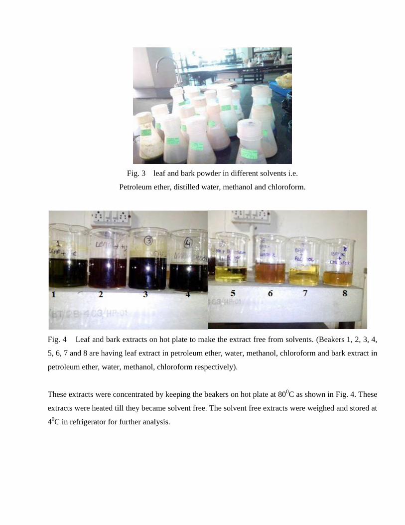
Fig. 3 leaf and bark powder in different solvents i.e.
Petroleum ether, distilled water, methanol and chloroform.
Fig. 4 Leaf and bark extracts on hot plate to make the extract free from solvents. (Beakers 1, 2, 3, 4,
5, 6, 7 and 8 are having leaf extract in petroleum ether, water, methanol, chloroform and bark extract in
petroleum ether, water, methanol, chloroform respectively).
These extracts were concentrated by keeping the beakers on hot plate at 800C as shown in Fig. 4. These
extracts were heated till they became solvent free. The solvent free extracts were weighed and stored at
40C in refrigerator for further analysis.

For Chemical Characterization- All the plant extracts were subjected to phytochemical analysis. All
the extracts were analyzed for the presence of phytochemicals like alkaloids, phenols, saponins
flavonoids and reducing sugars.
Test for alkaloids-
In solvent free extract 5 ml of dilute hydrochloric acid was added and mixed properly and then this
extract was filtered using filter paper 1. The filtrate obtained was used for detection of alkaloids using
alkaloidal reagents as follows-
(a)Wagner’s test
1 ml of filtrate was taken and few drops of Wagner’s reagent were added. Formation of red brown
precipitates was observed.
(b) Dragendorff’s test
1 ml of filtrate was taken and few drops of Dragendorff’s reagent were added. Formation of yellow
colored precipitations was observed.
Test for phenolic compounds
5 ml distilled water was dissolved in the solvent free extract. 100µl of ferric chloride (5%) solution was
added. Formation of dark green color was observed.
Test of saponins
10 ml of distilled water in added in 2ml of extract. And shake the test tube for 10 minutes. Formation
of layer of foam at the top of mixture was observed.
Test of Flavonoids
Aqueous solution of extract (1 ml) is added with 5ml of ammonia solution (10%). Formation of yellow
colored fluorescence was observed.
Test of reducing sugars
The presence of reducing sugars was checked using Fehling solution A & B. 1 ml of mixture of
Fehling solution A & B was added to extracts .Brick red colored were observed which shows the
presence of reducing sugars in extract.

Total phenolic content in crude extracts-
1 ml crude extract were added to 5 ml distilled water and 2 ml of Folin-ciocalteu’s phenolic reagent
(diluted with distilled water in the ratio 1:1). Mix them and keep at room temperature for 5 min. In this
mixture 2 ml sodium carbonate (20% in 0.1 N NaOH Solution). Keep them at water bath for 30 min.
Cool the test tubes and check the absorbance at 680 nm in spectrophotometer. Gallic acid is used as
standard for calibration of curve.
Total flavonoid content in crude extracts –
Take 0.5 ml of plant extract and mix it with 1.5 ml of methanol and add 0.1 ml of 10% aluminium
chloride and mix it. Now add 0.1 ml of potassium acetate (1M). Now make the final volume of tubes
by adding 2.8 ml of distilled water. Incubate these tubes to 37oC for 30 min. The observation of
reaction mixture was measured at 415nm. Quercetin was used for plotting calibration curve with its
various concentrations.
Antibacterial activity –
Disc diffusion method-to study the antimicrobial activity of plant extracts, agar diffusion method was
used against different microorganisms. In this method the pure culture of microorganism is sub
cultured nutrient broth and is kept for incubation at 37oC and the stock culture is revived. For
preparation of discs (6 mm) Whatman’s filter paper was used. These disc were left to dry under
laminar air flow in different plant extracts overnight. Gentamycin in concentration 0.1 mg/ml was used
as standard to check antimicrobial activity. For negative control the discs were prepared in respective
solvent. To check the antimicrobial activity the media used is Muller Hilton agar. The test organisms
were spread over the solidified Muller Hilton agar plate. Then the discs already prepared in different
extracts were positioned on MH Agar media. . Then these plates were kept in microbiological
incubator at 37oC for 24 hours. Antimicrobial activity was interpreted by the size of diameter of zone
of inhibition i.e the clear area around the disc
For Micropropagation-
Media preparation-MS (Murashige and Skoog) Media containing 3% (w/v) sucrose was prepared in
autoclaved distilled water. Plant growth hormones like 2, 4-D and BA was added in media for callus
induction. The pH of media was 5.8 using 0.1N NaOH and 0.1 N HCl. After this, agar (gelling

material) was added in the concentration of 0.8% (w/v). This media was then autoclaved at 121oC
temperature, 15psig pressure for 15 minutes.
Washing of explants- For micropropagation branches of bael were taken and then leaves were
removed. The nodal segment was washed with water thoroughly for 15 minutes. These nodal explants
were further washed with teepol solution for 5 minutes and then washed with distilled water. These
washed nodal explants were kept in laminar flow to provide aseptic conditions. For the surface
sterilization nodal explants were treated with 0.1% MgCl2 solution for 5 minutes. Then it is washed
with sterile distilled water for 3-4 times.
Inoculation of explants in media for callus induction- For callus induction MS media was
supplemented with combination of two growth hormones i.e. 2,4-D (1.5 mg/l) + BA (0.5 mg/l). Under
laminar air flow the media was solidified and the washed explants were inoculated in the media. These
culture tubes were kept at particular photoperiod for callus induction.
Media for shoot generation- After about 40 days the green colored callus was induced from the nodal
explant. This callus was then sub cultured in new media having different concentrations of growth
hormone. For shoot generation, MS media was supplemented with BA (1.5 mg/l).
Thin layer chromatography-
Preparation of TLC plates- To perform TLC the Silica Gel G was used as solid phase. Slurry of silica
gel was prepared using distilled water so that it can be easily poured over the glass plate. 20cm * 5 cm
glass slides were taken and the slurry was poured over the plates. The prepared plates were allowed to
set or dry at room temperature. Then the dried plates were put into hot air over for 1 hr at 110o C so
that plates get activated. Mark a line with pencil 1 cm above the bottom of the plate and make a spot of
extract with the help of small capillary tube.
Preparation of mobile phase - Three different solvents were used to make the mobile phase. Ethyl
acetate, toluene and formic acid were taken in ratio 12:36:5 respectively. Solution was kept in TLC
chamber to saturate it.
Sample spotted TLC plates were put in chamber and it was allowed to run 2/3 rd
of the plate
height. Plates were kept at room temperature to dry. Then these plates were observed under
transeluminator. Different bands were observed and the distance travelled by solvent and sample was
noted.

High performance liquid chromatography – Out of all the extracts, four extracts are having
significant number of compounds. These four extracts are methanol and chloroform extract of leaf and
bark. These extracts were sent in CDRI, Lucknow.

Chapter 6
Result and Discussion
In extraction of plant part i.e. leaves and bark, different amounts of crude extracts were
obtained. It was observed that all the extracts in different solvents are having different yield of crud
extract. The amount of crude extract of plant part in different solvents is shown in Table 1. In all the
extracts the percentage extractive values were calculated using the following formula-
Table 1: Amount of crude extract of leaves and bark in different solvents.
Sample S.No. Solvent Amount of crude extract
(gms.)/10 gms
Percent extracts
(%)
Leaf
1. Petroleum ether 0.140 gms 1.40
2. Water 2.70 gms 27.0
3. Alcohol 1.208 gms 12.0
4. Chloroform 0.341 gms 3.41
Bark
5. Petroleum ether 0.261 gms 2.61
6. Water 0.114 gms 1.14
7. Alcohol 0.180 gms 1.80
8. Chloroform 0.256 gms 2.56
From Table 1, it is clear that out of all the extracts, leaf in distilled water had given maximum
yield (2.70 gms) of crude extract. For leaf extraction distilled water is the better solvent. After water,
alcohol is good solvent but petroleum ether had given less amount (0.140 gms) of extract. For bark
extraction petroleum ether (0.261 gms) and chloroform (0.256 gms) are good solvents as they had
given much amount of extract than alcohol (0.180 gms) and water (0.114 gms). From this it is clear
those leaves and bark contain certain compounds which can be extracted using specific solvent system.
Shailesh et al., (2013) also found that in case of leaf, the water extract has high extraction
percentage as compare to the chloroform extract.

Leaf and bark extracts in different solvent i.e. petroleum ether, distilled water, methanol and
chloroform were checked for the presence of phytochemicals. Phytochemical tests performed indicated
the presence of different classes of secondary metabolites like alkaloids, saponins, flavonoids and
reducing sugars etc. It shows that for the isolation of specific secondary metabolite specific solvent
should be used. Plants are an important source of active natural products and these products differ
widely in their structures, biological properties and mechanisms of actions.
Test for alkaloids- All the extracts were tested for presence of alkaloids. The observation and
inference of all the extracts are shown in Table 2.
Table 2: Chemical tests to check the presence of alkaloids in different extract of Aegle marmelos.
Plant part Sample
No.
Solvent
used for
extraction
Observation
for Wagner
test
Inference Observation
for
Dragendorff
test
Inference
LEAF
1. Petroleum
ether
Red brown
ppts
Present Yellowish
ppts
present
2. Distilled
water
Red brown
ppts
Present Yellowish
ppts
present
3. Methanol Red brown
ppts
Present Yellowish
ppts
present
4. Chloroform Red brown
ppts
Present Yellowish
ppts
Present
BARK
5. Petroleum
ether
Red brown
ppts
Present Yellowish
ppts
Present
6. Distilled
water
Red brown
ppts
Present Yellowish
ppts
Present
7. Methanol Red brown
ppts
Present Yellowish
ppts
present
8. Chloroform red brown
ppts
Present Yellowish
ppts
Present

From Table 2, it is clear that all the extracts are having alkaloids. Alkaloid test was performed using
two different reagents and these reagents were Wagner’s reagent and Dragendorff‘s reagent. Wagner’s
reagent gave red brown colored precipitates which indicate the presence of alkaloids. Dragendorff test
gives yellowish precipitates on reaction with alkaloids present in sample. Using these reagents all the
extracts was tested and it was observed that all the extracts are having alkaloids
Fig. 5 Test to check the presence of alkaloids (Wagner test) (Tube 1, 2, 3, 4, 5, 6, 7 and 8 are leaf
extract in petroleum ether, water, methanol, chloroform and bark extract in petroleum ether, water,
methanol, chloroform respectively).
Fig. 6 Test to check the presence of alkaloids (Dragendorff test). (Tube 1, 2, 3, 4, 5, 6, 7 and 8 are leaf
extract in petroleum ether, water, methanol, chloroform and bark extract in petroleum ether, water,
methanol, chloroform respectively).
Mariya et al., (2013) also found that water, methanol and chloroform extract of leaves are
having alkaloids.

Test for phenolic compounds- All the leaf and bark extracts were tested for the presence of phenolic
compounds. The observations and inference are shown in Table 3.
Table 3: Chemical tests to check phenolic compounds in different extract of Aegle marmelos.
Plant part S No. Solvent for
extraction
Observation Inference
Leaves
1. Petroleum ether Green color Present
2. Distilled water Green color Present
3. Methanol Green color Present
4. Chloroform Green color Present
Bark
5. Petroleum ether No green color Absent
6. Distilled water No green color Absent
7. Methanol Green color Present
8. Chloroform Green color Present
From Table 3 it is clear that phenolic compounds were present in some extracts but phenols were
absent in other extracts. In case of leaves, the phenolic compounds were observed in petroleum ether,
water, methanol and chloroform extract. Similarly in case of bark, phenols were observed in methanol
and chloroform extract. In petroleum ether and water extract of bark phenols were not observed.
Fig. 7 Test to check the presence of phenolic content in plant extracts (Tube1, 2, 3, 4, 5, 6, 7, 8 are leaf
extract in petroleum ether, water, methanol, chloroform and bark extract in petroleum ether, water,
methanol, chloroform respectively).

Saroj et al., (2011) also found that leaf extract in petroleum ether, methanol and chloroform
contain phenolic compounds. Shailesh et al., (2013) found that phenols are present in water extract of
bael. Mariya et al., (2013) also observe the same results for water, methanol and chloroform extract of
leaves.
Test of saponins- All the extracts of leaf and bark were tested to check the presence of saponins.
Saponins forms a layer of foam on shaking continuously. The observations and inference of saponins is
shown in Table 4.
Table 4: Chemicals test to check the presence of saponins in plant extracts.
Plant part S No. Solvent used for
extraction
Observation Inference
Leaf
1. Petroleum ether No foam
formation
Absent
2. Distilled water Foam formation Present
3. Methanol Foam formation Present
4. Chloroform No foam
formation
Absent
Bark
5. Petroleum ether Foam formation Present
6. Distilled water Foam formation Present
7. Methanol Foam formation Present
8. Chloroform Foam formation Present
From Table 4, it is clear that saponins are present in most of the extracts but absent in
petroleum ether and chloroform extract of leaf. In case of bark saponins were observed in all the
extracts i.e. petroleum ether, water, methanol and chloroform extract.
Saroj et al., (2011) also observed the same result for leaf extracts. They observed that saponins
are absent in petroleum ether and chloroform extract of leaves and saponins are present in water and
methanol extract of leaf.

Fig 8- Test to check the presence of saponins in leaf extracts. (Tube 1, 2, 3, 4 are leaf extract in
petroleum ether, water, methanol, chloroform respectively).
Fig 9- Test to check the presence of saponins in Bark extracts. (Tube5, 6, 7 and 8 are bark extract in
petroleum ether, water, methanol, chloroform respectively.)
Test of reducing sugars- All the leaf and bark extracts were tested to check the presence of reducing
sugars. For this Fehling solution A and B were used. The observation and inference for reducing sugars
is shown in Table 5.

Table 5: Chemical test to check the presence of reducing sugars in extracts.
Plant part S No Solvent used for
extraction
Observation Inference
Leaf
1. Petroleum ether No precipitate Absent
2. Distilled water Brick red precipitate Present
3. Methanol Brick red precipitate Present
4. Chloroform No ppts Absent
Bark
5. Petroleum ether Brick red precipitate Present
6. Distilled water Brick red precipitate Present
7. Methanol Brick red precipitate Present
8. Chloroform Brick red precipitate Present
From Table 5, it is clear that reducing sugars are present in petroleum ether and water extract of
leaves but reducing sugars were absent in methanol and chloroform extract of leaf. Reducing sugars are
observed in all the four extracts of bark. Layer of foam was observed in petroleum ether, water,
methanol and chloroform extract of bark.
Fig. 10 Test to check the presence of reducing sugars. (Tube 1, 2, 3, 4, 5, 6, 7, 8 are leaf extract in
petroleum ether, water, methanol, chloroform and bark extract in petroleum ether, water, methanol,
chloroform respectively).

Vanitha et al., (2012) had also observed the same results for leaf extracts. They observed that
reducing sugars are present in water and methanol extract of leaf and petroleum ether. But reducing
sugars are absent in petroleum ether and chloroform extract.
Phenolic and Flavonoid content –
Antioxidant activity is mainly shown because of the presence of phenolic contents in plant
extracts. Total phenolic content was checked in leaf and bark extract with different solvents like
petroleum ether, distilled water, methanol and chloroform. The absorbance of unknown samples were
taken and calibrated in standaerd curve to check phenolic content in samples. Standard graph of gallic
acid is shown in Fig 11. Standard graph of gallic acid is having regression value, R2= 0.996, y= 0.012x
+ 0.002. The amount of phenols in leaf and bark extract is shown in Table 6. In methanol extract of
leaf the amount of phenol is maximum i.e. 66.4 mg/grams of dried leaves. In chloroform extract of leaf
minimum amount of phenols was observed i.e. 16 mg/grams of dried leaves. In case of bark, methanol
extract is having high amount (54.8 mg/g) of phenols. In bark, petroleum ether was having minimum
amount (12.8 mg/g) of phenols. Tupe et al., (2012) also found that total phenolic content in methanolic
extract is 63.01 mg GAE/ gm.
Fig. 11 Gallic acid standard curve for estimation of phenolic content

Table 6: Total phenolic and flavonoid content of different extracts of bael plant
Plant Part S. No. Solvent used Total phenolic
compound (TPC)
mg/g
Total flavonoid
compound (TFC)
mg/g
Leaf
1. Petrolem ether 32 77
2. Distilled water 48 7
3. Methanol 66.4 98
4. Chloroform 16 241
Bark
5. Petrolem ether 12.8 1.5
6. Distilled water 30.4 5
7. Methanol 54.8 55.5
8. Chloroform 26.4 73.5
Total flavonoid content estimation-
Flavonoids are having hydroxyl groups that are responsible for radical scavenging activity in
plants. Standard graph for flavonoid content is shown in Fig 12.
Fig. 12 Quercetin standard curve for estimation of flavonoid contents.

By calibrating the O.D of unknown samples in standard graph of flavonoids the amount of
flavonoids in unknown samples was calculated. It was found that chloroform extract of laves was
having maximum amount (241 mg/g) of flavonoids. Minimum amount of flavonoids was found to be
in petroleum ether extract (1.5 mg/g) of bark. High amount of flavonoids was found in leaf extracts. In
bark extract flavonoids were present in less amount as compare to the leaf extract. Overall, the extracts
are having high amount of flavonoids as compare to the phenolic compounds.
Antimicrobial activity
The antimicrobial activity of petroleum ether, distiller water, methanol and chloroform extract
of leaf and bark was studied by disc diffusion method. Test organisms for antimicrobial activity are E.
coli, Bacillus cereus, Bacillus subtilis, Staphylococcus pneumonia. The activity of extract against a
particular microorganism is calculated by the diameter of zone of inhibition. Higher diameter of zone
of inhibition means the extract is more effective against that microorganism. No zone of inhibition
means the microorganism to be tested is resistant against that extract. Gentamycin is an antibiotic
which is used as standard to check the antimicrobial activity of plant extracts. Effect of different plant
extracts on E. coli is shown in Fig 14.

Fig. 13 Effect of leaf and bark extract on E. coli (S1-lesf+petroleum ether, S2- leaf+water,S3 – leaf +
methanol, S4 – leaf +chloroform, S5- bark+ petroleum ether, S6- bark + water , S7 – bark+ methanol,
S8- bark +chloroform).
From Fig 13 it is clear that maximum activity against E. coli is shown by bark extract in
petroleum ether and chloroform solvents. There was no activity in water and methanol extract of bark.
Leaf extract in petroleum ether, water, methanol and chloroform had shown the antimicrobial effect. In
case of petroleum ether and chloroform extract the zone of inhibition was found to be 10 mm. In
Gentamycin the zone of inhibition was having diameter of 26mm. When compared with the standard,
the petroleum ether and chloroform extracts had shown 38.46 % inhibition. Whereas the water and
methanol extracts had not shown any activity against E. coli.
Table 7: Effect of different extracts on E. coli and Bacillus subtilis bacteria
Plant
part
S. No. Solvent for
extraction
Diameter of
zone of
inhibition in E.
coli (in mm)
Percentag
e
inhibition
Diameter of
zone of
inhibition in
Bacillus subtilis
(in mm)
Percentag
e
inhibition
Leaf
1. petroleum ether 8 mm 30.76% 11 mm 44 %
2. Distilled water 9 mm 34.61% 8 mm 32 %
3. Methanol 8 mm 30.76 8 mm 32 %
4. Chloroform 7.5 mm 28.84% 11 mm 44 %
Bark
5. petroleum ether 10mm 38.46% ----- ----
6. Distilled water ----- ----- ----- -----
7. Methanol ----- ------ ----- ------
8. chloroform 10 mm 38.46% 8 mm 32 %
Gentamycin 26mm 25 mm
Antimicrobial activity against Bacillus subtilis was checked using same method. It was found
that petroleum ether and chloroform extract of leaves had shown maximum activity against Bacillus
subtilis. The diameter of zone of inhibition in case of petroleum ether and chloroform extract of leaves
was 11 mm. The diameter of zone of inhibition in standard was 25 mm. When compared with standard
the petroleum ether and chloroform extracts shown 44% antimicrobial activity against Bacillus subtilis.
But in case of bark, only the chloroform extract had shown antimicrobial activity against Bacillus
subtilis. Chloroform extract of bark had shown 32% antimicrobial activity against Bacillus subtilis.
Bacillus subtilis had shown resistance against petroleum ether, methanol and chloroform extract of
bael.

Fig. 14 Effect of leaf and bark extract on Bacillus subtilis(S1-lesf+petroleum ether, S2- leaf+water,S3
– leaf + methanol, S4 – leaf +chloroform, S5- bark+ petroleum ether, S6- bark + water , S7 – bark+
methanol, S8- bark +chloroform).
Antimicrobial activity against Staphylococcus pneumonia was checked using disc diffusion
method. It was found that maximum activity against Staphylococcus pneumonia was shown by the
petroleum ether extract of leaf. The zone of inhibition of leaf extract in petroleum ether was 15 mm.
This zone of inhibition was compared with the standard and it was found that the extract had shown
71.42 % antimicrobial activity against Staphylococcus pneumonia. Leaf extract in methanol and bark
extract in petroleum ether had shown minimum activity against Staphylococcus pneumonia . In both
the cases the zone of inhibition was having diameter 8mm. leaf in methanol and bark in petroleum
ether had shown 38.09% antimicrobial activity. Staphylococcus pneumonia was found to be resistant
against water extract of leaves and bark. No zone of inhibition was observed in water extract of leaves
and bark.

Fig. 15 Effect of leaf and bark extract on Staphylococcus pneumonia (S1-lesf+petroleum ether, S2-
leaf+water,S3 – leaf + methanol, S4 – leaf +chloroform, S5- bark+ petroleum ether, S6- bark + water ,
S7 – bark+ methanol, S8- bark +chloroform

Table 8: Effect of different extracts on Staphylococcus pneumonia and Bacillus cereus
Plant
part
S.
No.
Solvent for
extraction
Diameter of zone
of inhibition in
Staphylococcus
pneumonia (in
mm)
Percentage
inhibition
Diameter of
zone of
inhibition in
Bacillus cereus
(in mm)
Percentage
inhibition
(%)
Leaf
1. petroleum ether 15mm 71.42% 9 mm 37.5
2. Distilled water ----- ----- 11 mm 45.83
3. Methanol 8 mm 38.09% ----- -----
4. Chloroform 9 mm 42.85% 13 mm 54.16
Bark
5. petroleum ether 8 mm 38.09% 15 mm 62.5
6. Distilled water ----- ---- 12 mm 50
7. Methanol 12 mm 57.14% 16 mm 66.66
8. Chloroform 9 mm 42.85% 8mm 33.33
Gentamycin 21 mm 24 mm

Fig. 16 Effect of leaf and bark extract on Bacillus cereus (S1-leaf+petroleum ether, S2- leaf+water,S3
– leaf + methanol, S4 – leaf +chloroform, S5- bark+ petroleum ether, S6- bark + water , S7 – bark+
methanol, S8- bark +chloroform
Antimicrobial activity was checked against the microorganism Bacillus cereus. Maximum zone
of inhibition was observed in case of methanol extract of bark with the diameter of zone of inhibition
16 mm. When compared with the standard having zone of inhibition 24 mm it was found that the bark
extract in methanol had shown 66.66% inhibition. Bark extracted in chloroform had shown minimum
activity against Bacillus cereus with zone of inhibition f 8mm. when compared with the standard the
extract was found to be having 33.33% inhibition. Methanol extract of leaves had not shown any zone
of inhibition. It indicates that Bacillus cereus is resistant against methanol extract of leaves. All the
bark extracts in petroleum ether, water, methanol and chloroform had shown antimicrobial activity.
From this, it is clear that maximum activity against E. coli is shown by the petroleum ether and
chloroform extract of bark. For Bacillus subtilis maximum activity was observed by petroleum ether
and chloroform extract of leaves. For Staphylococcus pneumonia, leaves extracted in petroleum ether
had shown more activity. For Bacillus cereus, bark extracted in methanol had shown maximum
activity.
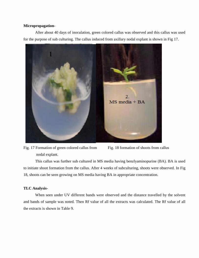
Micropropagation-
After about 40 days of inoculation, green colored callus was observed and this callus was used
for the purpose of sub culturing. The callus induced from axillary nodal explant is shown in Fig 17.
Fig. 17 Formation of green colored callus from Fig. 18 formation of shoots from callus
nodal explant.
This callus was further sub cultured in MS media having benzlyaminopurine (BA). BA is used
to initiate shoot formation from the callus. After 4 weeks of subculturing, shoots were observed. In Fig
18, shoots can be seen growing on MS media having BA in appropriate concentration.
TLC Analysis-
When seen under UV different bands were observed and the distance travelled by the solvent
and bands of sample was noted. Then Rf value of all the extracts was calculated. The Rf value of all
the extracts is shown in Table 9.

Fig. 19 TLC analysis of different extracts of Aegle marmelos. (1-leaf+petroleum ether, 2- leaf+ water,
3 – leaf + methanol, 4 – leaf +chloroform, 5- bark+ petroleum ether, 6- bark + water , 7 – bark+
methanol, 8- bark +chloroform).
From Fig. 19 it is clear that samples 3, 4, 7 and 8 are having more compounds as compare to 1,
2, 3 and 4. In petroleum ether and water extract of leaf and bark, less number of bands was observed.
In methanol and chloroform extract of leaf and bark, more number of bands was observed. The Rf
values of these extracts were compared with the Rf values of standard and these extracts were analyzed
for the presence of different compounds.
Rf values of the extracts were calculated using the formula-

Table 9: Rf values found in different extracts of Aegle marmelos and compounds present in extracts.
Plant
part
used
Sample
no.
Solvent
used
Rf values Name of the compound present in
extract
Leaves
1. Petroleum
ether
0.44, 0.77 Apigenin, 3Hydroxyflavone
2. Distilled
water
0.56,0.67,0.76 Ferulic acid, 6 -Hydroxyflavone, 3-
Hydroxyflavone
3. Methanol 0.51,0.56,0.62,0.6
5,0.77,0.88
Kaempferol, Ferulic acid, Chrysin,
Galangin, 3-Hydroxyflavone, Flavone
4. Chloroform 0.46,0.55,0.62,0.6
5,0.67,0.88
7-Hydroxyflavone,o/p Coumaric acid,
Chrysin, Galangin, 6-Hydroxyflavone,
Flavone
Bark
5. Petroleum
ether
0.77, 0.88 3-Hydroxyflavone, Flavone
6. Distilled
water
0.55 o/p Coumaric acid
7. Methanol 0.23,0.39,0.46,0.5
6,0.88
Morin, Quercetin, 7-Hydroxyflavone,
Ferulic acid, Flavone
8. Chloroform 0.23,0.55,0.77,
0.88
Morin, o/p Coumaric acid, 3-
Hydroxyflavone, Flavone
When these Rf values were compared with the standard it was found that the extracts contain
different compounds like morin, Quercetin, apigenin, caffeic acid, Quercetin, apigenin,
hydroxyflavone, dihydroxyflavone, coumaric acid, galangin, flavones etc. The compounds present in
different extracts are shown in Table. 9. From Table 9, it is clear that sample 1(leaf in petroleum ether)
contain apigenin and 3 hydroxyflavone. Sample 2(leaf in distilled water) contain ferulic acid, 3
hydroxyflavone and 6 hydroxyflavone. Sample 3 contain six compounds i.e. Kaempferol, ferulic acid,
Chrysin, galangin, 3 hydroxyflavone, flavone. Sample 4 (leaf in chloroform) contain 7Hydroxyflavone,
o/p Coumaric acid, Chrysin, Galangin, 6Hydroxyflavone and Flavone. Sample 5 (bark in petroleum
ether) contain 3Hydroxyflavone and Flavone only. Sample 6 (bark in water) is having o-Coumaric acid
and p-coumaric acid. Sample 7 (bark in methanol) contain Morin, Quercetin, 7-Hydroxyflavone,
Ferulic acid and Flavone. Sample 8 (bark in chloroform) is having Morin, ortho and para coumaric
acid, 3Hydroxyflavone and Flavone.
It is clear that leaf extraction in methanol and chloroform is showing presence of many
compounds. After these the bark extraction in methanol is giving many compounds as compare to bark
in chloroform. For both leaf and bark methanol is giving good range of compounds. Te samples having
good range of compounds were further used for HPLC analysis.
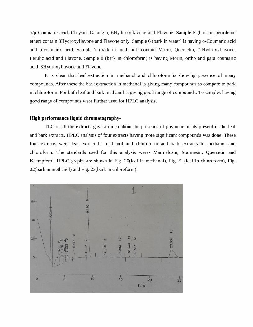
High performance liquid chromatography-
TLC of all the extracts gave an idea about the presence of phytochemicals present in the leaf
and bark extracts. HPLC analysis of four extracts having more significant compounds was done. These
four extracts were leaf extract in methanol and chloroform and bark extracts in methanol and
chloroform. The standards used for this analysis were- Marmelosin, Marmesin, Quercetin and
Kaempferol. HPLC graphs are shown in Fig. 20(leaf in methanol), Fig 21 (leaf in chloroform), Fig.
22(bark in methanol) and Fig. 23(bark in chloroform).

Fig. 20 HPLC analysis of leaf extract in methanol
Fig 21 HPLC analysis of leaf extract in chloroform

Fig. 22 HPLC analysis of bark extract in methanol
Fig. 23 HPLC analysis of bark extract in chloroform.

From Figs. ,20,21,22,23 it is clear that all the four extracts are having Marmelosin, Marmesin,
Quercetin and Kaempferol. The amount of these extracts in sample is shown in Table 10.
Table 10: Amount of Marmelosin, Marmesin, Quercetin and Kaempferol in samples.
Sample
No.
Plant part/solvent used Marmelosin
(mg/g)
Marmesin
(mg/g)
Quercetin
(mg/g)
Kaempferol
(mg/g)
1. Leaf / Methanol
67.5 80.0 40.2 11.5
2. Leaf / Chloroform
65.5 78.2 20.4 10.5
3. Bark / Methanol
46.5 50.1 15.7 8.5
4. Bark / Chloroform
42.0 48.7 12.6 7.5
From Table 10, it is clear that Marmelosin is present in all the four extracts. Maximum amount
of Marmelosin is observed in methanol extract of leaf (67.5mg/g). In case of leaf, methanol is having
more Marmelosin as compared to the chloroform extract. In case of bark also the methanol extract
(46.5 mg/g) is having high yield of Marmelosin then the chloroform extract. Marmesin is also present
in all the four extracts but higher amount of Marmesin is observed in methanol extract of leaf.
Methanol extract yield higher Marmesin (80 mg/g) as compare to the chloroform extract (78.2mh/g).
Similarly, methanol extract of bark yield higher (50.1 mg/g) Marmesin as compare to chloroform
extract (48.7 mg/g). Quercetin is present in high amount in Methanol extract of leaf (40.2 mg/g) and
lowest mount was observed in bark extract in chloroform (12.6 mg/g). Kaempferol is also present in all
the four extracts. Maximum amount was observed in methanol extract of leaf (11.5 mg/g) and lowest
amount was observed in chloroform extract of bark (7.5 mg/g). It indicates that methanol extract of leaf
had given maximum yield of compounds. So for extraction purpose methanol is best solvent. There
were some peaks also observed in HPLC graphs. These can be considered as unknown compound.
Umadevi et al., (2012) had also observed the same results in case of alcoholic extract of leaves
of bael. But instead of Kaempferol they found Ursolic acid in the alcoholic extract of leaves.

CHAPTER 7
CONCLUSION
Aegle marmelos contains different classes of secondary metabolites and these metabolites are
further used as herbal and ayurvedic medicines. For the chemical characterization the selection of
solvent should be done carefully because there are some solvents in which the phytochemical test
shows positive result and same extract shows negative result in other solvent. It was found that the
bioactive compounds isolated from plants are used in almost 25% of prescribed drugs. From the study
on bael tree it is clear that bael has became the major source of medicine for curing various diseases in
humans and animals. In conservation of medicinal plants and reintroduction to nature’s endangered
species micropropagation played an important role. The medicinal plants which are aromatic and of
medicinal use need to be multiplied in short span of time and establishment in their natural habitat. The
conventional methods for plant regeneration are slow and it was found that the seeds thus produced are
no much viable so researchers are now trying to increase yield of secondary metabolites, production
and to make the plant resistant to adverse environmental conditions.
The results obtained from the phytochemical analysis had shown similarities with the literature.
But in quantitative analysis of plant extract, differences in amount of TPC & TFC were observed.
These differences may be due to change in geographical region. In anitimicrobial activity, the extracts
had shown their inhibitory effect. But some extracts had shown no antimicrobial activity. In TLC there
were some bands for which there is no standard available to know the name of these compounds. So
further studies can be carried to know about the unknown compounds present in bands. Similarly in
HPLC of the extracts, it was observed that there are some peaks for which no standard compound has
been identified. So further studies can be done to know about the unknown compounds shown by the
peaks of HPLC graph. By doing this we will be having a wide range of bioactive compounds. Further
these bioactive compounds can be isolated and used in preparation of traditional medicines.

CHAPTER 8
REFERENCES
Abirami H. and Kumar P.S. (2013). In Vitro Regeneration and Extraction of Secondary
Metabolites In Aegle marmelos (L.) Correa. Asian Journal of Plant Science and Research,
3(2):99-106.
Bansal Y. and Bansal G. (2011). Analytical Methods For Standardization Of Aegle marmelos:
A Review. Journal of Pharmacy Education And Research, 2(2); 37-44
Charoensiddhi, S. and Anprung P. (2008). Bioactive compounds and volatile compounds of
Thai bael fruit (Aegle marmelos (L.) Correa) as a valuable source for functional food
ingredients. International Food Research Journal, 15(3): 287-295
Chavda N., Mujapara A., Mehta S.K. and Dodia P.P. (2012). Primary Identification of certain
Phytochemical Constituents of Aegle marmelos (L.) Corr. Serr Responsible for Antimicrobial
Activity against Selected Vegetable and Clinical Pathogen. International Journal of Philosophy
and Social Sciences, 2(6); 190-206.
Dhankhar S., Ruhil S., Balhara M., Dhankhar S. And Chhillar A.K. (2011). Aegle marmelos
(Linn.) Correa: A Potential Source Of Phytomedicine. Journal of Medicinal Plants Research,
5(9):1497-1507.
Janarthanan U. K., Varadharajan V. and Krishnamurthy V. (2012). Physicochemical evaluation,
Phytochemical screening and chromatographic fingerprint profile of Aegle marmelos (l.) Leaf
extracts. World Journal of Pharmaceutical Research, 1(3); 813-837.
Janarthanan U. K., Varadharajan V. and Krishnamurthy V. (2012). Physicochemical
Evaluation, Phytochemical Screening and Chromatographic Fingerprint Profile of Aegle
marmelos (L.) Leaf Extracts. World Journal Of Pharmaceutical Research, 1(3):813-837.
Joglekar M., Mandal M., Paralakoti Somaiah M. and Murthy S. (2012). Comparative analysis
of antioxidant and antibacterial properties of Aegle marmelos, Coriandrum sativum and
Trigonella foenum graecum. Research Article, Acta Biologica Indica, 1(1); 105-108.

Jyothi S.K. and Subba Rao B. (2010). Antibacterial Activity of Extracts from Aegle marmelos
against Standard Pathogenic Bacterial Strains. International Journal of PharmTech Research,
2(3); 1824-1826
Kothari S., Mishra V., Bharat S. and Tonpay S.D. (2011). Antimicrobial activity and
Phytochemical screening of serial extracts from leaves of Aegle marmelos (linn.). Acta
Poloniae Pharmaceutica, 68(5); 687-692.
Kumar D., Dhurandhar K., Verma R., Barman S. and Kumar A. (2013). To Evaluation of Total
Phenolics and Flavonoids in Different Plant of Chhattisgarh. Journal of Pharmacognosy and
phytochemistry, 2(4); 116-118.
Kumar S., K.N. and Hemalatha S. (2013). Phytochemical evaluation of leaf extracts of Aegle
marmelos. International Journal of Development Research, 3(7); 29-33.
Mathur G., Roy N. And Mathur A. (2013). In Vitro Analysis Of Aegle marmelos Leaf Extracts
On Skin Pathogens. Journal of Applied Pharmaceutical Science, 3 (10);097-100
Medic-Saric M., Jasprica I., Smolcic- Bubalo A. and Mornar A.(2004). Optimization of
Chromatographic Conditions in Thin Layer Chromatography of Flavonoids and Phenolic
Acids. International Standard Serial Number, 77(1-2); 361-366
Narayan T.W., Pankaj K., Julfikar A., Soni M.L. and Rakesh P.(2010). Tissue culture of
endangered Bael tree (Aegle marmelos ): A Review. Journal of Advanced Scientific Research,
1(2); 34-40
Natarajan E., Purushothaman B., Anija, Krishna A. and Gowri (2012). Antimutagenic and
antibacterial activity of Aegle marmelos (L.) Corr. International Journal of Advanced
Biotechnology and Research, 3(3); 670-679.
Patel P.K., Sahu J., Sahu L., Prajapati N. K. and Dubey B.K. (2012). Aegle marmelos: A
Review on its Medicinal Properties. International Journal of Pharmaceutical and
Phytopharmacological Research, 1(5); 332-341.
Poonkothai M. and Saravanan M. (2008). Antibacterial activity of Aegle marmelos against leaf,
bark and fruit extracts. Ancient Science of Life, 17(3); 15-8.
Puhan P. and Rath S.P. (2012). In vitro propagation of Aegle marmelos (L.) corr., a medicinal
plant through axillary bud multiplication. Advances in Bioscience and Biotechnology, 3; 121-
125

Rahman S. and Parvin R. (2014). Therapeutic Potential of Aegle marmelos (L.)-An Overview.
Asian pacific Journal of Tropical Disease, 4(1); 71-77.
Ramya M., Rekha S. and Parvathi A. (2013). Evaluation of phytoconstituents in aqueous and
organic extracts of Aegle marmelos (L.) Correa. And Punica granatum L. International Journal
of Pharmaceutical, Chemical and Biological Sciences, 3(4); 1024-1031.
Reddy P. V., Sahana N. And Urooj A. (2012). Antioxidant activity of Aegle marmelos and
Psidium guajava leaves. International Journal of Medicinal and Aromatic plants, 2(1); 105-108
Sarkar B.K. and Solanki S.S. (2011). Isolation, characterization and antimicrobial activity of
leaves extracts of bael (Aegle marmelos). International Journal of Pharmacy & Life Sciences,
2(12); 1303-1305.
Sathya K., Tamil Selvi S., Bharathidasan R., Rajkumar K., Ilakkiya R. and Prabakaran M.
(2013). Evaluation of phenolic content and antioxidant activity of Aegle marmelos (L.) Corr.
Serr. Journal of Natural product and Plant resources, 3(4); 24-28
Sekar D.K., Kumar G., Karthik L. and Bhaskar Rao K.V. (2011). A review on pharmacological
and phytochemical properties of Aegle marmelos (L.) Corr. Serr. (Rutaceae). Asian Journal of
Plant Science and Research, 1(2); 8-17
Sharma G.N., Dubey S.K., Sati N. and Sanadya J. (2011). Medicinal Values of Bael (Aegle
marmelos) (L.) Corr.: A Review. International Journal of Current Pharmaceutical Review and
Research, 1(3); 12-22.
Sharma G.N., Dubey S.K., Sati N. and Sanadya J. (2011). Anti-inflammatory Activity and
Total Flavonoid Content of Aegle marmelos Seeds. International Journal of Pharmaceutical
Sciences and Drug Research, 3(3); 214-218
Siddique N.A., Mujeeb M., Najmi A.K. and Akram M. (2010). Evaluation of antioxidant
activity, quantitative estimation of phenols and flavonoids in different parts of Aegle marmelos.
African Journal of Plant Science, 4(1); 001-005.
Singh N. and Kumar S. (2015). TLC AND HPLC Fingerprint development of Aegle marmelos
Corr. And its polyherbal marketed formulations. International Journal of Pharma Sciences and
Research, 6(1); 162-167.
Tupe, R.S., Kemse, N.G. and Khaire, A.A. (2013). Evaluation of antioxidant potentials and
total phenolic contents of selected Indian herbs powder extracts. International Food Research
Journal, 20(3); 1053-1063

Varughese B. and Tripathi J. (2013). Phytochemical Evaluation of different Solvent Extracts of
Aegle marmelos fruit at different Stages of its Ripening. Advances in Life Science and
Technology, 8; 8-13
Venkatesh P., Md. Begum C., Revathi A., Revathi P. and Jhansi B. (2014). Phytochemical
analysis, total phenol content and in vitro anticancer activity of immature bark of Aegle
marmelos. International Journal of Pharmaceutical resesrch and Novel Sciences, 1(1); 22-26
Victoria D.T., Rao K.K. and Sampot A.V.(2014). Antibacterial Activity and phytochemical
screening of Aegle marmelos. International Journal of Pharma and Bio Sciences, 5(4); 895-902