chicken major histocompatibility complex-encodedb-g antigens important
TRANSCRIPT

Proc. Natl. Acad. Sci. USAVol. 88, pp. 1359-1363, February 1991Immunology
Chicken major histocompatibility complex-encoded B-G antigensare found on many cell types that are important for theimmune system
(B-lymphocyte selection/B complex/bursa/caecal tonsil/thrombocyte)
JAN SALOMONSEN*, DOMINIQUE DUNON*, KARSTEN SKJ0DTtf, DIANE THORPE*, OLLI VAINIO*§,AND JIM KAUFMAN**Basel Institute for Immunology, Grenzacherstrasse 487, CH-4005 Basel, Switzerland; and tInstitute for Experimental Immunology, University of Copenhagen,N0rre Alle 71, DK-2100 Copenhagen 0., Denmark
Communicated by Niels K. Jerne, October 31, 1990 (received for review July 18, 1990)
ABSTRACT B-G antigens are a polymorphic multigenefamily of cell surface molecules encoded by the chicken majorhistocompatibility complex (MHC). They have previously beendescribed only on cells of the erythroid lineage. By using flowcytometry, section staining, and immunoprecipitation withmonoclonal antibodies and rabbit antisera to B-G moleculesand by using Northern blots with B-G cDNA clones, wedemonstrate here that B-G molecules and RNA are present inmany other cell types: thrombocytes, peripheral B and Tlymphocytes, bursal B cells and thymocytes, and stromal cellsin the bursa, thymus, and caecal tonsil of the intestine. Thereactions also identify at least one polymorphic B-G determi-nant encoded by the B-F/B-L region of the chicken MHC. Theserology and tissue distribution of B-G molecules are as com-plex as those ofmammalian MHC class I and class II molecules.These facts, taken with certain functional data, lead us tosuggest that B-G molecules have an important role in theselection of B cells in the chicken bursa.
The major histocompatibility complex (MHC) of mammalsincludes two multigene families that encode the MHC classI and class II molecules. Some of these cell surface het-erodimeric glycoproteins are highly polymorphic and areresponsible for recognition of antigen by T lymphocytes forimportant immunological phenomena. Others are not sopolymorphic and may be involved in other biological func-tions (for instance, as an intestinal Fc receptor). The tissuedistributions of these molecules are different and complex,changing during ontogeny and the activation state of partic-ular cells, and only partly reflect their functional roles (1, 2).The chicken MHC, called the B complex, contains a third
multigene family, which encodes polymorphic cell surfacedimeric proteins called the B-G antigens (3-5). Several linesof evidence suggest that B-G molecules are recognized in aspecial way by the immune systems of chickens and mam-mals (6, 7): the presence of "natural antibodies" to thesenonglycosylated molecules in many species (8), the rapid andlarge immunoglobulin response to B-G antigens as opposed toclass I or class II antigens (7, 9, 10), and the "adjuvant effect"of B-G molecules for the humoral response to other cellsurface alloantigens (7, 11, 12). The responses in thesephenomena are generally to polymorphic determinants ofB-G, which suggests that they are recognized by antigen-specific receptors on B or T cells.A role for the polymorphic B-G antigens in the immune
system has never been seriously considered, because theyhave been described only on erythrocytes and erythrocyteprecursors (13-15). In light of their similarity to MHC class
I and class II molecules, we thought that it was important todetermine their tissue distribution. We have previously usedmonoclonal antibodies (mAbs) and rabbit antisera to isolateand characterize B-G molecules and B-G cDNA clones(15-17). Here we use these reagents, as well as new mAbsderived for the purpose, to show that B-G antigens are foundon various cell types that are important for the chickenimmune system.¶
MATERIALS AND METHODSChicken strains have been described (15, 16, 19). The mAbsto B-G molecules called gl (I-8D8, y2a), g2 (14-7C11, it), g3(I-19A5, IL), g4 (I-17A8, pu), g5 (15-3D7, y2b), g6 (I-8D8a,y2a), g7 (5-7D7, y3), g8 (5-6C8, y2a), and g9 (18-6G2, y2a),and the rabbit antisera have been described (15, 16) or will bedescribed in another publication. The mAbs to chicken classII (2G11, yl), class I (F21-2, yl), and CD3 (CT3, 'yl) havebeen described (4, 20, 21).
Erythroblasts were isolated as described (15, 16). Periph-eral blood leukocytes were separated from erythrocytes byFicoll-Paque density centrifugation of heparinized blood asdescribed (20). Thrombocytes were partially purified bydifferential centrifugation of diluted heparinized blood (50 xg, 20 min, room temperature, ref. 22) followed by Ficoll-Paque density centrifugation of the cells in the pellet, with80%o thrombocytes and 20% CD3+ cells found at the inter-face. Single-cell suspensions from thymus and bursa weremade by pushing tissue slices through a screen, with thestromal cells considered to be the tissue left over. Caecaltonsils were identified as lymphoid regions in the appendicesat the junction of the small and large intestine.
Staining for flow cytometry was done with hybridomaculture supernatants followed by sheep anti-mouse immuno-globulin F(ab)2 coupled to fluorescein (Silenus, Hawthorn,Australia). For double staining, the stained cells were treatedwith 1% mouse serum and then stained either with 2G11coupled to biotin followed by streptavidin coupled to phy-coerythrin or CT3 followed by sheep anti-mouse yl coupledto phycoerythrin (Southern Biotechnology Associates, Bir-mingham, AL). Cells were analyzed by FACscan or sorted bya FACS440 (Becton Dickinson).
All other techniques were as previously described (15-17,20, 23-25).
Abbreviations: mAb, monoclonal antibody; MHC, major histocom-patibility complex.TPresent address: Institute for Medical Microbiology, University ofOdense, Campusvej 55, DK-5230 Odense C., Denmark.§Present address: Department of Medical Microbiology, Universityof Turku, Kiinamyllynkatu 13, SF-20520 Turku 52, Finland.1A preliminary account of this work has been published (18).
1359
The publication costs of this article were defrayed in part by page chargepayment. This article must therefore be hereby marked "advertisement"in accordance with 18 U.S.C. §1734 solely to indicate this fact.

1360 Immunology: Salomonsen et al.
RESULTSmAbs Raised to Erythrocyte B-G Molecules Recognize Sub-
sets of Erythrocyte B-G Molecules Encoded by the B-G Region.Erythrocyte B-G molecules purified by our first mAbs (15)were used to make rabbit antisera and hundreds ofnew mAbs.On erythrocytes, the rabbit antisera recognize non-glycosylated disulfide-linked dimers that are encoded by theB-G region of the MHC. The considerable size heterogeneityfound in these molecules between and within haplotypes couldbe due either to different genes or to alternative splicing in thecytoplasmic region (17). Many of the new mAbs bind to thesurface of erythrocytes and immunoprecipitate subsets of theerythrocyte B-G molecules recognized by rabbit antisera(illustrated in Fig. 1). Thus, certain erythrocyte moleculesencoded by a particular MHC haplotype have different extra-cellular regions and are probably encoded by different genes.To determine the genetic origin of these molecules, we
used two groups of chicken lines in which the B-F/B-L (classI/class II) region has been genetically separated from the B-Gregion (see Table 1 and ref. 19). Some of these mAbs bind topolymorphic determinants on erythrocytes (e.g., g2, g3, g5),others bind to monomorphic determinants (e.g., gl, g4, g6),and others do not bind to the cell surface at all (e.g., g9).However, all of these mAbs immunoprecipitate polymorphicpatterns of erythrocyte molecules that were mapped to theB-G region as defined by congenic and recombinant chickenlines (e.g., gi, g6, and g9 in Fig. 1).Thrombocytes Synthesize B-G Molecules That Are Very
Similar to Erythrocyte B-G Molecules. Chicken thrombocytesare nucleated cells which participate in clotting (like enucle-ated mammalian platelets) and are quite phagocytic (26, 27).Many mAbs to erythrocyte B-G molecules react with throm-bocyte determinants that were mapped to the B-G region ofthe MHC (Table 1), as illustrated for g6 (Fig. 2 Upper). Onlya subset of erythrocyte B-G determinants are found onthrombocytes (Table 1).These reactions are due to B-G molecules synthesized by
the thrombocytes themselves, rather than acquired fromerythrocytes by phagocytosis, since Northern blots ofthrom-bocytes, like anemic bone marrow cells (primarily erythro-blasts), show B-G mRNA (Fig. 3). Also, purification of B-G'cells from blood leukocytes by FACS with g8 gave virtuallyall thrombocytes, as identified by morphological and histo-logical criteria (26). Unsorted and B-G' cells, but not B-G-cells, synthesized B-G molecules with the same size as someof those on erythrocytes (Fig. 3). Parenthetically, thrombo-cytes, although reported not to bear class II molecules (28),have intracellular class II molecules (Figs. 2 Upper and 3;other data not shown).Some Molecules Crossreactive with Erythrocyte B-G Mole-
cules Are Found on Peripheral Lymphocytes. The mAbs wereall screened for reaction to peripheral blood lymphocytes bydouble staining. The mAbs g2, g3, and g4 recognize mono-morphic determinants on lymphocytes (B cells or both B andT cells) by flow cytometry but react with polymorphicmolecules on erythrocytes determined by the B-G region(Table 1, Figs. 1 and 2 Lower). Some of these erythrocyte
mAbg 6 mAbg3 rnAbg91 2 3 4 1 2 3 4 1 2 .3 4
-~~
FIG. 1. Immunoprecipitates from detergent lysates of cell-surface-iodinated erythrocytes by B-G mAbs analyzed by SDS gelelectrophoresis under reducing conditions. Lanes 1, H.B15 strain;lanes 2, H.B21 strain; lanes 3, R4 strain; lanes 4, R5 strain. Arrow-heads indicate standards: approximately 95, 70, 45, 30, and 20 kDa.
molecules are different from those recognized by mAbs thatreact only with erythrocytes and thrombocytes (Fig. 1,compare g3 and g9). None ofthese monomorphic lymphocytemAbs react with thrombocytes (Table 1).The mAbs g5 and g6 recognize B-G molecules on eryth-
rocytes and thrombocytes that are determined by the B-Gregion, but they crossreact with polymorphic determinantson B cells by flow cytometry that were mapped to theB-F/B-L region (Table 1, Fig. 2 Upper). These mAbs do notcrossreact with 82-microglobulin or class I or class II mole-cules in cocapping experiments (data not shown). Moreover,g5 reacts with mouse L cells transfected with a B-G genelocated in the B-F/B-L region (ref. 16 and unpublishedobservations), previously identified as the 8.5 gene expressedin the B-cell tumor RP9 (29).mAbs That Crosreact with Peripheral Lymphocytes Also
React with Lymphoid and Stromal Cells in the Bursa, Thymus,and Caecal Tonsil. Suspensions of thymic and bursal cells alsostained with B-G mAbs that recognize peripheral lymphocytes(Fig. 2 Lower) but not with those that recognize only eryth-rocytes (data not shown). Although the specificity was thesame as for peripheral lymphocytes, the levels ofstaining werequite different. Peripheral B cells are stained by g2, g3, and g4at roughly the same levels; bursal B cells are stained morestrongly by g2 and g3 and less strongly by g4. Thymocytes arestained by g4; the determinant recognized did not cocap withT-cell antigen receptor or CD3 (data not shown).Very striking patterns were found by staining bursa and
thymus sections with the B-G mAbs that recognize lympho-cytes (Fig. 4) but not with isotype-matched B-G mAbs thatrecognize only erythrocytes and thrombocytes (data notshown). It appears that these reactions are with stromal cells,based on morphology and the fact that these mAbs stain thelymphoid cells in a homogeneous way (see Fig. 2 Lower).
In the bursa (Fig. 4), the class II mAb strongly stainsoccasional cells in the medulla and many more in the cortexand stains very strongly at the corticomedullaryjunction. Theinverse staining is seen with g3; many cells in the medulla, butno cells in the cortex, are strongly stained. These cells,
Table 1. Reactions of mAbs to B-G with blood cells measured by flow cytometry
CC B4 CB B12 R1 B12rl R2 B4rJ H.B15 BIS H.B21 B21 R4 B21r3 R5 Bi5rimAbs F4, G4 Ff2, G12 Ff2, G4 F4, G12 FI5, GI5 F21, G21 F21, G15 FI5, G21
gl E thr E Thr E thr E Thr E Thr E E Thr Eg2, g3 B B B B B E B B E Bg4 E B T E B T E B T E B T E B T E B T E B T E B Tg5 e thr E Thr b e thr b E Thr - e - eg6 E thr E Thr b E thr b E Thr E Thr E E Thr E
The first line for each column heading gives the strain and MHC haplotype; the second line gives genetic origin of the two MHC regions.Reaction intensity: erythrocyte, E > e; thrombocyte, Thr > thr; B cell, B > b; T cell, T > t; -, no reaction.
Proc. Natl. Acad. Sci. USA 88 (1991)

Proc. Natl. Acad. Sci. USA 88 (1991) 1361
leukocytes erythro-unsorted B-G- B-G+ cytes1 2 3 4 1 2 3 4 1 2 3 4 1 2
0.
PBL Q PBL
l-aeBursa , Bursa w, Bursa
1 _-______ II__II1bi 162163mAbg2 mAbg3 mAbg4
anti B-G
6 CO PBL° 1-
.1 11
mAbg4
FIG. 3. (Left) GB-1 (B13) leukocytes unsorted, sorted negatively,or sorted positively for reaction with g8 (with erythroblast-sized cellsgated away) were biosynthetically labeled. GB-1 erythrocytes werecell surface iodinated. Immunoprecipitates with mAbs from detergentlysates were analyzed by SDS gel electrophoresis under reducingconditions. Leukocyte lanes 1, negative control; lanes 2, g7 to B-G;lanes 3, F21-2 to class I; lanes 4, 2G11 to class 1I. Erythrocyte lane 1,g7 to B-G; lane 2, g9 to B-G. Arrowheads indicate standards: 200, 95,70, 45, 30, 20, and 14 kDa. (Right) Northern blot of RNA from bonemarrow ofanemic H.B19 chickens (presumably erythroblasts, E, lane1) and purified H.B19 thrombocytes (purified by the two-step centrif-ugation procedure, T, lane 2) with B-G cDNA clone G5. Arrowheadsindicate standards: 9.5, 7.5, 4.4, 2.4, and 1.4 kilobases.
FIG. 2. Flow cytometric analysis by double staining with class IImAb (for B cells) or CD3 mAb (for T cells) and with four B-G mAbs.Thrombocytes (identified in other experiments) are on the horizontalbase line and stained thrombocyte populations are indicated witharrows. (Upper) Blood leukocytes of MHC congenic and recombi-nant strains (Lower). CB peripheral blood leukocytes (PBL) and cellsfrom H.B21 bursa and thymus. The small population stained stronglyby mAb 2G11 in the presence of g2 and g4 is only occasionallyobserved and is apparently due to debris.
although not extensively reticulated, are probably not lym-phoid cells (see Fig. 2 Lower). The mAbs g2 and g4 stain yetanother pattern, similar but distinct for the two mAbs (Fig. 4).Some reticulated cells in medulla and cortex are stained,particularly strongly with g2. The corticomedullary junctionand the borders of the follicles are very heavily stained, withg2 staining basal lamina and g4 staining both basal lamina andstromal (presumed epithelial) cells. In addition, both mAbsstain circular structures outside the corticomedullary junc-tion, which may represent capillary blood vessels (30).Thymus sections also stain with these mAbs, particularly
in the medulla and along the trabeculae in the cortex (Fig. 4Lower), with nearly confluent staining of the stromal cells inthe medulla and fine reticular staining in the cortex. Epithelialcells in the intestine, particularly in the caecal tonsils, alsostain strongly (Fig. 4 Lower), as reported (31) during thepreparation of this manuscript. This staining is not due toerythrocytes: aside from morphology, some of the mAbs donot react with erythrocytes of this haplotype. The only strongstaining in the spleen is with erythrocytes in the red pulp (datanot shown).No B-G molecules from peripheral lymphoid cells or
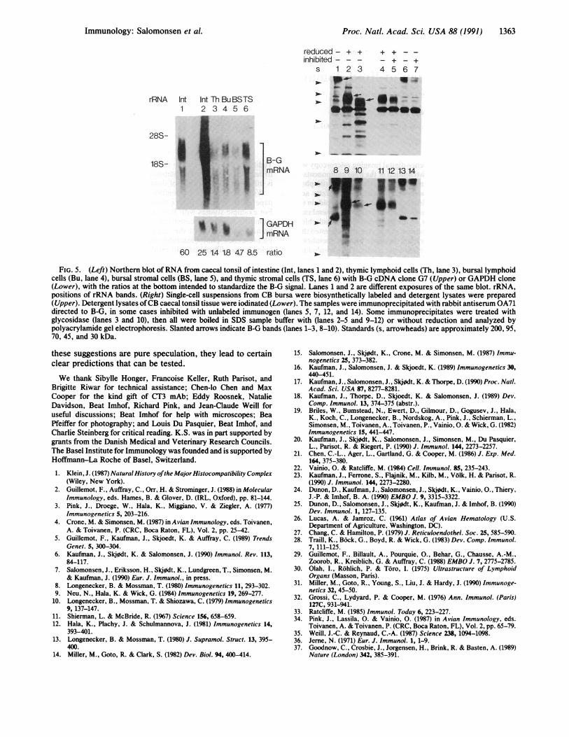
lymphoid tissues were detected by immunoprecipitation orWestern blot with these mAbs, presumably because theaffinity of crossreaction was too low. Three lines of evidencesuggest that these mAbs specifically crossreact with lympho-cyte B-G molecules. First, mAbs with different stainingpatterns in the periphery have very similar patterns in thelymphoid organs (g3 and g4) and vice versa (g2 and g3).Second, Northern blots show that B-G mRNA is present inboth stromal and lymphoid cells in these tissues (Fig. 5 Left).Third, some rabbit antisera to erythrocyte B-G moleculesrecognize molecules from bursal cells and caecal tonsils (Fig.
5 Right), with the specific reactions inhibited by unlabeledimmunogen. These molecules are larger than erythrocyteB-G molecules of the same haplotype, perhaps because theyare glycosylated. Some of these molecules without reductionare disulfide-linked multimers, while others show the markedincrease in electrophoretic mobility relative to reduced mol-ecules indicative of intrachain disulfide bonds. The relation-ship of the molecules recognized by the rabbit antisera andthe mAbs remains to be clarified.
DISCUSSIONWe previously showed that there are many different MHC-encoded B-G proteins on erythrocytes (15, 17), and that B-Gis an expressed multigene family at the level of DNA andRNA (16). Here we demonstrated that the erythrocyte pro-teins are almost certainly products of different genes, andthat similar molecules are differentially expressed on manycells types that are important for the immune system. Notonly does this finding represent a further point of similaritybetween the class I, class II, and B-G multigene families, butthe extended tissue distribution ofB-G supports the idea thatat least some B-G molecules play an important role in theimmune system.Perhaps the most obvious possibility is that certain B-G
molecules are T-cell restriction elements, consistent withtheir presence on thymic stromal cells, B cells, and (phago-cytic) thrombocytes. Some B-G molecules might berestriction elements for T-cell cytotoxicity of nucleatederythrocytes. However, we have been unable to demonstrateT cells that respond to allogeneic B-G (unpublished obser-vations).We favor the possibility that certain B-G molecules are
involved in selection of B cells and the resulting antibodyrepertoire in the chicken (6, 7). The mechanism of antibodydiversification by gene conversion of a single rearranging V(variable region) gene, the inefficiency ofthe gene conversionmechanism, the apparent extensive cell death in the bursa,the unimportance of exogenous antigen for bursal B celldifferentiation, and the lack of germline V gene expression inperipheral B cells (32-35) are all consistent with the presenceof selection on self antigens like B-G molecules. Such B-cell
)
C.)
C
RNAE T1 2
anti B-G
6f"C,)cnC,)a
cu14ii
Immunology: Salomonsen et al.
404a10-
I--
M.- 4m- ".
40 0 ]I--m
- wo
I-
10PBL
-t

1362 Immunology: Salomonsen et al.
A ,1,..
I.A.SS W bs;
.,~~ ~ a' #j~~~~4*Al
qr~ ~~~~*(
60~
anti classIl (2G11)
BURSA
anti B-G (mAbg3)
THYMUS
, AL
...,
anti B-G (mAbg2)
THYMUS
BURSA INTESTINE
mAbg4 mAbg4anti B-G
INTESTINE
mAbg3
FIG. 4. Tissue sections of bursa (all six panels of Upper and two left panels of Lower), thymus (Lower), and caecal tonsils of the intestine(Lower) from a 12-week-old CB chicken were stained with mAbs. Stain is brown, intense stain is black, counterstain is blue. m, Medulla; c,cortex; vertical arrows in bursa, follicle boundaries; horizontal arrows in bursa, corticomedullary boundaries; slanted arrows in bursa, circularstructures outside of corticomedullary border. (Upper, x20 and x 100; Lower, x70, except lower left, x200.)
selection, whether negative [as postulated by Jerne (36) forprimary lymphoid organs], positive, or both, could lead to anantibody repertoire which would crossreact with closelyrelated molecules (e.g., allogeneic B-G) and would thusaccount for the "natural antibodies," the exceptional immu-noglobulin response, and the "allogeneic effect." In addi-tion, the B-cell repertoire would be slanted by the selectingmolecule, and the survival of varying individuals might selectfor B-G polymorphism. In this light, selection of antibodyV regions by polymorphic B-G molecules in the bursa
would be analogous to selection of T-cell receptor V regionsby polymorphic MHC class I and class II molecules in thethymus.
Like mammalian Peyer's patches, the caecal tonsils in thechicken intestine contain many developing B cells, and theB-G molecules present there might also be involved in B-cellselection. B-G antigens on erythrocytes and other cells in theperiphery might be involved in fail-safe negative selection(37), in which contact ofperipheral B cells with crossreactingantigen in the absence ofhelper factors leads to anergy. While
Proc. Natl. Acad Sci. USA 88 (1991)
Proc. Natl. Acad. Sci. USA 88 (1991) 1363
rRNA Int Int Th BuBSTS1 2 3 4 5 6
28S-
18S-st
reduced- + + + + - -inhibited--- - + - +
s 1 2 3 4 5 6 7_^_ _ ..
- _
Ds as .- -.-
a10 W. w
_0-1__p
B-GmRNA 8 9 10 1112 1314
__~~ Ile*
GAPDHmRNA
60 25 14 1.8 4.7 8.5 ratio
FIG. 5. (Left) Northern blot ofRNA from caecal tonsil of intestine (Int, lanes 1 and 2), thymic lymphoid cells (Th, lane 3), bursal lymphoidcells (Bu, lane 4), bursal stromal cells (BS, lane 5), and thymic stromal cells (TS, lane 6) with B-G cDNA clone G7 (Upper) or GAPDH clone(Lower), with the ratios at the bottom intended to standardize the B-G signal. Lanes 1 and 2 are different exposures of the same blot. rRNA,positions of rRNA bands. (Right) Single-cell suspensions from CB bursa were biosynthetically labeled and detergent lysates were prepared(Upper). Detergent lysates ofCB caecal tonsil tissue were iodinated (Lower). The samples were immunoprecipitated with rabbit antiserum OA71directed to B-G, in some cases inhibited with unlabeled immunogen (lanes 5, 7, 12, and 14). Some immunoprecipitates were treated withglycosidase (lanes 3 and 10), then all were boiled in SDS sample buffer with (lanes 2-5 and 9-12) or without reduction and analyzed bypolyacrylamide gel electrophoresis. Slanted arrows indicate B-G bands (lanes 1-3, 8-10). Standards (s, arrowheads) are approximately 200, 95,70, 45, and 30 kDa.
these suggestions are pure speculation, they lead to certainclear predictions that can be tested.
We thank Sibylle Honger, Francoise Keller, Ruth Parisot, andBrigitte Riwar for technical assistance; Chen-lo Chen and MaxCooper for the kind gift of CT3 mAb; Eddy Roosnek, NatalieDavidson, Beat Imhof, Richard Pink, and Jean-Claude Weill foruseful discussions; Beat Imhof for help with microscopes; BeaPfeiffer for photography; and Louis Du Pasquier, Beat Imhof, andCharlie Steinberg for critical reading. K.S. was in part supported bygrants from the Danish Medical and Veterinary Research Councils.The Basel Institute for Immunology was founded and is supported byHoffmann-La Roche of Basel, Switzerland.
1. Klein, J. (1987) Natural History ofthe Major Histocompatibility Complex(Wiley, New York).
2. Guillemot, F., Auffray, C., Orr, H. & Strominger, J. (1988) in MolecularImmunology, eds. Hames, B. & Glover, D. (IRL, Oxford), pp. 81-144.
3. Pink, J., Droege, W., Hala, K., Miggiano, V. & Ziegler, A. (1977)Immunogenetics 5, 203-216.
4. Crone, M. & Simonsen, M. (1987) in Avian Immunology, eds. Toivanen,A. & Toivanen, P. (CRC, Boca Raton, FL), Vol. 2, pp. 25-42.
5. Guillemot, F., Kaufman, J., Skjoedt, K. & Auffray, C. (1989) TrendsGenet. 5, 300-304.
6. Kaufman, J., Skj0dt, K. & Salomonsen, J. (1990) Immunol. Rev. 113,84-117.
7. Salomonsen, J., Eriksson, H., Skjodt, K., Lundgreen, T., Simonsen, M.& Kaufman, J. (1990) Eur. J. Immunol., in press.
8. Longenecker, B. & Mossman, T. (1980) Immunogenetics 11, 293-302.9. Neu, N., Hala, K. & Wick, G. (1984) Immunogenetics 19, 269-277.
10. Longenecker, B., Mossman, T. & Shiozawa, C. (1979) Immunogenetics9, 137-147.
11. Shierman, L. & McBride, R. (1%7) Science 156, 658-659.12. Hala, K., Plachy, J. & Schulmannova, J. (1981) Immunogenetics 14,
393-401.13. Longenecker, B. & Mossman, T. (1980) J. Supramol. Struct. 13, 395-
400.14. Miller, M., Goto, R. & Clark, S. (1982) Dev. Biol. 94, 400-414.
15. Salomonsen, J., Skjodt, K., Crone, M. & Simonsen, M. (1987) Immu-nogenetics 25, 373-382.
16. Kaufman, J., Salomonsen, J. & Skjoedt, K. (1989) Immunogenetics 30,440-451.
17. Kaufman, J., Salomonsen, J., Skjodt, K. & Thorpe, D. (1990) Proc. Natl.Acad. Sci. USA 87, 8277-8281.
18. Kaufman, J., Thorpe, D., Skjoedt, K. & Salomonsen, J. (1989) Dev.Comp. Immunol. 13, 374-375 (abstr.).
19. Briles, W., Bumstead, N., Ewert, D., Gilmour, D., Gogusev, J., Hala,K., Koch, C., Longenecker, B., Nordskog, A., Pink, J., Schierman, L.,Simonsen, M., Toivanen, A., Toivanen, P., Vainio, 0. & Wick, G. (1982)Immunogenetics 15, 441-447.
20. Kaufman, J., Skj0dt, K., Salomonsen, J., Simonsen, M., Du Pasquier,L., Parisot, R. & Riegert, P. (1990) J. Immunol. 144, 2273-2257.
21. Chen, C.-L., Ager, L., Gartland, G. & Cooper, M. (1986) J. Exp. Med.164, 375-380.
22. Vainio, 0. & Ratcliffe, M. (1984) Cell. Immunol. 85, 235-243.23. Kaufman, J., Ferrone, S., Flajnik, M., Kilb, M., Volk, H. & Parisot, R.
(1990) J. Immunol. 144, 2273-2280.24. Dunon, D., Kaufman, J., Salomonsen, J., Skj0dt, K., Vainio, O., Thiery,
J.-P. & Imhof, B. A. (1990) EMBO J. 9, 3315-3322.25. Dunon, D., Salomonsen, J., Skjodt, K., Kaufman, J. & Imhof, B. (1990)
Dev. Immunol. 1, 127-135.26. Lucas, A. & Jamroz, C. (1961) Atlas of Avian Hematology (U.S.
Department of Agriculture, Washington, DC).27. Chang, C. & Hamilton, P. (1979) J. Reticuloendothel. Soc. 25, 585-590.28. Traill, K., Bock, G., Boyd, R. & Wick, G. (1983) Dev. Comp. Immunol.
7, 111-125.29. Guillemot, F., Billault, A., Pourquie, O., Behar, G., Chausse, A.-M.,
Zoorob, R., Kreiblich, G. & Auffray, C. (1988) EMBO J. 7, 2775-2785.30. Olah, I., Rohlich, P. & Toro, I. (1975) Ultrastructure of Lymphoid
Organs (Masson, Paris).31. Miller, M., Goto, R., Young, S., Liu, J. & Hardy, J. (1990) Immunoge-
netics 32, 45-50.32. Grossi, C., Lydyard, P. & Cooper, M. (1976) Ann. Immunol. (Paris)
127C, 931-941.33. Ratcliffe, M. (1985) Immunol. Today 6, 223-227.34. Pink, J., Lassila, 0. & Vainio, 0. (1987) in Avian Immunology, eds.
Toivanen, A. & Toivanen, P. (CRC, Boca Raton, FL), Vol. 2, pp. 65-79.35. Weill, J.-C. & Reynaud, C.-A. (1987) Science 238, 1094-1098.36. Jerne, N. (1971) Eur. J. Immunol. 1, 1-9.37. Goodnow, C., Crosbie, J., Jorgensen, H., Brink, R. & Basten, A. (1989)
Nature (London) 342, 385-391.
Immunology: Salornonsen et al.