cholesterol regulation of pulmonary endothelial …
TRANSCRIPT

University of New MexicoUNM Digital Repository
Biomedical Sciences ETDs Electronic Theses and Dissertations
Winter 12-13-2017
CHOLESTEROL REGULATION OFPULMONARY ENDOTHELIAL CALCIUMENTRY FOLLOWING CHRONIC HYPOXIABojun ZhangUniversity of New Mexico
Follow this and additional works at: https://digitalrepository.unm.edu/biom_etds
Part of the Cellular and Molecular Physiology Commons, and the Medicine and Health SciencesCommons
This Dissertation is brought to you for free and open access by the Electronic Theses and Dissertations at UNM Digital Repository. It has beenaccepted for inclusion in Biomedical Sciences ETDs by an authorized administrator of UNM Digital Repository. For more information, please [email protected].
Recommended CitationZhang, Bojun. "CHOLESTEROL REGULATION OF PULMONARY ENDOTHELIAL CALCIUM ENTRY FOLLOWINGCHRONIC HYPOXIA." (2017). https://digitalrepository.unm.edu/biom_etds/180

i
Bojun Zhang
Candidate
Cell Biology and Physiology
Department
This dissertation is approved, and it is acceptable in quality
and form for publication:
Approved by the Dissertation Committee:
Thomas C. Resta Ph.D., Chairperson
Benjimen R. Walker Ph.D.
Nikki L. Jernigan Ph.D.
William S. Garver Ph.D.

ii
CHOLESTEROL REGULATION OF PULMONARY
ENDOTHELIAL CALCIUM ENTRY FOLLOWING CHRONIC
HYPOXIA
BY
Bojun Zhang
B.S. Biology, Wuhan University, 2010
DISSERTATION
Submitted in Partial Fulfillment of the
Requirements for the Degree of
Doctor of Philosophy
Biomedical Science
The University of New Mexico
Albuquerque, New Mexico
May, 2018

iii
DEDICATION
Dedicated to my beloved mom who carried me all the way and gave me all her love.
I really miss you.

iv
ACKNOWLEDGEMENTS
I acknowledge Dr. Benjimen Walker, my mentor, for his patient guidance and care
through my graduate study. I will forever be in debt to Ben for his support when I was in
the most difficult time of my life. I would also like to thank my co-mentor and dissertation
chair, Dr. Thomas Resta, for his support and encouragement that help me overcame all
obstacles through my graduate career. It is impossible for me to complete my dissertation
without them.
I would additionally like to thank my two other committee members, Dr. Nikki
Jernigan and Dr. William Garver, for their expertise in the development of this project and
their invaluable advises aiding me to make progress throughout the Ph.D. program.
To Vascular Physiology Group and Cell Biology and Physiology Department; thank
you so much for the amazing training environment and fantastic assistance that enabled
me to focus on my work. You are the most friendly and supportive people I ever met.
Finally, I want to thank my family members, especially my wife, for their selfless love
and support for past years.

v
CHOLESTEROL REGULATION OF PULMONARY ENDOTHELIAL
CALCIUM ENTRY FOLLOWING CHRONIC HYPOXIA
by
Bojun Zhang
B.S.
Ph.D.
ABSTRACT
Chronic hypoxia (CH)-induced pulmonary hypertension (PH) is associated with
diminished ATP-induced endothelial Ca2+ entry as well as membrane cholesterol in
pulmonary arteries. Store-operated Ca2+ entry (SOCE) and depolarization-induced Ca2+
entry are major components of the response to ATP and are similarly decreased after CH.
Because endothelium-dependent vasodilation is closely associated with pulmonary
endothelial [Ca2+]i, the blunted agonist-induced Ca2+ influx in pulmonary artery endothelial
cells (PAEC) may contribute to the development of CH-induced PH. Interestingly,
impaired agonist-induced Ca2+ influx in PAEC following CH can be restored by membrane
cholesterol supplementation. In the current studies, we hypothesized that impaired Ca2+
entry in PAEC following CH is due to decreased membrane cholesterol.
We demonstrated that substitution of cholesterol with its functionally inactive epimer
epicholesterol, greatly attenuated ATP-induced Ca2+ influx in PAEC from control rats.
Whereas epicholesterol similarly blunted endothelial SOCE in PAEC from both groups,
cholesterol supplementation restored diminished SOCE in PAEC from CH rats while
having no effect in controls. Similar effects of cholesterol manipulation on T-type Ca2+

vi
channel-mediated Ca2+ influx were observed in PAEC. Additionally, the role of cholesterol
in SOCE mediated by Orai1, a Ca2+ selective ion channel, was examined in PAEC.
Whereas cholesterol restored endothelial SOCE in CH rats, both epicholesterol and the
Orai1 inhibitor, AnCoA4, attenuated SOCE only in normoxic controls. The Orai1 inhibitor
had no further effect in cells pretreated with epicholesterol. In cultured pulmonary
endothelial cells, using pharmacological inhibition and siRNA knockdown of Orai1, we
found that epicholesterol, AnCoA4 and Orai1 siRNA each inhibited SOCE compared to
their respective controls. Epicholesterol had no additional effect following knockdown of
Orai1. Finally, we found that endothelial stromal interaction molecule 1 (STIM1)-Orai1
interaction, which is essential for SOCE, requires membrane cholesterol. Our studies
support a novel regulatory role for membrane cholesterol in agonist-induced Ca2+ entry and
its components. Our observation also demonstrated that altered membrane cholesterol
homeostasis may contribute to impaired endothelial Ca2+ influx pathways following CH.

vii
TABLE OF CONTENTS
CHAPTER 1: Introduction ............................................................................................ 1
Mechanisms of chronic hypoxia-induced pulmonary hypertension ........................................... 1
Endothelium in the pulmonary circulation .................................................................................. 3
Regulation of endothelial Ca2+ levels ........................................................................................... 5
Agonist-induced Ca2+ entry .......................................................................................................... 6
Store-operated Ca2+ entry ............................................................................................................ 7
Depolarization-induced Ca2+ entry............................................................................................. 10
Impaired pulmonary endothelial Ca2+ influx following CH ........................................................ 12
Membrane cholesterol .............................................................................................................. 13
Membrane cholesterol regulates ion channel function ............................................................ 14
Membrane cholesterol regulates Ca2+ influx ............................................................................. 16
Rationale and specific aims ........................................................................................................ 17
Figure 1 .................................................................................................................................. 20
Figure 2 .................................................................................................................................. 21
Figure 3 .................................................................................................................................. 22
References ................................................................................................................................. 23
CHAPTER 2: Reduced Membrane Cholesterol Limits Pulmonary Endothelial Ca2+
Entry Following Chronic Hypoxia .......................................................................... 45
Abstract ...................................................................................................................................... 46
Introduction ............................................................................................................................... 48
Methods ..................................................................................................................................... 50
Results ........................................................................................................................................ 55
Discussion .................................................................................................................................. 58
Figure 1. ................................................................................................................................. 67
Figure 2 .................................................................................................................................. 68
Figure 3 .................................................................................................................................. 69
Figure 4 .................................................................................................................................. 70
Figure 5 .................................................................................................................................. 71
Figure 6 .................................................................................................................................. 72
Figure 7 .................................................................................................................................. 73
Figure 8 .................................................................................................................................. 74

viii
Figure 9 .................................................................................................................................. 75
References ................................................................................................................................. 76
CHAPTER 3: Reduced Membrane Cholesterol Following Chronic Hypoxia Limits
Orai1-Mediated Pulmonary Endothelial Ca2+ Entry............................................... 85
Abstract ...................................................................................................................................... 86
Introduction ............................................................................................................................... 88
Methods ..................................................................................................................................... 90
Results ........................................................................................................................................ 96
Discussion ................................................................................................................................ 100
Figure 1 ................................................................................................................................ 108
Figure 2 ................................................................................................................................ 109
Figure 3 ................................................................................................................................ 110
Figure 4 ................................................................................................................................ 111
Figure 5 ................................................................................................................................ 112
Figure 6 ................................................................................................................................ 113
Figure 7 ................................................................................................................................ 114
Figure 8. ............................................................................................................................... 115
References ............................................................................................................................... 116
CHAPTER 4: DISCUSSION ........................................................................................ 128
Effect of hypoxia on pulmonary endothelial [Ca2+]i ................................................................. 129
Pulmonary endothelium-mediated vasodilation following CH ............................................... 132
Membrane cholesterol: structural component vs. signaling mediator ................................... 134
Caveolae, caveolin-1, and membrane cholesterol regulation of Ca2+ channels ...................... 137
Cholesterol regulation of STIM1, Orai1, and TRPC. ................................................................. 139
Mechanisms of diminished membrane cholesterol by CH ...................................................... 142
Membrane cholesterol and hypercholesterolemia ................................................................. 143
Physiological significance of current studies ........................................................................... 145
Limitations of studies ............................................................................................................... 147
Summary .................................................................................................................................. 148
References ............................................................................................................................... 150

1
CHAPTER 1: Introduction
Mechanisms of chronic hypoxia-induced pulmonary hypertension
Pulmonary hypertension (PH) is the elevation of blood pressure in the pulmonary
circulation and is a leading cause of morbidity and mortality in patients with a number of
cardiovascular and respiratory diseases. Human PH is clinically defined as a mean arterial
pressure ≥ 25 mmHg at rest (74). Multiple factors may lead to the development of PH,
including genetic defects, disease, or environmental exposure. Interestingly, persistent or
intermittent hypoxia is highly associated with many forms of PH, such as in chronic
obstructive pulmonary disease, interstitial lung disease, and obstructive sleep apnea (65,
96, 138). The chronic hypoxia (CH)-induced PH animal model is one of the most
commonly used to mimic human disease (165).
Under normal conditions, the pulmonary circulation is a low-resistance and low-
pressure system. These characteristics allow blood to easily travel from the right ventricle
through the lungs for gas exchange. Pulmonary vascular resistance (R) can be calculated
as the ratio of the pressure gradient (∆P) across all vessels to flow (Q):
R = ∆P / Q
Thus, increased pulmonary arterial resistance would lead to elevated pulmonary arterial
pressure that must be overcome by the right heart to pump blood through pulmonary
circulation. There are various factors that can influence vascular resistance. Assuming a
vascular system consists of straight, non-distensible cylindrical tubes that have laminar
flow, then vascular resistance R of a single tube is equal to the product of the length of the
tube (l) and viscosity (η) and a constant divided by the product of π and the fourth power
of the internal radius:

2
R = (l * η *8) / (π *r4)
This is known as the Hagen-Poiseuille’s law. Under pathological conditions such as
prolonged exposure to hypoxia, however, there are functional and structural changes in the
pulmonary circulation, including polycythemia, acute hypoxia-induced vasoconstriction of
small pulmonary arteries, and pulmonary arterial remodeling with medial hypertrophy (2,
60). According to Hagen-Poiseuille’s law, all of these changes lead to increase in
pulmonary vascular resistance.
Polycythemia, an increase in the volume percentage of circulating red bloods cells
in whole blood, results in increased in blood viscosity, thereby increasing vascular
resistance. Chronic exposure to hypoxia may activate hypoxia inducible factor (HIF)-1α
which induces erythropoiesis that leads to the polycythemic response (34). CH-induced
active pulmonary vasoconstriction and arterial remodeling are the consequences of several
complex physiological mechanisms. One major cause of these changes to the pulmonary
vasculature is pulmonary arterial endothelial dysfunction, which is usually observed in the
development of PH (10; 35). The dysfunction of endothelial cells may be triggered by
multiple sources: shear stress, inflammation, hypoxia, and other unknown factors. This
impaired endothelial function is generally believed to result in imbalanced production of
various vasoactive factors (20, 49, 153). The increased production of vasoconstrictors with
decreased production of vasodilators is common in most forms of PH. Although the
pulmonary endothelium plays a major role in PH, the mechanisms by which impaired
endothelial function leads to the development of PH are not well defined. Understanding
the mechanisms of endothelial dysfunction leading to and contributing to PH is central to
designing effective and specific treatment for these pathologies.

3
Endothelium in the pulmonary circulation
The vascular endothelium is the innermost layer of blood vessels and serves as a
semi-selective barrier that allows exchange of fluid and macromolecules between the
circulation and surrounding tissues. As a major part of the vascular system, the endothelium
plays a crucial role in several physiological activities, such as maintaining tissue-fluid
homeostasis, regulating angiogenesis and vascular tone, and preventing inflammation (54,
75, 107, 125, 139). In the pulmonary circulation, endothelial cells help maintain low
resistance and prevent proliferation/migration of smooth muscle cells.
Pulmonary endothelial cells regulate vascular tone by the balanced production of
various vasoactive compounds. Vascular tone reflects the result of smooth muscle
contraction due to phosphorylation of myosin light chain (MLC). MLC phosphorylation is
generally regulated by Ca2+-dependent and -independent pathways: activation of myosin
light chain kinase via Ca2+/calmodulin or inhibition of myosin light chain phosphatase
through activated small GTPase RhoA and Rho-associated protein kinase (15).
Endothelium can produce vasoconstrictors like endothelin-1 and thromboxane that act as
paracrine factors to stimulate G protein-coupled receptors and cause Ca2+ influx and
subsequent contraction of smooth muscle cells (37, 131, 160). On the other hand, the
pulmonary endothelium also releases vasodilators such as nitric oxide and prostacyclin in
response to shear stress and agonist stimulation (52, 68). These vasodilators decrease both
intracellular Ca2+ levels and Ca2+ sensitivity of smooth muscle cells, thereby causing
vasorelaxation (86, 93).

4
NO is the most well studied vasodilator and plays an important role in maintaining
low resistance in the pulmonary circulation (137). NO causes vasodilation primarily
through activation of soluble guanylyl cyclase (cGC) in pulmonary artery smooth muscle
cells (PASMC) (93), which leads to the production of cGMP from GTP. cGMP then
activates protein kinase G (PKG). Intracellular calcium levels ([Ca2+]i) are crucial for
myosin light chain (MLC) phosphorylation and contraction in PASMC. PKG activation
causes smooth muscle relaxation primarily through lowering [Ca2+]i via various
mechanisms including inhibition of IP3 receptors (129), activation of sarco/endoplasmic
reticulum Ca2+-ATPase (SERCA) (93, 123), or activation of plasma membrane Ca2+-
ATPase (113). Stimulated PKG may also lead to activation of Ca2+ -dependent K+ channels
(BKCa) and PASMC hyperpolarization (5, 40, 46, 167), which inhibits voltage-gated Ca2+
channel (VGCC) -mediated Ca2+ influx and promotes MLC dephosphorylation and
subsequent vasorelaxation. cGMP may also inhibit calcium channels like TRPCs and
VGCC thereby causing vasorelaxation. PKG activation also leads to vasorelaxation
through Ca2+-independent mechanisms. Studies suggest that PKG activates MLC
phosphatase, which inhibits MLC phosphorylation and subsequent contraction (78, 97).
PKG may also phosphorylate RhoA, thereby inhibiting RhoA-mediated Ca2+ sensitization
(16, 17, 127). In addition, NO could directly activate BKCa and potentially cause smooth
muscle relaxation independent of PKG (15). Besides its role in regulating vascular tone,
endothelium-derived NO is also known to inhibit smooth muscle cell (SMC) proliferation
and migration (62, 126), which is important in preventing vascular remodeling in responses
to stimuli such as inflammation, oxidative stress and apoptosis.

5
Production of many endothelium-dependent vasoactive substances as well as
regulation of membrane potential are largely a function of pulmonary endothelial [Ca2+]i
(1). The production of NO, for example, is catalyzed by endothelial nitric oxide synthase
(eNOS) in the endothelium. Activation of eNOS requires increased endothelial [Ca2+]i and
formation of a Ca2+/calmodulin complex (89). Along with other cofactors like NADPH and
BH4, eNOS converts L-arginine into NO and L-citrulline (26, 89). Similarly, production
of endothelium-derived prostacyclin requires intracellular Ca2+ and calmodulin (89, 130).
The activation of cytosolic phospholipase A2, the key enzyme to release arachidonic acid
from plasma membrane, is Ca2+-dependent. Once arachidonic acid is liberated, it could be
enzymatically converted by cyclooxygenase-1 to prostacyclin after multiple reactions.
Arachidonic acid is also substrate for production of vasoconstrictor metabolites, such as
thromboxanes, leukotrienes, and epoxyeicosatrienoic acids.
Regulation of endothelial Ca2+ levels
Endothelial Ca2+ is important second messenger in various signaling cascades and
is carefully regulated by complex and dynamic pathways. Endothelial [Ca2+]i reflects the
sum of Ca2+ influx from extracellular space, Ca2+ release and sequestration by intracellular
stores, and Ca2+ extrusion from the cell. Endothelial Ca2+ influx is mainly mediated by
three mechanisms: 1) Ca2+ entry via receptor-mediated cation channels; 2) Ca2+ leak
through cation channels down its electrochemical gradient; and 3) Ca2+ entry via stretch-
activated cation channels. Steady-state [Ca2+]i may be rapidly altered by stimuli such as
shear stress or agonists, which activate membrane-bound receptors and cation channels and
leads to Ca2+ entry (3, 161). This major Ca2+ influx signaling cascade also generates the

6
second messenger, IP3, which can activate downstream ionotropic IP3 receptors on the
membrane of the ER, causing rapid and transient release of internal Ca2+ stores. Once ER
Ca2+ stores are depleted, the ATPase pump refills the intracellular Ca2+ pool by
sequestering Ca2+ from the cytosol, which may also be facilitated by a Ca2+ leak through
cation channels on the plasma membrane (1). Meanwhile, the Na+- Ca2+ exchanger on the
plasma membrane helps remove excessive intracellular Ca2+ after agonist stimulation (23,
39, 122). Overall, these pathways work together to maintain [Ca2+]i homeostasis in
endothelial cells.
Agonist-induced Ca2+ entry
Receptor-mediated Ca2+ influx is one of the major pathways leading to increased
[Ca2+]i. The binding of an agonist to its receptor mediates the activation of phospholipase
C (PLC), which then leads to the production of diacylglycerol and IP3 from hydrolysis of
phosphatidylinositol 4,5-bisphosphate (PIP2) (11). Cytosolic IP3 then binds to IP3 receptors
on the membrane of the ER and causes Ca2+ efflux from the ER store. The depletion of ER
stores triggers Ca2+ influx across the plasma membrane, which is known as store-operated
Ca2+ entry (SOCE) (150). Agonist binding may also elicit Ca2+ entry through channels that
are not activated by store emptying. This is known as receptor-operated calcium entry
(ROCE) (120).
ATP, as one of the important agonists that meditates endothelial [Ca2+]i, serves as
a regulator of vascular tone. Studies found that red blood cells (RBCs) and endothelial cells
(ECs) can release ATP under certain conditions to maintain blood flow. In RBCs, when
acutely exposed to low PO2, low pH, or mechanical deformation, there is an increase of

7
ATP release (38, 92, 136). ATP then causes endothelium-dependent vasorelaxation in
various species and tissues (25). It is likely that RBC- produced ATP binds to purinergic
receptors on ECs and stimulates NO production (84). In endothelial cells, shear stress
induces the production of ATP, which serves as an autocrine factor and stimulates
endothelial NO production (4, 69). It is proposed that extracellular ATP binds to P2Y
purinergic receptors on plasma membrane of ECs, which leads to IP3 production, internal
Ca2+ store release, Ca2+ influx and subsequent increase in [Ca2+]i, leading to increased
eNOS activation and NO production (50). Additionally, ATP may also enhance endothelial
cell barrier function by Rac and cortactin-dependent activation of the cytoskeleton (59).
Store-operated Ca2+ entry
SOCE is a Ca2+ influx response when internal SR/ER Ca2+ stores are depleted.
Under physiological conditions, agonists may bind to transmembrane receptors and
activate PLC to produce IP3 from hydrolysis of PIP2. IP3 then activates IP3 receptors on
membrane of SR/ER, leading to Ca2+ efflux, depletion of intracellular Ca2+ stores, and
activation of cation channels on the plasma membrane (150). SOCE was first proposed in
1986 by Putney and colleagues and was later found in many cell types. In rat parotid acinar
cells, the intracellular Ca2+ store depletion by muscarinic-cholinergic agonists causes Ca2+
influx (144). If the intracellular Ca2+ store is depleted independent of activation of PLC,
Ca2+ influx is still observed (143). Later in mast cells, in response to IP3 -induced Ca2+
store depletion, a highly Ca2+ selective inwardly rectifying current was found (57). This
store-operated current (Isoc) with a high Ca2+ selectivity is known as calcium release -
activated calcium (CRAC) current or Icrac.
8
SOCE has been demonstrated to be present in virtually all excitable and non-
excitable cells and is involved in mediating many important physiological activities such
as neurotransmitter secretion, skeletal muscle contraction, lymphocyte activation, and
vascular tone (41, 79, 110, 146). Although the physiological importance of SOCE has been
recognized for some time, the molecular mechanism of SOCE remained unclear until
recent years. A key finding was the identification of stromal interacting molecule (STIM)
proteins on the membrane of the ER. STIM1, as one isoform of STIM, was first found in a
library screen which was developed to identify molecules in stromal cells that bind to pre-
B lymphocytes (98). However, the role of STIM as the molecular component of SOCE was
revealed more recently through RNA interference (RNAi) screens by two independent
groups (83, 119). In Drosophila S2 cells, RNAi-mediated knockdown of STIM causes
significantly reduction of Ca2+ entry induced by the SERCA inhibitor, thapsigargin (TG)
(119). Similar observation that STIM1 and STIM2 are required for agonist- and TG-
induced SOCE was found in human HeLa cell (83). Later, continued genome-wide RNAi
screens in Drosophila S2 cells revealed Orai proteins as key components of the CRAC
channel (111, 156, 164). The presence of STIM1 and Orai1 are sufficient to reconstitute
functional SOCE in HEK293 cells (134).
The exploration for other potential components of SOCE also identified the
involvement of non-selective Ca2+ -permeable mammalian transient receptor potential
canocical (TRPC) channels (155). All TRPC channels are activated via the PLC signaling
pathway (155). However, only TRPC1, TRPC3, and TRPC4 are activated mostly by store
depletion, whereas the activation TPRC2, TRPC5, TRPC6, and TRPC7 is mainly through
store-independent mechanisms (155). The finding that STIM1 also gates TPRC1 in SOCE
9
(58) generates a new question in regard to the role of TRPC1 and Orai1 in SOCE. Later
studies then demonstrated that activation of TRPC1/STIM1-mediated SOCE requires
functional Orai1 (67, 73). In PAEC, TRPC1 and TRPC4 also form heteromultimeric
channels and interact with Orai1 via protein 4.1 in caveolin-rich membrane fractions (32).
This TRPC1/4 complex and Orai1 interaction is important for channel activation and
calcium selectivity (32). With the identification of these key components of SOCE, the
molecular mechanism of SOCE is better understood (27). The EF-hand motif in the N
terminus of STIM1 is localized in the ER lumen and senses ER Ca2+ stores. Upon the
depletion of the ER Ca2+ store, the SAM domain near the EF-hand motif mediates STIM1
oligomerization and leads to translocation of STIM1 to the plasma membrane. This
conformational change of STIM1 allows its interaction with Orai1 and forms ER-PM
junctions, which are crucial in Ca2+ release-activated Ca2+ influx.
SOCE in both SMC and ECs is important in regulating vascular tone. In vascular
SMC, Ca2+ influx induced by SR Ca2+ store depletion via SR Ca2+ -ATPase inhibition is
associated with increased vascular tone in different blood vessels (79). Interestingly,
SOCE-mediated vascular tone shows varying sensitivity to voltage-gated Ca2+ channel
inhibitors between vascular beds. For example, most of the SOCE -induced contraction is
nifedipine-sensitive in the rat aorta, which is not found in the rat pulmonary artery (79).
These observations suggest that SOCE may modulate vascular tone in a direct or indirect
manner. Many studies also indicate that increased [Ca2+]i through SOCE contributes to
smooth muscle proliferation and migration (13, 108, 141, 142). In cultured proliferative
migratory rat aortic VSMCs, both SOCE and expression of STIM1 and Orai1 are elevated
compared with non-proliferative controls (108). Knocking down STIM1 and Orai1 greatly

10
inhibits proliferation and migration of these active VSMCs (108). Besides its role in
mediating vascular endothelial permeability (147), endothelial SOCE, as part of agonist-
induced Ca2+ entry, significantly contributes to the increase in [Ca2+]i and subsequent
release of vasodilators like NO and prostacyclin (1, 70, 77, 82). In fact, pharmacological
inhibition of SOC by SKF-96365 blunts the shear stress-induced NO response (4).
Endothelial TRPC4 knockdown also impairs agonist-induced vasodilation by reducing
SOCE (44).
Depolarization-induced Ca2+ entry
VGCCs are a group of Ca2+ permeable channels that are activated by depolarized
membrane potential. Based on the requirement of a different level of depolarization for
activation, these channels can be classified into two major groups, high voltage-activated
(L-, P/Q-, N-, R- type) and low voltage-activated (T-type) calcium channels (150). These
channels have been found in neurons, muscle, endocrine cells, and many other cell types
(29, 145, 150, 162). They are involved in many different physiological activities including
muscle contraction, excitation of neurons, upregulation of gene expression, release of
hormones or neurotransmitters (28).
L-type VGCC were first described in neurons and cardiac cells and later found
widely expressed in all excitable and some non-excitable cells (152). Ca2+ influx through
L-type VGCC is the major source of Ca2+ entry in cardiac, skeletal, and smooth muscles.
In cardiac muscle, activation of L-type VGCC is required to initiate Ca2+-induced Ca2+
release and subsequent muscle contraction (14). In skeletal muscle, although L-type VGCC
are not involved in the initiation of muscle contraction, they are important in mediating

11
Ca2+ release from intracellular stores, which then facilitates mobilization of the
myofilaments and contraction (117). In vascular smooth muscle cells, Ca2+ influx through
L-type VGCC also serves as one of the Ca2+ sources for Ca2+/calmodulin-dependent
phosphorylation of MLC, which leads to smooth muscle contraction. Indeed, L-type
VGCC are important in mediating blood pressure and myogenic tone (35, 92).
Although L-type VGCC are considered the major VGCC that regulate the
myogenic response, T-type VGCC in vascular smooth muscle cells are receiving more
attention in recent years. In small resistance vessels, T-type VGCC are involved in
regulating vascular tone (154). T-type VGCC are also involved in the development of CH-
induced PH. Either chronic treatment of a T-type VGCC inhibitor or global deletion of the
T-type VGCC gene (Cav3.1) protects mice from developing CH-induced PH (30).
T-type VGCC are normally found expressed in electrically excitable cells.
However, their expression has also been described in pulmonary microvascular endothelial
cells and PAEC from rats and mice (101, 157, 159, 166). In PMVECs, Wu et al (159)
observed voltage-dependent currents with a more depolarized window current when
compared to typical T-type channels. These currents were sensitive to pharmacological T-
type channel blockers. Wang et al (157) found that acute hypoxia induced membrane
depolarization and a subsequent increase of endothelial [Ca2+]i in both alveolar capillaries
and upstream arterioles. The endothelial [Ca2+]i response to acute hypoxia was inhibited
by the T-type channel blocker mibefradil in both pulmonary capillaries and arterioles. They
proposed that membrane depolarization induced by acute hypoxia in lung capillaries is
conducted upstream to arterioles via gap junctions and activates T-type channels. Increased
endothelial [Ca2+]i then leads to vasoconstriction by activation of Ca2+-dependent

12
phospholipase A2-mediated vasoconstrictor production. Although T-type VGCC are not
found in cultured rat PAEC (159), their expression and function have been described in
freshly dispersed rat PACEs (101). Paffett et al. (101) have shown that T-type VGCCs
contribute to depolarization-induced Ca2+ entry and ATP-induced Ca2+ entry. These
findings suggest that T-type VGCCs are important mediators of depolarization-induced
Ca2+ entry in the pulmonary endothelium.
Impaired pulmonary endothelial Ca2+ influx following CH
Impaired NO release is often observed in diseases such as chronic obstructive
pulmonary disease and congestive heart failure and is a key feature of pulmonary
hypertension (8, 90, 115, 140, 148). However, eNOS protein expression is increased in
pulmonary hypertensive patients and CH-induced pulmonary hypertensive rats (48, 51,
116). The controversial observation that increased eNOS protein expression and impaired
endothelium-derived vasorelaxation simultaneously occur in CH-induced PH rats could be
the result of blunted eNOS activity. Murata et al confirmed this assumption by reporting
that CH impairs posttranslational regulation of eNOS activity via blunting agonist-induced
Ca2+ influx (95).
Previous work from our laboratory also shows that both basal [Ca2+] i and agonist-
induced Ca2+ influx are lower in PAEC from CH rats compared to those of control animals
(99, 100). CH similarly inhibits endothelial SOCE and depolarization-induced Ca2+ influx
through T-type VGCC, which are major components of agonist-induced Ca2+ entry in
isolated PAEC (99, 101). These findings suggest that store-operated channels and T-type
VGCC are important mediators of ATP-induced Ca2+ entry and that impaired Ca2+ entry
and may contribute to reduced basal [Ca2+]i in PAEC after CH, which may lead to

13
dysfunction of posttranslational eNOS regulation and subsequent impairment of
endothelium-derived NO production in CH-induced PH.
Membrane cholesterol
Cholesterol is a major component of the plasma membrane. The importance of
membrane cholesterol has been shown in regulating neurotransmission, cell signaling, and
protein sorting (85, 128, 132). Additionally, many diseases such as type II diabetes (31),
Alzheimer’s disease (6), and cancer (91) are associated with abnormal cellular cholesterol
levels. The homeostasis of cellular cholesterol levels is maintained mainly by four
pathways: cellular cholesterol synthesis, cholesterol uptake from extracellular sources,
cholesterol efflux, and esterification and storage in the lipid droplets (31). The ER is the
major site of cholesterol de novo synthesis, which requires multiple enzymes and cofactors
to covert acetyl-CoA to cholesterol in a complex series of reactions. Once synthesized,
cholesterol is bound with caveolins and transported from the ER to plasma membrane.
More than 90% of free cholesterol is located in the plasma membrane (66, 76) and is
distributed to microdomains known as lipid rafts, which are rich in cholesterol,
sphingolipids, and various proteins. Once in the plasma membrane, cholesterol maintains
membrane integrity and regulates many signaling pathways.
Membrane cholesterol also serves as an important structural component of the
plasma membrane by regulating membrane fluidity and permeability. Cholesterol may help
maintains membrane fluidity by interfering with the interactions between phospholipid
fatty acid chains. However, cholesterol may also increase membrane rigidity by reducing
the flexibility of phospholipid fatty acid chains (55). This regulation of membrane fluidity

14
is crucial in controlling solute movement across the membrane when membrane
composition or temperature are altered. In addition, positive electrostatic potential in the
plasma membrane is increased when cholesterol content is augmented, which may change
the membrane composition of charged components and alter the permeability of cations
and anions (55). Membrane cholesterol is also required for the formation of specific lipid
rafts known as caveolae. Caveolae are flask-shape invaginations of the plasma membrane
that contain caveolin, cholesterol, sphingolipid, and various signaling molecules and
receptors. They are involved in endocytosis, cholesterol transport, and signal transduction
(42, 106). Besides caveolins, which serve as the structural component of caveolae (36, 112,
114), cholesterol is also essential in maintaining stable and functional caveolae. Both
inhibition of cholesterol synthesis and acute membrane cholesterol depletion lead to
caveolar disruption and inhibition of cell signaling (64, 94, 105, 124, 168).
Membrane cholesterol regulates ion channel function
Besides its structural role, membrane cholesterol is also important in regulating
membrane-bound protein structure and function. Cholesterol is a polycyclic amphipathic
molecule with a polar section consisting of a single β-hydroxyl group which can interact
with membrane lipids or proteins through formation of hydrogen bonds (105). Various
membrane-bound proteins have been shown to be regulated by membrane cholesterol, such
as receptors, transporters, peptides, and ion channels (56, 80, 102, 151, 163).
The effect of membrane cholesterol on ion channel function may vary depending
on the type of ion channel. Many studies demonstrate an inhibitory role of membrane
cholesterol on ion channel activity via decreased open probability, unitary conductance,

15
and the number of active channels (80). For example, cholesterol decreases the open
probability of many K+ channels, as well as voltage-gated Na+ and Ca2+ channels (33, 81,
149). In contrast, other ion channels such as nicotinic acetylcholine receptor, GABAA
receptors, epithelial Na+ channels and TRPC channels are inhibited by removal of
membrane cholesterol (7, 9, 10, 72, 135), indicating that cholesterol may have an essential
functional role in regulating these channels.
Cholesterol may additionally modulate the function of membrane proteins via
either direct interaction or through altering the properties of lipid microdomains (80).
Direct interaction between sterols and ion channels has been suggested by the sensitivity
of inwardly rectifying potassium (Kir) channels (118) and large conductance, Ca2+-
activated potassium (BKCa) channels (24) to various sterol analogues. In addition,
cholesterol binding regions exist in both Kir channels and BKCa channels (121, 133).
Meanwhile, cholesterol may also indirectly regulate ion channels by modulating bilayer
stiffness and hydrophobic interaction between the membrane proteins and the lipid bilayer
(9). Thus, when determining the role of membrane cholesterol on channel function,
extreme caution is needed to discriminate the different mechanisms by which cholesterol
regulates certain types of ion channels. The enantiomer of cholesterol, epicholesterol,
differs from cholesterol only in the stereochemistry of the hydroxyl group and has similar
effects on membrane fluidity and lipid domain formation as those of cholesterol, but lacks
the regulatory influences of cholesterol on membrane proteins (80). Thus epicholesterol
has been used as a tool to distinguish the direct and indirect effect of cholesterol (80).
16
Membrane cholesterol regulates Ca2+ influx
The importance of membrane cholesterol in regulating Ca2+ entry has been shown
in many different types of cells. In VSMCs, membrane cholesterol depletion by methyl-β-
cyclodextrin reduces TRPC1-mediated Ca2+ entry in response to endothelin-1 (9).
Similarly, cholesterol depletion decreases STIM1 clustering, prevents activation of
TRPC1, and blunts SOCE in HSG and HEK293 cells (9). In addition, depletion of
membrane cholesterol also disrupts the association of Orai1 with TRPC1 and STIM1, and
attenuates SOCE in human platelets (61). This observation is also confirmed in
lymphocytes, in which a reduction of membrane cholesterol decreases SOCE. Cholesterol
enrichment, on the other hand, increases agonist-induced Ca2+ influx in cultured VSMCs
(12).
Under pathological conditions, the importance of membrane cholesterol in
regulating endothelial Ca2+ influx has also been revealed. Our laboratory’s previous studies
show that both membrane cholesterol levels and ATP- induced Ca2+ entry are reduced in
PAEC following CH (100). Interestingly, cholesterol repletion restores CH-impaired
endothelial ATP- induced Ca2+ influx (100), suggesting that membrane cholesterol may
facilitate endothelial agonist-induced Ca2+ entry. In PAEC, ATP induces both SOCE and
T-type VGCC blocker-sensitive Ca2+ influx (101). Although the role of membrane
cholesterol in regulating SOCE has been implicated in many studies (10, 47), the effect of
membrane cholesterol in regulation of endothelial depolarization-induced Ca2+ entry is
unknown. Considering that SOCE and depolarization-induced Ca2+ entry are major
components of ATP-induced Ca2+ entry, and that both Ca2+ influx pathways are blunted

17
following CH (99, 101), it is possible that membrane cholesterol also regulates these two
components of the ATP-induced Ca2+ response.
Rationale and specific aims
As summarized above, endothelial dysfunction is closely associated with many forms
of PH. In CH-induced PH, for example, increased pulmonary vascular resistance and
myogenic tone are observed with decreased production of endothelial vasodilatory factors
(21, 63, 95, 158). [Ca2+]i is a key regulator of the synthesis of many endothelial vasoactive
factors. CH decreases [Ca2+]i by impairing Ca2+ influx in pulmonary artery endothelium
(99–101). Diminished endothelial Ca2+ entry may contribute to elevated vascular tone by
limiting the activity of eNOS and other Ca2+ -sensitive vasodilatory pathways (26, 43, 53,
87, 88, 95). Many Ca2+ channels are localized in cholesterol-enriched lipid rafts (19, 45, 94,
104). Cellular cholesterol homeostasis affects membrane lipid content and thus the function
of Ca2+ channels and subsequent signaling pathways (10, 18, 22, 103, 109) by mechanisms
that are not fully understood.
There are multiple Ca2+ entry pathways that could directly affect intracellular Ca2+
levels in the endothelium. Agonist-induced calcium entry, as one of the major Ca2+ entry
pathways, has several components including SOCE and receptor-operated Ca2+ entry
(ROCE). Studies from our laboratory have shown that both endothelial SOCE and ROCE
are reduced in CH-induced PH (99, 101). Our previous work also identified a membrane
potential-sensitive Ca2+ entry pathway, which contributes to ROCE under control
conditions, but is lacking in CH-induced PH (101). This depolarization-induced Ca2+ entry
appears to involve T-type VGCCs which are expressed mesenteric and cerebral
18
endothelium but not reported in endothelium from other systemic vascular beds (71).
Additionally, CH leads to diminished membrane cholesterol that can be restored by
cholesterol supplementation (100). Interestingly, impaired agonist-induced Ca2+ entry is
also rescued by cholesterol restoration (100). However, it is not clear if membrane
cholesterol affects different Ca2+ entry pathways similarly. Furthermore, it is unknown
whether the effect of membrane cholesterol on Ca2+ entry is due to altered interaction of
channels with cholesterol directly or with other factors. Understanding the effect of CH on
cholesterol homeostasis and its impact on endothelial function may lead to new treatment
strategies for PH. Therefore, the proposed studies will test the central hypothesis (Figure
1) that impaired Ca2+ entry in pulmonary artery endothelial cells following chronic
hypoxia is due to decreased membrane cholesterol.
To test this hypothesis, we addressed the following Specific Aims:
Specific Aim 1: Determine the role of membrane cholesterol homeostasis in
impaired pulmonary endothelial Ca2+ entry following CH (Figure 2).
We hypothesized that 1) membrane cholesterol facilitates SOCE and depolarization-
induced Ca2+ entry in PAEC; and 2) reduced endothelial Ca2+ influx following CH is due
to loss of membrane cholesterol. To test these hypotheses, we administered cholesterol or
epicholesterol to acutely isolated PAEC from control and CH (4 wk, 380 Torr) rats to either
supplement or replace native cholesterol, respectively. The efficacy of membrane
cholesterol manipulation was confirmed by filipin staining. Ca2+ influx in PAEC was
measured in response to ATP, store-depletion, or a depolarizing stimulus. Additional
experiments examined the effect of cholesterol manipulation on endothelial caveolar
number in cultured pulmonary endothelial cells.

19
Specific Aim 2: Identify the contribution of membrane cholesterol to regulation
of Orai1-mediated SOCE in pulmonary endothelial cells (Figure 3).
We hypothesized that cholesterol facilitates the interaction of Orai1 with STIM1 to
mediate SOCE in PAEC. Experiments in this aim assessed the role of cholesterol in Orai1-
mediated SOCE using CH exposure in rats as a physiological stimulus to decrease PAEC
cholesterol. Effects of Orai1 inhibition with AnCoA4 on SOCE were examined in isolated
PAEC sheets from control and CH rats following cholesterol supplementation,
epicholesterol substitution, or vehicle treatment. We further determined the role of
cholesterol in Orai1-mediated SOCE by measuring SOCE in cholesterol-manipulated
cultured pulmonary endothelial cells following either pharmacological inhibition of Orai1
or Orai1 siRNA knockdown. In addition, the effect of cholesterol manipulation on STIM1-
Orai1 interaction was assessed by proximity ligation assay.

20
Figure 1: Schematic representation of the central hypothesis. We hypothesized that chronic
hypoxia (CH) reduces membrane cholesterol levels in pulmonary artery endothelial cells
and thus impairs agonist-induced Ca2+ entry and its components, store-operated Ca2+ entry
and depolarization-induced Ca2+ entry. Changes of these Ca2+ entry pathways lead to
reduced [Ca2+]i in pulmonary artery endothelial cells following CH.

21
Figure 2: Schematic representation of Aim 1. Cholesterol (Chol), endoplasmic reticulum
(ER), inositol trisphosphate receptor (IP3R), store-operated Ca2+ channel (SOC), T-type
voltage gated calcium channel (T-type VGCC), receptor (R).

22
Figure 3: Schematic representation of Aim 2. Cholesterol (Chol), endoplasmic reticulum
(ER), inositol trisphosphate receptor (IP3R), agonist (A), receptor (R).

23
References
1. Adams D, Barakeh J, Laskey R, Van Breeman C. Ion channels and regulation
of intracellular calcium in vascular endothelial cells. Biol J Fed Am Soc Exp 3:
2389–2400, 1989.
2. Anand IS. Hypoxia and the pulmonary circulation. Thorax 49: Suppl:S19-24,
1994.
3. Ando J, Yamamoto K. Flow detection and calcium signalling in vascular
endothelial cells. Cardiovasc Res 99: 260–268, 2013.
4. Andrews AM, Jaron D, Buerk DG, Barbee KA. Shear stress-induced NO
production is dependent on ATP autocrine signaling and capacitative calcium
entry. Cell Mol Bioeng 7: 510–520, 2015.
5. Archer SL, Huang JMC, Hampl V, Nelson DP, Shultz PJ, Weir EK. Nitric
oxide and cGMP cause vasorelaxation by activation of a charybdotoxin-sensitive
K channel by cGMP-dependent protein kinase. Physiology 91: 7583–7587, 1994.
6. ARISPE N. Plasma membrane cholesterol controls the cytotoxicity of
Alzheimer’s disease AbetaP (1-40) and (1-42) peptides. FASEB J 16: 1526–1536,
2002.
7. Barberà JA, Peinado VI, Santos S. Pulmonary hypertension in chronic
obstructive pulmonary disease. Eur Respir J 21: 892–905, 2003.
8. Barberà JA, Peinado VI, Santos S, Ramirez J, Roca J, Rodriguez-Roisin R.
Reduced expression of endothelial nitric oxide synthase in pulmonary arteries of
smokers. Am J Respir Crit Care Med 164: 709–13, 2001.
9. Barrantes FJ. Cholesterol effects on nicotinic acetylcholine receptor: Cellular
24
aspects. Subcell Biochem 51: 467–487, 2010.
10. Bergdahl A, Gomez MF, Dreja K, Xu SZ, Adner M, Beech DJ, Broman J,
Hellstrand P, Swärd K. Cholesterol depletion impairs vascular reactivity to
endothelin-1 by reducing store-operated Ca2+ entry dependent on TRPC1. Circ
Res 93: 839–847, 2003.
11. Berridge MJ, Bootman MD, Roderick HL. Calcium: Calcium signalling:
dynamics, homeostasis and remodelling. Nat Rev Mol Cell Biol 4: 517–529, 2003.
12. Bialecki RA, Tulenko TN, Colucci WS. Cholesterol enrichment increases basal
and agonist-stimulated calcium influx in rat vascular smooth muscle cells. J Clin
Invest 88: 1894–1900, 1991.
13. Bisaillon JM, Motiani RK, Gonzalez-Cobos JC, Potier M, Halligan KE,
Alzawahra WF, Barroso M, Singer HA, Jourd’heuil D, Trebak M. Essential
role for STIM1/Orai1-mediated calcium influx in PDGF-induced smooth muscle
migration. Am J Physiol Cell Physiol 298: C993-1005, 2010.
14. Bodi I, Mikala G, Koch SE, Akhter S a, Schwartz A. The L-type calcium
channel in the heart : the beat goes on. J Clin Invest 115: 3306–3317, 2005.
15. Bolotina VM, Najibi S, Palacino JJ, Pagano PJ, Cohen R a. Nitric oxide
directly activates calcium-dependent potassium channels in vascular smooth
muscle. Nature 368: 850–853, 1994.
16. Bolz SS, Vogel L, Sollinger D, Derwand R, De Wit C, Loirand G, Pohl U.
Nitric oxide-induced decrease in calcium sensitivity of resistance arteries is
attributable to activation of the myosin light chain phosphatase and antagonized by
the RhoA/Rho kinase pathway. Circulation 107: 3081–3087, 2003.

25
17. Bonnevier J, Arner A. Actions downstream of cyclic GMP/protein kinase G can
reverse protein kinase C-mediated phosphorylation of CPI-17 and Ca2+
sensitization in smooth muscle. J Biol Chem 279: 28998–29003, 2004.
18. Bowles DK, Heaps CL, Turk JR, Maddali KK, Price EM.
Hypercholesterolemia inhibits L-type calcium current in coronary macro-, not
microcirculation. J Appl Physiol 96: 2240–2248, 2004.
19. Brazer SW, Singh BB, Liu X, Swaim W, Ambudkar IS. Caveolin-1 contributes
to assembly of store-operated Ca2+ influx channels by regulating plasma
membrane localization of trpc1. J Biol Chem 278: 27208–27215, 2003.
20. Brian W. Christman, M.D., Charles D. McPherson, M.D., John H. Newman,
M.D., Gayle A. King, M.S., Gordon R. Bernard, M.D., Bertron M. Groves,
M.D., and James E. Loyd MD. An imbalance between the excretion of
thromboxane and prostacyclin metabolites in pulmonary hypertension. N Engl J
Med 327: 70–75, 1992.
21. Broughton BRS, Walker BR, Resta TC. Chronic hypoxia induces Rho kinase-
dependent myogenic tone in small pulmonary arteries. Am J Physiol Lung Cell
Mol Physiol 294: L797-806, 2008.
22. Brownlow SL, Sage SO. Transient receptor potential protein subunit assembly
and membrane distribution in human platelets. Thromb Haemost 94: 839–845,
2005.
23. Budel S, Beny JL. The role of the sodium-calcium exchanger for calcium
extrusion in coronary arteries. Life Sci 67: 549–557, 2000.
24. Bukiya AN, Belani JD, Rychnovsky S, Dopico AM. Specificity of cholesterol
26
and analogs to modulate BK channels points to direct sterol-channel protein
interactions. J Gen Physiol 137: 93–110, 2011.
25. Burnstock G, Kennedy C. A dual function for adenosine 5’-triphosphate in the
regulation of vascular tone. Excitatory cotransmitter with noradrenaline from
perivascular nerves and locally released inhibitory intravascular agent. Circ Res
58: 319–30, 1986.
26. Busse R, Mülsch A. Calcium-dependent nitric oxide synthesis in endothelial
cytosol is mediated by calmodulin. FEBS Lett 265: 133–136, 1990.
27. Cahalan MD. STIMulating store-operated Ca(2+) entry. Nat Cell Biol 11: 669–
677, 2009.
28. Catterall WA. Voltage-Gated Calcium Channels. Cold Spring Harb Perspect Biol
3: a003947–a003947, 2011.
29. Catterall WA, Few AP. Calcium Channel Regulation and Presynaptic Plasticity.
Neuron 59: 882–901, 2008.
30. Chevalier M, Gilbert G, Roux E, Lory P, Marthan R, Savineau JP, Quignard
JF. T-type calcium channels are involved in hypoxic pulmonary hypertension.
Cardiovasc Res 103: 597–606, 2014.
31. Cho W-J, Trikha S, Jeremic AM. Cholesterol regulates assembly of human islet
amyloid polypeptide on model membranes. J Mol Biol 393: 765–775, 2009.
32. Cioffi DL, Wu S, Chen H, Alexeyev M, St. Croix CM, Pitt BR, Uhlig S,
Stevens T. Orai1 determines calcium selectivity of an endogenous trpc
heterotetramer channel. Circ Res 110: 1435–1444, 2012.
33. Crowley JJ. Cholesterol antagonizes ethanol potentiation of human brain BKCa

27
channels reconstituted into phospholipid bilayers. Mol Pharmacol 64: 365–372,
2003.
34. Daniell H. Regulation of erythropoiesis by hypoxia-inducible factors. Blood Rev
76: 211–220, 2012.
35. Davis MJ, Hill M a. Signaling mechanisms underlying the vascular myogenic
response. Physiol Rev 79: 387–423, 1999.
36. Drab M. Loss of caveolae, vascular dysfunction, and pulmonary defects in
caveolin-1 gene-disrupted mice. Science (80- ) 293: 2449–2452, 2001.
37. Dupuis J, Hoeper MM. Endothelin receptor antagonists in pulmonary arterial
hypertension. Eur Respir J 31: 407–415, 2008.
38. Ellsworth ML, Forrester T, Ellis CG, Dietrich HH. The erythrocyte as a
regulator of vascular tone. Am J Physiol - Hear Circ Physiol 269: H2155-61, 1995.
39. Eszter D, Abbott NJ, Adam-vizi V. Na+ - Ca 2+ exchange and its implications
for calcium homeostasis in primary cultured rat brain microvascular endothelial
cells. J Physiol 515: 147–155, 1999.
40. Fellner SK, Arendshorst WJ. Complex interactions of NO / cGMP / PKG
systems on Ca 2+ signaling in afferent arteriolar vascular smooth muscle. Am J
Physiol - Hear Circ Physiol 298: H144–H151, 2010.
41. Feske S. Calcium signalling in lymphocyte activation and disease. Nat Rev
Immunol 7: 690–702, 2007.
42. Fielding CJ, Fielding PE. Cholesterol and caveolae: Structural and functional
relationships. Biochim Biophys Acta - Mol Cell Biol Lipids 1529: 210–222, 2000.
43. Förstermann U, Pollock JS, Schmidt HH, Heller M, Murad F. Calmodulin-

28
dependent endothelium-derived relaxing factor/nitric oxide synthase activity is
present in the particulate and cytosolic fractions of bovine aortic endothelial cells.
Proc Natl Acad Sci 88: 1788–1792, 1991.
44. Freichel M, Suh SH, Pfeifer a, Schweig U, Trost C, Weissgerber P, Biel M,
Philipp S, Freise D, Droogmans G, Hofmann F, Flockerzi V, Nilius B. Lack of
an endothelial store-operated Ca2+ current impairs agonist-dependent
vasorelaxation in TRP4-/- mice. Nat Cell Biol 3: 121–127, 2001.
45. Fujimoto T, Nakade S, Miyawaki A, Mikoshiba K, Ogawa K. Localization of
inositol 1,4,5-trisphosphate receptor-like protein in plasmalemmal caveolae. J Cell
Biol 119: 1507–1513, 1992.
46. Fukao M, Mason HS, Britton FC, Kenyon JL, Horowitz B, Keef KD. Cyclic
GMP-dependent Protein Kinase Activates Cloned BK Ca Channels Expressed in
Mammalian Cells by Direct Phosphorylation at Serine 1072*. Biochemistry 274:
10927–10935, 1999.
47. Galan C, Woodard GE, Dionisio N, Salido GM, Rosado JA. Lipid rafts
modulate the activation but not the maintenance of store-operated Ca2+ entry.
Biochim Biophys Acta - Mol Cell Res 1803: 1083–1093, 2010.
48. Giaid A, Saleh D. Reduced expression of endothelial nitric oxide synthase in the
lungs of patients with pulmonary hypertension. N Engl J Med 333: 214–221, 1995.
49. Giaid A SD. Reduced expression of endothelial nitric oxide synthase in the lungs
of patients with pulmonary hypertension. Nitric Oxide 333: 214–221, 2008.
50. Gonçalves Da Silva C, Specht A, Wegiel B, Ferran C, Kaczmarek E.
Mechanism of purinergic activation of endothelial nitric oxide synthase in
29
endothelial cells. Circulation 119: 871–879, 2009.
51. Hampl V, Herget J. Role of nitric oxide in the pathogenesis of chronic pulmonary
hypertension. Physiol Rev 80: 1337–1372, 2000.
52. Hanada T, Hashimoto M, Nosaka S, Sasaki T, Nakayama K, Masumura S,
Yamauchi M, Tamura K. Shear stress enhances prostacyclin release from
endocardial endothelial cells. Life Sci 66: 215–220, 1999.
53. Hinton JM, Langton PD. Inhibition of EDHF by two new combinations of K+ -
channel inhibitors in rat isolated mesenteric arteries. Br J Pharmacol 138: 1031–
1035, 2003.
54. Hoeben ANN, Landuyt B, Highley MSM, Wildiers H, Oosterom ATVAN,
Bruijn EADE, Van Oosterom AT, De Bruijn EA. Vascular endothelial growth
factor and angiogenesis. Pharmacol Rev 56: 549–580, 2004.
55. Holthuis JCM, Menon AK. Lipid landscapes and pipelines in membrane
homeostasis. Nature 510: 48–57, 2014.
56. Hong WC, Amara SG. Membrane cholesterol modulates the outward facing
conformation of the dopamine transporter and alters cocaine binding. J Biol Chem
285: 32616–32626, 2010.
57. Hoth M, Penner R. Depletion of intracellular calcium stores activates a calcium
current in mast cells. Nature 355: 353–356, 1992.
58. Huang GN, Zeng W, Kim JY, Yuan JP, Han L, Muallem S, Worley PF.
STIM1 carboxyl-terminus activates native SOC, I(crac) and TRPC1 channels. Nat
Cell Biol 8: 1003–1010, 2006.
59. Jacobson JR, Dudek SM, Singleton PA, Kolosova IA, Verin AD, Garcia JG.

30
Endothelial cell barrier enhancement by ATP is mediated by the small GTPase Rac
and cortactin. AmJ Physiol Lung Cell MolPhysiol 291: L289–L295, 2006.
60. Janssens SP, Thompson BT, Spence CR, Hales CA. Polycythemia and vascular
remodeling in chronic hypoxic pulmonary hypertension in guinea pigs. J Appl
Physiol (Bethesda, Md 1985) 71: 2218–2223, 1991.
61. Jardin I, Salido GM, Rosado JA. Role of lipid rafts in the interaction between
hTRPC1, Orai1 and STIM1. Channels 2: 401–403, 2008.
62. Jeremy JY, Rowe D, Emsley AM, Newby AC. Nitric oxide and the proliferation
of vascular smooth muscle cells. 43: 580–594, 1999.
63. Jernigan NL, Walker BR, Resta TC. Reactive oxygen species mediate
RhoA/Rho kinase-induced Ca2+ sensitization in pulmonary vascular smooth
muscle following chronic hypoxia. Am J Physiol - Lung Cell Mol Physiol 295:
L515–L529, 2008.
64. Jury EC, Isenberg DA, Mauri C, Ehrenstein MR. Atorvastatin restores Lck
expression and lipid raft-associated signaling in T cells from patients with
systemic lupus erythematosus. J Immunol 177: 7416–7422, 2006.
65. Kholdani C, Fares WH, Mohsenin V. Pulmonary hypertension in obstructive
sleep apnea: is it clinically significant? A critical analysis of the association and
pathophysiology. Pulm Circ 5: 220–7, 2015.
66. Kim JA, Maxwell K, Hajiar DP, Berliner JA. p-VCDL increases endothelial
cell plasma membrane cholesterol. J Lipid Res 32: 1125–1132, 1991.
67. Kim MS, Zeng W, Yuan JP, Shin DM, Worley PF, Muallem S. Native store-
operated Ca2+ influx requires the channel function of Orai1 and TRPC1. J Biol

31
Chem 284: 9733–9741, 2009.
68. Kolluru GK, Sinha S, Majumder S, Muley A, Siamwala JH, Gupta R,
Chatterjee S. Shear stress promotes nitric oxide production in endothelial cells by
sub-cellular delocalization of eNOS: A basis for shear stress mediated
angiogenesis. Nitric Oxide - Biol Chem 22: 304–315, 2010.
69. Korenaga R, Ando J, Tsuboi H, Yang W, Sakuma I, Toyo-oka T, Kamiya A.
Laminar flow stimulates ATP- and shear stress-dependent nitrix oxide production
in cultured bovine endothelial cells. Biochem. Biophys. Res. Commun. 198: 213–
219, 1994.
70. Kruse HJ, Grunberg B, Siess W, Weber PC. Formation of biologically active
autacoids is regulated by calcium influx in endothelial cells. Arter Thromb 14:
1821–1828, 1994.
71. Kuo IY-T, Wölfle SE, Hill CE. T-type calcium channels and vascular function:
the new kid on the block? J Physiol 589: 783–95, 2011.
72. Kwiatek AM, Minshall RD, Cool DR, Skidgel RA, Malik AB. Caveolin-1
regulates store-operated Ca 2+ influx by binding of its scaffolding domain to
transient receptor potential channel-1 in endothelial cells. Mol Pharmacol 70:
1174–1183, 2006.
73. Kwong TC, Liu X, Hwei LO, Ambudkar IS. Functional requirement for Orai1 in
store-operated TRPC1-STIM1 channels. J Biol Chem 283: 12935–12940, 2008.
74. Lai Y-C, Potoka KC, Champion HC, Mora AL, Gladwin MT. Pulmonary
Arterial Hypertension: The Clinical Syndrome. Circ Res 115: 115–130, 2014.
75. Lamalice L, Le Boeuf F, Huot J. Endothelial cell migration during angiogenesis.

32
Circ Res 100: 782–794, 2007.
76. Lange Y. Disposition of intracellular cholesterol in human fibroblasts. J Lipid Res
32: 359–361, 1991.
77. Lantoine F, Iouzalen L, Devynck M, Millanvoye-Van Brussel E, David-
Dufilho M. Nitric oxide production in human endothelial cells stimulated by
histamine requires Ca2+ influx. Biochem J 330: 695–699, 1998.
78. Lee MR, Li L, Kitazawa T. Cyclic GMP causes Ca 2+ desensitization in vascular
smooth muscle by activating the myosin light chain phosphatase. J Biol Chem 272:
5063–8, 1997.
79. Leung FP, Yung LM, Yao X, Laher I, Huang Y. Store-operated calcium entry in
vascular smooth muscle. Br J Pharmacol 153: 846–857, 2008.
80. Levitan I, Singh DK, Rosenhouse-Dantsker A. Cholesterol binding to ion
channels. Front Physiol 5 FEB: 1–14, 2014.
81. Lin M-W, Wu AZ, Ting W-H, Li C-L, Cheng K-S, Wu S-N. Changes in
membrane cholesterol of pituitary tumor (GH3) cells regulate the activity of large-
conductance Ca2+-activated K+ channels. Chin J Physiol 49: 1–13, 2006.
82. Lin S, Fagan KA, Li KX, Shaul WPW, Cooper DM, Rodman DM. Sustained
endothelial nitric oxide-synthase activatio requires capacitative Ca2+ entry. J Biol
Chem 275: 17979–17985, 2000.
83. Liou J, Kim ML, Won DH, Jones JT, Myers JW, Ferrell JE, Meyer T. STIM
is a Ca2+ sensor essential for Ca2+-store- depletion-triggered Ca2+ influx. Curr
Biol 15: 1235–1241, 2005.
84. Lockwood SY, Erkal JL, Spence DM. Endothelium-derived nitric oxide

33
production is increased by ATP released from red blood cells incubated with
hydroxyurea. Nitric Oxide - Biol Chem 38: 1–7, 2014.
85. Lundbæk JA, Andersen OS, Werge T, Nielsen C. Cholesterol-Induced Protein
Sorting: An Analysis of Energetic Feasibility. Biophys J 84: 2080–2089, 2003.
86. Majed BH, Khalil RA. Molecular mechanisms regulating the vascular
prostacyclin pathways and their adaptation during pregnancy and in the newborn.
Pharmacol Rev 64: 540–82, 2012.
87. Marrelli SP, Eckmann MS, Hunte MS. Role of endothelial intermediate
conductance KCa channels in cerebral EDHF-mediated dilations. Am J Physiol
Hear Circ Physiol 285: H1590-9, 2003.
88. Michiels C, Arnould T. Stimulation of prostaglandin synthesis by human
endothelial cells exposed to hypoxia. Am J Physiol 264: C866-74, 1993.
89. Mitchell JA, Ali F, Bailey L, Moreno L, Harrington LS. Role of nitric oxide
and prostacyclin as vasoactive hormones released by the endothelium. Exp Physiol
93: 141–147, 2008.
90. Mohri M, Egashira K, Tagawa T, Kuga T, Tagawa H, Harasawa Y,
Shimokawa H, Takeshita A. Basal release of nitric oxide is decreased in the
coronary circulation in patients with heart failure. Hypertension 30: 50–56, 1997.
91. Montero J, Morales A, Llacuna L, Lluis JM, Terrones O, Basañez G,
Antonsson B, Prieto J, García-Ruiz C, Colell A, Fernández-Checa JC.
Mitochondrial cholesterol contributes to chemotherapy resistance in hepatocellular
carcinoma. Cancer Res 68: 5246–5256, 2008.
92. Moosmang S, Schulla V, Welling a, Feil R, Feil S, Wegener JW, Hofmann F,

34
Klugbauer N. Dominant role of smooth muscle L-type calcium channel Cav1.2
for blood pressure regulation. Embo J 22: 6027–6034, 2003.
93. Morgado M, Cairrão E, Santos-Silva AJ, Verde I. Cyclic nucleotide-dependent
relaxation pathways in vascular smooth muscle. Cell Mol Life Sci 69: 247–266,
2012.
94. Murata T, Lin MI, Stan R V., Bauer PM, Yu J, Sessa WC. Genetic evidence
supporting caveolae microdomain regulation of calcium entry in endothelial cells.
J Biol Chem 282: 16631–16643, 2007.
95. Murata T, Sato K, Hori M, Ozaki H, Karaki H. Decreased endothelial nitric-
oxide synthase (eNOS) activity resulting from abnormal interaction between eNOS
and its regulatory proteins in hypoxia-induced pulmonary hypertension. J Biol
Chem 277: 44085–44092, 2002.
96. Naeije R. Pulmonary hypertension and right heart failure in chronic obstructive
pulmonary disease. Proc Am Thorac Soc 2: 20–22, 2005.
97. Nakamura K, Koga Y, Sakai H, Homma K, Ikebe M. cGMP-dependent
relaxation of smooth muscle is coupled with the change in the phosphorylation of
myosin phosphatase. Circ Res 101: 712–722, 2007.
98. Oritani K, Kincade PW. Identification of stromal cell products that interact with
pre-B cells. J Cell Biol 134: 771–782, 1996.
99. Paffett ML, Naik JS, Resta TC, Walker BR. Reduced store-operated Ca2+ entry
in pulmonary endothelial cells from chronically hypoxic rats. Am J Physiol Lung
Cell Mol Physiol 293: L1135-42, 2007.
100. Paffett ML, Naik JS, Riddle MA, Menicucci SD, Gonzales AJ, Resta TC,

35
Walker BR. Altered membrane lipid domains limit pulmonary endothelial
calcium entry following chronic hypoxia. Am J Physiol Hear Circ Physiol 301:
1331–1340, 2011.
101. Paffett ML, Riddle MA, Kanagy NL, Resta TC, Walker BR. Altered protein
kinase C regulation of pulmonary endothelial store- and receptor-operated Ca2+
entry after chronic hypoxia. J Pharmacol Exp Ther 334: 753–760, 2010.
102. Paila YD, Chattopadhyay A. Membrane cholesterol in the function and
organization of g-protein coupled receptors. Subcell Biochem 51: 439–466, 2010.
103. Pani B, Hwei LO, Liu X, Rauser K, Ambudkar IS, Singh BB. Lipid rafts
determine clustering of STIM1 in endoplasmic reticulum-plasma membrane
junctions and regulation of store-operated Ca2+ entry (SOCE). J Biol Chem 283:
17333–17340, 2008.
104. Pani B, Singh BB. Lipid rafts/caveolae as microdomains of calcium signaling.
Cell Calcium 45: 625–633, 2009.
105. Parpal S, Karlsson M, Thorn H, Strålfors P. Cholesterol depletion disrupts
caveolae and insulin receptor signaling for metabolic control via insulin receptor
substrate-1, but not for mitogen-activated protein kinase control. J Biol Chem 276:
9670–9678, 2001.
106. Parton RG, del Pozo MA. Caveolae as plasma membrane sensors, protectors and
organizers. Nat Rev Mol Cell Biol 14: 98–112, 2013.
107. Pober JS, Sessa WC. Evolving functions of endothelial cells in inflammation. Nat
Rev Immunol 7: 803–15, 2007.
108. Potier M, Gonzalez JC, Motiani RK, Abdullaev IF, Bisaillon JM, Singer H a,

36
Trebak M. Evidence for STIM1- and Orai1-dependent store-operated calcium
influx through ICRAC in vascular smooth muscle cells: role in proliferation and
migration. FASEB J 23: 2425–2437, 2009.
109. Pouvreau S, Berthier C, Blaineau S, Amsellem J, Coronado R, Strube C.
Membrane cholesterol modulates dihydropyridine receptor function in mice fetal
skeletal muscle cells. J Physiol 555: 365–81, 2004.
110. Powis DA, Clark CL, Brien KJO. Depleted internal store-activated Ca 2+ entry
can trigger neurotransmitter release in bovine chromaffin cells. Neurosci Lett 204:
165–168, 1996.
111. Prakriya M, Feske S, Gwack Y, Srikanth S, Rao A, Hogan PG. Orai1 is an
essential pore subunit of the CRAC channel. Nature 443: 230–233, 2006.
112. Quest AF., Leyton L, Párraga M. Caveolins, caveolae, and lipid rafts in cellular
transport, signaling, and disease. Biochem Cell Biol 82: 129–144, 2004.
113. Rashatwar SS, Cornwell TL, Lincolnt TM. Effects of 8-bromo-cGMP on Ca2+
levels in vascular smooth muscle cells: Possible regulation of Ca2+-ATPase by
cGMP-dependent protein kinase (phosphorylation/smooth muscle relaxation).
Biochemistry 84: 5685–5689, 1987.
114. Razani B, Combs TP, Wang XB, Frank PG, Park DS, Russell RG, Li M, Tang
B, Jelicks LA, Scherer PE, Lisanti MP. Caveolin-1-deficient mice are lean,
resistant to diet-induced obesity, and show hypertriglyceridemia with adipocyte
abnormalities. J Biol Chem 277: 8635–8647, 2002.
115. Recchia FA, McConnell PI, Bernstein RD, Vogel TR, Xu X, Hintze TH.
Reduced nitric oxide production and altered myocardial metabolism during the

37
decompensation of pacing-induced heart failure in the conscious dog. Circ Res 83:
969–79, 1998.
116. Resta TC, Gonzales RJ, Dail WG, Sanders TC, Walker BR. Selective
upregulation of arterial endothelial nitric oxide synthase in pulmonary
hypertension. Am J Physiol - Cell Physiol Hear Circ Physiol 272: H806–H813,
1997.
117. Rios E, Brum G. Involvement of dihydropyridine receptors in excitation–
contraction coupling in skeletal muscle. Nature 325: 717–720, 1987.
118. Romanenko VG, Rothblat GH, Levitan I. Modulation of endothelial inward-
rectifier K+ current by optical isomers of cholesterol. Biophys J 83: 3211–3222,
2002.
119. Roos J, DiGregorio PJ, Yeromin A V., Ohlsen K, Lioudyno M, Zhang S,
Safrina O, Kozak JA, Wagner SL, Cahalan MD, Veliçelebi G, Stauderman
KA. STIM1, an essential and conserved component of store-operated Ca 2+
channel function. J Cell Biol 169: 435–445, 2005.
120. Rosado J a, Sage SO. Protein kinase C activates non-capacitative calcium entry in
human platelets. J Physiol 529 Pt 1: 159–69, 2000.
121. Rosenhouse-Dantsker A, Noskov S, Durdagi S, Logothetis DE, Levitan I.
Identification of novel cholesterol-binding regions in Kir2 channels. J Biol Chem
288: 31154–31164, 2013.
122. Sage BYS, Breemen CVAN, Cannellt MB. Sodium-calcium exchange in
cultured bovine pulmonary artery endothelial cells. J Physiol 440: 569–580, 1991.
123. Salvemini D, Misko TP, Masferrer JL, Seibert K, Currie MG, Needleman P.

38
Mechanism of Nitric Oxide–Induced Vasodilatation. Proc Natl Acad Sci U S A 90:
7240–7244, 1993.
124. Sánchez-Wandelmer J, Dávalos A, Herrera E, Giera M, Cano S, de la Peña G,
Lasunción MA, Busto R. Inhibition of cholesterol biosynthesis disrupts lipid
raft/caveolae and affects insulin receptor activation in 3T3-L1 preadipocytes.
Biochim Biophys Acta - Biomembr 1788: 1731–1739, 2009.
125. Sandoo A, Veldhuijzen van Zanten JJCS, Metsios GS, Carroll D, Kitas GD.
The endothelium and its role in regulating vascular tone. Open Cardiovasc Med J
4: 302–312, 2010.
126. Sarkar R, Meinberg EG, Stanley JC, Gordon D, Clinton Webb R, Webb RC,
Clinton Webb R, Webb RC. Nitric oxide reversibly inhibits the migration of
cultured vascular smooth muscle cells. Circ Res 78: 225–30, 1996.
127. Sauzeau V, Le Jeune H, Cario-Toumaniantz C, Smolenski A, Lohmann SM,
Bertoglio J, Chardin P, Pacaud P, Loirand G. Cyclic GMP-dependent protein
kinase signaling pathway inhibits RhoA- induced Ca2+ sensitization of contraction
in vascular smooth muscle. J Biol Chem 275: 21722–91729, 2000.
128. Schengrund CL. Lipid rafts: Keys to neurodegeneration. Brain Res Bull 82: 7–17,
2010.
129. Schlossmann J, Ammendola a, Ashman K, Zong X, Huber a, Neubauer G,
Wang GX, Allescher HD, Korth M, Wilm M, Hofmann F, Ruth P. Regulation
of intracellular calcium by a signalling complex of IRAG, IP3 receptor and cGMP
kinase Ibeta. Nature 404: 197–201, 2000.
130. Seid M, Macneil S, Tomlinson S. Calcium, calmodulin, and the production of
39
prostacyclin by cultured vascular endothelial cells. Biosci Rep 3: 1007–1015,
1983.
131. Sellers MM, Stallone JN. Sympathy for the devil: the role of thromboxane in the
regulation of vascular tone and blood pressure. Am J Physiol Heart Circ Physiol
294: H1978–H1986, 2008.
132. Sheng R, Chen Y, Yung Gee H, Stec E, Melowic HR, Blatner NR, Tun MP,
Kim Y, Källberg M, Fujiwara TK, Hye Hong J, Pyo Kim K, Lu H, Kusumi A,
Goo Lee M, Cho W. Cholesterol modulates cell signaling and protein networking
by specifically interacting with PDZ domain-containing scaffold proteins. Nat
Commun 3: 1249, 2012.
133. Singh AK, McMillan J, Bukiya AN, Burton B, Parrill AL, Dopico AM.
Multiple cholesterol recognition/interaction amino acid consensus (CRAC) motifs
in cytosolic C tail of Slo1 subunit determine cholesterol sensitivity of Ca2+- and
voltage-gated K+ (BK) channels. J Biol Chem 287: 20509–20521, 2012.
134. Soboloff J, Spassova MA, Tang XD, Hewavitharana T, Xu W, Gill DL. Orai1
and STIM reconstitute store-operated calcium channel function. J Biol Chem 281:
20661–20665, 2006.
135. Sooksawate T, Simmonds MA. Influence of membrane cholesterol on
modulation of the GABA A receptor by neuroactive steroids and other
potentiators. Br J Pharmacol 134: 1303–1311, 2001.
136. Sprague RS, Ellsworth ML, Stephenson a H, Kleinhenz ME, Lonigro a J.
Deformation-induced ATP release from red blood cells requires CFTR activity.
Am J Physiol 275: H1726–H1732, 1998.

40
137. Stamler JS, Loh E, Roddy MA, Currie KE, Creager MA. Nitric oxide regulates
basal systemic and pulmonary vascular resistance in healthy humans. Circulation
89: 2035–40, 1994.
138. Strange C, Highland KB. Pulmonary hypertension in interstitial lung disease.
Curr Opin Pulm Med 11: 452–5, 2005.
139. Sukriti S, Tauseef M, Yazbeck P, Mehta D. Mechanisms regulating endothelial
permeability. Pulm Vasc Reseach Inst 4: 535–551, 2015.
140. Sumino H, Sato K, Sakamaki T, Masuda H, Nakamura T, Kanda T, Nagai R.
Decreased basal production of nitric oxide in patients with heart disease. Chest
113: 317–322, 1998.
141. Sweeney M, McDaniel SS, Platoshyn O, Zhang S, Yu Y, Lapp BR, Zhao Y,
Thistlethwaite P a, Yuan JX-J. Role of capacitative Ca2+ entry in bronchial
contraction and remodeling. J Appl Physiol 92: 1594–602, 2002.
142. Sweeney M, Yu Y, Platoshyn O, Zhang S, McDaniel SS, Yuan JX. Inhibition of
endogenous TRP1 decreases capacitative Ca2+ entry and attenuates pulmonary
artery smooth muscle cell proliferation. Am J Physiol Lung Cell Mol Physiol 283:
L144-55, 2002.
143. Takemura H, Hughes AR, Thastrup O, Putney JW. Activation of calcium entry
by the tumor promoter thapsigargin in parotid acinar cells. J Biol Chem 264:
12266–12271, 1989.
144. Takemura H, Putney JW. Capacitative calcium entry in parotid acinar cells.
Biochem J 258: 409–12, 1989.
145. Tanabe T, Mikami A, Niidome T, Numa S, Adams BA, Beam KG. Structure

41
and function of voltage - dependent calcium channels from muscle. Ann N Y Acad
Sci 707: 81–86, 1993.
146. Thornton AM, Zhao X, Weisleder N, Brotto LS, Bougoin S, Nosek TM, Reid
M, Hardin B, Pan Z, Ma J, Parness J, Brotto M. Store-Operated Ca 2+ Entry
(SOCE) contributes to normal skeletal muscle contractility in young but not in
aged skeletal muscle. Aging (Albany NY) 3: 621–634, 2011.
147. Tiruppathi C, Ahmmed GU, Vogel SM, Malik AB. Ca 2+ Signaling, TRP
Channels, and Endothelial Permeability. Microcirculation 13: 693–708, 2006.
148. Tonelli AR, Haserodt S, Aytekin M, Dweik RA. Nitric oxide deficiency in
pulmonary hypertension: Pathobiology and implications for therapy. PulmCirc 3:
20–30, 2013.
149. Toselli M, Biella G, Taglietti V, Cazzaniga E, Parenti M. Caveolin-1 expression
and membrane cholesterol content modulate N-type calcium channel activity in
NG108-15 cells. Biophys J 89: 2443–57, 2005.
150. Tran QK, Ohashi K, Watanabe H. Calcium signalling in endothelial cells.
Cardiovasc Res 48: 13–22, 2000.
151. Trikha S, Jeremic AM. Clustering and internalization of toxic amylin oligomers
in pancreatic cells require plasma membrane cholesterol. J Biol Chem 286: 36086–
36097, 2011.
152. Tsien RW, Ellinor PT, Horne WA. Molecular diversity of voltage-dependent
Ca2+ channels. Trends Pharmacol. Sci. 12: 349–354, 1991.
153. Tuder RM, Cool CD, Geraci MW, Wang J, Abman SH, Wright L, Badesch D,
Voelkel NF. Prostacyclin synthase expression is decreased in lungs from patients

42
with severe pulmonary hypertension. Am J Respir Crit Care Med 159: 1925–1932,
1999.
154. VanBavel E, Sorop O, Andreasen D, Pfaffendorf M, Jensen BL. Role of T-type
calcium channels in myogenic tone of skeletal muscle resistance arteries. Am J
Physiol Heart Circ Physiol 283: H2239-43, 2002.
155. Venkatachalam K, Montell C. TRP channels. Annu Rev Biochem 76: 387–417,
2007.
156. Vig M, Peinelt C, Beck A, Koomoa DL, Rabah D, Koblan-Huberson M, Kraft
S, Turner H, Fleig A, Penner R, Kinet J-P. CRACM1 is a plasma membrane
protein essential for store-operated Ca2+ entry. Science 312: 1220–3, 2006.
157. Wang L, Yin J, Nickles HT, Ranke H, Tabuchi A, Hoffmann J, Tabeling C,
Barbosa-sicard E, Chanson M, Kwak BR, Shin H, Wu S, Isakson BE,
Witzenrath M, Wit C De, Fleming I, Kuppe H, Kuebler WM. Hypoxic
pulmonary vasoconstriction requires endothelial signal conduction via connexin
40. J Clin Invest 122: 4218–4230, 2012.
158. Weigand L, Sylvester JT, Shimoda LA. Mechanisms of endothelin-1-induced
contraction in pulmonary arteries from chronically hypoxic rats. Am J Physiol
Lung Cell Mol Physiol 290: L284–L290, 2006.
159. Wu S, Haynes J, Taylor JT, Obiako BO, Stubbs JR, Li M, Stevens T. CaV3.1
(α1G) T-type Ca2+ channels mediate vaso-occlusion of sickled erythrocytes in
lung microcirculation. Circ Res 93: 346–353, 2003.
160. Wynne BM, Chiao C-W, Webb RC. Vascular Smooth Muscle Cell Signaling
Mechanisms for Contraction to Angiotensin II and Endothelin-1. J Am Soc
43
Hypertens 3: 84–95, 2009.
161. Yamamoto K, Sokabe T, Ohura N, Nakatsuka H, Kamiya A, Ando J.
Endogenously released ATP mediates shear stress-induced Ca 2 + influx into
pulmonary artery endothelial cells. Am J Physiol - Hear Circ Physiol 285: H793–
H803, 2003.
162. Yang S-N, Berggren P-O. The role of voltage-gated calcium channels in
pancreatic β-cell physiology and pathophysiology. Endocr Rev 27: 621–676, 2006.
163. Yeagle PL, Young J, Rice D. Effects of cholesterol on (Na+, K+)-ATPase ATP
hydrolyzing activity in bovine kidney. Biochemistry 27: 6449–6452, 1988.
164. Zhang SL, Yeromin A V, Zhang XH-F, Yu Y, Safrina O, Penna A, Roos J,
Stauderman KA, Cahalan MD. Genome-wide RNAi screen of Ca2+ influx
identifies genes that regulate Ca2+ release-activated Ca2+ channel activity. Proc
Natl Acad Sci 103: 9357–9362, 2006.
165. Zhao L. Chronic hypoxia-induced pulmonary hypertension in rat: The best animal
model for studying pulmonary vasoconstriction and vascular medial hypertrophy.
Drug Discov Today Dis Model 7: 83–88, 2010.
166. Zhou C, Wu S. T-type calcium channels in pulmonary vascular endothelium.
Microcirculation 13: 645–656, 2006.
167. Zhou X-B, Ruth P, Schlossmann J, Hofmann F, Korth M. Protein phosphatase
2A is essential for the activation of Ca2+-activated K+ currents by cGMP-
dependent protein kinase in tracheal smooth muscle and chinese hamster ovary
cells. J Biol Chem 271: 19760–19767, 1996.
168. Zhuang L, Kim J, Adam RM, Solomon KR, Freeman MR. Cholesterol

44
targeting alters lipid raft composition and cell survival in prostate cancer cells and
xenografts. J Clin Invest 115: 959–968, 2005.
45
CHAPTER 2:
Reduced Membrane Cholesterol Limits Pulmonary Endothelial
Ca2+ Entry Following Chronic Hypoxia1
Bojun Zhang, Jay S. Naik, Nikki L. Jernigan, Benjimen R. Walker and Thomas C. Resta
Vascular Physiology Group
Department of Cell Biology and Physiology
University of New Mexico Health Sciences Center
Albuquerque, NM
Running Head: Pulmonary Endothelial Ca2+ Entry Following Chronic Hypoxia
Corresponding Author: Thomas C. Resta, Ph.D.
Department of Cell Biology and Physiology
University of New Mexico Health Sciences Center
MSC 08-4750, 1 University of New Mexico
Albuquerque, NM 87131-0001
Phone: 505-272-8822
Fax: 505-272-6649
e-mail: [email protected]
1 This work has been published as: Zhang B, Naik JS, Jernigan NL, Walker BR, Resta TC. Reduced
membrane cholesterol limits pulmonary endothelial Ca2+ entry following chronic hypoxia. Am J Physiol
312(6):H1176-H1184, 2017.

46
Abstract
Chronic hypoxia (CH)-induced pulmonary hypertension is associated with diminished
production of endothelium-derived Ca2+-dependent vasodilators such as nitric oxide.
Interestingly, ATP-induced endothelial Ca2+ entry as well as membrane cholesterol (Chol)
are decreased in pulmonary arteries from CH rats (4 wk, PB = 380 Torr) compared to
normoxic controls. Store-operated Ca2+ entry (SOCE) and depolarization-induced Ca2+
entry are major components of the response to ATP and are similarly decreased after CH.
We hypothesized that membrane Chol facilitates both SOCE and depolarization-induced
pulmonary endothelial Ca2+ entry, and that CH attenuates these responses by decreasing
membrane Chol. To test these hypotheses, we administered Chol or epicholesterol
(Epichol) to acutely isolated pulmonary arterial endothelial cells (PAEC) from control and
CH rats to either supplement or replace native Chol, respectively. The efficacy of
membrane Chol manipulation was confirmed by filipin staining. Epichol greatly reduced
ATP-induced Ca2+ influx in PAEC from control rats. Whereas Epichol similarly blunted
endothelial SOCE in PAEC from both groups, Chol supplementation restored diminished
SOCE in PAEC from CH rats while having no effect in controls. Similar effects of Chol
manipulation on PAEC Ca2+ influx were observed in response to a depolarizing stimulus
of KCl. Furthermore, KCl-induced Ca2+ entry was inhibited by the T-type Ca2+ channel
antagonist, mibefradil, but not the L-channel inhibitor, diltiazem. We conclude that PAEC
membrane Chol is required for ATP -induced Ca2+ entry and its two components, SOCE
and depolarization-induced Ca2+ entry, and that reduced Ca2+ entry after CH may be due
to loss of this key regulator.

47
Key Words: pulmonary hypertension, T-type calcium channels, ATP, store-operated Ca2+
entry, depolarization-induced Ca2+ entry.
New & Noteworthy: This research is the first to examine the direct role of membrane
cholesterol in regulating pulmonary endothelial agonist -induced Ca2+ entry and its
components. The results provide a potential mechanism by which chronic hypoxia impairs
pulmonary endothelial Ca2+ influx which may contribute to pulmonary hypertension.

48
Introduction
Pulmonary vascular dysfunction resulting from chronic hypoxia (CH) leads to
increased vascular resistance and pulmonary hypertension in patients with chronic lower
respiratory diseases, sleep apnea, and in residents at high altitude. Vasoconstriction and
vascular remodeling associated with dysregulation of endothelium-derived mediators are
the major components of elevated vascular resistance in CH-induced pulmonary
hypertension. Production of many endothelium-dependent vasoactive substances as well
as regulation of membrane potential are largely a function of pulmonary endothelial
intracellular calcium levels ([Ca2+]i). For example, [Ca2+]i is a key regulatory factor in the
activity of endothelial nitric oxide synthase (9, 17, 35, 42) phospholipase 2 (PLA2) (52,
53), and the small and intermediate conductance Ca2+-activated potassium channels (SKCa
and IKCa, respectively) that are responsible for endothelial cell hyperpolarization upon
activation by agonists (22, 30). Diminished pulmonary endothelial [Ca2+]i may limit
production of endothelium-derived vasodilators and anti-mitogenic substances including
nitric oxide, prostacyclin, and endothelium-derived hyperpolarizing factors. Although
endothelial dysfunction and an associated reduction in pulmonary artery endothelial [Ca2+]i
and endothelium-derived nitric oxide (33, 38–40) may be contributing factors to the
development of CH-induced pulmonary hypertension, the mechanisms by which
endothelial [Ca2+]i is reduced following CH are incompletely understood.
Our previous studies demonstrate that both basal [Ca2+]i and agonist-induced Ca2+
influx are lower in pulmonary artery endothelial cells (PAEC) from CH rats compared to
those of control animals (38, 39). CH similarly inhibits store-operated calcium entry
(SOCE) and depolarization-induced Ca2+ influx through T-type voltage-gated Ca2+

49
channels (VGCC), which are major components of agonist-induced Ca2+ entry in isolated
PAEC (38, 40). Furthermore, agonist-induced Ca2+ influx along with membrane
cholesterol levels are reduced in PAEC from rats exposed to CH. This impaired agonist-
induced Ca2+ influx in PAEC can be restored by both membrane cholesterol
supplementation and by administration of a caveolin-1 (Cav-1) scaffolding domain peptide
(39). The number and structure of caveolae, however, are not altered in PAECs from CH
rats compared to controls (39). These data suggest that cholesterol per se may affect Ca2+
entry in these cells. However, questions remain as to whether cholesterol directly
modulates endothelial agonist-induced Ca2+ entry and whether its major components,
SOCE and depolarization-induced Ca2+ entry, are differentially affected.
The membrane cholesterol depleting agent, methyl-β-cyclodextrin (MβCD), has been
used to investigate the importance of membrane cholesterol in cellular signaling pathways
(58). However, by removing membrane cholesterol, MβCD can also disrupt caveolar
structure (41). Consequently, this approach often raises the question of whether membrane
cholesterol regulates signaling pathways by direct interaction with membrane proteins or
rather by altering the properties of lipid microdomains. To address this problem, the
enantiomer of cholesterol (epicholesterol), which has similar effects on membrane fluidity
and lipid domain formation as those of cholesterol but lacks the regulatory influences of
cholesterol on ion channel function, has been used as a tool to study cholesterol-ion channel
interaction (1, 20, 29, 56). In the present study, we hypothesized that 1) membrane
cholesterol facilitates SOCE and depolarization-induced Ca2+ entry in PAEC; and 2)
reduced endothelial Ca2+ influx following CH is due to loss of membrane cholesterol. We
tested this hypothesis by examining the effect of either membrane
50
cholesterol supplementation or cholesterol substitution with epicholesterol on endothelial
Ca2+ entry in freshly isolated PAEC cells from normoxic and CH rats.
Methods
Animals and Chronic Hypoxic Exposure Protocol
Male Sprague-Dawley rats (200-250 g) were used for all studies. Rats exposed to CH were
placed in a hypobaric chamber with barometric pressure maintained at ≈380 Torr for 4 wk.
Age-matched control rats were housed in similar cages under ambient barometric pressure
(≈630 Torr). The hypobaric chamber was opened three times per week to provide fresh rat
chow, water, and clean bedding. All animals were maintained on a 12:12-h light-dark cycle.
All protocols used in this study were reviewed and approved by the Institutional Animal
Care and Use Committee of the University of New Mexico Health Sciences Center.
Preparation of cholesterol and epicholesterol solutions
MβCD is a cyclic oligomer of glucose that when saturated with either cholesterol
or epicholesterol, can effectively deliver these sterols to the plasma membrane (20). This
approach has been used to enrich or substitute endogenous membrane cholesterol in
neurons (50) and aortic endothelial cells (45). MβCD -cholesterol or -epicholesterol
complexes were generated as described earlier (10). Briefly, the cyclodextrin-sterol
solutions were prepared by the addition of sterols to MβCD (10 mM) in the molar ratio of
1:5 and dissolution in HEPES buffer containing the following (in mM): 150 NaCl, 6 KCl,
1 MgCl2, 1.8 CaCl2, 10 HEPES, and 10 glucose (pH 7.4). Each solution was vortexed and
sonicated using a bath sonicator for 10-15 min. The saturated cyclodextrin-sterol solution

51
was then placed in a rotating incubator at 37 °C overnight. This stock solution was filtered
through a 0.22 μm syringe filter, aliquoted, and stored at -80 °C.
Isolation and preparation of pulmonary artery endothelial cells
Following CH or normoxic exposure, rats were euthanized with pentobarbital
sodium (200 mg/kg ip) and the heart and lungs were exposed by midline thoracotomy. The
left lung was rapidly excised and placed in ice-cold HEPES buffer solution. Intrapulmonary
arteries (3rd and 4th order, 200-400 μm inner diameter) were dissected from the superior
region of the left lung, and the parenchymal lung tissue was carefully removed. Arteries
were then cut longitudinally and treated with 0.2 mg/ml dithiothreitol and 2 U/ml papain
in HEPES buffer for 45 min at 37 °C. Vessels were carefully removed from the digestion
solution and placed in 1 ml of HEPES buffer containing 2 mg/ml bovine serum albumin.
Pulmonary artery endothelial cell sheets were then released by gentle trituration with a
small-bore fire-polished Pasteur pipette and stored at 4 °C. One to two drops of the solution
containing freshly isolated rat PAEC were placed on a poly-L-lysine-coated glass cover
slip and incubated at 37 °C in the presence vehicle, cholesterol, or epicholesterol.
Cholesterol supplementation was performed in previously untreated endothelial sheets
isolated from rats by incubation with cholesterol:MβCD (Chol:MβCD) solution for 30 min
at 37 °C. Epicholesterol substitution was similarly achieved by incubating isolated PAEC
with epicholesterol:MβCD (EpiChol:MβCD) solution for 30 min at 37 °C.
Membrane cholesterol content

52
Rat PAEC were freshly isolated and prepared on poly-L-lysine-coated glass cover
slips before treatment with vehicle, cholesterol, or epicholesterol. Briefly, cholesterol
supplementation and epicholesterol substitution were performed by incubating PAEC with
Chol:MβCD or EpiChol:MβCD solutions, respectively, for 30 min at 37 °C. Treated PAEC
were then washed with PBS and fixed with 2% paraformaldehyde in PBS for 15 min at
room temperature. Endothelial cell membrane cholesterol was detected by incubating cells
with the fluorescent cholesterol marker filipin III (Sigma, 20 μg/ml) for 15 min at room
temperature under light-protected conditions, and coverslips were mounted on the slides
using mounting media (39). Slides were air-dried at 4 °C and stored at -20 °C until analysis.
The samples were imaged by fluorescence confocal microscopy (Zeiss LSM 510
AxioObserver; Göttingen, Germany) using 405-nm laser (excitation), a 420-nm long pass
filter (emission), and a Plan-Neofluor X40/1.3 oil objective. Filipin staining was quantified
using NIH Image J. A total of 25-100 cells per rat were analyzed. Fluorescence intensity
was quantified by setting a threshold using blank control (filipin-untreated group).
Fluorescence of each PAEC sheet was calculated and averaged to determine mean
fluorescence for each animal.
Endothelial caveolar number
Cultured rat pulmonary microvascular endothelial cells (PMVEC, passage 6,
cultured in MCDB-131 complete media, VEC Technologies) were treated with vehicle,
MβCD, Chol:MβCD, or EpiChol:MβCD at 37 °C for 30 min. Cells were washed in HEPES
buffer for 10 min and prepared for transmission electron microscopy (TEM) by fixation
with 3% formaldehyde, 2% glutaraldehyde, and 1.5 mM CaCl2 in 0.1 M sodium cacodylate

53
buffer. Cells were post-fixed in reduced osmium tetroxide (1% OsO4 and 0.5% potassium
ferrocyanide), dehydrated, embedded in epoxy resin, sectioned, and stained with uranyl
acetate (saturated, aqueous).
Caveolae between 60 and 100 nm in diameter were counted at the membrane of
endothelial cells and divided by the length of cell membrane in μm using NIH Image J
software. A total of 100 images encompassing 38 cells and 438 μm of membrane were
analyzed by a person who was blinded to treatment.
Endothelial fura-2 loading
Following vehicle, cholesterol, or epicholesterol treatment, freshly isolated PAEC
sheets were plated and loaded with fura-2 AM (3 μM and 0.05% pluronic acid) in HEPES
buffer for 7 min at room temperature (≈23 °C) and washed for 15 min at 37 °C. Ratiometric
changes in endothelial cell [Ca2+]i were determined by alternating a xenon arc lamp light
source between 340- and 380- nm bandpass filters at 1 Hz (IonOptix Hyperswitch), and
the interleaved fura-2 fluorescence emissions at 510 nm were detected with a
photomultiplier tube.
Agonist-induced Ca2+ influx and SOCE in freshly isolated endothelial cells
Agonist-induced Ca2+ influx and SOCE were measured in PAEC sheets as
described previously (39). After fura-2 loading and washing, fura-2-loaded endothelial
sheets were superfused with Ca2+-free HEPES buffer for 5 min, then stimulated with ATP
(20 μM) or cyclopiazonic acid (CPA, sarco/endoplasmic reticulum Ca2+-ATPase inhibitor,
10 μM) to deplete intracellular Ca2+ stores. Calcium entry was then induced by repletion

54
of extracellular Ca2+ (1.8 mM) in the continued presence of ATP or CPA. Ca2+ influx was
quantified as the area under curve (AUC) for the 5 minutes following reintroduction of
extracellular Ca2+.
Measurement of PAEC Membrane Potential (Em)
Endothelial Em was measured in en face small pulmonary arteries superfused with
physiological salt solution (37 °C, equilibrated with 10% O2, 6% CO2) using glass
microelectrodes (tip resistance 40–80 MΩ) filled with 3 M KCl. A Neuroprobe amplifier
(A-M Systems) was used for recording Em. Analog output from the amplifier was low pass
filtered at 1 kHz and recorded and analyzed using Axoscope software (Axon Instruments).
Em was measured under baseline conditions and in response to 10 μM ATP. Criteria for
acceptance of Em recordings included: 1) an abrupt negative deflection of potential as the
microelectrode was advanced into a cell; 2) a stable membrane potential for at least 1 min;
and 3) an abrupt change in potential to 0 mV after the electrode was retracted from the cell.
Depolarization-induced Ca2+ influx in freshly isolated endothelial cells
The ratiometric changes in fura-2 fluorescence represent the sum of changes in
intracellular [Ca2+]i, which includes Ca2+ influx, Ca2+ uptake by organelles, and Ca2+
extrusion from the cell. To specifically assess Ca2+ entry, Mn2+ is used as a Ca2+ surrogate
due to its unique property to traverse most Ca2+ -permeable channels (2), irreversibly bind
to fura-2, and quench fura-2 fluorescence (wavelength at ~360 nm). Endothelial cell
depolarization-induced Ca2+ influx was determined by Mn2+ quenching of fura-2
fluorescence in freshly isolated PAEC sheets as describe previously (40). This preparation

55
was excited at the isosbestic wavelength (360 nm), and emission recorded at 510 nm.
Similar to the previous protocol, fura-2-loaded endothelial sheets were superfused with
Ca2+-free HEPES buffer for 5 min and administered KCL (60 mM) to elicit membrane
depolarization. Calcium entry represented by the influx of the Ca2+ surrogate Mn2+ was
then determined upon addition of extracellular Mn2+ (500 μM) in the continued presence
of KCl. Depolarization-induced Ca2+ entry was quantified by the percentage of the Mn2+
quenched fluorescence at 120s after administration of Mn2+.
Calculations and statistics
All data are expressed as means ± SE. Values of n refer to the number of cells for
experiment examining caveolar number or to the number of animals for other experiments.
Percentage data were converted to normal distributions by arcsine transformation before
parametric analysis. An unpaired t-test, one-way ANOVA, two-way ANOVA or Kruskal-
Wallis H test were used where appropriate for statistical comparisons. If differences were
detected by ANOVA or the Kruskal-Wallis H test, individual groups were compared with
the Student-Newman-Keuls or Dunn’s multiple comparison tests, respectively. A
probability of < 0.05 was accepted as statistically significant for all comparisons.
Results
Effect of cholesterol manipulation on endothelial membrane cholesterol content
Filipin is a polyene antibiotic that binds to membrane cholesterol via hydrophobic
interaction (37), but cannot bind to epicholesterol (14). Consistent with our previously
published data (39), filipin fluorescence was less in cells from CH rats compared with
56
normoxic rats (Fig. 1). Cholesterol supplementation restored diminished membrane
cholesterol in cells from CH animals while having no effect in normoxic controls.
Furthermore, epicholesterol treatment significantly reduced filipin fluorescence in freshly
isolated PAEC from both control and CH rats. These data suggest that epicholesterol
treatment is an effective approach to substitute endogenous membrane cholesterol for
epicholesterol in this preparation.
Effect of cholesterol manipulation on endothelial caveolar number
Effects of cholesterol depletion, supplementation or substitution treatments on
caveolar number and structure were examined in cultured rat PMVEC using transmission
electron microscopy. MβCD, as a cholesterol carrier, can sequester membrane cholesterol
and disrupt caveolar structure when used alone (21). Consistent with previous findings,
MβCD decreased the incidence of caveolae (Fig. 2). In contrast, neither cholesterol
supplementation nor epicholesterol substitution altered caveolar number compared to
vehicle control. These results support an effect of epicholesterol treatment to substitute
rather than deplete endogenous membrane cholesterol.
Epicholesterol substitution attenuates endothelial ATP-induced Ca2+ entry
ATP-induced Ca2+ entry and SOCE were assessed using ratiometric fura-2
measurement as described previously (38, 39). Ca2+ entry was quantified as the AUC for
the 5 minutes following reintroduction of extracellular Ca2+, in the continued presence of
ATP or CPA (Fig. 3). ATP-induced Ca2+ entry was diminished in isolated PAEC from
normoxic rats after EpiChol:MβCD treatment compared with vehicle controls, but was not

57
significantly altered by cholesterol supplementation (Chol:MβCD) (Fig.4). These data
suggest that endothelial membrane cholesterol is necessary for agonist-induced Ca2+ entry.
Cholesterol supplementation restores endothelial SOCE following CH
To assess the importance of membrane cholesterol in SOCE, we examined effects
of cholesterol supplementation and epicholesterol substitution on CPA-induced Ca2+ influx
in isolated PAEC. SOCE was diminished in cells from CH rats compared with controls and
was rescued by cholesterol repletion (Fig.5). Furthermore, epicholesterol substitution
greatly inhibited CPA-induced Ca2+ entry in PAEC from both normoxic and CH rats when
compared with their respective vehicle controls.
ATP causes endothelial membrane depolarization
We have previously demonstrated that CH inhibits depolarization-induced Ca2+
entry in PAEC (40). However, it is unclear if ATP elicits depolarization in these cells that
could evoke this pathway. Consistent with this possibility, we found that ATP caused
membrane depolarization in endothelium of freshly isolated pulmonary arteries from
control rats (Fig. 6).
Decreased membrane cholesterol leads to reduced endothelial depolarization-induced
Ca2+ entry following CH
Endothelial cell depolarization-induced Ca2+ influx was determined by Mn2+
quenching of fura-2 fluorescence in freshly isolated PAEC. Introduction of Mn2+ caused a
steady decline in fura-2 fluorescence in the absence of KCl in PAEC from both control and

58
CH rats, indicative of basal Ca2+ entry (Fig. 7). A depolarizing stimulus of KCl (60 mM)
increased entry of the Ca2+ surrogate in isolated PAECs from control rats. However, this
response to KCl was absent in in PAECs from CH rats.
To confirm the involvement of T-type VGCCs in depolarization-induced Ca2+
influx (40), we repeated these experiments in the presence of the L-type Ca2+ channel
inhibitor, diltiazem, or the T-type inhibitor, mibefradil. Whereas diltiazem did not affect
responses to KCl in cells from either normoxic or CH rats, mibefradil abolished KCl-
induced Mn2+ quenching of fura-2 fluorescence in PAEC from normoxic controls, while
having no effect in cells from CH rats (Fig. 8).
Consistent with a requirement for membrane cholesterol in depolarization-induced
Ca2+ entry, epicholesterol substitution prevented depolarization-induced Mn2+ entry in
PAEC from normoxic rats, whereas cholesterol supplementation was without effect in
these cells (Fig. 9). In addition, cholesterol repletion partially restored the KCl-induced
Mn2+ influx in PAEC from CH rats, suggesting that reduced membrane cholesterol
contributes to impaired depolarization-induced Ca2+ entry following CH.
Discussion
Our laboratory has previously shown that reduced membrane cholesterol following
CH is associated with decreased ATP-induced Ca2+ entry in intrapulmonary artery
endothelial cells. However, it is unclear if membrane cholesterol regulates endothelial Ca2+
entry through direct interaction with signaling molecules or through changes in physical
properties of the plasma membrane. The goal of the present study was to determine the
contribution of membrane cholesterol to agonist-induced Ca2+ entry and its major
components, store-operated Ca2+ entry and Ca2+ entry through voltage-gated Ca2+ channels.
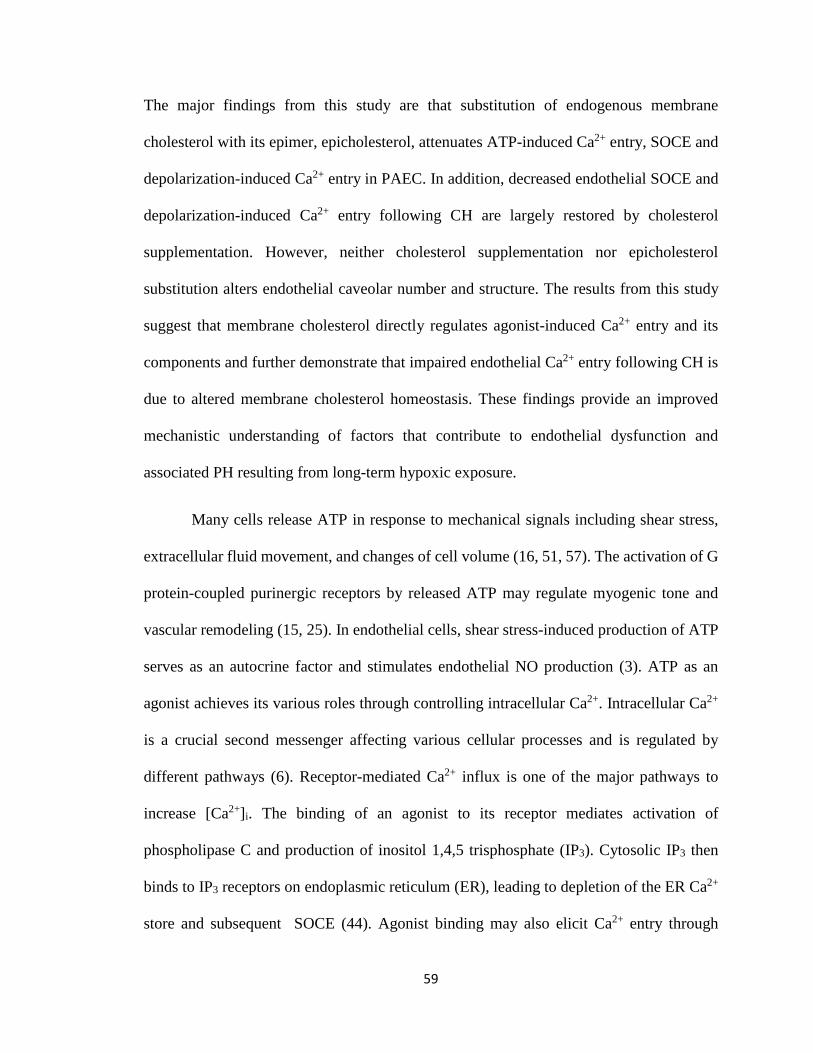
59
The major findings from this study are that substitution of endogenous membrane
cholesterol with its epimer, epicholesterol, attenuates ATP-induced Ca2+ entry, SOCE and
depolarization-induced Ca2+ entry in PAEC. In addition, decreased endothelial SOCE and
depolarization-induced Ca2+ entry following CH are largely restored by cholesterol
supplementation. However, neither cholesterol supplementation nor epicholesterol
substitution alters endothelial caveolar number and structure. The results from this study
suggest that membrane cholesterol directly regulates agonist-induced Ca2+ entry and its
components and further demonstrate that impaired endothelial Ca2+ entry following CH is
due to altered membrane cholesterol homeostasis. These findings provide an improved
mechanistic understanding of factors that contribute to endothelial dysfunction and
associated PH resulting from long-term hypoxic exposure.
Many cells release ATP in response to mechanical signals including shear stress,
extracellular fluid movement, and changes of cell volume (16, 51, 57). The activation of G
protein-coupled purinergic receptors by released ATP may regulate myogenic tone and
vascular remodeling (15, 25). In endothelial cells, shear stress-induced production of ATP
serves as an autocrine factor and stimulates endothelial NO production (3). ATP as an
agonist achieves its various roles through controlling intracellular Ca2+. Intracellular Ca2+
is a crucial second messenger affecting various cellular processes and is regulated by
different pathways (6). Receptor-mediated Ca2+ influx is one of the major pathways to
increase [Ca2+]i. The binding of an agonist to its receptor mediates activation of
phospholipase C and production of inositol 1,4,5 trisphosphate (IP3). Cytosolic IP3 then
binds to IP3 receptors on endoplasmic reticulum (ER), leading to depletion of the ER Ca2+
store and subsequent SOCE (44). Agonist binding may also elicit Ca2+ entry through
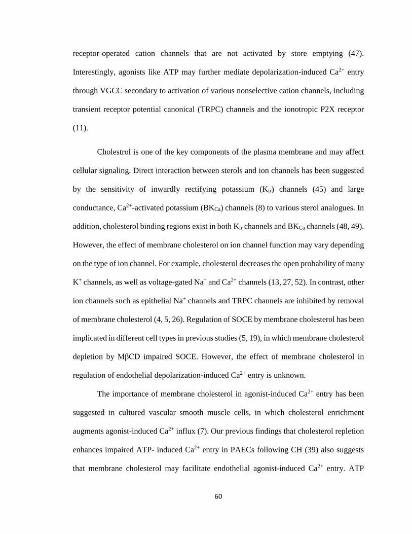
60
receptor-operated cation channels that are not activated by store emptying (47).
Interestingly, agonists like ATP may further mediate depolarization-induced Ca2+ entry
through VGCC secondary to activation of various nonselective cation channels, including
transient receptor potential canonical (TRPC) channels and the ionotropic P2X receptor
(11).
Cholestrol is one of the key components of the plasma membrane and may affect
cellular signaling. Direct interaction between sterols and ion channels has been suggested
by the sensitivity of inwardly rectifying potassium (Kir) channels (45) and large
conductance, Ca2+-activated potassium (BKCa) channels (8) to various sterol analogues. In
addition, cholesterol binding regions exist in both Kir channels and BKCa channels (48, 49).
However, the effect of membrane cholesterol on ion channel function may vary depending
on the type of ion channel. For example, cholesterol decreases the open probability of many
K+ channels, as well as voltage-gated Na+ and Ca2+ channels (13, 27, 52). In contrast, other
ion channels such as epithelial Na+ channels and TRPC channels are inhibited by removal
of membrane cholesterol (4, 5, 26). Regulation of SOCE by membrane cholesterol has been
implicated in different cell types in previous studies (5, 19), in which membrane cholesterol
depletion by MβCD impaired SOCE. However, the effect of membrane cholesterol in
regulation of endothelial depolarization-induced Ca2+ entry is unknown.
The importance of membrane cholesterol in agonist-induced Ca2+ entry has been
suggested in cultured vascular smooth muscle cells, in which cholesterol enrichment
augments agonist-induced Ca2+ influx (7). Our previous findings that cholesterol repletion
enhances impaired ATP- induced Ca2+ entry in PAECs following CH (39) also suggests
that membrane cholesterol may facilitate endothelial agonist-induced Ca2+ entry. ATP

61
induces both SOCE and T-type VGCC blocker -sensitive Ca2+ influx in PAECs (40). In
this study, we confirmed that ATP causes membrane depolarization in pulmonary artery
endothelium, which may lead to T-type channel activation and Ca2+ influx. Considering
that SOCE and depolarization-induced Ca2+ entry are major components of ATP- induced
Ca2+ entry, and that both Ca2+ influx pathways are blunted following CH (38, 40), it is
possible that membrane cholesterol also regulates these two components of the ATP-
induced Ca2+ response.
Considering the wide range of effects of MβCD such as altering caveolar structure
and disrupting other lipid microdomains (21), the direct contribution of membrane
cholesterol to regulation of ion channels remains unclear. To investigate the direct role of
membrane cholesterol in mediating endothelial Ca2+ influx, we used epicholesterol, the
enantiomer of cholesterol, to substitute membrane cholesterol. Although epicholesterol has
effects similar to cholesterol on physical properties of the cell membrane (56), few studies
have examined the effect of EpiChol:MβCD on caveolae. Here we provide evidence that
MβCD alone nearly abolished the incidence of caveolae, whereas neither Chol:MβCD nor
EpiChol:MβCD affected caveolar number in cultured PMVEC. It has previously been
shown that administration of an EpiChol:MβCD solution to bovine aortic endothelial cells
effectively substitutes epicholesterol for endogenous membrane cholesterol (46). To verify
this observation in our preparation, we assessed effects of epicholesterol substitution on
membrane cholesterol content in freshly isolated PAEC using the fluorescent cholesterol
marker, filipin. Epicholesterol does not interact with filipin due to a different orientation
of the 3-hydroxyl group compared to cholesterol (14, 37). As expected, we found that
epicholesterol treatment greatly decreased filipin fluorescence intensity in isolated PAEC

62
from both normoxic and CH rats, consistent with epicholesterol substitution of endogenous
membrane cholesterol. In addition, cholesterol treatment restored membrane cholesterol
content in PAEC from CH rats to the level of normoxic controls, consistent with our
previous findings (39). These findings suggest that EpiChol:MβCD treatment substitutes
endogenous membrane cholesterol for epicholesterol without disrupting caveolae.
Consequently, the observed effects of epicholesterol treatment on Ca2+ influx are likely due
to loss of ion channel regulation by cholesterol, rather than to changes in caveolar density.
Using epicholesterol to substitute endogenous cholesterol, we demonstrated a direct
role of membrane cholesterol to facilitate ATP-induced Ca2+ entry in PAEC. Our data are
consistent with findings that depletion of membrane cholesterol impairs this Ca2+ response
in PAEC (39). We also provide evidence that SOCE, as the major component of the ATP-
induced Ca2+ response, is similarly regulated by membrane cholesterol. This observation
implies that membrane cholesterol may either directly affect store-operated cation channels
(SOCs) or interact with a signaling pathway that activates SOCs. The finding that reduced
endothelial SOCE in pulmonary arteries following CH was acutely restored by membrane
cholesterol supplementation suggests that CH limits SOCE by reducing membrane
cholesterol rather than by decreasing ion channel expression. Although this study has not
identified the specific cation channel(s) involved in pulmonary endothelial SOCE,
candidates ion channels include TRPC1, TRPC4 and Orai1, each of which have been
implicated in endothelial SOCE and demonstrate cholesterol-sensitivity (5, 12, 23, 32).
Wu et al (54) first demonstrated that cultured rat PMVECs express mRNA of
CaV3.1 and possess voltage-dependent current that are sensitive to T-type channel blockers.
They also reported that T-type VGCCs contribute to both agonist-induced Ca2+ entry and

63
SOCE in PMVECs. Although T-type VGCCs are not found in cultured rat PAECs (54),
their expression and function are described in freshly dispersed rat PAECs (40). Paffett et
al showed that T-type VGCCs contribute to depolarization-induced Ca2+ entry and
receptor-operated Ca2+ entry in response to ATP. Following exposure to CH, the mibefradil
sensitive component of ATP-induced Ca2+ entry is greatly reduced. The effect of CH on
this Ca2+ entry is not due to altered membrane K+ permeability, since the impaired
endothelial depolarization-induced Ca2+ entry following CH persists even when membrane
K+ permeability is equivalently clamped by K+ ionophore, valinomycin. These findings
suggest that T-type VGCCs are important mediators of depolarization-induced Ca2+ entry
and that impaired Ca2+ entry via these channels may contribute to reduced basal [Ca2+]i in
PAECs after CH (40). Using the Mn2+ quenching technique to selectively assess Ca2+ entry,
we confirmed that depolarization-induced Ca2+ entry was sensitive to T-type VGCC
inhibition and Ca2+ entry was eliminated following CH. Additionally, depolarization-
induced Ca2+ entry following CH was partially restored by cholesterol supplementation. A
similar effect of cholesterol on VGCCs has been reported in other cell types (55). Although
studies on cholesterol regulation of VGCC are limited, our finding that epicholesterol
substitution abolished depolarization-induced Ca2+ entry in control cells suggests that there
may be a direct interaction between membrane cholesterol and T-type VGCCs that controls
channel activity.
Two potential mechanisms of decreased membrane cholesterol following CH
include: 1) CH inhibits de novo cholesterol biosynthesis; and 2) CH induces membrane
cholesterol oxidation. Mukodani et al. first reported that hypoxia induces lipid
accumulation and impairs cholesterol synthesis in cultured rabbit skin fibroblasts (31). The

64
mechanism by which hypoxia affects cholesterol synthesis was later explored by Nguyen
et al (36). They reported that hypoxia induces accumulation of cholesterol biosynthetic
intermediates and activates HIF1-α-mediated induction of ER membrane proteins called
insulin-induced gene. These two signaling pathways lead to rapid degradation of HMG-
CoA reductase and subsequently limit synthesis of cholesterol (36). Chronic hypoxia can
also increase the production of reactive oxygen species (ROS) (18, 28), which may
facilitate membrane cholesterol oxidation. Because filipin cannot be used to label oxidized
cholesterol (43), it is possible that reduced filipin staining in PAECs of CH group is the
result of membrane cholesterol oxidation by hypoxia-induced ROS. Cholesterol oxidation
not only has potential to disrupt the interaction between cholesterol and ion channels and
many regulatory proteins, but may also to inhibit de novo cholesterol synthesis (34). Future
studies are required to evaluate the potential contributions of these mechanisms to reduced
PAEC membrane cholesterol following CH.
Regulation of intracellular Ca2+ levels is a complex process. It is interesting that
CH impairs agonist-induced Ca2+ entry in PAEC via altered cholesterol regulation of
potential ion channels. This decreased endothelial ATP-induced Ca2+ influx following CH
is not associated with changes in ATP-induced Ca2+ mobilization (39), suggesting that
attenuated Ca2+ entry is not due to altered Ca2+ release of Ca2+ loading of the ER after CH
exposure. However, whether CH similarly affect other intracellular Ca2+ handling
pathways like Ca2+ sequestration and efflux remains to be determined. It should also be
noted that the impaired Ca2+ entry following CH is limited to endothelial cells of large
pulmonary arteries and may not reflect those of smaller arteries and arterioles that likely
contribute more to regulating vascular resistance in hypertensive pulmonary circulation.
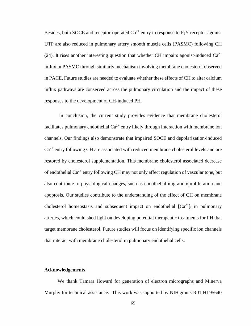
65
Besides, both SOCE and receptor-operated Ca2+ entry in response to P2Y receptor agonist
UTP are also reduced in pulmonary artery smooth muscle cells (PASMC) following CH
(24). It rises another interesting question that whether CH impairs agonist-induced Ca2+
influx in PASMC through similarly mechanism involving membrane cholesterol observed
in PACE. Future studies are needed to evaluate whether these effects of CH to alter calcium
influx pathways are conserved across the pulmonary circulation and the impact of these
responses to the development of CH-induced PH.
In conclusion, the current study provides evidence that membrane cholesterol
facilitates pulmonary endothelial Ca2+ entry likely through interaction with membrane ion
channels. Our findings also demonstrate that impaired SOCE and depolarization-induced
Ca2+ entry following CH are associated with reduced membrane cholesterol levels and are
restored by cholesterol supplementation. This membrane cholesterol associated decrease
of endothelial Ca2+ entry following CH may not only affect regulation of vascular tone, but
also contribute to physiological changes, such as endothelial migration/proliferation and
apoptosis. Our studies contribute to the understanding of the effect of CH on membrane
cholesterol homeostasis and subsequent impact on endothelial [Ca2+]i in pulmonary
arteries, which could shed light on developing potential therapeutic treatments for PH that
target membrane cholesterol. Future studies will focus on identifying specific ion channels
that interact with membrane cholesterol in pulmonary endothelial cells.
Acknowledgements
We thank Tamara Howard for generation of electron micrographs and Minerva
Murphy for technical assistance. This work was supported by NIH grants R01 HL95640

66
(B.R. Walker), R01 HL132883 and R01 HL088192 (T.C. Resta), and American Heart
Association grants 15GRNT21080001 (B.R. Walker) and 16GRNT27700010 (T.C. Resta).

67
Figure 1. Epicholesterol reduces endogenous membrane cholesterol of freshly isolated
PAEC sheets from both normoxic (Nor) and CH rats. A) Representative images of
membrane cholesterol indicated by filipin fluorescence in PAEC isolated from each group.
Cells were pretreated with vehicle, cholesterol (Chol), or epicholesterol (Epichol). Scale
bars = 20 μm. B) Mean filipin fluorescence (arbitrary units; A.U.) in PAEC sheets from
each group. Two-way ANOVA followed by the Student-Newman-Keuls post-hoc test was
used to compare between groups. Values are means ± SE; n = 3 animals/group. *P < 0.05
vs. Nor vehicle; #P < 0.05 vs. CH vehicle.

68
Figure 2. Caveolae number is reduced by cholesterol depletion with MΒCD, but not by
cholesterol or epicholesterol treatment in cultured PMVEC. A) Representative images of
caveolae in cultured PMVEC from each treatment group. B) Mean number of caveolae per
length of cell membrane (caveolae/μm). Data were compared by the Kruskal-Wallis H test
and Dunn’s multiple comparison test. Values are means ± SE; n=7-11 cells (indicated in
bars); *P < 0.05 versus all other treatments. A total of 100 images encompassing 38 cells
and 438 μm of membrane were analyzed.

69
Figure 3. Experimental protocol for measuring ATP- and CPA- induced Ca2+ entry in
isolated PAEC sheets. Depletion of intracellular Ca2+ store was induced by 20 μM ATP or
10 μM CPA in a Ca2+-free HEPES buffer. Intracellular Ca2+ is expressed as the fura-2
340/380 nm emission ratio. Ca2+ entry was assessed by calculating area under the curve
(AUC) for the 5 min following reintroduction of extracellular Ca2+ (indicated by rectangle).

70
Figure 4. Epicholesterol substitution reduces ATP-induced Ca2+ entry in PAEC sheets
from normoxic rats. Ca2+ influx was assessed by ratiometric analysis of fura-2
fluorescence. One-way ANOVA followed by the Student-Newman-Keuls test was used to
compare between groups. Values are means ± SE; n = 5 animals/group. *P < 0.05 vs.
vehicle.
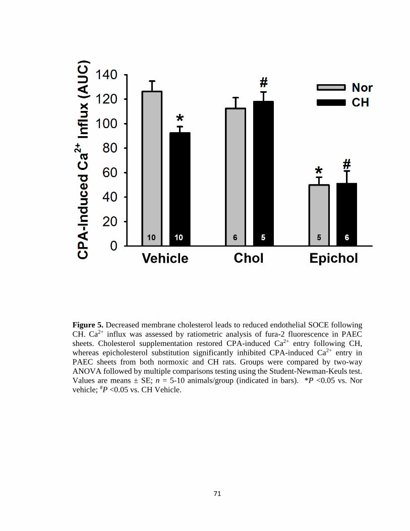
71
Figure 5. Decreased membrane cholesterol leads to reduced endothelial SOCE following
CH. Ca2+ influx was assessed by ratiometric analysis of fura-2 fluorescence in PAEC
sheets. Cholesterol supplementation restored CPA-induced Ca2+ entry following CH,
whereas epicholesterol substitution significantly inhibited CPA-induced Ca2+ entry in
PAEC sheets from both normoxic and CH rats. Groups were compared by two-way
ANOVA followed by multiple comparisons testing using the Student-Newman-Keuls test.
Values are means ± SE; n = 5-10 animals/group (indicated in bars). *P <0.05 vs. Nor
vehicle; #P <0.05 vs. CH Vehicle.
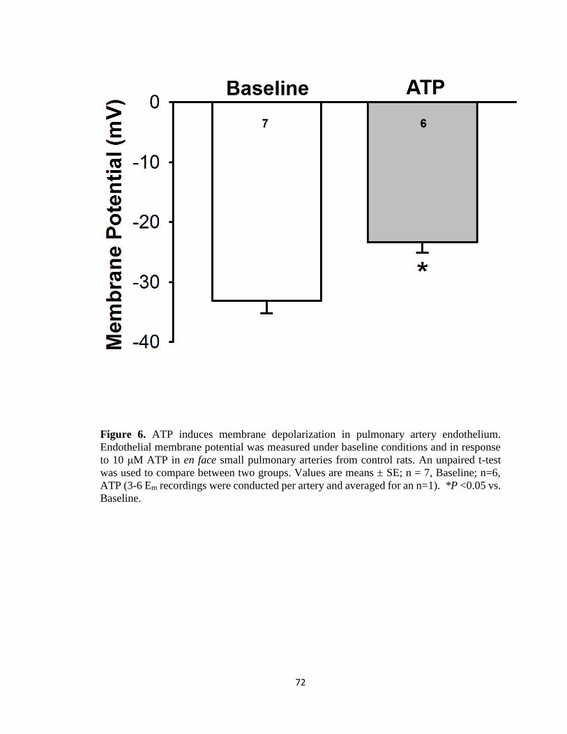
72
Figure 6. ATP induces membrane depolarization in pulmonary artery endothelium.
Endothelial membrane potential was measured under baseline conditions and in response
to 10 μM ATP in en face small pulmonary arteries from control rats. An unpaired t-test
was used to compare between two groups. Values are means ± SE; n = 7, Baseline; n=6,
ATP (3-6 Em recordings were conducted per artery and averaged for an n=1). *P <0.05 vs.
Baseline.

73
Figure 7. Exposure to CH abolishes endothelial depolarization-induced Ca2+ entry. A
measure of endothelial Ca2+ entry was assessed by Mn2+ quenching of fura-2 fluorescence
in PAEC sheets from control and CH rats treated with either vehicle (time control) or 60
mM KCl (depolarizing stimulus). F, fluorescence intensity at 360 nm excitation; F0,
fluorescence intensity at time zero. Two-way ANOVA and the Student-Newman-Keuls
test were used to compare between groups at each time point. Values are means ± SE; n =
5, Nor Vehicle; n = 5, CH Vehicle; n = 7, Nor KCl; n = 6, CH KCl. *P < 0.05 vs. Nor KCl
over the range of 20 – 120 s.

74
Figure 8. CH attenuates depolarization-induced Ca2+ entry through T-type Ca2+ channels
in PAEC. Data represent Mn2+-quenched fura-2 fluorescence (at 90 s time point) in
response to KCl (60 mM) in PAEC sheets from control and CH rats. Diltiazem (50 μM)
and mibefradil (10 μM) were used to selectively inhibit L-type and T-type Ca2+ channels,
respectively. Data are expressed as ΔF/F0 (%) from time control. Two-way ANOVA and
the Student-Newman-Keuls test were used to compare between groups. Values are means
± SE; n = 4-7 animals/group (indicated in bars). *P < 0.05 vs. Nor vehicle; #P < 0.05 vs.
Nor diltiazem.

75
Figure 9. Decreased membrane cholesterol leads to reduced endothelial depolarization-
induced Ca2+ entry following CH. Data represent Mn2+-quenched fura-2 fluorescence (at
90 s time point) in response to KCl (60 mM) in PAEC sheets from control and CH rats.
Cholesterol repletion partially restored depolarization-induced Ca2+ entry following CH.
Epicholesterol greatly inhibited KCl-induced Ca2+ entry in PAEC from both normoxic and
CH rats. Data are expressed as ΔF/F0 (%) from time control. Statistical comparisons were
made using two-way ANOVA and the Student-Newman-Keuls post-hoc test. Values are
means ± SE; n = 5-7 animals/group (indicated in bars). *P < 0.05 vs. Nor vehicle; #P <
0.05 vs. Nor chol; ∆P < 0.05 vs. CH vehicle.

76
References
1. Alakoskela JM, Sabatini K, Jiang X, Laitala V, Covey DF, Kinnunen PKJ.
Enantiospecific interactions between cholesterol and phospholipids. Langmuir 24:
830–836, 2008.
2. Anderson M. Mn ions pass through calcium channels. A possible explanation. J
Gen Physiol 81: 805–827, 1983.
3. Andrews AM, Jaron D, Buerk DG, Barbee KA. Shear stress-induced NO
production is dependent on ATP autocrine signaling and capacitative calcium
entry. Cell Mol Bioeng 7: 510–520, 2015.
4. Barberà JA, Peinado VI, Santos S. Pulmonary hypertension in chronic
obstructive pulmonary disease. Eur Respir J 21: 892–905, 2003.
5. Bergdahl A, Gomez MF, Dreja K, Xu SZ, Adner M, Beech DJ, Broman J,
Hellstrand P, Swärd K. Cholesterol depletion impairs vascular reactivity to
endothelin-1 by reducing store-operated Ca2+ entry dependent on TRPC1. Circ
Res 93: 839–847, 2003.
6. Berridge MJ, Lipp P, Bootman MD. The versatility and universality of calcium
signalling. Nat Rev Mol Cell Biol 1: 11–21, 2000.
7. Bialecki RA, Tulenko TN, Colucci WS. Cholesterol enrichment increases basal
and agonist-stimulated calcium influx in rat vascular smooth muscle cells. J Clin
Invest 88: 1894–1900, 1991.
8. Bukiya AN, Belani JD, Rychnovsky S, Dopico AM. Specificity of cholesterol

77
and analogs to modulate BK channels points to direct sterol-channel protein
interactions. J Gen Physiol 137: 93–110, 2011.
9. Busse R, Mülsch A. Calcium-dependent nitric oxide synthesis in endothelial
cytosol is mediated by calmodulin. FEBS Lett 265: 133–136, 1990.
10. Christian EA, Haynes MP, Phillips MC, George H. Use of cyclodextrins for
manipulating cellular cholesterol content. J Lipid Res 38: 2264–2272, 1997.
11. Chung HS, Park KS, Cha SK, Kong ID, Lee JW. ATP-induced [Ca(2+)](i)
changes and depolarization in GH3 cells. Br J Pharmacol 130: 1843–52, 2000.
12. Cioffi DL, Wu S, Stevens T. On the endothelial cell ISOC. Cell Calcium 33: 323–
336, 2003.
13. Crowley JJ. Cholesterol antagonizes ethanol potentiation of human brain BKCa
channels reconstituted into phospholipid bilayers. Mol Pharmacol 64: 365–372,
2003.
14. Elias PM, Friend DS, Goerke J. Membrane sterol heterogeneity. Freeze-fracture
detection with saponins and filipin. J Histochem Cytochem 27: 1247–1260, 1979.
15. Erlinge D, Burnstock G. P2 receptors in cardiovascular regulation and disease.
Purinergic Signal 4: 1–20, 2008.
16. Fitz JG. Regulation of cellular ATP release. Trans Am Clin Clim Assoc 118: 199–
208, 2007.
17. Förstermann U, Pollock JS, Schmidt HH, Heller M, Murad F. Calmodulin-
dependent endothelium-derived relaxing factor/nitric oxide synthase activity is

78
present in the particulate and cytosolic fractions of bovine aortic endothelial cells.
Proc Natl Acad Sci 88: 1788–1792, 1991.
18. Fresquet F, Pourageaud F, Leblais V, Brandes RP, Savineau JP, Marthan R,
Muller B. Role of reactive oxygen species and gp91phox in endothelial
dysfunction of pulmonary arteries induced by chronic hypoxia. Br J Pharmacol
148: 714–723, 2006.
19. Galan C, Woodard GE, Dionisio N, Salido GM, Rosado JA. Lipid rafts
modulate the activation but not the maintenance of store-operated Ca2+ entry.
Biochim Biophys Acta - Mol Cell Res 1803: 1083–1093, 2010.
20. Gimpl G, Burger K, Fahrenholz F. Cholesterol as modulator of receptor
function. Biochemistry 36: 10959–10974, 1997.
21. Graziani A, Bricko V, Carmignani M, Graier WF, Groschner K. Cholesterol-
and caveolin-rich membrane domains are essential for phospholipase A 2-
dependent EDHF formation. Cardiovasc Res 64: 234–242, 2004.
22. Hinton JM, Langton PD. Inhibition of EDHF by two new combinations of K+ -
channel inhibitors in rat isolated mesenteric arteries. Br J Pharmacol 138: 1031–
1035, 2003.
23. Iskandar F. Abdullaev, Jonathan M. Bisaillon, Marie Potier, Jose C.
Gonzalez, Rajender K. Motiani MT. Stim1 and Orai1 mediate CRAC currents
and store-operated calcium entry important for endothelial cell proliferation. Circ
Res 103: 1289–1299, 2009.

79
24. Jernigan NL, Broughton BRS, Walker BR, Thomas C, Resta TC, No-
dependent I. Impaired NO-dependent inhibition of store- and receptor-operated
calcium entry in pulmonary vascular smooth muscle after chronic hypoxia. Am J
Physiol Lung Cell Mol Physiol 290: 517–525, 2006.
25. Koltsova S V., Maximov G V., Kotelevtsev S V., Lavoie JL, Tremblay J,
Grygorczyk R, Hamet P, Orlov SN. Myogenic tone in mouse mesenteric arteries:
Evidence for P2Y receptor-mediated, Na+, K+, 2Cl- cotransport-dependent
signaling. Purinergic Signal 5: 343–349, 2009.
26. Kwiatek AM, Minshall RD, Cool DR, Skidgel RA, Malik AB. Caveolin-1
regulates store-operated Ca 2+ influx by binding of its scaffolding domain to
transient receptor potential channel-1 in endothelial cells. Mol Pharmacol 70:
1174–1183, 2006.
27. Lin M-W, Wu AZ, Ting W-H, Li C-L, Cheng K-S, Wu S-N. Changes in
membrane cholesterol of pituitary tumor (GH3) cells regulate the activity of large-
conductance Ca2+-activated K+ channels. Chin J Physiol 49: 1–13, 2006.
28. Liu JQ, Zelko IN, Erbynn EM, Sham JSK, Folz RJ. Hypoxic pulmonary
hypertension: role of superoxide and NADPH oxidase (gp91phox). AJP - Lung
Cell Mol Physiol 290: L2-10, 2006.
29. Mannock DA, McIntosh TJ, Jiang X, Covey DF, McElhaney RN. Effects of
natural and enantiomeric cholesterol on the thermotropic phase behavior and
structure of egg sphingomyelin bilayer membranes. Biophys J 84: 1038–1046,
2003.

80
30. Marrelli SP, Eckmann MS, Hunte MS. Role of endothelial intermediate
conductance KCa channels in cerebral EDHF-mediated dilations. Am J Physiol
Hear Circ Physiol 285: H1590-9, 2003.
31. Mukodani J, Ishikawa Y, Fukuzaki H. Effects of hypoxia on sterol synthesis,
acyl-CoA:cholesterol acyltransferase activity, and efflux of cholesterol in cultured
rabbit skin fibroblasts. Arteriosclerosis 10: 106–110, 1990.
32. Murata T, Lin MI, Stan R V., Bauer PM, Yu J, Sessa WC. Genetic evidence
supporting caveolae microdomain regulation of calcium entry in endothelial cells.
J Biol Chem 282: 16631–16643, 2007.
33. Murata T, Sato K, Hori M, Ozaki H, Karaki H. Decreased endothelial nitric-
oxide synthase (eNOS) activity resulting from abnormal interaction between eNOS
and its regulatory proteins in hypoxia-induced pulmonary hypertension. J Biol
Chem 277: 44085–44092, 2002.
34. Murphy RC, Johnson KM. Cholesterol, reactive oxygen species, and the
formation of biologically active mediators. J Biol Chem 283: 15521–15525, 2008.
35. Nakane M, Mitchell J, Förstermann U, Murad F. Phosphorylation by calcium
calmodulin-dependent protein kinase II and protein kinase C modulates the activity
of nitric oxide synthase. Biochem Biophys Res Commun 180: 1396–1402, 1991.
36. Nguyen AD, McDonald JG, Bruick RK, DeBose-Boyd RA. Hypoxia stimulates
degradation of 3-hydroxy-3-methylglutaryl-coenzyme A reductase through
accumulation of lanosterol and hypoxia-inducible factor-mediated induction of
insigs. J Biol Chem 282: 27436–27446, 2007.

81
37. Norman AW, Demel RA, Kruyff B De, Deenen LLM Van. Studies on the
biological properties of polyene antibiotics: evidence for the direct interaction of
filipin with cholesterol of polyene anitbiotics. J Biol Chem 47(6): 1918–1929,
1972.
38. Paffett ML, Naik JS, Resta TC, Walker BR. Reduced store-operated Ca2+ entry
in pulmonary endothelial cells from chronically hypoxic rats. Am J Physiol Lung
Cell Mol Physiol 293: L1135-42, 2007.
39. Paffett ML, Naik JS, Riddle MA, Menicucci SD, Gonzales AJ, Resta TC,
Walker BR. Altered membrane lipid domains limit pulmonary endothelial
calcium entry following chronic hypoxia. Am J Physiol Hear Circ Physiol 301:
1331–1340, 2011.
40. Paffett ML, Riddle MA, Kanagy NL, Resta TC, Walker BR. Altered protein
kinase C regulation of pulmonary endothelial store- and receptor-operated Ca2+
entry after chronic hypoxia. J Pharmacol Exp Ther 334: 753–760, 2010.
41. Parpal S, Karlsson M, Thorn H, Strålfors P. Cholesterol depletion disrupts
caveolae and insulin receptor signaling for metabolic control via insulin receptor
substrate-1, but not for mitogen-activated protein kinase control. J Biol Chem 276:
9670–9678, 2001.
42. Pollock JS, Förstermann U, Mitchell JA, Warner TD, Schmidt HH, Nakane
M, Murad F. Purification and characterization of particulate endothelium-derived
relaxing factor synthase from cultured and native bovine aortic endothelial cells.
Proc Natl Acad Sci U S A 88: 10480–4, 1991.
82
43. Pörn MI, Slotte JP. Localization of cholesterol in sphingomyelinase-treated
fibroblasts. Biochem J 308 (Pt 1): 269–74, 1995.
44. Putney JW, Broad LM, Braun FJ, Lievremont JP, Bird GS. Mechanisms of
capacitative calcium entry. J Cell Sci 114: 2223–2229, 2001.
45. Romanenko VG, Rothblat GH, Levitan I. Modulation of endothelial inward-
rectifier K+ current by optical isomers of cholesterol. Biophys J 83: 3211–3222,
2002.
46. Romanenko VG, Rothblat GH, Levitan I. Sensitivity of volume-regulated anion
current to cholesterol structural analogues. J Gen Physiol 123: 77–87, 2004.
47. Rosado J a, Sage SO. Protein kinase C activates non-capacitative calcium entry in
human platelets. J Physiol 529 Pt 1: 159–69, 2000.
48. Rosenhouse-Dantsker A, Noskov S, Durdagi S, Logothetis DE, Levitan I.
Identification of novel cholesterol-binding regions in Kir2 channels. J Biol Chem
288: 31154–31164, 2013.
49. Singh AK, McMillan J, Bukiya AN, Burton B, Parrill AL, Dopico AM.
Multiple cholesterol recognition/interaction amino acid consensus (CRAC) motifs
in cytosolic C tail of Slo1 subunit determine cholesterol sensitivity of Ca2+- and
voltage-gated K+ (BK) channels. J Biol Chem 287: 20509–20521, 2012.
50. Sooksawate T, Simmonds MA. Effects of membrane cholesterol on the
sensitivity of the GABA(A) receptor to GABA in acutely dissociated rat
hippocampal neurones. Neuropharmacology 40: 178–184, 2001.

83
51. Tatur S, Groulx N, Orlov SN, Grygorczyk R. Ca2+-dependent ATP release
from A549 cells involves synergistic autocrine stimulation by coreleased uridine
nucleotides. J Physiol 584: 419–35, 2007.
52. Toselli M, Biella G, Taglietti V, Cazzaniga E, Parenti M. Caveolin-1 expression
and membrane cholesterol content modulate N-type calcium channel activity in
NG108-15 cells. Biophys J 89: 2443–57, 2005.
53. Tuder RM, Chacon M, Alger L, Wang J, Taraseviciene-Stewart L, Kasahara
Y, Cool CD, Bishop AE, Geraci M, Semenza GL, Yacoub M, Polak JM,
Voelkel NF. Expression of angiogenesis-related molecules in plexiform lesions in
severe pulmonary hypertension: Evidence for a process of disordered
angiogenesis. J Pathol 195: 367–374, 2001.
54. Wu S, Haynes J, Taylor JT, Obiako BO, Stubbs JR, Li M, Stevens T. CaV3.1
(α1G) T-type Ca2+ channels mediate vaso-occlusion of sickled erythrocytes in
lung microcirculation. Circ Res 93: 346–353, 2003.
55. Xia F, Xie L, Mihic A, Gao X, Chen Y, Gaisano HY, Tsushima RG. Inhibition
of cholesterol biosynthesis impairs insulin secretion and voltage-gated calcium
channel function in pancreatic beta-cells. Endocrinology 149: 5136–5145, 2008.
56. Xu X, London E. The effect of sterol structure on membrane lipid domains
reveals how cholesterol can induce lipid domain formation. Biochemistry 39: 843–
849, 2000.
57. Yamamoto K, Furuya K, Nakamura M, Kobatake E, Sokabe M, Ando J.
Visualization of flow-induced ATP release and triggering of Ca 2+ waves at

84
caveolae in vascular endothelial cells. J Cell Sci 124: 3477–3483, 2011.
58. Zidovetzki R, Levitan I. Use of cyclodextrins to manipulate plasma membrane
cholesterol content: Evidence, misconceptions and control strategies. Biochim
Biophys Acta - Biomembr 1768: 1311–1324, 2007.

85
CHAPTER 3:
Reduced Membrane Cholesterol Following Chronic Hypoxia
Limits Orai1-Mediated Pulmonary Endothelial Ca2+ Entry2
Bojun Zhang, Jay S. Naik, Nikki L. Jernigan, Benjimen R. Walker and Thomas C. Resta
Vascular Physiology Group
Department of Cell Biology and Physiology
University of New Mexico Health Sciences Center
Albuquerque, NM
Running Head: Pulmonary Endothelial Ca2+ Entry Following Chronic Hypoxia
Corresponding Author: Thomas C. Resta, Ph.D.
Department of Cell Biology and Physiology
University of New Mexico Health Sciences Center
MSC 08-4750
1 University of New Mexico
Albuquerque, NM 87131-0001
Phone: 505-272-8822
Fax: 505-272-6649
e-mail: [email protected]
2 This work has been published as: Zhang B, Naik JS, Jernigan NL, Walker BR, Resta TC. Reduced
membrane cholesterol following chronic hypoxia limits Orai1-mediated pulmonary endothelial Ca2+ entry.
Am J Physiol 2017 Nov 3:ajpheart.00540.2017. doi: 10.1152/ajpheart.00540.2017. [Epub ahead of print].

86
Abstract
Endothelial dysfunction in chronic hypoxia (CH)-induced pulmonary hypertension
is characterized by reduced store-operated Ca2+ entry (SOCE) and diminished Ca2+-
dependent production of endothelium-derived vasodilators. We recently reported that
SOCE in pulmonary arterial endothelial cells (PAEC) is tightly regulated by membrane
cholesterol, and that decreased membrane cholesterol is responsible for impaired SOCE
following CH. However, the ion channels involved in cholesterol-sensitive SOCE are
unknown. We hypothesized that cholesterol facilitates SOCE in PAEC through the
interaction of Orai1 and stromal interaction molecule 1 (STIM1). The role of cholesterol
in Orai1-mediated SOCE was initially assessed using CH exposure in rats (4 wk, 380
mmHg) as a physiological stimulus to decrease PAEC cholesterol. Effects of Orai1
inhibition with AnCoA4 on SOCE were examined in isolated PAEC sheets from control
and CH rats following cholesterol supplementation, substitution of endogenous cholesterol
with epicholesterol (Epichol), or vehicle treatment. Whereas cholesterol restored
endothelial SOCE in CH rats, both Epichol and AnCoA4 attenuated SOCE only in
normoxic controls. The Orai1 inhibitor had no further effect in cells pretreated with
Epichol. Using cultured pulmonary endothelial cells to allow better mechanistic analysis
of the molecular components of cholesterol-regulated SOCE, we found that Epichol,
AnCoA4 and Orai1 siRNA each inhibited SOCE compared to their respective controls.
Epichol had no additional effect following knockdown of Orai1. Furthermore, Epichol
substitution significantly reduced STIM1-Orai1 interaction assessed by proximity ligation
assay. We conclude that membrane cholesterol is required for the STIM1-Orai1 interaction

87
necessary to elicit endothelial SOCE. Furthermore, reduced PAEC membrane cholesterol
following CH limits Orai1-mediated SOCE.
Key Words: pulmonary hypertension, STIM1, store-operated Ca2+ entry.
New & Noteworthy: This research demonstrates a novel contribution of cholesterol to
regulate the interaction of Orai1 and STIM1 required for pulmonary endothelial agonist-
induced Ca2+ entry. The results provide a mechanistic basis for impaired pulmonary
endothelial Ca2+ influx following CH that may contribute to pulmonary hypertension.

88
Introduction
Endothelial dysfunction in pulmonary hypertension is characterized by imbalanced
production of endothelium-derived vasocontrictors and vasodilators leading to
vasoconstriction, vascular remodeling, and subsequent elevation of pulmonary vascular
resistance (8, 21, 75). The production of many endothelium-derived vasodilators and anti-
mitogenic compounds, including nitric oxide (NO), prostacyclin, and endothelium-derived
hyperpolarizing factors is closely regulated by endothelial intracellular calcium levels
([Ca2+]i) (9, 17, 25, 45, 47). Diminished pulmonary endothelial [Ca2+]i and limited
production of endothelium-derived NO are associated with the development of chronic
hypoxia (CH)-induced pulmonary hypertension (51, 56). However, the mechanisms of
reduced pulmonary endothelial [Ca2+]i following CH exposure are not well-investigated.
Endothelial store-operated Ca2+ entry (SOCE) is required for endothelium-
dependent vasorelaxation in systemic arteries (28), in part through production of NO (3).
SOCE is initiated when a stimulated G protein-coupled receptor activates phospholipase C
(PLC), which produces inositol 1,4,5 trisphosphate (IP3) by hydrolyzing
phosphatidylinositol bisphosphate (PIP2). IP3, as a second messenger, activates IP3
receptors on the endoplasmic reticulum (ER) membrane, leading to rapid ER Ca2+ release.
ER Ca2+ store depletion then activates store-operated cation channels (SOC), which leads
to sustained Ca2+ influx (64). Stromal interaction molecule 1 (STIM1) has been identified
as an ER Ca2+ sensor and was suggested as the essential link between Ca2+ store depletion
and SOC activation (72). As ER Ca2+ levels fall, STIM1 molecules oligomerize on the ER
membrane and interact with SOC on the cell membrane, which include Orai channels (15).
Orai1, one of three isoforms of Orai found in mammalian cells, mediates SOCE when

89
activated by STIM1 (86). The STIM1-Orai1 interaction occurs in cholesterol-rich caveolae
on the cell membrane. Interestingly, intact caveolae are required for activation of Orai1-
mediated SOCE in platelets (14). Depletion of membrane cholesterol not only inhibits
SOCE, but also interupts STIM1 clustering near the cell membrane (59). However, whether
membrane cholesterol directly faciliates STIM1-Orai1 interaction and SOCE activation or
indirectly modulates this Ca2+ entry pathway by contributing to lipid raft integrity is not
clear.
We have previously reported that decreased endothelial SOCE following CH is
associated with diminished membrane cholesterol and this reduced SOCE is restored by
cholesterol supplementation (56, 57, 84). Furthermore, to study the functional role of
membrane cholesterol in regulating SOCE, epicholesterol (Epichol) was used to replace
native cholesterol. As the enantiomer of cholesterol, Epichol has similar effects on
membrane fluidity and lipid domain formation as those of cholesterol but lacks the
regulatory influences of cholesterol on ion channel function (22, 65, 83). Substitution of
endogenous cholesterol with Epichol mimics the effect of CH in reducing endothelial
SOCE while having no effect on caveolar number (84). However, the mechanisms by
which reduced endothelial membrane cholesterol limits SOCE following CH are not clear.
In the present study, we hypothesized that cholesterol facilitates the interaction of Orai1
with STIM1 to mediate SOCE in PAEC. We tested this hypothesis by examining the effects
of either cholesterol or Epichol treatment combined with Orai1 inhibition on SOCE in both
freshly isolated pulmonary arterial endothelial cells (PAEC) from normoxic and CH rats
and in cultured pulmonary microvascular endothelial cells (PMVEC).

90
Methods
Animals and Chronic Hypoxic Exposure Protocol
Male Sprague-Dawley rats (200-250 g) were used for in vivo and ex vivo studies.
Rats exposed to CH were placed in a hypobaric chamber with barometric pressure
maintained at ≈ 380 mmHg (inspired PO2 ≈ 70 mmHg) for 4 wk (84). Age-matched control
rats were housed in similar cages under ambient barometric pressure (≈ 630 mmHg in
Albuquerque, NM). The hypobaric chamber was opened three times per week to provide
fresh rat chow, water, and clean bedding. All animals were maintained on a 12:12-h light-
dark cycle. All protocols used in this study were reviewed and approved by the Institutional
Animal Care and Use Committee of the University of New Mexico Health Sciences Center.
Isolation and preparation of pulmonary artery endothelial cells
Following CH or normoxic exposure, rats were euthanized with pentobarbital
sodium (200 mg/kg ip) and the heart and lungs were exposed by midline thoracotomy. The
left lung was rapidly excised and placed in ice-cold HEPES buffer solution. Intrapulmonary
arteries (3rd and 4th order, 200-400 μm inner diameter) were dissected from the superior
region of the left lung, and the parenchymal lung tissue was carefully removed. Arteries
were then cut longitudinally and treated with 0.2 mg/ml dithiothreitol and 2 U/ml papain
in HEPES buffer for 45 min at 37 °C. Vessels were carefully removed from the digestion
solution and placed in 1 ml of HEPES buffer containing 2 mg/ml bovine serum albumin.
PAEC sheets were then released by gentle trituration with a small-bore fire-polished
Pasteur pipette and stored at 4 °C. One to two drops of the solution containing freshly
isolated rat PAEC were placed on a poly-L-lysine-coated glass cover slip and incubated at

91
37 °C for 30 min prior to experimentation (84). PAEC sheets collected using this approach
are identified by their distinct morphology (57, 84) and uptake of Dil-Ac-LDL (56).
Cell culture
To complement experiments using freshly isolated PAEC sheets, cultured
pulmonary microvascular endothelial cells (PMVEC; VEC Technologies) were also used
in this study. Cultured cells permit the use of genetic manipulation with siRNA and
determination of protein-protein interactions (Orai1-STIM1) using proximity ligation
assay for better mechanistic analysis of the molecular components of cholesterol-regulated
SOCE. PMVEC were maintained in a humidified incubator at 37 °C with 5% CO2 in
MCDB-131 complete media (VEC Technologies). Cultures were split every 4-6 days and
cells studied between passages 5 and 10. Depending on the experimental protocol, cells
were seeded onto round poly-L-lysine-coated glass coverslips in 6-well plates, 6-well
plates coated with attachment factors, or 18-well slides coated with attachment factors.
Preparation of cholesterol and epicholesterol solutions
Solutions were prepared by saturating methyl-β-cyclodextrin (MβCD) with either
cholesterol or Epichol, as decribed previously (11). Briefly, the cyclodextrin-sterol
solutions were prepared by the addition of sterols to MβCD (10 mM) in a molar ratio of
1:5 and dissolution in HEPES buffer containing the following (in mM): 150 NaCl, 6 KCl,
1 MgCl2, 1.8 CaCl2, 10 HEPES, and 10 glucose (pH 7.4). Each solution was vortexed and
sonicated using a bath sonicator for 10-15 min. The saturated cyclodextrin-sterol solution
was then placed in a rotating incubator at 37 °C overnight. This stock solution was filtered
through a 0.22 μm syringe filter, aliquoted, and stored at -80 °C.

92
Endothelial cell membrane cholesterol manipulation
Cholesterol supplementation was performed in untreated PAEC or PMVEC by
incubation with the cholesterol/MβCD solution for 30 min at 37 °C (84). Epichol
substitution was similarly achieved by incubating isolated PAEC or cultured PMVEC with
the Epichol/MβCD solution under the same conditions. Preliminary studies revealed that,
in contrast to effects of membrane cholesterol manipulation in freshly dispersed PAEC
from normoxic rats (84), cholesterol supplementation increased SOCE in cultured PMVEC
and conferred sensitivity to Epichol substitution (data not shown), suggesting that
membrane cholesterol content is reduced under cell culture conditions compared to the
native state. Therefore, cultured PMVEC were pretreated with cholesterol/MβCD solution
prior to experimentation to mimic responses of native PAEC cells.
Ca2+ influx in freshly isolated and cultured pulmonary endothelial cells
Following vehicle, cholesterol, or Epichol treatment, freshly isolated PAEC sheets
or cultured PMVEC (passages 5-10) were loaded with fura-2 AM (3 μM and 0.05%
pluronic acid) in HEPES buffer for 7 min at room temperature (≈23 °C) and washed for 15
min at 37 °C. SOCE, depolarization-induced Ca2+ influx, and receptor-operated Ca2+ influx
were measured by Mn2+-quenching of fura-2 fluorescence in PAEC sheets or PMVEC as
previously described (84). Mn2+ enters the cell as a Ca2+ surrogate and reduces fura-2
fluorescence upon binding to the fluorophore. The preparation was excited at the isosbestic
wavelength (360 nm) at 1 Hz (IonOptix Hyperswitch), and emission recorded at 510 nm
with a photomultiplier tube. At this excitation wavelength, fura-2 fluorescence intensity is
not influenced by changes in [Ca2+]i, thus providing a measure of Ca2+ influx as reflected
by Mn2+ uptake independent of ER Ca2+ release, sequestration or efflux across the cell

93
membrane (31). Fura-2-loaded endothelial cells were superfused with Ca2+-free HEPES
buffer in the presence of vehicle or AnCoA4 (20 μM, Orai1 inhibitor (67)) for 5 min and
administered the sarco/endoplasmic reticulum Ca2+-ATPase inhibitor, cyclopiazonic acid
(CPA, 10 μM) to deplete intracellular Ca2+ stores and activate store-operated channels. To
confirm the specificity of AnCoA4 as an inhibitor of SOCE, KCl (60 mM) or 1-oleoyl-2-
acetyl-sn-glycerol (OAG, diacylglycerol analog, 50 μM) were applied in separate studies
to examine the effect of AnCoA4 on depolarization-induced and receptor-operated Ca2+
influx (58), respectively. Ca2+ entry represented by influx of the Ca2+ surrogate Mn2+ was
then determined upon addition of extracellular Mn2+ (500 μM) in the continued presence
of AnCoA4 or vehicle. SOCE was quantified by the percentage of the Mn2+ quenched
fluorescence at 120 s after administration of Mn2+.
Membrane cholesterol content
We have previously used the fluorescent cholesterol marker, filipin, to determine
the efficacy of cholesterol manipulation protocols in freshly dispersed PAEC (84). To
confirm similar effectiveness of cholesterol manipulation in PMVEC, cultures (passage 6)
were treated with vehicle, MβCD, cholesterol:MβCD, or Epichol:MβCD at 37 °C for 30
min, then washed with PBS and fixed with 2% paraformaldehyde in PBS for 15 min at
room temperature. Endothelial cell membrane cholesterol was detected by incubating cells
with filipin III (Sigma, 20 μg/ml) for 15 min at room temperature under light-protected
conditions, and coverslips were mounted on the slides using mounting media (57). Slides
were air-dried at 4 °C and stored at -20 °C until analysis. The samples were imaged by
fluorescence confocal microscopy (Zeiss LSM 510 AxioObserver; Göttingen, Germany)
using 405-nm laser (excitation), a 420-nm long pass filter (emission), and a Plan-Neofluor

94
X40/1.3 oil objective. Filipin fluorescence intensity was quantified using NIH Image J, and
calculated as that above threshold assessed using a blank control (filipin-untreated group).
Fluorescence of each PMVEC was calculated and averaged to determine mean
fluorescence for each treatment group. Cellular cholesterol content was also examined
using an Amplex Red cholesterol assay kit (Molecular Probes) following manufacturer
instructions.
Orai1 siRNA knockdown
A cocktail of three different Orai1 siRNAs was used (Thermofisher, RSS357633-
357635). Transfection in PMVEC (passages 6-10) was achieved using lipofectamine
(Invitrogen) according to the manufacturer’s instructions. A scrambled sequence
(Dharmacon) was used as a non-targeting (NT) control. Cells seeded onto either round
glass coverslips (for Ca2+ imaging) or 6-well plates (for Western blotting) were transfected
with 9 μg of siRNA in each well and were assayed 72 hr after transfection. There were no
apparent effects of either siRNA or NT treatments on cell morphology.
Western blotting for Orai1
PMVEC were homogenized in a buffered solution (255 mM sucrose, 10 mM Tris
HCl, 2 mM EDTA, 12 μM leupeptin, 1 μM pepstatin A, and 0.3 μM aprotinin) and
centrifuged at 500 x g for 5 min at 4 °C. Cell lysate protein content was quantified using a
NanoDrop (NanoDrop 2000, Thermofisher) and 50 g of protein were separated by SDS-
PAGE (12% Tris/glycine) and transferred onto polyvinylidene fluoride membranes. After
blocking with 5% nonfat milk dissolved in Tri-buffered saline with 0.1% Tween 20 (TBS-
T) for 1 hr at room temperature, the membrane was probed with primary antibody (1:400,
rabbit anti-Orai1, ACC-062, Alomone Labs) in TBS-T containing 5% non-fat milk

95
overnight at 4 °C. After washing, the membrane was incubated with secondary antibody
(IgG-horseradish peroxidase-conjugated goat anti-rabbit, 1:3000, Bio-Rad) in TBS-T
containing 0.5% non-fat milk for 1 h at room temperature. Anti-β-actin (1:5000) was used
for loading control experiments in which the same membrane probing for Orai1 above was
washed and reprobed for β-actin. Detection was performed with the enhanced
chemiluminescence reagent (ECL Western blotting detection reagents, Pierce) and
chemiluminescence-sensitive film (GeneMate). All bands of targeted size were quantified
by densitometry using the ImageJ software.
Duolink proximity ligation assay
STIM1-Orai1 interaction is required for the activation of Orai1 channels and
SOCE. To determine whether cell membrane cholesterol regulates this pivotal step in
SOCE, the interaction of STIM1 and Orai1 was assessed in PMVEC using the Duolink in
situ proximity ligation assay (PLA) according to manufacturer’s instructions (Sigma-
Aldrich). Briefly, PMVEC were plated on 18-well slides (Ibidi) and grown to 80-90%
confluency. PMVEC pretreated with cholesterol or Epichol were then treated with either
vehicle or CPA (10 μM; 5 min) before fixing with 2% paraformaldehyde. PMVEC were
incubated with Duolink blocking buffer for 30 min at 37°C then incubated overnight with
rabbit anti-STIM1 (1:250; ab106531, Abcam) and goat anti-Orai1 (1:100; sc-74778, Santa
Cruz Biotechnology). Cells were then incubated with anti-rabbit PLUS and anti-goat
MINUS PLA probes (1:5) for 1 hr at 37°C. Negative controls were completed by 1)
omission of primary antibody, and 2) incubation with each primary antibody individually.
Samples were amplified with Duolink In Situ Detection Reagent Orange
(excitation/emission: 554/579 nm; Sigma-Aldrich) for 100 min at 37°C. SYTOX Green

96
(1:5,000; Invitrogen) was used as a nuclear stain. Samples were mounted with Duolink
mounting media and Z-stack images of the PLA interaction were acquired using a confocal
microscope (TCS SP5; Leica). The number of puncta per cell were determined using Image
J (NIH).
Calculations and statistics
All data are expressed as means ± SE. Values of n refer to the number of animals
for experiments using freshly isolated PAEC or to the number of groups as indicated in
figure legends for other experiments. Percentage data were converted to normal
distributions by arcsine transformation before parametric analysis. An unpaired t-test, one-
way ANOVA, two-way ANOVA or Kruskal-Wallis H test were used where appropriate
for statistical comparisons. If differences were detected by ANOVA or the Kruskal-Wallis
H test, individual groups were compared with the Student-Newman-Keuls or Dunn’s
multiple comparison tests, respectively. A probability of < 0.05 was accepted as
statistically significant for all comparisons.
Results
Impaired pulmonary endothelial SOCE following CH is restored by cholesterol
supplementation
The importance of membrane cholesterol in diminished SOCE following CH was
confirmed by examining effects of cholesterol supplementation and Epichol substitution
on CPA-induced Ca2+ influx in freshly isolated PAEC from control and CH rats using the
Mn2+ quenching technique. Although cholesterol treatment did not affect endothelial
SOCE in normoxic rats, Epichol substitution greatly inhibited this Ca2+ entry pathway (Fig.

97
1A). In contrast, cholesterol repletion increased SOCE in PAEC of CH rats (Fig. 1B).
Treatment with Epichol did not further attenuate SOCE in PAEC from CH rats compared
to vehicle treated cells (Fig. 1B). These data are summarized in Figure 1C. Exposure to CH
significantly blunted SOCE compared to normoxic controls, which was restored by
cholesterol supplementation (Fig. 1C). In cells from normoxic animals, treatment with
Epichol reduced SOCE, mimicking the effects of CH exposure.
Orai1 mediates pulmonary endothelial SOCE
We initially assessed the contribution of Orai1 to pulmonary endothelial SOCE
using the Orai1 inhibitor, AnCoA4 (REF). AnCoA4 was originally identified as an
inhibitor of Orai1 using minimal functional domains of Orai1 and STIM1 to screen small-
molecule microarrays. This compound was found to directly bind to the C-terminus of
Orai1 and interfere not only with channel gating but also with the interaction of Stim1 and
Orai1. The specificity of AnCoA4 to inhibit SOCE was confirmed by comparing its
effectiveness to attenuate SOCE vs. other forms of Ca2+ entry not linked to Orai1, including
depolarization-induced Ca2+ entry mediated by T-type voltage-gated Ca2+ channels (REF)
and receptor-operated Ca2+ entry in response to the diacylglycerol analog, OAG (REF). In
PAEC from normoxic rats. AnCoA4 significantly attenuated CPA-induced SOCE without
affecting Ca2+ entry to a depolarizing stimulus of KCl (Fig. 2A). AnCoA4 similarly
reduced SOCE without affecting receptor-operated Ca2+ influx elicited by OAG (Fig. 2B)
in cultured PMVEC where this pathway of influx is more demonstrable. Together, these
data establish the specificity of AnCoA4 as a SOCE-specific inhibitor.
CH impairs Orai1-mediated pulmonary endothelial SOCE: role of membrane cholesterol
98
The effect of CH on Orai1-mediated endothelial SOCE was examined using
AnCoA4 in freshly isolated PAEC from normoxic and CH rats. Orai1 inhibition
significantly reduced endothelial SOCE in PAEC from normoxic rats (Fig. 3A, B), while
having no effect in cells from CH animals (Fig. 3A, C). However, AnCoA4-sensitive
SOCE was restored by cholesterol supplementation in PAEC from CH rats (Fig. 3C),
mirroring the efficacy of Orai1 inhibition in cholesterol-treated cells from normoxic rats
(Fig. 3B). In contrast, Orai1 inhibition was without effect on Ca2+ influx following Epichol
substitution in PAEC from either normoxic (Fig. 3B) or CH rats (Fig. 3C).
Effect of cholesterol manipulation on membrane cholesterol content in cultured PMVEC.
To further explore the mechanism by which membrane cholesterol regulates Orai1-
mediated endothelial SOCE, we focused on cultured PMVEC where genetic approaches
and assessment of protein-protein association are technically more feasible. We first
examined the effect of cholesterol manipulation on endothelial cell membrane cholesterol
levels. Consistent with previous observations in freshly isolated PAEC (84), both MβCD
and Epichol treatment significantly reduced filipin fluorescence (Fig.4A, B). Interestingly,
cholesterol supplementation increased filipin fluorescence in cultured PMVEC (Fig.4A,
B), which is different from our previous observation in freshly isolated PAEC where
cholesterol treatment did not further augment filipin fluorescence (57). We also examined
endothelial cholesterol content using an Amplex Red cholesterol assay. Consistently,
cholesterol treatment augmented endothelial cholesterol content while MβCD and Epichol
treatment reduced native cholesterol content (Fig.4C). Since these results suggest that
cultured cells are relatively cholesterol deplete, PMVEC used in the following studies were

99
first supplemented with cholesterol prior to subsequent manipulation to better mimic native
cells.
Epicholesterol substitution reduces SOCE in PMVEC
SOCE was assessed in cultured PMVEC after cholesterol manipulation. Similar to
that observed in native cells, Epichol substitution significantly reduced endothelial SOCE
compared to the cholesterol-treated group (Fig. 5).
Orai1 siRNA knockdown inhibits membrane cholesterol-mediated SOCE in PMVEC
Orai1 siRNA was employed as a genetic approach to complement pharmacological
inhibition studies using AnCoA4 and further control for potential off-target effects of the
inhibitor. The efficacy of Orai1 siRNA knockdown was confirmed by western blotting.
Orai1 siRNA had a moderate but significant effect to reduce Orai1 protein expression
compared with the NT control siRNA (Fig. 6A, B). Consistent with pharmacologic Orai1
inhibition in both native and cultured cells (Fig. 2A, B), Orai1 siRNA knockdown
attenuated SOCE in PMVEC (Fig. 6C). Furthermore, Epichol substitution greatly reduced
endothelial SOCE of the NT group without affecting that of Orai1 siRNA-treated cells
(Fig. 6C). Together these findings confirm the involvement of Orai1 in cholesterol-
sensitive SOCE.
Epicholesterol substitution reduces STIM1-Orai1 interaction in PMVEC
The role of membrane cholesterol in regulating STIM1-Orai1 interaction was
examined in PMVEC. CPA-induced ER store depletion increased STIM1-Orai1 co-
localization (i.e., increased the number of red puncta) (Fig. 7A, B). Epichol treatment,
however, nearly abolished STIM1-Orai1 co-localization when compared to the cholesterol-

100
treated group (Fig.7A, B), supporting a role for membrane cholesterol to facilitate the
interaction of STIM1 and Orai1 in PMVEC.
Discussion
Our laboratory has previously shown that reduced membrane cholesterol following
CH is associated with impaired SOCE in intrapulmonary artery endothelial cells, and that
endothelial membrane cholesterol facilitates SOCE through direct interaction with
signaling molecules. However, the mechanism by which membrane cholesterol-regulated
endothelial Ca2+ entry via SOC has not previously been addressed. The goal of the present
study was to determine the contribution of membrane cholesterol to Orai1-mediated SOCE
in pulmonary endothelial cells. The major findings from this study are that 1) restoration
of SOCE by cholesterol supplementation in PAEC from CH rats is sensitive to Orai1
inhibition; 2) both substitution of endogenous membrane cholesterol with its epimer,
Epichol, and inhibition of Orai1 by AnCoA4 attenuate SOCE without additive effects in
isolated PAEC and cultured PMVEC; 3) Epichol treatment does not further reduce SOCE
in PMVEC following Orai1 siRNA knockdown; and 4) membrane cholesterol is required
for the interaction of STIM1 and Orai1 in response to ER Ca2+ store depletion in PMVEC.
The results from this study suggest that membrane cholesterol directly regulates Orai1-
mediated endothelial SOCE by facilitating the interaction of STIM1 and Orai1, and further
demonstrate that impaired pulmonary endothelial Ca2+ entry following CH is due to altered
membrane cholesterol homeostasis that limits Orai1 activity.
One of the hallmarks of CH-induced pulmonary hypertension is pulmonary arterial
endothelial dysfunction. Pulmonary arterial smooth muscle cell contraction and

101
proliferation is regulated by vasoactive factors secreted from the endothelium. The
increased production of vasoconstrictors/proliferative factors and decreased synthesis of
vasodilatory/anti-mitogenic factors contribute to enhanced vascular tone and remodeling
in CH-induced pulmonary hypertension (71). Production of many endothelium-dependent
vasodilators as well as regulation of membrane potential are largely a function of
pulmonary endothelial intracellular calcium levels ([Ca2+]i). The activity of endothelial
nitric oxide synthase (9, 17, 53, 62), phospholipase 2 (PLA2) (43, 69), and the small and
intermediate conductance Ca2+-activated potassium channels (SKCa and IKCa, respectively)
that are responsible for endothelial cell hyperpolarization upon activation by agonists (25,
45) are regulated by [Ca2+]i. Thus, diminished pulmonary endothelial [Ca2+]i may limit
production of these endothelium-derived vasoactive factors. In CH-induced pulmonary
hypertension, for example, posttranslational regulation of eNOS activity is impaired due to
reduced agonist-induced Ca2+ influx (51). Our previous work also shows that both basal
[Ca2+]i and agonist-induced Ca2+ influx are lower in PAEC from CH rats compared to those
of control animals (56, 57). CH similarly inhibits endothelial SOCE, receptor-operated
Ca2+ entry, and T-type VGCC-mediated depolarization-induced Ca2+ influx, which are
major components of agonist-induced Ca2+ entry in isolated PAEC (56, 58). These findings
suggest that store-operated channels and T-type VGCCs are important in determining
endothelial Ca2+ influx, and impaired Ca2+ entry through these channels may contribute to
reduced basal [Ca2+]i in PAEC after CH. Previous studies from our laboratory suggest that
CH inhibits endothelial Ca2+ influx through alterations in membrane lipid domains, which
represent key regulatory sites of ion channel function in PAEC (57, 84).

102
Cholesterol is a polycyclic amphipathic molecule with a polar section consisting of
a single β-hydroxyl group that can interact with membrane lipids or proteins through
formation of hydrogen bonds (61). Cholesterol-enriched caveolar microdomains are signal
transduction platforms where many ion channels and their regulatory factors reside (71).
Membrane cholesterol can inhibit some ion channels by decreasing open probability,
unitary conductance, and the number of active channels (38). In contrast, ion channels such
as the nicotinic acetylcholine receptor, GABA receptors, epithelial Na+ channels and TRPC
channels are inhibited by removal of membrane cholesterol (4–6, 32, 73), indicating an
important role for cholesterol in normal channel function. Consistent with this regulatory
function, our previous studies show that membrane cholesterol facilitates major Ca2+ entry
pathways in PAEC, including agonist-induced Ca2+ entry, SOCE, and depolarization-
induced Ca2+ entry, and that CH impairs endothelial Ca2+ influx by reducing membrane
cholesterol levels (84). However, the mechanisms by which cholesterol regulates
endothelial Ca2+ influx are not well-investigated.
The current study focused on exploring the ion channel involved in membrane
cholesterol-dependent endothelial SOCE, a major component of agonist-induced Ca2+
entry, in the pulmonary circulation. Orai1 and transient receptor potential canonical
channels (TPRC) have been reported as putative SOC (18, 40, 60, 82, 87). Depletion of ER
Ca2+ stores causes STIM1 oligomerization (15, 39), which recruits Orai1 into
microdomains and ensures the physical interaction between STIM1 and Orai1, leading to
Orai1 activation and Ca2+ influx (86). TRPC1 and TRPC4 channels also localize to
caveolar microdomains and regulate endothelial Ca2+ entry in murine PMVEC (50).
However, TRPC1-mediated SOCE is less Ca2+ selective compared to Orai1-mediated

103
SOCE (35) and requires functional Orai1 (34). Orai1 also interacts with TPRC4 and
regulates TRPC1/4 heterotetramer channel activation and Ca2+ selectivity in lung
endothelial cells (12). The current study investigated Orai1 as a candidate ion channel in
mediating cholesterol-dependent endothelial SOCE. We found that either pharmacological
inhibition or gene silencing of Orai1 significantly decreased endothelial SOCE,
demonstrating that Orai1 contributes to SOCE in both PAEC and PMVEC. Furthermore,
AnCoA4-sensitive SOCE was abolished in PAEC from CH rats, suggesting that the
reduction in SOCE following CH results from impaired Orai1 activity. Interestingly, our
finding that cholesterol replenishment rescues Ca2+ entry in PAEC from CH rats suggests
that altered expression of Orai1 or STIM1 does not explain this deficit, but rather regulation
of Orai1 activity by the molecular composition of the membrane. Such influences of
cholesterol may occur through direct interaction with Orai1 and STIM1, indirectly through
regulation of caveolin-1 or other lipid raft components, or by altering Orai1/STIM1
trafficking and membrane localization.
Although AnCoA4 is characterized as a selective inhibitor of Orai1 (ref), whether
AnCoA4 similarly inhibits Orai2 or Orai3 is unknown. These related Ca2+ channel
isoforms have been implicated as store-operated channels in a variety of cell types (16, 33,
68, 77), and mediate SOCE when over-expressed with STIM1 in heterologous expression
systems (48). However, in contrast to the established contribution of Orai1 to SOCE in
pulmonary endothelial cells, store-depletion induced Ca2+ entry is independent of Orai2
and Orai3 in endothelial cells from several vascular beds (30, 74, 85). Furthermore,
whether these channels are expressed and play a functional role in PAECs remains to be
established. Therefore, while we cannot exclude a possible inhibitory effect of AnCoA4 on
104
Orai2 or Orai3 in the present study, the similar effects of AnCoA4 and Orai1 siRNA to
inhibit SOCE are supportive of a contribution of Orai1 to this response.
Cholesterol may modulate the function of membrane proteins via either direct
interaction or through altering the properties of lipid microdomains (38). Several groups
have reported that membrane cholesterol depletion by MβCD attenuates SOCE in a variety
of cell types (14, 20, 24, 29, 59). Interestingly, however, recent reports by Derler et al (13)
and Pacheco et al (55) reported cholesterol-binding sites on Orai1 and STIM1, respectively,
and demonstrated an inhibitory effect of membrane cholesterol on SOCE in cultured
human embryonic kidney 293 cells and rat basophilic leukemia 2H3 cells. Both studies
further showed that the cholesterol-Orai1 and cholesterol-STIM1 interaction was
attenuated by mutating the cholesterol-binding site of the target protein. Although many
studies have employed the cholesterol-depleting agents MβCD or filipin to evaluate
functional roles for membrane cholesterol, an important limitation of these agents is that
they may exert off-target effects by disrupting caveolar stucture, thereby altering
biophysical properties of the plasma membrane (2, 61, 84). Thus, these approaches may
interrupt SOCE in a non-specific manner unrelated to direct interaction of membrane
cholesterol with ion channels, which may limit the interpretation of how cholesterol
regulates SOC. In our current studies, we substituted endogenous cholesterol with Epichol,
an approach that alters membrane cholesterol content without disrupting caveoli (84), and
found that cholesterol is required for Orai1-mediated SOCE in both isolated PAEC and
cultured PMVEC. We also showed that Epichol substitution significantly reduced the
STIM1-Orai1 interaction, suggesting that membrane cholesterol regulates endothelial
SOCE by facilitating this interaction that is required for Orai1 activation. The reason for
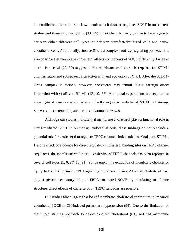
105
the conflicting observations of how membrane cholesterol regulates SOCE in our current
studies and those of other groups (13, 55) is not clear, but may be due to heterogeneity
between either different cell types or between transfected/cultured cells and native
endothelial cells. Additionally, since SOCE is a complex muti-step signaling pathway, it is
also possible that membrane cholesterol affects components of SOCE differently. Galan et
al and Pani et al (20, 59) suggested that membrane cholesterol is required for STIM1
oligmerization and subsequent interaction with and activation of Orai1. After the STIM1-
Orai1 complex is formed, however, cholesterol may inhibit SOCE through direct
interaction with Orai1 and STIM1 (13, 20, 55). Additional experiments are required to
investigate if membrane cholesterol directly regulates endothelial STIM1 clustering,
STIM1-Orai1 interaction, and Orai1 activation in PAECs.
Although our studies indicate that membrane cholesterol plays a functional role in
Orai1-mediated SOCE in pulmonary endothelial cells, these findings do not preclude a
potential role for cholesterol to regulate TRPC channels independent of Orai1 and STIM1.
Despite a lack of evidence for direct regulatory cholesterol binding sites on TRPC channel
sequences, the membrane cholesterol sensitivity of TRPC channels has been reported in
several cell types (1, 6, 37, 50, 81). For example, the extraction of membrane cholesterol
by cyclodextrins impairs TRPC1 signaling processes (6, 42). Although cholesterol may
play a pivotal regulatory role in TRPC1-mediated SOCE by regulating membrane
structure, direct effects of cholesterol on TRPC functions are possible.
Our studies also suggest that loss of membrane cholesterol contributes to impaired
endothelial SOCE in CH-induced pulmonary hypertension (84). Due to the limitation of
the filipin staining approach to detect oxidized cholesterol (63), reduced membrane

106
cholesterol following CH is possibly the result of impaired cholesterol de novo synthesis
(54), oxidative modification of cholesterol (52) or decreased membrane cholesterol
trafficking. Nguyen et al (54) have explored the mechanism by which hypoxia affects de
novo cholesterol biosynthesis. They reported that hypoxia induces accumulation of
cholesterol biosynthetic intermediates and rapid degradation of HMG-CoA reductase,
which contribute to a reduction of cholesterol production. Given the importance of reactive
oxygen species to the development of CH-induced pulmonary hypertension (19, 26, 27,
41), endothelial dysfunction in this setting may alternatively result from oxidative
modifications to cholesterol that alter membrane lipid domains and interfere with ion
channel function.
Based on our findings that CH attenuates pulmonary endothelial Ca2+ entry by
depleting membrane cholesterol, it may be predicted that targeted depletion of PAEC
cholesterol would minimally or adversely affect the development of PH in this setting. In
apparent contrast to this prediction, HMG-CoA reductase inhibitors (statins) have
demonstrated beneficial effects in attenuating the development of CH-induced pulmonary
hypertension in animal models (23, 36, 49). However, the mechanisms by which statins
mediate this protective influence are thought to be independent of their cholesterol
lowering properties (10, 46, 70, 78, 80). These include inhibition of RhoA that can not only
decrease vascular smooth muscle contractility (76), proliferation and migration (79), but
also increase eNOS expression and activity (70). Additional evidence suggests that statins
increase NO production by inhibiting expression and activity of NADPH oxidase subunits,
and thus production of endothelial reactive oxygen species (44, 78, 80), rather than through
effects on endothelial cholesterol content. Such non-specific effects of statins likely result
107
from decreased production of intermediates of the mevalonate pathway that are necessary
for posttranslational modification of many signaling proteins (7). In contrast to these
animal studies, however, clinical trials of statin therapy in patients with pulmonary arterial
hypertension have been largely inconclusive (66).
In conclusion, the current study demonstrates a novel effect of membrane
cholesterol to regulate pulmonary endothelial SOCE by facilitating the interaction of
STIM1 and Orai1 (Figure 8). These observations advance our basic understanding of how
membrane cholesterol regulates endothelial [Ca2+]i homeostasis, and have potentially
broader implications for cholesterol-dependent regulation of a wide range of vasoactive
and mitogenic pathways. These findings may additionally provide a mechanistic basis to
explain the CH-induced diminution of endothelial Ca2+ influx and associated endothelial
dysfunction that is central to the pathogenesis of pulmonary hypertension. Challenges of
future studies are to identify the potential contribution of reactive oxygen species to
decreased endothelial cholesterol following CH, either through direct cholesterol oxidation
or impaired sterol trafficking, and the contribution of these responses to increased
vasoconstrictor reactivity, arterial smooth muscle mitogenesis, and the development of
pulmonary hypertension.
Acknowledgements
We thank Minerva Murphy, Lindsay Herbert, and Tamara Howard for technical
assistance. This work was supported by NIH grants R01 HL95640 (B.R. Walker), R01
HL132883 and R01 HL088192 (T.C. Resta), and American Heart Association grant
15GRNT21080001 (B.R. Walker).
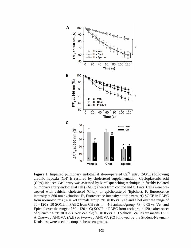
108
Figure 1. Impaired pulmonary endothelial store-operated Ca2+ entry (SOCE) following
chronic hypoxia (CH) is restored by cholesterol supplementation. Cyclopiazonic acid
(CPA)-induced Ca2+ entry was assessed by Mn2+ quenching technique in freshly isolated
pulmonary artery endothelial cell (PAEC) sheets from control and CH rats. Cells were pre-
treated with vehicle, cholesterol (Chol), or epicholesterol (Epichol). F, fluorescence
intensity at 360 nm excitation; F0, fluorescence intensity at time zero. A) SOCE in PAEC
from normoxic rats.; n = 5-8 animals/group. *P <0.05 vs. Veh and Chol over the range of
30 - 120 s. B) SOCE in PAEC from CH rats. n = 4-8 animals/group. *P <0.05 vs. Veh and
Epichol over the range of 60 - 120 s. C) SOCE in PAEC from each group 120 s after onset
of quenching. *P <0.05 vs. Nor Vehicle; #P <0.05 vs. CH Vehicle. Values are means ± SE.
A One-way ANOVA (A,B) or two-way ANOVA (C) followed by the Student-Newman-
Keuls test were used to compare between groups.

109
Figure 2. Orai1 mediates pulmonary endothelial SOCE. A) AnCoA4 inhibits SOCE but
not depolarization-induced Ca2+ entry in freshly dispersed PAEC from normoxic rats. B)
AnCoA4 attenuates SOCE but not OAG (diacylglycerol analog)-induced Ca2+ entry in
cultured pulmonary microvascular endothelial cells (PMVEC). An unpaired t-test was
used to compare between groups at 120 s after onset of quenching. *P < 0.05 vs. Veh CPA.
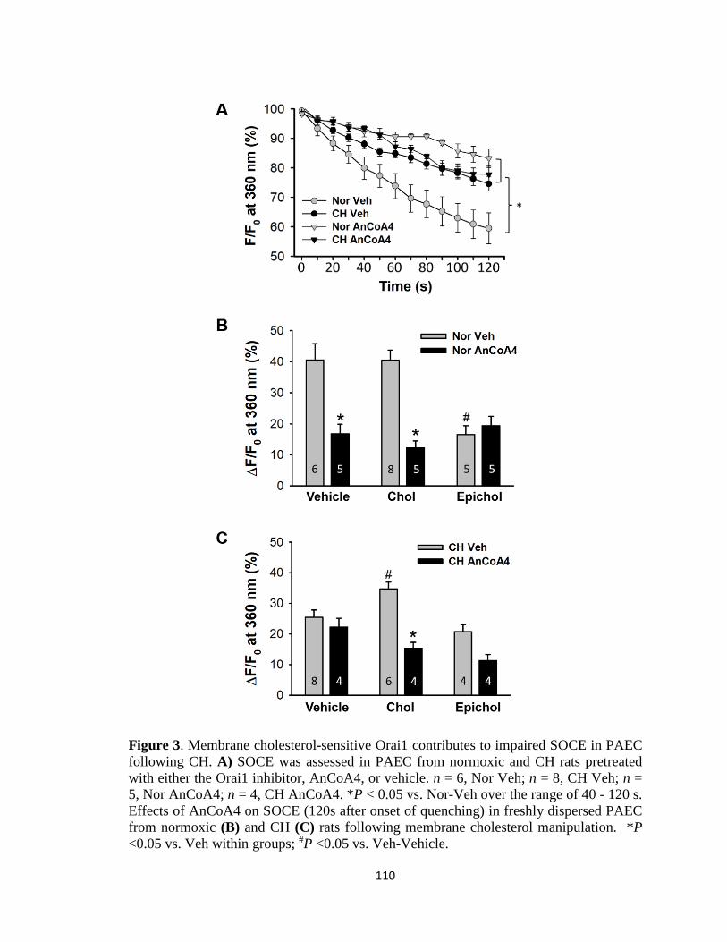
110
Figure 3. Membrane cholesterol-sensitive Orai1 contributes to impaired SOCE in PAEC
following CH. A) SOCE was assessed in PAEC from normoxic and CH rats pretreated
with either the Orai1 inhibitor, AnCoA4, or vehicle. n = 6, Nor Veh; n = 8, CH Veh; n =
5, Nor AnCoA4; n = 4, CH AnCoA4. *P < 0.05 vs. Nor-Veh over the range of 40 - 120 s.
Effects of AnCoA4 on SOCE (120s after onset of quenching) in freshly dispersed PAEC
from normoxic (B) and CH (C) rats following membrane cholesterol manipulation. *P
<0.05 vs. Veh within groups; #P <0.05 vs. Veh-Vehicle.

111
Figure 4. Epicholesterol reduces endogenous membrane cholesterol content of cultured
PMVEC. A) Representative images of filipin fluorescence from PMVEC treated with
vehicle, MβCD, cholesterol, or epicholesterol. B) Mean filipin fluorescence (arbitrary
units; A.U.) in PMVEC from each group. Data were compared by the Kruskal-Wallis H
test and Dunn’s multiple comparison test. n = 5 (filipin fluorescence of 4-16 cells were
measured per field of interest and averaged for an n=1). *P < 0.05 vs. Vehicle; #P <0.05
vs. MβCD and Epichol. C) Amplex Red cholesterol assay to detect cholesterol
concentration in PMVEC. Data were compared by one-way ANOVA followed by the
Student-Newman-Keuls test. n = 3. *P < 0.05 vs. Vehicle; #P <0.05 vs. MβCD and Epichol.

112
Figure 5. Epicholesterol substitution reduces SOCE in PMVEC pretreated with
cholesterol. n = 5, Chol; n = 5, Epichol; n = 3, Time Control. *P < 0.05 vs. Chol over the
range of 50 - 120 s.
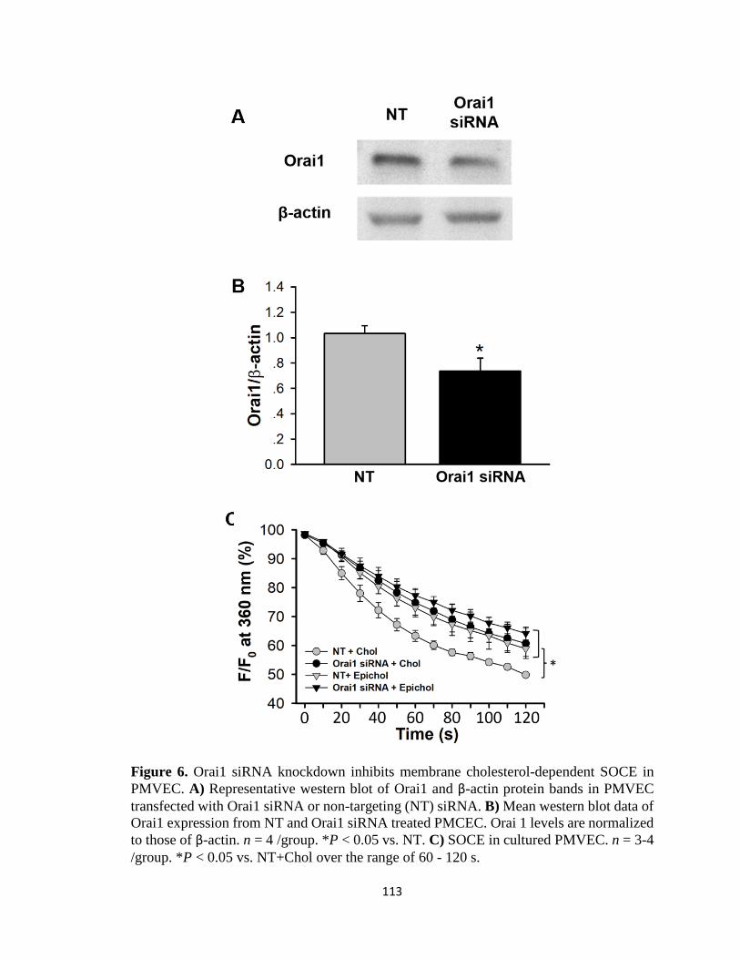
113
Figure 6. Orai1 siRNA knockdown inhibits membrane cholesterol-dependent SOCE in
PMVEC. A) Representative western blot of Orai1 and β-actin protein bands in PMVEC
transfected with Orai1 siRNA or non-targeting (NT) siRNA. B) Mean western blot data of
Orai1 expression from NT and Orai1 siRNA treated PMCEC. Orai 1 levels are normalized
to those of β-actin. n = 4 /group. *P < 0.05 vs. NT. C) SOCE in cultured PMVEC. n = 3-4
/group. *P < 0.05 vs. NT+Chol over the range of 60 - 120 s.

114
Figure 7. Epicholesterol substitution reduces STIM1-Orai1 interaction in PMVEC as
assessed by in situ proximity ligation assay. A) Representative images of STIM1-Orai1
interactions (red puncta) in response to CPA in cultured PMVEC with manipulated
membrane cholesterol. Nuclei area labeled with SYTOX (green). B) Summarized data of
STIM1-Orai1 interaction expressed as average number of puncta/cell in cultured PMVEC.
Cholesterol supplementation increased the CPA-induced STIM1-Orai1 interaction,
whereas epicholesterol substitution significantly inhibited this response. Groups were
compared by two-way ANOVA followed by multiple comparisons testing using the
Student-Newman-Keuls test. n = 5/group. *P <0.05 vs. Chol Veh; #P <0.05 vs. Chol CPA.

115
Figure 8. Proposed mechanism by which reduced cell membrane cholesterol (Chol)
following CH attenuates SOCE in PAECs. Cholesterol facilitates the interaction of STIM1
and Orai1 in response to depletion of intracellular Ca2+ stores, e.g. by receptor (R)-
mediated activation of inositol trisphosphate receptors (IP3R), or inhibition of the
sarco/endoplasmic reticulum Ca2+ -ATPase (SERCA). CH inhibits this mechanism by
depletion of cell membrane cholesterol. Endoplasmic reticulum (ER), agonist (A).

116
References
1. Alkhani H, Ase AR, Grant R, O Donnell D, Groschner K, Séguéla P.
Contribution of TRPC3 to store-operated calcium entry and inflammatory
transductions in primary nociceptors. Mol Pain 10: 43, 2014.
2. Anderson RGW. the Caveolae Membrane System. Annu Rev Biochem 67: 199–
225, 1998.
3. Andrews AM, Jaron D, Buerk DG, Barbee KA. Shear stress-induced NO
production is dependent on ATP autocrine signaling and capacitative calcium
entry. Cell Mol Bioeng 7: 510–520, 2015.
4. Barberà JA, Peinado VI, Santos S. Pulmonary hypertension in chronic
obstructive pulmonary disease. Eur Respir J 21: 892–905, 2003.
5. Barrantes FJ. Cholesterol effects on nicotinic acetylcholine receptor: Cellular
aspects. Subcell Biochem 51: 467–487, 2010.
6. Bergdahl A, Gomez MF, Dreja K, Xu SZ, Adner M, Beech DJ, Broman J,
Hellstrand P, Swärd K. Cholesterol depletion impairs vascular reactivity to
endothelin-1 by reducing store-operated Ca2+ entry dependent on TRPC1. Circ
Res 93: 839–847, 2003.
7. Bonetti PO, Lerman LO, Napoli C, Lerman A. Statin effects beyond lipid
lowering--are they clinically relevant? Eur Heart J 24: 225–48, 2003.
8. Brian W. Christman, M.D., Charles D. McPherson, M.D., John H. Newman,
M.D., Gayle A. King, M.S., Gordon R. Bernard, M.D., Bertron M. Groves,
M.D., and James E. Loyd MD. An imbalance between the excretion of
thromboxane and prostacyclin metabolites in pulmonary hypertension. N Engl J

117
Med 327: 70–75, 1992.
9. Busse R, Mülsch A. Calcium-dependent nitric oxide synthesis in endothelial
cytosol is mediated by calmodulin. FEBS Lett 265: 133–136, 1990.
10. Chen IC, Tan MS, Wu BN, Chai CY, Yeh JL, Chou SH, Chen IJ, Dai ZK.
Statins ameliorate pulmonary hypertension secondary to left ventricular
dysfunction through the Rho-kinase pathway and NADPH oxidase. Pediatr
Pulmonol 52: 443–457, 2017.
11. Christian EA, Haynes MP, Phillips MC, George H. Use of cyclodextrins for
manipulating cellular cholesterol content. J Lipid Res 38: 2264–2272, 1997.
12. Cioffi DL, Wu S, Chen H, Alexeyev M, St. Croix CM, Pitt BR, Uhlig S,
Stevens T. Orai1 determines calcium selectivity of an endogenous trpc
heterotetramer channel. Circ Res 110: 1435–1444, 2012.
13. Derler I, Jardin I, Stathopulos PB, Muik M, Fahrner M, Zayats V, Pandey
SK, Poteser M, Lackner B, Absolonova M, Schindl R, Groschner K, Ettrich
R, Ikura M, Romanin C. Cholesterol modulates Orai1 channel function. Sci
Signal 9: 1–11, 2016.
14. Dionisio N, Galán C, Jardín I, Salido GM, Rosado JA. Lipid rafts are essential
for the regulation of SOCE by plasma membrane resident STIM1 in human
platelets. Biochim Biophys Acta - Mol Cell Res 1813: 431–437, 2011.
15. Elizabeth D. Covington, Minnie M. Wu and RSL. Essential role for the CRAC
activation domain in store-dependent oligomerization of STIM1. Mol Biol Cell 21:
1897–1907, 2010.
16. Fernandez RA, Wan J, Song S, Smith KA, Gu Y, Tauseef M, Tang H, Makino

118
A, Mehta D, Yuan JX-J. Upregulated expression of STIM2, TRPC6, and Orai2
contributes to the transition of pulmonary arterial smooth muscle cells from a
contractile to proliferative phenotype. Am J Physiol - Cell Physiol 308: C581–
C593, 2015.
17. Förstermann U, Pollock JS, Schmidt HH, Heller M, Murad F. Calmodulin-
dependent endothelium-derived relaxing factor/nitric oxide synthase activity is
present in the particulate and cytosolic fractions of bovine aortic endothelial cells.
Proc Natl Acad Sci 88: 1788–1792, 1991.
18. Freichel M, Suh SH, Pfeifer a, Schweig U, Trost C, Weissgerber P, Biel M,
Philipp S, Freise D, Droogmans G, Hofmann F, Flockerzi V, Nilius B. Lack of
an endothelial store-operated Ca2+ current impairs agonist-dependent
vasorelaxation in TRP4-/- mice. Nat Cell Biol 3: 121–127, 2001.
19. Fresquet F, Pourageaud F, Leblais V, Brandes RP, Savineau JP, Marthan R,
Muller B. Role of reactive oxygen species and gp91phox in endothelial
dysfunction of pulmonary arteries induced by chronic hypoxia. Br J Pharmacol
148: 714–723, 2006.
20. Galan C, Woodard GE, Dionisio N, Salido GM, Rosado JA. Lipid rafts
modulate the activation but not the maintenance of store-operated Ca2+ entry.
Biochim Biophys Acta - Mol Cell Res 1803: 1083–1093, 2010.
21. Giaid A SD. Reduced expression of endothelial nitric oxide synthase in the lungs
of patients with pulmonary hypertension. Nitric Oxide 333: 214–221, 2008.
22. Gimpl G, Burger K, Fahrenholz F. Cholesterol as modulator of receptor
function. Biochemistry 36: 10959–10974, 1997.

119
23. Girgis RE, Li D, Zhan X, Garcia JGN, Tuder RM, Hassoun PM, Johns RA.
Attenuation of chronic hypoxic pulmonary hypertension by simvastatin. Am J
Physiol Heart Circ Physiol 285: H938-45, 2003.
24. Gwozdz T, Dutko-Gwozdz J, Schafer C, Bolotina VM. Overexpression of Orai1
and STIM1 proteins alters regulation of store-operated Ca2+ entry by endogenous
mediators. J Biol Chem 287: 22865–22872, 2012.
25. Hinton JM, Langton PD. Inhibition of EDHF by two new combinations of K+ -
channel inhibitors in rat isolated mesenteric arteries. Br J Pharmacol 138: 1031–
1035, 2003.
26. Jernigan NL, Walker BR, Resta TC. Endothelium-derived reactive oxygen
species and endothelin-1 attenuate NO-dependent pulmonary vasodilation
following chronic hypoxia. Am J Physiol Lung Cell Mol Physiol 287: L801-8,
2004.
27. Jernigan NL, Walker BR, Resta TC. Reactive oxygen species mediate RhoA /
Rho kinase-induced Ca 2 ϩ sensitization in pulmonary vascular smooth muscle
following chronic hypoxia. Am J Physiol - Lung Cell Mol Physiol 1: 515–529,
2008.
28. Kassan M, Zhang W, Aissa KA, Stolwijk J, Trebak M, Matrougui K.
Differential role for stromal interacting molecule 1 in the regulation of vascular
function. Pflugers Arch Eur J Physiol 467: 1195–1202, 2014.
29. Kato N, Nakanishi M, Hirashima N. Cholesterol depletion inhibits store-
operated calcium currents and exocytotic membrane fusion in RBL-2H3 cells.
Biochemistry 42: 11808–11814, 2003.

120
30. Kito H, Yamamura H, Suzuki Y, Yamamura H, Ohya S, Asai K, Imaizumi Y.
Regulation of store-operated Ca2+ entry activity by cell cycle dependent up-
regulation of Orai2 in brain capillary endothelial cells. Biochem Biophys Res
Commun 459: 457–462, 2015.
31. Kwakye GF, Li D, Kabobel OA, Bowman AB. Cellular fura-2 Manganese
Extraction Assay (CFMEA). Curr Protoc Toxicol 2651: 1–26, 2011.
32. Kwiatek AM, Minshall RD, Cool DR, Skidgel RA, Malik AB. Caveolin-1
regulates store-operated Ca 2+ influx by binding of its scaffolding domain to
transient receptor potential channel-1 in endothelial cells. Mol Pharmacol 70:
1174–1183, 2006.
33. Kwon J, An H, Sa M, Won J, Shin JI, Lee CJ. Orai1 and Orai3 in combination
with Stim1 mediate the majority of store-operated calcium entry in astrocytes. Exp
Neurobiol 26: 42, 2017.
34. Kwong TC, Liu X, Hwei LO, Ambudkar IS. Functional requirement for Orai1 in
store-operated TRPC1-STIM1 channels. J Biol Chem 283: 12935–12940, 2008.
35. Kwong Tai Chen, Hwei Ling Ong, Xibao Liu and ISA. Contribution of TRPC1
and Orai1 to Ca2+ entry activated by store depletion. 704: 731–747, 2011.
36. Laufs U, Fata V La, Plutzky J, Liao JK. Upregulation of endothelial nitric oxide
synthase by HMG-CoA reductase inhibitors. Circulation 97: 1129–1135, 1998.
37. Lei L, Lu S, Wang Y, Kim T, Mehta D, Wang Y. The role of mechanical tension
on lipid raft dependent PDGF-induced TRPC6 activation. Biomaterials 35: 2868–
2877, 2014.
38. Levitan I, Singh DK, Rosenhouse-Dantsker A. Cholesterol binding to ion

121
channels. Front Physiol 5 FEB: 1–14, 2014.
39. Liou J, Kim ML, Heo W Do, Jones JT, Myers JW, James E, Jr F, Meyer T.
STIM Is a Ca2+ Sensor Essential for Ca2+-Store-DepletionTriggered Ca2+ Influx.
15: 1235–1241, 2011.
40. Liu X, Singh BB, Ambudkar IS. TRPC1 is required for functional store-operated
Ca2+ channels: Role of acidic amino acid residues in the S5-S6 region. J Biol
Chem 278: 11337–11343, 2003.
41. Liu Z-Q, Shan H-Y. Cholesterol, not polyunsaturated fatty acids, is target
molecule in oxidation induced by reactive oxygen species in membrane of human
erythrocytes. Cell Biochem Biophys 45: 185–193, 2006.
42. Lockwich TP, Liu X, Singh BB, Jadlowiec J, Weiland S, Ambudkar IS.
Assembly of Trp1 in a signaling complex associated with caveolin- scaffolding
lipid raft domains. J Biol Chem 275: 11934–11942, 2000.
43. Majed BH, Khalil RA. Molecular mechanisms regulating the vascular
prostacyclin pathways and their adaptation during pregnancy and in the newborn.
Pharmacol Rev 64: 540–82, 2012.
44. Margaritis M, Sanna F, Antoniades C. Statins and oxidative stress in the
cardiovascular system. Curr Pharm Des Sep 26: doi:
10.2174/1381612823666170926130338. Epub ahead, 2017.
45. Marrelli SP, Eckmann MS, Hunte MS. Role of endothelial intermediate
conductance KCa channels in cerebral EDHF-mediated dilations. Am J Physiol
Hear Circ Physiol 285: H1590-9, 2003.
46. Mason RP, Walter MF, Jacob RF. Effects of HMG-CoA reductase inhibitors on

122
endothelial function: role of microdomains and oxidative stress. Circulation 109:
II34-41, 2004.
47. Michiels C, Arnould T. Stimulation of prostaglandin synthesis by human
endothelial cells exposed to hypoxia. Am J Physiol 264: C866-74, 1993.
48. Moccia F, Zuccolo E, Soda T, Tanzi F, Guerra G, Mapelli L, Lodola F,
D’Angelo E. Stim and Orai proteins in neuronal Ca(2+) signaling and excitability.
Front Cell Neurosci 9: 153, 2015.
49. Murata T, Kinoshita K, Hori M, Kuwahara M, Tsubone H, Karaki H, Ozaki
H. Statin protects endothelial nitric oxide synthase activity in hypoxia-induced
pulmonary hypertension. Arterioscler Thromb Vasc Biol 25: 2335–2342, 2005.
50. Murata T, Lin MI, Stan R V., Bauer PM, Yu J, Sessa WC. Genetic evidence
supporting caveolae microdomain regulation of calcium entry in endothelial cells.
J Biol Chem 282: 16631–16643, 2007.
51. Murata T, Sato K, Hori M, Ozaki H, Karaki H. Decreased endothelial nitric-
oxide synthase (eNOS) activity resulting from abnormal interaction between eNOS
and its regulatory proteins in hypoxia-induced pulmonary hypertension. J Biol
Chem 277: 44085–44092, 2002.
52. Murphy RC, Johnson KM. Cholesterol, reactive oxygen species, and the
formation of biologically active mediators. J Biol Chem 283: 15521–15525, 2008.
53. Nakane M, Mitchell J, Förstermann U, Murad F. Phosphorylation by calcium
calmodulin-dependent protein kinase II and protein kinase C modulates the activity
of nitric oxide synthase. Biochem Biophys Res Commun 180: 1396–1402, 1991.
54. Nguyen AD, McDonald JG, Bruick RK, DeBose-Boyd RA. Hypoxia stimulates
123
degradation of 3-hydroxy-3-methylglutaryl-coenzyme A reductase through
accumulation of lanosterol and hypoxia-inducible factor-mediated induction of
insigs. J Biol Chem 282: 27436–27446, 2007.
55. Pacheco J, Domingu L, Bohórquez-herná A, Asanov A, Vaca L. OPEN A
cholesterol-binding domain in STIM1 modulates STIM1-Orai1 physical and
functional interactions. Sci Rep 6: 1–16, 2016.
56. Paffett ML, Naik JS, Resta TC, Walker BR. Reduced store-operated Ca2+ entry
in pulmonary endothelial cells from chronically hypoxic rats. Am J Physiol Lung
Cell Mol Physiol 293: L1135-42, 2007.
57. Paffett ML, Naik JS, Riddle MA, Menicucci SD, Gonzales AJ, Resta TC,
Walker BR. Altered membrane lipid domains limit pulmonary endothelial
calcium entry following chronic hypoxia. Am J Physiol Hear Circ Physiol 301:
1331–1340, 2011.
58. Paffett ML, Riddle MA, Kanagy NL, Resta TC, Walker BR. Altered protein
kinase C regulation of pulmonary endothelial store- and receptor-operated Ca2+
entry after chronic hypoxia. J Pharmacol Exp Ther 334: 753–760, 2010.
59. Pani B, Hwei LO, Liu X, Rauser K, Ambudkar IS, Singh BB. Lipid rafts
determine clustering of STIM1 in endoplasmic reticulum-plasma membrane
junctions and regulation of store-operated Ca2+ entry (SOCE). J Biol Chem 283:
17333–17340, 2008.
60. Parekh a. B, Putney Jr JW. Store-operated calcium channels. Physiol Rev 85:
757–810, 2005.
61. Parpal S, Karlsson M, Thorn H, Strålfors P. Cholesterol depletion disrupts
124
caveolae and insulin receptor signaling for metabolic control via insulin receptor
substrate-1, but not for mitogen-activated protein kinase control. J Biol Chem 276:
9670–9678, 2001.
62. Pollock JS, Förstermann U, Mitchell JA, Warner TD, Schmidt HH, Nakane
M, Murad F. Purification and characterization of particulate endothelium-derived
relaxing factor synthase from cultured and native bovine aortic endothelial cells.
Proc Natl Acad Sci U S A 88: 10480–4, 1991.
63. Pörn MI, Slotte JP. Localization of cholesterol in sphingomyelinase-treated
fibroblasts. Biochem J 308 (Pt 1): 269–74, 1995.
64. Putney JW, Broad LM, Braun FJ, Lievremont JP, Bird GS. Mechanisms of
capacitative calcium entry. J Cell Sci 114: 2223–2229, 2001.
65. Romanenko VG, Rothblat GH, Levitan I. Modulation of endothelial inward-
rectifier K+ current by optical isomers of cholesterol. Biophys J 83: 3211–3222,
2002.
66. Rysz-Gorzynska M, Gluba-Brzozka A, Sahebkar A, Serban M-C, Mikhailidis
DP, Ursoniu S, Toth PP, Bittner V, Watts GF, Lip GYH, Rysz J, Catapano
AL, Banach M. Efficacy of statin therapy in pulmonary arterial hypertension: a
systematic review and meta-analysis. Sci Rep 6: 30060, 2016.
67. Sadaghiani AM, Lee SM, Odegaard JI, Leveson-Gower DB, McPherson OM,
Novick P, Kim MR, Koehler AN, Negrin R, Dolmetsch RE, Park CY.
Identification of Orai1 channel inhibitors by using minimal functional domains to
screen small molecule microarrays. Chem Biol 21: 1278–1292, 2014.
68. Saliba Y, Keck M, Marchand A, Atassi F, Ouillé A, Cazorla O, Trebak M,
125
Pavoine C, Lacampagne A, Hulot JS, Farès N, Fauconnier J, Lompré AM.
Emergence of Orai3 activity during cardiac hypertrophy. Cardiovasc Res 105:
248–259, 2015.
69. Seid M, Macneil S, Tomlinson S. Calcium, calmodulin, and the production of
prostacyclin by cultured vascular endothelial cells. Biosci Rep 3: 1007–1015,
1983.
70. Shiga N, Hirano K, Hirano M, Nishimura J, Nawata H, Kanaide H. Long-term
inhibition of RhoA attenuates vascular contractility by enhancing endothelial NO
production in an intact rabbit mesenteric artery. Circ Res 96: 1014–1021, 2005.
71. Shimoda LA, Sham JSK, Sylvester JT. Altered pulmonary vasoreactivity in the
chronically hypoxic lung. Physiol Res 49: 549–560, 2000.
72. Soboloff J, Rothberg BS, Madesh M, Gill DL. STIM proteins: dynamic calcium
signal transducers. Nat Publ Gr 13: 549–565, 2012.
73. Sooksawate T, Simmonds MA. Influence of membrane cholesterol on
modulation of the GABA A receptor by neuroactive steroids and other
potentiators. Br J Pharmacol 134: 1303–1311, 2001.
74. Sundivakkam PC, Freichel M, Singh V, Yuan JP, Vogel SM, Flockerzi V,
Malik AB, Tiruppathi C. The Ca2+ sensor stromal interaction molecule 1
(STIM1) is necessary and sufficient for the store-operated Ca2+ entry function of
transient receptor potential canonical (TRPC) 1 and 4 channels in endothelial cells.
Mol Pharmacol 81: 510–526, 2012.
75. Tuder RM, Cool CD, Geraci MW, Wang J, Abman SH, Wright L, Badesch D,
Voelkel NF. Prostacyclin synthase expression is decreased in lungs from patients
126
with severe pulmonary hypertension. Am J Respir Crit Care Med 159: 1925–1932,
1999.
76. Uehata M, Ishizaki T, Satoh H, Ono T, Kawahara T, Morishita T, Tamakawa
H, Yamagami K, Inui J, Maekawa M, Narumiya S. Calcium sensitization of
smooth muscle mediated by a Rho-associated protein kinase in hypertension.
Nature 389: 990–4, 1997.
77. Vaeth M, Yang J, Yamashita M, Zee I, Eckstein M, Knosp C, Kaufmann U,
Karoly Jani P, Lacruz RS, Flockerzi V, Kacskovics I, Prakriya M, Feske S.
ORAI2 modulates store-operated calcium entry and T cell-mediated immunity. Nat
Commun 8: 14714, 2017.
78. Wagner AH, Köhler T, Rückschloss U, Just I, Hecker M. Improvement of
nitric oxide-dependent vasodilation by HMG-CoA reductase inhibitors through
attenuation of endothelial superoxide anion formation. Arterioscler Thromb Vasc
Biol nicht bekannt 20: 61–69, 2000.
79. Walker J, Undem C, Yun X, Lade J, Jiang H, Shimoda LA. Role of Rho kinase
and Na + /H + exchange in hypoxia‐induced pulmonary arterial smooth muscle cell
proliferation and migration. Physiol Rep 4: e12702, 2016.
80. Wassmann S, Laufs U, Muller K, Konkol C, Ahlbory K, Baumer AT, Linz W,
Bohm M, Nickenig G. Cellular Antioxidant Effects of Atorvastatin In Vitro and
In Vivo. Arter Thromb Vasc Biol 22: 300–305, 2002.
81. Weerth SH, Holtzclaw LA, Russell JT. Signaling proteins in raft-like
microdomains are essential for Ca2+ wave propagation in glial cells. Cell Calcium
41: 155–167, 2007.

127
82. Wu X, Babnigg G, Villereal MLCN-C. Functinal significance of human Trp1
and Trp3 in store-operated Ca2+ entry in HEK-293 cells. Am J Physiol Cell
Physiol 278: 526–536, 2000.
83. Xu X, London E. The effect of sterol structure on membrane lipid domains
reveals how cholesterol can induce lipid domain formation. Biochemistry 39: 843–
849, 2000.
84. Zhang B, Naik XJS, Jernigan NL, Walker BR, Resta TC. Reduced membrane
cholesterol limits pulmonary endothelial Ca 2+ entry after chronic hypoxia. Am J
Physiol - Hear Circ Physiol 312: 1176–1184, 2017.
85. Zhou MH, Zheng H, Si H, Jin Y, Peng JM, He L, Zhou Y, Muñoz-Garay C,
Zawieja DC, Kuo L, Peng X, Zhang SL. Stromal interaction molecule 1
(STIM1) and Orai1 mediate histamine-evoked calcium entry and nuclear factor of
activated T-cells (NFAT) signaling in human umbilical vein endothelial cells. J
Biol Chem 289: 29446–29456, 2014.
86. Zhou Y, Wang X, Wang X, Loktionova NA, Cai X, Nwokonko RM, Vrana E,
Wang Y, Rothberg BS, Gill DL. STIM1 dimers undergo unimolecular coupling
to activate Orai1 channels. Nat Commun 6: 8395, 2015.
87. Zitt C, Zobel A, Obukhov AG, Harteneck C, Kalkbrenner F, Lückhoff A,
Schultz G. Cloning and functional expression of a human Ca2+-permeable cation
channel activated by calcium store depletion. Neuron 16: 1189–1196, 1996.

128
CHAPTER 4: DISCUSSION
The goal of this dissertation was to investigate the role of membrane cholesterol in
pulmonary endothelial Ca2+ entry in CH-induced pulmonary hypertensive rats. In the
current studies we tested the hypotheses that 1) membrane cholesterol facilitates SOCE
and depolarization-induced pulmonary endothelial Ca2+ entry; 2) membrane cholesterol
regulates Orai1-mediated SOCE through facilitating STIM1-Orai1 interaction, and 3)
reduced endothelial Ca2+ influx following CH is due to a loss of membrane cholesterol.
We identified the role of membrane cholesterol in regulating agonist-induced Ca2+ entry
and its components in PAECs following CH through measurement of [Ca2+]i using a
ratiometric fluorescent Ca2+ indicator in live cells with cholesterol supplementation or
substitution of cholesterol with its epimer, epicholesterol. The rationale behind these
studies emerges from the observation in CH-induced PH that dysregulation of
endothelium-derived mediators contributes to elevated vascular resistance in the
pulmonary circulation. The production of many endothelium-derived vasodilators and anti-
mitogenic substances like NO (18, 39, 89, 102) and prostacyclin (60, 144) are largely a
function of endothelial [Ca2+]i. Therefore, it is possible that reduced endothelial [Ca2+]i
limits the production of NO, prostacyclin, and other endothelium-derived hyperpolarizing
factors. Our laboratory previously reported that both basal [Ca2+]i and agonist-induced Ca2+
influx are lower in PAECs from CH rats compared to their normoxic controls (95). Both
endothelial SOCE and depolarization-induced Ca2+ entry through T-type VGCCs, two
major components of ATP-induced Ca2+ influx, are also reduced following CH exposure
(94, 96). Interestingly, membrane cholesterol levels along with the agonist-induced Ca2+
entry are decreased in PAECs of chronic hypoxic rats. This impaired Ca2+ influx is restored
129
simply by either membrane cholesterol supplementation or administration of caveolin-1
scaffolding domain peptide (95). Because membrane cholesterol and caveolin-1 are major
structural components of caveolae, the diminished Ca2+ influx could be the result of
decreased caveolae after CH. However, the number of caveolae are not altered in PAECs
from CH rats compared to controls (95). These observations suggest a role of membrane
cholesterol in mediating endothelial Ca2+ influx.
The major findings from the current studies are that substitution of endogenous
membrane cholesterol with epicholesterol attenuates ATP-induced Ca2+ entry, SOCE and
depolarization-induced Ca2+ entry in PAEC. In addition, decreased endothelial SOCE and
depolarization-induced Ca2+ entry following CH are largely restored by cholesterol
supplementation. Importantly, neither cholesterol supplementation nor epicholesterol
substitution alters endothelial caveolar number. In addition, membrane cholesterol
regulates Orai1-mediated SOCE in both native and cultured cells. Epicholesterol
substitution greatly reduces SOCE and STIM1-Orai1 interaction in cultured PMVECs.
These data suggest that membrane cholesterol directly regulates agonist-induced Ca2+ entry
and its components. These observations also demonstrate that altered membrane
cholesterol homeostasis may contribute to impaired endothelial Ca2+ entry following CH.
Effect of hypoxia on pulmonary endothelial [Ca2+]i
The mechanisms by which hypoxia affects endothelial [Ca2+]i are not well-
investigated. Studies investigating the effect of acute hypoxia on endothelial [Ca2+]i in vitro
show similar results in cells from the systemic and pulmonary vasculatures. In human
umbilical vein endothelial cells (HUVEC) (6, 11), as well as in bovine (53) and porcine
(58) PAECs, acute hypoxia increases endothelial [Ca2+]i. The significant increase of [Ca2+]i
130
in HUVECs is due to energy deficiency and subsequent influx of extracellular Ca2+ (6).
Berna et al (11) later discovered that the Na+/ Ca2+ exchanger is involved in this hypoxia-
induced Ca2+ influx. By using an inhibitor of Na+-glucose cotransport, they proposed that
energy deficiency activates glycolysis and subsequent activation of Na+-glucose
cotransport, which leads to a series of actions including Na+ influx, Na+/ Ca2+ exchanger
activation, and Ca2+ influx. In PAECs, however, although acute hypoxia (5 min) causes a
rapid and transient increase in [Ca2+]i by releasing Ca2+ from ER stores (53), a longer
duration of hypoxia (15 min) decreases [Ca2+]i likely by causing membrane depolarization
and reducing the electrochemical driving force for Ca2+ entry (124). While it is certainly
possible that hypoxia has a dual-phase effect of releasing Ca2+ from internal stores and
inhibiting Ca2+ influx in bovine PAECs, it should be noted that Hampl et al (53) used
bovine cells of high passage (16-25), whereas Stevens et al (124) used primary bovine
PAECs. The increased passage number of cells may cause changes in ion channel
expression and activity (55, 70). In rat PAECs, for example, although T-type VGCCs are
not found in cultured cells (145), their expression and function are described in freshly
dispersed PAECs (96). This difference may lead to opposite effects of hypoxia on
endothelial Ca2+ entry, as hypoxia-induced membrane depolarization may blunt Ca2+ influx
driven by electrochemical gradient in cultured PAECs while increasing Ca2+ entry through
T-type VGCCs in native PAECs.
In human PAEC, prolonged exposure to hypoxia (3% O2, 60-72h) causes an
increase in basal [Ca2+]i, which is due to enhanced expression of TRPC-encoded SOC
channels and Ca2+ influx (53). However, it is possible that exposure of hypoxia to cultured
cells may not mimic the in vivo effect of CH on PAECs. It is also likely that CH impairs

131
the regulation of pulmonary endothelial [Ca2+]i as PH develops. Murata et al (87) showed
that in 1 week CH-induced pulmonary hypertensive rats, agonist-induced Ca2+ influx in the
pulmonary endothelium is significantly less compared to normoxic control rats.
Consistently, in rats exposed to 4 weeks CH, our previous data demonstrate that, in PAECs,
both basal [Ca2+]i and agonist-induced Ca2+ influx with its major components, SOCE and
depolarization-induced Ca2+ influx through T-type VGCCs, are lower compared to those
of normoxic control animals (94–96). In addition, membrane cholesterol levels are also
reduced in PAEC from rats exposed to CH. The most exciting discovery is that the impaired
agonist-induced Ca2+ influx in PAEC can be restored by both membrane cholesterol
supplementation and by administration of a caveolin-1 (Cav-1) scaffolding domain peptide
(95). This finding suggests that the reduced endothelial agonist-induced Ca2+ influx
following CH is not likely due to reduced expression of the relevant Ca2+ channels. In the
current studies, we found that the diminished SOCE and depolarization-induced Ca2+
influx can also be restored by membrane cholesterol supplementation (Figure 5 and 9:
Chapter 3). These data suggest that cholesterol per se may affect Ca2+ entry in these cells.
By using epicholesterol, we also determined that cholesterol directly modulates endothelial
agonist-induced Ca2+ entry, SOCE and depolarization-induced Ca2+ entry independent of
its structural role in the plasma membrane, which will be discussed later in “Membrane
cholesterol: structural component vs. signaling mediator”.
Although we did not examine the change of [Ca2+]i in early stages of CH-induced
PH, it is possible that hypoxia augments Ca2+ influx and [Ca2+]i in PAEC during the early
stages of hypoxic exposure (135). However, after prolonged hypoxia, endothelial Ca2+
entry may be gradually diminished due to altered cholesterol homeostasis, leading to

132
decreased [Ca2+]i in PAECs. The finding that reduced endothelial Ca2+ entry in pulmonary
arteries following CH was acutely restored by membrane cholesterol supplementation
suggests that CH limits Ca2+ entry in PAECs by reducing membrane cholesterol rather than
by decreasing ion channel expression. The physiological consequences of these changes in
pulmonary endothelial Ca2+ influx and [Ca2+]i will be discussed in detail later in
“Physiological significance of current studies”.
Pulmonary endothelium-mediated vasodilation following CH
The results of our current and previous studies (94) show that agonist-induced Ca2+
entry as well as basal [Ca2+]i are reduced in PAECs following CH. Although we did not
examine the physiological consequences of decreased pulmonary endothelial [Ca2+]i, the
reduction of [Ca2+]i may limit the synthesis and release of vasodilators such as NO, which
would impair endothelium-dependent vasodilation in pulmonary arteries. However,
opposite to this assumption, early studies from our and other laboratory demonstrated that
CH enhances pulmonary artery endothelium-derived nitric oxide (EDNO)-dependent
vasodilation (32, 59, 108, 115). In these studies using isolated perfused lungs, there was an
increase in AVP/histamine/ET-1-induced EDNO-dependent vasodilation in rats exposed
to 4 weeks of CH (32, 108). However, CH did not affect the vasodilation to an exogenous
NO donor, suggesting that the change of agonist-induced vasodilatory responses after CH
was due to reduced bioavailability of NO. Later studies also found that eNOS protein
expression is elevated in CH rats compared to their normoxic controls (107). Thus, the
apparent contradictory observation of impaired pulmonary endothelial [Ca2+]i and
augmented EDNO-dependent vasodilation following CH warrants discussion.

133
Because endothelium-derived NO is the result of multiple signaling cascades
involving Ca2+-dependent and Ca2+-independent pathways, receptor stimulation by
agonists may lead to NO production through different mechanisms. In addition, since the
production of other endothelium-derived vasodilators like prostacyclin and EDHF are also
closely regulated by endothelial [Ca2+]i (56, 79, 117), NO may not fully responsible for
agonist-induced endothelium-dependent vasodilation. The receptors responsible for
AVP/histamine/ET-1 -induced EDNO are mainly V1-receptors (114)/H1 and H2-receptor
(115, 126)/ETB receptors (33). V1-receptors, H1-receptors, and ETB receptors are Gq-
protein coupled receptors (27, 69, 127, 128), the activation of which leads to Ca2+ entry
though similar pathways as ATP-induced Ca2+ influx described in the introduction. V2-
and H2-receptors are Gs-protein coupled receptors that may be involved in NO production
through Ca2+-independent mechanisms (65, 66). The stimulation of Gs-protein coupled
receptors activates adenylyl cyclase and subsequently produces cyclic AMP (cAMP) from
ATP. cAMP may phosphorylate eNOS-Ser (bovine eNOS-Ser1179 or human eNOS-Ser1177)
through AMP-activated protein kinase (22) or protein kinase B (PKB) (149) signaling
pathways. Phosphorylation of eNOS-Ser increases the activity of eNOS by reducing the
relative requirement of Ca2+ for its activation, which is achieved by increasing the rate of
electron flux from the reductase to the oxygenase domain of the protein and reduces (82).
Thus, with multiple signaling pathways leading to vasodilation, the physiological effect of
reduced agonist-induced Ca2+ entry after CH may not be reflected when potential
compensatory mechanisms are present. In fact, H2-receptor -mediated vasodilatory
responses to histamine were augmented in the pulmonary circulation following CH (115).
Besides, since EDHF also mediates histamine-induced vasodilation in the pulmonary

134
circulation (54), it is also possible that this vasodilatory pathway is augmented after CH,
which may explain why non-EDNO-mediated vasodilation by histamine was increased
after CH (108).
Despite the potential compensatory mechanisms by which CH increases agonist-
induced NO production and vasodilation, Ca2+-dependent EDNO production may still be
increased as a result of enhanced expression of eNOS (107) and receptors for agonists (122)
in PAECs from CH rats. Because endothelial ROS production is elevated following CH
(62), enhanced Ca2+-dependent EDNO production may be a compensatory mechanism to
increase NO bioavailability. In addition, based on studies by Murata et al (87), in rats
exposed to 1 week chronic hypoxia, agonist-induced Ca2+ entry and NO production are
significantly reduced in pulmonary endothelium while no change of eNOS expression is
detected. The reduced NO production could be the result of both decreased Ca2+-dependent
eNOS activation and impaired eNOS activity due to tight coupling of eNOS/caveolin-1.
This evidence suggests that endothelial Ca2+ entry and associated NO production are
impaired in the early stage of CH-induced PH, which may activate potential compensatory
mechanisms that increase agonist-induced EDNO production.
Membrane cholesterol: structural component vs. signaling mediator
The role of membrane cholesterol in regulating ion channel function has received
considerable attention during the last decade. It is now well-accepted that cholesterol is a
crucial regulator of various ion channels and plays different roles depending on the type of
ion channel. In general, membrane cholesterol may decrease the number of active channels
or the activity of several K+ channels, voltage-gated Na+ and Ca2+ channels, and volume-

135
regulated anion channels (25, 74, 112, 130). However, other ion channels such as the
nicotinic acetylcholine receptor, GABAA receptors, epithelial Na+ channels and TRPC
channels are inhibited by removal of membrane cholesterol (8–10, 71, 123). These dynamic
roles of membrane cholesterol in regulating ion channel function are possibly achieved
through two mechanisms: 1) indirect regulation by altering the properties of lipid
microdomains; and 2) direct interaction with ion channels. The first mechanism was
proposed by Lundbaek et al (78) that membrane cholesterol may affect ion channels by
modulating bilayer stiffness and hydrophobic interactions between membrane proteins and
the lipid bilayer. The stiffer membrane may lead to increased membrane deformation
energy that affects the conformational change of ion channels when transitioning between
open and closed states (78). A change in the lipid bilayer properties may also affect other
signaling cascades within the lipid raft microdomains, which could indirectly modulate ion
channel function. The concept of direct interaction between cholesterol and ion channels
was first proposed by Marsh and Barrantes in a “lipid belt” model (78). The direct
interaction between membrane cholesterol and ion channels was not demonstrated until the
use of the enantiomer of the sterol, epicholesterol (111) and the identification of a
cholesterol binding motif in several ion channels (30, 113, 120). Considering that direct
and indirect effects of cholesterol on ion channels may exist simultaneously, discrimination
between these two possibilities is challenging but important when exploring the
mechanisms of cholesterol regulation of ion channels.
Many studies have shown that membrane cholesterol is important in regulating Ca2+
channels and that its effects on Ca2+ influx are diverse (14, 17, 104). In HSG and HEK293
cells, cholesterol depletion by MβCD blunts SOCE by suppression of STIM1 clustering

136
and activation of TRPC1 (97). In VSMCs, MβCD also reduces endothelin-1-induced Ca2+
entry through TRPC1 (10). Our previous and current findings showed that, in PAECs,
agonist-induced Ca2+ influx along with membrane cholesterol levels are reduced following
CH (95, 148). This impaired endothelial Ca2+ influx was restored by membrane cholesterol
supplementation (95, 148). Our current studies also show that cholesterol directly
modulates endothelial agonist-induced Ca2+ entry and its major components, SOCE and
depolarization-induced Ca2+ entry. The direct functional effect of cholesterol was examined
by replacing cholesterol with its enantiomer, epicholesterol, that has similar effects on
membrane fluidity and lipid domain formation but differential effects on ion channel
function. We found that substitution of endogenous membrane cholesterol with its epimer
attenuates ATP-induced Ca2+ entry, SOCE and depolarization-induced Ca2+ entry in PAEC
(Figure 4, 5 and 9: Chapter 3). In addition, decreased endothelial SOCE and
depolarization-induced Ca2+ entry following CH are largely restored by cholesterol
supplementation (Figure 5 and 9: Chapter 3). These data suggest that membrane
cholesterol directly regulates agonist-induced Ca2+ entry and its components and further
demonstrate that impaired endothelial Ca2+ entry following CH is due to altered membrane
cholesterol homeostasis.
It should be noted that the rationale of using epicholesterol in this aim is based on the
assumption that epicholesterol should have very similar effects to cholesterol on the
physical properties of the plasma membrane but interact with Ca2+ channels differently.
Our observation that neither cholesterol supplementation nor epicholesterol substitution
alters endothelial caveolar number (Figure 2: Chapter 3) support this rationale. However,
although other studies also showed that cholesterol and epicholesterol have similar effects

137
on membrane fluidity (46) and lipid domain formation (146), these two sterols may still
have differential effects on the properties of plasma membrane (28, 29, 84). Parallel studies
using the other enantiomer, ent-cholesterol that differs from cholesterol in the
configuration of each of the eight stereocenters, might provide additional insight to how
membrane cholesterol directly modulates pulmonary endothelial Ca2+ entry.
Caveolae, caveolin-1, and membrane cholesterol regulation of Ca2+ channels
Caveolae are small (50-100 nm) invaginations of the plasma membrane that are rich
in cholesterol and the structural protein Cav-1. They are one type of lipid raft with dynamic
assemblies of proteins, ion channels, and high levels of cholesterol and glycosphingolipids.
Due to their unique characteristics as a platform for various signaling molecules, caveolae
regulate many signaling pathways, including Ca2+ entry. In fact, Ca2+ signaling
components such as G-protein coupled receptors, PLC, IP3R, and SOC are found in lipid
raft domains/caveolae (15, 43, 86, 99). Both TRPC1 and 4, as SOCs, are not only located
in caveolae, but also interact with Cav-1, which is crucial for SOCE (15, 86).
Cav-1, as one of three isoforms of caveolin, is abundantly expressed in endothelial
cells (116). Cav-1 is localized to caveolae and serves as one of the basic structural
components of caveolae (36, 106). Besides its structural role in caveolar formation, Cav-1
also plays a regulatory role in mediating Ca2+ influx, as suggested by studies using mutated
Cav-1 in cultured cells or Cav-1 knockout mice indicating that Cav-1 is required for SOCE
by interacting with Ca2+ channels (15, 86). Thus, similar to membrane cholesterol, Cav-1
may regulate endothelial Ca2+ influx through its structural modulation of caveolae and its
functional interaction with ion channels. What makes the interpretation of how Cav-1

138
affects endothelial Ca2+ influx more complicated is that Cav-1 may also regulate cholesterol
homeostasis via cholesterol influx/efflux and intracellular trafficking (38, 40, 151). As
reported in our previous studies (95), agonist-induced Ca2+ entry is diminished in PAECs
of CH rats and can be restored to levels of normoxic controls by either cholesterol
supplementation or administration of the Cav-1 scaffolding domain peptide, AP-Cav.
Because there is no change of caveolae number in pulmonary endothelium after CH
exposure (95), any structural changes in caveolae may have minimal effects on endothelial
Ca2+ channel function. Based on the findings of our current studies, membrane cholesterol
may directly regulate Ca2+ channels or signaling pathways leading to channel activation.
Because indirect measurements of Cav-1 expression by immunofluorescence labeling
show no obvious changes in Cav-1 levels after CH (95), AP-Cav may restore the
diminished endothelial Ca2+ influx in CH rats by facilitating cholesterol trafficking from
ER lipid droplets to the plasma membrane. This potential mechanism of ion channel
regulation is supported by studies of Toselli et al (130). They found that Cav-1-transfected
NG108-15 cells have increased membrane cholesterol content and significantly reduced
N-type Ca2+ current compared to wild-type control. This effect was mimicked in wild-type
cells by cholesterol supplementation, suggesting that membrane cholesterol, not Cav-1, is
required for N-type Ca2+ channel inhibition.
Nevertheless, with evidence that Cav-1 may interact with large conductance, Ca2+-
activated K+ channels (BK) (136), we cannot exclude the possibility that Cav-1 may also
directly modulate certain Ca2+ channels function in PAEC. Besides, membrane cholesterol
not only regulates Cav-1 expression (52) but also affects Cav-1 trafficking between the
plasma membrane and Golgi apparatus (101, 121). Membrane cholesterol may also help

139
stabilize Cav-1 on the plasma membrane (41). Thus, membrane cholesterol may indirectly
affect endothelial Ca2+ influx through regulation of Cav-1 expression and trafficking. This
regulation of Cav-1 by membrane cholesterol may be observed in PASMC, in which both
SOCE and ROCE, as well as agonist-induced contraction in pulmonary artery rings are
augmented due to increased Cav-1 following CH (63). These Ca2+ entry pathways appear
to be independent of membrane cholesterol as cholesterol supplementation does not
increase these responses. The disruption of caveolae by MβCD, however, inhibits all Ca2+
responses, potentially through reduction of cholesterol-mediated surface expression of
Cav-1. Considering the regulatory effect of membrane cholesterol and Cav-1 on each other,
it is very likely that both components of caveolae are involved in the changes of pulmonary
endothelial Ca2+ homeostasis during CH exposure.
Cholesterol regulation of STIM1, Orai1, and TRPC.
The essential role of membrane cholesterol in regulating SOCE has been shown in
many cell types as membrane cholesterol depletion by MβCD attenuates SOCE (31, 45, 51,
64, 97). In the present studies, we substituted endogenous cholesterol with epicholesterol,
and found that cholesterol is required for Orai1-mediated SOCE in both isolated PAEC and
cultured PMVEC. We also showed that epicholesterol substitution significantly reduced
the STIM1-Orai1 interaction, suggesting that membrane cholesterol regulates endothelial
SOCE by facilitating this interaction that is required for Orai1 activation. Interestingly,
Derler et al (30) and Pacheco et al (93) reported cholesterol-binding sites on Orai1 and
STIM1, respectively, and demonstrated an inhibitory effect of membrane cholesterol on
SOCE in cultured human embryonic kidney 293 (HEK293) cells and rat basophilic

140
leukemia 2H3 cells. Both studies further showed that the cholesterol-Orai1 and cholesterol-
STIM1 interaction was attenuated by mutating the cholesterol-binding site of the target
protein. The reason for the conflicting observations of how membrane cholesterol regulates
SOCE in our current studies and those of other groups (30, 93) is not clear, but may be due
to heterogeneity between either different cell types or between transfected/cultured cells
and native endothelial cells. Additionally, since SOCE is a complex multi-step signaling
pathway, it is also possible that membrane cholesterol affects components of SOCE
differently. Galan et al (45) showed that membrane cholesterol depletion by MβCD
attenuates SOCE and STIM1-Orai1 association in HEK293 cells. However, if stores are
depleted prior to MβCD treatment, SOCE is enhanced while STIM1-Orai1 association
remain unaffected by cholesterol depletion. Pani et al (97) found that MβCD treatment
attenuates store depletion-induced STIM1 clustering and SOCE. Together, these findings
suggest that plasma membrane cholesterol is required for STIM1 oligmerization and
subsequent interaction with and activation of Orai1. Whether the cholesterol regulation of
STIM1 clustering is achieved through maintaining caveolar structure or direct modulation
of trafficking is not clear. However, since the silencing of Cav-1, which reduces the number
of caveolae (26), does not affect STIM puncta formation in response to ER store depletion
(98), it is possible that cholesterol on the plasma membrane or ER membrane directly
affects STIM1 oligmerization. After the STIM1-Orai1 complex is formed, however,
cholesterol may inhibit SOCE through direct interaction with Orai1 and STIM1 (30, 45,
93).
TRPC channels are also activated by stimuli that lead to PIP2 hydrolysis, and are
identified as components of SOCE. Our current findings do not preclude a potential role

141
for cholesterol to regulate TRPC channels independent of Orai1 and STIM1. Despite a lack
of evidence for direct regulatory cholesterol binding sites on TRPC channel sequences, the
membrane cholesterol sensitivity of TRPC channels has been reported in several cell types
(1, 10, 73, 86, 140). For example, the extraction of membrane cholesterol by cyclodextrins
impairs TRPC1 signaling processes (10, 77). In addition, both TRPC1 and TRPC4 are
expressed in murine lung endothelial cells (86). In these cells, Cav-1 regulates the
subcellular distribution of TRPC1 and 4 to a low-density cholesterol-enriched membrane
fraction. Cav-1 has also been shown to modulate surface expression of TRPC1 in cultured
HSG cells (98). In endothelial cells from endothelium-specific Cav-1 knockout mice,
SOCE is significantly blunted, which is restored in cells from Cav-1 reconstructed mice
(86). Considering the regulatory role of Cav-1 on cholesterol homeostasis, although
cholesterol may play a pivotal regulatory role in TRPC1-mediated SOCE by regulating
membrane structure, direct effects of cholesterol on TRPC trafficking and functions are
possible.
Cholesterol may regulate surface expression of many membrane-bound proteins and
ion channels. It has been shown that endocytosis (13, 138), proteins distribution in
membrane microdomains (129), and endosomal mobility (20) are closely associated with
membrane cholesterol levels. In rat atrial myocytes, lowering of membrane cholesterol by
MβCD enhances outward potassium current by recruiting Kv1.5 channels from recycling
endosomes (7). In addition, the role of membrane cholesterol in ion channel surface
expression may vary depending on the channel isoform (44). In our previous and current
studies, CH significantly reduced membrane cholesterol in PAECs (95, 148). It is not clear
if chronic hypoxia changes the surface expression of certain Ca2+ channels through

142
membrane cholesterol-regulated pathways. Current literature does, however, demonstrate
the inhibitory effect of hypoxia on K+ channel trafficking to the plasma membrane (23, 90).
With the possibility that cholesterol modulates trafficking of cation channels, whether the
effect of cholesterol supplementation of restoring diminished Ca2+ entry in PAECs is
achieved through facilitating Ca2+ channel surface expression remains to be determined.
Potential Mechanisms of diminished membrane cholesterol by CH
Dietary uptake of cholesterol and endogenous biosynthesis are two major sources of
cholesterol for mammalian cells. Cholesterol is synthesized in the ER and cytoplasm from
acetyl coenzyme A (acetyl-CoA) to 3-hydroxy-3-methylglutaryl-CoA (HMG-CoA) in the
initial steps (49). Then HMG-CoA is converted to mevalonic acid by the rate-limiting
enzyme, HMG-CoA reductase, which is the target of cholesterol lowering drugs like statins
(61). Mevalonic acid is converted to squalene, and then to lanosterol through multiple steps.
An additional 19 steps are required before cholesterol is synthesized from lanosterol. After
its synthesis, cholesterol is either transferred to the plasma membrane or is converted to
cholesterol esters, which prevent over-accumulation of intracellular free cholesterol (19).
Excessive cholesterol is also prevented by negative feedback control mechanisms, one of
which involves rapid degradation of HMG-CoA reductase through ER membrane proteins
known as insulin-induced gene (Insig) -1 and -2 (118). An accumulation of sterol in the
ER membrane causes binding of HMG-CoA reductase to Insigs and subsequent
ubiquitination of reductase to degradation (118). Two potential mechanisms of decreased
membrane cholesterol following CH include: 1) inhibition of de novo cholesterol
biosynthesis; and 2) membrane cholesterol oxidation. Mukodani et al. first reported that

143
hypoxia induces lipid accumulation and impairs cholesterol synthesis in cultured rabbit
skin fibroblasts (83). The mechanism by which hypoxia affects cholesterol synthesis was
later explored by Nguyen et al. (92), who reported that hypoxia induces both accumulation
of cholesterol biosynthetic intermediates and activation of HIF1-α-mediated induction of
Insigs. These two signaling pathways lead to rapid degradation of HMG-CoA reductase
and subsequently limit synthesis of cholesterol (92). In addition, CH can also increase the
production of reactive oxygen species (ROS) (42, 75), which may facilitate membrane
cholesterol oxidation. Because filipin cannot be used to label oxidized cholesterol (103), it
is possible that reduced filipin staining in PAECs of CH group is the result of membrane
cholesterol oxidation by hypoxia-induced ROS. Cholesterol oxidation not only has
potential to disrupt the interaction between cholesterol and ion channels and many
regulatory proteins, but may also inhibit de novo cholesterol synthesis (88). In addition,
cholesterol oxidation causes Cav-1 accumulation in the Golgi apparatus (121), which may
contribute to impaired Ca2+ channel function. With very limited evidence of how
cholesterol oxidation by ROS may lead to endothelial dysfunction in pulmonary arteries,
future studies are required to evaluate the potential contributions of this mechanism to
reduced PAEC membrane cholesterol and Ca2+ entry following CH.
Membrane cholesterol and hypercholesterolemia
In addition to effects on serum cholesterol, hypercholesterolemia also leads to the
increase of membrane choleterol levels of vascular endothelial cells (34), smooth muscle
cells (21), and erythrocytes (131). Studies suggest that hypercholesterolemia increases
[Ca2+]i of VSMCs and augments contraction (24, 142). Increased [Ca2+]i of VSMCs could

144
be the result of excessive membrane cholesterol in hypercholesterolemia, as suggested by
studies looking into the effect of cholesterol enrichment on basal and agonist-induced Ca2+
entry in these cells (12, 48). Augmented sensitivity of VSMCs to contractile stimuli may
also be associated with ROS produced during hypercholesterolemia (109, 125).
Meanwhile, hypercholesterolemia is also reported to impair endothelium-dependent
relaxation (119, 147), which may also be caused by excessive membrane cholesterol added
during hyperchoelsterolemia that reduces SOCE, eNOS phosphorylation, and NO
production (4).
However, our current observation that cholesterol supplementation rescues blunted
endothelial Ca2+ entry in PAEC from CH rats suggests a different role of cholesterol in
regulating Ca2+ entry pathways. Although it is reported that CH does not affect total
cholesterol levels in blood of pulmonary hypertensive rats (85), our studies do show the
reduction of both serum cholesterol (unpublished data) and membrane cholesterol in
PAECs after CH exposure (95, 148). This observation generates an interesting question of
whether inhibition of cholesterol synthesis by HMG-CoA reductase inhibitors may mimic
the effect of CH in reducing Ca2+ entry in PAECs. Rather, studies from other groups using
statins, HMG-CoA reductase inhibitors to treat hypercholesterolemia, demonstrate a
beneficial effect of this cholesterol-lowering drug in attenuating the development of CH-
induced PH (47, 85). Statins could promote endothelial function through several
mechanisms (80). For example, statin treatment may restore the reduced endothelial NO
through multiple mechanisms including increasing eNOS expression (72), enhancing
eNOS activity (37, 85), or antioxidant effects (134, 139). Studies also suggests that, by
decreasing cholesterol synthesis and Cav-1 expression, statins reduce the inhibitory effect

145
of caveolin on eNOS and enhance NO release (37). Since chronic treatment of statins does
not reduce NO production and endothelium-dependent vasodilatory responses to agonist
under normoxic conditions (85) and they have pharmacological actions other than just
inhibiting cholesterol synthesis, statins may not serve as a useful tool to investigate the
mechanism by which cholesterol regulate Ca2+ entry in PAECs. However, the antioxidant
effects of statins that increase NO bioavailability in pulmonary endothelium during CH-
induced PH suggest their potential role in preventing cholesterol oxidation (76, 88, 100).
Considering the fact that ROS are also associated with the development of atherosclerosis,
it is possible that oxidized cholesterol may contribute to impaired endothelial Ca2+ entry in
these diseases.
Physiological significance of current studies
The current studies advance our mechanistic understanding of impaired Ca2+ entry in
PAECs as a key feature of endothelial dysfunction during CH-induced PH. Since agonist-
induced Ca2+ entry, SOCE, and depolarization-induced Ca2+ entry are important in
mediating pulmonary endothelial [Ca2+]i, restoring Ca2+ influx in PAECs may help increase
endothelial Ca2+-dependent vasodilation in pulmonary arteries following CH. The
discovery that membrane cholesterol is important in mediating these Ca2+ entry pathways
thus provides membrane cholesterol as a potential new therapeutic target in treating CH-
induced PH.
Our current studies also add potential insights in understanding the mechanisms of
acute hypoxic pulmonary vasoconstriction (HPV). Pulmonary arteries constrict in response
to acute hypoxia, which is responsible for limiting the ventilation-perfusion ratio mismatch
146
during localized alveolar hypoxia. One of the hypothesized mechanisms suggests that HPV
is achieved primarily through activation of voltage-dependent L-type Ca2+ channels in
VSMCs in response to membrane depolarization as a result of inhibition of KV channels
by hypoxia (5, 81, 141). However, studies using isolated perfused lungs and isolated
pulmonary arteries show that HPV is not very sensitive to blockers of L-type Ca2+ channels
(110, 137). In addition, because VSMCs are located in pulmonary arterioles and arteries
that are rather remote from the capillaries adjacent to alveoli, the connection between
oxygen sensing in areas of gas exchange and vasoconstriction remained undefined. Studies
from Kuebler group in recent years filled this gap between oxygen sensor and effector in
HPV (50, 135). In their proposed mechanism of HPV, alveolar capillaries sense hypoxia
and conducts the depolarization response retrogradely along the endothelial cell layer
through connexin-40 gap junctions to upstream arterioles and large arteries. Membrane
depolarization activates T-type VGCCs and increases production of arachidonic acid (AA)
metabolites epoxyeicosatrienoic acids (EETs) in a Ca2+-dependent manner. EETs then
diffuse to VSMC and cause vasoconstriction potentially through activation of TRPC6 and
TPRV4. In CH-induced PH, HPV is greatly reduced (132, 143, 150). Given the importance
of endothelial T-type VGCCs in HPV, our observations that CH impairs T-type channel-
mediated Ca2+ entry by reducing membrane cholesterol suggest that the loss of T-type
channel function may be one of causes to diminished HPV in CH-induced PH (148).
In addition to hypoxia, other physiological stimulus may also cause membrane
depolarization and activate T-type channel. Shear stress, for example, induces the
production of ATP in endothelial cells (3, 67), which may serve as an autocrine factor that
depolarizes endothelial cells. In addition, shear stress may initiate SOCE though increased
147
IP3 production (105). Since store depletion leads to Ca2+ entry through T-type channels
(145) in PMVECs, shear stress could potentially activate T-type channels indirectly.
Another source of endothelial depolarizing stimuli is the membrane depolarization of
VSMC. This is achieved by direct electrical communication between these two type of
cells through gap junctions (2). In pulmonary arteries, depolarized VSMC may activate T-
type channels on endothelial cells and Ca2+-induced endothelium-dependent
vasorelaxation to relieve the tone. However, in spite of increased shear stress (133) and
PASMC depolarization (16) after CH, these potential mechanisms of vasodilation could be
impaired in pulmonary arteries due to reduced cholesterol-mediated T-type channel
function.
Cholesterol is rarely considered “good” in many diseases (35, 91), especially in
atherosclerosis (57). Adding cholesterol to endothelial cells from aorta reduces both ATP-
induced Ca2+ entry and SOCE, along with flow-induced NO production (4), which is the
opposite of what we found in PAECs. Our findings suggest that the contribution of
membrane cholesterol to endothelial dysfunction during development of various vascular
diseases may be different between the pulmonary circulation and systemic vasculature.
Limitations of studies
In our current studies, isolated native PAECs and cultured PMVECs were investigated.
PAECs were isolated in normoxic conditions, which removed the hypoxic stimulus to cells
from CH animals and enables us to study effects of CH independent of acute hypoxic
influences. Although these changes may not affect expression or synthesis of ion channels,
proteins, and lipids, they do have potential impact to trafficking and compartmentation of

148
these components involved in the endothelial Ca2+ entry. It should also be noted that by
studying cells, the effect of many physiological stimuli, such as stretch of pulmonary
arteries associated with lung movement during breathing and vasoactive compounds
released by erythrocytes, were not included, which may also influence the endothelial Ca2+
influx.
We used fura-2-AM as a fluorescent Ca2+ indicator to measure endothelial Ca2+ influx.
The ratiometric measurement of fura-2 only provides information in global change of
[Ca2+]i without indication of localized Ca2+ events, which may be important in regulating
the production of EDHF through activation of small and intermediate conductance Ca2+-
activated K+ channels (68). Thus, how a change of membrane cholesterol would affect Ca2+
responses in caveolae or areas close to lipid rafts is unknown. Although Mn2+ quenching
technique is a clean approach in measuring Ca2+ entry, limited information of kinetics of
stimulated Ca2+ influx was acquired. Additionally, all cells were stimulated in the absence
of extracellular Ca2+ for a period of time, and Ca2+ responses were examined afterwards.
This approach certainly did not mimic native agonist-induced Ca2+ influx, which happens
immediately upon stimulation and store depletion. Overall, these limitations of approaches
should be considered when interpreting findings of current studies.
Summary
To summarize, membrane cholesterol contributes to the normal function of many Ca2+
influx pathways in PAECs. The stimulation with an agonist like ATP not only initiates
SOCE by activating Orai1 through clustered STIM1 in response to ER Ca2+ store depletion,
but also causes membrane depolarization and subsequent Ca2+ entry via T-type VGCCs.

149
These agonist-induced Ca2+ influx components become inactive following CH due to
reduced membrane cholesterol levels and the loss of functional regulation of ion channels.
Impaired cholesterol-dependent endothelial Ca2+ influx may contribute to decreased
production and release of endothelium-derived vasodilators and anti-mitogenic factors
following CH. These features of endothelial dysfunction may additionally contribute to
increased vasoconstrictor reactivity, arterial smooth muscle mitogenesis, and development
of PH.
Challenges of future studies are to identify the potential contribution of ROS to
decreased endothelial cholesterol following CH, either through direct cholesterol oxidation
or impaired sterol trafficking, and the potential impact of these responses to trafficking and
surface expression of Cav-1 and Ca2+ channels including TRPC1 and Orai1. Additionally,
it is important to explore the effect of CH on ER STIM1 clustering in pulmonary artery
endothelium. Finally, the physiological effect of cholesterol supplementation on NO
production and endothelium-mediated vasodilation in pulmonary arteries after CH
exposure remains to be identified.
To conclude, elevated cellular membrane cholesterol has been linked to many
diseases (35, 57, 91). In contrast, CH-induced PH appears to be associated with
diminished membrane cholesterol in PAECs. Our current studies clearly establish a
prominent role of membrane cholesterol in the regulation of agonist-induced Ca2+ entry in
PAECs. These studies also support a role for reduced membrane cholesterol in impaired
Orai1-mediated SOCE following CH. These findings may provide a mechanistic basis to
explain CH-induced diminution of endothelial Ca2+ influx and associated endothelial
dysfunction that is central to the pathogenesis of pulmonary hypertension.
150
References
1. Alkhani H, Ase AR, Grant R, O Donnell D, Groschner K, Séguéla P.
Contribution of TRPC3 to store-operated calcium entry and inflammatory
transductions in primary nociceptors. Mol Pain 10: 43, 2014.
2. Allen T, Iftinca M, Cole WC, Plane F. Smooth muscle membrane potential
modulates endothelium-dependent relaxation of rat basilar artery via myo-
endothelial gap junctions. J Physiol 545: 975–86, 2002.
3. Andrews AM, Jaron D, Buerk DG, Barbee KA. Shear stress-induced NO
production is dependent on ATP autocrine signaling and capacitative calcium
entry. Cell Mol Bioeng 7: 510–520, 2015.
4. Andrews AM, Muzorewa TT, Zaccheo KA, Buerk DG, Jaron D, Barbee KA.
Cholesterol enrichment impairs capacitative calcium entry, eNOS phosphorylation
& shear stress-induced NO production. Cell Mol Bioeng 10: 30–40, 2017.
5. Archer S, Michelakis E. The mechanism(s) of hypoxic pulmonary
vasoconstriction: potassium channels, redox O(2) sensors, and controversies. News
Physiol Sci 17: 131–137, 2002.
6. Arnould T, Michiels C, Alexandre I, Remacle J. Effect of hypoxia upon
intracellular calcium concentration of human endothelial cells. J Cell Physiol 152:
215–21, 1992.
7. Balse E, El-Haou S, Dillanian G, Dauphin A, Eldstrom J, Fedida D,
Coulombe A, Hatem SN. Cholesterol modulates the recruitment of Kv1.5
channels from Rab11-associated recycling endosome in native atrial myocytes.

151
Proc Natl Acad Sci U S A 106: 14681–14686, 2009.
8. Barberà JA, Peinado VI, Santos S. Pulmonary hypertension in chronic
obstructive pulmonary disease. Eur Respir J 21: 892–905, 2003.
9. Barrantes FJ. Cholesterol effects on nicotinic acetylcholine receptor: Cellular
aspects. Subcell Biochem 51: 467–487, 2010.
10. Bergdahl A, Gomez MF, Dreja K, Xu SZ, Adner M, Beech DJ, Broman J,
Hellstrand P, Swärd K. Cholesterol depletion impairs vascular reactivity to
endothelin-1 by reducing store-operated Ca2+ entry dependent on TRPC1. Circ
Res 93: 839–847, 2003.
11. Berna N, Arnould T, Remacle J, Michiels C. Hypoxia-induced increase in
intracellular calcium concentration in endothelial cells: role of the Na(+)-glucose
cotransporter. J Cell Biochem 84: 115–131, 2001.
12. Bialecki RA, Tulenko TN, Colucci WS. Cholesterol enrichment increases basal
and agonist-stimulated calcium influx in rat vascular smooth muscle cells. J Clin
Invest 88: 1894–1900, 1991.
13. Borroni V, Baier CJ, Lang T, Bonini I, White MM, Garbus I, Barrantes FJ.
Cholesterol depletion activates rapid internalization of submicron-sized
acetylcholine receptor domains at the cell membrane. Mol Membr Biol 24: 1–15,
2007.
14. Bowles DK, Heaps CL, Turk JR, Maddali KK, Price EM.
Hypercholesterolemia inhibits L-type calcium current in coronary macro-, not

152
microcirculation. J Appl Physiol 96: 2240–2248, 2004.
15. Brazer SW, Singh BB, Liu X, Swaim W, Ambudkar IS. Caveolin-1 contributes
to assembly of store-operated Ca2+ influx channels by regulating plasma
membrane localization of trpc1. J Biol Chem 278: 27208–27215, 2003.
16. Broughton BRS, Jernigan NL, Norton CE, Walker BR, Resta TC. Chronic
hypoxia augments depolarization-induced Ca2+ sensitization in pulmonary
vascular smooth muscle through superoxide-dependent stimulation of RhoA. Am J
Physiol Lung Cell Mol Physiol 298: L232-42, 2010.
17. Brownlow SL, Sage SO. Transient receptor potential protein subunit assembly
and membrane distribution in human platelets. Thromb Haemost 94: 839–845,
2005.
18. Busse R, Mülsch A. Calcium-dependent nitric oxide synthesis in endothelial
cytosol is mediated by calmodulin. FEBS Lett 265: 133–136, 1990.
19. Chang TY, Chang CC, Cheng D. Acyl-coenzyme A:cholesterol acyltransferase.
Annu Rev Biochem 66: 613–638, 1997.
20. Chen H, Yang J, Low PS, Cheng J-X. Cholesterol level regulates endosome
motility via Rab proteins. Biophys J 94: 1508–20, 2008.
21. Chen M, Mason RP, Tulenko TN. Atherosclerosis alters the composition,
structure and function of arterial smooth muscle cell plasma membranes. Biochim
Biophys Acta 1272: 101–12, 1995.
22. Chen ZP, Mitchelhill KI, Michell BJ, Stapleton D, Rodriguez-Crespo I,

153
Witters LA, Power DA, Ortiz de Montellano PR, Kemp BE. AMP-activated
protein kinase phosphorylation of endothelial NO synthase. FEBS Lett 443: 285–9,
1999.
23. Chimote AA, Kuras Z, Conforti L. Disruption of Kv1.3 channel forward
vesicular trafficking by hypoxia in human T lymphocytes. J Biol Chem 287: 2055–
2067, 2012.
24. Cox DA, Cohen ML. Selective enhancement of 5-hydroxytryptamine-induced
contraction of porcine coronary artery by oxidized low-density lipoprotein. J
Pharmacol Exp Ther 276: 1095–103., 1996.
25. Crowley JJ. Cholesterol antagonizes ethanol potentiation of human brain BKCa
channels reconstituted into phospholipid bilayers. Mol Pharmacol 64: 365–372,
2003.
26. D’Alessio A, Kluger MS, Li JH, Al-Lamki R, Bradley JR, Pober JS. Targeting
of tumor necrosis factor receptor 1 to low density plasma membrane domains in
human endothelial cells. J Biol Chem 285: 23868–23879, 2010.
27. Deliu E, Brailoiu GC, Mallilankaraman K, Wang H, Madesh M, Undieh AS,
Koch WJ, Brailoiu E. Intracellular endothelin type B receptor-driven Ca2+ signal
elicits nitric oxide production in endothelial cells. J Biol Chem 287: 41023–41031,
2012.
28. Demel RA, Bruckdorfer KR, van Deenen LLM. Structural requirements of
sterols for the interaction with lecithin at the air-water interface. BBA - Biomembr
255: 311–320, 1972.

154
29. Demel RA, Bruckdorfer KR, Van Deenen LLM. The effect of sterol structure
on the permeability of lipomes to glucose, glycerol and Rb+. BBA - Biomembr
255: 321–330, 1972.
30. Derler I, Jardin I, Stathopulos PB, Muik M, Fahrner M, Zayats V, Pandey
SK, Poteser M, Lackner B, Absolonova M, Schindl R, Groschner K, Ettrich
R, Ikura M, Romanin C. Cholesterol modulates Orai1 channel function. Sci
Signal 9: 1–11, 2016.
31. Dionisio N, Galán C, Jardín I, Salido GM, Rosado JA. Lipid rafts are essential
for the regulation of SOCE by plasma membrane resident STIM1 in human
platelets. Biochim Biophys Acta - Mol Cell Res 1813: 431–437, 2011.
32. Eichinger MR, Walker BR. Enhanced pulmonary arterial dilation to arginine
vasopressin in chronically hypoxic rats. Am J Physiol Hear Circ Physiol 267:
H2413–H2419, 1994.
33. Fagan KA, McMurtry IF, Rodman DM. Role of endothelin-1 in lung disease.
Respir Res 2: 1, 2001.
34. Fang Y, Mohler ER, Hsieh E, Osman H, Hashemi SM, Davies PF, Rothblat
GH, Wilensky RL, Levitan I. Hypercholesterolemia suppresses inwardly
rectifying K+ channels in aortic endothelium in vitro and in vivo. Circ Res 98:
1064–1071, 2006.
35. Félix-Redondo FJ, Grau M, Fernández-Bergés D. Cholesterol and
cardiovascular disease in the elderly. Facts and gaps. Aging Dis 4: 154–69, 2013.
155
36. Fernandez I, Ying Y, Albanesi J, Anderson RGW. Mechanism of caveolin
filament assembly. Proc Natl Acad Sci U S A 99: 11193–11198, 2002.
37. Feron O, Dessy C, Desager J, Balligand J. Hydroxy-methylglutaryl–coenzyme
A reductase inhibition promotes endothelial nitric oxide synthase activation
through a decrease in caveolin abundance. Louv Med 103: 113–118, 2001.
38. Fielding PE, Fielding CJ. Plasma membrane caveolae mediate the efflux of
cellular free cholesterol. Biochemistry 34: 14288–14292, 1995.
39. Förstermann U, Pollock JS, Schmidt HH, Heller M, Murad F. Calmodulin-
dependent endothelium-derived relaxing factor/nitric oxide synthase activity is
present in the particulate and cytosolic fractions of bovine aortic endothelial cells.
Proc Natl Acad Sci 88: 1788–1792, 1991.
40. Frank PG, Cheung MW, Pavlides S, Llaverias G, Park DS, Lisanti MP,
Philippe G, Pav- S. Caveolin-1 and regulation of cellular cholesterol homeostasis.
Am J Physiol Circ Physiol 19107, 2006.
41. Frank PG, Marcel YL, Connelly MA, Lublin DM, Franklin V, Williams DL,
Lisanti MP. Stabilization of caveolin-1 by cellular cholesterol and scavenger
receptor class B type I. Biochemistry 41: 11931–11940, 2002.
42. Fresquet F, Pourageaud F, Leblais V, Brandes RP, Savineau JP, Marthan R,
Muller B. Role of reactive oxygen species and gp91phox in endothelial
dysfunction of pulmonary arteries induced by chronic hypoxia. Br J Pharmacol
148: 714–723, 2006.
156
43. Fujimoto T, Nakade S, Miyawaki A, Mikoshiba K, Ogawa K. Localization of
inositol 1,4,5-trisphosphate receptor-like protein in plasmalemmal caveolae. J Cell
Biol 119: 1507–1513, 1992.
44. Furst O, D’Avanzo N, Fürst O, D’Avanzo N. Isoform dependent regulation of
human HCN channels by cholesterol. Sci Rep 5: 14270, 2015.
45. Galan C, Woodard GE, Dionisio N, Salido GM, Rosado JA. Lipid rafts
modulate the activation but not the maintenance of store-operated Ca2+ entry.
Biochim Biophys Acta - Mol Cell Res 1803: 1083–1093, 2010.
46. Gimpl G, Burger K, Fahrenholz F. Cholesterol as modulator of receptor
function. Biochemistry 36: 10959–10974, 1997.
47. Girgis RE, Li D, Zhan X, Garcia JGN, Tuder RM, Hassoun PM, Johns RA.
Attenuation of chronic hypoxic pulmonary hypertension by simvastatin. Am J
Physiol Heart Circ Physiol 285: H938-45, 2003.
48. Gleason MM, Medow MS, Tulenko TN. Excess membrane cholesterol alters
calcium movements, cytosolic calcium levels, and membrane fluidity in arterial
smooth muscle cells. Circ Res 69: 216–227, 1991.
49. Goedeke L, Fernández-Hernando C. Regulation of cholesterol homeostasis. Cell
Mol Life Sci 69: 915–930, 2012.
50. Goldenberg NM, Wang L, Ranke H, Liedtke W, Tabuchi A, Kuebler WM.
TRPV4 is required for hypoxic pulmonary vasoconstriction. Anesthesiology 122:
1338–1348, 2015.
157
51. Gwozdz T, Dutko-Gwozdz J, Schafer C, Bolotina VM. Overexpression of Orai1
and STIM1 proteins alters regulation of store-operated Ca2+ entry by endogenous
mediators. J Biol Chem 287: 22865–22872, 2012.
52. Hailstones D, Sleer LS, Parton RG, Stanley KK. Regulation of caveolin and
caveolae by cholesterol in MDCK cells. J Lipid Res 39: 369–79, 1998.
53. Hampl V, Cornfield DN, Cowan NJ, Archer SL. Hypoxia potentiates nitric
oxide synthesis and transiently increases cytosolic calcium levels in pulmonary
artery endothelial cells. Eur Respir J 8: 515–522, 1995.
54. Hasunuma K, Yamaguchi T, Rodman DM, O’Brien RF, McMurtry IF.
Effects of inhibitors of EDRF and EDHF on vasoreactivity of perfused rat lungs.
Am J Physiol 260: L97-104, 1991.
55. Hdud IM, El-Shafei AA, Loughna P, Barrett-Jolley R, Mobasheri A.
Expression of transient receptor potential vanilloid (TRPV) channels in different
passages of articular chondrocytes. Int J Mol Sci 13: 4433–4445, 2012.
56. Hinton JM, Langton PD. Inhibition of EDHF by two new combinations of K+ -
channel inhibitors in rat isolated mesenteric arteries. Br J Pharmacol 138: 1031–
1035, 2003.
57. HS K. Lipoprotein cholesterol and atherosclerosis. Curr Mol Med 1: 633–53,
2001.
58. Hu QH, Wang DX. Hypoxia increases cytosolic free calcium in porcine
pulmonary arterial endothelial cells [Online]. J Tongji Med Univ 13: 14–17, 1993.

158
http://www.ncbi.nlm.nih.gov/pubmed/8392110.
59. Isaacson TC, Hampl V, Weir EK, Nelson DP, Archer SL. Increased
endothelium-derived NO in hypertensive pulmonary circulation of chronically
hypoxic rats. J Appl Physiol 76: 933–940, 1994.
60. Jaffe E a, Grulich J, Weksler BB, Hampel G, Watanabe K. Correlation
between thrombin-induced prostacyclin production and inositol trisphosphate and
cytosolic free calcium levels in cultured human endothelial cells. [Online]. J Biol
Chem 262: 8557–65, 1987. http://www.ncbi.nlm.nih.gov/pubmed/3110148.
61. Jasińska M, Owczarek J, Orszulak-Michalak D. Statins: A new insight into
their mechanisms of action and consequent pleiotropic effects. Pharmacol. Reports
59: 483–499, 2007.
62. Jernigan NL, Walker BR, Resta TC. Endothelium-derived reactive oxygen
species and endothelin-1 attenuate NO-dependent pulmonary vasodilation
following chronic hypoxia. Am J Physiol Lung Cell Mol Physiol 287: L801-8,
2004.
63. Jiao H-X, Mu Y-P, Gui L-X, Yan F-R, Lin D-C, Sham JSK, Lin M-J. Increase
in caveolae and caveolin-1 expression modulates agonist-induced contraction and
store- and receptor-operated Ca2+ entry in pulmonary arteries of pulmonary
hypertensive rats. Vascul Pharmacol 84: 55–66, 2016.
64. Kato N, Nakanishi M, Hirashima N. Cholesterol depletion inhibits store-
operated calcium currents and exocytotic membrane fusion in RBL-2H3 cells.
Biochemistry 42: 11808–11814, 2003.

159
65. Kaufmann JE, Iezzi M, Vischer UM. Desmopressin (DDAVP) induces NO
production in human endothelial cells via V2 receptor- and cAMP-mediated
signaling. J Thromb Haemost 1: 821–828, 2003.
66. Kishi F, Nakaya Y, Ito S. Histamine H2-receptor-mediated nitric oxide release
from porcine endothelial cells. J Cardiovasc Pharmacol 32: 177–82, 1998.
67. Korenaga R, Ando J, Tsuboi H, Yang W, Sakuma I, Toyo-oka T, Kamiya A.
Laminar flow stimulates ATP- and shear stress-dependent nitrix oxide production
in cultured bovine endothelial cells. Biochem. Biophys. Res. Commun. 198: 213–
219, 1994.
68. Kroigaard C, Dalsgaard T, Nielsen G, Laursen BE, Pilegaard H, Kohler R,
Simonsen U. Activation of endothelial and epithelial K(Ca) 2.3 calcium-activated
potassium channels by NS309 relaxes human small pulmonary arteries and
bronchioles. Br J Pharmacol 167: 37–47, 2012.
69. Kühn B, Kühn B, Schmid A, Schmid A, Harteneck C, Harteneck C,
Gudermann T, Gudermann T, Schultz G, Schultz G. G proteins of the Gq
family couple the H2 histamine receptor to phospholipase C. Mol Endocrinol 10:
1697–1707, 1996.
70. Kurejová M, Uhrík B, Sulová Z, Sedláková B, Križanová O, Lacinová L.
Changes in ultrastructure and endogenous ionic channels activity during culture of
HEK 293 cell line. Eur J Pharmacol 567: 10–18, 2007.
71. Kwiatek AM, Minshall RD, Cool DR, Skidgel RA, Malik AB. Caveolin-1
regulates store-operated Ca 2+ influx by binding of its scaffolding domain to

160
transient receptor potential channel-1 in endothelial cells. Mol Pharmacol 70:
1174–1183, 2006.
72. Laufs U, Fata V La, Plutzky J, Liao JK. Upregulation of endothelial nitric oxide
synthase by HMG-CoA reductase inhibitors. Circulation 97: 1129–1135, 1998.
73. Lei L, Lu S, Wang Y, Kim T, Mehta D, Wang Y. The role of mechanical tension
on lipid raft dependent PDGF-induced TRPC6 activation. Biomaterials 35: 2868–
2877, 2014.
74. Lin M-W, Wu AZ, Ting W-H, Li C-L, Cheng K-S, Wu S-N. Changes in
membrane cholesterol of pituitary tumor (GH3) cells regulate the activity of large-
conductance Ca2+-activated K+ channels. Chin J Physiol 49: 1–13, 2006.
75. Liu JQ, Zelko IN, Erbynn EM, Sham JSK, Folz RJ. Hypoxic pulmonary
hypertension: role of superoxide and NADPH oxidase (gp91phox). AJP - Lung
Cell Mol Physiol 290: L2-10, 2006.
76. Liu Z-Q, Shan H-Y. Cholesterol, not polyunsaturated fatty acids, is target
molecule in oxidation induced by reactive oxygen species in membrane of human
erythrocytes. Cell Biochem Biophys 45: 185–193, 2006.
77. Lockwich TP, Liu X, Singh BB, Jadlowiec J, Weiland S, Ambudkar IS.
Assembly of Trp1 in a signaling complex associated with caveolin- scaffolding
lipid raft domains. J Biol Chem 275: 11934–11942, 2000.
78. Lundbæk JA, Birn P, Girshman J, Hansen AJ, Andersen OS. Membrane
stiffness and channel function. Biochemistry 35: 3825–3830, 1996.

161
79. Marrelli SP, Eckmann MS, Hunte MS. Role of endothelial intermediate
conductance KCa channels in cerebral EDHF-mediated dilations. Am J Physiol
Hear Circ Physiol 285: H1590-9, 2003.
80. Mason RP, Walter MF, Jacob RF. Effects of HMG-CoA reductase inhibitors on
endothelial function: role of microdomains and oxidative stress. Circulation 109:
II34-41, 2004.
81. Mauban JRH, Remillard C V, Yuan JX-J. Hypoxic pulmonary
vasoconstriction: role of ion channels. J Appl Physiol 98: 415–20, 2005.
82. McCabe TJ, Fulton D, Roman LJ, Sessa WC. Enhanced electron flux and
reduced calmodulin dissociation may explain “calcium-independent” eNOS
activation by phosphorylation. J Biol Chem 275: 6123–6128, 2000.
83. Mukodani J, Ishikawa Y, Fukuzaki H. Effects of hypoxia on sterol synthesis,
acyl-CoA:cholesterol acyltransferase activity, and efflux of cholesterol in cultured
rabbit skin fibroblasts. Arteriosclerosis 10: 106–110, 1990.
84. Murari R, Murari MP, Baumann WJ. Sterol Orientations in
Phosphatidyleholine Liposomes As Determined by Deuterium NMR. Biochemistry
25: 1062–1067, 1986.
85. Murata T, Kinoshita K, Hori M, Kuwahara M, Tsubone H, Karaki H, Ozaki
H. Statin protects endothelial nitric oxide synthase activity in hypoxia-induced
pulmonary hypertension. Arterioscler Thromb Vasc Biol 25: 2335–2342, 2005.
86. Murata T, Lin MI, Stan R V., Bauer PM, Yu J, Sessa WC. Genetic evidence

162
supporting caveolae microdomain regulation of calcium entry in endothelial cells.
J Biol Chem 282: 16631–16643, 2007.
87. Murata T, Sato K, Hori M, Ozaki H, Karaki H. Decreased endothelial nitric-
oxide synthase (eNOS) activity resulting from abnormal interaction between eNOS
and its regulatory proteins in hypoxia-induced pulmonary hypertension. J Biol
Chem 277: 44085–44092, 2002.
88. Murphy RC, Johnson KM. Cholesterol, reactive oxygen species, and the
formation of biologically active mediators. J Biol Chem 283: 15521–15525, 2008.
89. Nakane M, Mitchell J, Förstermann U, Murad F. Phosphorylation by calcium
calmodulin-dependent protein kinase II and protein kinase C modulates the activity
of nitric oxide synthase. Biochem Biophys Res Commun 180: 1396–1402, 1991.
90. Nanduri J, Bergson P, Wang N, Ficker E, Prabhakar NR. Hypoxia inhibits
maturation and trafficking of hERG K+ channel protein: Role of Hsp90 and ROS.
Biochem Biophys Res Commun 388: 212–216, 2009.
91. Nelson RH. Hyperlipidemia as a risk factor for cardiovascular disease. Prim Care
40: 195–211, 2014.
92. Nguyen AD, McDonald JG, Bruick RK, DeBose-Boyd RA. Hypoxia stimulates
degradation of 3-hydroxy-3-methylglutaryl-coenzyme A reductase through
accumulation of lanosterol and hypoxia-inducible factor-mediated induction of
insigs. J Biol Chem 282: 27436–27446, 2007.
93. Pacheco J, Domingu L, Bohórquez-herná A, Asanov A, Vaca L. OPEN A

163
cholesterol-binding domain in STIM1 modulates STIM1-Orai1 physical and
functional interactions. Sci Rep 6: 1–16, 2016.
94. Paffett ML, Naik JS, Resta TC, Walker BR. Reduced store-operated Ca2+ entry
in pulmonary endothelial cells from chronically hypoxic rats. Am J Physiol Lung
Cell Mol Physiol 293: L1135-42, 2007.
95. Paffett ML, Naik JS, Riddle MA, Menicucci SD, Gonzales AJ, Resta TC,
Walker BR. Altered membrane lipid domains limit pulmonary endothelial
calcium entry following chronic hypoxia. Am J Physiol Hear Circ Physiol 301:
1331–1340, 2011.
96. Paffett ML, Riddle MA, Kanagy NL, Resta TC, Walker BR. Altered protein
kinase C regulation of pulmonary endothelial store- and receptor-operated Ca2+
entry after chronic hypoxia. J Pharmacol Exp Ther 334: 753–760, 2010.
97. Pani B, Hwei LO, Liu X, Rauser K, Ambudkar IS, Singh BB. Lipid rafts
determine clustering of STIM1 in endoplasmic reticulum-plasma membrane
junctions and regulation of store-operated Ca2+ entry (SOCE). J Biol Chem 283:
17333–17340, 2008.
98. Pani B, Ling H, Brazer S-CW, Liu X, Rauser K, Singh BB, Ambudkar IS,
Ong HL. Activation of TRPC1 by STIM1 in ER-PM microdomains involves
release of the channel from its scaffold caveolin-1. Proc Natl Acad Sci U S A 106:
20087–92, 2009.
99. Pani B, Singh BB. Lipid rafts/caveolae as microdomains of calcium signaling.
Cell Calcium 45: 625–633, 2009.
164
100. Peterson TE, Poppa V, Ueba H, Wu A, Yan C, Berk BC. Opposing effects of
reactive oxygen species and cholesterol on endothelial nitric oxide synthase and
endothelial cell caveolae 2. CircRes 85: 29–37, 1999.
101. Pol A, Martin S, Fernández MA, Ingelmo-Torres M, Ferguson C, Enrich C,
Parton RG. Cholesterol and fatty acids regulate dynamic caveolin trafficking
through the Golgi complex and between the cell surface and lipid bodies. Mol Biol
Cell 16: 2091–2105, 2005.
102. Pollock JS, Förstermann U, Mitchell JA, Warner TD, Schmidt HH, Nakane
M, Murad F. Purification and characterization of particulate endothelium-derived
relaxing factor synthase from cultured and native bovine aortic endothelial cells.
Proc Natl Acad Sci U S A 88: 10480–4, 1991.
103. Pörn MI, Slotte JP. Localization of cholesterol in sphingomyelinase-treated
fibroblasts. Biochem J 308 (Pt 1): 269–74, 1995.
104. Pouvreau S, Berthier C, Blaineau S, Amsellem J, Coronado R, Strube C.
Membrane cholesterol modulates dihydropyridine receptor function in mice fetal
skeletal muscle cells. J Physiol 555: 365–81, 2004.
105. Prasad a R, Logan S a, Nerem RM, Schwartz CJ, Sprague E a. Flow-related
responses of intracellular inositol phosphate levels in cultured aortic endothelial
cells. Circ Res 72: 827–836, 1993.
106. Razani B, Engelman JA, Wang XB, Schubert W, Zhang XL, Marks CB,
Macalusol F, Russell RG, Li M, Pestell RG, Di Vizio D, Hou H, Kneitz B,
Lagaud G, Christ GJ, Edelmann W, Lisanti MP. Caveolin-1 null mice are

165
viable but show evidence of hyperproliferative and vascular abnormalities. J Biol
Chem 276: 38121–38138, 2001.
107. Resta TC, Gonzales RJ, Dail WG, Sanders TC, Walker BR. Selective
upregulation of arterial endothelial nitric oxide synthase in pulmonary
hypertension. Am J Physiol - Cell Physiol Hear Circ Physiol 272: H806–H813,
1997.
108. Resta TC, Walker BR. Chronic hypoxia selectively augments endothelium-
dependent pulmonary arterial vasodilation. Am J Physiol 270: H888-96, 1996.
109. Del Rio M, Chulia T, Ruiz E, Tejerina T. Action of probucol in arteries from
normal and hypercholesterolaemic rabbits. Br J Pharmacol 118: 1639–1644, 1996.
110. Robertson TP, Dipp M, Ward JP, Aaronson PI, Evans AM. Inhibition of
sustained hypoxic vasoconstriction by Y-27632 in isolated intrapulmonary arteries
and perfused lung of the rat. Br J Pharmacol 131: 5–9, 2000.
111. Romanenko VG, Rothblat GH, Levitan I. Modulation of endothelial inward-
rectifier K+ current by optical isomers of cholesterol. Biophys J 83: 3211–3222,
2002.
112. Romanenko VG, Rothblat GH, Levitan I. Sensitivity of volume-regulated anion
current to cholesterol structural analogues. J Gen Physiol 123: 77–87, 2004.
113. Rosenhouse-Dantsker A, Noskov S, Durdagi S, Logothetis DE, Levitan I.
Identification of novel cholesterol-binding regions in Kir2 channels. J Biol Chem
288: 31154–31164, 2013.
166
114. Russ RD, Resta TC, Walker BR. Pulmonary vasodilatory response to
neurohypophyseal peptides in the rat. JApplPhysiol 73: 473–478, 1992.
115. Russell PC, Wright CE, Barer GR, Howard P. Histamine induced pulmonary
vasodilation in the rats: site of action and changes in rate of hypoxia. Eur Respir J
7: 1138–1144, 1994.
116. Schlegel A, Volonté D, Engelman JA, Galbiati F, Mehta P, Zhang XL, Scherer
PE, Lisanti MP. Crowded little caves: Structure and function of caveolae. Cell.
Signal. 10: 457–463, 1998.
117. Seid M, Macneil S, Tomlinson S. Calcium, calmodulin, and the production of
prostacyclin by cultured vascular endothelial cells. Biosci Rep 3: 1007–1015,
1983.
118. Sever N, Yang T, Brown MS, Goldstein JL, DeBose-Boyd RA. Accelerated
degradation of HMG CoA reductase mediated by binding of insig-1 to its sterol-
sensing domain. Mol Cell 11: 25–33, 2003.
119. Shimokawa H, Vanhoutte PM. Impaired endothelium-dependent relaxation to
aggregating platelets and related vasoactive substances in porcine coronary arteries
in hypercholesterolemia and atherosclerosis. Circ Res 64: 900–914, 1989.
120. Singh AK, McMillan J, Bukiya AN, Burton B, Parrill AL, Dopico AM.
Multiple cholesterol recognition/interaction amino acid consensus (CRAC) motifs
in cytosolic C tail of Slo1 subunit determine cholesterol sensitivity of Ca2+- and
voltage-gated K+ (BK) channels. J Biol Chem 287: 20509–20521, 2012.

167
121. Smart EJ, Ying YS, Conrad PA, Anderson RGW. Caveolin moves from
caveolae to the Golgi apparatus in response to cholesterol oxidation. J Cell Biol
127: 1185–1197, 1994.
122. Soma S, Takahashi H, Muramatsu M, Oka M, Fukuchi Y. Localization and
distribution of endothelin receptor subtypes in pulmonary vasculature of normal
and hypoxia-exposed rats. Am J Respir Cell Mol Biol 20: 620–630, 1999.
123. Sooksawate T, Simmonds MA. Influence of membrane cholesterol on
modulation of the GABA A receptor by neuroactive steroids and other
potentiators. Br J Pharmacol 134: 1303–1311, 2001.
124. Stevens T, Cornfield DN, McMurtry IF, Rodman DM. Acute reductions in PO2
depolarize pulmonary artery endothelial cells and decrease [Ca2+]i. Am J Physiol
266: H1416-21, 1994.
125. Stewart-Lee AL, Ferns GA, Anggard EE. Differences in onset of impaired
endothelial responses and in effects of vitamin E in the hypercholesterolemic
rabbit carotid and renal arteries. J Cardiovasc Pharmacol 25: 906–13., 1995.
126. Szarek JL, Bailly DA, Stewart NL, Gruetter CA. Histamine H1-receptors
mediate endothelium-dependent relaxation of rat isolated pulmonary arteries. Pulm
Pharmacol 5: 67–74, 1992.
127. Tang L, Luo B, Patel R. Modulation of pulmonary endothelial endothelin B
receptor expression and signaling: implications for experimental hepatopulmonary
syndrome. Am J … 5: 1467–1472, 2007.
168
128. Thibonnier M, Goraya T, Berti-Mattera L. G protein coupling of human
platelet V1 vascular vasopressin receptors. Am J Physiol 264: C1336-44, 1993.
129. Tikku S, Epshtein Y, Collins H, Travis AJ, Rothblat GH, Levitan I.
Relationship between Kir2.1/Kir2.3 activity and their distributions between
cholesterol-rich and cholesterol-poor membrane domains. Am J Physiol Cell
Physiol 293: C440-50, 2007.
130. Toselli M, Biella G, Taglietti V, Cazzaniga E, Parenti M. Caveolin-1 expression
and membrane cholesterol content modulate N-type calcium channel activity in
NG108-15 cells. Biophys J 89: 2443–57, 2005.
131. Vayá A, Martínez Triguero M, Réganon E, Vila V, Martínez Sales V, Solá E,
Hernández Mijares A. Erythrocyte membrane composition in patients with
primary hypercholesterolemia. Clin Hemorheol Microcirc 40: 289–294, 2008.
132. Voelkel NF, Morris KG, McMurtry IF, Reeves JT. Calcium augments hypoxic
vasoconstriction in lungs from high-altitude rats. J Appl Physiol 49: 450–455,
1980.
133. Voelkel NF, Tuder RM. Cellular and molecular mechanisms in the pathogenesis
of severe pulmonary hypertension. Eur. Respir. J. 8: 2129–2138, 1995.
134. Wagner AH, Köhler T, Rückschloss U, Just I, Hecker M. Improvement of
nitric oxide-dependent vasodilation by HMG-CoA reductase inhibitors through
attenuation of endothelial superoxide anion formation. Arterioscler Thromb Vasc
Biol nicht bekannt 20: 61–69, 2000.

169
135. Wang L, Yin J, Nickles HT, Ranke H, Tabuchi A, Hoffmann J, Tabeling C,
Barbosa-Sicard E, Chanson M, Kwak BR, Shin HS, Wu S, Isakson BE,
Witzenrath M, De Wit C, Fleming I, Kuppe H, Kuebler WM. Hypoxic
pulmonary vasoconstriction requires connexin 40-mediated endothelial signal
conduction. J Clin Invest 122: 4218–4230, 2012.
136. Wang XL, Ye D, Peterson TE, Cao S, Shah VH, Katusic ZS, Sieck GC, Lee
HC. Caveolae targeting and regulation of large conductance Ca 2+-activated K+
channels in vascular endothelial cells. J Biol Chem 280: 11656–11664, 2005.
137. Ward JPT, Aaronson PI. Mechanisms of hypoxic pulmonary vasoconstriction:
Can anyone be right? Respir Physiol 115: 261–271, 1999.
138. Wasser CR, Ertunc M, Liu X, Kavalali ET. Cholesterol-dependent balance
between evoked and spontaneous synaptic vesicle recycling. J Physiol 579: 413–
429, 2007.
139. Wassmann S, Laufs U, Muller K, Konkol C, Ahlbory K, Baumer AT, Linz W,
Bohm M, Nickenig G. Cellular Antioxidant Effects of Atorvastatin In Vitro and
In Vivo. Arter Thromb Vasc Biol 22: 300–305, 2002.
140. Weerth SH, Holtzclaw LA, Russell JT. Signaling proteins in raft-like
microdomains are essential for Ca2+ wave propagation in glial cells. Cell Calcium
41: 155–167, 2007.
141. Weir EK, Archer SL. The mechanism of acute hypoxic pulmonary
vasoconstriction: the tale of two channels. FASEB J 9: 183–189, 1995.

170
142. Weisbrod RM, Griswold MC, Du Y, Bolotina VM, Cohen RA. Reduced
responsiveness of hypercholesterolemic rabbit aortic smooth muscle cells to nitric
oxide. Arterioscler Thromb Vasc Biol 17: 394–402., 1997.
143. Weissmann N, Nollen M, Gerigk B, Ardeschir Ghofrani H, Schermuly RT,
Gunther A, Quanz K, Fink L, Hanze J, Rose F, Seeger W, Grimminger F.
Downregulation of hypoxic vasoconstriction by chronic hypoxia in rabbits: effects
of nitric oxide. Am J Physiol Hear Circ Physiol 284: H931-8, 2003.
144. Whorton AR, Willis CE, Kent RS, Young SL. The role of calcium in the
regulation of prostacyclin synthesis by porcine aortic endothelial cells. Lipids 19:
17–24, 1984.
145. Wu S, Haynes J, Taylor JT, Obiako BO, Stubbs JR, Li M, Stevens T. CaV3.1
(α1G) T-type Ca2+ channels mediate vaso-occlusion of sickled erythrocytes in
lung microcirculation. Circ Res 93: 346–353, 2003.
146. Xu X, London E. The effect of sterol structure on membrane lipid domains
reveals how cholesterol can induce lipid domain formation. Biochemistry 39: 843–
849, 2000.
147. Zeiher AM, Drexler H, Wollschläger H, Just H. Modulation of coronary
vasomotor tone in humans. Progressive endothelial dysfunction with different
early stages of coronary atherosclerosis. Circulation 83: 391–401, 1991.
148. Zhang B, Naik XJS, Jernigan NL, Walker BR, Resta TC. Reduced membrane
cholesterol limits pulmonary endothelial Ca 2+ entry after chronic hypoxia. Am J
Physiol - Hear Circ Physiol 312: 1176–1184, 2017.

171
149. Zhang X-P, Hintze TH. cAMP signal transduction induces eNOS activation by
promoting PKB phosphorylation. Am J Physiol Heart Circ Physiol 290: H2376-84,
2006.
150. Zhao L, Crawley DE, Hughes JM, Evans TW, Winter RJ. Endothelium-
derived relaxing factor activity in rat lung during hypoxic pulmonary vascular
remodeling. J Appl Physiol 74: 1061–1065, 1993.
151. Zhou M, Parr RD, Petrescu AD, Payne HR, Atshaves BP, Kier AB, Ball JM,
Schroeder F. Sterol carrier protein-2 directly interacts with caveolin-1 in vitro and
in vivo. Biochemistry 43: 7288–7306, 2004.