chronic lymphocytic leukemia e highly similar genomic
TRANSCRIPT
Haematologica | 2018; 103(5) 865
Received: August 8, 2017.
Accepted: February 7, 2018.
Pre-published: February 15, 2018.
©2018 Ferrata Storti Foundation
Material published in Haematologica is covered by copyright.All rights are reserved to the Ferrata Storti Foundation. Use ofpublished material is allowed under the following terms andconditions: https://creativecommons.org/licenses/by-nc/4.0/legalcode. Copies of published material are allowed for personal or inter-nal use. Sharing published material for non-commercial pur-poses is subject to the following conditions: https://creativecommons.org/licenses/by-nc/4.0/legalcode,sect. 3. Reproducing and sharing published material for com-mercial purposes is not allowed without permission in writingfrom the publisher.
Correspondence: [email protected]
Ferrata StortiFoundation
Haematologica 2018Volume 103(5):865-873
ARTICLEChronic Lymphocytic Leukemia
doi:10.3324/haematol.2017.177212
Check the online version for the most updatedinformation on this article, online supplements,and information on authorship & disclosures:www.haematologica.org/content/103/5/865
Despite the recent discovery of recurrent driver mutations in chron-ic lymphocytic leukemia, the genetic factors involved in diseaseonset remain largely unknown. To address this issue, we per-
formed whole-genome sequencing in 11 individuals with monoclonal B-cell lymphocytosis, both of the low-count and high-count subtypes, and5 patients with ultra-stable chronic lymphocytic leukemia (>10 yearswithout progression from initial diagnosis). All three entities were indis-tinguishable at the genomic level exhibiting low genomic complexityand similar types of somatic mutations. Exonic mutations were not fre-quently identified in putative chronic lymphocytic leukemia driver genesin all settings, including low-count monoclonal B-cell lymphocytosis. Tocorroborate these findings, we also performed deep sequencing in 11known frequently mutated genes in an extended cohort of 28 monoclon-al B-cell lymphocytosis/chronic lymphocytic leukemia cases.Interestingly, shared mutations were detected between clonal B cells andpaired polymorphonuclear cells, strengthening the notion that at least afraction of somatic mutations may occur before disease onset, likely atthe hematopoietic stem cell level. Finally, we identified previously unre-ported non-coding variants targeting pathways relevant to B-cell andchronic lymphocytic leukemia development, likely associated with theacquisition of the characteristic neoplastic phenotype typical of bothmonoclonal B-cell lymphocytosis and chronic lymphocytic leukemia.
Highly similar genomic landscapes in monoclonal B-cell lymphocytosis and ultra-stable chronic lymphocytic leukemia withlow frequency of driver mutations Andreas Agathangelidis,1* Viktor Ljungström,2* Lydia Scarfò,1 Claudia Fazi,1
Maria Gounari,1,3 Tatjana Pandzic,2 Lesley-Ann Sutton,2,4
Kostas Stamatopoulos,3 Giovanni Tonon,5 Richard Rosenquist2,4**
and Paolo Ghia1**
1Strategic Research Program on CLL and B-cell Neoplasia Unit, Division of ExperimentalOncology, Università Vita-Salute San Raffaele and IRCCS Istituto Scientifico SanRaffaele, Milan, Italy; 2Department of Immunology, Genetics and Pathology, Science forLife Laboratory, Uppsala University, Sweden; 3Institute of Applied Biosciences, Center forResearch and Technology Hellas, Thessaloniki, Greece; 4Department of MolecularMedicine and Surgery, Karolinska Institutet, Stockholm, Sweden and 5FunctionalGenomics of Cancer Unit, Division of Experimental Oncology, IRCCS Istituto ScientificoSan Raffaele, Milan, Italy*AA and VL contributed equally as first authors. **RR and PG contributed equally as last authors
ABSTRACT
Introduction
Chronic lymphocytic leukemia (CLL), the most common adult leukemia in theWest, is a clinically heterogeneous disease.1 At one end of the spectrum, CLLpatients present with an indolent disease that does not require therapy fordecades. At the other end of the spectrum, patients experience a rapidly progres-sive disease, need early treatment, and frequently relapse.2,3
High-throughput studies14,15 have established that, though displaying a markedlylower mutational burden compared to solid tumors,16 CLL is characterized by adiverse genetic landscape with driver gene mutations in pathways considered cen-tral for disease pathogenesis, e.g. NOTCH and NF-κB signaling.7,9,17 The frequencyof most driver gene mutations in CLL tends to increase in aggressive/refractorycases supporting their involvement mainly in disease progression.18-20
Chronic lymphocytic leukemia is preceded by a condition termed monoclonalB-cell lymphocytosis (MBL) that is characterized by the presence of circulatingmonoclonal B cells with a CLL phenotype, however, at a lower concentration than
required for a clinical diagnosis of CLL (≥5x10⁹/L).21-24MBL, found in otherwise healthy individuals, is dividedinto 2 subtypes based on the number of circulating cells:‘high-count MBL’ (HC-MBL: 0.5-5x10⁹/L) that evolves intoCLL requiring therapy at a rate of 1%/year,25 and ‘low-count MBL’ (LC-MBL: <0.5x10⁹/L) that has not beenobserved to progress into a clinical disease,26 yet persistsover time.26,27 Several typical CLL driver gene mutationshave been reported in HC-MBL9,28,29 even years before thetransition to CLL,30 and these correlate with adverse dis-ease course.31 Such mutations have been reported in mul-tipotent hematopoietic progenitor CD34+ cells frompatients with CLL,32 suggesting that such aberrations mayalso be implicated in CLL onset. Here, we aimed to gain insight into the genetic lesions
that may be involved in the transformation from MBL toCLL, analyzing LC-MBL cases for the first time. To thisend, we used whole-genome sequencing (WGS) and tar-geted re-sequencing to profile LC-MBL, HC-MBL and aparticularly indolent subset of CLL, i.e. patients with ultra-stable disease for more than ten years, thus, clinically anal-ogous to MBL. Moreover, in order to explore the possibleorigin of genetic lesions at the hematopoietic progenitorcell level, we analyzed polymorphonuclear (PMN) cellsfrom the study participants. We report that the genomic profiles of ultra-stable CLL
patients are very similar to individuals with LC-MBL andHC-MBL, characterized by infrequent CLL driver genemutations that, however, were not associated with dis-ease progression. Furthermore, we observed non-codingvariants (NCVs) that target key pathways/cellular process-es relevant to normal and neoplastic B-cell development,thus, potentially contributing to the leukemic transforma-tion. We also found shared somatic mutations betweenMBL/CLL and PMN cells, strengthening the notion that atleast a proportion of somatic mutations may occur beforethe onset of CLL.
Methods
The research protocol was approved by the Institutional EthicsCommittee and all participants gave written informed consent inaccordance with the Declaration of Helsinki.
Study populationThe study cohort comprised 9 subjects with LC-MBL, 13 sub-
jects with HC-MBL, and 7 patients with Rai stage 0 CLL, hereincalled ‘ultra-stable’ CLL. Detailed information about the studycohort is provided in the Online Supplementary Appendix.
Cell samplesChronic lymphocytic leukemia cells were stained with anti-
CD19, anti-CD5 and anti-CD20 antibodies. CD19+CD5+CD20dim
cells were sorted using a High Speed FACS Sorter MoFLo(Beckman Coulter) according to previously published methods.26
PMN cells were sorted based on physical parameters. Buccal cellswere collected with the use of appropriate buccal swabs(Epicentre, Madison, USA).
DNA extractionThe NucleoSpin® Tissue XS kit (Macherey-Nagel, Germany)
was used for DNA extraction in samples with less than 5x10⁴ cellsand the QIAamp DNA Micro kit (Qiagen, Germany) in sampleswith cell numbers ranging between 5x10⁴ and 1x10⁶. The QIAamp
DNA Blood Mini kit (Qiagen, Germany) was used for sampleswith more than 1x10⁶ cells as well as for the buccal samples. DNAquantity and quality were assayed using the Qubit dsDNA HSAssay Kit (Life Technologies, USA).
WGS: library preparation The Nextera technology was utilized for the library construc-
tion (Nextera™ DNA Sample Prep Kit, Illumina, USA) as itrequires low input material whilst maintaining library complexity.Fifty ng of genomic DNA were used for the construction oflibraries that were sequenced in paired-end mode 2x100bp on aHiSeq 2000 (Illumina, USA).A variant allele frequency (VAF) of 10% was used as threshold
for variant calling. More detailed information regarding the bioin-formatics analysis is given in the Online Supplementary Appendix.
Targeted re-sequencing: library preparation Probes targeting all coding exons or hotspot regions of 11
known or postulated CLL driver genes (ATM, BIRC3, MYD88,NOTCH1, SF3B1, TP53, EGR2, POT1, NFKBIE, XPO1, FBXW7)(Online Supplementary Table S1) were designed using Agilent’sSureDesign service (https://earray.chem.agilent.com/suredesign/home.htm). The target regions were captured using the HaloPlex HS tar-geting enrichment kit (Agilent Technologies, USA). Paired-endsequencing (150 bp reads) was performed on the NextSeq instru-ment with the use of the 500/550 High Output Kit (Illumina,USA).
Gene enrichment analysisThe identification of genes/gene pathways (gene enrichment
analysis, GEA) enriched within the targets of NCVs and motif-breaking events caused by NCVs was performed with Enrichr31
using the KEGG 2016 gene database.
Results
WGS reveals highly similar mutational profiles in MBLand ultra-stable CLL Whole-genome sequencing was performed on 6 individ-
uals with LC-MBL, 5 individuals with HC-MBL, and 5patients with ultra-stable CLL. For each individual/patient,samples from MBL/CLL cells and PMN cells were evaluat-ed against buccal (control) cells resulting in a total of 48samples sequenced with an average autosomal coverageof 32X (Online Supplementary Table S2). Basic demographicand biological characteristics of the MBL/CLL casesincluded in the WGS analysis are provided in OnlineSupplementary Table S3. Overall, 37,033 somatic variantswere detected in MBL/CLL samples with an average of2040 somatic variants in LC-MBL (range: 298-2871), 2558in HC-MBL (range: 1428-3483), and 2400 in CLL (range:1650-3176), respectively. Notably, 2792 variants wereidentified in the 15 PMN control samples compared withbuccal DNA, with an average of 186 variants/sample (thePMN sample from case CLL_3 was excluded from theanalysis due to tumor cell contamination) (Figure 1A).Highly analogous mutation rates were observed in HC-MBL and CLL (0.79 and 0.74 mutations per Mb, respec-tively), while a slightly lower rate was seen in LC-MBL(0.63 mutations per Mb); this latter finding was due tosample LC-MBL_1 (excluding this sample, the averagemutation rate for LC-MBL would have been 0.74 muta-tions per Mb) (Figure 1B). The ratio of single-nucleotidevariants (SNVs)/small indels was again almost identical in
A. Agathangelidis et al.
866 haematologica | 2018; 103(5)

the three entities (ranging from 11.4 to 12.6), whereas itwas significantly lower in the PMN samples (3.9 for LC-MBL and CLL and 4.1 for HC-MBL, respectively) (P<0.005for all cases) (Figure 1C). The transition to transversion (Ti/Tv) ratio ranged from
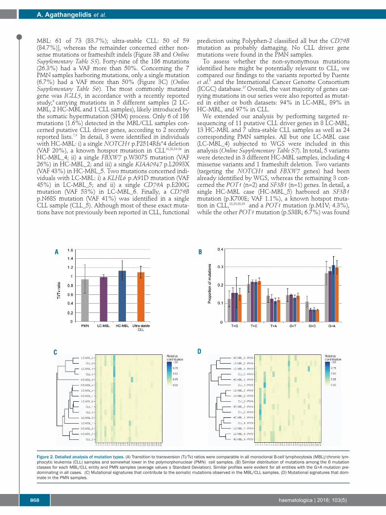
0.99 to 1.13 in the MBL/CLL samples, while it was slightlylower in the PMN samples (0.94) (Figure 2A). No clear dif-ferences were observed between MBL/CLL and PMNsamples when the distribution of mutations among the sixtypes was examined (Figure 2B). We then evaluated thesequence context of each mutation by incorporating infor-mation on the bases immediately upstream and down-stream of the mutated base, hence leading to 96 possiblemutation types in this classification16 (OnlineSupplementary Table S4). Almost all major differences wereidentified between MBL/CLL samples and PMN sampleswith the former group exhibiting mainly C>T mutationsat NpCpG trinucleotides (P<0.05 for ultra-stable CLL andHC-MBL). The MutationalPatterns package33 was used to delineate
the mutational signatures in our cohort. Mutational pat-terns identified in the MBL/CLL samples resembled thosereported by Puente et al.;9 this finding was corroborated by
calculating the pairwise similarity with the 30 previouslypublished signatures, where signature 9 and, to someextent, signature 1 where the main contributors (Figure2C). The same analysis in the PMN samples gave differentresults, with a strong impact of mutational signatures 3and 5 (Figure 2D). Signature 3 had previously been identi-fied in solid tumors and is associated with failure of DNAdouble-strand break-repair by homologous recombina-tion.16 Signature 5 exhibits transcriptional strand bias forC>T and T>C mutations at ApTpN context and displaysa correlation between smoking history and mutation con-tribution.16
MBL and ultra-stable CLL display a paucity of mutations in putative CLL driver genes Whole-genome sequencing identified 186 non-synony-
mous exonic variants amongst MBL/CLL samples and 15amongst PMN samples. The average number was 8.9 forLC-MBL (range: 1-16), 14.8 for HC-MBL (range: 9-27),11.6 for ultra-stable CLL (range: 7-19), and 0.9 for thePMN samples (range: 0-6), respectively (Figure 3A). InMBL/CLL samples, the vast majority of non-synonymousmutations were missense [LC-MBL: 47 of 53 (88.7%); HC-
WGS in MBL and ultra-stable CLL
haematologica | 2018; 103(5) 867
Figure 1. Somatic mutational analysis of ultra-stable chronic lymphocytic leukemia (CLL), high-count monoclonal B-cell lymphocytosis (HC-MBL), low-count mon-oclonal B-cell lymphocytosis (LC-MBL) and control polymorphonuclear (PMN) cell samples. (A) Total number of somatic mutations identified by whole-genomesequencing (WGS) in CLL cell samples from MBL, CLL and the respective PMN samples. All samples carried similar mutational loads with the exception of a singleLC-MBL sample (LC-MBL_1) that displayed a very low number of mutation events; as can be seen, the corresponding PMN sample had a mutation load similar tothe other PMN samples, where comparison of mutation profiles between the MBL and PMN sample showed few common hits, thus excluding the likelihood of con-tamination. Concerning PMN control samples, they were also characterized by high homogeneity regarding the mutational load. There was a single sample with avery high mutational load; detailed comparison against its respective CLL sample showed a high overlap of mutations indicating potential tumor cell contamination,hence this sample was removed from downstream analysis. (B) Average mutation rates ± Standard Deviation (SD) for LC-MBL, HC-MBL and CLL. Highly analogousmutation rates were observed in the HC-MBL (0.79 mutations per Mb) and CLL (0.74 mutations per Mb) samples, while LC-MBL samples had a slightly lower ratio(0.63 mutations per Mb). (C) Average SNV to small indels ratio ± SD for all sample groups. All 3 entities displayed similar ratios in clear contrast to the PMN sampleswhere the ratio was much lower.
A
B C
MBL: 61 of 73 (83.7%); ultra-stable CLL: 50 of 59(84.7%)], whereas the remainder concerned either non-sense mutations or frameshift indels (Figure 3B and OnlineSupplementary Table S5). Forty-nine of the 186 mutations(26.3%) had a VAF more than 50%. Concerning the 7PMN samples harboring mutations, only a single mutation(6.7%) had a VAF more than 50% (Figure 3C) (OnlineSupplementary Table S6). The most commonly mutatedgene was IGLL5, in accordance with a recently reportedstudy,6 carrying mutations in 5 different samples (2 LC-MBL, 2 HC-MBL and 1 CLL samples), likely introduced bythe somatic hypermutation (SHM) process. Only 6 of 186mutations (1.6%) detected in the MBL/CLL samples con-cerned putative CLL driver genes, according to 2 recentlyreported lists.7,9 In detail, 3 were identified in individualswith HC-MBL: i) a single NOTCH1 p.P2514Rfs*4 deletion(VAF 20%), a known hotspot mutation in CLL10,28,34-36 inHC-MBL_4; ii) a single FBXW7 p.W307S mutation (VAF26%) in HC-MBL_2; and iii) a single KIAA0947 p.L2093X(VAF 43%) in HC-MBL_5. Two mutations concerned indi-viduals with LC-MBL: i) a KLHL6 p.A91D mutation (VAF45%) in LC-MBL_5; and ii) a single CD79A p.E200Gmutation (VAF 53%) in LC-MBL_6. Finally, a CD79Bp.N68S mutation (VAF 41%) was identified in a singleCLL sample (CLL_5). Although most of these exact muta-tions have not previously been reported in CLL, functional
prediction using Polyphen-2 classified all but the CD79Bmutation as probably damaging. No CLL driver genemutations were found in the PMN samples.To assess whether the non-synonymous mutations
identified here might be potentially relevant to CLL, wecompared our findings to the variants reported by Puenteet al.9 and the International Cancer Genome Consortium(ICGC) database.37 Overall, the vast majority of genes car-rying mutations in our series were also reported as mutat-ed in either or both datasets: 94% in LC-MBL, 89% inHC-MBL, and 97% in CLL.We extended our analysis by performing targeted re-
sequencing of 11 putative CLL driver genes in 8 LC-MBL,13 HC-MBL and 7 ultra-stable CLL samples as well as 24corresponding PMN samples. All but one LC-MBL case(LC-MBL_4) subjected to WGS were included in thisanalysis (Online Supplementary Table S7). In total, 5 variantswere detected in 3 different HC-MBL samples, including 4missense variants and 1 frameshift deletion. Two variants(targeting the NOTCH1 and FBXW7 genes) had beenalready identified by WGS, whereas the remaining 3 con-cerned the POT1 (n=2) and SF3B1 (n=1) genes. In detail, asingle HC-MBL case (HC-MBL_5) harbored an SF3B1mutation (p.K700E; VAF 1.1%), a known hotspot muta-tion in CLL,18,29,38,39 and a POT1 mutation (p.M1V; 4.3%),while the other POT1mutation (p.S38R; 6.7%) was found
A. Agathangelidis et al.
868 haematologica | 2018; 103(5)
Figure 2. Detailed analysis of mutation types. (A) Transition to transversion (Ti/Tv) ratios were comparable in all monoclonal B-cell lymphocytosis (MBL)/chronic lym-phocytic leukemia (CLL) samples and somewhat lower in the polymorphonuclear (PMN) cell samples. (B) Similar distribution of mutations among the 6 mutationclasses for each MBL/CLL entity and PMN samples (average values ± Standard Deviation). Similar profiles were evident for all entities with the G>A mutation pre-dominating in all cases. (C) Mutational signatures that contribute to the somatic mutations observed in the MBL/CLL samples. (D) Mutational signatures that dom-inate in the PMN samples.
A B
C D
in HC-MBL_2, which also harbored the FBXW7mutation.Two somatic non-synonymous variants were identified in2 different PMN samples: an ATM variant (p.R337C; VAF20%) detected in an LC-MBL case, frequently reportedand probably with a low functional impact, and a TP53mutation (p.G245A, VAF 3%) found in a HC-MBL case,previously reported in several human cancers and lym-phomas40 (Online Supplementary Table S8).
Non-coding variants in MBL and ultra-stable CLL targetgenes in pathways relevant to CLL pathogenesisCoding and non-coding regions enriched for mutations
were detected using Fishhook29 in 10 kilobase windowsacross the genome and with compensation for replicationtiming. In line with previous findings,9 the analysisrevealed highest mutational enrichment in the IG loci andwithin sites known to be recurrently affected by off-targetsomatic hypermutation (e.g. BTG2, BCL6 and TCL1A)(Online Supplementary Figure S1). Funseq2,41 a bioinformatics tool investigating the link-
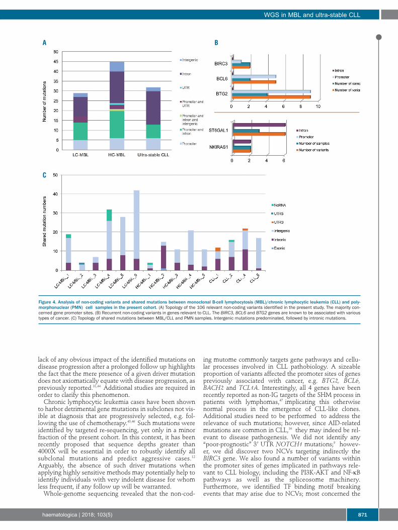
age between NCVs and target genes using integratedbisulfite sequencing, ChIP-Sequencing, and RNA-sequenc-ing data from the Roadmap Epigenomics Project, wasused for the examination of the NCVs. This analysisrevealed a total of 1517 variants in the MBL/CLL samplesand 39 in the PMN samples. After stringent filtering, 106NCVs of potential relevance to MBL and CLL were iden-tified (Online Supplementary Table S9): 29 in LC-MBL (aver-age 4.8), 45 in HC-MBL (average 9), and 32 in CLL samples(average 6.4), respectively; only 4 NCVs were found in 2PMN samples. Since we intentionally selected for NCVs intranscription factor (TF) highly occupied regions (seeOnline Supplementary Appendix), not unexpectedly mostvariants were located in gene promoter sites (Figure 4A).Twenty-nine variants (26.4%) concerned 16 cancer-associ-ated genes and were evenly distributed amongst the 3entities: 9 in LC-MBL samples, 11 in HC-MBL, and 9 inultra-stable CLL samples. Three of these cancer-associatedgenes were recurrently targeted: 9 variants concerned theBTG2 gene in 4 samples (2 CLL, 1 HC-MBL and 1 LC-MBL), 5 variants involved the BCL6 gene in 2 samples (1HC-MBL and 1 LC-MBL), and 2 variants targeted theBIRC3 gene in 2 samples (1 CLL and 1 HC-MBL). We alsoidentified 6 variants concerning the ST6GAL1 gene in 3CLL samples and the same NKIRAS1-related variant in 2CLL samples (Figure 4B). Moreover, pathway analysiswith Enrichr31 showed that 30 of 110 (27.3%) of the vari-ants targeted genes were implicated in key CLL pathwaysand cellular processes, such as the PI3K-AKT pathway(TCL1A, CCND1, BCL2, PKN1, DDIT4 and SGK3)(P<0.05), the NF-κB pathway (BIRC3, BCL2 and PLAU)(P<0.05) and the spliceosome machinery (DDX46 andHSPA2). In most of these cases (22 of 30, 73.3%), the vari-ants were located at promoter sites. Comparison to theseries by Puente et al.9 identified 4 common gene targets:BTG2, BCL6, BACH2 and TCL1A; none of our samplescarried variants affecting either the 3’ UTR of theNOTCH1 gene or the PAX5 gene enhancer.We also analyzed the predicted impact of the NCVs on
TF binding and found that 72 of 110 (65.5%) of the vari-ants could result in a motif-breaking event (LC-MBL:n=21, HC-MBL: n=33, ultra-stable CLL: n=18) (OnlineSupplementary Table S10). We subsequently investigatedgenes and gene pathways that may be affected by such TFmotif breaks. In 55 of 72 (76.4%) cases, variants disrupted
a DNase I hypersensitive site, while enrichment analysisof the implicated target genes using Enrichr31 led to theidentification of genes participating in pathways relevantto CLL pathogenesis, such as the MAPK, WNT and AP-1pathways (P<0.0005) (Online Supplementary Table S11). Moreover, we examined the potential relation of the
NCVs that affect TF binding to AID activity by checkingif they occurred in the known hotspots (WRCY, RGYW,WA, TW). According to our findings, 21 of 72 (29.2%)NCVs were located at AID hotspots. Gene enrichmentanalysis of the remaining 51 target genes revealed similarpathways as in the original analysis (namely AP-1 andDNA damage response pathways).
Shared mutations between CLL and polymorphonuclearcells indicate that somatic variants can arise beforeCLL onsetShared mutations between MBL/CLL samples and their
respective PMN samples were identified in all samplesirrespective of origin. Regarding exonic mutations, thesame synonymous GSE1 mutation was found in an HC-MBL case and its paired PMN sample with comparableVAF (28% vs. 26%). In addition, a LC-MBL sample and itspaired PMN sample carried an identical mutation withinthe ncRNA gene LOC339874, though with different VAF(16% vs. 31%). In the case of non-exonic mutations, 179shared NCVs were identified between MBL/CLL andPMN samples (Online Supplementary Table S12); the aver-age number per sample was 15.8 for LC-MBL, 8.2 for HC-MBL, and 9 for ultra-stable CLL (range: 2-34), respectively.Most of these mutations were intergenic (128 of 179,71.5%) (Figure 4C). Interestingly, 6 NCVs were recurrent-ly found in more than one MBL/CLL-PMN sample pair: 3were intergenic and the other 3 were intronic (OnlineSupplementary Table S13). Finally, we also examined themutational signatures for shared mutations betweenMBL/CLL and PMN samples but did not observe clear cor-relations with any signature (data not shown). In order to exclude the possibility of contamination of
the PMN cell fraction by MBL/CLL DNA, we designedallele-specific primers (Online Supplementary Table S14) andperformed PCR amplification of the clonotypic IGH generearrangement in both the MBL/CLL and the respectivePMN samples in 11 of 16 cases with available material.We identified the clonotypic rearrangement in all 11MBL/CLL samples but in none of the corresponding PMNsamples examined, effectively ruling out the possibilitythat the observed results were due to contamination.
Somatic copy-number analysissCNA analysis was performed in 3 samples from each
entity and their respective PMN control samples, as wellas in 4 additional PMN samples from 1 HC-MBL and 3LC-MBL cases. In total, 16 sCNAs were identified in theMBL/CLL samples (average: 1.8, range: 1-6): 7 in LC-MBL,4 in HC-MBL, and 5 in the ultra-stable CLL samples, allbut one concerning deletion events (Online SupplementaryTable S15). Of the recurrent cytogenetic aberrationsincluded in the Döhner hierarchical model,42 del(17p),del(11q) and trisomy 12 were not identified in any of thesamples, whereas del(13q) was detected in 7 of 9MBL/CLL cases (2 LC-MBL, 3 HC-MBL and 2 CLL cases).FISH analysis gave concordant results in 5 of 7 caseswhere data from both techniques were available; in theremaining 2 cases del(13q) was detected with a single
WGS in MBL and ultra-stable CLL
haematologica | 2018; 103(5) 869

technique each (Online Supplementary Table S16). All othersCNAs represented unique events. In terms of distributionacross the chromosomes, 7 of the 32 (21.9%) sCNAs werefound in the vicinity of centromeres, whereas 11 of 32(34.4%) were located close to a telomere (distance<10x107 bp). None of the PMN samples demonstratedsCNAs typical of CLL. Only one MBL case showed ashared del(8)(p11.22) between the LC-MBL sample and itspaired PMN sample.
Discussion
Limited information is available concerning the genomiclandscape at the very early or indolent phases of CLL. Tothis end, we compared the genomes of ultra-stable CLLcases, defined as those cases stable for more than ten yearsafter diagnosis, to genomes from individuals with: i) LC-MBL, a condition that does not progress into a clinicallyrelevant leukemia;26 and, ii) HC-MBL, a clinically identifi-able pre-leukemic state.25 Both types of MBL and ultra-stable CLL exhibited the
same low level of genomic complexity, similar genome-wide mutation rates, and average number of exonic muta-tions, which were distinct from those of the control sam-ples. Reflecting this similarity, analysis relating to pub-lished mutational signatures revealed similar patterns insamples from all 3 entities. In more detail, signature 9 thatpredominated in the MBL/CLL cohort has been previous-ly identified in CLL and B-cell lymphomas and is attrib-uted to polymerase η that is involved in AID-induced
somatic hypermutation.16 The second ranking signature 1is an age-related signature stemming from spontaneousdeamination of 5-methylcytosine that has been detectedin many cancer types.16 Analogies between MBL and ultra-stable CLL extended also to sCNAs in that all samples,irrespective of origin, carried very few sCNA. Del(13q)predominated in all three entities, as shown in previousstudies.26 Most of the sCNAs were located in close prox-imity to either centromeres or telomeres, in keeping withprevious findings reporting significant over-representa-tions in these regions due to duplication rates.43 Thus,most of the sCNAs identified here may not be directlyrelated to the MBL/CLL phenotype.Interestingly, PMN cells harbored a significantly higher
load of mutations compared to buccal cells. Mutationsdetected in the PMN samples were characterized by thedominance of distinct mutational signatures compared tothe MBL/CLL cohort. However, these samples carriedshared somatic mutations with the respective MBL/CLLcell samples in all analyzed cases. Most shared mutationsconcerned intergenic regions, yet we also identified a sin-gle shared exonic mutation. This finding supports thenotion that some mutations present in the CLL clonecould be acquired prior to disease onset, as previously sug-gested.32Almost all genes that were found mutated in HC-MBL
and/or LC-MBL had been previously described as recur-rently mutated in CLL.9,37 In contrast to our recent WESstudy on relapsing CLL,8 where the great majority of casescarried at least one CLL driver mutation, such mutationswere relatively scarce in our cohort. Most importantly, the
A. Agathangelidis et al.
870 haematologica | 2018; 103(5)
Figure 3. Exonic mutations in our monoclonal B-cell lymphocytosis (MBL)/chronic lymphocyticleukemia (CLL) cohort and polymorphonuclear(PMN) cell samples. (A) Average numbers ofexonic non-synonymous mutations in MBL/CLLentities and PMN samples. (B) The vast majorityof non-synonymous mutations were missense inall 3 MBL/CLL entities. PMN samples failed toshow such predominance. (C) Average numbersof mutations with VAF≥50% for all 3 MBL/CLLentities were comparable. A single PMN samplecarried a clonal mutation.
A B
C
lack of any obvious impact of the identified mutations ondisease progression after a prolonged follow up highlightsthe fact that the mere presence of a given driver mutationdoes not axiomatically equate with disease progression, aspreviously reported.31,44 Additional studies are required inorder to clarify this phenomenon.Chronic lymphocytic leukemia cases have been shown
to harbor detrimental gene mutations in subclones not vis-ible at diagnosis that are progressively selected, e.g. fol-lowing the use of chemotherapy.45,46 Such mutations wereidentified by targeted re-sequencing, yet only in a minorfraction of the present cohort. In this context, it has beenrecently proposed that sequence depths greater than4000X will be essential in order to robustly identify allsubclonal mutations and predict aggressive cases.12Arguably, the absence of such driver mutations whenapplying highly sensitive methods may potentially help toidentify individuals with very indolent disease for whomless frequent, if any follow up will be warranted.Whole-genome sequencing revealed that the non-cod-
ing mutome commonly targets gene pathways and cellu-lar processes involved in CLL pathobiology. A sizeableproportion of variants affected the promoter sites of genespreviously associated with cancer, e.g. BTG2, BCL6,BACH2 and TCL1A. Interestingly, all 4 genes have beenrecently reported as non-IG targets of the SHM process inpatients with lymphomas,47 implicating this otherwisenormal process in the emergence of CLL-like clones.Additional studies need to be performed to address therelevance of such mutations; however, since AID-relatedmutations are common in CLL,16 they may indeed be rel-evant to disease pathogenesis. We did not identify any“poor-prognostic” 3’ UTR NOTCH1 mutations;9 howev-er, we did discover two NCVs targeting indirectly theBIRC3 gene. We also found a number of variants withinthe promoter sites of genes implicated in pathways rele-vant to CLL biology, including the PI3K-AKT and NF-κBpathways as well as the spliceosome machinery.Furthermore, we identified TF binding motif breakingevents that may arise due to NCVs; most concerned the
WGS in MBL and ultra-stable CLL
haematologica | 2018; 103(5) 871
Figure 4. Analysis of non-coding variants and shared mutations between monoclonal B-cell lymphocytosis (MBL)/chronic lymphocytic leukemia (CLL) and poly-morphonuclear (PMN) cell samples in the present cohort. (A) Topology of the 106 relevant non-coding variants identified in the present study. The majority con-cerned gene promoter sites. (B) Recurrent non-coding variants in genes relevant to CLL. The BIRC3, BCL6 and BTG2 genes are known to be associated with varioustypes of cancer. (C) Topology of shared mutations between MBL/CLL and PMN samples. Intergenic mutations predominated, followed by intronic mutations.
A B
C

MAPK, WNT and AP-1 signaling pathways. In this con-text, preliminary results (data not shown) from our ongoinghigh-throughput study on aggressive CLL cases showed agreat degree of consistency in the targeting of NCVs: thesame “CLL-relevant” gene pathways were again amongthe most common targets of NCVs, further corroboratingour present findings. Having said that, some of these vari-ants could represent bystander SHM targets of unknownsignificance or minor contributors to disease pathogenesis,therefore requiring further studies before definitive con-clusions can be drawn regarding their actual significance.It is important to note that a recent study on the epige-
netic profile of CLL48 reported a novel pathogenic role ofTF dysregulation in CLL, with increased activity of EGRand NFAT as well as loss of EBF and AP-1, causing imbal-ances in the normal B-cell epigenetic program.Interestingly, certain members of these networks (e.g.EBF1, JUN and FOS) were among the most commonlyaffected TFs across all sample types tested. Collectively,our findings support the notion that gene pathways couldbe indirectly targeted by NCVs with the targets beingeither the genes themselves or other interacting genes, e.g.TFs. Limitations of the present work involve the relatively
small size of the cohort, mainly due to the rarity of sam-ples meeting the selection criteria. In particular, CLLpatients had to have stable disease after a prolonged fol-low up, whereas all individuals with MBL had to have apersistent monoclonal B-cell population. Concerning LC-MBL, low CLL cell number was an additional challengingfactor. Furthermore, although our targeted re-sequencingapproach covered almost 50% of reported mutations inputative driver genes (as reported by Puente et al.),9 by def-inition this approach is not exhaustive.
In summary, we report that MBL and ultra-stable CLLare virtually indistinguishable at the genomic level. Whilethis may be reflective of a passive and slow accumulationof mutations, we identified both exonic and NCV-targetedpathways central for B-cell biology and CLL development,likely linked to the acquisition of the MBL/CLL pheno-type. Importantly, ultra-stable CLL cases carried fewknown driver gene mutations, even after ten years of fol-low up, perhaps reflecting the central role of microenvi-ronmental signals rather than cell-intrinsic defects in shap-ing clonal behavior. In other words, cell-extrinsic trigger-ing, specifically mediated through the B-cell receptor,might represent the major driving force in the early stagesof CLL, whereas disease progression will require acquisi-tion of genetic driver mutations.
FundingThis research project was supported by the Associazione
Italiana per la Ricerca sul Cancro, AIRC (Investigator Grant#15189 to PG and Special Program Molecular ClinicalOncology – 5 per mille #9965), Milano, Italy, RicercaFinalizzata 2010 (#2318823 to PG); Swedish Cancer Society,the Swedish Research Council, Uppsala University, UppsalaUniversity Hospital, Lion’s Cancer Research Foundation, andSelander’s Foundation, Uppsala; and, H2020 “AEGLE, Ananalytics framework for integrated and personalized healthcareservices in Europe”, by the European Union; “MEDGENET,Medical Genomics and Epigenomics Network” (No.692298) bythe European Union; “GCH-CLL” funded by the GeneralSecretariat for Research and Technology (GSRT) of Greece andthe Italian Ministry of Health (MoH); and IMI2 “HARMO-NY”, funded by the European Union. AA is a fellow ofAssociazione Italiana per la Ricerca sul Cancro AIRC (Triennialfellowship “Guglielmina Lucatello é Gino Mazzega”).
A. Agathangelidis et al.
872 haematologica | 2018; 103(5)
References
1. Chiorazzi N, Rai KR, Ferrarini M. Chroniclymphocytic leukemia. N Engl J Med.2005;352(8):804-815.
2. Parikh SA, Shanafelt TD. Prognostic factorsand risk stratification in chronic lympho-cytic leukemia. Semin Oncol. 2016;43(2):233-240.
3. Scarfo L, Ferreri AJ, Ghia P. Chronic lym-phocytic leukaemia. Crit Rev OncolHematol. 2016;104:169-182.
4. Fabbri G, Khiabanian H, Holmes AB, et al.Genetic lesions associated with chroniclymphocytic leukemia transformation toRichter syndrome. J Exp Med.2013;210(11):2273-2288.
5. Fabbri G, Rasi S, Rossi D, et al. Analysis ofthe chronic lymphocytic leukemia codinggenome: role of NOTCH1 mutational acti-vation. J Exp Med. 2011;208(7):1389-1401.
6. Kasar S, Kim J, Improgo R, et al. Whole-genome sequencing reveals activation-induced cytidine deaminase signatures dur-ing indolent chronic lymphocyticleukaemia evolution. Nat Commun. 2016;6(8866).
7. Landau DA, Tausch E, Taylor-Weiner AN,et al. Mutations driving CLL and their evo-lution in progression and relapse. Nature.2015;526(7574):525-530.
8. Ljungstrom V, Cortese D, Young E, et al.Whole-exome sequencing in relapsingchronic lymphocytic leukemia: clinicalimpact of recurrent RPS15 mutations.Blood. 2016;127(8):1007-1016.
9. Puente XS, Bea S, Valdes-Mas R, et al. Non-coding recurrent mutations in chronic lym-phocytic leukaemia. Nature. 2015;526(7574):519-524.
10. Puente XS, Pinyol M, Quesada V, et al.Whole-genome sequencing identifies recur-rent mutations in chronic lymphocyticleukaemia. Nature. 2011;475(7354):101-105.
11. Quesada V, Conde L, Villamor N, et al.Exome sequencing identifies recurrentmutations of the splicing factor SF3B1 genein chronic lymphocytic leukemia. NatGenet. 2012;44(1):47-52.
12. Rose-Zerilli MJ, Gibson J, Wang J, et al.Longitudinal copy number, whole exomeand targeted deep sequencing of 'good risk'IGHV-mutated CLL patients with progres-sive disease. Leukemia. 2016;30(6):1301-1310.
13. Schuh A, Becq J, Humphray S, et al.Monitoring chronic lymphocytic leukemiaprogression by whole genome sequencingreveals heterogeneous clonal evolution pat-terns. Blood. 2012;120(20):4191-4196.
14. Wang L, Lawrence MS, Wan Y, et al. SF3B1and other novel cancer genes in chronic
lymphocytic leukemia. N Engl J Med.2011;365(26):2497-2506.
15. Burns A, Alsolami R, Becq J, et al. Whole-genome sequencing of chronic lymphocyt-ic leukaemia reveals distinct differences inthe mutational landscape betweenIgHV(mut) and IgHV(unmut) subgroups.Leukemia. 2018;32(2):332-342.
16. Alexandrov LB, Nik-Zainal S, Wedge DC,et al. Signatures of mutational processes inhuman cancer. Nature. 2013;500(7463):415-421.
17. Landau DA, Carter SL, Stojanov P, et al.Evolution and impact of subclonal muta-tions in chronic lymphocytic leukemia.Cell. 2013;152(4):714-726.
18. Rossi D, Bruscaggin A, Spina V, et al.Mutations of the SF3B1 splicing factor inchronic lymphocytic leukemia: associationwith progression and fludarabine-refrac-toriness. Blood. 2011;118(26):6904-6908.
19. Rossi D, Fangazio M, Rasi S, et al.Disruption of BIRC3 associates with flu-darabine chemorefractoriness in TP53wild-type chronic lymphocytic leukemia.Blood. 2012;119(12):2854-2862.
20. Schnaiter A, Paschka P, Rossi M, et al.NOTCH1, SF3B1, and TP53 mutations infludarabine-refractory CLL patients treatedwith alemtuzumab: results from theCLL2H trial of the GCLLSG. Blood.2013;122(7):1266-1270.
21. Landgren O, Albitar M, Ma W, et al. B-cellclones as early markers for chronic lym-phocytic leukemia. N Engl J Med. 2009;360(7):659-667.
22. Marti GE, Rawstron AC, Ghia P, et al.Diagnostic criteria for monoclonal B-celllymphocytosis. Br J Haematol. 2005;130(3):325-332.
23. Dagklis A, Fazi C, Scarfo L, et al.Monoclonal B lymphocytosis in the generalpopulation. Leuk Lymphoma. 2009;50(3):490-492.
24. Ghia P, Prato G, Scielzo C, et al.Monoclonal CD5+ and CD5- B-lympho-cyte expansions are frequent in the periph-eral blood of the elderly. Blood. 2004;103(6):2337-2342.
25. Rawstron AC, Bennett FL, O'Connor SJ, etal. Monoclonal B-cell lymphocytosis andchronic lymphocytic leukemia. N Engl JMed. 2008;359(6):575-583.
26. Fazi C, Scarfo L, Pecciarini L, et al. Generalpopulation low-count CLL-like MBL per-sists over time without clinical progression,although carrying the same cytogeneticabnormalities of CLL. Blood. 2011;118(25):6618-6625.
27. Matos DM, Furtado FM, Falcao RP.Monoclonal B-cell lymphocytosis in indi-viduals from sporadic (non-familial) chron-ic lymphocytic leukemia families persistsover time, but does not progress to chronicB-cell lymphoproliferative diseases. RevBras Hematol Hemoter. 2015;37(5):292-295.
28. Rasi S, Monti S, Spina V, et al. Analysis ofNOTCH1 mutations in monoclonal B-celllymphocytosis. Haematologica. 2012;97(1):153-154.
29. Imielinski M, Guo G, Meyerson M.Insertions and Deletions Target Lineage-Defining Genes in Human Cancers. Cell.2017;168(3):460-472 e414.
30. Kreutzer DA, Essigmann JM. Oxidized,
deaminated cytosines are a source of C -->T transitions in vivo. Proc Natl Acad SciUSA. 1998;95(7):3578-3582.
31. Kuleshov MV, Jones MR, Rouillard AD, etal. Enrichr: a comprehensive gene setenrichment analysis web server 2016update. Nucleic Acids Res. 2016;44(W1):W90-97.
32. Damm F, Mylonas E, Cosson A, et al.Acquired initiating mutations in earlyhematopoietic cells of CLL patients. CancerDiscov. 2014;4(9):1088-1101.
33. Blokzijl FJ, R.; van Boxtel, R.; Cuppen, E.MutationalPatterns: comprehensivegenome-wide analysis of mutationalprocesses. bioRxiv 071761; doi: https://doi.org/10.1101/071761.
34. Baliakas P, Hadzidimitriou A, Sutton LA, etal. Recurrent mutations refine prognosis inchronic lymphocytic leukemia. Leukemia.2015;29(2):329-336.
35. Sutton LA, Young E, Baliakas P, et al.Different spectra of recurrent gene muta-tions in subsets of chronic lymphocyticleukemia harboring stereotyped B-cellreceptors. Haematologica. 2016;101(8):959-967.
36. Lionetti M, Fabris S, Cutrona G, et al. High-throughput sequencing for the identifica-tion of NOTCH1 mutations in early stagechronic lymphocytic leukaemia: biologicaland clinical implications. Br J Haematol.2014;165(5):629-639.
37. Zhang J, Baran J, Cros A, et al. InternationalCancer Genome Consortium Data Portal--aone-stop shop for cancer genomics data.Database (Oxford). 2011;2011:bar026.
38. Strefford JC, Sutton LA, Baliakas P, et al.Distinct patterns of novel gene mutationsin poor-prognostic stereotyped subsets ofchronic lymphocytic leukemia: the case ofSF3B1 and subset #2. Leukemia. 2013;27(11):2196-2199.
39. Jeromin S, Weissmann S, Haferlach C, et al.
SF3B1 mutations correlated to cytogeneticsand mutations in NOTCH1, FBXW7,MYD88, XPO1 and TP53 in 1160 untreatedCLL patients. Leukemia. 2014;28(1):108-117.
40. Wilda M, Bruch J, Harder L, et al.Inactivation of the ARF-MDM-2-p53 path-way in sporadic Burkitt's lymphoma inchildren. Leukemia. 2004;18(3):584-588.
41. Fu Y, Liu Z, Lou S, et al. FunSeq2: a frame-work for prioritizing noncoding regulatoryvariants in cancer. Genome Biol. 2014;15(10):480.
42. Dohner H, Stilgenbauer S, Benner A, et al.Genomic aberrations and survival in chron-ic lymphocytic leukemia. N Engl J Med.2000;343(26):1910-1916.
43. Nguyen DQ, Webber C, Ponting CP. Bias ofselection on human copy-number variants.PLoS Genet. 2006;2(2):e20.
44. Hurtado AM, Chen-Liang TH,Przychodzen B, et al. Prognostic signatureand clonality pattern of recurrently mutat-ed genes in inactive chronic lymphocyticleukemia. Blood Cancer J. 2015;5:e342.
45. Ojha J, Ayres J, Secreto C, et al. Deepsequencing identifies genetic heterogeneityand recurrent convergent evolution inchronic lymphocytic leukemia. Blood.2015;125(3):492-498.
46. Rossi D, Khiabanian H, Spina V, et al.Clinical impact of small TP53 mutated sub-clones in chronic lymphocytic leukemia.Blood. 2014;123(14):2139-2147.
47. Khodabakhshi AH, Morin RD, Fejes AP, etal. Recurrent targets of aberrant somatichypermutation in lymphoma. Oncotarget.2012;3(11):1308-1319.
48. Oakes CC, Seifert M, Assenov Y, et al.DNA methylation dynamics during B cellmaturation underlie a continuum of diseasephenotypes in chronic lymphocyticleukemia. Nature Genetics. 2016;48(3):253-264.
WGS in MBL and ultra-stable CLL
haematologica | 2018; 103(5) 873