circulating progenitor cells identify peripheral arterial...
TRANSCRIPT

DOI: 10.1161/CIRCRESAHA.116.308802 1
Circulating Progenitor Cells Identify Peripheral Arterial Disease in Patients With Coronary Artery Disease
Salim S. Hayek1, James MacNamara2, Ayman Tahhan2, Mosaab Awad1, Adithya Yadalam1, Yi-An Ko3, Sean Healy1, Iraj Hesaroieh1, Hina Ahmed1, Brandon Gray1, Salman S. Sher,1 Nima Ghasemzadeh1,
Riyaz Patel1, Jinhee Kim4, Edmund K. Waller4, and Arshed Quyyumi1
1Division of Cardiology, Emory University School of Medicine, Atlanta, GA; 2Department of Internal Medicine, Emory University School of Medicine, Atlanta, GA; 3Department of Biostatistics and
Bioinformatics, Emory University, Atlanta, GA, and; 4Department of Hematology and Oncology, Winship Cancer Institute, Emory University, Atlanta, GA.
Running title: Arterial Disease and Progenitor Cells in CAD
Subject Terms: Stem Cells Peripheral Vascular Disease Coronary Artery Disease Address correspondence to: Dr. Arshed A. Quyyumi Division of Cardiology Department of Medicine Emory University School of Medicine 1462 Clifton Rd. NE, Suite 507 Atlanta, GA 30322 Tel: (404) 727-3655 Fax: (404) 712-8785 [email protected] In May 2016, the average time from submission to first decision for all original research papers submitted to Circulation Research was 14.93 days.
by guest on June 28, 2018http://circres.ahajournals.org/
Dow
nloaded from
by guest on June 28, 2018http://circres.ahajournals.org/
Dow
nloaded from
by guest on June 28, 2018http://circres.ahajournals.org/
Dow
nloaded from
by guest on June 28, 2018http://circres.ahajournals.org/
Dow
nloaded from
by guest on June 28, 2018http://circres.ahajournals.org/
Dow
nloaded from
by guest on June 28, 2018http://circres.ahajournals.org/
Dow
nloaded from
by guest on June 28, 2018http://circres.ahajournals.org/
Dow
nloaded from

DOI: 10.1161/CIRCRESAHA.116.308802 2
ABSTRACT Rationale: Peripheral arterial disease (PAD) is a clinical manifestation of extra-coronary atherosclerosis. Despite sharing the same risk factors, only 20-30% of patients with coronary artery disease (CAD) develop PAD. Declines in the number of bone-marrow derived circulating progenitor cells (PCs) is thought to contribute to the pathogenesis of atherosclerosis. Whether specific changes in PCs differentiate patients with both PAD and CAD from those with CAD alone is unknown. Objective: Determine whether differences exist in PCs counts of CAD patients with and without known PAD. Methods and Results: 1497 patients (mean age 65, 62% male) with known CAD were identified in the Emory Cardiovascular Biobank. Presence of PAD (n=308) was determined by history, review of medical records or imaging, and was classified as carotid (53%), lower extremity (41%), upper extremity (3%) and aortic disease (33%). Circulating PCs were enumerated by flow cytometry. Patients with CAD and PAD had significantly lower PC counts compared to those with only CAD. In multivariable analysis, a 50% decrease in CD34+ or CD34+/VEGFR2+ counts were associated with a 31% (P=0.032) and 183% (P=0.002) increase in the odds of having PAD, respectively. CD34+ and CD34+/VEGFR2+ counts significantly improved risk prediction metrics for prevalent PAD. Low CD34+/VEGFR2+ counts were associated with a 1.40-fold (95%CI, 1.03, 1.91) and a 1.64-fold (95%CI 1.07, 2.50) increase in the risk of mortality and PAD-related events, respectively. Conclusions: PAD is associated with low CD34+ and CD34+/VEGFR2+ PC counts. Whether low PC counts are useful in screening for PAD needs to be investigated. Keywords: Atherosclerosis, stem cells, CD34, CD133, VEGFR2, KDR, carotid, peripheral arterial disease, aortic aneurysm, progenitor cell, coronary artery disease Nonstandard Abbreviations and Acronyms: CAD Coronary Artery Disease CD Cluster of Differentiation CXCR4 Chemokine (C-X-C Motif) Receptor 4 FITC Fluorescein isothiocyanate PAD Peripheral Arterial Disease PC Progenitor Cells PerCP Peridinin Chlorophyll Protein Complex SSDI Social Security Death Index VEGFR2 Vascular Endothelial Growth Factor Receptor-2
by guest on June 28, 2018http://circres.ahajournals.org/
Dow
nloaded from

DOI: 10.1161/CIRCRESAHA.116.308802 3
INTRODUCTION
Peripheral arterial disease (PAD) is a clinical manifestation of atherosclerosis that leads to obstruction of blood flow by embolism, thrombosis or narrowing of peripheral arteries. It may involve one or multiple vascular beds including the cerebrovascular, aorta, renal, or the upper and lower extremities.1 An accurate estimate of the incidence of PAD is difficult to ascertain because it is often asymptomatic, but it is thought to be present in 10-20% of the population >60 years old.1-5 Clinical syndromes of PAD share common risk factors such as older age, diabetes mellitus, smoking, hypertension and hyperlipidemia.6 These factors, in addition to endothelial dysfunction and inflammation only partially explain the pathogenesis of atherosclerosis. Moreover, despite sharing the same etiologic risk factors, only 20-30% of patients with coronary artery disease (CAD) develop PAD.7-9 Why some patients are predisposed to CAD, others to PAD, and some to both, despite similar predisposing factors, remains unknown.
Recently, a pivotal role for progenitor cells (PCs) in vascular repair and regeneration was
uncovered.10-13 Circulating PCs are mononuclear, originate primarily but not exclusively from the bone marrow, and have been described as having the potential to differentiate into hematopoietic, endothelial, and other lineages, and contribute to vascular repair and regeneration through both direct angiogenic and local paracrine mechanisms.11, 14-16 A relatively rare population of bone marrow-derived mononuclear cells expressing cluster of differentiation 34 (CD34) are enriched for PCs that can differentiate into hematopoietic, endothelial, and other lineages.11, 14, 16-18 CD34-expressing mononuclear cells include hematopoietic, endothelial, and non-hematopoietic (mesenchymal, lacking CD45 expression) PCs.19 CD133 is a 5-transmembrane antigen of primitive stem cells that is lost during maturation and dual expression of these markers (CD34+/CD133+) identifies a PC-enriched subpopulation,17, 20 while co-expression of vascular endothelial growth factor receptor-2 (VEGFR2) appears to identify a rarer subpopulation of PCs further enriched for endothelial progenitors.21-23 Finally, co-expression of Chemokine (C-X-C Motif) Receptor 4 (CXCR4), which promotes homing of PC to stromal-derived factor-rich hypoxic environments, may further characterize CD34+ PC with capacity for tissue repair.24
Lower circulating PC counts and impaired PC activity, measured by colony forming and migration
assays, have been reported in subjects with cardiovascular risk factors or CAD in some but not all studies.25-
27 We and others have also shown that lower circulating PC levels in patients with CAD is associated with an increased risk of adverse CAD events.28, 29 Previous studies investigating the role of PCs in diabetics with PAD found significantly decreased CD34+/VEGFR2+ cell counts and proliferation compared to healthy or diabetic subjects without PAD.30-32 It remains unclear whether the observed impairment in PC counts is specific for PAD or whether it occurs in all individuals with atherosclerosis including those with CAD. In order to address this, we investigated whether circulating PC counts in patients with both CAD and PAD differed from those with only CAD but no known PAD. We hypothesized that abnormalities in endogenous regenerative capacity, enumerated as differences in circulating PC numbers would contribute to the development of extensive atherosclerosis and be lower in patients with more extensive disease (PAD plus CAD) compared with patients with CAD and no known PAD.
by guest on June 28, 2018http://circres.ahajournals.org/
Dow
nloaded from
DOI: 10.1161/CIRCRESAHA.116.308802 4
METHODS Study design and subjects. We compared PC counts in patients with CAD and no known PAD with counts in those with both CAD and PAD in at least one site (upper or lower extremity PAD, carotid stenosis, thoracic or abdominal aortic aneurysms). We identified 1497 subjects with CAD who had PC counts measured and were enrolled in the Emory Cardiovascular Biobank, a prospective registry of adult patients undergoing cardiac catheterization at three Emory Healthcare sites in Atlanta, GA, (Online Figure I, Table 1).29 Subjects presenting with acute myocardial infarction were excluded. PC counts were measured at the time of enrollment from blood samples obtained at the time of catheterization. CAD was defined by the presence of atherosclerotic plaque on the coronary angiogram, and obstructive CAD as the presence of ≥50% stenosis in at least one major coronary artery. Demographic characteristics, medical history, medication use, and behavioral habits were documented as previously described.29 Patients were followed-up for outcomes. The study was approved by the Institutional Review Board at Emory University (Atlanta, GA). All subjects provided written informed consent. Defining peripheral arterial disease. We extensively reviewed patients’ self-reported as well as physician-documented medical history and imaging reports to identify the presence of PAD. PAD was defined as a history of symptomatic or asymptomatic non-coronary atherosclerotic disease in at least one of the following arteries: carotid, thoracic or abdominal aorta, subclavian, brachial, iliac, femoral or popliteal arteries. No routine testing was performed to screen for asymptomatic PAD as part of this study. PAD of the lower extremities was diagnosed when at least one of the following were present: documented ankle-brachial index <0.90; lower limb revascularization; atherosclerotic plaques or stenosis on imaging (CT, ultrasound or fluoroscopy) in the iliac, femoral, or popliteal arteries; history of amputation for critical limb ischemia. PAD of the carotid artery was diagnosed if there was ≥20% stenosis in any carotid artery on imaging (ultrasound, CT or MRA). Aortic disease was diagnosed when there was a history of abdominal or thoracic aneurysms (excluding subjects with aortic root aneurysm associated with bicuspid aortic valves) or evidence of atherosclerotic plaques of the aorta or renal arteries on CT imaging. Progenitor cell assays. Venous blood was collected in EDTA tubes and incubated with fluorochrome-labeled monoclonal anti-human mouse antibodies within 4 hours. Cell populations enriched for circulating PCs were enumerated using flow cytometry as CD45dim cells co-expressing CD34+, CD133+, VEGFR2+, or CXCR4+.33-36 We incubated 300 µL of peripheral blood with 7 µL of FITC-CD34 (BD Biosciences), PerCP-CD45 (BD Biosciences), PE-VEGFR2 (R&D system) and 5 µL APC-CD133 (Miltenyi), and 3ul PE-Cy7-conjugated anti-CXCR4 (EBioscience, clone 12G5) in the dark for 15 minutes. Then 1.5 mL ammonium chloride lysing buffer was added to lyse red blood cells. 1.5 mL staining medium (PBS with 3% heat-inactivated serum and 0.1% sodium azide) was added to stop the lysing reaction. Prior to flow cytometry, 100 µL of AccuCheck Counting Beads (Invitrogen, Cat#: PCB100) were added to act as an internal standard for direct estimation of the concentration of target cell subsets. At least 2.5 million events were acquired from the cytometer. Flow data were analysed with Flowjo software (Treestar, Inc.) (Online Figure II). Absolute mononuclear cell count was estimated as the sum of lymphocytes and monocytes using a Coulter ACT/Diff cell counter (Beckman Coulter). PC populations are reported as cell counts/mL. In 20 samples that were repeatedly analyzed on two occasions by the same technician, the coefficients of variation of the cell types were: CD34+ 2.9%; CD34+/CD133+ 4.8%; CD34+/CXCR4+ 6.5% and CD34+/CD133+/CXCR4+ 7.5%, CD34+/VEGFR2+ cells 21.6%. There were significant correlations between the PC subtypes, with moderate-strong correlations between CD34+, CD34+/CD133+, CD34+/CXCR4+ (r range 0.75-0.91, P<0.001), and weak correlations (r range 0.12-0.34, P<0.001) between CD34+/VEGFR2+ subtypes and the aforementioned PCs (Online Table I).
by guest on June 28, 2018http://circres.ahajournals.org/
Dow
nloaded from

DOI: 10.1161/CIRCRESAHA.116.308802 5
Follow-up and outcomes. We conducted follow-up as previously described to identify pre-specified incident adverse cardiovascular outcomes including death and myocardial infarction.29 In brief, follow-up and adjudication were conducted by personnel blinded to the PC data by phone, electronic medical record review, and social security death index (SSDI) and state records. PAD-related events such as peripheral revascularization and amputation were identified using standard CPT codes for vascular procedures.37 We examined the association between PC counts and death, PAD-related events, and a composite outcome of death, myocardial infarction and PAD-related events. Statistical analysis. Subject characteristics were reported as descriptive statistics with means, medians, standard deviations and ranges. Differences between groups were assessed using t-tests for continuous variables, and chi-square or Fischer exact tests for categorical variables where appropriate. For non-normally distributed variables such as circulating PC counts, the Mann-Whitney U test was used to compare groups in unadjusted analyses. For multivariable analyses, CD34+, CD133+ and CXCR4+ cell counts were log-transformed (base 10) to a normal distribution, while CD34+/VEGFR2+ cell counts were analyzed as a dichotomous variable using the median as a cutoff. Independent predictors of PAD were identified using binary logistic regression modeling accounting for age, gender, race, body mass index, smoking history, hypertension, diabetes, hyperlipidemia, history of heart failure, statin use, angiotensin pathway antagonist use, estimated glomerular filtration rate at enrollment, and obstructive CAD. All multivariable analyses incorporated the aforementioned covariates and specific PC subsets. Sensitivity analyses was performed to explore the interactions between clinical variables significantly associated with PAD and PC subtypes. The incremental value of PC counts to prediction of PAD was tested by addition of individual PC subsets dichotomized to high versus low using the median as a cutoff, to a clinical model with the risk factors including the aforementioned variables. The c-statistic, continuous net reclassification improvement (NRI), and integrated discrimination improvement (IDI) were calculated to evaluate the improvement in predictive ability of the models with and without PCs.38 Cox regression analyses examined the association between PC counts and all-cause death, PAD-related events and the combined endpoint of death, myocardial infarction and PAD-related events, adjusting for the aforementioned variables including PAD. Sensitivity analyses explored whether the association with outcomes differed between patients with and without known PAD. Two-tailed P-value ≤0.05 were considered statistically significant. Analyses were performed using IBM SPSS Statistics Version 22, (Armonk, NY, USA).
by guest on June 28, 2018http://circres.ahajournals.org/
Dow
nloaded from

DOI: 10.1161/CIRCRESAHA.116.308802 6
RESULTS
Characteristics of the 1189 subjects with CAD and 308 with both CAD and PAD are shown in Table 1. Patients with both CAD and PAD were more likely to be older, smokers, hypertensives, diabetics, with heart failure, lower BMI and higher incidence of obstructive CAD (Table 1). Among patients with PAD, 53% had carotid disease, 41% had lower extremity PAD, 3% had upper extremity PAD, and 33% had either abdominal or thoracic aortic disease. Most patients (74%) had only one site of documented PAD, 69 (22%) had two, and 10 (3%) had at least three. In multivariable analyses, age, lower BMI, a history of smoking, statin use, heart failure and lower estimated glomerular filtration rate were independently associated with PAD (Table 2).
Relationship between PCs and PAD.
In unadjusted analyses, cell populations enriched for hematopoietic progenitors (CD34+,
CD34+/CD133+, and CD34+/CXCR4+ cells) as well as those enriched for endothelial progenitors (CD34+/VEGFR2+ cells) were lower in patients with PAD compared to those without PAD (Table 3). Notably CD34+/VEGFR2+ cells were close to 2-fold lower in number in those with PAD compared to those without (Table 3). There were no significant differences in PC counts among patients with PAD according to the location of disease (Table 3). On multivariable analyses adjusting for aforementioned clinical covariates and analyzing each PC subset separately, CD34+ and CD34+/VEGFR2+ cell counts (Models 2 and 5), but not CD34+/CD133+ (Model 3) or CD34+/CXCR4+ (Model 4) counts were independently associated with the presence of PAD (Table 2). Thus, a 50% decrease in CD34+ or CD34+/VEGFR2+ cell counts was associated with a 31% (OR 1.31, P=0.032) and 183% (OR 2.83, P=0.002) increase in the odds of having PAD respectively.
Given the weak correlation between CD34+ and CD34+/VEGFR2+ cell counts (r=0.22, P<0.001),
we examined their association with PAD in the same multivariable model and found them to be both associated with PAD independent of each other (OR 1.35 for CD34+ and OR 1.49 for CD34+/VEGFR2+, Table 2, Model 6). Moreover, patients with both low (≤median) CD34+ and CD34+/VEGFR2+ had a higher prevalence of PAD (28% versus 15%, P<0.001) compared to those with higher than median counts in both subsets (Figure 1), as well as higher odds (1.65, P=0.002) of having PAD (Model 7, Table 2). Subjects with low levels in only one cell subset had intermediate prevalence of PAD (Figure 1).
Sensitivity analyses.
We performed sensitivity analyses in order to determine whether the association between PCs and PAD differed according to conventional PAD risk factors; age, gender, diabetes and smoking status, and presence of obstructive CAD (Figure 2). We found a significant interaction between CD34+ and smoking status in the prediction of PAD (interaction P=0.019). Patients with a history of smoking and low CD34+ (≤1652 cells/mL) had significantly higher odds of having PAD (OR 1.69, P=0.003), while non-smokers with low CD34+ cells did not (OR 0.90, P=0.68). There were otherwise no interactions between the remainder of the characteristics and CD34+ or CD34+/VEGFR2+ cell counts.
Prediction performance.
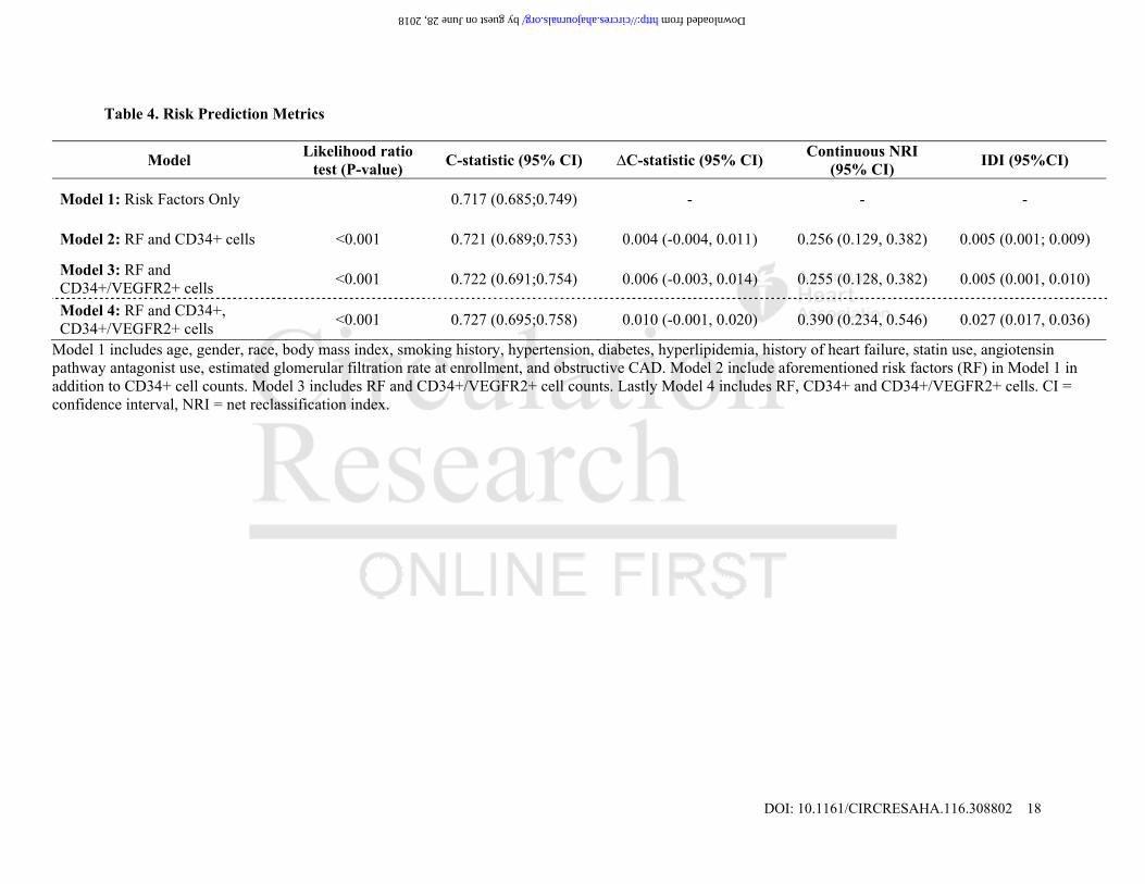
To determine the potential of PCs as biomarkers of PAD, we compared the likelihood, c-statistic, net NRI and IDI between the model with traditional risk factors only (Model 1) and three models incorporating PC counts (Models 2, 3 and 4) in addition to demographics and risk factors (Table 4). Addition of either CD34+ counts (Model 2) or CD34+/VEGFR2+ counts (Model 3) to the risk factor model was associated with a significant improvement in the likelihood ratio, NRI as well as IDI (Table 4). The
by guest on June 28, 2018http://circres.ahajournals.org/
Dow
nloaded from
DOI: 10.1161/CIRCRESAHA.116.308802 7
largest improvement was noted when both CD34+ and CD34+/VEGFR2+ PC counts were added to the clinical model together (Model 4), with a NRI of 0.390 (95%CI 0.234, 0.543) and an IDI of 0.027 (95%CI 0.017, 0.036). The improvement in c-statistic with addition of both cells counts to the clinical model was not statistically significant (estimated change=0.010, 95%CI -0.001, 0.020).
Progenitor cell counts and outcomes. Lastly, we examined the association between CD34+, CD34+/VEGFR2+ cell counts and incident adverse cardiovascular outcomes (Table 5). There were 217 deaths (14%), 67 myocardial infarctions (4%) and 142 PAD-related events (9%) during a median follow-up period of 2 years [1.2-2.9]. Patients with PAD were more likely to die (21% versus 12%, P<0.001), suffer from a myocardial infarction (8% versus 3%, P<0.001) or undergo a vascular procedure (27% versus 4%, P<0.001) compared to those without known PAD at enrollment. When dichotomized by median, patients with low CD34+ counts (≤1652 cells/mL, Log-Rank P=0.012) or low CD34+/VEGFR2+ counts (≤33 cells/mL, Log-Rank P=0.002) had greater mortality compared to those with higher counts. Only subjects with low CD34+/VEGFR2+counts experienced a higher rate of PAD-related events (Log-Rank P<0.001). In Cox regression analyses adjusting for the aforementioned covariates and PAD history, a low CD34+/VEGFR2+ cell count was associated with a 1.43-fold increase in risk of death, a 1.64-fold increased risk of PAD-related events, and a 1.65-fold increased risk of the composite event rate of death, myocardial infarction, and PAD events (Table 5). There was no interaction with PAD status suggesting that this cell type was predictive of events in both subjects with and without PAD. We did not find an association between CD34+ cell counts, dichotomized by median value, and future PAD-related events. DISCUSSION
In the large study in patients with known CAD to date, we have identified an association between low CD34+, CD34+/VEGFR2+ PC counts and the presence of PAD. Subjects with both CAD and PAD had a 2-fold lower CD34+/VEGFR2+ cell count compared to subjects with only CAD and no known PAD. After adjusting for known risk factors for PAD, low CD34+ (≤1652 cells/mL) and CD34+/VEGFR2+ (≤33 cells/mL) cell counts were associated with a 41% and 55% increase in the odds of having PAD, respectively. Moreover, subjects with both low CD34+ and low CD34+/VEGFR2+ cell counts had a 65% increase in the odds of PAD and improved risk discrimination metrics when added to a model with traditional risk factors. Most importantly, low CD34+/VEGFR2+ cell counts were associated with increased mortality and risk of incident PAD-related events. These findings build on the growing body of evidence indicating an important role for circulating PCs in the pathogenesis of atherosclerosis, and may explain why, despite similar risk factors, certain patients develop isolated CAD while others have more widespread atherosclerosis of the peripheral circulation.
There was no evidence suggesting an association between CD34+ cells expressing the CD133 or
CXCR4 epitopes and the co-occurrence of PAD and CAD in this population. We and others have previously shown these cells to be predictive of outcomes in patients with CAD.27, 29 While peripheral blood CD34+ cells are heterogeneous, they are enriched for cells with endothelial lineage potential, express endothelial marker genes, and form endothelial structures in vitro and in vivo.11, 39 In our study, CD34+ cells of interest were predominantly (>95%) CD45dim, and thus largely represent cells of the hematopoietic lineage. While the additional expression of VEGFR2 receptor on CD34+ cells is often considered to define a subset enriched for endothelial PCs, this remains a subject of controversy.17, 40-43
Our findings are consistent with prior smaller studies showing similarly lower levels of circulating
CD34+/VEGFR2+ cells in patients with PAD.30-32, 44 Shaffer et al. and Bitterli et al. noted similar findings
by guest on June 28, 2018http://circres.ahajournals.org/
Dow
nloaded from

DOI: 10.1161/CIRCRESAHA.116.308802 8
when comparing patients with PAD to healthy subjects,30, 44 while Fadini et al. reported decreased counts in diabetic subjects with lower extremity PAD or carotid stenosis compared to diabetics without PAD.32 These studies were limited by small sample size and most importantly the inability to account for the presence or absence of CAD. Our study examined the association between PCs and PAD in a much larger cohort of patients with CAD, with and without diabetes or obstructive CAD. Moreover, we demonstrated that the association between lower CD34+/VEGFR2+ PC counts extends to forms of PAD beyond diabetic vasculopathy, lower extremity PAD and carotid stenosis, as 33% of our subjects with PAD had aortic disease. While the association between PC counts and risk of death and myocardial infarction has been previously described,28, 29 our findings that low CD34+/VEGFR2+ PC counts are predictive of incident PAD-related events are novel. Experimental studies have shown that disruption of the bone marrow is a major contributor to the pathogenesis of atherosclerosis.45-47 In humans with critical limb ischemia, examination of the bone marrow demonstrated profound changes including microvascular disruption and reduced CD34+ cells, indicating that changes in peripheral blood we described are likely associated with similar disruption of PCs in the bone marrow in PAD.48, 49
Strengths of our study include (1) a large cohort study design to limit heterogeneity, (2) use of
commonly used high-throughput technology (flow cytometry) for quantification of PCs by the same technical team, (3) exploration of several CD34+ cell sub-populations enriched for both hematopoietic and endothelial PCs, and (4) the association with incident cardiac and vascular events. Limitations include the lack of systematic screening for PAD. Thus, it is possible that some patients with undiagnosed or asymptomatic PAD are unaccounted for and may be included with the group of CAD only patients. Nevertheless, our findings suggest that PC counts could help identify a subset of patients with CAD at high risk for underlying PAD. Although our findings imply that depletion of circulating PC pool may be associated with more extensive atherosclerosis, and in particular PAD, the cohort design prevents us from establishing causation.
Clinical implications.
Measuring PC counts may be useful as a screening test in subjects without known PAD. Several measures have been found to increase mobilization of PCs, such as lifestyle modification, intensifying statin therapy, cilostazol, and exercise.31, 50,25, 26 Thus, identifying subjects at risk for PAD may allow for earlier interventions, and potentially abrogation of that risk. A low CD34+/VEGF2R+ PC cell count is indicative of worse long term prognosis, especially from vascular events. Given the significant impact of PAD on morbidity and mortality, whether a sustained decrease in PC counts precedes development of PAD is worthy of further study.
by guest on June 28, 2018http://circres.ahajournals.org/
Dow
nloaded from

DOI: 10.1161/CIRCRESAHA.116.308802 9
ACKNOWLEDGEMENTS We would like to thank all members of the Emory ACTSI for their support in performing this study. We are grateful to Ernestine Maher, Hilary Rosenthal and Wayne Harris for their assistance with flow cytometry. SOURCES OF FUNDING AAQ is supported by 5P01HL101398-02, 1P20HL113451-01, 1R56HL126558-01, 1RF1AG051633-01, R01 NS064162-01, R01 HL89650-01, HL095479-01, 1U10HL110302-01, 1DP3DK094346-01, 2P01HL086773-06A1. SSH is supported by the Abraham J. & Phyllis Katz Foundation (Atlanta, GA). DISCLOSURES None of the authors have conflicts of interest to disclose. REFERENCES 1. Criqui MH and Aboyans V. Epidemiology of Peripheral Artery Disease. Circulation research. 2015;116:1509-1526. 2. Savji N, Rockman CB, Skolnick AH, Guo Y, Adelman MA, Riles T and Berger JS. Association between advanced age and vascular disease in different arterial territories: a population database of over 3.6 million subjects. Journal of the American College of Cardiology. 2013;61:1736-43. 3. Ostchega Y, Paulose-Ram R, Dillon CF, Gu Q and Hughes JP. Prevalence of peripheral arterial disease and risk factors in persons aged 60 and older: data from the National Health and Nutrition Examination Survey 1999-2004. J Am Geriatr Soc. 2007;55:583-9. 4. Norgren L, Hiatt WR, Harris KA, Lammer J and Group TIW. TASC II section F on revascularization in PAD. J Endovasc Ther. 2007;14:743-4. 5. Go AS, Mozaffarian D, Roger VL, Benjamin EJ, Berry JD, Borden WB, Bravata DM, Dai S, Ford ES, Fox CS, Franco S, Fullerton HJ, Gillespie C, Hailpern SM, Heit JA, Howard VJ, Huffman MD, Kissela BM, Kittner SJ, Lackland DT, Lichtman JH, Lisabeth LD, Magid D, Marcus GM, Marelli A, Matchar DB, McGuire DK, Mohler ER, Moy CS, Mussolino ME, Nichol G, Paynter NP, Schreiner PJ, Sorlie PD, Stein J, Turan TN, Virani SS, Wong ND, Woo D, Turner MB, American Heart Association Statistics C and Stroke Statistics S. Heart disease and stroke statistics--2013 update: a report from the American Heart Association. Circulation. 2013;127:e6-e245. 6. Creager MA, White CJ, Hiatt WR, Criqui MH, Josephs SC, Alberts MJ, Pearce WH, Gray BH, Rocha-Singh KJ and Association AH. Atherosclerotic Peripheral Vascular Disease Symposium II: executive summary. Circulation. 2008;118:2811-25. 7. Golomb BA, Dang TT and Criqui MH. Peripheral Arterial Disease: Morbidity and Mortality Implications. Circulation. 2006;114:688-699. 8. Ness J and Aronow WS. Prevalence of coexistence of coronary artery disease, ischemic stroke, and peripheral arterial disease in older persons, mean age 80 years, in an academic hospital-based geriatrics practice. J Am Geriatr Soc. 1999;47:1255-6. 9. Kownator S, Cambou J-P, Cacoub P, Léger P, Luizy F, Herrmann M-A and Priollet P. Prevalence of unknown peripheral arterial disease in patients with coronary artery disease: data in primary care from the IPSILON study. Arch Cardiovasc Dis. 2009;102:625-31. 10. Rauscher FM, Goldschmidt-Clermont PJ, Davis BH, Wang T, Gregg D, Ramaswami P, Pippen AM, Annex BH, Dong C and Taylor DA. Aging, progenitor cell exhaustion, and atherosclerosis. Circulation. 2003;108:457-63. 11. Asahara T, Murohara T, Sullivan A, Silver M, van der Zee R, Li T, Witzenbichler B, Schatteman G and Isner JM. Isolation of putative progenitor endothelial cells for angiogenesis. Science. 1997;275:964-7.
by guest on June 28, 2018http://circres.ahajournals.org/
Dow
nloaded from

DOI: 10.1161/CIRCRESAHA.116.308802 10
12. Lin Y, Weisdorf DJ, Solovey A and Hebbel RP. Origins of circulating endothelial cells and endothelial outgrowth from blood. J Clin Invest. 2000;105:71-7. 13. Quyyumi AA. Endothelial function in health and disease: new insights into the genesis of cardiovascular disease. The American journal of medicine. 1998;105:32S-39S. 14. Asahara T, Masuda H, Takahashi T, Kalka C, Pastore C, Silver M, Kearne M, Magner M and Isner JM. Bone Marrow Origin of Endothelial Progenitor Cells Responsible for Postnatal Vasculogenesis in Physiological and Pathological Neovascularization. Circ Res. 1999;85:221-228. 15. Lin Y, Weisdorf DJ, Solovey A and Hebbel RP. Origins of circulating endothelial cells and endothelial outgrowth from blood. Journal of Clinical Investigation January. 2000;105:71-77. 16. Urbich C and Dimmeler S. Endothelial progenitor cells: characterization and role in vascular biology. Circ Res. 2004;95:343-53. 17. Gehling UM, Ergun S, Schumacher U, Wagener C, Pantel K, Otte M, Schuch G, Schafhausen P, Mende T, Kilic N, Kluge K, Schafer B, Hossfeld DK and Fiedler W. In vitro differentiation of endothelial cells from AC133-positive progenitor cells. Blood. 2000;95:3106-12. 18. Kawamoto A, Iwasaki H, Kusano K, Murayama T, Oyamada A, Silver M, Hulbert C, Gavin M, Hanley A, Ma H, Kearney M, Zak V, Asahara T and Losordo DW. CD34-positive cells exhibit increased potency and safety for therapeutic neovascularization after myocardial infarction compared with total mononuclear cells. Circulation. 2006;114:2163-9. 19. Waller EK, Olweus J, Lund-Johansen F, Huang S, Nguyen M, Guo GR and Terstappen L. The "common stem cell" hypothesis reevaluated: human fetal bone marrow contains separate populations of hematopoietic and stromal progenitors. Blood. 1995;85:2422-35. 20. Yin AH, Miraglia S, Zanjani ED, Almeida-Porada G, Ogawa M, Leary AG, Olweus J, Kearney J and Buck DW. AC133, a novel marker for human hematopoietic stem and progenitor cells. Blood. 1997;90:5002-12. 21. Fadini GP, Losordo D and Dimmeler S. Critical reevaluation of endothelial progenitor cell phenotypes for therapeutic and diagnostic use. Circ Res. 2012;110:624-37. 22. Case J, Mead LE, Bessler WK, Prater D, White HA, Saadatzadeh MR, Bhavsar JR, Yoder MC, Haneline LS and Ingram DA. Human CD34+AC133+VEGFR-2+ cells are not endothelial progenitor cells but distinct, primitive hematopoietic progenitors. Experimental hematology. 2007;35:1109-18. 23. Alaiti MA, Ishikawa M and Costa MA. Bone marrow and circulating stem/progenitor cells for regenerative cardiovascular therapy. Translational research : the journal of laboratory and clinical medicine. 2010;156:112-29. 24. Seeger FH, Rasper T, Koyanagi M, Fox H, Zeiher AM and Dimmeler S. CXCR4 expression determines functional activity of bone marrow-derived mononuclear cells for therapeutic neovascularization in acute ischemia. Arterioscler Thromb Vasc Biol. 2009;29:1802-9. 25. Hu T, She Q, Jiang Y, Su L and Yin Y. Level of CD14+-endothelial progenitor cells is not associated with coronary artery disease or cardiovascular risk factors. Age. 2008;30:319-26. 26. Hill JM, Zalos G, Halcox JP, Schenke WH, Waclawiw MA, Quyyumi AA and Finkel T. Circulating endothelial progenitor cells, vascular function, and cardiovascular risk. The New England journal of medicine. 2003;348:593-600. 27. Fadini GP, Maruyama S, Ozaki T, Taguchi A, Meigs J, Dimmeler S, Zeiher AM, de Kreutzenberg S, Avogaro A, Nickenig G, Schmidt-Lucke C and Werner N. Circulating progenitor cell count for cardiovascular risk stratification: a pooled analysis. PloS one. 2010;5:e11488. 28. Rigato M, Avogaro A and Fadini GP. Levels of Circulating Progenitor Cells, Cardiovascular Outcomes and Death: A Meta-Analysis of Prospective Observational Studies. Circulation research. 2016. 29. Patel RS, Li Q, Ghasemzadeh N, Eapen DJ, Moss LD, Janjua AU, Manocha P, Al Kassem H, Veledar E, Samady H, Taylor WR, Zafari AM, Sperling L, Vaccarino V, Waller EK and Quyyumi AA. Circulating CD34+ progenitor cells and risk of mortality in a population with coronary artery disease. Circ Res. 2015;116:289-97.
by guest on June 28, 2018http://circres.ahajournals.org/
Dow
nloaded from
DOI: 10.1161/CIRCRESAHA.116.308802 11
30. Bitterli L, Afan S, Bühler S, DiSanto S, Zwahlen M, Schmidlin K, Yang Z, Baumgartner I, Diehm N and Kalka C. Endothelial progenitor cells as a biological marker of peripheral artery disease. Vascular medicine. 2016;21:3-11. 31. Saber R, Liu K, Ferrucci L, Criqui MH, Zhao L, Tian L, Guralnik JM, Liao Y, Domanchuk K, Kibbe MR, Green D, Perlman H and McDermott MM. Ischemia-related changes in circulating stem and progenitor cells and associated clinical characteristics in peripheral artery disease. Vascular medicine. 2015;20:534-43. 32. Fadini GP, Sartore S, Albiero M, Baesso I, Murphy E, Menegolo M, Grego F, Vigili de Kreutzenberg S, Tiengo A, Agostini C and Avogaro A. Number and function of endothelial progenitor cells as a marker of severity for diabetic vasculopathy. Arteriosclerosis, thrombosis, and vascular biology. 2006;26:2140-6. 33. Krause DS, Fackler MJ, Civin CI and May WS. CD34: structure, biology, and clinical utility [see comments]. Blood. 1996;87:1-13. 34. Berenson RJ. Transplantation of CD34+ hematopoietic precursors: clinical rationale. Transplantation Proceedings. 1992;24:3032-4. 35. Baum CM, Weissman IL, Tsukamoto AS, Buckle AM and Peault B. Isolation of a candidate human hematopoietic stem-cell population. Proceedings of the National Academy of Sciences of the United States of America. 1992;89:2804-8. 36. Miraglia S, Godfrey W, Yin AH, Atkins K, Warnke R, Holden JT, Bray RA, Waller EK and Buck DW. A novel five-transmembrane hematopoietic stem cell antigen: isolation, characterization, and molecular cloning. Blood. 1997;90:5013-21. 37. Goodney PP, Travis LL, Nallamothu BK, Holman K, Suckow B, Henke PK, Lucas FL, Goodman DC, Birkmeyer JD and Fisher ES. Variation in the use of lower extremity vascular procedures for critical limb ischemia. Circulation Cardiovascular quality and outcomes. 2012;5:94-102. 38. Pencina MJ, D'Agostino RB, D'Agostino RB and Vasan RS. Evaluating the added predictive ability of a new marker: from area under the ROC curve to reclassification and beyond. Stat Med. 2008;27:157-72; discussion 207-12. 39. Masuda H, Alev C, Akimaru H, Ito R, Shizuno T, Kobori M, Horii M, Ishihara T, Isobe K, Isozaki M, Itoh J, Itoh Y, Okada Y, McIntyre BA, Kato S and Asahara T. Methodological development of a clonogenic assay to determine endothelial progenitor cell potential. Circ Res. 2011;109:20-37. 40. Case J, Mead LE, Bessler WK, Prater D, White HA, Saadatzadeh MR, Bhavsar JR, Yoder MC, Haneline LS and Ingram DA. Human CD34+AC133+VEGFR-2+ cells are not endothelial progenitor cells but distinct, primitive hematopoietic progenitors. Exp Hematol. 2007;35:1109-18. 41. Friedrich EB, Walenta K, Scharlau J, Nickenig G and Werner N. CD34-/CD133+/VEGFR-2+ endothelial progenitor cell subpopulation with potent vasoregenerative capacities. Circ Res. 2006;98:e20-5. 42. Madeddu P, Emanueli C, Pelosi E, Salis MB, Cerio AM, Bonanno G, Patti M, Stassi G, Condorelli G and Peschle C. Transplantation of low dose CD34+KDR+ cells promotes vascular and muscular regeneration in ischemic limbs. Faseb J. 2004;18:1737-9. 43. Ohtani K, Vlachojannis GJ, Koyanagi M, Boeckel JN, Urbich C, Farcas R, Bonig H, Marquez VE, Zeiher AM and Dimmeler S. Epigenetic regulation of endothelial lineage committed genes in pro-angiogenic hematopoietic and endothelial progenitor cells. Circ Res. 2011;109:1219-29. 44. Shaffer RG, Greene S, Arshi A, Supple G, Bantly A, Moore JS, Parmacek MS and Mohler ER, 3rd. Effect of acute exercise on endothelial progenitor cells in patients with peripheral arterial disease. Vascular medicine. 2006;11:219-26. 45. Iwata H, Manabe I and Nagai R. Lineage of bone marrow-derived cells in atherosclerosis. Circ Res. 2013;112:1634-47. 46. Sata M, Saiura A, Kunisato A, Tojo A, Okada S, Tokuhisa T, Hirai H, Makuuchi M, Hirata Y and Nagai R. Hematopoietic stem cells differentiate into vascular cells that participate in the pathogenesis of atherosclerosis. Nat Med. 2002;8:403-9.
by guest on June 28, 2018http://circres.ahajournals.org/
Dow
nloaded from

DOI: 10.1161/CIRCRESAHA.116.308802 12
47. Shimizu K, Sugiyama S, Aikawa M, Fukumoto Y, Rabkin E, Libby P and Mitchell RN. Host bone-marrow cells are a source of donor intimal smooth- muscle-like cells in murine aortic transplant arteriopathy. Nat Med. 2001;7:738-41. 48. Spinetti G, Cordella D, Fortunato O, Sangalli E, Losa S, Gotti A, Carnelli F, Rosa F, Riboldi S, Sessa F, Avolio E, Beltrami AP, Emanueli C and Madeddu P. Global remodeling of the vascular stem cell niche in bone marrow of diabetic patients: implication of the microRNA-155/FOXO3a signaling pathway. Circ Res. 2013;112:510-22. 49. Teraa M, Fledderus JO, Rozbeh RI, Leguit RJ, Verhaar MC and Group JS. Bone marrow microvascular and neuropathic alterations in patients with critical limb ischemia. Circ Res. 2014;114:311-4. 50. Chao T-H, Tseng S-Y, Chen I-C, Tsai Y-S, Huang Y-Y, Liu P-Y, Ou H-Y, Li Y-H, Wu H-L, Cho C-L, Tsai L-M and Chen J-H. Cilostazol enhances mobilization and proliferation of endothelial progenitor cells and collateral formation by modifying vasculo-angiogenic biomarkers in peripheral arterial disease. International journal of cardiology. 2014;172:e371-4.
by guest on June 28, 2018http://circres.ahajournals.org/
Dow
nloaded from

DOI: 10.1161/CIRCRESAHA.116.308802 13
FIGURE LEGENDS Figure 1. Prevalence of Peripheral Arterial Disease Stratified by CD34+ and CD34+/VEGFR2+ Cell Counts. Three-dimensional bar plot depicting the prevalence of peripheral vascular disease (Y axis) stratified by the median counts of CD34+ and CD34+/VEGFR2+ cells. Figure 2. Sensitivity Analyses. Forest plot of interaction with traditional risk factors and median CD34+ cell counts (panel A), and median CD34+/VEGFR2+ cell counts for predicting the presence of PAD. There was a significant interaction between smoking and CD34+ cell counts (highlighted in box, panel A).
by guest on June 28, 2018http://circres.ahajournals.org/
Dow
nloaded from
DOI: 10.1161/CIRCRESAHA.116.308802 14
Novelty and Significance What Is Known?
Despite sharing risk factors, peripheral arterial disease (PAD) and coronary artery disease (CAD) co-occur in only 20-30% of patients.
Circulating progenitor cells (PC) are thought to be involved in vascular repair and regeneration. Mononuclear cells expressing CD34 and VEGFR2 are enriched for endothelial PCs.
In comparison with healthy patients, patients with PAD have been shown to have lower circulating
levels of CD34+/VEGFR2+ and progenitor cells.
What New Information Does This Article Contribute?
Compared with patients with CAD alone, p those with both PAD and CAD have at least 30% lower CD34+/VEGFR2+ PC counts, independent of demographics and clinical characteristics.
Addition of CD34+/VEGFR2+ PC counts to a traditional risk factor model significantly improved risk prediction metrics for PAD.
Patients with low CD34+/VEGFR2+ PCs were at a higher risk of cardiovascular outcomes including death, myocardial infarction and PAD-related events.
Although atherosclerosis is one of the most studied human diseases, there is still much we do not understand of its pathogenesis. While both PAD and CAD share similar risk factors, only 20-30% of patients with CAD develop PAD. Why some patients are predisposed to CAD, others to PAD, and some to both, despite similar risk factors, is unknown. Circulating PCs are thought to be involved in vascular repair, and are decreased in patients with PAD compared to healthy counterparts. We investigated whether PC counts could distinguish between patients with PAD and CAD and those with CAD alone. We found that patients with PAD had significantly lower CD34+, and CD34+/VEGFR2+ PCs, independent of demographics and clinical characteristics. CD34+/VEGR2+ counts added incremental value to traditional risk factors in predicting PAD. Moreover, we found that low levels of CD34+/VEGFR2+ cells were associated with worse cardiovascular outcomes, and PAD-related events. These findings imply a disruption in endogenous regenerative potential, may underlie the pathogenesis of PAD, and suggest that PC counts could be used to identify patients with CAD who are at high risk of PAD, provide prognostic information, and potentially guide early interventions.
by guest on June 28, 2018http://circres.ahajournals.org/
Dow
nloaded from

DOI: 10.1161/CIRCRESAHA.116.308802 15
Table 1. Characteristics of Patients with CAD with and without Peripheral Vascular Disease
Variables Without Peripheral Vascular
Disease (n=1189) Peripheral Vascular Disease (n=308) P-value
Age, years 65 (13) 71 (11) <0.001
Male, n (%) 725 (61%) 199 (65%) 0.264
African American, n (%) 254 (21%) 58 (19%) 0.346
Body Mass Index, kg/m2 30 (6) 28 (6) <0.001
Clinical Characteristics
Smoking History, n (%) 773 (65%) 225 (73%) 0.008
Hypertension, n (%) 1063 (90%) 298 (97%) <0.001
Diabetes Mellitus, n (%) 489 (42%) 148 (50%) 0.026
Hyperlipidemia, n (%) 899 (76%) 245 (80%) 0.268
Statin use, n (%) 400 (34%) 138 (45%) <0.001
Low-Density Lipoprotein, mg/dL 91 (39) 86 (35) 0.054
High-Density Lipoprotein, mg/dL 45 (15) 45 (13) 0.404
Heart Failure, n (%) 270 (23%) 101 (33%) <0.001
Ejection fraction, % 53 (13) 51 (14) 0.017
Obstructive Coronary Artery Disease, n (%) 722 (61%) 229 (74%) <0.001
ACEi/ARB use, n (%) 323 (27%) 111 (36%) 0.003
Estimated glomerular filtration rate, mL/min/1.73 m2 70 (26) 60 (25) <0.001 Values are mean (SD), n (%) as noted. Obstructive coronary artery disease denotes the presence of at least 50% obstruction in any of the coronary arteries on angiogram.
by guest on June 28, 2018 http://circres.ahajournals.org/ Downloaded from

DOI: 10.1161/CIRCRESAHA.116.308802 16
Table 2. Independent Predictors of Peripheral Vascular Disease Variables β, P-value 95% CI Model 1: Risk Factors
Age, per 10 years 1.41, <0.001 1.24, 1.604 Male 1.10, 0.523 0.82, 1.48 African American 1.00, 0.982 0.69, 1.46 Body Mass Index, per kg/m2 0.95, <0.001 0.93, 0.98 Smoking History 1.43, 0.021 1.06, 1.95 Hypertension 2.33, 0.026 1.11, 4.90 Diabetes Mellitus 1.30, 0.070 0.98, 1.74 Hyperlipidemia 0.83, 0.300 0.58, 1.18 Statin use 1.47, 0.020 1.06, 2.03 Heart Failure 1.30, 0.085 0.96, 1.76 Obstructive Coronary Artery Disease 1.65, 0.002 1.19, 2.27 ACEi/ARB use 1.18, 0.325 0.85, 1.66 eGFR, per mL/min/1.73 m2 0.99, 0.001 0.98, 0.99
Model 2-5: Risk Factors + Individual PC subtypes CD34+, ≤1652 cells/mL 1.41, 0.015 1.07, 1.87 CD34+/CD133+, ≤762 cells/mL 1.15, 0.329 0.87, 1.53 CD34+/CXCR4+, ≤799 cells/mL 1.13, 0.376 0.59, 1.50 CD34+/VEGFR2+ ≤33 cells/mL 1.55, 0.005 1.14, 2.10
Model 6: Risk Factors and CD34+, CD34+/VEGFR2+ CD34+ ≤1652 cells/mL 1.35, 0.037 1.02, 1.79 CD34+/VEGFR2+ ≤33 cells/mL 1.49, 0.012 1.09, 2.03
Model 7: RF and Low CD34+, CD34+/VEGFR2+ CD34+ ≤1652 cells/mL and CD34+/VEGFR2+ ≤33 cells/mL 1.65, 0.002 1.19, 2.29
Progenitor cell subtypes were each entered into separate models incorporating demographics and risk factors. The odds ratio and confidence intervals reported for the demographics and clinical characteristics are derived from the model not incorporating any PCs.
by guest on June 28, 2018 http://circres.ahajournals.org/ Downloaded from

DOI: 10.1161/CIRCRESAHA.116.308802 17
Table 3. Circulating Progenitor Cell Counts Stratified by Peripheral Vascular Disease
Variables, cells/mL Without Peripheral
Vascular Disease (n=1189)
Peripheral Vascular Disease
(n=308)
#P-value Carotid Disease
(n=162) Lower Extremity Disease (n=127)
Aortic Disease (n=100)
†P-value
CD34+ 1696 (1080, 2622) 1456 (867, 2253) <0.001 1495 (850,2194) 1417 (889,2286) 1260 (843,2202) 0.795
CD34+/CD133+ 786 (474, 1251) 671 (398, 1138) 0.004 682 (0392,1070) 665 (383,1109) 649 (384,1191) 0.928
CD34+/CXCR4+ 829 (501, 1370) 725 (417, 1231) 0.005 697 (394,0829 726 (450,1268) 713 (400,1117) 0.105
CD34+/VEGFR2+ 39 (11, 125) 22 (8, 85) <0.001 23 (8,77) 33 (8,121) 25 (7,86) 0.601
Progenitor cell counts are reported as median (25th, 75th percentiles). # denotes P-value for comparison between patients with and without peripheral vascular disease. † denotes P-value for ANOVA comparing progenitor cell counts among patients with various types of peripheral vascular disease. Of note, patient overlap exists between carotid, lower extremity and aortic disease columns.
by guest on June 28, 2018 http://circres.ahajournals.org/ Downloaded from
DOI: 10.1161/CIRCRESAHA.116.308802 18
Table 4. Risk Prediction Metrics
Model Likelihood ratio
test (P-value) C-statistic (95% CI) ∆C-statistic (95% CI)
Continuous NRI (95% CI)
IDI (95%CI)
Model 1: Risk Factors Only 0.717 (0.685;0.749) - - -
Model 2: RF and CD34+ cells <0.001 0.721 (0.689;0.753) 0.004 (-0.004, 0.011) 0.256 (0.129, 0.382) 0.005 (0.001; 0.009)
Model 3: RF and CD34+/VEGFR2+ cells
<0.001 0.722 (0.691;0.754) 0.006 (-0.003, 0.014) 0.255 (0.128, 0.382) 0.005 (0.001, 0.010)
Model 4: RF and CD34+, CD34+/VEGFR2+ cells
<0.001 0.727 (0.695;0.758) 0.010 (-0.001, 0.020) 0.390 (0.234, 0.546) 0.027 (0.017, 0.036)
Model 1 includes age, gender, race, body mass index, smoking history, hypertension, diabetes, hyperlipidemia, history of heart failure, statin use, angiotensin pathway antagonist use, estimated glomerular filtration rate at enrollment, and obstructive CAD. Model 2 include aforementioned risk factors (RF) in Model 1 in addition to CD34+ cell counts. Model 3 includes RF and CD34+/VEGFR2+ cell counts. Lastly Model 4 includes RF, CD34+ and CD34+/VEGFR2+ cells. CI = confidence interval, NRI = net reclassification index.
by guest on June 28, 2018 http://circres.ahajournals.org/ Downloaded from

DOI: 10.1161/CIRCRESAHA.116.308802 19
Table 5. Progenitor cells and incident adverse cardiovascular outcomes
Variables Death PAD-related Events
Death, myocardial infarction and PAD-related events
HR, P-value 95% CI HR, P-value 95% CI HR, P-value 95% CI Model 1: Risk Factors
Age, per 10 years 1.14, 0.036 1.01, 1.29 0.85, 0.050 0.72, 1.00 1.01, 0.830 0.91, 1.12 Male 0.92, 0.589 0.69, 1.24 1.09, 0.654 0.74, 1.61 1.04, 0.739 0.82, 1.32 African American race 0.97, 0.867 0.67, 1.40 0.54, 0.015 0.33, 0.89 0.85, 0.268 0.63, 1.14 Body Mass Index, per kg/m2 0.96, 0.003 0.93, 0.99 0.99, 0.418 0.95, 1.02 0.97, 0.006 0.95, 0.99 Smoking History 1.36, 0.061 0.99, 1.88 0.93, 0.699 0.63, 1.37 1.12, 0.394 0.87, 1.43 Hypertension 1.02, 0.936 0.58, 1.80 1.32, 0.491 0.60, 2.87 1.48, 0.125 0.90, 2.45 Diabetes Mellitus 1.18, 0.270 0.88, 1.60 1.34, 0.116 0.93, 1.94 1.14, 0.279 0.90, 1.44 Hyperlipidemia 1.01, 0.961 0.71, 1.44 0.92, 0.703 0.58, 1.45 0.97, 0.834 0.73, 1.29 Statin use 0.73, 0.044 0.53, 0.99 1.43, 0.157 0.87, 2.36 1.07, 0.613 0.82, 1.41 Heart Failure 1.78, <0.001 1.33, 2.38 1.06, 0.780 0.71, 1.57 1.36, 0.011 1.07, 1.73 Obstructive Coronary Artery Disease 0.47, 0.043 0.23, 0.98 1.01, 0.988 0.31, 3.31 0.76, 0.473 0.35, 1.62 ACEi/ARB use 0.72, 0.029 0.54, 0.97 1.12, 0.573 0.76, 1.64 0.93, 0.518 0.73, 1.17 eGFR, per mL/min/1.73 m2 0.99, 0.001 0.99, 1.00 1.00, 0.441 1.00, 1.01 1.00, 0.065 0.99, 1.00 Peripheral Vascular Disease 1.59, 0.004 1.16, 2.19 9.11, <0.001 6.16, 13.48 2.94, <0.001 2.31, 3.75
Model 2: Risk Factors + Individual PC subtypes CD34+, ≤1652 cells/mL 1.22, 0.171 0.92, 1.64 1.29, 0.179 0.89, 1.86 1.03, 0.810 0.82, 1.29 CD34+/VEGFR2+ ≤33 cells/mL 1.43, 0.022 1.05, 1.94 1.64, 0.023 1.07, 2.50 1.65, <0.001 1.28, 2.13
Sensitivity Analysis Peripheral Vascular Disease*CD34+/VEGFR2+ P=0.580 P=0.191 P=0.918
Progenitor cell subtypes were each entered into separate models incorporating demographics and risk factors. The odds ratio and confidence intervals reported for the demographics and clinical chracteristics are derived from the model not incorporating any PC.
by guest on June 28, 2018 http://circres.ahajournals.org/ Downloaded from
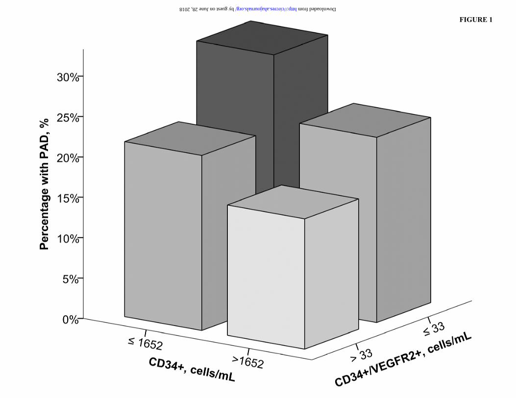
FIGURE 1
by guest on June 28, 2018 http://circres.ahajournals.org/ Downloaded from

FIGURE 2
by guest on June 28, 2018 http://circres.ahajournals.org/ Downloaded from

Riyaz S Patel, Jinhee Kim, Edmund K Waller and Arshed A QuyyumiYi-an Ko, Sean Healy, Iraj Ghaini, Hina Ahmed, Brandon Gray, Salman Sher, Nima Ghasemzadeh,
Salim S Hayek, James MacNamara, Ayman Samman Tahhan, Mosaab Awad, Adithya Yadalam,Artery Disease
Circulating Progenitor Cells Identify Peripheral Arterial Disease in Patients With Coronary
Print ISSN: 0009-7330. Online ISSN: 1524-4571 Copyright © 2016 American Heart Association, Inc. All rights reserved.is published by the American Heart Association, 7272 Greenville Avenue, Dallas, TX 75231Circulation Research
published online June 6, 2016;Circ Res.
http://circres.ahajournals.org/content/early/2016/06/06/CIRCRESAHA.116.308802World Wide Web at:
The online version of this article, along with updated information and services, is located on the
http://circres.ahajournals.org/content/suppl/2016/06/06/CIRCRESAHA.116.308802.DC1Data Supplement (unedited) at:
http://circres.ahajournals.org//subscriptions/
is online at: Circulation Research Information about subscribing to Subscriptions:
http://www.lww.com/reprints Information about reprints can be found online at: Reprints:
document. Permissions and Rights Question and Answer available in the
Request Permissions in the middle column of the Web page under Services. Further information about this process isOffice. Once the online version of the published article for which permission is being requested is located, click
can be obtained via RightsLink, a service of the Copyright Clearance Center, not the EditorialCirculation Research Requests for permissions to reproduce figures, tables, or portions of articles originally published inPermissions:
by guest on June 28, 2018http://circres.ahajournals.org/
Dow
nloaded from

1
Supplemental Material
Title: Circulating Progenitor Cells Identify Peripheral Arterial Disease in Patients with Coronary Artery Disease
Contents
Supplementary Methods and Figure Legends………………………………………………2
Online Table I:………………………………………………………………………………3
Online Figure I:……………………………………………………………………………...4
Online Figure II:……………………………………………………………………………..5

2
Supplementary Methods Progenitor Cell Quantification by Flow Cytometry
We incubated 300µl of peripheral blood with 7ul of the same fluorochrome-labeled
monoclonal anti-human mouse antibodies but with the addition of 3µl PE-Cy7-conjugated anti-
CXCR4 (EBioscience, clone 12G5). Prior to flow cytometry, 100µl of AccuCheck Counting Beads
(Invitrogen, Cat#: PCB100) were added to act as an internal standard for direct estimation of the
concentration of target cell subsets. At least 2.5 million events were acquired from the Cytometer.
Flow data were analyzed with Flowjo software (Treestar, Inc.). Absolute mononuclear cell count was
estimated as the sum of lymphocytes and monocytes using a Coulter ACT/Diff cell counter (Beckman
Coulter). CD45med cells, also referred to as CD45dim cells exclude CD45bright and CD45-(negative)
cells. By excluding CD45- we exclude non-hematopoioetic progenitors. By excluding the rare
CD45bright cells we exclude lymphoblasts.
Figure Legends
Online Figure I. Flow diagram describing subject selection for analysis.
Online Figure II. Flow cytometric analysis of human peripheral blood. Panel A: forward scatter and
side scatter gates following lyse-no wash of blood and the addition of fluorescent counting beads (left
upper corner in plot). Panel B: gating of CD34+, low side scatter cells from blood leukocytes shown
in panel A. Panel C: histograms of CD45 expression in the CD34+ low side scatter cells (red
histogram) shown in panel B. Panel C: the pattern of co-expression of CD34 and CD45dim on blood
progenitors gated. Panel D: the co-expression of CD133 and VEGFR2 on CD34+CD45dim blood
progenitors.

3
Online Table I. Correlations between Progenitor Cell Subtypes
CD34+ CD34+/CD133+ CD34+/CXCR4+ CD34+/VEGFR2+ CD34+ 0.908 0.848 0.217 CD34+/CD133+ 0.908 0.745 0.124 CD34+/CXCR4+ 0.848 0.745 0.341 CD34+/VEGFR2+ 0.217 0.124 0.341 All correlations were statistically significant at P<0.001

4
Online Figure I. Flow Diagram

5
Online Figure II. Flow Cytometry Analysis Of Blood Progenitor Cells