stimulation of cholesterol accumulation dna...
TRANSCRIPT

311
Triggerlike Stimulation of CholesterolAccumulation and DNA and ExtracellularMatrix Synthesis Induced by AtherogenicSerum or Low Density Lipoprotein in
Cultured CellsAlexander N. Orekhov, Vladimir V. Tertov,
Sergey A. Kudryashov, and Vladimir N. Smirnov
A 72-hour incubation of cultured cells with blood sera or plasma of patients suffering fromcoronary heart disease (CHD) with angiographically assessed coronary atherosclerosis causeda threefold to fourfold elevation of intracellular cholesterol. An elevated cholesterol level in thecells precultured with patients' sera was retained several days after the removal of the examinedserum from culture. The accumulation of intracellular cholesterol was accompanied byenhanced synthesis of DNA, total protein, collagen, sulfated glycosaminoglycans, and hyal-uronic acid. Enhanced DNA and total protein synthesis was retained for at least 9 days afterthe serum had been removed from culture. The obtained results suggest that the sera of CHDpatients possess an atherogenic potential that manifests itself at the arterial cell level in thestable stimulation of atherosclerotic cellular processes: proliferation, lipidosis, and fibrosis.The examined sera of healthy donors were devoid of such an atherogenic potential. The lowdensity lipoprotein (LDL) fraction (density, 1.030-1.050 g/cm3) obtained from an atherogenicserum had the same atherogenic potential as a whole serum. Atherosclerotic alterations incultured intimal cells caused by atherogenic LDL were retained for at least 3 days after theremoval of the lipoprotein from culture. Preincubation of intimal cells with LDL obtained fromhealthy donors had no effect on the intracellular cholesterol level or the synthesis of DNA andextracellular matrix. One may assume that the atherogenic potential of CHD patients' sera isrelated to the presence of LDLs that are qualitatively different from the LDL of healthysubjects. (Circulation Research 1990;66:311-320)
W e have recently found that the blood sera ofmost (>90%) patients with angiographi-cally assessed coronary atherosclerosis
are able to induce the accumulation of lipids incultured smooth muscle cells obtained from grosslynormal intima of human aorta.'-4 This property ofpatients' sera was termed "atherogenicity." Athero-genic sera caused a significant elevation of freecholesterol, triglycerides, and, especially, cholesterylesters in cultured cells. The degree of intracellularlipid accumulation depended on the concentration ofserum added to culture: the total cholesterol level
From the Institute of Experimental Cardiology, USSR Cardiol-ogy Research Center, Moscow.Address for correspondence: Alexander N. Orekhov, PhD,
Institute of Experimental Cardiology, USSR Cardiology ResearchCenter, 3rd Cherepkovskaya Street 15A, 121552 Moscow, Russia.
Received August 1, 1988; accepted August 18, 1989.
reached the maximum at 20-40% atherogenicserum. The sera of most healthy donors had noatherogenicity.
Subsequently, it was demonstrated that low densitylipoprotein (LDL) isolated from an atherogenicserum possesses the capacity to induce the accumu-lation of intracellular cholesterol in intimal cellcultures.2-4 Most preparations of other lipoproteinclasses as well as LDL obtained from healthy donorswere nonatherogenic.
In addition to the accumulation of intracellularlipids, atherosclerotic lesions also are characterizedby cell proliferation activity and connective tissueaccumulation. We investigated whether the sera andLDLs obtained from the blood of patients withcoronary heart disease (CHD) also possessed theability to stimulate the other two essential athero-genic processes: cell proliferation and extracellular
by guest on June 27, 2018http://circres.ahajournals.org/
Dow
nloaded from

312 Circulation Research Vol 66, No 2, February 1990
matrix formation. For this purpose, we compared theeffects of the atherogenic sera obtained from CHDpatients with the effects of nonatherogenic sera takenfrom healthy donors.
Materials and MethodsReagentsThe reagents were purchased from Sigma Chemi-
cal, St. Louis, Missouri, if not stated otherwise.
Donors' Sera and PlasmaFor this study, we selected the sera of six CHD
patients with angiographically documented coronaryatherosclerosis and the sera of six healthy donors,each selected on the basis of a single criterion: theability or inability of the serum to induce the accu-mulation of cholesterol in cultured cells (atherogenicand nonatherogenic sera, respectively). All theselected sera of CHD patients were atherogenic, andall the sera of healthy subjects were nonatherogenic.During 24 hours of incubation, the sera of CHDpatients raised the cholesterol level in the cells by75-262% (average, 150%). The atherogenicity ofcorresponding plasma was lower (49-162%: average,98%). Ability of patients' sera to induce the accumu-lation of cholesterol was within the variation rangecharacteristic of atherogenic sera.' Neither sera norplasma of healthy donors' blood caused a statisticallysignificant accumulation of intracellular cholesterol.The characteristics of CHD patients and healthydonors were described in detail elsewhere.12 Thegroup of patients and healthy donors was comparablewith respect to such parameters as sex and age, andnone of the 12 participants had diabetes mellitus.The mean cholesterol level in both types of the serawas the same: 217±6 mg/dl for patients and 219±7mg/dl for healthy subjects.
In the morning, before meals, blood was drawnfrom the cubital vein into plastic tubes with caps. Forserum preparation, the blood was incubated for 1hour at 37° C and centrifuged for 20 minutes at 3,000rpm. Blood sera were used immediately after prepa-ration. For preparation of plasma, the blood wascollected into 1 mg/ml Na2EDTA and then centri-fuged. The lipoprotein-deficient fraction wasobtained by centrifuging the serum at 300,000g (den-sity, 1.250 g/cm3) for 48 hours at +40 C according toLindgren5 and dialyzing as described below. Theobtained sera and plasma were sterilized by filtration(pore size, 0.22 gm).
LipoproteinsLDL (density, 1.030-1.050 g/cm3) was isolated from
the plasma obtained from patients and healthy donorsaccording to the conventional method, describedelsewhere,2,4 of ultracentrifugation in a stepwise gra-dient of NaBr3. Protein content in the lipoproteinpreparations was determined according to Lowry etal.6 Lipid composition of LDL preparation was esti-mated as described earlier.7 The percent composition
by weight of the 1.030-1.050 g/cm3 fraction was 22%protein, 52% cholesterol, 6% triglycerides, and 20%phospholipid for healthy donors' LDL. The corre-sponding ratio for CHD patients' LDL was 22%, 50%,8%, and 20%, respectively (not statistically differentfrom the values for the LDL of healthy donors). LDLsobtained from healthy donors were modified by mal-ondialdehyde according to Haberland et al.8 InsolubleLDL complexes with heparin, fibronectin, and gelatinwere prepared according to Falcone et a19 asdescribed elsewhere.10 Lipoprotein preparations andthe lipoprotein-deficient serum were dialyzed for 24hours against 2,000 vol phosphate-buffered saline(PBS), sterilized by filtration, and stored for not morethan 2 weeks at +40 C. Lipoproteins were used within1-4 days of preparation.
Cell CulturesCells were obtained from thoracic aorta of 40- to
60-year-old men and women within 1.5-3 hours ofsudden death caused mainly by myocardial infarction.Subendothelial smooth muscle cells were isolatedfrom a grossly normal intima by dispersion of aortictissue with collagenase and were cultured as describedelsewhere.7,11 A suspension of the isolated cells wasresuspended in the growth medium containingMedium 199, 10% fetal calf serum, 2mM L-glutamine,100 U/ml penicillin, 100 ,ug/ml streptomycin, and 2.5,ug/ml fungizone (all reagents from GIBCO, GrandIsland, New York), and seeded into Linbro® 24-welltissue culture plates (Flow Laboratories, UK) with adensity of 2-4x10⁴ cells/cm2 of the growth area. Thecells were cultured at 370 C in an atmosphere of 95%air-5% CO2 in a humidified CO2-incubator. The pri-mary cultures contained a mixed cell population com-posed primarily of typical and modified smooth mus-cle cells.7 The medium was changed every day. Alladditions were diluted to the final concentration withMedium 199. During the experiments, the medium inall cultures was changed every day with standardgrowth medium or replaced with growth mediumcontaining 40% serum under investigation or 10%lipoprotein-deficient human serum, combined with100 gg LDL protein/ml. Twenty-four hours before theend of the experiment radioactively labeled precursorswere added to culture to determine the synthesis ofDNA and extracellular matrix.
CholesterolBefore cholesterol determination the cells were
rinsed three times with PBS, treated with 0.025%Trypsin-EDTA for 5 minutes, and washed with PBSfive times (all reagents were from GIBCO). The cellswere removed from the substrate with 0.25%Tryspin-EDTA and washed twice by centrifugation(200g for 10 minutes). Lipids were extracted fromcells with a chloroform-methanol mixture (1:2 vol/vol) according to Bligh and Dyer.12 The total choles-terol content in the lipid extracts was determinedusing Boehringer-Mannheim Monotest®, Choles-terol CHOD-PAP Method (catalogue No. 236691,
by guest on June 27, 2018http://circres.ahajournals.org/
Dow
nloaded from

Orekhov et al Atherogenicity of CHD Patients' Sera 313
TABLE 1. Effects of Patients' Sera on Atherosclerotic Parameters of Subendothelial Cells Cultured From Grossly Normal Intima ofHuman Aorta
SulfatedCholesterol Total protein Collagen glycosaminoglycan Hyaluroniccontent Proliferation synthesis synthesis synthesis acid synthesis
Serum (,ug/mg*) (dpm/lig*) (dpm/gg*) (dpm/lg*) (dpm/,.g*) (dpm/pg*)Version 1 Studied serum was added on days 6, 7, 8, and 9;
cellular indexes were measured on day 10Nonatherogenic 64+13 185+13 1,412+98 306+21 2,093+115 1,027±115Atherogenic 208±15t 452±42t 1,422±94 297±23 2,239+139 1,275±87tVersion 2 Studied serum was added on days 6, 7, and 8;
studied serum was removed on day 9; cellularindexes were measured on day 10
Nonatherogenic 55±7 142±11 423±28 22±2 994±72 721±53Atherogenic 172±13t 234±19t 606±52t 37±3t 1,524±106t 1,327±97tVersion 3 Studied serum was added on days 3, 4, and 5;
studied serum was removed on day 6; cellularindexes were measured on day 10
Nonatherogenic 65±6 107±9 258±30 8±1 606±32 402±43Atherogenic 161±14t 240±18t 405±22t 23±2t 590±60 711±50tVersion 4 (control) Cultivation under standard conditions from days 1 to 10; cellular
indexes were measured on day 10Fetal calf serum 55±6 96±11 363±33 11±1 565±58 337±11
Atherogenic sera of patients and nonatherogenic sera of healthy donors were used. For each examined serum, atherosclerotic cellularindexes were determined in three separate wells. Version 1, studied serum was added on days 6, 7, 8, and 9; cellular indexes were measuredon day 10. Version 2, studied serum was added on days 6, 7, and 8; removed on day 9; and cellular indexes were measured on day 10. Version3, studied serum was added on days 3, 4, and 5; removed on day 6; and cellular indexes were measured on day 10. Version 4 (control),cultivation under standard conditions from days 1 to 10; cellular indexes were measured on day 10.
Values listed are means of the respective index for cells cultured with the sera of patients and healthy donors±SEM.*Micrograms (milligrams) of cell protein.tSignificant difference between the sera of patients and healthy subjects (p<0.05).
Boehringer-Mannheim GmbH, Mannheim, FRG).Cholesterol stock standard (stock No. 965-25, SigmaChemical) was used for standard preparation.
DNA SynthesisDNA synthesis was evaluated by the incorporation
of [3H]thymidine into the acid-insoluble cell fractionas described elsewhere.13
Total Protein SynthesisAfter incubation with 1 ,uCi/ml [4,5-3H]leucine (135
Ci/mmol, Amersham International, Amersham, UK),the incubation medium and the cells, suspended witha rubber policeman, were heated for 10 minutes at800 C, and proteins were precipitated by 10% tri-chloroacetic acid (TCA). The precipitate was washedtwice with 5% TCA and twice with diethyl ether,dissolved with 0.5N KOH, and precipitated again by10% TCA. The precipitate was resuspended in 5%TCA, and its radioactivity was measured.
Collagen SynthesisThe synthesis of secreted collagen was determined
according to Peterkofsky and Diegelmann14 on
['H]proline incorporation in the collagenase-sensitive fraction.
Glycosaminoglycan SynthesisThe synthesis of sulfated glycosaminoglycans and
hyaluronic acid was determined according to Saarni
and Tammi15 on incorporation of [3H]glucosaminehydrochloride.
Statistical AnalysisThe significance of differences was evaluated by
dispersion analysis methods using a statistical pro-gram package.16
ResultsWe compared the atherosclerosis-related effects of
the atherogenic sera taken from CHD patients withthe effects of nonatherogenic sera taken from healthydonors. Our experiments were threefold. First, ath-erosclerotic indexes were measured in the presenceof the serum (Table 1, version 1). Second, the sameindexes were determined on the next day after serumremoval (Table 1, version 2). Third, the determina-tion of atherosclerotic indexes was performed 3 daysafter serum removal (Table 1, version 3). As acontrol, we used cells cultured under standard con-ditions, that is, in the presence of fetal calf serum(Table 1, version 4).
Cultivation of intimal subendothelial cells in thepresence of atherogenic sera for 96 hours (from day6 to day 9 in primary culture) led to a nearly fourfoldrise in the total intracellular cholesterol by day 10(Table 1, compare versions 1 and 4). Similar expo-sure of nonatherogenic sera did not significantly alterthe cholesterol level in cultured cells.
by guest on June 27, 2018http://circres.ahajournals.org/
Dow
nloaded from

314 Circulation Research Vol 66, No 2, February 1990
Cultivation of intimal cells in the presence ofhuman sera stimulated the DNA synthesis. Incorpo-ration of ['H]thymidine into the DNA of cells cul-tured with nonatherogenic sera of healthy subjectswas nearly twofold higher compared with the['H]thymidine incorporation into cells culturedunder standard conditions, that is, with fetal calfserum (Table 1, compare versions 1 and 4). Athero-genic sera had an even stronger stimulative effect onthe DNA synthesis of cultured cells. The incorpo-ration of [3H]thymidine in the presence of athero-genic sera was substantially higher than in the pres-ence of nonatherogenic sera.
Cultivation of intimal cells with both atherogenicand nonatherogenic sera sharply increased the syn-thesis of total protein, collagen, sulfated glycosami-noglycans, and hyaluronic acid. As compared withthe cells cultured in the presence of fetal calf serum,the above-mentioned indexes in the cells culturedwith human sera were several times higher (Table 1,compare versions 1 and 4). Atherogenic and non-atherogenic sera were no different with respect totheir ability to increase the synthesis of total protein,collagen, and sulfated glycosaminoglycans. However,atherogenic sera were much more powerful in thestimulation of hyaluronic acid synthesis as comparedwith nonatherogenic sera.To find out how the presence of serum affects the
parameters in question, we modified the experimentso that cells were cultured with atherogenic ornonatherogenic sera for 72 hours, from day 6 to day8. On day 9, the examined sera were replaced with astandard fetal calf serum and cells were cultured foranother 24 hours. Then, on day 10, the cholesterollevel and the synthesis of DNA, total protein, colla-gen, sulfated glycosaminoglycans, and hyaluronicacid were measured (Table 1, version 2). Under theseconditions, we revealed a nearly threefold elevationof cholesterol in the cells cultured with atherogenicsera (Table 1, compare versions 2 and 4). Preexpo-sure to nonatherogenic sera had no effect on intra-cellular cholesterol (Table 1, version 2).A 3-day preexposure of intimal cells to atherogenic
sera had a significantly higher stimulative effect onthe synthesis of DNA, total protein, collagen, sul-fated glycosaminoglycans, and hyaluronic acid com-pared with the preexposure to nonatherogenic sera(Table 1, version 2). Thus, preexposure of intimalcells to atherogenic sera of CHD patients led to theaccumulation of intracellular cholesterol, enhancedsynthesis of DNA, and extracellular matrix, that is,all the major manifestations of atherosclerosis at thecellular level. On the other hand, preexposure tononatherogenic sera of healthy donors had little orno effect on atherosclerotic parameters.We investigated whether the activation of athero-
sclerotic cellular manifestations induced by anatherogenic serum is retained for a certain period oftime. A new modification of the experiment was asfollows (Table 1, version 3). On days 3, 4, and 5, theculture medium was replaced with a fresh one con-
taining atherogenic or nonatherogenic sera. On day6, (i.e., after 72 hours of incubation with the exam-ined sera), the medium was again replaced with astandard one containing fetal calf serum, and thecells were cultured under standard conditions foranother 72 hours (i.e., until day 10). Then, all theatherosclerotic indexes were measured.On day 10, none of the indexes in the cells
preincubated with nonatherogenic sera of healthydonors from day 3 to day 6 significantly differed(p>0.05) from the respective characteristics of thecells cultured under standard conditions in thepresence of fetal calf serum for the whole experi-mental period, that is, from days 1 to 10 (Table 1,compare versions 3 and 4). Thus, a 72-hour preex-posure of intimal cells to nonatherogenic sera doesnot affect atherosclerotic cellular indexes even 3days after the removal of nonatherogenic sera fromculture.A 3-day preexposure of cells to atherogenic sera of
CHD patients led to stable alterations in atheroscle-rotic cellular indexes. Even 3 days after the transferof culture into standard conditions, the cells preex-posed to atherogenic sera had a much higher choles-terol level and a higher synthesis of DNA, totalprotein, collagen, and hyaluronic acid than the con-trol cells (Table 1, compare versions 3 and 4). Of allthe atherosclerotic parameters, only the synthesis ofsulfated glycosaminoglycans showed nonsignificantdifferences from the control (standard) culture.
In connection with the ability of atherogenic serato cause stable atherosclerotic alterations in intimalcells, it was essential to determine the durability ofthese alterations. Intimal cells were cultured withatherogenic or nonatherogenic sera from day 3 to day6. On day 6, the medium was replaced with astandard one containing fetal calf serum. From day 7to day 14, the three major parameters were mea-sured: total intracellular cholesterol and the synthe-sis of DNA and total protein.From day 7 to day 10, the cholesterol level in the
cells precultured with atherogenic sera from day 3 today 6 was 2.2-fold higher than in the cells pre-cultured with nonatherogenic sera (Figure 1). Afterday 10, the intracellular cholesterol level decreased,although by day 14 it significantly exceeded thecholesterol content in the cells preexposed tononatherogenic sera. The cholesterol content in thecells preexposed to nonatherogenic sera showed nochanges from day 7 to day 14.The [3H]thymidine incorporation into intimal cells
increased with a period of cultivation (Figure 1). Thisreflects the process of gradual involvement of cul-tured human aortic cells into proliferation. As wasshown earlier,11'13 this process is especially activeduring the second week in primary culture. From day8 to day 14, the [3H]thymidine incorporation into theDNA of the cells preexposed to nonatherogenic seraincreased from 188±15 dpm/,g cell protein to379±19 dpm/,g cell protein while in the cells preex-posed to atherogenic sera it increased from 291+±15
by guest on June 27, 2018http://circres.ahajournals.org/
Dow
nloaded from

Orekhov et al Atherogenicity of CHD Patients' Sera 315
CHOLESTEROL200
1SO0
4 12 16
PROLIFERATION600
400
300
200 -
*...1., ,.,4 8 12 16
PROTEIN SYNTHESIS
500~~~~~~~~~T000 _
,o f~i,I,,t_-'-*---
000 -
SOO -I aIE1
4 8 12 16
FIGURE 1. Total intracellular cholesterol content, prolifera-tive activity, and total protein synthesis in cultures of intimalcells preexposed to the sera of patients with coronaty heartdisease or healthy donors. In a 3-day primary culture ofsubendothelial cells isolated from grossly normal intima ofhuman aorta, the standard culture medium was replaced witha medium containing 40% serum of coronary heart diseasepatients or 40% serum ofhealthy donors. On days 4 and 5, theculture medium was replaced with a fresh one containing thetested serum. Starting at day 6, the culture medium wasreplaced daily with fresh standard medium containing 10%fetal calfserum. Twenty-four hours before the determination ofatherosclerotic parameters, radioactively labeled precursors[3H]thymidine or [3H]proline were added to the respectiveculture. The total cholesterol level andprecursor incorporationwere determined on days 7, 8, 10, 12, and 14. The respectiveindexes for each tested serum were determined in three sepa-rate wells. Values listed are mean +SEM of the correspondingindex for the cells preexposed to patients' sera (solid line) orhealthy donors' sera (broken line). Asterisks mark significantdifferences between the indexes ofcellspreexposed to these sera(p<0.OS).
DAYS IN CULTURE
dpm4l,g cell protein to 495+±38 dpm/Iug cell protein.Any time the measurements were taken (up to day14), the DNA synthesis in the cells preexposed toatherogenic sera was significantly higher than in thecells preexposed to nonatherogenic sera (Figure 1).
Atherogenic sera caused a stable stimulation oftotal protein synthesis. At least until day 14 inprimary culture, the intimal cells preexposed to
atherogenic sera from day 3 to day 6 synthesizedprotein at a considerably higher rate as comparedwith the cells preexposed to nonatherogenic sera(Figure 1).Thus, the interaction of atherogenic CHD
patients' sera with subendothelial cells of humanaortic intima for 3 days led to a long-term activationof lipidosis, proliferation, and fibrosis, that is, exactly
c'E
=
a
c
'a
0
0.0P
2
2
P-#%~
l
l by guest on June 27, 2018
http://circres.ahajournals.org/D
ownloaded from
316 Circulation Research Vol 66, No 2, February 1990
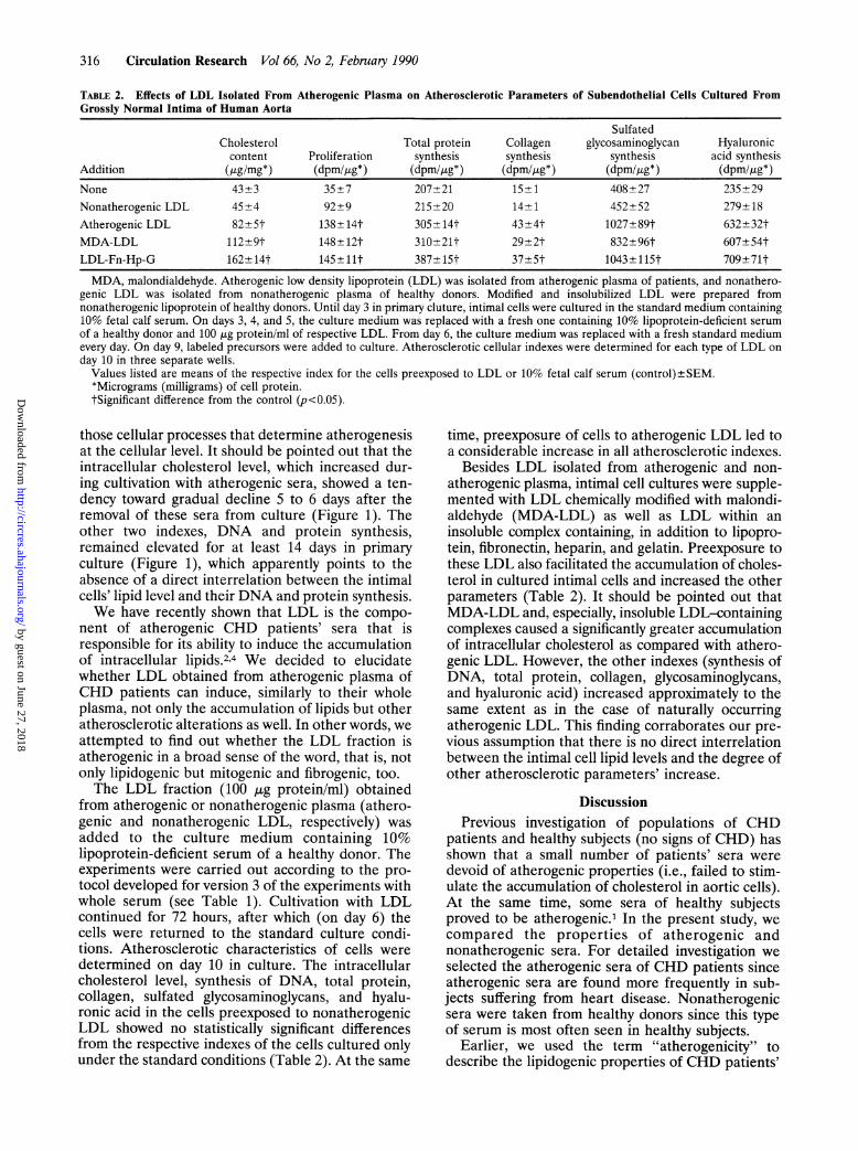
TABLE 2. Effects of LDL Isolated From Atherogenic Plasma on Atherosclerotic Parameters of Subendothelial Cells Cultured FromGrossly Normal Intima of Human Aorta
SulfatedCholesterol Total protein Collagen glycosaminoglycan Hyaluroniccontent Proliferation synthesis synthesis synthesis acid synthesis
Addition (,ug/mg*) (dpm/4g*) (dpm/4g*) (dpm/gg*) (dpm/Ipg*) (dpm/4g*)None 43+3 35+7 207+21 15+1 408+27 235+29Nonatherogenic LDL 45+4 92+9 215+20 14±1 452+52 279±18Atherogenic LDL 82±5t 138-+-14t 305-+14t 43±4t 1027±89t 632+32tMDA-LDL 112±9t 148±12t 310±21t 29±2t 832±96t 607±54tLDL-Fn-Hp-G 162±14t 145±11t 387±15t 37±5t 1043±115t 709±71tMDA, malondialdehyde. Atherogenic low density lipoprotein (LDL) was isolated from atherogenic plasma of patients, and nonathero-
genic LDL was isolated from nonatherogenic plasma of healthy donors. Modified and insolubilized LDL were prepared fromnonatherogenic lipoprotein of healthy donors. Until day 3 in primary cluture, intimal cells were cultured in the standard medium containing10% fetal calf serum. On days 3, 4, and 5, the culture medium was replaced with a fresh one containing 10% lipoprotein-deficient serumof a healthy donor and 100 ,ug protein/ml of respective LDL. From day 6, the culture medium was replaced with a fresh standard mediumevery day. On day 9, labeled precursors were added to culture. Atherosclerotic cellular indexes were determined for each type of LDL onday 10 in three separate wells.
Values listed are means of the respective index for the cells preexposed to LDL or 10% fetal calf serum (control)±SEM.*Micrograms (milligrams) of cell protein.tSignificant difference from the control (p<0.05).
those cellular processes that determine atherogenesisat the cellular level. It should be pointed out that theintracellular cholesterol level, which increased dur-ing cultivation with atherogenic sera, showed a ten-dency toward gradual decline 5 to 6 days after theremoval of these sera from culture (Figure 1). Theother two indexes, DNA and protein synthesis,remained elevated for at least 14 days in primaryculture (Figure 1), which apparently points to theabsence of a direct interrelation between the intimalcells' lipid level and their DNA and protein synthesis.We have recently shown that LDL is the compo-
nent of atherogenic CHD patients' sera that isresponsible for its ability to induce the accumulationof intracellular lipids.2'4 We decided to elucidatewhether LDL obtained from atherogenic plasma ofCHD patients can induce, similarly to their wholeplasma, not only the accumulation of lipids but otheratherosclerotic alterations as well. In other words, weattempted to find out whether the LDL fraction isatherogenic in a broad sense of the word, that is, notonly lipidogenic but mitogenic and fibrogenic, too.The LDL fraction (100 ,ug protein/ml) obtained
from atherogenic or nonatherogenic plasma (athero-genic and nonatherogenic LDL, respectively) wasadded to the culture medium containing 10%lipoprotein-deficient serum of a healthy donor. Theexperiments were carried out according to the pro-tocol developed for version 3 of the experiments withwhole serum (see Table 1). Cultivation with LDLcontinued for 72 hours, after which (on day 6) thecells were returned to the standard culture condi-tions. Atherosclerotic characteristics of cells weredetermined on day 10 in culture. The intracellularcholesterol level, synthesis of DNA, total protein,collagen, sulfated glycosaminoglycans, and hyalu-ronic acid in the cells preexposed to nonatherogenicLDL showed no statistically significant differencesfrom the respective indexes of the cells cultured onlyunder the standard conditions (Table 2). At the same
time, preexposure of cells to atherogenic LDL led toa considerable increase in all atherosclerotic indexes.
Besides LDL isolated from atherogenic and non-atherogenic plasma, intimal cell cultures were supple-mented with LDL chemically modified with malondi-aldehyde (MDA-LDL) as well as LDL within aninsoluble complex containing, in addition to lipopro-tein, fibronectin, heparin, and gelatin. Preexposure tothese LDL also facilitated the accumulation of choles-terol in cultured intimal cells and increased the otherparameters (Table 2). It should be pointed out thatMDA-LDL and, especially, insoluble LDL-containingcomplexes caused a significantly greater accumulationof intracellular cholesterol as compared with athero-genic LDL. However, the other indexes (synthesis ofDNA, total protein, collagen, glycosaminoglycans,and hyaluronic acid) increased approximately to thesame extent as in the case of naturally occurringatherogenic LDL. This finding corraborates our pre-vious assumption that there is no direct interrelationbetween the intimal cell lipid levels and the degree ofother atherosclerotic parameters' increase.
DiscussionPrevious investigation of populations of CHD
patients and healthy subjects (no signs of CHD) hasshown that a small number of patients' sera weredevoid of atherogenic properties (i.e., failed to stim-ulate the accumulation of cholesterol in aortic cells).At the same time, some sera of healthy subjectsproved to be atherogenic.1 In the present study, wecompared the properties of atherogenic andnonatherogenic sera. For detailed investigation weselected the atherogenic sera of CHD patients sinceatherogenic sera are found more frequently in sub-jects suffering from heart disease. Nonatherogenicsera were taken from healthy donors since this typeof serum is most often seen in healthy subjects.
Earlier, we used the term "atherogenicity" todescribe the lipidogenic properties of CHD patients'
by guest on June 27, 2018http://circres.ahajournals.org/
Dow
nloaded from

Orekhov et al Atherogenicity of CHD Patients' Sera 317
sera manifested in the elevation of intracellularlipids.1-4 The use of this term proved to be quitejustified. We have shown in this study that CHDpatients' sera are not only lipidogenic but are mito-genic and fibrogenic, too. In other words, they pos-
sess a universal atherogenicity manifested under theconditions of culture. Thus, blood serum of mostCHD patients contains all the elements necessary forthe onset of atherogenesis in the vessel wall.
Sera of healthy donors (subjects with no signs ofCHD) were nonlipidogenic, but had mitogenic andfibrogenic properties (i.e., stimulated the synthesis ofDNA and extracellular matrix components). How-ever, the mitogenic and fibrogenic effects of healthydonors' sera proved to be substantially lower afterthe serum was removed from culture, and completelydisappeared 3 days later. Consequently, to manifestthese properties, the sera of healthy donors require a
constant contact with the cells. At the same time, theeffects exerted on the intimal cells by CHD patients'sera were noticeable for at least 8 days after theserum was removed from culture. This means thatthe CHD patients' sera may act as a trigger, inducingcellular atherogenesis. Subsequently, the atherogenicprocess can develop even in the absence of thetrigger.The cellular indexes associated with atherosclero-
sis were investigated in many studies performed on
different cultures. These studies focused on theeffects of hyperlipidemic and diabetic sera upon themajor atherosclerotic cellular indexes: accumulationof lipids, proliferative activity, and extracellularmatrix synthesis. A summary of the obtained resultsfollows.
Hyperlipidemic sera from cholesterol-fed animalsinduced lipid accumulation in various tissue culturecells and organ cultures.17-29 The results of studies onthe effect of hyperlipidemic serum on the growth ofculture cells are conflicting. Many authors observedthat hyperlipidemic sera from patients and cholesterol-fed animals enhanced proliferation of cultured cellsof various origins30-37 and promoted labeling of cellsin organ arterial culture with [3H]thymidine.27,38-40At the same time, other workers showed that duringincubation, human hyperlipidemic serum either didnot stimulate or slightly inhibited human arterialsmooth muscle cell proliferation.41'42 Sera of type 2diabetics and sera of diabetic rabbits stimulated thegrowth of homologous smooth muscle cells more
than normal sera.3343 Hyperlipidemic serum did notaffect or slightly inhibited the synthesis of collagen bycultured arterial smooth muscle cells and isolatedaortas.32,42,44-48 However, it was found that hyperlip-idemic serum stimulated the synthesis of collagen byfreshly isolated cells.32,49 Sera from type 1 diabeticpatients significantly increased the production ofcollagen by cultured cells,50 but in the case of sera
from type 2 diabetic patients any stimulatory effectwas absent.5' On the other hand, a clearcut increasein the synthesis of hyaluronic acid was found in thepresence of type 2 diabetic sera.5' The synthesis of
hyaluronic acid by cultured smooth muscle cells wasreduced in the presence of hyperlipidemic sera.41
It can be concluded from these findings that hyper-lipidemia and diabetes mellitus, important risk fac-tors for atherosclerosis and CHD, are characterizedby the presence of blood components that facilitatethe development of certain atherogenic manifesta-tions at the vascular cell level. However, very oftenatherosclerosis and CHD are not accompanied bydiabetes mellitus and/or hyperlipidemia. Recently,we have reported that the blood sera of patients withCHD demonstrate lipidogenic properties irrespectiveof the presence or absence of concomitant diabetesmellitus.52 The sera used in present study were notmarkedly hyperlipidemic. Consequently, diabetesmellitus and/or hyperlipidemia cannot be regarded asprerequisites for the realization of CHD patients'serum atherogenic potential at the arterial cell level.
Certain differences between the data obtained inthe present study using the sera ofCHD patients withangiographically assessed atherosclerosis fromresults obtained using hyperlipidemic or diabeticserum may be explained by the two factors. First, weused a homologous system including human serumand primary culture of human aortic subendothelialcells, that is, exactly those cells in which primaryatherosclerotic alterations occur. The cited studies ofother researchers were performed on medial smoothmuscle cells or a heterologous combination of thesera and cells. Second, the difference of our resultsfrom other published studies may reflect specificproperties of CHD patients' sera that cause it todiffer from hyperlipidemic and diabetic sera.Recently, Laughton et a129 confirmed our earlierobservations.' They found that sera from patientswith angiographically documented coronary athero-sclerosis cause marked lipid accumulation in culturedhuman arterial smooth muscle cells.
It was demonstrated that certain atherogenic prop-erties of the serum are mostly associated with lipo-proteins contained in these sera. Avogaro et a153 haverecently found a subfraction of LDL in normalsubjects which causes the deposition of free choles-terol in cultured macrophages. This atherogenicmodified LDL differed from native LDL by a highernegative charge and the somewhat altered lipid com-position. Many studies have demonstrated that LDLobtained from normolipemic, hyperlipemic, or dia-betic serum are mitogenic for arterial cells.30,3454-63Others have shown little or no mitogenic effect ofnormal LDL34,64 and an inhibitory effect of diabeticLDL.65 It was suggested that LDL are not themselvesgrowth factors but rather may supply lipids requiredfor maximal growth of mitogen-stimulated cells.61
Earlier, we demonstrated that LDL, unlike otherclasses of lipoproteins isolated from atherogenicsera, possesses lipidogenicity.2,4 In the present study,we have found that this LDL possesses mitogenicityand fibrogenicity as well. Consequently, the LDLobtained from the blood of CHD patients withcoronary atherosclerosis has the same atherogenic
by guest on June 27, 2018http://circres.ahajournals.org/
Dow
nloaded from

318 Circulation Research Vol 66, No 2, February 1990
potential as whole serum. At the same time, prein-cubation of cells with LDL obtained from nonathero-genic plasma did not induce the accumulation ofintracellular lipids or increase the rate of DNA andextracellular matrix synthesis. The presence ofatherogenic potential in LDL obtained from theblood of CHD patients allows us to assume that thislipoprotein fraction is responsible for the atheroge-nicity of their whole blood serum.The basic differences in the properties of LDL
obtained from CHD patients and healthy donors thatmanifest themselves during the interaction with inti-mal cells led to the assumption that these types ofLDL are qualitatively different. It is known that inculture, normal LDL fails to induce the accumulationof intracellular lipids,66-69 whereas chemically modi-fied LDL or LDL insolubilized by forming a complexwith extracellular matrix components is able to causemassive deposition of lipids within the cells.7,8,66,69-73In present work, we have tested two types of modifiedlipoproteins: MDA-LDL and LDL insolubilized byfibronectin, heparin, and gelatin. In both cases, weobserved the accumulation of cholesterol in culturedintimal cells accompanied with increased prolifera-tion and production of connective tissue matrix.Thus, not only LDL obtained from CHD patients'blood but initially normal LDL modified by differentmethods, too, possesses an atherogenic potentialmanifested in cell culture.
Recently, we have demonstrated that the differenceof CHD patients' LDL from native LDL of healthydonors is explained by low sialic acid content.74Atherogenic patients' LDL had a twofold to fivefoldlower level of sialic acid as compared with native LDL.On the other hand, initially native LDL modified bypartial desialylation with neuraminidase caused lipidaccumulation in cultured cells. A detailed study ofnaturally occurring modified (desialylated) LDL is thesubject of our current investigations.
In our view, the fact that the blood of CHDpatients contains LDL that possesses atherogenicproperties constitutes a very important finding. As isknown, LDL can penetrate through the endotheliallining and accumulate within the intima.75 One mayassume that once within the intima atherogenic LDL,through interaction with subendothelial cells, cancause a whole range of atherosclerotic processesoccurring at the cellular level: lipidosis, enhancedproliferation, and fibrosis.We assume that the major event inducing athero-
genesis is primary cellular lipidosis, that is, the accu-mulation of intracellular lipids induced by athero-genic LDL. In our view, stimulation of cellproliferation and enhanced connective tissue synthe-sis accompanying primary lipidosis are the conse-quences of intracellular lipid deposition. The mito-genic and fibrogenic effects of cell preincubation withatherogenic LDL were unrelated to the presence oflipoprotein in culture, since these effects were man-ifested 3 days after the removal of LDL from theculture medium. This finding brought us to the
conclusion that the stimulation of proliferation andenhanced production of the extracellular matrix com-ponents arise from the deposition of intracellularcholesterol caused by atherogenic LDL. We thinkthat the absence of a direct relation between mito-genicity, fibrogenicity, and the degree of primarycellular lipidosis means that the decisive factor is thedeposition of excess lipid and not the amount.
Furthermore, our data allow the assumption thatthe development of cellular atherogenesis does notrequire constant transport of atherogenic LDL intothe intima, since atherosclerotic alterations causedby this lipoprotein proved to be rather stable. Thissuggests two important conclusions. First, a brieflocal disturbance in the permeability of the endothe-lial lining, leading to massive penetration of athero-genic LDL within the subendothelial intima, mayinitiate atherogenesis even if normal permeability issubsequently restored. Second, constant presence ofatherogenic LDL in the circulation is not necessaryfor the development of atherogenesis in the vesselwall. The emergence of atherogenicity in the bloodeven for a short time can be sufficient for the onset ofatherosclerotic alterations in the vessel, after whichatherogenesis would develop even if atherogenicfactors disappear. This assumption may account forthe fact that in a broad population of patients withcoronary atherosclerosis, we found about 10% ofpatients whose blood had no atherogenic propertiesat the moment of study.1
AcknowledgmentsThe authors thank Dr. S.N. Pokrovsky and his
coworkers for their assistance with LDL isolation andMiss I. Beskhlebnova for illustrative work.
References1. Chazov El, Tertov VV, Orekhov AN, Lyakishev AA, Perova
NV, Kurdanov KhA, Khashimov KhA, Novikov ID, SmirnovVN: Atherogenicity of blood serum from patients with coro-nary heart disease. Lancet 1986;2:595-598
2. Orekhov AN, Tertov VV, Pokrovsky SN, Adamova IYu,Martsenyuk ON, Lyakishev AA, Smirnov VN: Blood serumatherogenicity associated with coronary atherosclerosis: Evi-dence for nonlipid factor providing atherogenicity of low-density lipoproteins and an approach to its elimination. CircRes 1988;62:421-429
3. Chazov El, Orekhov AN, Tertov VV, Pokrovsky SN, AdamovaIYu, Lyakishev AA, Gratsianski NA, Nechaev AS, Perova NV,Khashimov KhA, Kurdanov KhA, Kukharchuk VV, SmirnovVN: Atherogenicity of blood plasma from patients with coro-nary atherosclerosis and its correction. Atherosclerosis Rev1988;7:9-20
4. Tertov VV, Orekhov AN, Martsenyuk ON, Perova NV,Smirnov VN: Low-density lipoproteins isolated from the bloodof patients with coronary heart disease induce the accumula-tion of lipids in human aortic cells. Exp Mol Pathol 1989;50:337-347
5. Lindgren Fr: Preparative ultracentrifugal laboratory proce-dures and suggestions for lipoprotein analysis, in Perkins EG(ed): Analysis ofLipids and Lipoproteins. New York, AmericanOil Chemical Society, 1975, pp 205-224
6. Lowry OH, Rosenbrough NJ, Farr AL, Randall RJ: Proteinmeasurement with the Folin phenol reagent. J Biol Chem1951;193:265-275
by guest on June 27, 2018http://circres.ahajournals.org/
Dow
nloaded from

Orekhov et al Atherogenicity of CHD Patients' Sera 319
7. Orekhov AN, Tertov VV, Novikov ID, Krushinsky AV, Andre-eva ER, Lankin VZ, Smirnov VN: Lipids in cells of athero-sclerotic and uninvolved human aorta: I. Lipid composition ofaortic tissue and enzyme isolated and cultured cells. Exp MolPathol 1985;42:117-137
8. Haberland ME, Fogelman AM, Edwards PA: Specficity ofreceptor-mediated recognition of malondialdehyde-modifiedlow density lipoprotein. Proc Natl Acad Sci USA 1982;79:1712-1716
9. Falcone DJ, Mated N, Shio H, Minick CR: Lipoprotein-heparin-fibronectin-denatured collagen complexes enhancecholesteryl ester accumulation in macrophages. J Cell Biol1984;99:1266-1274
10. Orekhov AN, Tertov VV, Mukhin DN, Koteliansky VE,Glukhova MA, Khashimov KhA, Smirnov VN: Association oflow-density lipoprotein with particulate connective tissuematrix components enhances cholesterol accumulation in cul-tured subendothelial cells of human aorta. Biochim BiophysActa 1987;928:251-258
11. Orekhov AN, Kosykh VA, Repin VS, Smirnov VN: Cellproliferation in normal and atherosclerotic human aorta: II.Autoradiographic observation in deoxyribonucleic acid syn-thesis in primary cell culture. Lab Invest 1983;48:749-754
12. Bligh EG, Dyer WJ: A rapid method of total lipid extractionand purification. Can J Biochem Physiol 1959;37:911-917
13. Orekhov AN, Tertov VV, Kudryashov SA, Khashimov KhA,Smirnov VN: Primary culture of human aortic intima cells as amodel for testing antiatherosclerotic drugs: Effects of cyclicAMP, prostaglandins, calcium antagonists, antioxidants, andlipid-lowering agents. Atherosclerosis 1986;60:101-110
14. Peterkofsky B, Diegelmann R: Use of a mixture of proteinase-free collagenases for the specific assay of radioactive collagenin the presence of other proteins. Biochemistry 1971;10:988-994
15. Saarni H, Tammi M: A rapid method for separation and assayof radiolabelled mucopolysaccharides from cell culturemedium. Anal Biochem 1977;81:40-46
16. Dixon WJ, Brown MB: Biomedical Computer Programs. P-Series. Berkeley, University of California Press, 1977, pp185-198
17. Chen RM, Fischer-Dzoga K: Effect of hyperlipemic serumlipoproteins on the lipid accumulation and cholesterol flux ofrabbit aortic medial cells. Atherosclerosis 1977;28:339-353
18. Bates SR, Wissler RW: Effect of hyperlipemic serum oncholesterol accumulation in monkey aortic medial cells. Bio-chim Biophys Acta 1976;450:78-88
19. StClair RW, Leight MA: Differential effects of isolated lipo-proteins from normal and hypercholesterolemic rhesus mon-keys on cholesterol esterification and accumulation in arterialsmooth muscle cells in culture. Biochem Biophys Acta 1978;530:279-291
20. Pearson JD: Lipid metabolism in cultured smooth muscle cellsand comparison with other types: Part 1. Composition of cellsgrown in hyperlipemic serum. Atherosclerosis 1976;24:233-242
21. Warhurst G, Wynn CH: Subcellular changes associated withculture of Chinese hamster fibroblasts in hyperlipemicmedium. Atherosclerosis 1981;38:383-393
22. Rothblat GH, Arbogast L, Kritchevsky D, Naftulin M: Cho-lesteryl ester metabolism in tissue culture cells: Part II. Sourceof accumulated esterified cholesterol in Fu5AH rat hepatomacells. Lipids 1976;11:97-108
23. Mahley R, Innerarity TL, Weisgraber KH, Fry DL: Caninehyperlipoproteinemia and atherosclerosis: Accumulation oflipid by aortic medial cells in vivo and in vitro. Am J Pathol1977;87:205-225
24. Bater SR: Source of the cholesterol ester accumulated inmonkey arterial smooth muscle cells grown in hyperlipemicserum. Circ Res 1979;45:821-828
25. StClair RW, Harpold GJ: Stimulation of cholesterol esterifi-cation in vitro in organ cultures of normal pigeon aorta. ExpMol Pathol 1975;22:207-219
26. Fischer-Dzoga K, Chen R, Wissler RW: Effect of serumlipoproteins on the morphology, growth, and metabolism of
arterial smooth muscle cells. Adv Exp Med Biol 1977;43:299-311
27. Nikkari T, Pietila K, Salo M: Increased cholesterol contentand esterification in rabbit aortic medial cells cultured inhyperlipidemic serum. Med Biol 1976;54:264-271
28. Rothblat GH: Cholesteryl ester metabolism in tissue culturecells: I. Accumulation in Fu5AH rat hepatoma cells. Lipids1974;9:526-535
29. Laughton CW, Ruddle DL, Bedord CJ, Alderman EL: Seracontaining elevated nonesterified fatty acids from patientswith angiographically documented coronary atherosclerosiscause marked lipid accumulation in cultured human arterialsmooth muscle-derived cells. Atherosclerosis 1988;70:233-246
30. Fischer-Dzoga K, Wissler RW: Stimulation in stationary pri-mary cultures of monkey aortic smooth muscle cells: Part 2.Effects of varying concentrations of hyperlipemic serum andlow density lipoproteins of varying fat origins. Atherosclerosis1976;24:515-525
31. Jarvelainen H, Perltonen J, Ronnemaa T: The growth-promoting effect of human whole blood serum on culturedhuman aortic smooth muscle cells is a constant serum donor-dependent property. In Vitro Cell Devel Biol 1986;22:515-518
32. Ronnemaa T, Doherty NS: Effect of serum and liver extractsfrom hypercholesterolemic rats on the synthesis of collagen byisolated aortas and cultured aortic smooth muscle cells. Ath-erosclerosis 1977;26:261-272
33. Ledet T, Fischer-Dzoga K, Wissler RW: Growth of rabbitaortic smooth muscle cells cultured in media containing dia-betic and hyperlipemic serum. Diabetes 1976;25:207-215
34. Robertson AL Jr: Role of circulating lipoproteins in theproliferative phase of atherogenesis (abstract). Am J Pathol1974;74:94a
35. Bourdillon MC, Boissel JP, Crouzet B: Proliferation of pri-mary cultures from rat aortic media: Effects of hyperlipemicserum. Prog Biochem Pharnacol 1977;13:103-110
36. Fischer-Dzoga K, Kuo Y-F, Wissler RW: The proliferativeeffect of platelets and hyperlipidemic serum on stationaryprimary cultures. Atherosclerosis 1983;47:35-43
37. Fischer-Dzoga K, Wissler RW, Vesselinovitch D: The effect ofestradiol on the proliferation of rabbit aortic medial tissueculture cells induced by hyperlipemic serum. Exp Mol Pathol1983;39:355-363
38. Daoud AS, Fritz KE, Jarmolych J: Increased DNA synthesis inaortic explants from swine fed a high-cholesterol diet. Exp MolPathol 1970;13:377-384
39. Daoud AS, Fritz KE, Jarmolych J, Augustyn JM: Use of aorticmedial explants in the study of atherosclerosis. Exp Mol Pathol1973;18:177-189
40. Florentin RA, Choi BH, Lee KT, Thomas WA: Stimulation ofDNA synthesis and cell division in vitro by serum fromcholesterol-fed swine. J Cell Biol 1969;41:641-645
41. Jarvelainen H, Ronnemaa T, Tammi M, Vihersaari T, Leh-tonen A, Viikari J: Type IIA hyperlipoproteinemic seradecrease the synthesis of hyaluronic acid by cultured humanaortic smooth muscle cells. Atherosclerosis 1981;39:61-69
42. Jarvelainen H, Halme T, Lehtonen A, Ronnemaa T: Serumfrom type IIA hyperlipoproteinemic patients does not stimu-late proliferation of and collagen synthesis in human fetalaortic smooth muscle cells in culture. Atherosclerosis 1985;56:199-211
43. Koschinsky T, Bunting CE, Schwippert B, Gries FA: Increasedgrowth of human fibroblasts and arterial smooth muscle cellsfrom diabetic patients related to diabetic serum factors andcell origin. Atherosclerosis 1979;33:245-252
44. Pietila K, Nikkari T: Effect of phase of growth and hyperlip-idemic serum on the synthesis of collagen in rabbit aorticsmooth muscle cells in culture. Med Biol 1978;56:11-16
45. Ronnemaa T, Jarvelainen H, Lehtonen A, Gronroos M,Marniemi J, Rautio A: Growth of human aortic smoothmuscle cells cultured with human serum is retarded whenserum lipids are lowered by medroxyprogesterone. Atheroscle-rosis 1987;67:223-228
46. StClair RW, Jones DC, Hester SH: Failure of hypercholester-olemic serum to stimulate collagen synthesis in aortic smooth
by guest on June 27, 2018http://circres.ahajournals.org/
Dow
nloaded from
320 Circulation Research Vol 66, No 2, February 1990
muscle cells from two species of nonhuman primates havingdifferent rates of collagen synthesis. Proc Soc Exp Biol Med1983;174:137-142
47. Holderbaum D, Ehrhart LA: Inhibition of collagen andnoncollagen protein synthesis in cultured aortic smooth mus-
cle cells by hyperlipoproteinemic serum. Artery 1980;7:16-3148. Holderbaum D, Ehrhart LA, McCullagh KG: Effects of hyper-
lipoproteinemic serum and exogenous proline concentrationon collagen synthesis by isolated rabbit aortas. Proc Soc ExpBiol Med 1975;150:363-367
49. Ronnemaa T, Juva K, Kulonen E: Effect of hyperlipidemic ratserum on the synthesis of collagen by chick embryo fibroblasts.Atherosclerosis 1975;21:315-324
50. Ledet T, Vuust J: Arterial procollagen type 1, type III andfibronectin: Effects of diabetic serum, glucose, insulin, ketoneand growth hormone studied on rabbit aortic myomedial cellcultures. Diabetes 1980;29:964-970
51. Jarvelainen H, Ronnemaa T, Viikari J: Effect of sera frommale type 2 (non-insulin-dependent) diabetics on humanaortic smooth muscle cells in culture. Med Biol 1986;64:361-363
52. Slavina ES, Madanat AYa, Pankov YuA, Syrkin AL, TertovVV, Orekhov AN: Diabetes mellitus and atherosclerosis. NEngl J Med 1987;317:836
53. Avogaro P, Bittolo Bon G, Cazzolato G: Presence of a
modified low density lipoprotein in humans. Arteriosclerosis1988;8:79-87
54. Oikawa S-I, Hori S, Sano R, Suzuki N, Fujii Y, Abe R, GotoY: Effect of low density lipoprotein on DNA synthesis ofcultured human arterial smooth muscle cells. Atherosclerosis1987;64:7-12
55. Chen J-K, Hoshi H, McClure DB, McKeehan WL: Role oflipoproteins in growth of human adult endothelial and smoothmuscle cells in low lipoprotein-deficient serum. J Cell Physiol1986;129:207-214
56. Fless GM, Kirchhausen T, Fischer-Dzoga K, Wissler RW,Seanu AM: Serum low density lipoproteins with mitogeniceffect on cultured aortic smooth muscle cells. Atherosclerosis1982;41:171-183
57. Brown BG, Mahley R, Assmann G: Swine aortic smoothmuscle in tissue culture: Some effects of purified swine lipo-proteins on cell growth and morphology. Circ Res 1976;39:415-424
58. Ross R, Glomset JA: Atherosclerosis and arterial smoothmuscle cell. Science 1973;180:1332-1339
59. Gospodarowicz D, Hirabayashi K, Giguere L, Tauber J-P:Factors controlling the proliferative rate, final cell density, andlife span of bovine vascular smooth muscle cells in culture. JCell Biol 1981;89:568-578
60. Giguere L, Cheng J, Gospodarowicz D: Factors involved in thecontrol of proliferation of bovine endothelial cells maintainedin serum-free medium. J Cell Physiol 1982;110:72-80
61. Libby P, Miao P, Ordovas JM, Schaefer EJ: Lipoproteinsincrease growth of mitogen-stimulated arterial smooth musclecells. J Cell Physiol 1985;124:1-8
62. Fischer-Dzoga K, Fraser R, Wissler RW: Stimulation ofproliferation in stationary primary cultures of monkey and
rabbit aortic smooth muscle cells. Exp Mol Pathol 1976;24:346-359
63. Petty RG, Pearson JD, Morgan DML, Mahler RF: Stimula-tion of endothelial cell growth by sera from diabetic patientswith retinopathy. Lancet 1988;1:208-211
64. Augustyn JM, Fritz KE, Daoud AS, Jarmolych J: Effect oflipoprotein on in vitro synthesis of DNA in aortic tissue.Atherosclerosis 1977;27:179-188
65. Hessler JR, Robertson AL Jr, Chisolm GM III: LDL-inducedcytotoxicity and its inhibition by HDL in human vascularsmooth muscle and endothelial cells in culture. Atherosclerosis1979;32:213-229
66. Goldstein JL, Ho YK, Basu SK, Brown MS: Binding site onmacrophages that mediates uptake and degradation of acety-lated low density lipoprotein, producing massive cholesteroldeposition. Proc Natl Acad Sci USA 1979;76:333-337
67. Brown MS, Goldstein JL, Krieger M, Ho YK, AndersonRGW: Reversible accumulation of cholesterol esters in mac-rophage incubated with acetylated lipoproteins. J Cell Biol1979;82:598-613
68. Traber MG, Kayden HJ: Low density lipoprotein receptoractivity in human monocyte-derived macrophages and itsrelation to atheromatous lesions. Proc Natl Acad Sci USA1980;77:5466-5470
69. Shechter I, Fogelman AM, Haberland ME, Seager J, HokomM, Edwards PA: The metabolism of native andmalondialdehyde-altered low density lipoproteins by humanmonocyte-macrophage. J Lipid Res 1981;22:63-71
70. Mahley RW, Innerarity TL, Weisgraber KH, Oh SY: Alteredmetabolism (in vivo and in vitro) of plasma lipoproteins afterselective chemical modification of lysine residues of apolipo-proteins. J Clin Invest 1979;64:743-750
71. Fogelman AM, Shechter I, Seager J, Hokom M, Child JS,Edwards PA: Malondialdehyde alterations of low density oflipoproteins leads to cholesteryl ester accumulation in humanmonocyte-macrophage. Proc Natl Acad Sci USA 1980;77:2214-2218
72. Salisbury BGJ, Falcone DJ, Minick CR: Insoluble low-densitylipoprotein-proteoglycan complexes enhance cholesteryl esteraccumulation in macrophages. Am J Pathol 1985;120:6-11
73. Vijayagopal P, Srinivasan SR, Jones KM, Radhakrishnamur-thy B, Berenson GS: Complexes of low-density lipoproteinsand arterial proteoglycan aggregates promote cholesteryl esteraccumulation in mouse macrophages. Biochim Biophys Acta1985;837:251-261
74. Orekhov AN, Tertov VV, Mukhin DN, Mikhailenko IA:Modification of low density lipoprotein by desialylation causeslipid accumulation in cultured cells: Discovery of desialylatedlipoprotein with altered cellular metabolism in the blood ofatherosclerotic patients. Biochem Biophys Res Commun 1989;62:206-211
75. Hoff H, Morton RE: Lipoproteins containing apo B extractedfrom human aortas. Structure and function. Ann NYAcad Sci1985;454:183-194
KEY WORDS * blood serum * coronary atherosclerosis intimalcells * fibrogenicity * lipidogenicity * mitogenicity * primarycell culture * human aorta * atherogenicity * low densitylipoprotein
by guest on June 27, 2018http://circres.ahajournals.org/
Dow
nloaded from

A N Orekhov, V V Tertov, S A Kudryashov and V N Smirnovsynthesis induced by atherogenic serum or low density lipoprotein in cultured cells.
Triggerlike stimulation of cholesterol accumulation and DNA and extracellular matrix
Print ISSN: 0009-7330. Online ISSN: 1524-4571 Copyright © 1990 American Heart Association, Inc. All rights reserved.is published by the American Heart Association, 7272 Greenville Avenue, Dallas, TX 75231Circulation Research
doi: 10.1161/01.RES.66.2.3111990;66:311-320Circ Res.
http://circres.ahajournals.org/content/66/2/311World Wide Web at:
The online version of this article, along with updated information and services, is located on the
http://circres.ahajournals.org//subscriptions/
is online at: Circulation Research Information about subscribing to Subscriptions:
http://www.lww.com/reprints Information about reprints can be found online at: Reprints:
document. Permissions and Rights Question and Answer about this process is available in the
located, click Request Permissions in the middle column of the Web page under Services. Further informationEditorial Office. Once the online version of the published article for which permission is being requested is
can be obtained via RightsLink, a service of the Copyright Clearance Center, not theCirculation Researchin Requests for permissions to reproduce figures, tables, or portions of articles originally publishedPermissions:
by guest on June 27, 2018http://circres.ahajournals.org/
Dow
nloaded from