cirrhotic cardiomyopathy
TRANSCRIPT

Review
Cirrhotic cardiomyopathy
Søren Møller*, Jens H. Henriksen
Department of Clinical Physiology and Nuclear Medicine, 239, Hvidovre Hospital, Faculty of Health Sciences, University of Copenhagen, Denmark
Increased cardiac output was first described in patients with Introduction
cirrhosis more than fifty years ago. Later, various observationshave indicated the presence of a latent cardiac dysfunction,which includes a combination of reduced cardiac contractilitywith systolic and diastolic dysfunction and electrophysiologicalabnormalities. This syndrome is termed cirrhotic cardiomyopa-thy. Results of experimental studies indicate the involvementof several mechanisms in the pathophysiology, such asreduced b-adrenergic receptor signal transduction, alteredtransmembrane currents and electromechanical coupling, nitricoxide overproduction, and cannabinoid receptor activation.Systolic incompetence in patients can be revealed by pharma-cological or physical strain and during stressful procedures,such as transjugular intrahepatic portosystemic shunt insertionand liver transplantation. Systolic dysfunction has recentlybeen implicated in development of renal failure in advanceddisease. Diastolic dysfunction reflects delayed left ventricularfilling and is partly attributed to ventricular hypertrophy, sub-endocardial oedema, and altered collagen structure. The QTinterval is prolonged in about half of the cirrhotic patientsand it may be normalised by b-blockers. No specific therapyfor cirrhotic cardiomyopathy can be recommended, but treat-ment should be supportive and directed against the cardiacdysfunction. Future research should better describe the preva-lence, impact on morbidity and survival, and look for potentialtreatments.� 2010 European Association for the Study of the Liver. Publishedby Elsevier B.V. All rights reserved.Journal of Hepatology 20
Keywords: Cardiac failure; Portal hypertension; Cirrhotic cardiomyopathy; Hyp-erdynamic circulation; Myocardial dysfunction.* Corresponding author at: Department of Clinical Physiology and NuclearMedicine, 239, Hvidovre Hospital, DK-2650 Hvidovre, Faculty of Health Sciences,University of Copenhagen, Denmark. Tel.: +45 3632 3568; fax: +45 3632 3750.E-mail address: [email protected] (S. Møller).Abbreviations: ANP, atrial natriuretic peptide; BNP, B-type natriuretic peptide; CO,carbon monoxide; CB1, cannabinoid-1-receptor; Gai, inhibitory G-protein; Gas,stimulatory G-protein; HO, haemoxygenase; HRS, hepatorenal syndrome; L-NA-ME, L-nitro-arginine-methyl-ester; NO, nitric oxide; NOS, nitric oxide synthase;PDE, phosphodiestrase; RAAS, renin-angiotensin-aldosterone system; RGS, regu-lator of G-protein signalling; SNS, sympathetic nervous system; SBP, spontaneousbacterial peritonitis; TIPS, transjugular intrahepatic portosystemic shunt; TNF-a,tumour necrosis factor-a.
Deterioration of liver function was first associated with the func-tion of the cardio-vascular system via a hyperdynamic circulationthat was described in these patients more than fifty years ago [1].Later it became clear that cirrhotic patients exhibit a circulatoryand cardiac dysfunction predominantly governed by peripheralvasodilatation [2,3]. There is now substantial evidence thatimpaired liver function and portal hypertension with splanchnicvasodilatation lead to the development of a hyperdynamic syn-drome [3,4]. Redistribution of the circulating blood volumeresults in a reduced central blood volume with central or ‘‘effec-tive” hypovolaemia [3]. Low effective blood volume (central andarterial volume) in combination with arterial hypotension, leadto volume- and baroreceptor activation of potent vasoconstrict-ing systems, such as for example the sympathetic nervous system[5]. This further aggravates the hyperdynamic circulation andcardiac strain. Results of experimental and clinical studies haveshown impaired myocardial contractility as well as electrophys-iological abnormalities in cirrhosis, which have led to a clinicalentity called ‘‘cirrhotic cardiomyopathy” [6,7]. This term denotesa chronic cardiac dysfunction, characterised by blunted contrac-tile responsiveness to stress and altered diastolic relaxation withelectrophysiological abnormalities, such as prolongation of theQT interval, all occurring in the absence of any other cardiac dis-ease [8,9]. This cardiac dysfunction may affect the prognosis ofthe patients and aggravate the course during invasive proceduressuch as surgery, insertion of a transjugular intrahepatic portosys-temic shunts (TIPS), and liver transplantation [10,11]. On theother hand, liver transplantation has also been shown to amelio-rate the cardiac and circulatory disturbances [12].
This review seeks to describe the elements of the cirrhotic car-diomyopathy, the pathophysiological background, the impact onthe course of the disease, aspects of treatment, and futurestrategies.
Experimental evidence of cirrhotic cardiomyopathy
Physiological and biochemical abnormalities in cirrhotic modelsmay differentially affect cardiac function with respect to the con-trol of heart rate, contractility, conduction, and repolarisation.Arterial vasodilatation, increased cardiac output, and increasedheart rate characterise the circulation in cirrhotic animal models[13]. Effective hypovolaemia activates the sympathetic nervoussystem (SNS) and the renin-angiotensin-aldosterone system(RAAS) both of which significantly contribute to the hyperdynamic
10 vol. 53 j 179–190

Review
circulatory state in cirrhosis [14–16]. In addition to being hyperdy-namic, the circulation in cirrhosis is also hyporeactive withreduced vascular reactivity to adrenaline and angiotensin-II mostlikely because of increased release of nitric oxide (NO) [17–21].An autonomic dysfunction that involves the sympathetic as wellas the parasympathetic branch also seems to play a role [22–24].This has been verified in cirrhotic patients by applying standardcardiovascular reflex tests, for example to measure heart rate var-iability, response to head-up tilt, and baroreflex-sensitivity[5,25,26]. Most of the results of these tests are impaired in cirrhosisand the autonomic dysfunction may therefore interact with thecardiac performance.Results of many experimental studies in cirrhotic models haveshown reduced cardiac performance with impaired cardiac con-tractility and limited preload reserve, but of different pathophys-iological mechanisms (Fig. 1).
In the face of an activated SNS, Gerbes et al. found a decreaseddensity of b-adrenoceptors in leucocytes from cirrhotic patientsas evidence of the down-regulation of b-adrenoceptors [27]. Lee
AC
SR
Ca++ release
-adrenergic receptors
L-type Ca++ channels
C
K+ channels
G i G s
RGS
Coll
cAMP
PKA
PDE
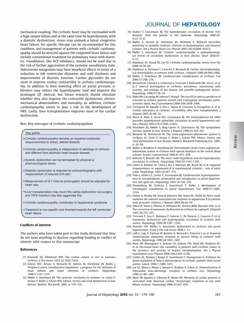
Fig. 1. Potential mechanisms involved in the impaired contractile function of the creceptors with decreased content of G-protein (Gai: inhibitory G-protein; Gas: stiincreased inhibitory effects of cardiodepressant substances such as haemoxygenase(NO) release, and tumour necrosis factor-a (TNF-a). Many post-receptor effects are medsignalling; PDE: phosphodiesterase; PKA: protein kinase A). Sarcoplasmatic reticulum (SRL-type calcium channels, and increased fluidity of the plasma membrane (increasedcontractility together with altered ratio of collagens and titins.
180 Journal of Hepatology 201
et al. expanded these results in a cirrhotic rat model by showingevidence of desensitisation of myocardial b-adrenergic receptors,indicating that down-regulation of b-adrenergic receptors couldbe responsible for the myocardial hyporesponsiveness to cate-cholamines [28]. Another important mechanism to be consideredis the nitration of proteins, which may be harmful to their func-tion. Thus, Mani et al. recently showed that nitration of cardiacproteins led to an abnormal cardiac chronotropic function andthereby impairment of control of heart rate in cirrhotic cardiomy-opathy [29].
The contractility of the heart muscle cell can be disturbed indifferent ways, but the molecular mechanisms of the decreasedcontractility are not completely understood. There is however,experimental evidence of decreased responsiveness of cardio-myocytes to b-adrenergic agonists. The b-adrenergic receptorsare located in the cell membrane, and altered physical propertieswith increased fluidity of the cardiomyocyte plasma membranemay be associated with the decreased b-adrenoceptor function[30]. Thus, changes in the cardiomyocyte plasma membrane
NO
TNF-
annabinoid 1-receptor
HO
CO
Myofibril
NOS
agens and titins
ATP
Plasma membrane
ardiomyocyte in cirrhotic cardiomyopathy: Down-regulation of b-adrenergicmulatory G-protein); up-regulation of cannabinoid 1-receptor stimulation;(HO), carbon monoxide (CO), nitric oxide synthase (NOS)-induced nitric oxideiated by adenylcyclase (AC) inhibition or stimulation. (RGS: regulator of G-protein), Altered function and reduced conductance of potassium channels, inhibition of
cholesterol/phospholipid ratio) also contribute to reduced calcium release and
0 vol. 53 j 179–190

JOURNAL OF HEPATOLOGY
may affect b-adrenergic receptors, signalling function, and car-diac contractility [31,32]. Recently, Ceolotto and co-workersdemonstrated that the reduced b-adrenergic-dependent inotro-pic effect could partly be attributed to an over-expression ofproteins such as inhibitory G-protein (Gai) and regulators of G-protein signalling (RGS2) that inhibit the adenylate cyclase andthose that accelerate degradation of cAMP such as phosphodies-terase (PDE2) [33], Fig. 1. Thus, abnormal gene expression of theb-adrenergic system may be involved in the abnormal signaltransduction and altered myocardial contractility in cirrhotic car-diomyopathy [33]. In addition, there is experimental evidence forthe involvement of the parasympathetic nervous system,although parasympathetic dysfunction appears somewhat com-plex. The myocardial contractile responsiveness to muscarinicstimulation is attenuated in cirrhotic rats but the changes arelikely compensatory, suggesting post-receptor factors [34]. Thus,it is less likely that the muscarinic over-activity is directlyinvolved in the cirrhotic cardiomyopathy.The endogenous and exogenous cannabinoids (CB) belong to asystem of cellular signalling pathways acting via CB1 receptors,whose activation induces arterial hypotension. The endocannab-inoids influence the vascular tone by a vasodilatory effect thatactivates G-proteins; an effect that is amplified by the ability ofendocannabinoids to induce apoptosis of hepatic stellate cells,thus favouring the development of portal hypertension andhyperdynamic circulation [35,36]. There is evidence of increasedlocal ventricular endocannabinoid production in cirrhosis andthat activation of CB1 receptors by endogenous anandamide con-tributes to the reduced cardiac contractility in cirrhosis [37].Antagonist blockade of the CB1 receptors may reverse theimpaired cardiac contractility, and studies in a model of CCl4-induced cirrhotic rats by the use of CB1 receptor antagonists haveshown an improvement in the contractile function in cirrhoticcardiomyopathy [37,38]. Future clinical studies should focus onthe potential therapeutic benefits of CB1 antagonism.
As mentioned above, there is experimental evidence that NOis involved in the vascular hyporesponsiveness to vasoconstric-tors [18,19]. Furthermore, NO has been shown to modify cardiacperformance with significant impairment of the cardiaccontractilityin bile duct-ligated cirrhotic rats [39,40]. Results of additionalexperimental studies have indicated that the cytokine–NO path-way occurs in cirrhotic rat hearts with enhanced expression ofthe NO synthase [20,41] and that inhibition of the NO synthesisby the NO inhibitor L-NAME reverses the impaired cardiac con-tractility [39,41]. In a model of chronic bile duct ligation-inducedcirrhosis, portal hypertension induced a marked left ventricularhypertrophy with increased myocardial NO synthesis, but in thismodel without any functional impairment [42]. Defects in con-tractile and connective proteins may play a role in the impairedsystolic and diastolic function. In bile duct-ligated rats, over-expression of the b-myosin heavy chain in the cirrhotic heartseems to play a role in the impaired systolic function [43], andan altered ratio of the stiffer collagen I and the more compliantcollagen III along with titins could play a role in the decreasedcompliance in the cirrhotic heart and the diastolic dysfunction[44]. Activation of the haemoxygenase-carbon monoxide path-way may also occur in cirrhotic rat hearts with expression ofhaemoxygenase-1 [45]. Inhibition of the effects of carbon monox-ide improves cardiac contractility and may play an important rolein the pathogenesis of cirrhotic cardiomyopathy [40,45]. Nuclearfactor-jB regulates different cytokines such as tumour necrosis
Journal of Hepatology 201
factor-a (TNF-a) and seems activated in cirrhotic rat hearts[46]. Activation of nuclear factor-jB, a transcription factor thatregulates inflammatory processes, suppresses cardiac contractil-ity and its inhibition improves systolic and diastolic contractility,which emphasises a role of cytokine expression in cirrhotic car-diomyopathy [46].
Abnormalities in the properties of the plasma membraneaffect repolarisation and conduction. The plasma membrane flu-idity of the cardiomyocyte is changed in models of cirrhotic car-diomyopathy and the membrane lipid content is abnormal withan increased cholesterol and thereby an increased cholesterol/phospholipid ratio [47]. As this determines the characteristicsof the membrane and its membrane-bound proteins, the functionof ion channels, for instance, may be significantly changed. Thus,Ward et al. found a decreased density of potassium currents inventricular myocytes, which may contribute to prolong the QTinterval [48]. In the same model of bile duct-ligated rats, thesame group found a reduced expression and density of L-typeCa++ channels and of inward cellular calcium current; thereforemodifications of the cellular calcium regulatory system withreduced calcium influx contributes to the reduced cardiac con-tractility (Fig. 1) [49]. In a model of portal vein-ligated rats,Zavecz et al. similarly reported a reduction of the density of L-type Ca++ channels and the sarcoplasmic reticulum Ca++ pool asa cause of changed excitation–contraction coupling [50]. Changesin the calcium channels also contribute to the prolonged QTinterval, which has an arrythmogenic effect. However, in differ-ent rat models, NO reduces the susceptibility to adrenaline-induced arrhythmia, which could counteract the pro-arrythmo-genic effect of a QT prolongation [51].
In conclusion, cirrhotic models supply a considerable amountof experimental evidence for the reduced cardiac contractility.The pathophysiology behind the reduced contractility is complex,but different findings have contributed towards a better under-standing of the pathophysiological background of cirrhotic car-diomyopathy, such as the down-regulation of b-receptors alongwith an impaired b-adrenergic signalling, changed plasma mem-brane fluidity with altered potassium, calcium channels and elec-trophysiological abnormalities, activation of the cannabinoid, NOand cytokine systems, as well as abnormal myofilaments.
Cirrhotic cardiomyopathy in patients
Systolic dysfunction
The systolic function closely relates to the size of the stroke vol-ume, heart rate, and cardiac output. In cirrhosis, the circulatorydysfunction has been expressed as a hyperdynamic unloaded fail-ure of the heart [52]. Despite the characteristic high cardiac out-put, a systolic dysfunction is included in the working definition ofthe cirrhotic cardiomyopathy (Table 1) [9]. However, at rest, thecardiac pressures are normal in the majority of cirrhotic patients[52,53]. Physical exercise, however, increases left ventricularpressures in some patients [54,55] and induces a relatively smal-ler increase in the left-ventricular ejection fraction and heart rate,Table 2 [56]. Although sympathetic nervous tone is increasedduring exercise, cardiac performance is reduced because ofreduced cardiovascular reactivity [57].
Administration of vasoconstrictors, such as angiotensin IIand terlipressin, increases the systemic vascular resistance
0 vol. 53 j 179–190 181

Table 1. Proposal for diagnostic and supportive criteria for cirrhotic cardio-myopathy agreed upon at a working party held at the 2005 World Congress ofGastroenterology in Montreal.
A working definition of cirrhotic cardiomyopathyA cardiac dysfunction in patients with cirrhosis characterised byimpaired contractile responsiveness to stress and/or altered diastolicrelaxation with electrophysiological abnormalities in the absence ofother known cardiac disease
Diagnostic criteriaSystolic dysfunction
� Blunted increase in cardiac output with exercise, volume challenge orpharmacological stimuli� Resting EF <55%
Diastolic dysfunction� E/A ratio <1.0 (age-corrected)� Prolonged deceleration time (>200 msec)� Prolonged isovolumetric relaxation time (>80 msec)
Supportive criteria� Electrophysiological abnormalities� Abnormal chronotropic response� Electromechanical uncoupling/dyssynchrony� Prolonged QTc interval� Enlarged left atrium� Increased myocardial mass� Increased BNP and pro-BNP� Increased troponin I
BNP, brain natriuretic peptide; E/A, early diastolic/atrial filling ratio; EF, left-ventricular ejection fraction.
Review
and thereby the left ventricular afterload [3,58–60]. Pharmaco-logical or physical stress may unmask a latent left ventriculardysfunction in patients with cirrhosis, as evidenced by anincrease in left ventricular end-diastolic volume and a decrease
Table 2. Potential cardiovascular changes in cirrhotic cardiomyopathy at restand during pharmacological or physical stress.
At restHeart
Heart rate "Cardiac output "Left atrial volume "Left ventricular volume ? (")Right atrial volume? " ;Right atrial pressure ? "Right ventricular end-diastolic pressure ?Pulmonary artery pressure ? "Pulmonary capillary wedge pressure ?Left atrial pressure ?Left ventricular end-diastolic pressure ?
Systemic circulationPlasma volume "Total blood volume "Non-central blood volume "Central and arterial blood volume ? ;Cardiac output (?) " (;)Arterial blood pressure ? ;Heart rate "Systemic vascular resistance ;
During stressPeak heart rate ;Peak work rate ;Peak exercise blood pressure ;Blood pressure � heart rate product ;Left atrial pressure "Left ventricular volume "Peak cardiac output ;Peak left-ventricular ejection fraction ;
", ?, ; denote increased, unchanged, and decreased values compared to controls.Arrows in parentheses describe less typical changes.
182 Journal of Hepatology 201
in left-ventricular ejection fraction (Table 2) [61]. Infusion ofangiotensin II in cirrhotic patients elicits an abnormal responsein terms of a 30% increase in the left ventricular afterload andresults in a doubling of the pulmonary capillary wedge pressurewithout changes in the cardiac output [58]. Dobutamine infu-sion increases the cardiac work by elevating the cardiac outputand heart rate but with smaller changes in cardiac pressures[59]. In cirrhotic patients, a dobutamine stress test could beused to reveal a cardiac dysfunction [62]. In the preoperativeassessment of patients with end-stage liver disease, a dobuta-mine stress echocardiography revealed ventricular dysfunctionin fewer than 10% of the patients, but the predictive values ofthis test are not known [60]. In accordance with these findings,recent data from our group indicate that cirrhotic cardiomyop-athy may be less frequently revealed by dobutamine, at least inpatients with mild disease [63]. Therefore, at present the dobu-tamine test cannot be recommended for the diagnosis of cir-rhotic cardiomyopathy, but should be reserved for severeischaemic heart disease before transplantation.
Analogously to the normal resting cardiac pressures in cirrho-sis, changes in the size of the cardiac volumes are only modest. Asassessed by magnetic resonance imaging, there seems to be atrend towards slightly increased left ventricular end-diastolicand left atrial volumes, probably in relation to the presence ofdiastolic dysfunction [64–66]. Increased atrial natriuretic peptide(ANP) in some patients with cirrhosis may reflect distension ordistortion of the atria, a finding that may coexist with effectivehypovolaemia [67,68].
The ventricular myocardial mass is increased in some patientsin particular with septal hypertrophy [69]. Troponin I is a thin fil-ament-associated protein of the myocyte, which reflects cardiacinjury. This marker is elevated in patients with myocardialischaemia and in some patients with cirrhosis who showincreased serum concentrations [70]. These patients have a sig-nificantly lower ventricular stroke volume and left ventricularmass index, indicating subclinical myocardial injury [70].
ANP and B-type natriuretic peptide (BNP) are secreted fromthe cardiac atria and ventricles, respectively. ANP signals todecrease blood pressure and BNP to decrease hypertrophy andlocally reduce ventricular fibrosis [71]. BNP and its pro-hormone,pro-BNP, are sensitive markers of even mild myocardial injury,and have been found elevated in both compensated and decom-pensated cirrhosis [72]. These peptides seem to correlate with theseverity of cirrhosis, degree of cardiac dysfunction and myocardi-ac hypertrophy, but not to the degree of hyperdynamic circula-tion [73] (Fig. 2) [74,75]. Since BNP and pro-BNP reflect thepresence of myocardial hypertrophy and cardiac dysfunctionthese peptides may be useful for screening patients for the pres-ence of cirrhotic cardiomyopathy [72,76].
The exercise capacity is reduced in a considerable number ofcirrhotic patients with a particularly low peak rate of work [77](Table 2). Together with the impaired cardiovascular responseto exercise, this is significantly associated with a chronotropicincompetence and failure to enhance systolic function [78]. Thenormal stepwise increase in cardiac output following gradedexercise testing by upright bicycle ergometry is significantly sup-pressed in patients with cirrhosis compared with controls, andthe inability to increased cardiac performance seems most pro-nounced in decompensated patients [79].
In advanced cirrhosis with pronounced vasodilatation, effec-tive hypovolaemia, and arterial hypotension, the RAAS is highly
0 vol. 53 j 179–190

0
10
20
30
40
50
60
70
Controls A B CCirrhosis
BNP (pmol/L)
***
0
10
20
30
40
50
60
70
Controls A B CCirrhosis
ProBNP (pmol/L)
** *
Fig. 2. Brain natriuretic peptide (BNP) and its pro-peptide (pro-BNP) in controls and in patients with cirrhosis, according to the Child–Turcotte classification. BNPand pro-BNP are significantly increased in cirrhosis and correlate with the prolonged QT interval, indicating that the cardiac generation of BNPs reflects cardiac dysfunctionin cirrhosis. Data from Henriksen et al. [73].
JOURNAL OF HEPATOLOGY
activated and such patients often develop a hepatorenal syn-drome (HRS) [15]. Patients with HRS have a low renal bloodflow, glomerular filtration rate, and sodium excretion thatappear to be related to a reduced systolic function. Thus, Ruiz-Del-Arbol et al. recently reported lower cardiac output and arte-rial blood pressure in patients with HRS, and a higher risk ofdeveloping HRS in patients with a cardiac output lower than6 L/min [80]. Maintenance of cardiac contractility thus seemsto be an important factor in the prevention of renal dysfunctionand HRS. Krag et al. recently demonstrated a significant relationbetween the degree of systolic and renal dysfunction andsurvival in patients with decompensated cirrhosis [81]. Thesestudies indicate that systolic dysfunction contributes tosodium and water-retention but on the other hand sodiumretention may also be involved in the pathophysiology ofsystolic as well as diastolic dysfunction [82]. However, theserelations need to be more carefully studied in the future. Releaseof inflammatory mediators such as tumour necrosis factor-a(TNF-a) and interleukins may also play a role in the systolicdysfunction. In patients with spontaneous bacterial peritonitis(SBP), cardiac output seems lower in patients who developrenal failure both before and after resolution of the infection[83].
In conclusion, systolic dysfunction is often latent in cirrhoticpatients, but is unmasked by physical or pharmacological strain.The reduced systolic function may have an impact on the devel-opment of complications, such as sodium and water-retention aswell as ascites formation, the development of renal dysfunctionand prognosis (see Box) [9,69].
Journal of Hepatology 201
Diastolic dysfunction
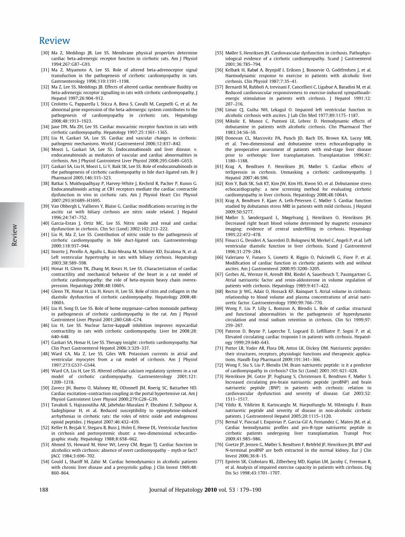
Abnormal left ventricular diastolic function, caused by decreasedleft ventricular compliance and relaxation, implies an abnormalfilling pattern of the ventricles. The transmitral blood flow ischanged, with an increased atrial contribution to the late ventric-ular filling [55] (Fig. 3). The pathophysiological background of thediastolic dysfunction in cirrhosis is an increased stiffness of themyocardial wall, most likely because of a combination of mildmyocardial hypertrophy, fibrosis, and subendothelial oedema[84]. There is experimental as well as clinical evidence thatincreased sodium intake may lead to the development of myocar-dial hypertrophy [82,85]. Therefore sodium retention per se maycontribute to diastolic dysfunction in cirrhosis. The increase inmyocardial stiffness is also reflected by other parameters of dia-stolic dysfunction, such as a prolonged time for the ventricles torelax after diastolic filling at a specific end-diastolic volume,which reflects an increased resistance to the ventricular inflow,Fig. 3 [55,65,69]. With Doppler echocardiography, Finucci et al.found impaired left ventricular relaxation, decreased E/A ratio,and delayed early diastolic transmittal filling in patients with cir-rhosis compared with controls [65]. This was later confirmed byPozzi et al. and Torregrosa et al., who revealed a complex patternof diastolic dysfunction in cirrhosis which was most pronouncedin patients with ascites [12,86].
ANP is regarded as a marker of volume overload and is foundincreased in decompensated cirrhosis [87]. ANP is released bystretch of the atrial fibers such as in patients with ascites wherethe right atrium has been reported increased partly because of
0 vol. 53 j 179–190 183

EA
AE
200 400 600 8000
25
50
75
100
cm/s
ec
Peak E
Peak A
DT
Time (msec)
Normal filling pattern Pattern of diastolic dysfunction
Fig. 3. Schematic illustration of the transmitral Doppler flow profile. Peak Edenotes the early filling of the ventricle and peak A the late atrial contribution.Diastolic dysfunction induces characteristic changes in the flow pattern, includingincreased E/A ratio and prolonged deceleration time (DT). The lower diagramsshow a normal filling pattern (left) and a filling pattern indicating diastolicdysfunction (right).
Review
volume overload with expanded blood volume [71,87]. However,the interpretation of the increased ANP in patients who from afunctional point of view suffer from effective hypovolaemia iscomplex. Nazar et al. recently reported a prevalence of 57%among 102 patients with cirrhosis, with a higher degree of liverfailure and circulatory dysfunction and higher concentrations ofANP [88]. The RAAS and ANP both contribute to volume regula-tion and compliance, and seem to be associated with the degreeof diastolic dysfunction in cirrhotic, as well as in non-cirrhotic,portal fibrosis [89]. On the other hand, volume and pressure over-load stretches myocardial fibres and activates the intracardiacrenin-angiotensin-system [90]. Therefore, the RAAS may bedirectly as well as indirectly involved in diastolic dysfunction incirrhosis. Patients with diastolic dysfunction are particularly sen-sitive to volume changes that occur, for example, in relation toinsertion of a TIPS. Portal decompression with a TIPS may leadto a further increase in the left atrial diameter and pulmonarycapillary wedged pressure, which indicates that the cirrhoticheart is unable to receive an adequate increased preload [91–93]. Thus, Cazzaniga et al. investigated the diastolic function in32 cirrhotic patients after TIPS insertion and found that it pre-dicted death after TIPS [94]. Changes in diastolic function appearmost prominent in patients with severe decompensation, and inthese patients the combination of myocardial hypertrophy, con-tractile dysfunction, changes in heart volumes, and diastolic dys-function may add to a cirrhotic cardiomyopathy [66,69,79,95].The increase in diastolic volumes after TIPS seems, however, tonormalise after months but with persistence of a mild left ven-tricular hypertrophy [93]. Moreover, reduced diastolic functionseems to be associated with slower mobilisation of ascites [11].After liver transplantation, diastolic function seems to improvein some patients, but the results are sparse [12,96]. Pozzi et al.recently demonstrated that anti-aldosterone treatment with K-canrenoate in cirrhosis ameliorated cardiac structure by reducingleft ventricular wall thickness and volume, but this treatment hadalmost no effects on systolic and diastolic function [97].
In conclusion, there is evidence that the diastolic function isimpaired particularly in patients with advanced cirrhosis and
184 Journal of Hepatology 201
large ascites, and that indicators of diastolic dysfunction maycontain prognostic information on the course of procedures thatmay affect filling of the ventricles such as TIPS insertion.
Prolongation of the QT interval
The main electrocardiographic change in cirrhosis is a prolonga-tion of the QT interval adjusted for heart rate. It has beenknown for a long time that it is prolonged in up to 50% ofpatients with cirrhosis [1]. In alcoholic patients, prolonged QTinterval is associated with an increased risk of sudden cardiacdeath [98]. In patients with cirrhosis, the prolonged QT intervalis unrelated to the aetiology of the liver disease, and it is seen inboth alcoholic and non-alcoholic liver diseases [98–100]. How-ever, in cirrhosis, the duration of the QT interval is associatedwith indicators of autonomic dysfunction and is partly revers-ible after liver transplantation [101–103]. In a minority of thepatients, the QT interval may worsen after liver transplantation[104,105]. In their study of 107 cirrhotic patients, Bernardi et al.showed that the prolonged QT interval was correlated with thedegree of liver dysfunction and circulating plasma noradrenaline[10]. In that study, the QT interval was also related to survival,but others have been unable to confirm an effect on mortality[106]. The QT interval is prolonged in both non-cirrhotic andcirrhotic portal hypertension, and the observation of a furtherincrease after TIPS insertion suggests that portosystemic shunt-ing may be responsible for the altered ventricular repolarisation[107,108]. The finding that the QT interval is also prolonged inpatients with only mild portal hypertension adds to theassumption that the portosystemic shunting per se is of impor-tance [109]. Acute non-selective b-blockade has been shown toreduce the prolonged QT interval towards normal values inpatients with cirrhosis [110]; it could therefore constitute afuture indication for b-blocker treatment. Recently, Zambruniet al. showed that chronic b-blockade shortened the QT intervalbut only in patients with a prolonged interval before treatment[111]. Life-threatening arrythmias are, however, uncommon incirrhosis and until now there is little evidence that treatmentwith b-blockers prevent their occurrence. At present, QT intervalprolongation per se in patients with cirrhosis is therefore not anindication for treatment with b-blockers. Studies on the disper-sion of the QT interval (i.e. difference between the longest andshortest interval) have shown a normal diurnal variation, andthe combination of a prolonged interval and a normal dispersionsuggests a delayed myocyte repolarisation in cirrhosis [112].Electromechanical uncoupling is a functional disturbancebetween the electrical and mechanical coupling, and has beenfound in experimental cirrhosis [31,50]. Bernardi et al. mea-sured systolic time intervals and reported a reduced cardiovas-cular responsiveness to exercise, despite enhanced sympatheticnervous activity [57]. These findings indicate that the impair-ment of cardiac contractility is partly based on an abnormalelectromechanical coupling. Among 48 cirrhotic patients and17 controls, we found a significant difference between the elec-trical and mechanical systole, the latter being significantlylonger in patients with cirrhosis, whereas, as expected, therewas a direct relation between the QT interval and the durationof the mechanical systole in the controls [113] (Fig. 4). This sug-gests an altered cardiac excitation–contraction coupling with acompromised association between the electrical and mechanicalfunction of the cirrhotic heart [8].
0 vol. 53 j 179–190

A B Pa (mmHg)
70
80
90
100
110
120
tp
ts
tRR
tD
m Volt
0
1
0.00 1.00 Time (sec)
QTQ
S
T
R
0
2
4
6
8
10
12
- 125 - 100 - 75 - 50 - 250
25 50 100 150 175
Number
Δt (msec)75 125
Control
0
2
4
6
8
10
12
- 125 - 100 - 75 - 50 - 250
25 50 100 150 175
Number
Δt (msec)75 125
Cirrhosis
Fig. 4. Mechanical and electrical time intervals from the aortic pressure curve and electrocardiogram. (A) Pa, arterial pressure as a function of time; tP, time to peakpressure; tS, systolic time; tD, diastolic time; tRR, time of one heart cycle; QT interval, the time from the start of the Q wave to the end of the T wave. (B) Difference betweenelectrical and mechanical systole time (Dt = QT � tS) in controls and patients with cirrhosis. Data from Henriksen et al. [113].
JOURNAL OF HEPATOLOGY
In conclusion, there is a large body of evidence of cardiac con-ductance abnormalities in cirrhosis, which may increase the riskof cardiac events. The electromechanical changes may be aggra-vated after TIPS insertion and may be partly reversible after livertransplantation.
Cardiac autonomic dysfunction
Reduced baroreflex-sensitivity has been shown to occur in cir-rhosis as part of a general cardiovascular autonomic dysfunction[3,24,25,100,112,114]. In a study of 105 patients with cirrhosis,we found a reduced baroreflex-sensitivity, which was signifi-cantly related to central haemodynamics and biochemicalcharacteristics [5]. These results suggest that a reduced barore-ceptor-sensitivity owing to the severity of the liver disease, isassociated with the cardiac dysfunction in cirrhosis [5]. Since reg-ulation of the arterial blood pressure plays an important role inthe development of fluid retention and renal function, a reducedbaroreflex-sensitivity will further impair renal sodium and waterexcretion in these patients. The baroreflex-sensitivity is reducedafter exposure to hypoxia such as, for example, a sojourn at highaltitude, but supply of oxygen to cirrhotic patients does not seemto ameliorate the low baroreflex-sensitivity [115]. A considerable
Journal of Hepatology 201
number of cirrhotic patients show a reduced heart rate variabil-ity, which correlates with the severity of the disease, central hyp-ovolaemia, and the degree of portal hypertension [26,108,112].
In conclusion, there are several indications that the reducedbaroreflex-sensitivity and heart rate variability contribute tothe cardiac dysfunction in cirrhosis.
Cardiac performance after TIPS
TIPS insertion leads to an acute increase in the right cardiac pre-load, because of acute translocation of portal venous blood intothe systemic circulation. This results in a worsening of the hyper-dynamic state with a further increase in cardiac output, strokevolume and left and right end-diastolic volumes and a decreasein the systemic vascular resistance [93,116–118]. It has beendebated whether the effects of TIPS are transient or more sus-tained. Studies that describes short-term effects on the hyperdy-namic circulation [91,119,120] as well as long-term effects(>6 months) have been published [92,121]. A sustained rise incardiac output and a reduction in the systemic vascular resis-tance may persist for at least 1 year post-TIPS. Furthermore, TIPSinsertion can lead to high-output congestive heart failure withrises in pulmonary arterial and capillary pulmonary wedge
0 vol. 53 j 179–190 185
Review
pressures [122–124], although clinically significant heart failureoccurs in relatively few patients [125]. The acute increase in car-diac output seems to attenuate, despite the persistence ofincreased pulmonary vascular resistances [119,126]. After 6–12 months, cardiac output and the systemic vascular resistancetend to normalise, despite an unchanged degree of portosystemicshunting [92,93,120]. The combined increase in left atrial diame-ter, the pulmonary capillary wedge pressure and total pulmonaryresistance most likely reflect a diastolic dysfunction of the leftventricle, as mentioned earlier, and therefore TIPS may unmaska cirrhotic cardiomyopathy [91,92]. Prolongation of the QT inter-val as seen in patients with cirrhosis and portal hypertension,may also worsen after TIPS insertion in cirrhotic, as well asnon-cirrhotic, portal hypertensive patients [107].In conclusion, TIPS insertion may acutely worsen the hyperdy-namic circulation, but it seems to attenuate over time. TIPS inser-tion is associated with an increased risk of heart failure and bothhaemodynamic and electrophysiological changes seem to beaffected.
%
**
**
*
Pre-LtxPost-Ltx
dHR dCl dEF- 20
0
20
40
60
80
100
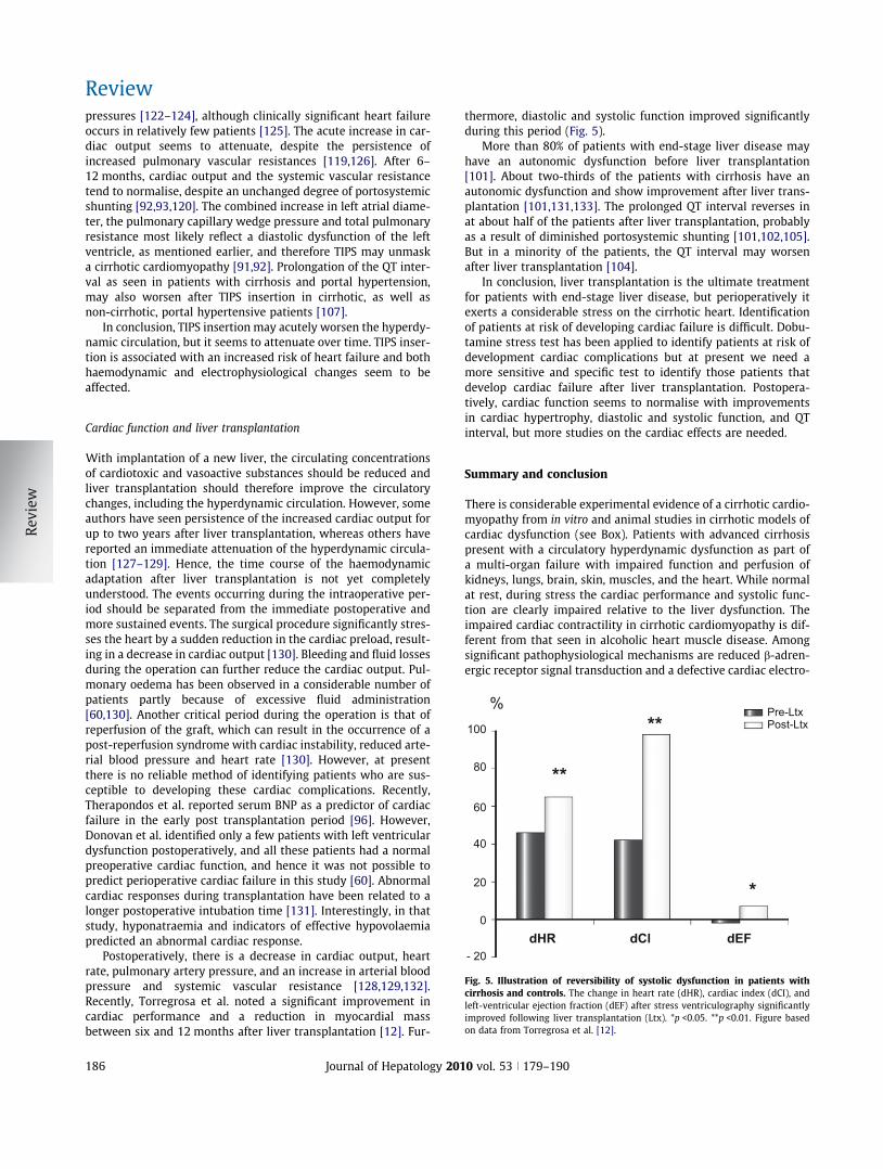
Fig. 5. Illustration of reversibility of systolic dysfunction in patients withcirrhosis and controls. The change in heart rate (dHR), cardiac index (dCI), andleft-ventricular ejection fraction (dEF) after stress ventriculography significantlyimproved following liver transplantation (Ltx). *p <0.05. **p <0.01. Figure basedon data from Torregrosa et al. [12].
Cardiac function and liver transplantation
With implantation of a new liver, the circulating concentrationsof cardiotoxic and vasoactive substances should be reduced andliver transplantation should therefore improve the circulatorychanges, including the hyperdynamic circulation. However, someauthors have seen persistence of the increased cardiac output forup to two years after liver transplantation, whereas others havereported an immediate attenuation of the hyperdynamic circula-tion [127–129]. Hence, the time course of the haemodynamicadaptation after liver transplantation is not yet completelyunderstood. The events occurring during the intraoperative per-iod should be separated from the immediate postoperative andmore sustained events. The surgical procedure significantly stres-ses the heart by a sudden reduction in the cardiac preload, result-ing in a decrease in cardiac output [130]. Bleeding and fluid lossesduring the operation can further reduce the cardiac output. Pul-monary oedema has been observed in a considerable number ofpatients partly because of excessive fluid administration[60,130]. Another critical period during the operation is that ofreperfusion of the graft, which can result in the occurrence of apost-reperfusion syndrome with cardiac instability, reduced arte-rial blood pressure and heart rate [130]. However, at presentthere is no reliable method of identifying patients who are sus-ceptible to developing these cardiac complications. Recently,Therapondos et al. reported serum BNP as a predictor of cardiacfailure in the early post transplantation period [96]. However,Donovan et al. identified only a few patients with left ventriculardysfunction postoperatively, and all these patients had a normalpreoperative cardiac function, and hence it was not possible topredict perioperative cardiac failure in this study [60]. Abnormalcardiac responses during transplantation have been related to alonger postoperative intubation time [131]. Interestingly, in thatstudy, hyponatraemia and indicators of effective hypovolaemiapredicted an abnormal cardiac response.
Postoperatively, there is a decrease in cardiac output, heartrate, pulmonary artery pressure, and an increase in arterial bloodpressure and systemic vascular resistance [128,129,132].Recently, Torregrosa et al. noted a significant improvement incardiac performance and a reduction in myocardial massbetween six and 12 months after liver transplantation [12]. Fur-
186 Journal of Hepatology 201
thermore, diastolic and systolic function improved significantlyduring this period (Fig. 5).
More than 80% of patients with end-stage liver disease mayhave an autonomic dysfunction before liver transplantation[101]. About two-thirds of the patients with cirrhosis have anautonomic dysfunction and show improvement after liver trans-plantation [101,131,133]. The prolonged QT interval reverses inat about half of the patients after liver transplantation, probablyas a result of diminished portosystemic shunting [101,102,105].But in a minority of the patients, the QT interval may worsenafter liver transplantation [104].
In conclusion, liver transplantation is the ultimate treatmentfor patients with end-stage liver disease, but perioperatively itexerts a considerable stress on the cirrhotic heart. Identificationof patients at risk of developing cardiac failure is difficult. Dobu-tamine stress test has been applied to identify patients at risk ofdevelopment cardiac complications but at present we need amore sensitive and specific test to identify those patients thatdevelop cardiac failure after liver transplantation. Postopera-tively, cardiac function seems to normalise with improvementsin cardiac hypertrophy, diastolic and systolic function, and QTinterval, but more studies on the cardiac effects are needed.
Summary and conclusion
There is considerable experimental evidence of a cirrhotic cardio-myopathy from in vitro and animal studies in cirrhotic models ofcardiac dysfunction (see Box). Patients with advanced cirrhosispresent with a circulatory hyperdynamic dysfunction as part ofa multi-organ failure with impaired function and perfusion ofkidneys, lungs, brain, skin, muscles, and the heart. While normalat rest, during stress the cardiac performance and systolic func-tion are clearly impaired relative to the liver dysfunction. Theimpaired cardiac contractility in cirrhotic cardiomyopathy is dif-ferent from that seen in alcoholic heart muscle disease. Amongsignificant pathophysiological mechanisms are reduced b-adren-ergic receptor signal transduction and a defective cardiac electro-
0 vol. 53 j 179–190
JOURNAL OF HEPATOLOGY
mechanical coupling. The cirrhotic heart may be overloaded witha high-output failure and at the same time be hyperdynamic witha diastolic dysfunction; strain may unmask a latent congestiveheart failure. No specific therapy can be recommended for thiscondition, and management of patients with cirrhotic cardiomy-opathy should be directed against the congested heart failure andinclude conventional treatment for pulmonary stasis with diuret-ics. Vasodilators, like ACE-inhibitors, should not be used due tothe risk of further aggravation of the systemic vasodilatory state.Aldosterone antagonists may have beneficial effects in terms of areduction in left ventricular dilatation and wall thickness andimprovement of diastolic function. Cardiac glycosides do notseem to improve cardiac contractility in cirrhotic cardiomyopa-thy. In addition to their lowering effect on portal pressure, b-blockers may reduce the hyperdynamic load and improve theprolonged QT interval, but future research should elucidatewhether they also improve the contractile dysfunction, electro-mechanical abnormalities, and mortality. In addition, cirrhoticcardiomyopathy seems to play a role in the development ofHRS. Lastly, liver transplantation improves most of the cardiacdysfunction.Box: Key messages of cirrhotic cardiomyopathies
Conflicts of interest
The authors who have taken part in this study declared that theydo not have anything to disclose regarding funding or conflict ofinterest with respect to this manuscript.
References
[1] Kowalski HJ, Abelmann WH. The cardiac output at rest in Laennecscirrhosis. J Clin Invest 1953;32:1025–1033.
[2] Schrier RW, Arroyo V, Bernardi M, Epstein M, Henriksen JH, Rodés J.Peripheral artery vasodilatation hypothesis: a proposal for the initiation ofrenal sodium and water retention in cirrhosis. Hepatology1988;5:1151–1157.
[3] Møller S, Henriksen JH. The systemic circulation in cirrhosis. In: Gines P,Arroyo V, Rodes J, Schrier RW, editors. Ascites and renal dysfunction in liverdisease. Malden: Blackwell; 2005. p. 139–155.
Journal of Hepatology 201
[4] Iwakiri Y, Groszmann RJ. The hyperdynamic circulation of chronic liverdiseases: from the patient to the molecule. Hepatology 2006;43:S121–S131.
[5] Møller S, Iversen JS, Henriksen JH, Bendtsen F. Reduced baroreflexsensitivity in alcoholic cirrhosis: relations to hemodynamics and humoralsystems. Am J Physiol Heart Circ Physiol 2007;292:H2966–H2972.
[6] Møller S, Henriksen JH. Cirrhotic cardiomyopathy: a pathophysiologi-cal review of circulatory dysfunction in liver disease. Heart 2002;87:9–15.
[7] Alqahtani SA, Fouad TR, Lee SS. Cirrhotic cardiomyopathy. Semin Liver Dis2008;28:59–69.
[8] Zambruni A, Trevisani F, Caraceni P, Bernardi M. Cardiac electrophysiolog-ical abnormalities in patients with cirrhosis. J Hepatol 2006;44:994–1002.
[9] Møller S, Henriksen JH. Cardiovascular complications of cirrhosis. Gut2008;57:268–278.
[10] Bernardi M, Calandra S, Colantoni A, Trevisani F, Raimondo ML, Sica G, et al.Q–T interval prolongation in cirrhosis: prevalence, relationship withseverity, and etiology of the disease and possible pathogenetic factors.Hepatology 1998;27:28–34.
[11] Rabie RN, Cazzaniga M, Salerno F, Wong F. The use of E/A ratio as a predictor ofoutcome in cirrhotic patients treated with transjugular intrahepatic porto-systemic shunt. Am J Gastroenterol 2009;104:2458–2466.
[12] Torregrosa M, Aguade S, Dos L, Segura R, Gonzalez A, Evangelista A, et al.Cardiac alterations in cirrhosis: reversibility after liver transplantation. JHepatol 2005;42:68–74.
[13] Wiest R, Shah V, Sessa WC, Groszmann RJ. NO overproduction by eNOSprecedes hyperdynamic splanchnic circulation in portal hypertensive rats.Am J Physiol 1999;276:G1043–G1051.
[14] Henriksen JH, Møller S, Ring-Larsen H, Christensen NJ. The sympatheticnervous system in liver disease. J Hepatol 1998;29:328–341.
[15] Bernardi M, Domenicali M. The renin-angiotensin-aldosterone system incirrhosis. In: Ginés P, Arroyo V, Rodes J, Schrier RW, editors. Ascites andrenal dysfunction in liver disease. Malden: Blackwell Publishing Ltd.; 2005.p. 43–54.
[16] Møller S, Bendtsen F, Henriksen JH. Determinants of the renin-angiotensin-aldosterone system in cirrhosis with special emphasis on the central bloodvolume. Scand J Gastroenterol 2006;41:451–458.
[17] Bomzon A, Blendis LM. The nitric oxide hypothesis and the hyperdynamiccirculation in cirrhosis. Hepatology 1994;20:1343–1350.
[18] Castro A, Jimenez W, Claria J, Ros J, Martinez JM, Bosch M, et al. Impairedresponsiveness to angiotensin-II in experimental cirrhosis – role of nitricoxide. Hepatology 1993;18:367–372.
[19] Polio J, Sieber CC, Lerner E, Groszmann RJ. Cardiovascular hyporesponsive-ness to norepinephrine, propranolol and nitroglycerin in portal-hyperten-sive and aged rats. Hepatology 1993;18:128–136.
[20] Hennenberg M, Trebicka J, Sauerbruch T, Heller J. Mechanisms ofextrahepatic vasodilation in portal hypertension. Gut 2008;57:1300–1314.
[21] Helmy A, Newby DE, Jalan R, Johnston NR, Hayes PC, Webb DJ. Nitric oxidemediates the reduced vasoconstrictor response to angiotensin II in patientswith preascitic cirrhosis. J Hepatol 2003;38:44–50.
[22] Dillon JF, Nolan J, Thomas H, Williams BC, Neilson JMM, Bouchier IAD, et al.The correction of autonomic dysfunction in cirrhosis by captopril. J Hepatol1997;26:331–335.
[23] Trevisani F, Sica G, Mainqua P, Santese G, De Notariis S, Caraceni P, et al.Autonomic dysfunction and hyperdynamic circulation in cirrhosis withascites. Hepatology 1999;30:1387–1392.
[24] Dumcke CW, Møller S. Autonomic dysfunction in cirrhosis and portalhypertension. Scand J Clin Lab Invest 2008:1–11.
[25] Laffi G, Lagi A, Cipriani M, Barletta G, Bernardi L, Fattorini L, et al. Impairedcardiovascular autonomic response to passive tilting in cirrhosis withascites. Hepatology 1996;24:1063–1067.
[26] Mani AR, Montagnese S, Jackson CD, Jenkins CW, Head IM, Stephens RC,et al. Decreased heart rate variability in patients with cirrhosis relates tothe presence and severity of hepatic encephalopathy. Am J PhysiolGastrointest Liver Physiol 2008;296:G330–G338.
[27] Gerbes AL, Remien J, Jüngst D, Sauerbruch T, Paumgartner G. Evidence fordown-regulation of beta-2-adrenoceptors in cirrhotic patients with severeascites. Lancet 1986;1:1409–1411.
[28] Lee SS, Marty J, Mantz J, Samain E, Braillon A, Lebrec D. Desensitization ofmyocardial beta-adrenergic receptors in cirrhotic rats. Hepatology1990;12:481–485.
[29] Mani AR, Ippolito S, Ollosson R, Moore KP. Nitration of cardiac proteins isassociated with abnormal cardiac chronotropic responses in rats withbiliary cirrhosis. Hepatology 2006;43:847–856.
0 vol. 53 j 179–190 187
Review
[30] Ma Z, Meddings JB, Lee SS. Membrane physical properties determinecardiac beta-adrenergic receptor function in cirrhotic rats. Am J Physiol1994;267:G87–G93.
[31] Ma Z, Miyamoto A, Lee SS. Role of altered beta-adrenoceptor signaltransduction in the pathogenesis of cirrhotic cardiomyopathy in rats.Gastroenterology 1996;110:1191–1198.
[32] Ma Z, Lee SS, Meddings JB. Effects of altered cardiac membrane fluidity onbeta-adrenergic receptor signalling in rats with cirrhotic cardiomyopathy. JHepatol 1997;26:904–912.
[33] Ceolotto G, Papparella I, Sticca A, Bova S, Cavalli M, Cargnelli G, et al. Anabnormal gene expression of the beta-adrenergic system contributes to thepathogenesis of cardiomyopathy in cirrhotic rats. Hepatology2008;48:1913–1923.
[34] Jaue DN, Ma ZH, Lee SS. Cardiac muscarinic receptor function in rats withcirrhotic cardiomyopathy. Hepatology 1997;25:1361–1365.
[35] Liu H, Gaskari SA, Lee SS. Cardiac and vascular changes in cirrhosis:pathogenic mechanisms. World J Gastroenterol 2006;12:837–842.
[36] Moezi L, Gaskari SA, Lee SS. Endocannabinoids and liver disease. v.endocannabinoids as mediators of vascular and cardiac abnormalities incirrhosis. Am J Physiol Gastrointest Liver Physiol 2008;295:G649–G653.
[37] Gaskari SA, Liu H, Moezi L, Li Y, Baik SK, Lee SS. Role of endocannabinoids inthe pathogenesis of cirrhotic cardiomyopathy in bile duct-ligated rats. Br JPharmacol 2005;146:315–323.
[38] Batkai S, Mukhopadhyay P, Harvey-White J, Kechrid R, Pacher P, Kunos G.Endocannabinoids acting at CB1 receptors mediate the cardiac contractiledysfunction in vivo in cirrhotic rats. Am J Physiol Heart Circ Physiol2007;293:H1689–H1695.
[39] Van Obbergh L, Vallieres Y, Blaise G. Cardiac modifications occurring in theascitic rat with biliary cirrhosis are nitric oxide related. J Hepatol1996;24:747–752.
[40] Garcia-Estan J, Ortiz MC, Lee SS. Nitric oxide and renal and cardiacdysfunction in cirrhosis. Clin Sci (Lond) 2002;102:213–222.
[41] Liu H, Ma Z, Lee SS. Contribution of nitric oxide to the pathogenesis ofcirrhotic cardiomyopathy in bile duct-ligated rats. Gastroenterology2000;118:937–944.
[42] Inserte J, Perello A, Agullo L, Ruiz-Meana M, Schluter KD, Escalona N, et al.Left ventricular hypertrophy in rats with biliary cirrhosis. Hepatology2003;38:589–598.
[43] Honar H, Glenn TK, Zhang M, Keurs H, Lee SS. Characterization of cardiaccontractility and mechanical behavior of the heart in a rat model ofcirrhotic cardiomyopathy: the role of beta-myosin heavy chain overex-pression. Hepatology 2008;48:1060F.
[44] Glenn TK, Honar H, Liu H, Keurs H, Lee SS. Role of titin and collagen in thediastolic dysfunction of cirrhotic cardiomyopathy. Hepatology 2008;48:1060F.
[45] Liu H, Song D, Lee SS. Role of heme oxygenase–carbon monoxide pathwayin pathogenesis of cirrhotic cardiomyopathy in the rat. Am J PhysiolGastrointest Liver Physiol 2001;280:G68–G74.
[46] Liu H, Lee SS. Nuclear factor-kappaB inhibition improves myocardialcontractility in rats with cirrhotic cardiomyopathy. Liver Int 2008;28:640–648.
[47] Gaskari SA, Honar H, Lee SS. Therapy insight: cirrhotic cardiomyopathy. NatClin Pract Gastroenterol Hepatol 2006;3:329–337.
[48] Ward CA, Ma Z, Lee SS, Giles WR. Potassium currents in atrial andventricular myocytes from a rat model of cirrhosis. Am J Physiol1997;273:G537–G544.
[49] Ward CA, Liu H, Lee SS. Altered cellular calcium regulatory systems in a ratmodel of cirrhotic cardiomyopathy. Gastroenterology 2001;121:1209–1218.
[50] Zavecz JH, Bueno O, Maloney RE, ODonnell JM, Roerig SC, Battarbee HD.Cardiac excitation–contraction coupling in the portal hypertensive rat. Am JPhysiol Gastrointest Liver Physiol 2000;279:G28–G39.
[51] Tavakoli S, Hajrasouliha AR, Jabehdar-Maralani P, Ebrahimi F, Solhpour A,Sadeghipour H, et al. Reduced susceptibility to epinephrine-inducedarrhythmias in cirrhotic rats: the roles of nitric oxide and endogenousopioid peptides. J Hepatol 2007;46:432–439.
[52] Keller H, Bezjak V, Stegaru B, Buss J, Holm E, Heene DL. Ventricular functionin cirrhosis and portosystemic shunt: a two-dimensional echocardio-graphic study. Hepatology 1988;8:658–662.
[53] Ahmed SS, Howard M, Hove Wt, Leevy CM, Regan TJ. Cardiac function inalcoholics with cirrhosis: absence of overt cardiomyopathy – myth or fact?JACC 1984;3:696–702.
[54] Gould L, Shariff M, Zahir M. Cardiac hemodynamics in alcoholic patientswith chronic liver disease and a presystolic gallop. J Clin Invest 1969;48:860–864.
188 Journal of Hepatology 201
[55] Møller S, Henriksen JH. Cardiovascular dysfunction in cirrhosis. Pathophys-iological evidence of a cirrhotic cardiomyopathy. Scand J Gastroenterol2001;36:785–794.
[56] Kelbæk H, Rabøl A, Brynjolf I, Eriksen J, Bonnevie O, Godtfredsen J, et al.Haemodynamic response to exercise in patients with alcoholic livercirrhosis. Clin Physiol 1987;7:35–41.
[57] Bernardi M, Rubboli A, trevisani F, Cancellieri C, Ligabue A, Baradini M, et al.Reduced cardiovascular responsiveness to exercise-induced sympathoadr-energic stimulation in patients with cirrhosis. J Hepatol 1991;12:207–216.
[58] Limas CJ, Guiha NH, Lekagul O. Impaired left ventricular function inalcoholic cirrhosis with ascites. J Lab Clin Med 1977;89:1175–1187.
[59] Mikulic E, Munoz C, Puntoni LE, Lebrec D. Hemodynamic effects ofdobutamine in patients with alcoholic cirrhosis. Clin Pharmacol Ther1983;34:56–59.
[60] Donovan CL, Marcovitz PA, Punch JD, Bach DS, Brown KA, Lucey MR,et al. Two-dimensional and dobutamine stress echocardiography inthe preoperative assessment of patients with end-stage liver diseaseprior to orthotopic liver transplantation. Transplantation 1996;61:1180–1188.
[61] Krag A, Bendtsen F, Henriksen JH, Møller S. Cardiac effects ofterlipressin in cirrhosis. Unmasking a cirrhotic cardiomyopathy. JHepatol 2007;46:S96.
[62] Kim Y, Baik SK, Suk KT, Kim JW, Kim HS, Kwon SO, et al. Dobutamine stressechocardiography: a new screening method for evaluating cirrhoticcardiomyopathy in liver cirrhosis. Hepatology 2008;48:1064F.
[63] Krag A, Bendtsen F, Kjaer A, Leth-Petersen C, Møller S. Cardiac functionstudied by dobutamin stress MRI in patients with mild cirrhosis. J Hepatol2009;50:S277.
[64] Møller S, Søndergaard L, Møgelvang J, Henriksen O, Henriksen JH.Decreased right heart blood volume determined by magnetic resonanceimaging: evidence of central underfilling in cirrhosis. Hepatology1995;22:472–478.
[65] Finucci G, Desideri A, Sacerdoti D, Bolognesi M, Merkel C, Angeli P, et al. Leftventricular diastolic function in liver cirrhosis. Scand J Gastroenterol1996;31:279–284.
[66] Valeriano V, Funaro S, Lionetti R, Riggio O, Pulcinelli G, Fiore P, et al.Modification of cardiac function in cirrhotic patients with and withoutascites. Am J Gastroenterol 2000;95:3200–3205.
[67] Gerbes AL, Wernze H, Arendt RM, Riedel A, Sauerbruch T, Paumgartner G.Atrial natriuretic factor and renin-aldosterone in volume regulation ofpatients with cirrhosis. Hepatology 1989;9:417–422.
[68] Rector Jr WG, Adair O, Hossack KF, Rainquet S. Atrial volume in cirrhosis:relationship to blood volume and plasma concentrations of atrial natri-uretic factor. Gastroenterology 1990;99:766–770.
[69] Wong F, Liu P, Lilly L, Bomzon A, Blendis L. Role of cardiac structuraland functional abnormalities in the pathogenesis of hyperdynamiccirculation and renal sodium retention in cirrhosis. Clin Sci 1999;97:259–267.
[70] Pateron D, Beyne P, Laperche T, Logeard D, Lefilliatre P, Sogni P, et al.Elevated circulating cardiac troponin I in patients with cirrhosis. Hepatol-ogy 1999;29:640–643.
[71] Potter LR, Yoder AR, Flora DR, Antos LK, Dickey DM. Natriuretic peptides:their structures, receptors, physiologic functions and therapeutic applica-tions. Handb Exp Pharmacol 2009;191:341–366.
[72] Wong F, Siu S, Liu P, Blendis LM. Brain natriuretic peptide: is it a predictorof cardiomyopathy in cirrhosis? Clin Sci (Lond) 2001;101:621–628.
[73] Henriksen JH, Gotze JP, Fuglsang S, Christensen E, Bendtsen F, Møller S.Increased circulating pro-brain natriuretic peptide (proBNP) and brainnatriuretic peptide (BNP) in patients with cirrhosis: relation tocardiovascular dysfunction and severity of disease. Gut 2003;52:1511–1517.
[74] Yildiz R, Yildirim B, Karincaoglu M, Harputluoglu M, Hilmioglu F. Brainnatriuretic peptide and severity of disease in non-alcoholic cirrhoticpatients. J Gastroenterol Hepatol 2005;20:1115–1120.
[75] Bernal V, Pascual I, Esquivias P, Garcia-Gil A, Fernandez C, Mateo JM, et al.Cardiac hemodynamic profiles and pro-B-type natriuretic peptide incirrhotic patients undergoing liver transplantation. Transpl Proc2009;41:985–986.
[76] Goetze JP, Jensen G, Møller S, Bendtsen F, Rehfeld JF, Henriksen JH. BNP andN-terminal proBNP are both extracted in the normal kidney. Eur J ClinInvest 2006;36:8–15.
[77] Epstein SK, Ciubotaru RL, Zilberberg MD, Kaplan LM, Jacoby C, Freeman R,et al. Analysis of impaired exercise capacity in patients with cirrhosis. DigDis Sci 1998;43:1701–1707.
0 vol. 53 j 179–190

JOURNAL OF HEPATOLOGY
[78] Grose RD, Nolan J, Dillon JF, Errington M, Hannan WJ, Bouchier IAD, et al.Exercise-induced left ventricular dysfunction in alcoholic and non-alco-holic cirrhosis. J Hepatol 1995;22:326–332.
[79] Wong F, Girgrah N, Graba J, Allidina Y, Liu P, Blendis L. The cardiac responseto exercise in cirrhosis. Gut 2001;49:268–275.
[80] Ruiz-Del-Arbol L, Monescillo A, Arocena C, Valer P, Gines P, Moreira V, et al.Circulatory function and hepatorenal syndrome in cirrhosis. Hepatology2005;42:439–447.
[81] Krag A, Bendtsen F, Henriksen JH, Møller S. Low cardiac output predictsdevelopment of hepatorenal syndrome and survival in patients withcirrhosis and ascites. Gut 2010;59:105–110.
[82] Fields NG, Yuan BX, Leenen FH. Sodium-induced cardiac hypertrophy.Cardiac sympathetic activity versus volume load. Circ Res1991;68:745–755.
[83] Ruiz-Del-Arbol L, Urman J, Fernandez J, Gonzalez M, Navasa M, MonescilloA, et al. Systemic, renal, and hepatic hemodynamic derangement incirrhotic patients with spontaneous bacterial peritonitis. Hepatology2003;38:1210–1218.
[84] Ma Z, Lee SS. Cirrhotic cardiomyopathy: getting to the heart of the matter.Hepatology 1996;24:451–459.
[85] Schmieder RE. Salt intake is related to the process of myocardial hyper-trophy in essential hypertension. JAMA 1989;262:1187–1188.
[86] Pozzi M, Carugo S, Boari G, Pecci V, de Ceglia S, Maggiolini S, et al. Evidenceof functional and structural cardiac abnormalities in cirrhotic patients withand without ascites. Hepatology 1997;26:1131–1137.
[87] LaVilla G, Laffi G. Atrial natriuretic peptide and other natriuretic factors incirrhosis. In: Gines P, Arroyo V, Rodes J, Schrier RW, editors. Ascites andrenal dysfunction. Malden: Blackwell Publishing; 2005. p. 73–83.
[88] Nazar A, Sitges M, Guevara M, Terra C, Marinelli A, Villa F, et al.Cardiomyopathy in patients with cirrhosis. Frequency, characteristics andrelationship with circulatory dysfunction and prognosis. Journal of Hepa-tology 2009;50:S85.
[89] De BK, Majumdar D, Das D, Biswas PK, Mandal SK, Ray S, et al. Cardiacdysfunction in portal hypertension among patients with cirrhosis and non-cirrhotic portal fibrosis. J Hepatol 2003;39:315–319.
[90] Raizada V, Skipper B, Luo W, Griffith J. Intracardiac and intrarenal renin-angiotensin systems: mechanisms of cardiovascular and renal effects. JInvestig Med 2007;55:341–359.
[91] Huonker M, Schumacher YO, Ochs A, Sorichter S, Keul J, Rôssle M. Cardiacfunction and haemodynamics in alcoholic cirrhosis and effects of thetransjugular intrahepatic portosystemic stent shunt. Gut 1999;44:743–748.
[92] Merli M, Valeriano V, Funaro S, Attili AF, Masini A, Efrati C, et al.Modifications of cardiac function in cirrhotic patients treated with trans-jugular intrahepatic portosystemic shunt (TIPS). Am J Gastroenterol2002;97:142–148.
[93] Kovacs A, Schepke M, Heller J, Schild HH, Flacke S. Short-term effects oftransjugular intrahepatic shunt on cardiac function assessed by cardiacMRI: Preliminary results. Cardiovasc Intervent Radiol 2010;33:290–296.
[94] Cazzaniga M, Salerno F, Pagnozzi G, Dionigi E, Visentin S, Cirello I, et al.Diastolic dysfunction is associated with poor survival in cirrhotic patientswith transjugular intrahepatic portosystemic shunt. Gut 2007;56:869–875.
[95] Alexander J, Mishra P, Desai N, Ambadekar S, Gala B, Sawant P. Cirrhoticcardiomyopathy: Indian scenario. J Gastroenterol Hepatol2007;22:395–399.
[96] Therapondos G, Flapan AD, Dollinger MM, Garden OJ, Plevris JN, Hayes PC.Cardiac function after orthotopic liver transplantation and the effects ofimmunosuppression: a prospective randomized trial comparing cyclo-sporin (Neoral) and tacrolimus. Liver Transpl 2002;8:690–700.
[97] Pozzi M, Redaelli E, Ratti L, Poli G, Guidi C, Milanese M, et al. Time-course ofdiastolic dysfunction in different stages of chronic HCV related liverdiseases. Minerva Gastroenterol Dietol 2005;51:179–186.
[98] Day PC, James FWO, Butler JT, Campbell RWF. Q–T prolongation and suddencardiac death in patients with alcoholic liver disease. Lancet1993;341:1423–1428.
[99] Kempler P, Varadi A, Kadar E, Szalay F. Autonomic and peripheralneuropathy in primary biliary cirrhosis: evidence of small sensory fibredamage and prolongation of the QT interval. J Hepatol 1994;21:1150–1151.
[100] Lazzeri C, Lavilla G, Laffi G, Vecchiarino S, Gambilonghi F, Gentilini P, et al.Autonomic regulation of heart rate and QT interval in nonalcoholiccirrhosis with ascites. Digestion 1997;58:580–586.
[101] Mohamed R, Forsey PR, Davies MK, Neuberger JM. Effect of livertransplantation on QT interval prolongation and autonomic dysfunctionin end-stage liver disease. Hepatology 1996;23:1128–1134.
Journal of Hepatology 201
[102] Garcia GM, Hernandez-Madrid A, Lopez-Sanroman A, Candela A, Nuno J,Barcena R. Reversal of QT interval electrocardiographic alterations incirrhotic patients undergoing liver transplantation. Transpl Proc 1999;31:2366–2367.
[103] Puthumana L, Chaudhry V, Thuluvath PJ. Prolonged QTc interval and itsrelationship to autonomic cardiovascular reflexes in patients with cirrho-sis. J Hepatol 2001;35:733–738.
[104] Carey EJ, Douglas DD. Effects of orthotopic liver transplantation on thecorrected QT interval in patients with end-stage liver disease. Dig Dis Sci2005;50:320–323.
[105] Adigun AQ, Pinto AG, Flockhart DA, Gorski JC, Li L, Hall SD, et al. Effect ofcirrhosis and liver transplantation on the gender difference in QT interval.Am J Cardiol 2005;95:691–694.
[106] Bal JS, Thuluvath PJ. Prolongation of QTc interval: relationship withetiology and severity of liver disease, mortality and liver transplantation.Liver Int 2003;23:243–248.
[107] Trevisani F, Merli M, Savelli F, Valeriano V, Zambruni A, Riggio O, et al. QTinterval in patients with non-cirrhotic portal hypertension and in cirrhoticpatients treated with transjugular intrahepatic porto-systemic shunt. JHepatol 2003;38:461–467.
[108] Genovesi S, Prata Pizzala DM, Pozzi M, Ratti L, Milanese M, Pieruzzi F, et al.QT interval prolongation and decreased heart rate variability in cirrhoticpatients: relevance of hepatic venous pressure gradient and serum calcium.Clin Sci (Lond) 2008;116:851–859.
[109] Ytting H, Henriksen JH, Fuglsang S, Bendtsen F, Møller S. Prolonged Q–T(c)interval in mild portal hypertensive cirrhosis. J Hepatol 2005;43:637–644.
[110] Henriksen JH, Bendtsen F, Hansen EF, Møller S. Acute non-selective beta-adrenergic blockade reduces prolonged frequency-adjusted Q–T interval(QTc) in patients with cirrhosis. J Hepatol 2004;40:239–246.
[111] Zambruni A, Trevisani F, Di Micoli A, Savelli F, Berzigotti A, Bracci E, et al.Effect of chronic beta-blockade on QT interval in patients with livercirrhosis. J Hepatol 2008;48:415–421.
[112] Hansen S, Møller S, Bendtsen F, Jensen G, Henriksen JH. Diurnal variationand dispersion in QT interval in cirrhosis: relation to haemodynamicchanges. J Hepatol 2007;47:373–380.
[113] Henriksen JH, Fuglsang S, Bendtsen F, Christensen E, Møller S. Dyssyn-chronous electrical and mechanical systole in patients with cirrhosis. JHepatol 2002;36:513–520.
[114] Veglio F, Melchio R, Calva S, Rabbia F, Gallo V, Melino P, et al. Noninvasiveassessment of spontaneous baroreflex sensitivity in patients with livercirrhosis. Liver 1998;18:420–426.
[115] Møller S, Iversen JS, Krag A, Bendtsen F. Relation between baroreflexsensitivity and pulmonary dysfunction in cirrhosis: effect of hyperoxia. JHepatol 2009;50:S84.
[116] Azoulay D, Castaing D, Dennison A, Martino W, Eyraud D, Bismuth H.Transjugular intrahepatic portosystemic shunt worsens the hyperdynamiccirculatory state of the cirrhotic patient – preliminary report of aprospective study. Hepatology 1994;19:129–132.
[117] Quiroga J, Sangro B, Nunez M, Bilbao I, Longo J, Garcia-Villarreal L, et al.Transjugular intrahepatic portal-systemic shunt in the treatment ofrefractory ascites: effect on clinical, renal, humoral, and hemodynamicparameters. Hepatology 1995;21:986–994.
[118] Wong F, Sniderman K, Liu P, Allidina Y, Sherman M, Blendis L. Transjugularintrahepatic portosystemic stent shunt: effects on hemodynamics andsodium homeostasis in cirrhosis and refractory ascites. Ann Intern Med1995;122:816–822.
[119] Colombato LA, Spahr L, Martinet JP, Dufresne MP, Lafortune M, FenyvesD, et al. Haemodynamic adaptation two months after transjugularintrahepatic portosystemic shunt (TIPS) in cirrhotic patients. Gut1996;39:600–604.
[120] Lotterer E, Wengert A, Fleig WE. Transjugular intrahepatic portosystemicshunt: short-term and long-term effects on hepatic and systemic hemo-dynamics in patients with cirrhosis. Hepatology 1999;29:632–639.
[121] Wong F, Pantea L, Sniderman K. Midodrine, octreotide, albumin, and TIPS inselected patients with cirrhosis and type 1 hepatorenal syndrome.Hepatology 2004;40:55–64.
[122] Braverman AC, Steiner MA, Picus D, White H. High-output congestive heartfailure following transjugular intrahepatic portal-systemic shunting. Chest1995;107:1467–1469.
[123] Rodriguez-Laiz JM, Banares R, Echenagusia A, Casado M, Camunez F,Perezroldan F, et al. Effects of transjugular intrahepatic portasystemicshunt (TIPS) on splanchnic and systemic hemodynamics, and hepaticfunction in patients with portal hypertension: preliminary results. Dig DisSci 1995;40:2121–2127.
0 vol. 53 j 179–190 189

Review
[124] Schwartz JM, Beymer C, Althaus SJ, Larson AM, Zaman A, Glickerman DJ,et al. Cardiopulmonary consequences of transjugular intrahepatic porto-systemic shunts: role of increased pulmonary artery pressure. J ClinGastroenterol 2004;38:590–594.
[125] Gines P, Uriz J, Calahorra B, Garcia-Tsao G, Kamath PS, del Arbol LR, et al.Transjugular intrahepatic portosystemic shunting versus paracentesis plusalbumin for refractory ascites in cirrhosis. Gastroenterology 2002;123:1839–1847.
[126] Vanderlinden P, Lemoine O, Ghysels M, Ortinez M, Deviere J. Pulmonaryhypertension after transjugular intrahepatic portosystemic shunt: effectson right ventricular function. Hepatology 1996;23:982–987.
[127] Henderson JM, Mackay GJ, Hooks M, Chezmar JL, Galloway JR, Dodson TF,et al. High cardiac output of advanced liver diseases persists afterorthotopic liver transplantation. Hepatology 1992;15:258–262.
[128] Navasa M, Feu F, Garciapagan JC, Jimenez W, Llach J, Rimola A, et al.Hemodynamic and humoral changes after liver transplantation in patientswith cirrhosis. Hepatology 1993;17:355–360.
190 Journal of Hepatology 201
[129] Piscaglia F, Zironi G, Gaiani S, Mazziotti A, Cavallari A, Gramantieri L, et al.Systemic and splanchnic hemodynamic changes after liver transplantationfor cirrhosis: a long-term prospective study. Hepatology 1999;30:58–64.
[130] Myers RP, Lee SS. Cirrhotic cardiomyopathy and liver transplantation. LiverTranspl 2000;6:S44–S52.
[131] Ripoll C, Catalina MV, Yotti R, Olmedilla L, Perez-Pena J, Lo IO, et al. Cardiacdysfunction during liver transplantation: incidence and preoperativepredictors. Transplantation 2008;85:1766–1772.
[132] Gadano A, Hadengue A, Widmann JJ, Vachiery F, Moreau R, Yang S,et al. Hemodynamics after orthotopic liver transplantation: study ofassociated factors and long-term effects. Hepatology 1995;22:458–465.
[133] Carey EJ, Gautam M, Ingall T, Douglas DD. The effect of liver transplantationon autonomic dysfunction in patients with end-stage liver disease. LiverTranspl 2008;14:235–239.
0 vol. 53 j 179–190